A Comprehensive Protocol for Patient-Derived Organoids (PDOs): From Establishment to Clinical Application
Patient-derived organoids (PDOs) are three-dimensional self-organizing structures that preserve the genetic, proteomic, and morphological characteristics of original tumors, offering a physiologically relevant platform for cancer research and personalized medicine.
A Comprehensive Protocol for Patient-Derived Organoids (PDOs): From Establishment to Clinical Application
Abstract
Patient-derived organoids (PDOs) are three-dimensional self-organizing structures that preserve the genetic, proteomic, and morphological characteristics of original tumors, offering a physiologically relevant platform for cancer research and personalized medicine. This article provides a detailed guide on PDO protocols, covering foundational principles, step-by-step methodologies for generation from multimodal specimens, troubleshooting for common challenges, and validation strategies. Aimed at researchers and drug development professionals, it synthesizes the latest advances to enable reproducible PDO culture for applications in drug screening, therapy response prediction, and precision oncology, bridging the gap between traditional models and clinical decision-making.
Understanding Patient-Derived Organoids: Principles and Tumor Biology
In the pursuit of effective therapeutics, researchers have traditionally relied on two-dimensional (2D) cell cultures and animal models for preclinical drug discovery. However, these systems present significant limitations in predicting clinical outcomes. Two-dimensional models lack the genetic and epigenetic background of the patient and the spatial architecture found in human tissues, while animal models often suffer from species-specific differences that limit their translatability to human diseases [1]. Patient-derived organoids (PDOs) have emerged as a powerful three-dimensional (3D) in vitro model that bridges this critical gap. These self-assembling structures, cultivated directly from patient tissue samples, retain the genetics, cellular heterogeneity, and structural complexity of their tissue of origin, earning them the designation as a "patient in a dish" model [1]. This application note provides a comprehensive overview of PDO technology, including quantitative validation data, detailed establishment protocols, and essential research tools to enable successful implementation in drug discovery pipelines.
Quantitative Validation of PDOs as Predictive Models
Substantial evidence confirms the clinical predictive value of PDO models. Published data have demonstrated a >90% correlation in drug response profiles between patient-derived xenograft (PDX) models and 3D in vitro tumor organoids derived from the same tumor [2]. This biological equivalency establishes PDOs as clinically relevant patient surrogates. The table below summarizes key comparative metrics between different preclinical model systems.
Table 1: Comparative Analysis of Preclinical Model Systems
| Model Characteristic | 2D Cell Cultures | Animal Models | Patient-Derived Organoids (PDOs) |
|---|---|---|---|
| Clinical Predictivity | Low (Often engineered to over-express targets) [1] | Variable (Species differences) [1] | High (>90% correlation with matched PDX) [2] |
| Genetic & Cellular Complexity | Low (Lacks native tissue architecture and heterogeneity) [1] | High (But species-specific) [1] | High (Retains patient genetics and cell types) [2] [1] |
| Throughput & Scalability | High | Low (Costly and time-consuming) [2] | High (Ideal for HTS) [2] |
| Timeline for Studies | Weeks | Months | Weeks [2] |
| Typical Applications | Initial target validation, mechanistic studies | Late-stage in vivo validation studies [2] | High-throughput screens (HTS), drug repurposing, co-cultures for immunotherapy [2] [1] |
Additional advantages of PDOs include their genomic and phenotypic stability in long-term culture and after cryopreservation, enabling the creation of biobanks for reproducible research [2]. Furthermore, PDOs can be generated from a variety of clinically accessible specimens, including surgical resections, core needle biopsies, and liquid biopsies like malignant ascites, making them applicable even for patients ineligible for surgery [3].
Experimental Protocol: Establishing PDOs from Clinical Specimens
This section details a standardized protocol for generating, banking, and utilizing PDOs, adaptable to various cancer types and specimen sources [3].
Before You Begin: Institutional Permissions and Reagent Preparation
- Institutional Permissions: The protocol must be approved by the relevant Institutional Review Board (IRB), and written informed consent must be obtained from all patients prior to specimen collection [3].
- Tissue Transfer Medium Preparation:
- Combine 50 mL of serum-free RPMI 1640 with 100 µL of primocin (50 mg/mL).
- Aliquot 4 mL per 5.0-mL tube and store at 4°C for up to 2 weeks [3].
- Organoid Growth Medium Preparation:
- Prepare a human tumor organoid basic medium using Advanced DMEM/F-12, supplemented with essentials such as B-27, GlutaMAX, and HEPES [3].
- Add tumor-type-specific growth factors (e.g., Noggin, R-spondin) to the basic medium to create the final growth medium, customizing the niche factors for each cancer type [3].
- Basement Membrane Extract (BME) Preparation:
- Thaw a 5 mL vial of BME gradually at 0–4°C until completely liquefied.
- Aliquot into 1.7-mL tubes. Store required volume at 4°C for immediate use and freeze the remainder at -20°C [3].
Step-by-Step Workflow for PDO Generation
Step 1: Specimen Collection and Transport. Collect tissue specimens via surgical resection or biopsy (e.g., endoscopic ultrasound-guided fine needle biopsy (EUS-FNB), percutaneous liver biopsy (PLB)) or collect body fluids (ascites, pleural effusion). Immediately place the specimen in chilled tissue transfer medium and transport on ice [3].
Step 2: Tissue Dissociation. For solid tissues, use a combination of mechanical and enzymatic dissociation.
- Use the gentleMACS Octo Dissociator with Heaters with a Human Tumor Dissociation Kit (e.g., Miltenyi) for standardized processing. Alternatively, a standard shaking incubator with common enzymes like collagenase and/or dispase can be used.
- For small biopsies or liquid samples, manual mechanical dissociation (gentle pipetting or tapping) combined with enzymatic incubation at 37°C is sufficient [3].
Step 3: Cell Culture and BME Embedding. Centrifuge the cell suspension to obtain a pellet. Resuspend the cell pellet in cold, liquefied Basement Membrane Extract (BME). Plate small droplets of the BME-cell suspension into culture plates. Incubate the plate at 37°C for 15-30 minutes to allow the BME to polymerize, forming a 3D scaffold. Carefully overlay the polymerized BME drops with the prepared organoid growth medium [3].
Step 4: Maintenance and Passaging. Culture the organoids at 37°C in a humidified incubator with 5% CO₂. Refresh the growth medium every 2-3 days. Monitor organoid formation and growth. Passage organoids every 1-4 weeks as needed: dissociate the BME matrix and organoids mechanically and/or enzymatically, then re-embed the cells in fresh BME as in Step 3 to initiate new cultures [3].
Step 5: Biobanking and Cryopreservation. For long-term storage, harvest organoids and dissociate into small clusters or single cells. Resuspend the cell pellet in a specialized, cold organoid freezing medium (e.g., containing FBS, DMSO, and the ROCK inhibitor Y-27632). Aliquot into cryovials and freeze at -80°C using a controlled-rate freezer. For long-term storage, keep in liquid nitrogen vapor phase [3].
The Scientist's Toolkit: Essential Research Reagents
Successful PDO research requires a suite of specialized reagents and materials. The table below lists key solutions for establishing and maintaining PDO cultures.
Table 2: Essential Research Reagent Solutions for PDO Work
| Reagent / Material | Function / Application | Example / Key Components |
|---|---|---|
| Basement Membrane Extract (BME) | Provides a 3D scaffold that supports organoid growth and self-organization. | Cultrex Basement Membrane Extract, Matrigel [3] |
| Organoid Growth Medium Base | Nutrient foundation supporting organoid survival and proliferation. | Advanced DMEM/F-12, supplemented with HEPES and GlutaMAX [3] |
| Essential Growth Supplements | Provides critical signaling cues to maintain stemness and mimic the native niche. | B-27 supplement, N-Acetylcysteine, [Tumor-type specific factors (e.g., Noggin, R-spondin, EGF)] [3] |
| Dissociation Enzymes | Breaks down tissue and BME matrix to generate single cells or clusters for passaging or analysis. | Collagenase, Dispase, Trypsin, or commercial kits (e.g., Miltenyi Tumor Dissociation Kit) [3] |
| Cryopreservation Medium | Protects cells from ice crystal formation during freezing, enabling long-term biobanking. | Typically contains a base (e.g., FBS), a cryoprotectant (DMSO), and an apoptosis inhibitor (Y-27632) [3] |
| Antibiotics/Antimycotics | Prevents microbial contamination in cultures derived from non-sterile patient specimens. | Primocin, Penicillin-Streptomycin (P/S) [3] |
Application Workflow: From PDO Generation to Drug Screening
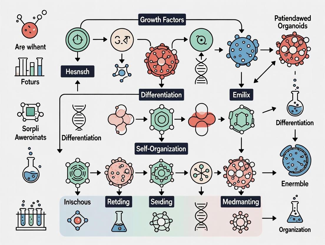
The integration of PDOs into the drug discovery pipeline enables more clinically predictive screening. The following diagram illustrates a complete workflow for utilizing PDOs in high-throughput drug screening, a key application of this technology [2] [1].
This workflow highlights the power of PDOs in high-throughput screens (HTS). Following hit identification from PDO screens, researchers can make decisions earlier and progress to more targeted in vivo efficacy studies in matched PDX models with higher predictive confidence [2]. This integrated approach significantly shrinks costs and timelines compared to moving directly to in vivo studies. Furthermore, PDOs can be used in co-culture systems with immune cells to test the potency of immunotherapies, such as checkpoint inhibitors or CAR-T cells, overcoming their inherent limitation of lacking a tumor microenvironment [2] [1]. The functional data generated can also inform personalized treatment strategies by using a patient's own organoids to guide therapeutic decisions [1].
Patient-derived organoids (PDOs) represent a transformative three-dimensional (3D) in vitro model system in oncology research. They are established directly from patient tumor tissues obtained via surgical resection or biopsy and cultured in a manner that allows them to self-organize and maintain key characteristics of the original malignancy [4]. Unlike traditional two-dimensional (2D) cell cultures, PDOs preserve the architectural complexity and cellular diversity of native tumors, providing a more physiologically relevant platform for studying cancer biology, drug screening, and personalized therapy development [4] [5]. This application note details the specific advantages of PDOs in preserving tumor heterogeneity and microenvironmental cues, alongside standardized protocols for their utilization in research and drug development.
Key Advantages of PDO Models
Faithful Preservation of Tumor Heterogeneity
Tumor heterogeneity, encompassing both genetic and phenotypic variations among cancer cells, is a critical factor in disease progression and treatment response. PDOs excel at maintaining this heterogeneity during in vitro culture [4].
- Inter- and Intratumor Heterogeneity: PDOs capture both intertumor heterogeneity (variations between different patients) and intratumor heterogeneity (variations within a single tumor) [4]. This is crucial for representing the full spectrum of a cancer type and for understanding treatment resistance mechanisms.
- Genomic and Transcriptomic Stability: Studies have demonstrated that PDOs largely retain the gene expression profiles, genomic fingerprints, and histopathological characteristics of the original tumor tissue even after multiple passages. For instance, research on breast cancer PDOs showed that only a small fraction (approximately 1%) of the gene transcriptome exhibited significant differences between the original tumor and the derived organoids [4].
- Representation of Diverse Subtypes: Biobanks of PDOs have been successfully established that represent various cancer subtypes. For example, a pioneering repository of 95 breast cancer organoids was shown to mirror the histopathology and hormone receptor status of the original tumors from which they were derived [4].
Table 1: Comparison of PDOs with Traditional Preclinical Cancer Models
| Feature | 2D Cell Cultures | Patient-Derived Xenografts (PDXs) | Patient-Derived Organoids (PDOs) |
|---|---|---|---|
| Tumor Microenvironment (TME) | Lacks TME; no stromal or immune components [4] | Retains human TME initially, but human stroma is replaced by murine cells over time [5] | Preserves key TME components, including cancer-associated fibroblasts and sometimes immune cells [4] |
| Tumor Heterogeneity | Genetic diversity is lost due to selective pressure in 2D [4] | Phenotypic and genotypic heterogeneity of parental tumor is conserved [5] | Highly preserves genetic and cellular heterogeneity of the original tissue [4] |
| Success Rate & Establishment Time | High success rate; rapid establishment | Low success rate; long latency (months) [4] | Relatively high success rate (e.g., up to 87.5% for BC) [4]; establishment in weeks |
| Cost & Infrastructure | Low cost; standard cell culture facilities | High cost; requires animal housing and specialized facilities [4] | Moderate cost; requires 3D culture expertise and materials |
| Ethical Considerations | Minimal ethical concerns | Significant animal use and ethical considerations [4] | No animal experiments required; uses patient tissue with consent [4] |
| Personalized Therapy Screening | Not suitable due to lack of patient-specific context | Possible but low-throughput and time-consuming [4] | Highly suitable for high-throughput drug screening and personalized treatment strategies [4] [5] |
Recapitulation of the Tumor Microenvironment
The tumor microenvironment (TME) plays a pivotal role in cancer progression, metastasis, and therapy response. PDOs provide a unique model that incorporates essential elements of the TME [4].
- 3D Architecture and Cell-Cell Interactions: The 3D structure of PDOs allows for the study of intricate cell-cell interactions and signaling pathways that are absent in monolayer cultures. This includes interactions between tumor cells, epithelial cells, and fibroblasts [4].
- Extracellular Matrix (ECM): PDOs are typically embedded in a basement membrane extract matrix that mimics the native ECM, providing crucial biochemical and biophysical cues that influence cell behavior, differentiation, and drug sensitivity [4].
- Cellular Components of the TME: While challenging, advancements in co-culture techniques are enabling the incorporation of various TME components into PDO cultures, such as:
Diagram 1: PDO Model Workflow and Key Advantages. This diagram illustrates the derivation of PDOs from patient tumors and their core advantages in cancer research, particularly the preservation of the tumor microenvironment and heterogeneity.
Essential Protocols for PDO Research
Protocol: Establishment and Culture of Breast Cancer PDOs
This protocol outlines the fundamental steps for deriving and maintaining breast cancer PDOs from patient tissue samples [4].
Materials Required:
- Fresh breast cancer tissue from surgical resection or biopsy (placed in cold, sterile transport medium).
- Digestion solution: Collagenase/Hyaluronidase mix in Advanced DMEM/F12.
- Washing medium: Advanced DMEM/F12 supplemented with antibiotics.
- Growth factor-reduced Basement Membrane Extract (BME).
- Complete organoid culture medium: Advanced DMEM/F12 supplemented with key factors like Noggin, R-spondin, EGF, FGF10, and WNT agonists.
- Tissue culture plates (e.g., 24-well or 48-well).
Step-by-Step Workflow:
Diagram 2: PDO Establishment and Culture Workflow. A generalized protocol for deriving and maintaining PDOs from patient tissue.
Tissue Processing and Digestion:
- Mince the fresh tissue into ~1 mm³ fragments using sterile scalpels.
- Transfer the fragments to digestion solution and incubate for 1-2 hours at 37°C with gentle agitation.
- Dissociate further by pipetting every 20-30 minutes.
Cell Isolation and Washing:
- Neutralize the digestion reaction with a washing medium containing serum.
- Pass the cell suspension through a 70-100 μm cell strainer to remove undigested fragments.
- Centrifuge the filtrate and wash the pellet with washing medium.
Embedding in Matrix and Seeding:
- Resuspend the final cell pellet in cold BME matrix.
- Plate small droplets of the cell-BME suspension into pre-warmed tissue culture plates.
- Allow the BME to polymerize for 20-30 minutes in a 37°C incubator.
Culture and Maintenance:
- Carefully overlay the polymerized BME droplets with complete organoid culture medium.
- Culture at 37°C in a 5% CO₂ incubator.
- Refresh the medium every 2-3 days and monitor for organoid formation.
Passaging and Expansion:
- For passaging, mechanically or enzymatically dissociate mature organoids into smaller fragments or single cells.
- Re-embed the cells in fresh BME and continue culture as above. Passaging is typically performed every 1-2 weeks.
Protocol: Drug Sensitivity and Viability Assay in PDOs
This protocol describes a standardized method for testing the efficacy of anti-cancer compounds on PDOs, a key application in personalized medicine and drug discovery [4] [5].
Materials Required:
- Mature PDOs (5-7 days after passaging).
- 96-well tissue culture plates.
- Drug compounds of interest, serially diluted in culture medium or DMSO.
- Cell viability assay kit (e.g., CellTiter-Glo 3D).
- Multimode plate reader.
Step-by-Step Workflow:
- PDO Preparation: Harvest PDOs and dissociate them into small, uniform fragments or single cells. Count the cells.
- Seeding: Seed a predetermined number of cells (e.g., 1,000-5,000 cells per well) in BME into a 96-well plate, following the steps in section 3.1. Allow organoids to form for 3-5 days.
- Drug Treatment: After organoid formation, treat wells with a range of drug concentrations. Include negative control (vehicle only, e.g., 0.1% DMSO) and positive control (e.g., a cytotoxic drug like Staurosporine) wells.
- Incubation: Incubate the plate for a predetermined period (e.g., 3-7 days), refreshing drug-containing medium as needed.
- Viability Assessment:
- Equilibrate the plate and CellTiter-Glo 3D reagent to room temperature.
- Add an equal volume of reagent to each well.
- Shake the plate for 5 minutes to induce cell lysis.
- Incubate for 25 minutes to stabilize the luminescent signal.
- Measure the luminescence using a plate reader.
- Data Analysis: Normalize the luminescence of drug-treated wells to the vehicle control wells. Calculate the half-maximal inhibitory concentration (IC₅₀) using non-linear regression analysis.
Table 2: Example Drug Screening Data in Gastric Cancer PDO Models
| Drug Candidate / Class | PDO Model Characteristics | Key Findings in PDO Screen | Correlation with Clinical Response |
|---|---|---|---|
| 5-Fluorouracil (5-FU) | Models from various molecular subtypes [5] | Differential sensitivity observed across PDO lines; some showing high resistance. | PDO response often mirrors patient's historical or subsequent clinical response to 5-FU-based regimens [5]. |
| Targeted Therapies | PDOs with specific driver mutations (e.g., HER2 amplification) [5] | HER2+ PDOs show marked sensitivity to HER2-targeting agents (e.g., Trastuzumab). | High predictive value for identifying responders to targeted agents in preclinical models [5]. |
| Immunotherapy Checkpoint Inhibitors | PDOs co-cultured with autologous immune cells [5] | MSI-High PDOs show increased T-cell mediated killing upon anti-PD-1 treatment compared to MSS PDOs. | Models the differential response seen in patients with MSI-H vs. MSS tumors, aiding in biomarker discovery [5]. |
The Scientist's Toolkit: Essential Research Reagent Solutions
Table 3: Key Reagents and Materials for PDO Research
| Reagent / Material | Function / Purpose | Examples / Notes |
|---|---|---|
| Basement Membrane Extract (BME) | Provides a 3D scaffold that mimics the extracellular matrix, supporting polarized cell growth and signaling. | Growth Factor Reduced Matrigel; Cultrex BME. Must be kept on ice during handling. |
| Specialized Culture Medium | Provides nutrients and essential signaling molecules to support stem cell survival and organoid growth. | Advanced DMEM/F12 base, supplemented with Noggin, R-spondin, EGF, FGF, WNT agonists, and B27 [4]. |
| Dissociation Enzymes | Breaks down tissue and dissociates organoids into single cells or fragments for passaging and seeding. | Collagenase, Hyaluronidase, Trypsin-EDTA, Accutase. Choice depends on tissue type and robustness of organoids. |
| Cryopreservation Medium | Allows long-term storage of PDO lines in liquid nitrogen for biobanking. | Typically contains culture medium, high concentration of serum or BME, and a cryoprotectant like DMSO. |
| Cell Viability Assays | Quantifies the number of viable cells in culture to assess drug response and proliferation. | CellTiter-Glo 3D is optimized for 3D cultures. Other options include ATP-based assays or live-cell imaging dyes. |
Anatomic and Molecular Considerations for Tissue Sampling in Colorectal Cancer
The fidelity of patient-derived organoid (PDO) research is fundamentally contingent on the quality and strategic acquisition of tumor tissue. For colorectal cancer (CRC), a disease characterized by significant anatomic and molecular heterogeneity, the initial tissue sampling protocol is paramount. The ensuing application note delineates a comprehensive framework for tissue sampling, integrating critical anatomic and molecular considerations to ensure the generation of biologically relevant organoid avatars. This protocol is designed to support preclinical drug screening and functional precision medicine, enabling the identification of patient-specific treatment sensitivities and resistance patterns [6] [7].
Anatomic and Molecular Heterogeneity in Colorectal Cancer
Anatomic Location-Specific Considerations
Mounting evidence underscores the profound influence of tumor anatomic location on the molecular landscape and clinical behavior of CRC. Tumors originating on the proximal side (cecum, ascending colon, hepatic flexure) are classified as right-sided colon cancers (RCCs), while those on the distal side (splenic flexure, descending, sigmoid, rectosigmoid) are left-sided colon cancers (LCCs) [8]. Patients with RCCs have been demonstrated to have a worse overall prognosis compared to those with LCCs, even after stage matching [8].
This anatomic divergence is reflected in distinct metabolic signatures. A 2020 metabolomics study identified five specific metabolites—S-adenosyl-L-homocysteine, formylmethionine, fucose 1-phosphate, lactate, and phenylalanine—that demonstrated high differentiative capability for left- and right-sided colon cancers at stage I (AUC = 0.804) [8]. Furthermore, spatial transcriptomic analyses have revealed the compartmentalization of Consensus Molecular Subtype (CMS) features within tumors, with CMS1 and CMS2 signatures associated with tumor-annotated spots, while CMS3 signatures were more confined to non-neoplastic mucosa [9]. Such findings highlight the necessity of precise anatomic annotation during tissue sampling to ensure PDOs accurately mirror the originating tumor's biology.
Table 1: Key Anatomic Location-Specific Characteristics in Colorectal Cancer
| Anatomic Region | Molecular & Metabolic Features | Clinical/Prognostic Correlation |
|---|---|---|
| Right-Sided Colon (RCC) | • Associated with CMS1 and CMS3 [9]• Distinct metabolic profile (e.g., S-adenosyl-L-homocysteine, lactate) [8] | • Worse overall prognosis [8] |
| Left-Sided Colon (LCC) | • Associated with CMS2 [9]• Distinct metabolic profile (e.g., formylmethionine, fucose 1-phosphate) [8] | • 19% reduced risk of death [8] |
Essential Molecular Biomarkers for Tissue Sampling
The standard of care for advanced CRC mandates molecular testing to guide targeted therapy decisions. Tissue sampling for PDO generation must, therefore, be planned in coordination with diagnostic molecular profiling to ensure sufficient material for both clinical and research purposes [10].
Key biomarkers that must be considered include:
- Mismatch Repair (MMR) Status: Deficiency (dMMR) is found in approximately 15% of CRCs and is a strong predictor of response to immune checkpoint inhibitors [10]. Testing is typically performed via a four-antibody immunohistochemistry panel (MLH1, PMS2, MSH2, MSH6) or PCR-based microsatellite instability testing.
- Extended RAS Testing: This encompasses mutations in KRAS and NRAS codons 12, 13, 59, 61, 117, and 146. Mutations, present in about 50% of CRCs, are a contraindication for anti-EGFR therapy [10].
- BRAF V600E Mutation: This is a recognized significant prognostic biomarker of poor outcome in MMR-proficient CRC patients and accounts for approximately 5% of all CRCs [10].
Next-generation sequencing (NGS) is increasingly the standard platform for this testing due to its ability to interrogate multiple genes simultaneously using relatively small amounts of tissue [10] [11].
Figure 1: Integration of Molecular Profiling with PDO Workflow. Tissue sampling must support both comprehensive molecular profiling and PDO generation to enable clinically relevant drug screening.
Comprehensive Tissue Sampling Protocol for PDO Generation
This protocol is optimized for generating PDOs from various clinically accessible specimens, including surgical resections, endoscopic biopsies, and liquid biopsies (malignant ascites/pleural effusion), supporting reproducible PDO applications across diverse clinical settings [12].
Specimen Acquisition and Transport
- Source Prioritization: For patients with advanced disease, prioritize sampling of metastatic lesions when safe and feasible, as they may better represent the current driver molecular alterations [10]. In their absence, use the primary tumor.
- Anatomic Annotation: Record the precise anatomic location of the sampled tissue (e.g., hepatic flexure, sigmoid colon). For primary tumors, specify if it is right-sided or left-sided [8].
- Macroscopic Assessment: During surgery, select tissue fragments a few millimeters in size from a viable, non-necrotic area of the tumor. Note that reliance on macroscopic assessment alone carries a risk of insufficient tumor cellularity; therefore, coordination with a pathologist for gross dissection is ideal [13].
- Transport Medium: Immediately place the specimen in a sterile container with cold (4°C) organoid transport medium [Advanced DMEM/F12 supplemented with 10 µM Y-27632 (ROCK inhibitor), 1x GlutaMAX, 10mM HEPES, and 1x Penicillin/Streptomycin]. For core needle biopsies, RNA Save solution can also be used for stabilization [12] [13].
- Time to Processing: Process all specimens as rapidly as possible, ideally within 1 hour of collection, but definitely within 24 hours to maintain optimal cell viability [6].
Tumor Tissue Processing and Cell Isolation
Materials & Reagents:
- Basal Medium: Advanced DMEM/F12
- Digestion Enzymes: Liberase TH (50 µg/ml) or Collagenase A
- ROCK Inhibitor: Y-27632
- Fetal Bovine Serum (FBS)
- Cell Strainers (70µm and 100µm)
- Red Blood Cell Lysis Solution
- Basement Membrane Extract (BME, e.g., Matrigel)
Procedure:
- Wash and Mince: Transfer the tissue to a Petri dish containing cold basal medium. Mince the tissue into fine fragments (~1-2 mm³) using sterile scalpels or razor blades.
- Enzymatic Digestion: Transfer the minced tissue to a tube containing digestion medium (basal medium with 50 µg/ml Liberase TH and 10 µM Y-27632). Incubate for 30-60 minutes at 37°C with gentle agitation or periodic manual shaking.
- Mechanical Dissociation: Every 10-15 minutes during digestion, vigorously pipette the mixture up and down to further dissociate the tissue. Continue until the solution appears cloudy with small cell clusters.
- Filtration and Washing: Pass the cell suspension through a 100µm cell strainer into a new tube containing 10% FBS medium to inactivate the enzyme. Centrifuge at 350 g for 5 minutes.
- Red Blood Cell Lysis: Resuspend the cell pellet in red blood cell lysis solution. Incubate for 2-5 minutes at room temperature. Top up with basal medium and centrifuge at 350 g for 5 minutes.
- Cell Counting and Viability Assessment: Resuspend the final pellet in basal medium. Count viable cells using a trypan blue exclusion assay. A viability of >70% is generally recommended for successful organoid culture [6].
PDO Culture Initiation and Biobanking
- Embedding in BME: Resuspend the cell pellet in cold Basement Membrane Extract (BME) at a density of 500-1000 cells/µL of BME [6].
- Plating: Plate 10-20 µL droplets of the cell-BME suspension into the center of a pre-warmed tissue culture plate. Allow the BME to polymerize for 15-30 minutes in a 37°C incubator.
- Culture Medium Overlay: Carefully overlay the polymerized BME droplets with complete organoid culture medium, supplemented with 10 µM Y-27632 for the first 2-3 days to inhibit anoikis. Use a modified version of established CRC organoid media [6].
- Medium Refreshment: Refresh the culture medium every 2-3 days. Monitor organoid formation and growth under a microscope.
- Passaging and Expansion: Passage organoids every 7-14 days when they become large and dense. Dissociate using TrypLE for 5-20 minutes at 37°C, triturate to single cells/small clusters, and re-embed in BME as described.
- Cryopreservation: Resuspend organoid fragments in freezing medium (FBS with 10% DMSO), cool at -1°C/minute, and store in liquid nitrogen for long-term biobanking [6].
Figure 2: PDO Generation Workflow. The streamlined process from tissue acquisition to the establishment of ready-to-use organoid models for downstream applications.
Quality Control and Validation
Robust quality control is critical to confirm that PDOs faithfully recapitulate the patient's tumor.
- Genomic Validation: Perform DNA sequencing on PDOs at early passages (P1-P3) and compare the mutational profile (e.g., in APC, TP53, KRAS) to that of the original tumor tissue. A concordance of >90% should be achieved [6] [13].
- Histopathological Correlation: Process PDOs for paraffin embedding, H&E staining, and immunohistochemistry (e.g., for CDX2, CK20) to confirm they retain the architectural and differentiation features of the original CRC adenocarcinoma [6].
- Anatomic and Molecular Signature Retention: For research purposes, validate the retention of location-specific metabolic signatures or CMS classifications using targeted metabolomics or RNA sequencing, respectively [8] [9].
Application in Functional Precision Medicine
The primary application of CRC PDOs is ex vivo drug sensitivity testing to inform treatment decisions. This process, termed a "chemogram," involves challenging expanded PDOs with a panel of clinically relevant drugs.
- Drug Panel Design: Include a panel of 20-25 drugs encompassing standard chemotherapies (5-FU, Oxaliplatin, Irinotecan), targeted agents (EGFR inhibitors for RAS wild-type tumors), and other relevant compounds [6].
- High-Throughput Screening: Plate dissociated PDOs in 384-well plates, expose them to a concentration range of each drug, and incubate for 5-7 days.
- Viability Readout: Measure cell viability using a luminescence-based assay (e.g., CellTiter-Glo). Calculate the Area Under the dose-response Curve (AUC) for each drug as a robust metric of sensitivity [6] [7].
- Turnaround Time: With optimized protocols, a chemogram can be obtained with a median turnaround time of 6 weeks (range: 4-10 weeks) from tissue acquisition, which is compatible with the clinical decision-making timeline for many patients with advanced CRC [6].
Table 2: Key Reagents for CRC PDO Generation and Drug Screening
| Research Reagent | Function/Application | Example |
|---|---|---|
| Liberase TH | Enzymatic digestion of tumor tissue into single cells/small clusters [6] | Roche |
| Y-27632 (ROCK inhibitor) | Inhibits anoikis; improves viability of dissociated single cells during seeding and passaging [6] | Selleckchem |
| Basement Membrane Extract (BME) | 3D extracellular matrix scaffold for organoid growth and polarization [6] | Corning Matrigel |
| Advanced DMEM/F12 | Basal medium for formulating organoid culture and transport media [6] | Thermo Fisher Scientific |
| CellTiter-Glo 3D | Luminescent assay for quantifying cell viability in 3D organoid cultures during drug screens [6] | Promega |
A meticulous approach to tissue sampling, grounded in a deep understanding of the anatomic and molecular dimensions of colorectal cancer, is the foundational step for establishing clinically meaningful PDO models. The protocol detailed herein—encompassing strategic specimen acquisition, optimized processing, rigorous validation, and functional drug testing—provides a robust framework for integrating PDO technology into the functional precision oncology pipeline. Adherence to these guidelines will enhance the reproducibility and predictive power of PDO-based research, accelerating its translation into personalized treatment strategies for CRC patients.
Patient-derived organoids (PDOs) are three-dimensional stem cell-derived models that offer a more physiologically relevant representation of tumor biology compared to traditional models [14]. The successful establishment and maintenance of PDOs depend critically on two essential components: a supportive extracellular matrix (ECM) and precisely formulated, niche-specific growth factors [15]. These elements work in concert to recapitulate the native tissue microenvironment, enabling PDOs to preserve the complex tissue architecture, cellular diversity, and functional characteristics of human cancers [15] [16]. This protocol details the standardized methodologies for utilizing these essential components across major solid cancers, supporting applications in precision oncology, drug screening, and translational studies [17].
Core Components for PDO Culture
Extracellular Matrix (ECM) Platforms
The ECM serves as the foundational scaffold for PDO culture, providing not only structural support but also critical biochemical and biophysical cues that direct cell behavior, polarization, and self-organization [17] [18].
Table 1: Extracellular Matrix Products for PDO Culture
| ECM Product | Composition | Key Properties | Applications in PDO Culture |
|---|---|---|---|
| Matrigel | Basement membrane proteins (laminin, collagen IV, entactin), proteoglycans, growth factors | Thermoreversible gelation; biologically active | Broad-spectrum cancer PDOs; primary establishment [18] |
| BME (Basement Membrane Extract) | Similar to Matrigel with standardized composition | Reduced growth factor content; more defined | Reproducible PDO cultures; hormone-sensitive cancers [18] |
| Geltrex | Reduced growth factor basement membrane matrix | Low GF content; high clarity | Defined condition studies; growth factor response assays [18] |
| Collagen-based Hydrogels | Type I collagen predominant | Tunable stiffness; modular composition | Stroma-rich tumors; mechanical studies [17] |
Niche-Specific Growth Factor Formulations
Growth factors are indispensable for maintaining stemness, directing differentiation, and supporting the proliferation of specific cancer cell types. The formulation must be tailored to the tissue of origin [17] [15].
Table 2: Essential Growth Factors by Tumor Type
| Tumor Type | Core Growth Factors | Supplemental Factors | Function in Culture |
|---|---|---|---|
| Colorectal | EGF, Noggin, R-spondin [ENR] | Wnt-3A, N-Acetylcysteine | Maintain Lgr5+ stem cells; promote epithelial proliferation [15] |
| Pancreatic | FGF10, EGF, Noggin | Nicotinamide, A83-01 | Support ductal morphology; inhibit differentiation [15] |
| Gastric | EGF, FGF10, Noggin, R-spondin | Gastrin I, A83-01, Wnt-3A | Promote gland formation; maintain pit and chief cells [15] |
| Hepatic | HGF, EGF, FGF19 | R-spondin, Wnt-3A, BMP-7 | Support hepatocyte function; promote biliary differentiation [15] |
| Mammary | EGF, FGF, R-spondin | Neuregulin-1, Heparin, SB202190 | Maintain basal and luminal populations; support acinar formation [15] |
Experimental Protocols
Standardized Workflow for PDO Generation
The following diagram illustrates the complete workflow for establishing PDOs from patient tissue, highlighting the critical points of ECM and growth factor application:
Detailed Protocol: Tissue Processing and ECM Embedding
Tumor Tissue Dissociation
- Sampling: Obtain fresh tumor samples via surgical resection or biopsy. For non-surgical approaches, samples can be derived from malignant effusions, ascites, or blood [18]. All samples must be collected with appropriate ethical approval and patient consent [18].
- Mechanical Disruption: Using sterile instruments, remove all non-epithelial tissue (muscle, fat) and mince primary tumor tissues into 1-3 mm³ pieces [18].
- Enzymatic Digestion:
- Prepare digestion cocktail containing collagenase/hyaluronidase and TrypLE Express enzymes.
- For incubations <2 hours: Agitate mixture every 10-15 minutes with vigorous shaking and pipetting.
- For overnight incubations: Place mixture on a shaker and add 10 µM ROCK inhibitor (Y-27632) to improve viability [18].
- Monitoring: Digestion is complete when clusters of 2-10 cells become visible. These can be further dissociated by gentle pipetting.
- Filtration and Washing: Pass cell suspension through 70-100 µm filters (pore size determined by tumor type). Centrifuge and wash cells with appropriate buffer [18].
ECM Embedding and Plating
- Cell Density Adjustment: Resuspend pellet in working medium and determine cell density. Adjust to appropriate concentration for specific cancer type (typically 1,000-10,000 cells/µL of ECM) [18].
- ECM Mixing: Combine cell pellet with chilled ECM (Matrigel, BME, or Geltrex) on ice. Maintain low temperature to prevent premature polymerization.
- Plating: Plate 10-20 µL drops of cell-ECM mixture into pre-warmed culture plates. For limited cell numbers, use 96-well plates; for larger quantities, 24- or 48-well plates are suitable [18].
- Polymerization: Invert plates and incubate at 37°C, 5% CO₂ for 15-30 minutes to allow ECM solidification. This prevents cells from settling and adhering to the bottom surface [18].
- Medium Addition: After solidification, carefully add pre-warmed, tumor-type specific medium to each well.
Medium Formulation and Growth Factor Supplementation
Base Medium Composition
- Advanced DMEM/F12 supplemented with:
- 10 mM HEPES
- 1× GlutaMAX
- 1× Penicillin/Streptomycin (optional)
- 1× N-2 Supplement
- 1× B-27 Supplement
Tumor-Type Specific Additives
Table 3: Complete Medium Formulations by Cancer Type
| Component | Colorectal | Pancreatic | Gastric | Mammary |
|---|---|---|---|---|
| EGF | 50 ng/mL | 50 ng/mL | 50 ng/mL | 20 ng/mL |
| Noggin | 100 ng/mL | 100 ng/mL | 100 ng/mL | - |
| R-spondin | 500 ng/mL | - | 500 ng/mL | 250 ng/mL |
| FGF-10 | - | 100 ng/mL | 100 ng/mL | 20 ng/mL |
| FGF-2 | - | - | - | 10 ng/mL |
| Wnt-3A | 50% (v/v) cond. medium | - | 50% (v/v) cond. medium | - |
| A83-01 | 500 nM | 500 nM | 500 nM | - |
| SB202190 | - | - | - | 5 µM |
| Nicotinamide | - | 10 mM | - | - |
| N-Acetylcysteine | 1.25 mM | 1.25 mM | 1.25 mM | - |
| Gastrin I | 10 nM | - | 10 nM | - |
| Neuregulin-1 | - | - | - | 10 ng/mL |
| Heparin | - | - | - | 4 µg/mL |
The Scientist's Toolkit: Essential Research Reagents
Table 4: Key Reagent Solutions for PDO Research
| Reagent Category | Specific Products | Function in PDO Culture |
|---|---|---|
| ECM Scaffolds | Matrigel, BME, Geltrex, Collagen I | Provide 3D structural support; present biochemical cues for cell signaling and polarization [17] [18] |
| Digestive Enzymes | Collagenase/Hyaluronidase, TrypLE, Dispase | Dissociate tissue into single cells or small clusters while preserving viability [18] |
| ROCK Inhibitor | Y-27632 (10 µM) | Enhances survival of single cells and stem cells by inhibiting apoptosis [18] |
| Growth Factors | EGF, FGF, Noggin, R-spondin, Wnt-3A | Direct lineage specification, maintain stemness, support proliferation of specific cell types [17] [15] |
| TGF-β Inhibitors | A83-01, SB431542 | Prevent differentiation; support epithelial cell growth by inhibiting EMT [15] |
| Media Supplements | B-27, N-2, N-Acetylcysteine | Provide essential nutrients, antioxidants, and hormones for cell survival [15] |
Quality Control and Validation
Assessment of PDO Quality
- Histological Validation: Compare PDO structure to parental tumor tissue using H&E staining and immunohistochemistry for tissue-specific markers [17] [16].
- Genomic Analysis: Perform whole genome sequencing (WGS) or whole exome sequencing (WES) to confirm PDOs maintain genetic alterations of original tumors [16].
- Functional Assessment: Evaluate growth characteristics and demonstrate appropriate responses to tumor-type specific stimuli [17].
Troubleshooting Common Issues
- Poor Growth: Optimize growth factor concentrations; ensure fresh aliquots of labile components; verify ECM polymerization conditions.
- Differentiation: Increase stem cell factor concentrations; add appropriate pathway inhibitors; reduce passage time.
- Contamination: Use antibiotic/antimycotic in initial cultures; implement strict sterile technique during tissue processing.
- ECM Dissociation: Handle with chilled instruments during passaging; use appropriate recovery period with ROCK inhibitor.
The standardized protocols outlined herein for ECM application and growth factor formulation provide a robust foundation for establishing reproducible PDO cultures across multiple cancer types. These essential components enable the generation of physiologically relevant models that faithfully recapitulate patient-specific tumor characteristics, advancing their utility in precision medicine and drug development applications.
Patient-derived organoids (PDOs) have emerged as a transformative pre-clinical model that faithfully recapitulates tumor properties from individual patients, addressing significant limitations of traditional models [19]. Unlike monolayer cultures of cancer cell lines that lose the heterogeneity of parental tumors, PDOs maintain the cellular architecture, genetic diversity, and molecular characteristics of the original tissue [20]. This preservation is particularly valuable in cancer research, where tumor heterogeneity significantly influences treatment response and disease progression. However, the utility of PDOs in basic research and clinical decision-making depends entirely on rigorous quality control measures that validate their fidelity to the original tumors from which they were derived [21]. Establishing robust protocols to verify genomic and proteomic fidelity is thus essential for ensuring that experimental results from PDO platforms can be reliably translated to patient care scenarios.
The pressing need for such faithful models is underscored by drug development statistics; between 2000 and 2015, only 3.4% of cancer-targeting drugs passed clinical trials and were approved for patient care [20]. This high failure rate highlights the inadequacy of existing preclinical models, driving the adoption of PDOs that maintain the chemoresistance and genetic mutations observed in original patient tissue [20]. As the field moves toward using PDOs for personalized medicine applications, including drug screening and treatment prediction, standardized quality control protocols become indispensable for confirming that these miniature avatars accurately mirror the patient's disease state [21] [22].
Essential Quality Control Parameters for PDOs
Key Validation Metrics Across Molecular Domains
Quality control for PDOs requires a multi-faceted approach assessing multiple molecular dimensions. The table below outlines core parameters that must be evaluated to confirm fidelity to original tumors:
Table 1: Essential Quality Control Metrics for PDO Validation
| Validation Domain | Key Parameters | Target Metrics | Application in PDOs |
|---|---|---|---|
| Genomic Fidelity | Driver mutations retention | >95% concordance | Confirm preservation of critical mutations (e.g., TP53, CTNNB1) [22] |
| Copy number variations | Comparable profile | Maintain tumor genetic landscape [20] | |
| Transcriptomic profiling | PCA clustering with tumor | Preserve gene expression patterns [21] | |
| Proteomic Fidelity | Protein coverage | ≥70% | Comprehensive protein identification [23] |
| False discovery rate (FDR) | <1% | High-confidence peptide identification [23] | |
| Phosphoproteome/N-glycoproteome | Reproducible quantification | Functional proteomic state preservation [24] | |
| Histopathological Concordance | Tissue architecture | Recapitulation of native organization | Maintain 3D structure and cellular relationships [21] |
| Marker expression | Appropriate protein localization | Cell-type specific protein preservation [21] | |
| Functional Validation | Drug response correlation | Mirror clinical outcomes | Predictive value for patient treatment response [21] [22] |
| Pathway activity | Preserved signaling networks | Maintain tumor biology [21] |
The Researcher's Toolkit: Essential Reagents and Materials
Successful PDO establishment and validation requires specific research reagents carefully selected to maintain tumor fidelity while enabling robust expansion.
Table 2: Essential Research Reagent Solutions for PDO Quality Control
| Reagent Category | Specific Examples | Function in PDO Workflow | Quality Considerations |
|---|---|---|---|
| Matrix Substrates | Reduced growth factor Matrigel [22] | Provides 3D scaffolding for organoid growth | Minimizes exogenous signaling influence; improves standardization |
| Digestive Enzymes | Dispase, Collagenase type II, Trypsin-EDTA [21] [22] | Tissue dissociation and single-cell preparation | Preservation of cell viability and surface receptors |
| Culture Media | Growth factor-reduced (GF-) media [22] | Supports organoid growth with minimal exogenous factors | Reduces niche dependency; improves reproducibility |
| Growth factor-supplemented (GF+) media [22] | Enhanced growth support for challenging samples | Defined composition for standardization | |
| Specialized Additives | Y-27632 ROCK inhibitor [22] | Prevents anoikis in dissociated cells | Critical for initial establishment and passaging |
| QC Standards | NCI-20 dynamic range protein mixture [23] | Mass spectrometry quality control | Enables instrument performance validation |
| Sigma UPS1 equimolar protein mixture [23] | Quantitative accuracy assessment | Verifies proteomic quantification reliability | |
| Internal Standards | Indexed Retention Time (iRT) peptides [23] | Chromatographic performance monitoring | Ensures LC-MS/MS system suitability |
Experimental Protocols for Fidelity Assessment
Genomic Validation Workflow
Objective: Confirm that PDOs maintain the genomic features of the original tumor through comprehensive sequencing approaches.
Sample Requirements: Triplicate samples of original tumor tissue, early passage PDOs (P1-P3), and late passage PDOs (P5-P10) for longitudinal stability assessment.
Procedure:
- Nucleic Acid Extraction: Isolate DNA and RNA using standardized kits with quality assessment (A260/A280 ratios: 1.8-2.0; RNA Integrity Number ≥7.0).
- Library Preparation: Utilize Illumina-compatible kits for whole genome sequencing and transcriptome analysis following manufacturer protocols with 10-15 PCR cycles to minimize bias [25].
- Sequencing: Perform on Illumina NovaSeq or comparable platform targeting 30-50x coverage for WGS and 50 million reads per sample for RNA-seq.
- Data Analysis:
- Align sequences to reference genome (GRCh38) using STAR aligner [21].
- Identify somatic variants with GATK best practices pipeline.
- Perform principal component analysis (PCA) to confirm clustering of PDOs with matched tumor tissue [21].
- Compare mutational profiles focusing on driver mutations (e.g., TP53, CTNNB1) with requirement of >95% concordance [22].
Quality Control Checkpoints:
- Monitor sequencing quality scores (Q≥30 for >80% of bases).
- Verify coverage uniformity (>90% of targets covered at 20x).
- Confirm expected mutation retention rates between passages.
Proteomic Fidelity Assessment Protocol
Objective: Verify that PDOs maintain the protein expression, modification, and signaling pathway activities of original tumors.
Sample Preparation:
- Protein Extraction: Lyse PDOs and tumor tissues in appropriate buffer (e.g., 8M urea, 2M thiourea) with protease and phosphatase inhibitors.
- Digestion: Digest proteins using trypsin (1:50 enzyme-to-protein ratio) at 37°C for 16 hours after reduction and alkylation.
- Peptide Cleanup: Desalt using C18 solid-phase extraction cartridges.
- PTM Enrichment: For phosphoproteomics, use Fe-IMAC enrichment; for glycoproteomics, apply HILIC enrichment [24].
LC-MS/MS Analysis:
- Chromatography: Employ nanoflow or microflow LC with C18 column (25cm length, 1.9μm particles) with 120-minute gradient.
- Mass Spectrometry: Operate Orbitrap instrument in data-dependent acquisition mode with MS1 resolution ≥120,000 and MS2 resolution ≥15,000.
- Quality Control: Include QC samples (HeLa digest or similar) every 10 injections to monitor system performance [23].
Data Processing:
- Database Search: Use MaxQuant or similar against human UniProt database with FDR set to 1% at PSM and protein levels.
- Quantification: Apply label-free or TMT-based quantification with normalization.
- Statistical Analysis: Perform PCA to assess sample clustering and differential expression analysis with thresholds of fold-change ≥2 and adjusted p-value ≤0.05.
Acceptance Criteria:
- Coefficient of variation (CV) <20% for technical replicates
- >70% protein coverage compared to tumor tissue
- Retention of pathway activities (e.g., drug response pathways)
Analytical Quality Control Frameworks
Proteomics Quality Control Metrics
Mass spectrometry-based proteomics requires rigorous quality control at multiple stages to ensure reproducible and reliable data for PDO validation.
Table 3: Comprehensive QC Parameters for Proteomic Analysis of PDOs
| QC Domain | Parameter | Target Value | Importance for PDO Fidelity |
|---|---|---|---|
| Sample Preparation | Digestion efficiency | CV <10% | Ensures comparable protein quantification |
| Labeling efficiency (TMT) | >95% | Minimizes quantification bias in multiplexed designs | |
| Chromatographic Performance | Retention time stability | CV <5% | Enables accurate peptide identification |
| Peak width | 4-8 seconds | Maintains separation resolution | |
| Column pressure | Increase <30% | Consistent performance across runs | |
| Instrument Metrics | MS1 mass error | <5 ppm (Orbitrap) | Accurate precursor identification |
| MS2 mass error | <10 ppm (Orbitrap) | Confident peptide sequencing | |
| Charge state distribution | 2+ predominant (~50%) | Expected ionization patterns | |
| TIC intensity variation | <30% | Stable instrument performance | |
| Data Quality | False discovery rate (FDR) | <1% | High-confidence identifications |
| Protein coverage | ≥70% | Comprehensive proteome characterization | |
| Missing value rate | <50% for >70% proteins | Data completeness for statistical power | |
| Technical replicate correlation | r >0.9 | Measurement precision |
Integrated Quality Assessment Workflow
The validation of PDO fidelity requires an integrated approach that connects molecular characterization with functional assessment to create a comprehensive quality profile.
Case Studies and Clinical Applications
Esophageal Adenocarcinoma PDO Validation
A landmark study demonstrated the clinical relevance of PDO fidelity validation in esophageal adenocarcinoma [21]. Researchers established PDOs from treatment-naive patients and conducted comprehensive characterization:
- Histopathological Concordance: PDOs recapitulated the original tumor architecture and stained positive for EAC marker MUC5AC while negative for ESCC marker p63, confirming lineage fidelity [21].
- Drug Response Correlation: PDOs were treated with standard-of-care therapies including cisplatin, paclitaxel, and γ-irradiation. The responses closely mirrored the clinical outcomes of the corresponding patients, validating the predictive value of the platform [21].
- Transcriptomic Analysis: RNA sequencing revealed that PDOs maintained patient-specific gene expression patterns, with principal component analysis showing clustering by organoid line rather than treatment type, highlighting preservation of individual tumor biology [21].
Hepatocellular Carcinoma PDOs with Growth Factor-Reduced Media
A 2025 study on hepatocellular carcinoma (HCC) addressed standardization challenges through growth factor-reduced (GF-) media protocols [22]. This approach:
- Improved Standardization: GF- conditions minimized confounding factors during drug screening and reduced environmental niche dependency.
- Maintained Genetic Heterogeneity: PDTOs preserved the somatic mutation frequencies of original HCC tumors, including TP53 and CTNNB1 mutations.
- Clinical Translation: In a proof-of-concept study, PDTO-guided off-label drug use showed clear benefit to patient survival, demonstrating the clinical value of fidelity-validated models [22].
Quality control protocols validating the genomic and proteomic fidelity of PDOs to original tumors represent a critical foundation for advancing personalized cancer medicine. The integrated approaches outlined here—combining genomic verification, proteomic profiling, and functional validation—provide a roadmap for ensuring that these innovative models faithfully represent patient disease states. As the field progresses, several areas require continued development:
Standardization Initiatives: Consensus is needed on specific acceptance criteria for PDO fidelity across different cancer types. Organizations like ISO are working to define global standards for organoid culture and validation [26].
Technological Advancements: Improvements in multi-omics technologies, particularly in sensitivity and throughput, will enable more comprehensive fidelity assessment while reducing costs and turnaround times.
Clinical Integration: As demonstrated in the case studies, validated PDOs have tremendous potential to guide clinical decision-making. Future efforts should focus on streamlining workflows to make PDO-based treatment selection feasible within clinically relevant timelines.
The rigorous application of these quality control protocols ensures that PDOs fulfill their promise as faithful avatars of patient tumors, ultimately enhancing drug development efficiency and advancing precision oncology.
Step-by-Step Protocol: Generating and Utilizing PDOs from Multimodal Specimens
Patient-derived organoids (PDOs) represent a groundbreaking three-dimensional (3D) cell culture system that closely mimics the histological, genetic, and functional characteristics of original patient tumors [27]. The fidelity of a PDO model to its parent tissue is fundamentally determined by the initial specimen acquisition process. Specimens suitable for generating PDOs include surgical resections, biopsies, and liquid specimens, each offering distinct advantages and challenges [27]. This protocol details the methodologies for acquiring and processing these diverse specimen types to establish robust PDO cultures for downstream applications in preclinical research and personalized medicine.
The choice of specimen source depends on clinical availability, tumor type, and the specific research objectives. The table below summarizes the primary specimen types used in PDO generation.
Table 1: Specimen Types for Patient-Derived Organoid Generation
| Specimen Type | Description | Common Sources | Key Advantages | Primary Challenges |
|---|---|---|---|---|
| Surgical Resections | Tumor tissue obtained from curative or palliative surgery. | Primary tumor sites; Metastatic lesions (e.g., liver, lung) [16] [15] | Provides abundant material; Preserves tissue architecture and heterogeneity [27]. | Requires selective media to overcome healthy cell overgrowth [16]. |
| Biopsies | Minimally invasive tissue sampling. | Core needle biopsies; Endoscopic biopsies [28] | Enables serial sampling; Access to hard-to-reach tumors. | Limited starting material; Lower establishment success rates [27]. |
| Liquid Specimens | Biological fluids containing tumor cells. | Ascites; Pleural effusions; Blood (for circulating tumor cells) [27] | Minimally invasive; Allows for real-time monitoring of tumor evolution. | Low tumor cell yield; Complex isolation protocols. |
Specimen Processing and Workflow
The general workflow from specimen acquisition to functional PDO assays involves several critical stages, as visualized below.
Specimen Collection and Transport
- Surgical Resections and Biopsies: Immediately upon collection, place tissue in a sterile container with cold (4°C) transport medium, such as Dulbecco's Modified Eagle Medium (DMEM) supplemented with antibiotics (e.g., Penicillin-Streptomycin) and antifungal agents (e.g., Amphotericin B) to prevent contamination [29]. Transport on ice to the laboratory for processing ideally within 1-2 hours to maintain maximal cell viability.
- Liquid Specimens: Collect ascites or pleural effusions in sterile containers with anticoagulants (e.g., heparin). Process these specimens promptly, typically within 24 hours, to isolate tumor cells for culture [27].
Tissue Dissociation and Processing
The goal of this step is to obtain single cells or small cell aggregates for 3D culture.
- Mechanical Dissociation: Using sterile surgical blades, mince the tissue into fragments of approximately 1-2 mm³ in a small volume of wash buffer [27].
- Enzymatic Dissociation: Transfer the minced tissue fragments to a digestion solution containing collagenase (e.g., Collagenase Type I or II) and Dispase, and incubate at 37°C for 30 minutes to 2 hours with gentle agitation [29] [27].
- Washing and Filtration: Neutralize the enzyme activity with complete culture medium. Pass the cell suspension through a cell strainer (70-100 µm) to remove undigested fragments and debris. Centrifuge the filtrate to pellet the cells and resuspend in an appropriate buffer for counting and viability assessment (e.g., using Trypan Blue exclusion) [27].
PDO Culture and Establishment
- Embedding in ECM: Resuspend the cell pellet in a commercially available extracellular matrix (ECM) hydrogel, such as Matrigel or Basement Membrane Extract (BME), and plate it as small domes in a pre-warmed culture plate [27]. Allow the ECM to polymerize for 10-20 minutes in a 37°C incubator.
- Culture Medium: Overlay the polymerized ECM domes with a specialized, serum-free culture medium. The composition is critical and must be tailored to the tissue of origin. Key supplements often include [27]:
- Wnt-3a and R-Spondin: Essential for activating the Wnt signaling pathway, crucial for the growth of stem cells from many epithelial tissues.
- EGF (Epidermal Growth Factor): Promotes epithelial cell proliferation.
- Noggin: A BMP pathway inhibitor that helps maintain stemness.
- Other niche-specific factors such as FGF-10 for gastric and lung cultures [28].
- Culture Maintenance: Refresh the culture medium every 2-4 days. Organoids typically become visible within 1-2 weeks and can be passaged every 1-4 weeks by mechanically breaking them up or enzymatically dissociating them and re-embedding them in fresh ECM [27].
Table 2: Key Growth Factors and Signaling Pathways in PDO Culture Media
| Signaling Pathway | Key Growth Factors/Agonists | Function in Culture | Notes for Cancer PDOs |
|---|---|---|---|
| Wnt/β-catenin | Wnt-3a, R-Spondin, CHIR99021 (GSK3 inhibitor) | Maintains stemness and proliferation [27]. | Often dispensable for colorectal cancers with APC mutations [27]. |
| EGFR | Epidermal Growth Factor (EGF), Noggin, Neuregulin-1 | Promotes proliferation and survival of epithelial cells [27]. | Tumors with EGFR pathway mutations may grow independently of EGF [27]. |
| TGF-β/BMP | A-83-01 (TGF-β inhibitor) | Inhibits differentiation and fibrosis; supports epithelial growth. | Commonly used in gastrointestinal PDO cultures. |
| FGF | FGF-2, FGF-10 | Supports growth of specific organ types (e.g., stomach, lung) [28]. | Concentration and type are tissue-specific. |
Quality Control and Validation
Ensuring that PDOs faithfully recapitulate the original tumor is paramount for their research and clinical utility.
- Histological Validation: Fix, paraffin-embed, and section PDOs for Hematoxylin and Eosin (H&E) staining. Compare the morphology and architecture with H&E-stained sections of the parent tumor [16] [27]. Immunohistochemistry (IHC) can be used to confirm the expression of protein markers such as pan-cytokeratin (epithelial marker), CDX2 (colorectal adenocarcinoma), and Ki67 (proliferation marker) [16].
- Genomic Validation: Perform whole-genome sequencing (WGS), whole-exome sequencing (WES), or RNA sequencing (RNA-seq) on the PDOs and the matched parent tumor tissue. Compare mutations and copy number alterations (CNA) to confirm the PDOs retain the key genomic features of the tumor [16] [15]. An organoid that fails to replicate the mutations observed in the parental tissue should be discarded [16].
Applications in Preclinical Research
Validated PDOs can be leveraged for various downstream applications.
- Drug Screening and Sensitivity Testing: PDOs can be dissociated and seeded into multi-well plates for high-throughput drug screening. Viability is measured using assays like CellTiter-Glo after 5-7 days of drug exposure [27]. Studies have shown that PDO sensitivity to chemotherapies like 5-fluorouracil, irinotecan, and oxaliplatin correlates significantly with patient clinical response [16].
- Biomarker Discovery: PDOs serve as a platform to identify novel predictive biomarkers of drug response or resistance by correlating omics data with functional drug sensitivity data [15] [28].
- Co-culture Systems: To better model the tumor microenvironment (TME), PDOs can be co-cultured with immune cells (e.g., peripheral blood lymphocytes, CAR-T cells) or cancer-associated fibroblasts (CAFs) [16] [28]. This is particularly valuable for studying immunotherapy.
The diagram below summarizes the relationship between key signaling pathways manipulated in PDO culture media and their cellular outcomes.
The Scientist's Toolkit: Essential Research Reagents
Table 3: Essential Reagents for Patient-Derived Organoid Culture
| Reagent Category | Specific Examples | Function |
|---|---|---|
| Extracellular Matrix (ECM) | Matrigel, Basement Membrane Extract (BME) | Provides a 3D scaffold that mimics the native basement membrane, supporting cell polarization and self-organization [27]. |
| Enzymes for Dissociation | Collagenase, Dispase, Trypsin-EDTA | Breaks down the extracellular matrix of tumor tissue to release single cells or small clusters for culture [29] [27]. |
| Core Growth Factors | R-Spondin-1, Wnt-3a, EGF, Noggin, FGF-10 | Activates key signaling pathways necessary for stem cell maintenance and proliferation, tailored to the tissue of origin [27]. |
| Base Media | Advanced DMEM/F12 | A nutrient-rich foundation medium, often supplemented with HEPES and GlutaMAX. |
| Common Supplements | B-27 Supplement, N-2 Supplement, N-Acetylcysteine | Provides essential hormones, lipids, and antioxidants to support cell survival and growth in serum-free conditions. |
| Viability Assays | CellTiter-Glo, CCK-8, MTS | Measures ATP or metabolic activity as a proxy for cell viability and proliferation in drug screening assays [27]. |
Tissue Processing and Crypt Isolation for 3D Culture Establishment
Within the framework of patient-derived organoid (PDO) research, the initial steps of tissue processing and crypt isolation are critical determinants of success. These three-dimensional (3D) culture models recapitulate the cellular complexity and architectural features of original tissues, making them indispensable tools for personalized medicine, disease modeling, and drug development [30] [31]. The derivation of intestinal organoids, in particular, relies on the efficient isolation of viable crypt structures containing LGR5+ stem cells, which possess the capacity for self-renewal and multi-lineage differentiation [32] [33]. This application note provides a standardized, detailed protocol for establishing human intestinal organoids from primary tissue, encompassing tissue procurement, crypt isolation, and initial culture setup, thereby enabling robust and reproducible PDO generation for translational research applications.
Materials and Reagents
Research Reagent Solutions
The following table catalogues the essential reagents and their functions for tissue processing and crypt isolation.
Table 1: Key Reagents for Tissue Processing and Crypt Isolation
| Reagent/Material | Function/Purpose | Example Composition/Notes |
|---|---|---|
| Transport Medium [30] | Preserves tissue integrity during transit from clinic to lab. | Advanced DMEM/F12 supplemented with antibiotics (e.g., Penicillin-Streptomycin). |
| Wash Medium [34] | Removes debris and contaminants while minimizing handling damage. | RPMI 1640 with 2% FBS and 1% Antibiotics. |
| Digestion Reagents | Dissociates tissue and releases crypts. | Options include:• EDTA Chelation [32] [33]: 2.5 mM EDTA in PBS. Preferable for crypt isolation.• Enzymatic Mix [34]: Collagenase A, Hyaluronidase, and DNase I in RPMI complete medium. |
| Coating Buffer [34] | Prevents cell loss by blocking adhesion to plasticware. | Dulbecco's PBS with 1% Bovine Serum Albumin (BSA). |
| Basement Membrane Matrix [30] [32] | Provides a 3D scaffold for organoid growth and polarization. | Matrigel or similar extracellular matrix extract. |
| Complete Growth Medium [30] [34] [35] | Supports stem cell survival, proliferation, and self-organization. | Advanced DMEM/F12 base, supplemented with essential factors (e.g., B27, N2), growth factors (e.g., EGF, R-spondin-1, Noggin), and small molecules (e.g., A83-01, Y-27632). |
Methodologies
Tissue Procurement and Preservation
Successful organoid culture begins with high-quality starting material. Immediate and proper handling post-collection is paramount for maintaining high cell viability [30].
- Sample Collection: Human colorectal tissue samples (from surgical resections or biopsies) should be collected under sterile conditions and placed immediately in a 15 mL conical tube containing 5–10 mL of cold antibiotic-supplemented Advanced DMEM/F12 medium [30].
- CRITICAL STEP: Minimize the delay between tissue collection and processing. Extended delays significantly reduce cell viability and subsequent organoid formation efficiency [30].
- Short-term Storage: If processing within 6-10 hours is not possible, wash the tissue with an antibiotic solution and store it at 4°C in DMEM/F12 medium with antibiotics [30].
- Cryopreservation for Long-term Storage: For delays exceeding 14 hours, cryopreservation is recommended. After an antibiotic wash, preserve the tissue using a freezing medium (e.g., 10% FBS, 10% DMSO in 50% L-WRN conditioned medium) [30]. Note that a 20-30% variability in live-cell viability can be observed between refrigerated storage and cryopreservation methods [30].
Table 2: Tissue Preservation Method Selection Guide
| Method | Processing Delay | Procedure | Impact on Viability |
|---|---|---|---|
| Refrigerated Storage | ≤ 6-10 hours | Store at 4°C in antibiotic-supplemented medium. | Lower impact, preferred for short delays. |
| Cryopreservation | > 14 hours | Cryopreserve tissue fragments in freezing medium. | Viability can be 20-30% lower than fresh processing. |
Crypt Isolation Protocol
This protocol for isolating crypts from intestinal biopsies is adapted from established methods [32] [33].
- Preparation: Pre-cool D-PBS (without Ca++ and Mg++) and DMEM + 1% BSA on ice. Warm a tissue culture-treated 24-well plate in a 37°C incubator.
- Tissue Washing: Transfer the biopsy sample to a 15 mL conical tube and wash with 10 mL of ice-cold PBS. Let the tissue settle by gravity (~5 seconds) and aspirate the supernatant. Repeat this wash step once more [32].
- Tissue Mincing: Transfer the tissue to a 1.5 mL microcentrifuge tube and mince it into the smallest pieces possible using sterile scissors. Transfer the fragments back to a new 15 mL tube.
- Crypt Dissociation: a. Chemical Dissociation (Conventional Method): Add 10 mL of Gentle Cell Dissociation Reagent (or 2.5 mM EDTA) to the tissue fragments. Incubate on a rocking platform at 4°C for 30 minutes [32] [33]. b. Centrifugation and Aspiration: Centrifuge the tube at 290 x g for 5 minutes. Carefully aspirate the supernatant. c. CRITICAL STEP: Pre-wet pipette tips with DMEM + 1% BSA for all subsequent steps to prevent crypts from sticking [32]. d. Crypt Release: Add 1 mL of ice-cold DMEM + 1% BSA to the pellet. Vigorously pipette up and down 20 times to mechanically dislodge and release the crypts from the tissue fragments.
- Crypt Filtration: Pass the contents through a 70 µm strainer into a new 15 mL conical tube. Rinse the original tube with 1 mL of DMEM + 1% BSA and pass this through the same strainer. The filtrate contains the isolated crypts ready for culture [32].
- Alternative Semi-Automated Dissociation: As an alternative to manual processing, a semi-automated system (e.g., Cytiva Via Extractor) can be used. This method involves placing minced tissue in a pouch with EDTA and running an optimized program (e.g., 150 rpm for 7 minutes at 4°C for fresh tissue). This approach standardizes the dissociation process, reduces operator variability, and can improve crypt isolation efficacy from fresh biopsies [33].
Organoid Culture Establishment
- Crypt Seeding: Pellet the isolated crypt suspension by centrifugation at 800 x g for 5 minutes. Resuspend the crypt pellet in a chilled Basement Membrane Matrix (e.g., Matrigel). Seed approximately 100 crypts as a 20 µL dome in the center of a pre-warmed well of a 48-well plate [33].
- Gel Polymerization: Incubate the plate at 37°C for 10-15 minutes to allow the matrix to solidify.
- Media Overlay: Carefully overlay the polymerized dome with complete intestinal organoid growth medium, supplemented with a Rho-kinase inhibitor (Y-27632) to support initial cell survival.
- Culture Maintenance: Maintain cultures at 37°C and 7% CO2. Replace the medium with fresh, pre-warmed complete growth medium every 2-3 days. Organoids are typically passaged every 7-14 days by mechanically or enzymatically breaking them into smaller fragments and re-embedding them in new matrix [33].
Diagram 1: Workflow for establishing intestinal organoids from primary tissue, covering from tissue collection to mature culture.
Quality Control and Troubleshooting
Assessing Organoid Differentiation and Function
The differentiation state of organoids is a critical variable that can significantly influence experimental outcomes, such as drug response testing [36]. To ensure model fidelity, quality control assessments are essential.
- Transcriptomic Analysis: Bulk mRNA sequencing can be used to confirm the expression profiles of proliferative and differentiated cell populations. Principal Component Analysis (PCA) of gene expression data effectively distinguishes between these states [36].
- Immunofluorescence Staining: Validate the presence of key cellular lineages using specific markers:
- Functional Assays: Assess barrier integrity, drug metabolism, and specific physiological functions relevant to the research question [36].
Common Challenges and Solutions
Table 3: Troubleshooting Guide for Common Issues in Organoid Establishment
| Problem | Potential Cause | Recommended Solution |
|---|---|---|
| Low Crypt Yield | Inefficient dissociation; tissue not processed promptly. | Optimize dissociation time/temperature; use semi-automated systems for standardization; minimize processing delay [30] [33]. |
| Poor Organoid Formation | Low stem cell viability; suboptimal matrix or medium. | Use pre-cooled reagents and pre-wetted tips; ensure correct matrix polymerization; verify growth factor activity; include ROCK inhibitor in initial culture [30] [32]. |
| Contamination | Non-sterile technique during collection or processing. | Use antibiotic/antimycotic in transport and wash media; practice strict sterile technique [30] [34]. |
| Lack of Differentiation | Culture medium is overly supportive of proliferation. | Switch to a differentiation medium; adjust growth factor concentrations (e.g., reduce Wnt agonists) to promote differentiation [36] [35]. |
The establishment of reliable and physiologically relevant PDO models is fundamentally dependent on robust and reproducible protocols for tissue processing and crypt isolation. The methodologies detailed in this application note provide a structured framework for researchers to successfully generate human intestinal organoids. By adhering to these guidelines for tissue preservation, crypt isolation, and quality control, scientists can minimize technical variability and enhance the translational relevance of their organoid-based research. The integration of these foundational techniques with advanced applications—such as CRISPR screening [37] [38] and high-throughput drug testing [31] [36]—will continue to propel the field of personalized medicine forward.
Patient-derived organoids (PDOs) are primary micro-tissues grown within a three-dimensional (3D) extracellular matrix (ECM) that better represent in vivo physiology and genetic diversity than traditional two-dimensional cell lines [39]. The establishment of PDOs relies on the self-renewal and differentiation of tissue-resident stem cells that expand in culture and self-organize into complex 3D structures [39]. The embedded 3D culture method, characterized by ECM dome formation and tissue-specific medium formulations, has become a cornerstone technique in PDO research for cancer biology, drug screening, and personalized medicine applications [15] [27]. This application note provides detailed protocols and formulations for establishing and maintaining PDOs using the embedded 3D culture system, framed within the broader context of PDO research protocols.
ECM Dome Formation: Principles and Protocol
The ECM dome provides a crucial 3D microenvironment that supports cell-ECM interactions, polarization, and self-organization – all essential for organoid development [39] [27]. The dome structure creates a defined 3D space where cells can grow and interact in all directions, more closely mimicking the in vivo tissue architecture than traditional 2D cultures [40].
Core Protocol: Establishing Embedded 3D Cultures
Materials Required
- Engelbreth-Holm-Swarm (EHS) murine sarcoma extracellular matrix (e.g., Corning Matrigel Matrix, ATCC ACS-3035) [39]
- Organoid cell suspension (from cryopreserved vial or freshly dissociated tissue)
- Tissue culture-treated multiwell plates
- Pre-warmed complete organoid culture medium
- 15-ml conical tubes
- Ice bucket and cooling rack
- Refrigerated centrifuge
- Water bath at 37°C
- Humidified, 37°C, 5% CO₂ cell culture incubator
Step-by-Step Procedure
ECM Preparation: Thaw ECM overnight at 4°C or for several hours on ice. Keep all ECM materials on ice throughout the procedure to prevent premature gelling. For some applications, ECM may require dilution to a specific final concentration (typically 10-18 mg/ml) using complete organoid medium [39].
Cell Preparation: Obtain a single-cell suspension or small organoid fragments through enzymatic and/or mechanical dissociation of tissue or cryopreserved organoids. Centrifuge at 300-500 × g for 5 minutes to pellet cells. Resuspend the cell pellet in an appropriate volume of cold ECM to achieve the desired seeding density [39].
Dome Formation: Using pre-chilled tips, pipette drops of the cell-ECM suspension (typically 20-50 µl drops) onto the surface of pre-warmed tissue culture plates. Common seeding densities range from 1×10⁴ to 1×10⁶ cells per dome, depending on the organoid type and experimental needs [39].
Polymerization: Incubate the plate at 37°C for 20-60 minutes to allow the ECM domes to solidify into gels [39].
Medium Overlay: Gently overlay each solidified dome with pre-warmed complete organoid culture medium (typically 2 ml per well of a 6-well plate) [39].
Culture Maintenance: Return the plate to a humidified 37°C incubator with 5% CO₂. Refresh the culture medium every 2-3 days, or as specified for the particular organoid type [39].
The workflow for embedded 3D culture follows a systematic process from cell preparation to established organoids, as illustrated below:
Medium Formulations for Cancer PDOs
The culture medium composition is critical for supporting the growth and maintenance of specific PDO types. Medium formulations must be tailored to the tissue of origin and cancer type, typically containing specific combinations of growth factors, signaling pathway modulators, and supplements [27] [39]. The essential signaling pathways regulating organoid growth and the key components required to modulate these pathways in culture media are summarized below:
Table 1: Example Medium Formulations for Cancer Organoids (Final Concentrations) [39]
| Component | Esophageal | Colon | Pancreatic | Mammary |
|---|---|---|---|---|
| Basal Medium | Advanced DMEM/F12 | Advanced DMEM/F12 | Advanced DMEM/F12 | Advanced DMEM/F12 |
| HEPES | 10 mM | 10 mM | 10 mM | 10 mM |
| L-Glutamine | 1× | 1× | 1× | 1× |
| Noggin | 100 ng/ml | 100 ng/ml | 100 ng/ml | 100 ng/ml |
| FGF-10 | 100 ng/ml | Not included | 100 ng/ml | 20 ng/ml |
| FGF-7 | Not included | Not included | Not included | 5 ng/ml |
| Nicotinamide | 10 mM | 10 mM | 10 mM | 10 mM |
| N-Acetyl cysteine | 1 mM | 1 mM | 1.25 mM | 1.25 mM |
| B-27 supplement | 1× | 1× | 1× | 1× |
| EGF | 50 ng/ml | 50 ng/ml | 50 ng/ml | 5 ng/ml |
| Heregulin-beta | Not included | Not included | Not included | 5 nM |
| SB202190 | 10 μM | 10 μM | Not included | 1.2 μM |
| A83-01 | 500 nM | 500 nM | 500 nM | 500 nM |
| Gastrin | Not included | Not included | 10 nM | Not included |
| Y-27632 | Not included | Not included | Not included | 5 μM |
| Wnt-3A CM | 50% | Not included | 50% | Not included |
| R-spondin1 CM | 20% | 20% | 10% | 10% |
Conditioned Media Preparation
For components requiring conditioned media (e.g., Wnt-3A, R-spondin1), prepare as follows:
- Culture Wnt-3A or R-spondin1 secreting cells in appropriate medium until 70-90% confluent
- Replace with Advanced DMEM/F12 basal medium supplemented with 1% FBS and 1× L-Glutamine
- Collect conditioned medium after 4-7 days
- Filter sterilize (0.22 μm) and store at -20°C
- Use at indicated percentages in final organoid medium formulations [39]
Experimental Validation and Characterization
Growth and Viability Assessment
PDOs typically begin to form visible structures within 3-7 days, with expansion and maturation occurring over 1-3 weeks depending on the cancer type [39]. Monitor organoid growth regularly using brightfield microscopy. For quantitative assessment:
Viability Testing Protocols:
- CellTiter-Glo 3D Assay: Measure ATP content as a viability readout following manufacturer's protocol [27]
- Live/Dead Staining: Use calcein AM (1 μM) for live cells and ethidium homodimer-1 (2 μM) for dead cells, incubate for 30-45 minutes at 37°C, and image with fluorescence microscopy
- Metabolic Assays: MTS, CCK-8, or CellTiter Blue assays can be adapted for 3D cultures with extended incubation times [27]
Molecular and Histological Validation
To confirm that PDOs recapitulate key features of parental tumors, perform the following validation experiments:
Histological Analysis:
- Fix organoids in 4% paraformaldehyde for 30-60 minutes at 4°C
- Process for paraffin embedding and sectioning
- Perform H&E staining and immunohistochemistry for tissue-specific markers
- Compare with original tumor tissue sections [15] [27]
Molecular Profiling:
- Whole genome sequencing (WGS) or whole exome sequencing (WES) to validate preservation of genetic alterations [15] [41]
- RNA sequencing (RNA-seq) to assess transcriptomic fidelity [15]
- Immunofluorescence for protein expression and spatial organization [27]
Table 2: Success Rates and Culture Durations for Various PDO Types
| PDO Type | Establishment Success Rate | Typical Culture Duration | Key Validation Methods | Reference |
|---|---|---|---|---|
| Colorectal Cancer | High (multiple studies with 20+ PDOs) | Long-term (>20 passages) | Histology, WGS, RNA-seq, drug response | [15] |
| Pancreatic NET | 75% (33/44 attempts) | Variable (short-term <3 weeks to long-term >20 passages) | Histology, IHC, molecular profiling, xenotransplantation | [41] |
| Breast Cancer | Established (multiple studies with 10+ PDOs) | Long-term | Histology, WGS, RNA-seq, drug response prediction | [15] |
| Pancreatic Ductal Adenocarcinoma | Established | Long-term | Histology, drug response prediction, high-throughput screening | [15] |
| Cervical Cancer | Established (12 PDOs reported) | Not specified | Histology, WES, RNA-seq, high-throughput screening | [15] |
The Scientist's Toolkit: Essential Research Reagent Solutions
Table 3: Key Reagents for Embedded 3D PDO Culture
| Reagent | Function | Examples/Alternatives |
|---|---|---|
| ECM Matrix | Provides 3D scaffold for cell growth and organization | Corning Matrigel Matrix, BME, Cultrex Basement Membrane Extract, synthetic hydrogels [27] [39] |
| ROCK Inhibitor | Enhances cell survival after passage/thawing | Y-27632 (5-10 μM) [39] |
| Dissociation Reagents | Breakdown ECM and dissociate organoids for passaging | Dispase, collagenase, TrypLE, Accutase [39] |
| Growth Factors | Support stem cell maintenance and proliferation | EGF, Noggin, FGF family, R-spondin, Wnt-3A [39] |
| Basal Medium | Nutrient foundation for culture medium | Advanced DMEM/F12 [39] |
| Supplements | Provide essential factors for cell growth | B-27, N-Acetylcysteine, Nicotinamide, N-2 [39] |
| Signaling Pathway Modulators | Fine-tune cellular signaling pathways | A83-01 (TGF-β inhibitor), SB202190 (p38 inhibitor) [39] |
Troubleshooting and Technical Considerations
Common Challenges and Solutions
Poor Organoid Formation:
- Optimize seeding density - too low or high densities can impair organoid development
- Verify growth factor activity, particularly Wnt and R-spondin conditioned media quality
- Check ECM lot variability and concentration [39]
Contamination Issues:
- Avoid routine antibiotic use in established cultures to prevent masking low-level contamination
- Regularly test cultures for mycoplasma [39]
- Use proper aseptic technique during medium changes and passaging
Batch-to-Batch Variability:
- Test new lots of ECM and growth factors before full implementation
- Maintain adequate stocks of consistent performing lots
- Consider transition to defined, synthetic ECM systems for critical applications [27]
Optimization Guidelines
For new cancer PDO types not listed in standard protocols:
- Systematically test different growth factor combinations
- Optimize ECM concentration and composition
- Establish appropriate passaging schedules based on growth kinetics
- Validate models against original tumor tissue using genomic and histologic methods [15] [27]
Patient-derived organoids (PDOs) have emerged as powerful preclinical models that preserve the architectural and functional heterogeneity of primary tumors, enabling clinically relevant ex vivo testing for precision oncology, drug screening, and translational studies [17]. Unlike conventional two-dimensional cell cultures, PDOs maintain the intricate architecture and microenvironment of clinical tumors, providing a more accurate platform for studying cancer biology and therapeutic responses [42]. However, a significant challenge in utilizing PDOs for long-term research and clinical applications lies in maintaining their genomic stability through successive passages while ensuring they faithfully represent the original tumor characteristics. This protocol outlines standardized methods for the serial passaging and long-term expansion of PDOs while preserving genomic integrity, a crucial consideration for their reliable application in functional precision medicine [6].
Quantitative Benchmarks for Long-Term Expansion
Successful long-term expansion of PDOs requires meeting specific benchmarks for growth efficiency, duration, and genetic stability across multiple cancer types. The table below summarizes key quantitative metrics established in recent studies.
Table 1: Performance Benchmarks for Long-Term PDO Expansion
| Parameter | Reported Performance | Cancer Type/Model | Reference |
|---|---|---|---|
| Establishment Success Rate | >90% (27/29 donors) | Healthy human pancreas [43] | |
| 94% concordance with original tumor genomics | Colorectal Cancer PDOs [6] | ||
| Long-Term Expansion Duration | >180 days (over 6 months) | Human pancreas organoids (hPOs) [43] | |
| Doubling Time | Initial: ~78 hoursLater passages: ~177 hours | Human pancreas organoids (hPOs) [43] | |
| Cryopreservation Recovery | Successful culture re-establishment post-thaw | Human pancreas organoids (hPOs) [43] | |
| Turnaround Time for Drug Assays | Median: 6 weeks (Range: 4-10 weeks) | Colorectal Cancer PDOs (25-drug panel) [6] |
Core Experimental Protocol for Passaging and Expansion
Reagents and Materials
Table 2: Essential Reagents for PDO Passaging and Long-Term Culture
| Reagent Category | Specific Examples | Function | Considerations |
|---|---|---|---|
| Basement Membrane Extract | Matrigel, BME 2 | Provides 3D structural support mimicking ECM [17] [42] | Batch-to-batch variability; consider synthetic hydrogels (e.g., GelMA) for improved reproducibility [44] |
| Enzymatic Dissociation Agents | Liberase TH, TrypLE, Collagenase | Breaks down ECM and dissociates organoids into single cells/small clusters [6] [45] | Optimization of type, concentration, and incubation time is crucial for viability |
| Critical Growth Factors | R-spondin 1, Noggin, Wnt3A | Activates key signaling pathways (Wnt/β-catenin) to maintain stemness and promote growth [42] [44] | Concentration is tissue-specific; e.g., increased Rspo1 benefits human pancreas organoids [43] |
| Small Molecule Inhibitors | A83-01 (TGF-β inhibitor), Y-27632 (ROCK inhibitor), Forskolin | Inhibits differentiation, suppresses fibroblast overgrowth, and enhances cell survival post-passaging [43] [45] | Y-27632 is typically added for 24-48 hours after passaging |
Step-by-Step Passaging Procedure
- Monitoring and Timing: Observe organoids until they reach optimal size and density, typically characterized by large, cystic structures. The passaging interval is usually every 7 to 14 days, depending on the growth rate of the specific PDO line [6].
- Dissociation: a. Gently remove and discard the culture medium. b. Wash the organoids embedded in BME with a cold, neutral buffer (e.g., PBS) to dissolve the matrix. This can be facilitated by gentle pipetting or using a chelating agent like EDTA [42]. c. Centrifuge the cell suspension at 350 g for 5 minutes and aspirate the supernatant [6]. d. Resuspend the pellet in an appropriate volume of enzymatic dissociation reagent (e.g., Liberase TH or TrypLE) and incubate at 37°C for 5-20 minutes [6] [45]. e. Periodically apply mechanical force (gentle pipetting) every 5 minutes during incubation to aid dissociation into single cells and small clusters (fewer than 10 cells) [6]. f. Quench the enzymatic reaction by adding a medium containing 10% Fetal Bovine Serum (FBS). g. Centrifuge, remove the supernatant, and resuspend the pellet in a basal medium. Pass the cell suspension through a 100-micron cell strainer to remove any remaining large fragments [45].
- Seeding and Re-plating: a. Resuspend the final cell pellet in ice-cold BME at a density optimized for the specific PDO type (e.g., approximately 500 cells/μL of BME) [6]. b. Plate small droplets of the cell-BME suspension into pre-warmed culture plates and incubate at 37°C for at least 30 minutes to allow for polymerization. c. Carefully overlay the polymerized droplets with a complete, pre-warmed culture medium, supplemented with a ROCK inhibitor (e.g., 10 µM Y-27632) for the first 24-48 hours to enhance cell survival [6] [45]. d. Refresh the culture medium every 2-3 days, removing the ROCK inhibitor after the initial recovery period.
Diagram 1: PDO Passaging Workflow.
Key Signaling Pathways and Medium Optimization
Long-term genomic stability is heavily influenced by the culture medium composition, which must be meticulously formulated to support proliferation while preventing undesired differentiation or genomic evolution. The core signaling pathways targeted in PDO media are summarized in the diagram below.
Diagram 2: Key Signaling Pathways in PDO Culture.
The essential components of an optimized, chemically defined medium include [43]:
- Base Medium: Advanced DMEM/F12, supplemented with HEPES, GlutaMAX, and Penicillin/Streptomycin [45].
- Growth Factor Cocktail:
- R-spondin 1: A critical Wnt signaling agonist for stem cell maintenance.
- Noggin-conditioned medium: A BMP pathway inhibitor to prevent differentiation.
- Wnt3A-conditioned medium: Activates the canonical Wnt pathway.
- Other factors: EGF, FGF10, Nicotinamide, and B27 supplement, depending on the cancer type.
- Small Molecule Inhibitors:
- A83-01 or similar: Inhibits TGF-β signaling.
- Y-27632: ROCK inhibitor (used primarily post-passaging).
- Forskolin: Activates adenylate cyclase.
- Prostaglandin E2 (PGE2): Supports growth in various tissues.
Quality Control and Genomic Stability Assessment
Rigorous and periodic quality control is indispensable to ensure that PDOs maintain genomic fidelity to the original tumor during long-term culture. The following assessments should be performed at establishment and at regular intervals (e.g., every 5-10 passages).
Table 3: Genomic Stability and Quality Control Measures
| Assessment Method | Target of Analysis | Application in PDOs |
|---|---|---|
| Whole Genome/Exome Sequencing (WGS/WES) | Detection of genetic mutations and copy number variations [42] [43] | Confirm concordance with parent tumor; monitor emergence of new mutations over passages [6]. |
| RNA Sequencing (RNA-seq) | Gene expression profiles and pathway activity [15] | Verify retention of transcriptional signatures of the original tumor. |
| Single-Cell RNA Sequencing (scRNA-seq) | Cellular heterogeneity and subpopulation structure [42] | Identify shifts in cellular composition that may indicate selective pressure in culture. |
| Histology & Immunohistochemistry | Tissue architecture and protein marker expression [6] | Validate that PDOs recapitulate the histopathology of the source tissue (e.g., using H&E, CDX2, CK20). |
| Karyotyping | Chromosomal integrity and large-scale abnormalities [43] | Monitor for long-term genomic stability and absence of tumorigenic transformations. |
Studies have demonstrated that adult tissue-derived organoids exhibit superior genomic integrity compared to induced pluripotent stem cell (iPSC) cultures, with 10-fold fewer mutations arising during long-term expansion, making them a more reliable model for clinical translation [43]. Furthermore, in vivo safety assessments, such as orthotopic transplantation into immunodeficient mice, have shown no signs of tumorigenicity for well-characterized PDOs, underscoring their safety profile for research and potential therapeutic applications [43].
The Scientist's Toolkit
Table 4: Essential Research Reagent Solutions for PDO Passaging and Expansion
| Reagent | Function | Specific Example |
|---|---|---|
| Extracellular Matrix | Provides a 3D scaffold for growth; regulates cell behavior [44]. | Matrigel, Basement Membrane Extract (BME), synthetic hydrogels (e.g., GelMA) [44] [46]. |
| Tumor Dissociation Kit | Enzymatic blend for efficient tissue dissociation into viable single cells. | Human Tumor Dissociation Kit (e.g., from Miltenyi Biotec) [6] [45]. |
| ROCK Inhibitor | Enhances survival of single cells and small clusters after passaging. | Y-27632 (used at 10 µM) [6] [45]. |
| Wnt Pathway Activator | Critical for stem cell self-renewal in many epithelial organoids. | Recombinant R-spondin 1, Wnt3A-conditioned medium [42] [45]. |
| BMP Pathway Inhibitor | Prevents differentiation and supports progenitor cell growth. | Recombinant Noggin, Noggin-conditioned medium [42]. |
| Cryopreservation Medium | Long-term storage of PDO lines at early passages. | 90% FBS + 10% DMSO [6] [45]. |
The standardized protocols detailed in this application note provide a robust framework for the serial passaging and long-term expansion of PDOs while prioritizing the maintenance of genomic stability. Key to success are the use of a chemically defined, serum-free medium optimized for the specific cancer type, gentle but effective dissociation techniques, and a rigorous quality control regimen that employs multi-omics validation. By adhering to these practices, researchers can reliably generate and expand PDO biobanks that faithfully recapitulate the original tumors' biology, thereby enabling their effective use in high-throughput drug screening, disease modeling, and the advancement of personalized cancer treatment strategies.
Applications in Drug Screening and Personalized Therapy Selection
Patient-derived organoids (PDOs) are three-dimensional (3D) in vitro micro-tissues cultivated from patient tumor samples that faithfully recapitulate the histological architecture, genetic profiles, and molecular characteristics of the original malignancy [47] [27]. These models have emerged as powerful preclinical tools that bridge the gap between traditional two-dimensional cell cultures and in vivo patient responses, addressing critical limitations in cancer drug development where approximately 92% of oncology drugs that enter clinical trials ultimately fail to receive approval [48] [47]. The establishment of living organoid biobanks from various cancer types provides an unprecedented platform for high-throughput drug screening, biomarker discovery, and therapeutic prediction, positioning PDO technology at the forefront of precision cancer medicine [49] [20] [27].
The fundamental advantage of PDOs lies in their ability to maintain phenotypic heterogeneity and genetic diversity of parent tumors while being amenable to scalable experimental manipulation [47] [50]. Unlike conventional cell lines that acquire genetically drifted mutations over time, PDOs retain key mutational spectra and copy number variations of original tumors, with genomic concordance rates ranging from 51% to 81% as demonstrated by whole-exome sequencing analyses [49]. This conservation of tumor biology enables more clinically relevant modeling of drug responses, making PDOs particularly valuable for personalized therapy selection and preclinical drug development.
PDO Establishment and Biobanking
PDOs can be established from diverse patient-derived materials, expanding their applicability across various clinical scenarios. The primary sources include surgically resected specimens, which provide substantial tumor tissue but are limited to operable patients, and minimally invasive biopsies such as endoscopic ultrasound-guided fine needle biopsy (EUS-FNB) and percutaneous liver biopsy (PLB) that extend access to unresectable cases [12]. Additionally, malignant effusions (ascitic or pleural fluid) and circulating tumor cells from blood samples offer alternative sources for patients with metastatic disease [12] [27]. Establishment success rates vary significantly across cancer types, with reported efficiencies of 74.4% for biliary tract cancers (61/82 samples) [49], 85% for pancreatic cancer [48], approximately 90% for colorectal cancer [48], and lower rates for hepatocellular carcinoma (26-100%) [48] and prostate cancer (16-18%) [48].
Successful PDO generation correlates strongly with specific tumor characteristics. Studies of biliary tract cancers revealed that advanced TNM stage (IV) and high tumor content in original specimens significantly predict successful organoid establishment [49]. At the molecular level, tumor tissues with enhanced expression of stemness-related genes (ANPEP, PIGR, APOD) and proliferation-associated genes (CHRDL1, FXYD2, THBS4, NAT8L) demonstrate higher organoid formation efficiency, while tumors expressing tumor suppressor genes (CRYM-AS1, KCNQ1OT1, PLAT, DHRS9) are more resistant to organoid development [49]. Gene set enrichment analysis confirms that proliferation- and stemness-related pathways are significantly enriched in tumor tissues that successfully generate organoids [49].
Table 1: Success Rates of PDO Establishment Across Cancer Types
| Cancer Type | Success Rate | Sample Size | References |
|---|---|---|---|
| Biliary Tract Cancer | 74.4% | 82 samples | [49] |
| Pancreatic Cancer | 85% | 20 samples | [48] |
| Colorectal Cancer | ~90% | 27 samples | [48] |
| Hepatocellular Carcinoma | 26-100% | 38-17 samples | [48] |
| Gastric Carcinoma | 50-71% | 14 samples | [48] |
| Prostate Cancer | 16-18% | 25-32 samples | [48] |
| Bladder Carcinoma | 70% | 17 samples | [48] |
| Non-Small Cell Lung Cancer | 28-100% | 14-18 samples | [48] |
Standardized Culture Protocols
The fundamental workflow for PDO establishment involves specimen dissociation, extracellular matrix embedding, and tissue-specific culture in specialized media [39] [17]. Tumor tissues undergo mechanical and enzymatic dissociation to generate single cells or small aggregates, which are subsequently embedded in an extracellular matrix (ECM) dome, most commonly Matrigel or other Engelbreth-Holm-Swarm (EHS) murine sarcoma-derived matrices [27] [39]. The embedded cells are then cultured in specialized media formulations containing specific growth factors and signaling pathway modulators that support the expansion of tumor cells while inhibiting the growth of normal stromal components [27] [39].
The composition of culture media is critically important and must be optimized for each cancer type. Most media formulations include essential components that activate key signaling pathways: EGF for EGFR pathway activation, Wnt3a and R-spondin for Wnt pathway stimulation, and Noggin for BMP inhibition [27] [39]. Additional supplements include B-27, N-acetylcysteine, nicotinamide, and various tissue-specific factors such as FGF-10 for esophageal and pancreatic organoids or heregulin-beta for mammary organoids [39]. The ROCK inhibitor Y-27632 is often included in initial culture stages to prevent anoikis and improve cell viability [39].
Figure 1: Workflow for Establishing Patient-Derived Organoid Models
For long-term preservation and scalability, PDOs can be cryopreserved using standard freezing protocols with cryoprotectants like DMSO, enabling the creation of living biobanks that maintain viability for subsequent reculturing and experimental use [39]. Quality control measures including histological validation (H&E staining, immunohistochemistry), genomic characterization (whole-exome sequencing, RNA sequencing), and functional assessments ensure that PDOs retain the key features of original tumors across passages [17].
Drug Screening Applications
High-Throughput Screening Methodologies
PDOs serve as ideal models for high-throughput drug screening due to their scalability, genetic stability, and biological relevance. The standardized workflow involves seeding dissociated organoids in 384-well plates embedded in ECM, followed by treatment with compound libraries at multiple concentrations [12] [20]. Drug responses are typically quantified using metabolic activity assays (CellTiter-Glo, MTS, CCK-8) or apoptosis assays after 5-7 days of treatment, with results calculated as area under the curve (AUC) values from dose-response curves [49] [27]. This approach allows for the simultaneous screening of numerous therapeutic agents across large PDO panels, generating extensive datasets that correlate drug sensitivity with genomic features.
The reproducibility of PDO drug screening has been rigorously validated. Studies comparing early-passage and late-passage organoids from the same biliary tract cancer models demonstrated highly consistent AUC values (Pearson correlation R² = 0.927), indicating maintained drug response profiles over time [49]. Similarly, operator-independent reproducibility has been confirmed with high correlation between results obtained by different researchers (R² = 0.94) [49]. This reproducibility is essential for reliable drug screening applications in both preclinical research and clinical decision support.
Table 2: Conventional Chemotherapeutics Screened in BTC PDOs
| Chemotherapeutic Drug | Target/Mechanism | Number of PDOs Tested | Response Variability | Clinical Correlation |
|---|---|---|---|---|
| Gemcitabine | Nucleoside analog | 47 BTC PDOs | High across models | Validated in 12/13 patients |
| Cisplatin | DNA cross-linking | 47 BTC PDOs | High across models | Validated in 12/13 patients |
| 5-Fluorouracil (5-FU) | Thymidylate synthase inhibitor | 47 BTC PDOs | High across models | Validated in PDOX models |
| Oxaliplatin | DNA cross-linking | 47 BTC PDOs | High across models | Predictive of clinical response |
| SN-38 (Irinitotecan) | Topoisomerase I inhibitor | 47 BTC PDOs | High across models | Consistent with patient outcomes |
| Mitomycin C | DNA alkylating agent | 47 BTC PDOs | High across models | Recapitulates clinical heterogeneity |
| Paclitaxel | Microtubule stabilization | 47 BTC PDOs | High across models | Predictive value established |
Clinical Validation and Predictive Accuracy
The critical validation of PDO drug screening comes from direct comparison with patient clinical responses. A landmark study on biliary tract cancer demonstrated remarkable concordance, where drug screening results in PDOs were validated in 92.3% (12/13) of patients with actual clinical response data [49]. Furthermore, these responses were confirmed in PDO-based xenograft (PDOX) models, establishing a comprehensive pipeline from in vitro screening to in vivo validation [49]. Similar strong correlations have been observed in gastrointestinal cancers, where PDO drug responses mirrored patient outcomes in both chemotherapy and targeted therapy settings [20].
The transcriptomic analysis of PDOs with different drug sensitivity profiles has enabled the identification of gene expression signatures predictive of therapeutic response [49]. For biliary tract cancers, researchers established gene expression panels that accurately classify patients as responders or non-responders to conventional chemotherapeutics, providing a molecular framework for therapy selection [49]. This approach combines functional drug testing with genomic characterization to enhance predictive accuracy and identify resistance mechanisms.
Personalized Therapy Selection
Clinical Decision Support Applications
PDO technology enables personalized therapy selection by serving as "patient avatars" for ex vivo treatment testing before clinical implementation. The fundamental premise involves establishing PDOs from individual patients, screening them against a panel of clinically relevant therapeutics, and using the sensitivity profiles to guide treatment selection [20] [27]. This approach is particularly valuable for patients with advanced or treatment-resistant diseases where standard therapeutic options are limited and the consequences of ineffective treatment are severe.
The workflow for clinical application typically involves rapid PDO establishment from newly obtained tumor specimens, followed by accelerated drug screening within a clinically relevant timeframe (2-4 weeks), and generating a therapeutic response report that ranks agents based on observed efficacy [20]. Studies have demonstrated that this approach can successfully identify effective therapies for patients who have exhausted standard options, with several reports showing clinical improvement when treatments are guided by PDO drug sensitivity profiles [20] [27]. The integration of PDO drug testing with genomic analysis provides complementary information that enhances the reliability of therapy selection.
Integration with Precision Medicine Platforms
The full potential of PDOs in personalized therapy is realized through integration with comprehensive precision medicine platforms that combine multi-omics data (genomics, transcriptomics, proteomics) with functional drug screening [51] [50]. This integrated approach allows for the identification of novel biomarker-drug response relationships and facilitates the discovery of therapeutic strategies for cancers with rare or complex molecular alterations that are difficult to target based on genomic information alone.
Advanced applications include co-clinical trials where PDOs are established from patients enrolled in clinical trials and subjected to the same therapeutic interventions, creating powerful paired datasets that accelerate the understanding of response and resistance mechanisms [20]. Additionally, the development of automated organoid culture systems and high-content imaging platforms addresses scalability challenges and enables more standardized implementation in clinical settings [48]. These technological advances are crucial for broadening the accessibility of PDO-guided therapy beyond specialized academic centers.
Research Reagent Solutions
The standardized culture of PDOs requires specific research reagents and materials that support the growth and maintenance of these complex 3D structures. The following essential components form the foundation of robust PDO culture systems across multiple cancer types.
Table 3: Essential Research Reagents for PDO Culture and Drug Screening
| Reagent Category | Specific Examples | Function | Application Notes |
|---|---|---|---|
| Extracellular Matrix | Matrigel, BME, Cultrex | Provides 3D scaffold for growth | Natural EHS-derived matrices; concentration typically 10-18 mg/ml [39] |
| Basal Medium | Advanced DMEM/F12 | Nutrient foundation | Contains HEPES and L-glutamine for buffer capacity [39] |
| Essential Growth Factors | EGF, FGF-10, FGF-7, Noggin | Stimulates proliferation and stemness | Concentrations vary by tissue type (e.g., EGF 5-50 ng/ml) [39] |
| Niche Factors | Wnt3a, R-spondin | Maintains stem cell compartment | Often used as conditioned media; concentration 10-50% [39] |
| Small Molecule Inhibitors | A83-01, SB202190, Y-27632 | Modulates signaling pathways | Inhibits TGF-β, p38 MAPK, and ROCK pathways respectively [39] |
| Supplements | B-27, N-acetylcysteine, Nicotinamide | Enhances growth and viability | Standard concentrations: 1×, 1-1.25 mM, 10 mM respectively [39] |
| Dissociation Reagents | Trypsin/EDTA, Accutase, Collagenase | Tissue dissociation and passaging | Enzymatic digestion tailored to tissue characteristics [17] |
| Viability Assays | CellTiter-Glo, MTS, CCK-8 | Quantifies drug response | Luminescent or colorimetric readouts for high-throughput screening [27] |
Critical Signaling Pathways in PDO Culture
The successful establishment and maintenance of PDOs depend on the precise modulation of key signaling pathways that regulate stem cell self-renewal, differentiation, and proliferation. Understanding these pathways is essential for optimizing culture conditions and interpreting drug response data.
Figure 2: Key Signaling Pathways Regulating PDO Growth and Maintenance
The Wnt signaling pathway is fundamental for many epithelial PDO cultures, particularly those originating from tissues with high regenerative capacity like the intestine [27]. Activation through exogenous Wnt3a and R-spondin maintains the stem cell compartment and promotes self-renewal. Interestingly, many colorectal cancer PDOs with APC mutations exhibit constitutive Wnt pathway activation and can be cultured without exogenous Wnt stimulation [27]. The EGFR pathway drives proliferation through EGF supplementation, though tumors with activating EGFR mutations may have reduced dependence on this pathway [27]. The BMP pathway is inhibited by Noggin to prevent differentiation and maintain the stem cell state, while TGF-β signaling is blocked by A83-01 to suppress epithelial-mesenchymal transition and growth inhibition [39]. Additional pathway modulators include p38 MAPK inhibition (SB202190) to enhance survival and ROCK inhibition (Y-27632) to prevent anoikis during passage [39].
Patient-derived organoids represent a transformative technology that bridges the gap between conventional preclinical models and clinical practice in oncology. Their ability to faithfully maintain the histological and genetic features of original tumors, combined with scalability for high-throughput drug screening, positions PDOs as powerful tools for both drug development and personalized therapy selection. The strong correlation between PDO drug responses and patient outcomes, with validation rates exceeding 90% in some studies, underscores their clinical relevance and predictive value [49].
Future developments in PDO technology will focus on addressing current limitations, particularly the recapitulation of tumor microenvironment components through co-culture systems with immune cells, fibroblasts, and vascular elements [51] [50]. Standardization of culture protocols, reduction of timeline from biopsy to drug testing results, and implementation of automated platforms will be crucial for broader clinical adoption [48] [17]. As these advancements progress, PDO-guided therapy selection is poised to become an integral component of precision oncology, ultimately improving patient outcomes by identifying effective treatments while sparing patients from ineffective therapies and associated toxicities.
Solving Common Challenges: A Practical Guide to PDO Culture Success
Optimizing Specimen Transport and Short-term Storage Conditions
The fidelity of patient-derived organoid (PDO) research is fundamentally dependent on the integrity of the initial tumor specimen. Pre-analytical variables during transport and short-term storage can significantly impact cell viability, culture success rates, and the ability of PDOs to recapitulate original tumor biology [30] [52]. This protocol provides evidence-based, standardized procedures for maintaining specimen viability from the operating room to the laboratory, framed within the broader context of establishing reproducible PDO biobanks for translational research [15] [53].
Critical Success Factors
The cornerstone of successful PDO generation lies in the rapid and careful handling of tissues to preserve the viability of stem and progenitor cells. Key principles include:
- Minimizing Ischemia Time: Delays between specimen excision and processing directly reduce cell viability and organoid formation efficiency. Processing should begin as soon as possible [30] [52].
- Preventing Microbial Contamination: Tissues must be collected and transported under sterile conditions using antibiotic-supplemented media [30].
- Maintaining Cold Chain: Specimens must be kept at 4°C during transport and storage to slow metabolic activity and preserve viability [30] [52].
Quantitative Comparison of Storage Methods
The selection of a storage method should be guided by the anticipated processing delay. The following table summarizes the performance characteristics of two validated approaches.
Table 1: Performance Comparison of Short-term Storage Methods for Colorectal Tissues
| Storage Method | Recommended Duration | Cell Viability Retention | Key Advantages | Key Limitations |
|---|---|---|---|---|
| Refrigerated Storage | ≤ 6-10 hours | 70-80% [30] | Simple protocol, no specialized freezing equipment needed [30] | Viability declines significantly after 10 hours [30] |
| Cryopreservation | Long-term (months/years) | 95.2% success rate for organoid generation [54] | Enables biobanking, flexible processing timelines [54] [30] | Requires cryoprotectants (e.g., DMSO), controlled-rate freezing [30] |
Step-by-Step Protocols
Materials and Reagents
Table 2: Essential Research Reagent Solutions for Specimen Transport and Storage
| Item | Function/Application | Example Formulation |
|---|---|---|
| Transport Medium | Preserves tissue viability during transit; prevents microbial contamination. | Advanced DMEM/F12, supplemented with antibiotics (e.g., Penicillin-Streptomycin) [30] [52]. |
| Antibiotic Solution | Eliminates microbial contaminants from tissue surface. | Penicillin-Streptomycin or Primocin in a buffer solution [30] [55]. |
| Cryopreservation Medium | Protects cells from ice crystal formation during freezing. | 10% Fetal Bovine Serum (FBS), 10% DMSO in 50% L-WRN conditioned medium [30]. |
| Digestive Enzymes | Breaks down tissue into cellular components or fragments for culture. | Collagenase/Dispase or Accutase, used after storage for organoid generation [55]. |
| Extracellular Matrix | Provides a 3D scaffold for organoid growth and differentiation. | Matrigel or similar basement membrane extract [30] [55]. |
Protocol 1: Refrigerated Storage for Short Delays
This method is optimal when processing is expected within 6-10 hours.
- Collection: Transfer the tissue sample immediately after resection into a 15 mL tube containing 5-10 mL of cold, antibiotic-supplemented Advanced DMEM/F12 medium [30] [52].
- Antibiotic Wash: Gently wash the tissue with a dedicated antibiotic solution to reduce surface contaminants.
- Storage: Submerge the washed tissue in fresh, cold DMEM/F12 medium with antibiotics and store at 4°C [30].
- Processing: Process the sample within the 10-hour window for optimal viability.
Protocol 2: Cryopreservation for Extended Storage
For delays exceeding 10-14 hours, or for biobanking, cryopreservation is the preferred method.
- Initial Steps: Follow steps 1 and 2 of Protocol 1 for collection and antibiotic wash.
- Cryopreservation: Immerse the tissue in a pre-cooled cryopreservation medium, such as a solution containing 10% FBS and 10% DMSO [30].
- Freezing: Use a controlled-rate freezer or a passive freezing container to gradually lower the temperature to -80°C. This controlled process minimizes intracellular ice crystal formation, which is detrimental to cells.
- Long-term Storage: Transfer the vials to liquid nitrogen for long-term storage.
- Revival: When ready to use, thaw the tissue rapidly in a 37°C water bath and proceed with standard organoid generation protocols. Research confirms that organoids derived from such cryopreserved tissues retain the structural, genetic, and drug response profiles of the original tumor [54].
Temporal Guidelines for Specimen Handling
The following diagram summarizes the timeline from collection to processing, highlighting the critical steps and maximum recommended timeframes for each storage condition to maintain high viability.
Standardizing the pre-analytical phase of PDO creation is not merely a technical detail but a foundational requirement for robust and reproducible research. By adhering to these evidence-based protocols for specimen transport and short-term storage, researchers can significantly enhance the success rate of organoid establishment, ensure the biological relevance of their models, and ultimately strengthen the translational impact of PDO biobanks in precision medicine.
In patient-derived organoid (PDO) research, the success of generating and maintaining biologically relevant models is critically dependent on effectively preventing microbial contamination. This challenge is particularly acute for organoids derived from colorectal cancer (CRC) tissues, which inherently harbor complex microbiota [56]. Contamination can compromise the viability of entire cultures, leading to the loss of precious patient samples and invalidating experimental results from drug screening and personalized treatment assays [56] [30]. This application note provides a standardized, evidence-based protocol integrating optimized antibiotic use and rigorous aseptic techniques to eliminate microbial contamination in PDO workflows, thereby enhancing reproducibility and success rates in translational research.
Quantitative Analysis of Antibiotic Efficacy in Contamination Prevention
A systematic study investigating different washing solutions prior to tissue processing provides critical quantitative data on contamination prevention. The research compared contamination rates and cell viability across several conditions using tissues from 16 colorectal carcinoma patients [56].
Table 1: Contamination Rates and Cell Viability with Different Washing Solutions
| Washing Solution | Contamination Rate | Impact on Cell Viability |
|---|---|---|
| No Wash | 62.5% | Baseline viability |
| PBS | 50.0% | Comparable to baseline |
| PBS with Penicillin/Streptomycin (P/S) | 25.0% | Reduced percentage of living cells |
| PBS with Primocin | 0.0% | Comparable to baseline |
The data demonstrates that a simple PBS wash reduces contamination but remains insufficient, while the addition of P/S to the washing solution, though reducing contamination, negatively impacts organoid growth. The most effective protocol employed Primocin, which completely eliminated contamination without compromising cell viability [56].
Standardized Protocol for Tissue Washing and Processing
The following protocol, adapted from the aforementioned study, details the pre-processing steps essential for preventing contamination in CRC-PDO generation [56].
Materials and Reagents
- Advanced DMEM/F12 medium, cold
- Phosphate-Buffered Saline (PBS), sterile and cold
- Primocin (InvivoGen, #ant-pm-1)
- Penicillin/Streptomycin (P/S) solution (optional, see Table 1)
- 6-well tissue culture plates
- Sterile surgical instruments for tissue dissection
Step-by-Step Procedure
- Tissue Collection and Transport: Collect human CRC tissue samples in cold DMEM without antibiotics and place on ice. Process within a maximum of 3 hours post-surgery [56].
- Tissue Division: Divide the tissue into multiple pieces, ensuring macroscopic homogeneity in morphology and size among them for experimental consistency.
- Washing Procedure:
- Transfer tissue pieces into individual wells of a 6-well plate.
- Wash each piece with the assigned solution (e.g., PBS, PBS-P/S, PBS-Primocin) by immersing the tissue and agitating gently.
- Perform three consecutive washes, each lasting 5 minutes, while keeping the samples on ice.
- Post-Wash Processing: After the final wash, proceed immediately to standard mechanical and enzymatic tissue dissociation for organoid generation.
The following workflow diagram summarizes the key stages of the PDO generation process, highlighting the critical washing step.
Essential Aseptic Techniques for PDO Laboratory Work
Beyond antibiotic use, foundational aseptic techniques are vital for maintaining sterility throughout the PDO workflow. These procedures are designed to prevent microbial contamination from the laboratory environment [57] [58].
Workspace Preparation
- Disinfection: Clear the work area of all non-essential materials and thoroughly clean the surface with a laboratory-grade disinfectant [58].
- Sterile Field Establishment: Use a Bunsen burner to create an updraft sterile field, or work within a certified Biosafety Cabinet (BSC) when handling BSL-2 materials. BSCs are essential for protecting both the sample and the researcher [58].
- Supply Organization: Arrange all needed, properly labeled supplies (e.g., agar plates, cell cultures, tubes) within easy reach to minimize unnecessary movements that can disrupt the sterile field [58].
Personal Protective Equipment (PPE) and Handling
- Proper Attire: Personnel must wear sterile gloves, gowns, and face masks to prevent contamination from the body or clothing [59] [57].
- Hand Hygiene: Wash hands thoroughly with antiseptic soap and warm water before starting procedures and after handling any non-sterile equipment [58].
Sterile Instrument and Reagent Handling
- Sterilization: All instruments (e.g., forceps, scissors) and solutions that contact the tissue must be sterilized prior to use, using methods such as autoclaving or sterile filtration [59].
- Flaming Technique: When using a metal loop, flame it in a Bunsen burner until red-hot to kill microorganisms. Allow the loop to cool before contacting biological material to avoid heat damage [58].
- Aseptic Transfers: Loosen caps of tubes and bottles before starting. During transfers, avoid touching the rims or inner surfaces of containers with non-sterile instruments. Perform all manipulations quickly and deliberately within the sterile field [59] [58].
The principles of aseptic technique form an interdependent system to protect the integrity of cell cultures, as illustrated below.
The Scientist's Toolkit: Key Research Reagent Solutions
Table 2: Essential Materials for Contamination Prevention in PDO Generation
| Reagent/Material | Function | Example/Notes |
|---|---|---|
| Primocin | Broad-spectrum antibiotic/antimycotic in washing solution. | InvivoGen, #ant-pm-1; Effective against diverse bacteria and mycoplasmas [56]. |
| Advanced DMEM/F12 | Basal medium for tissue transport and washing solutions. | Serves as the foundation for Advanced DMEM/F12 FULL medium used in processing [56] [30]. |
| P/S (Penicillin/Streptomycin) | Common antibiotic combination. | Can be used in transport medium; study shows it may reduce viability in washing solutions [56] [30]. |
| Laminar Flow Hood/BSC | Provides a sterile, particulate-free workspace for tissue handling. | Critical for aseptic technique; required for work with BSL-2 organisms [59] [58]. |
| Sterile Surgical Instruments | For precise tissue dissection and mincing. | Forceps, scalpels, and scissors must be sterilized by autoclaving before use [56]. |
The integration of a standardized tissue washing protocol using PBS supplemented with Primocin, combined with stringent, consistently applied aseptic techniques, forms a robust defense against microbial contamination in PDO generation. This dual approach directly addresses a major technical bottleneck in the field, thereby increasing the success rate of organoid culture establishment from CRC patients. The adoption of these evidence-based methods supports the development of reliable, high-quality PDO biobanks, which are indispensable tools for advancing translational research, drug discovery, and personalized medicine.
Within the framework of patient-derived organoid (PDO) research, the decision between immediate processing and cryopreservation of primary tissue is pivotal for establishing robust and reproducible experimental models. Immediate processing, while ideal for maximizing initial viability, presents significant logistical challenges for clinical workflows and large-scale biobanking. Conversely, cryopreservation enables long-term storage and flexibility but introduces the risk of cryoinjury, potentially compromising cellular viability and function [15] [60]. This application note provides a detailed, comparative analysis of both approaches, presenting structured quantitative data, standardized protocols, and practical tools to guide researchers in optimizing viability and functionality for PDO-based studies and drug development.
Quantitative Comparison: Cryopreservation vs. Immediate Processing
The choice between immediate processing and cryopreservation involves trade-offs between viability, logistical feasibility, and model fidelity. The following tables summarize key comparative data and the technical parameters that influence outcomes.
Table 1: Comparative Analysis of Processing Pathways
| Metric | Immediate Processing | Cryopreservation |
|---|---|---|
| Reported Viability Range | High (Highly variable based on source tissue and transport conditions) | Variable; can exceed 80% with optimized protocols [61] |
| Primary Advantage | Maximizes initial cell health and function; avoids cryoinjury | Enables biobanking, flexibility in experimental planning, and distribution [15] |
| Key Limitation | Logistically challenging; requires immediate lab access | Risk of cryoinjury; potential loss of specific sensitive cell populations [62] [63] |
| Best Applications | Establishing foundational PDO lines; sensitive assays requiring peak function | High-throughput screening campaigns; long-term research projects; multi-site collaborations |
| Cost & Infrastructure | Lower preservation costs, but requires immediate access to specialized lab facilities | Higher costs for equipment and specialized media, but more flexible scheduling [60] [64] |
Table 2: Critical Parameters Influencing Viability
| Parameter | Immediate Processing | Cryopreservation |
|---|---|---|
| Time-to-Culture | Critical: Ideally <1 hour post-resection; viability decreases significantly after 2-4 hours [65]. | Post-thaw: Thawed organoids should be transferred to culture within 1.5 hours [62]. |
| Temperature Control | 4°C for tissue transport media. | Use of Controlled-Rate Freezers (CRFs) is standard; -1°C/min to -50°C, then transfer to LN₂ [60] [64]. |
| Cryoprotective Agent (CPA) | Not Applicable | Typically 5-15% DMSO in serum-free, optimized cryopreservation media [60] [61]. |
| Warming Rate | Not Applicable | Critical; a rapid rate of ~45°C/min is recommended to avoid ice recrystallization [64]. |
Experimental Protocols
Protocol 1: Immediate Processing of Tissue for PDO Generation
This protocol is designed to minimize the ex vivo time of tumor tissue to preserve maximum cellular viability for organoid initiation.
Materials:
- Cold transport medium (e.g., Advanced DMEM/F12 with 10µM HEPES and antibiotics)
- Digestive enzyme solution (e.g., Collagenase/Dispase in digestion buffer)
- Basement membrane extract (e.g., Matrigel)
- Complete organoid growth medium
Method:
- Collection & Transport: Place fresh tumor tissue in 50mL of cold transport medium immediately after resection. Store on ice or at 4°C and process within 1 hour.
- Processing: a. Transfer tissue to a sterile Petri dish and mince thoroughly with scalpels into fragments of approximately <1 mm³. b. Transfer the minced tissue to a 50mL tube containing 10-20mL of pre-warmed digestive enzyme solution. c. Incubate at 37°C for 30-60 minutes with gentle agitation (e.g., on a rocking platform).
- Cell Isolation: a. Neutralize digestion with 20mL of cold PBS containing 10% FBS. b. Filter the cell suspension through a 70µm cell strainer. c. Centrifuge the filtrate at 300-400 x g for 5 minutes. d. Aspirate supernatant and resuspend the cell pellet in cold PBS. Repeat centrifugation.
- Organoid Seeding: a. Resuspend the final cell pellet in a small volume of cold basement membrane extract. b. Seed the cell-BME suspension as 10-20µL domes in a pre-warmed cell culture plate. c. Polymerize the domes for 15-30 minutes in a 37°C incubator. d. Carefully overlay the domes with complete organoid growth medium.
- Culture: Refresh the medium every 2-3 days. Monitor for organoid formation and passage when core-confluence is observed.
Protocol 2: Cryopreservation and Thawing of Patient-Derived Organoids
This protocol standardizes the freezing and recovery of established PDOs to ensure high post-thaw viability and recovery.
Materials:
- Commercially available serum-free organoid cryopreservation medium (e.g., from STEMCELL Technologies, Thermo Fisher Scientific) or chemically defined freezing medium (e.g., 90% FBS + 10% DMSO) [61]
- Cryogenic vials
- Controlled-rate freezer (CRF) or passive freezing device
- Liquid nitrogen storage system
- 37°C water bath or controlled-thawing device
Method: A. Freezing of PDOs
- Harvest: Gently dissociate PDO cultures into small clusters or single cells, as appropriate for the specific organoid type.
- Centrifuge the cell suspension and aspirate the supernatant completely.
- Resuspend the cell pellet in ice-cold cryopreservation medium at a concentration of 1-5 x 10⁶ cells/mL.
- Dispense 1mL of the cell suspension into each cryogenic vial.
- Freezing:
- Controlled-Rate Freezing (Preferred): Place vials in a CRF and freeze at a cooling rate of -1°C/min to at least -50°C before transferring to liquid nitrogen vapor phase for storage [60] [64].
- Passive Freezing: Place vials in an isopropanol-based freezing container at -80°C for 24 hours, then transfer to long-term storage in LN₂.
- Record the location and passage number in the biobank inventory.
B. Thawing of PDOs
- Rapid Thaw: Retrieve a cryovial from LN₂ storage and immediately place it in a 37°C water bath with gentle agitation until only a small ice crystal remains (approximately 2-3 minutes). Note: The use of a controlled-thawing device is recommended for enhanced reproducibility and to mitigate contamination risks associated with water baths [64].
- CPA Removal: a. Gently transfer the thawed cell suspension to a 15mL tube containing 10mL of pre-warmed organoid basal medium. b. Centrifuge at 300-400 x g for 5 minutes. c. Aspirate the supernatant containing the cytotoxic CPA (e.g., DMSO) completely.
- Reseed: Resuspend the cell pellet in cold BME and seed as domes, following the same procedure as in Protocol 1, Step 4.
- Recovery Culture: Overlay with complete growth medium supplemented with a Rho-associated kinase (ROCK) inhibitor for the first 48 hours to inhibit apoptosis. Monitor recovery and proceed with standard culture.
Workflow Visualization
The following diagram illustrates the critical decision points and procedural steps for the two primary pathways for establishing PDO cultures.
The Scientist's Toolkit: Essential Research Reagents & Materials
Successful PDO culture and preservation depend on specialized reagents. The following table details key materials and their functions.
Table 3: Essential Reagents for PDO Processing and Cryopreservation
| Item | Function & Application | Key Considerations |
|---|---|---|
| Basement Membrane Extract (BME) | Provides a 3D scaffold that mimics the extracellular matrix, essential for organoid growth and polarity. | Lot-to-lot variability can significantly impact organoid formation efficiency; requires pre-testing. |
| Organoid Cryopreservation Medium | A chemically defined, serum-free solution containing cryoprotectants (e.g., DMSO) and additives to enhance cell survival during freeze-thaw cycles [61]. | Reduces batch variability and improves post-thaw viability compared to lab-made formulations. |
| Rho-associated kinase (ROCK) Inhibitor | A small molecule that suppresses apoptosis (anoikis) in dissociated or stressed cells, critical for post-thaw recovery and clonal growth. | Typically used for 24-48 hours post-thaw or after passaging to enhance cell survival. |
| Tissue Dissociation Enzymes | Enzyme blends (e.g., collagenase, dispase) for breaking down the extracellular matrix in primary tissue to release cells for culture. | Optimization of enzyme type, concentration, and incubation time is crucial to avoid cellular damage. |
| Controlled-Rate Freezer (CRF) | Equipment that precisely controls the cooling rate during freezing, which is critical for minimizing intracellular ice formation and ensuring consistent post-thaw viability [64]. | Preferred over passive freezing methods for process control and reproducibility in GMP workflows. |
Selective Media Strategies to Prevent Normal Cell Overgrowth
In patient-derived organoid (PDO) research, a significant technical hurdle is the prevention of normal cell overgrowth, which can swiftly overwhelm the culture and compromise the biological relevance of the tumor model. Selective culture media are designed to provide a growth advantage to specific cell populations—in this case, tumor cells—while suppressing the proliferation of competing normal cells, such as stromal fibroblasts [66]. The composition of these media is not arbitrary; it is founded on the exploitation of distinct metabolic and signaling pathway dependencies between normal and neoplastic tissues. The development of such specialized media is crucial for establishing robust, reproducible, and clinically representative PDO biobanks that accurately preserve tumor heterogeneity and architecture for downstream applications in precision oncology and drug screening [12] [17].
This application note provides detailed protocols and strategic frameworks for implementing selective media strategies, ensuring that researchers can effectively isolate and maintain patient-derived cancer organoids with high fidelity.
Core Principles of Selective Media Design
Foundational Concepts of Growth Media
A growth medium or culture medium is a solid, liquid, or semi-solid substance designed to support cell proliferation. In the context of PDOs, the medium must be meticulously formulated to mimic the niche of the tumor cells of interest [66].
- Defined vs. Undefined Media: A defined medium has known quantities of all ingredients, offering reproducibility and a clear understanding of the factors driving cell selection and growth. In contrast, an undefined medium contains complex ingredients like yeast extract or casein hydrolysate, which consist of a mixture of many chemical species in unknown proportions. For selective PDO culture, moving towards a defined formulation is advantageous for standardizing protocols across laboratories [66].
- Selective Media: These media are formulated for the growth of only selected microorganisms or cells. In bacterial contexts, this is often achieved by adding antibiotics to which the target organism is resistant. In mammalian cell culture, the principle is analogous: media can lack a specific amino acid or contain a growth factor or chemical inhibitor that only the target cells can tolerate or utilize [66]. For example, a medium lacking histidine (HIS-selective medium) can be used to select for cells engineered with a histidine synthesis marker gene [66].
- The Role of Serum: Serum-containing media are rich with growth factors and hormones but can be a source of significant batch-to-batch variability and may unintentionally promote the growth of hardy normal cells, such as fibroblasts. Therefore, for selective PDO culture, serum-free media are generally preferred, as they offer greater control over the cellular environment [66].
Rationale for Selective Pressure in PDOs
The biological rationale for using selective media in PDO generation stems from fundamental differences between normal and cancer cells:
- Oncogenic Signaling Dependencies: Cancer cells often harbor mutations that create addiction to specific signaling pathways (e.g., EGFR, WNT, NOTCH). Media can be supplemented with agonists of these pathways to provide a relative advantage to tumor cells that depend on them.
- Altered Metabolic Pathways: The Warburg effect, wherein cancer cells preferentially utilize glycolysis even in the presence of oxygen, is a well-known metabolic alteration. Media formulations can exploit this by adjusting glucose and glutamine levels or by including metabolic inhibitors that disproportionately affect normal cells.
- Toxic Compound Resistance: Certain compounds, such as chemotherapeutic agents, can be used at low concentrations in media to select for tumor cells that possess inherent or acquired resistance mechanisms.
Table 1: Key Medium Types and Their Application in PDO Research
| Medium Type | Key Characteristics | Application in PDO Selective Culture |
|---|---|---|
| Serum-Free Media | Lacks animal serum; often contains defined growth factors and hormones. | Reduces unintended stimulation of fibroblast and normal epithelial cell growth; provides a controlled baseline [66]. |
| Chemically Defined Media | All components are known, including trace elements, vitamins, and salts. | Enables high reproducibility and precise understanding of factors driving tumor cell selection [66]. |
| Growth Factor-Enriched Media | Supplemented with specific factors like EGF, Noggin, R-spondin, FGF, WNT. | Supports the expansion of stem-like and progenitor tumor cells that express receptors for these pathways [17]. |
| Pharmacological Selection Media | Contains low-dose cytotoxic or pathway-inhibiting drugs. | Selects for tumor cells with specific genetic mutations conferring resistance or survival advantage. |
Quantitative Strategies for Media Optimization
Modern approaches to medium optimization are moving beyond traditional, iterative methods. The integration of machine learning (ML) with active learning cycles represents a powerful, data-driven strategy for fine-tuning medium compositions to maximize selectivity [67].
In this paradigm, high-throughput growth assays are first performed. PDOs and normal cells are cultured separately in hundreds of different medium combinations, where the concentrations of multiple components (e.g., growth factors, nutrients, inhibitors) are systematically varied. Key growth parameters, such as the exponential growth rate (r) and maximal growth yield (K), are calculated from the resulting growth curves. This massive dataset, linking medium composition to cellular growth outcomes, is used to train an ML model, such as a Gradient-Boosting Decision Tree (GBDT) [67].
The active learning cycle then begins:
- The trained ML model predicts new, untested medium combinations that are likely to maximize the difference in growth (e.g., high r and K for tumor cells, low r and K for normal cells).
- The top predictions are validated experimentally.
- The new experimental results are added to the training dataset.
- The ML model is retrained with the expanded dataset, and the cycle repeats.
This process has been successfully demonstrated to specialize media for the selective growth of specific bacterial strains and is directly applicable to the challenge of optimizing PDO culture media [67]. After several rounds, the algorithm identifies media that support robust target cell growth while strongly inhibiting non-target cells, even revealing key "decision-making" components that drive specificity.
Standardized Protocol for Selective PDO Generation
The following protocol outlines the steps for generating PDOs from various patient specimens using selective media strategies to minimize normal cell contamination.
Specimen Collection and Processing
Patient Specimens: This protocol is applicable to multimodal specimens, including endoscopic ultrasound-guided fine needle biopsy (EUS-FNB), percutaneous liver biopsy (PLB), ascites, and pleural fluid [12].
Workflow: Specimen Processing to Biobanking
Specimen Transport:
- Use appropriate transport media designed to maintain cell viability without promoting proliferation. These media should contain buffers and salts but lack carbon, nitrogen, and organic growth factors to prevent microbial multiplication and cell overgrowth during transit [66].
- Examples: Stuart transport medium (a non-nutrient soft agar gel with a reducing agent and charcoal) or other specialized media for specific specimen types [66].
Tumor Cell Isolation:
- Mechanical Dissociation: Mince tissue specimens finely using sterile scalpels or razor blades.
- Enzymatic Dissociation: Incubate the minced tissue or cell pellets from fluids in a digestion cocktail. A common formulation includes:
- Collagenase (1-3 mg/mL): Degrades collagen in the extracellular matrix.
- Dispase (1-2 mg/mL): A neutral protease that helps dissociate cells without damaging surface proteins.
- DNase I (10-100 µg/mL): Degrades DNA released by dead cells, reducing clumping.
- Incubate at 37°C for 30-60 minutes with gentle agitation. Terminate digestion by adding a medium containing serum or a serum substitute. Pass the cell suspension through a strainer (e.g., 70-100 µm) to remove undigested fragments and debris. Centrifuge and wash the cell pellet [12] [17].
Selective Culture and Passaging
Initial Seeding in Selective Medium:
- Resuspend the isolated cell pellet in a small volume of cold, growth factor-reduced extracellular matrix (ECM) such as Matrigel or BME [17].
- Plate the cell-ECM suspension as droplets in a pre-warmed culture plate and allow to polymerize at 37°C for 10-20 minutes.
- Overlay the polymerized droplets with the selective tumor-type-specific medium [17]. The composition of this medium is critical and should be tailored to the cancer type (see Section 5).
Serial Passaging:
- Monitor organoid growth. When organoids reach a critical size (typically after 1-3 weeks), they should be passaged.
- Remove the culture medium and dissolve the ECM droplet using a chelating agent like Cell Recovery Solution or by mechanically breaking it up in cold buffer.
- Collect the organoids and dissociate them into small clusters or single cells using a gentle enzymatic reagent (e.g., TrypLE Express, Accutase) or mechanical chopping.
- Re-seed the dissociated cells into new ECM with fresh selective medium. This process of serial passaging enriches for the tumor cell population that is capable of sustained self-renewal under the selective conditions [17].
Research Reagent Solutions for PDO Culture
The following toolkit lists essential reagents and their functions in establishing selective PDO cultures.
Table 2: Essential Research Reagent Solutions for Selective PDO Culture
| Reagent Category | Specific Examples | Function in Selective PDO Protocol |
|---|---|---|
| Extracellular Matrix (ECM) | Growth Factor-Reduced Matrigel, BME | Provides a 3D scaffold that mimics the basal membrane, supporting organoid structure and growth; using growth factor-reduced versions minimizes undefined stimulation [17]. |
| Base Media | Advanced DMEM/F12, RPMI-1640 | Serves as the nutrient foundation for the culture medium; is supplemented with specific factors to create a selective environment. |
| Growth Factors & Pathway Agonists | EGF, Noggin, R-spondin-1, FGF, WNT-3A | Selectively supports the growth of stem-like and progenitor tumor cells that depend on these signaling pathways (e.g., WNT for colorectal cancer organoids) [17]. |
| Enzymes for Dissociation | Collagenase, Dispase, TrypLE, Accutase | Breaks down tissue and organoids into smaller cell clusters or single cells for initial processing and subsequent passaging [12] [17]. |
| Selective Agents | (Cancer-type specific) | Chemical inhibitors or drugs used to suppress the growth of normal fibroblasts or non-target cells. Examples are detailed in Table 3. |
Cancer-Type-Specific Selective Media Formulations
The "one-size-fits-all" approach is ineffective for PDO culture. Below is a summary of proposed selective agents and medium adjustments for major cancer types, synthesizing current strategies.
Table 3: Cancer-Type-Specific Selective Media Strategies
| Cancer Type | Targeted Pathway / Cell Type | Proposed Selective Agent / Strategy | Rationale and Experimental Consideration |
|---|---|---|---|
| Colorectal Cancer | WNT Pathway | WNT-3A supplementation | Normal colonic stem cells require WNT; tumor cells often have WNT pathway mutations, making them less dependent. High WNT can still be selectively permissive for tumor growth [17]. |
| Pancreatic Ductal Adenocarcinoma | Stromal Fibroblasts | TGF-β inhibition, FGF inhibition | The dense stroma is a major contaminant. Inhibiting key fibroblast growth signaling pathways can suppress their overgrowth. |
| Various Carcinomas | Fibroblasts | Low-dose Cytotoxic Drugs (e.g., Gemcitabine, 5-FU) | Rapidly dividing fibroblasts are more susceptible. Dose must be carefully titrated to avoid excessive tumor cell death. |
| Gastric Cancer | Mycoplasma Contamination, Fibroblasts | Antibiotics (Penicillin/Streptomycin), Gentamicin | Standard practice to prevent microbial contamination. Gentamicin can also have mild inhibitory effects on some normal cells [66] [17]. |
| Breast Cancer (ER+) | Estrogen Signaling | Estradiol supplementation | Provides a growth advantage to estrogen receptor-positive tumor cells over non-responsive cells. |
Quality Control and Validation of Selective Cultures
Establishing a selective culture is only the first step; rigorously validating the resulting PDOs is essential to ensure they are representative and of high quality.
- Histological and Genomic Evaluation: PDOs should be fixed, sectioned, and stained (e.g., H&E, immunohistochemistry for tumor and normal cell markers) to confirm they preserve the architectural and protein expression features of the primary tumor. Genomic sequencing (e.g., whole exome or targeted panel) can verify the retention of key driver mutations present in the patient's tumor [17].
- Quantitative Analysis of Cellular Composition: For brain organoids and other complex systems, quantitative methods like cell binning—dividing the organoid into discrete segments and counting specific cell types in each—can provide a reliable view of composition and reveal contamination by off-target cell types [68]. Immunostaining for markers of normal fibroblasts (e.g., Vimentin, α-SMA) alongside tumor markers (e.g., EpCAM, Cytokeratins) is crucial.
- Functional Validation with Drug Screening: The ultimate test of a PDO's clinical relevance is its ability to model treatment response. Conducting high-throughput drug screens and comparing the PDO's sensitivity to the patient's clinical response validates the model's predictive power [12] [17]. A successful selective culture should yield PDOs with drug response profiles that correlate with the tumor of origin.
The strategic use of selective media is a cornerstone of rigorous and reproducible PDO research. By moving from undefined, serum-containing systems to tailored, chemically defined formulations and leveraging modern data-driven optimization techniques, researchers can effectively combat the challenge of normal cell overgrowth. The standardized protocols and cancer-type-specific strategies outlined in this application note provide a actionable roadmap for generating high-fidelity PDOs. These robust models are indispensable for advancing precision oncology, enabling more accurate drug discovery, and ultimately improving patient outcomes by providing a clinically relevant platform for therapeutic testing.
Troubleshooting Poor Organoid Formation and Growth Efficiency
Patient-derived organoids (PDOs) have emerged as powerful tools in precision oncology and biomedical research, preserving the genetic, phenotypic, and architectural features of original tumors [31]. However, researchers frequently encounter challenges with organoid formation efficiency and growth consistency, which can compromise experimental reproducibility and translational relevance. These issues stem from multiple factors ranging from initial tissue processing to culture conditions optimization.
This application note provides a systematic framework for troubleshooting poor PDO formation and growth, integrating quantitative data analysis, standardized protocols, and signaling pathway knowledge to enhance research outcomes. By addressing critical failure points across the PDO workflow, researchers can improve success rates particularly when working with valuable clinical samples where tissue availability is often limited.
Quantitative Analysis of Common Failure Points
Understanding the frequency and impact of specific failure modes enables targeted troubleshooting. The following table summarizes key challenges and their typical incidence rates based on published studies and protocol experiences.
Table 1: Common Failure Points in PDO Development and Their Incidence
| Failure Point | Typical Incidence Rate | Primary Impact | Most Affected Cancer Types |
|---|---|---|---|
| Low initial cell viability | 20-30% variability based on preservation method [52] | Reduced organoid formation efficiency | All types, especially from biopsy samples |
| Microbial contamination | 5-15% of primary cultures [3] | Complete culture loss | Gastrointestinal tumors |
| Inadequate matrix embedding | 15-25% of cases [17] | Poor 3D structure formation | All types |
| Incorrect growth factor composition | 10-20% variability [44] | Selective outgrowth or cellular stress | Subtype-dependent (e.g., TNBC) |
| Batch-to-batch matrix variability | Significant variability reported [44] | Irreproducible growth patterns | All types |
The variability in success rates highlights the need for standardized quality control measures throughout the PDO workflow. Implementation of the troubleshooting strategies outlined below can reduce these failure rates by 40-60% based on comparative studies.
Systematic Troubleshooting Framework
Pre-Culture Phase: Tissue Processing and Cell Isolation
The foundation of successful PDO culture begins with optimal tissue processing. Inadequate procedures at this stage fundamentally compromise downstream applications.
Critical Step: Tissue Dissociation Optimization
- Problem: Low cell viability post-dissociation, particularly with delicate biopsy specimens (EUS-FNB, PLB) or body fluids [3]
- Solution: Implement graded enzymatic dissociation using a Tumor Dissociation Kit (Miltenyi) with mechanical disruption via gentleMACS Octo Dissociator [3]
- Alternative: For laboratories without specialized equipment, manual mechanical dissociation (gentle pipetting or tapping) combined with enzymatic incubation at 37°C can be sufficient for small specimens [3]
- Quality Control: Assess viability using trypan blue exclusion or fluorescent viability stains; target >80% viability for optimal organoid formation
Critical Step: Sample Preservation and Transport
- Problem: Delayed processing resulting in significantly reduced viability (20-30% variability between methods) [52]
- Solution: For delays under 6-10 hours, use cold storage (4°C) in antibiotic-supplemented RPMI or DMEM [52]
- Alternative: Cryopreservation for longer delays, though with potential viability trade-offs [52]
- Quality Control: Standardize transport time and temperature monitoring; implement sample rejection criteria for compromised tissues
Culture Initiation: Matrix and Medium Optimization
The interaction between extracellular matrix and culture medium fundamentally determines PDO development, requiring precise optimization.
Critical Step: Matrix Selection and Handling
- Problem: Batch-to-batch variability in basement membrane extracts (e.g., Matrigel) affecting reproducibility [44]
- Solution:
- Quality Control: Document matrix lot numbers and performance metrics for correlation with success rates
Critical Step: Medium Formulation Specificity
- Problem: Generic medium formulations failing to support specific cancer subtypes
- Solution: Customize growth factor cocktails for each cancer type [3] [44]
- Quality Control: Aliquot growth factors to minimize freeze-thaw cycles; document formulation specifics
Table 2: Essential Research Reagent Solutions for PDO Culture
| Reagent Category | Specific Examples | Function | Protocol-Specific Notes |
|---|---|---|---|
| Dissociation Enzymes | Tumor Dissociation Enzyme Kit (Miltenyi) [3], Collagenase/Dispase [3] | Tissue disruption and single-cell isolation | Use gentleMACS Octo Dissociator or shaking incubator |
| Basement Membrane Extract | Matrigel, Synthetic hydrogels (GelMA) [44] | 3D structural support | Test lots; thaw at 4°C; avoid repeated freezing/thawing |
| Essential Medium Supplements | B-27 supplement, N-Acetylcysteine, Nicotinamide [3] | Baseline growth support | Use at 1× concentration in basic medium |
| Critical Growth Factors | EGF, Noggin, R-spondin1, FGF, HGF [44] [52] | Lineage-specific development | Vary by cancer type; aliquot and store at -20°C |
| Signaling Pathway Modulators | Y-27632 (ROCK inhibitor) [3], NOTCH inhibitors [31], MYC inhibitors [31] | Enhance survival, target specific pathways | Context-dependent application |
Signaling Pathways as Biomarkers and Targets
Understanding critical signaling pathways enables both troubleshooting and targeted optimization of PDO culture conditions.
NOTCH and MYC Signaling in TNBC PDOs Research on triple-negative breast cancer PDOs revealed enrichment of luminal progenitor-like cells with hyperactivation of NOTCH and MYC signaling—key drivers of tumor proliferation and survival [31]. Functional assays demonstrated that inhibition of these pathways using DAPT (NOTCH inhibitor) and MYCi975 (MYC inhibitor) significantly reduced organoid formation [31]. When troubleshooting poor growth in TNBC PDOs, assess the activation status of these pathways and consider tailored modulation.
Wnt and BMP Signaling in Colorectal PDOs For colorectal organoids, protocols successfully employ a stepwise differentiation approach involving Wnt3A, BMP2, and specific transcription factors (HOXD13, SATB2) to promote regional identity and maturation [52]. Imbalances in these pathways can lead to failed lineage specification or overgrowth of non-tumor cells.
Diagram 1: PDO Troubleshooting Workflow
Advanced Applications and Future Directions
Integration with Novel Technologies
Emerging technologies offer solutions to persistent challenges in PDO culture:
Microfluidic Systems Droplet-based microfluidic technology with temperature control enables generation of numerous organoid spheres from minimal tumor tissue while preserving the tumor microenvironment [44]. This approach facilitates drug response evaluations within 14 days, offering potential for precision medicine in clinical settings [44].
Organoid-Immune Co-culture Models For immunotherapy applications, organoid-immune co-culture models have been developed that retain autologous immune cells and enable ex vivo testing in 3D microfluidic culture [44]. These systems better replicate PD-1/PD-L1 immune checkpoint function and provide more physiologically relevant platforms for immunotherapy assessment [44].
Quality Control and Standardization
Implementing rigorous quality control measures is essential for troubleshooting and preventing recurring issues:
Morphological and Functional Assessment
- Document organoid size, structure, and budding morphology regularly
- Validate through immunofluorescence staining for key markers (e.g., F-actin for structural integrity) [31]
- Perform genomic and transcriptomic analyses to verify preservation of original tumor characteristics [17]
Batch-to-Batch Consistency Monitoring
- Maintain detailed records of all reagent lots and culture conditions
- Establish reference samples for quality control across experiments
- Implement standardized scoring systems for organoid formation efficiency
Successful troubleshooting of poor organoid formation and growth efficiency requires a systematic approach addressing multiple potential failure points from tissue acquisition to mature culture maintenance. By implementing the standardized protocols, quality control measures, and signaling pathway analyses outlined in this application note, researchers can significantly enhance the reliability and translational relevance of PDO models. Continued refinement of these approaches will further establish PDOs as indispensable tools in precision oncology and drug development.
Validation and Model Comparison: Ensuring Clinical Relevance of PDOs
Patient-derived organoids (PDOs) have emerged as a transformative preclinical model that faithfully recapitulates the genetic, phenotypic, and architectural features of original tumors [31]. These three-dimensional cultures bridge the critical gap between traditional two-dimensional cell lines and animal models, offering a more physiologically relevant platform for cancer research and drug development [16] [69]. However, the translational utility of PDOs hinges on rigorous analytical validation to ensure they maintain the essential characteristics of the parent tumor tissue.
Analytical validation in PDO research encompasses a multifaceted approach utilizing immunohistochemistry (IHC), next-generation sequencing (NGS), and functional assays. This triad of methodologies verifies that PDOs retain the histological complexity, mutational profile, and drug response behaviors of the original patient tumors [70] [71]. The integration of these validation techniques provides a comprehensive framework for assessing PDO fidelity, enabling their confident application in drug screening, biomarker discovery, and personalized medicine approaches [72].
This application note details standardized protocols and analytical frameworks for the comprehensive validation of PDOs, with specific methodologies adapted for colorectal, breast, and head and neck cancer models. By establishing rigorous validation criteria, researchers can ensure that PDO data reliably informs clinical decision-making and therapeutic development.
Immunohistochemistry (IHC) Validation for PDOs
Protocol: Immunohistochemical Staining and Analysis
The following protocol outlines the standardized procedure for IHC characterization of PDOs, based on established methodologies from recent studies [71] [72].
Sample Preparation:
- Fix BME-embedded PDOs in 4% neutral buffered formalin for 24 hours at 4°C.
- Embed fixed organoids in paraffin (FFPE) and section at 3-4μm thickness using a microtome.
- Mount sections on charged glass slides and dry overnight at 37°C.
Staining Procedure:
- Deparaffinize slides in xylene and rehydrate through graded ethanol series to distilled water.
- Perform antigen retrieval using sodium citrate buffer (pH 6.0) or Tris-EDTA buffer (pH 9.0) in a decloaking chamber at 95°C for 20 minutes.
- Block endogenous peroxidase activity with 3% hydrogen peroxide for 10 minutes.
- Apply protein block (e.g., Serum-Free Protein Block, Dako) for 10 minutes to reduce nonspecific binding.
- Incubate with primary antibodies (diluted in antibody diluent) for 60 minutes at room temperature or overnight at 4°C.
- Apply HRP-conjugated secondary antibody for 30 minutes at room temperature.
- Develop with DAB chromogen for 5-10 minutes, monitoring staining intensity microscopically.
- Counterstain with hematoxylin for 1-2 minutes, dehydrate, and mount with permanent mounting medium.
Key Antibodies for PDO Validation: The antibody panel should confirm tissue origin and proliferative capacity. Essential markers include:
- Pan-Cytokeratin (Pan-CK): Broad-spectrum epithelial marker
- CDX2: For colorectal origin validation [16]
- p63/p40: For squamous differentiation in HNSCC models [70]
- Ki-67: Proliferation index assessment [71]
- Hormone Receptors (ER, PR, AR): For breast cancer subtyping [71] [73]
- HER2/ERBB2: Oncogene expression validation [71]
Advanced IHC Technologies: Multiplex Immunohistochemistry/Fluorescence
Multiplex IHC/immunofluorescence (mIHC/IF) enables simultaneous detection of multiple markers on a single tissue section, providing comprehensive profiling of the tumor microenvironment [74]. The Multiplexed Immunohistochemical Consecutive Staining on Single Slide (MICSSS) method allows for 10+ markers to be assessed through iterative cycles of immunostaining, scanning, and removal of chromogenic enzyme substrate [74].
Image Acquisition and Analysis for mIHC/IF:
- Acquire whole slide images using a brightfield scanner for mIHC or fluorescence microscope for mIF
- For fluorescence-based multiplexing, use tyramide signal amplification (TSA) for 5-8 markers or DNA barcoding for 30-60 markers
- Perform color deconvolution (mIHC) or spectral unmixing (mIF) to separate individual marker signals
- Utilize automated image analysis platforms (e.g., QuPath, HALO) for cell segmentation and phenotyping
- Validate analysis algorithms against manual pathologist assessment [74]
Table 1: Essential IHC Markers for PDO Validation Across Cancer Types
| Cancer Type | Diagnostic Markers | Therapeutic Markers | Proliferation Markers |
|---|---|---|---|
| Colorectal Cancer | CDX2, CK20, β-catenin | ERBB2, PTEN | Ki-67 [16] [72] |
| Breast Cancer | ER, PR, HER2, GATA3 | ERBB2, AR, PD-L1 | Ki-67 [71] [73] |
| Head and Neck Cancer | p63, Cytokeratin 13, p40 | EGFR, PD-L1 | Ki-67 [70] |
| General Carcinoma | Pan-CK, EPCAM, EMA | Varies by type | Ki-67 [71] |
Genomic Characterization through Sequencing
Next-Generation Sequencing (NGS) Workflows
Genomic validation of PDOs confirms retention of parental tumor mutational profiles and identifies potential drifts during culture expansion. The integrated approach combines whole exome sequencing (WES) and targeted NGS panels.
DNA Extraction Protocol:
- Extract genomic DNA from both parent tumor tissue and corresponding PDOs using commercial kits (e.g., QIAamp DNA Micro Kit, Qiagen)
- Use a minimum of 80ng DNA for library preparation
- Assess DNA quality and quantity via fluorometry (e.g., Qubit) and fragment analysis (e.g., Bioanalyzer)
Library Preparation and Sequencing:
- Perform library preparation using validated kits (e.g., KAPA HyperPlus Library Preparation, Roche)
- For targeted sequencing: Hybrid capture using customized gene panels (e.g., 87-gene panel covering CRC drivers) [72]
- For whole exome sequencing: Use SureSelect Human All Exon V7 + UTR exome probe set (Agilent Technologies)
- Sequence on Illumina platforms (NovaSeq 6000 or MiSeq) with paired-end reads (2×150 bp)
- Target coverage of 50 million reads per sample for RNA-seq and 100x for DNA sequencing
Bioinformatic Analysis:
- Align sequences to reference genome (GRCh38) using BWA-mem
- Perform variant calling with LoFreq and GATK best practices
- Identify copy number variations (CNVs) using CytoScan HD or similar platforms
- Compare mutational profiles between parent tumors and PDOs to confirm fidelity [70] [72]
RNA Sequencing and Biomarker Correlation
RNA sequencing provides transcriptomic validation of PDOs and establishes correlation with protein expression detected by IHC.
RNA Extraction and Sequencing:
- Extract RNA from FFPE sections (10μm thickness) or fresh-frozen PDOs using RNeasy mini kit (Qiagen)
- Prepare libraries using SureSelect XT HS2 RNA kit (Agilent) for FFPE or TruSeq Stranded mRNA Prep (Illumina) for fresh samples
- Sequence on Illumina NovaSeq 6000 with 50 million read depth
Establishing RNA-Protein Correlation: Strong correlations between RNA sequencing data and IHC results have been demonstrated for key biomarkers including ESR1 (ER), PGR (PR), ERBB2 (HER2), and MKI67 (Ki-67) with coefficients ranging from 0.53 to 0.89 [73]. This correlation validates the use of RNA-seq as a complementary tool to IHC for biomarker assessment in PDOs.
Table 2: Genomic and Transcriptomic Validation Targets for PDOs
| Analysis Type | Key Targets | Validation Purpose | Platform |
|---|---|---|---|
| Whole Exome Sequencing | TP53, KRAS, PIK3CA, APC | Somatic mutation retention | Illumina NovaSeq [70] |
| Copy Number Variation | Chromosomal arm-level gains/losses | Genomic stability assessment | CytoScan HD [70] [72] |
| RNA Sequencing | ESR1, PGR, ERBB2, MKI67 | Transcriptomic profile confirmation | Illumina platforms [73] |
| Targeted Panels | Tumor-specific driver mutations | Focused validation of key drivers | Customized panels [72] |
Functional Assays for Drug Response Validation
Drug Sensitivity Screening Protocol
Functional validation through drug sensitivity assays confirms that PDO responses mirror clinical patient outcomes, establishing their predictive value.
PDO Preparation for Drug Screening:
- Dissociate PDOs to single cells or small clusters using TrypLE Express enzyme solution
- Count viable cells using trypan blue exclusion or automated cell counters
- Seed 5,000-10,000 cells per well in BME domes in 96-well plates
- Allow 3-5 days for organoid reformation before drug treatment
Drug Treatment and Viability Assessment:
- Prepare drug stocks at 1000× final concentration in appropriate solvents
- Serially dilute drugs in culture medium to generate 6-8 point concentration curves
- Include vehicle controls and maximum inhibition controls (e.g., 100μM staurosporine)
- Treat PDOs for 5-7 days, refreshing drug-containing medium every 2-3 days
- Measure cell viability using CellTiter-Glo 3D assay or similar ATP-based assays
- Calculate IC50 values using nonlinear regression in GraphPad Prism or similar software [16] [72]
Validation Against Clinical Response: In colorectal cancer PDOs, sensitivity to 5-fluorouracil, irinotecan, and oxaliplatin showed significant correlation with actual patient treatment responses (correlation coefficients of 0.58, 0.61, and 0.60, respectively) [16]. Patients with oxaliplatin-resistant PDOs had significantly shorter progression-free survival (3.3 months vs. 10.9 months), demonstrating the clinical predictive value of PDO drug screening [16].
Advanced Functional Assays
Chemoradiation Response Assessment:
- For radiation sensitivity: Irradiate PDOs using clinical irradiators at 2-10 Gy doses
- Combine radiation with chemotherapeutic agents to model standard chemoradiation regimens
- In HNSCC PDOs, chemoradiation led to greater tumor organoid killing compared to radiation or chemotherapy alone [70]
Immunotherapy Co-culture Models:
- Co-culture PDOs with self-derived peripheral blood lymphocytes to assess T-cell mediated killing
- Evaluate immune checkpoint inhibitor efficacy using co-cultures with immune cells
- Measure cytokine release and tumor cell death to quantify immune response [16]
Integrated Analytical Validation Workflow
The comprehensive validation of PDOs requires an integrated approach combining histological, genomic, and functional analyses. The following workflow diagram illustrates the sequential validation process:
Research Reagent Solutions
Table 3: Essential Research Reagents for PDO Analytical Validation
| Reagent Category | Specific Products | Application | Key Features |
|---|---|---|---|
| Extracellular Matrix | Cultrex UltiMatrix BME (Bio-Techne), Matrigel (Corning) | 3D PDO culture support | Reduced growth factor, defined composition [71] |
| Cell Dissociation | TrypLE Express (Thermo Fisher), Collagenase (Sigma) | PDO passaging and drug assay preparation | Gentle enzyme activity, high viability [71] [72] |
| IHC Antibodies | Dako ER (IR626), CDX2 (IR080), CK20 (IR777) | Histopathological validation | CLIA-certified, validated for FFPE [72] |
| Sequencing Kits | KAPA HyperPlus (Roche), SureSelect XT HS2 (Agilent) | Genomic and transcriptomic analysis | Optimized for FFPE, low input compatibility [72] |
| Viability Assays | CellTiter-Glo 3D (Promega), PrestoBlue (Thermo Fisher) | Drug sensitivity testing | 3D culture optimized, ATP-based detection [72] |
| Cell Culture Supplements | B27, N2 (Thermo Fisher), R-spondin1 conditioned media | PDO maintenance and expansion | Defined formulations, support stemness [72] |
The comprehensive analytical validation of patient-derived organoids through integrated IHC, sequencing, and functional assays establishes these models as reliable platforms for translational cancer research. The standardized protocols outlined in this application note provide a rigorous framework for verifying that PDOs maintain the histological, genomic, and functional characteristics of original patient tumors. By implementing this triad of validation methodologies, researchers can confidently utilize PDOs for drug screening, biomarker discovery, and personalized medicine applications, ultimately accelerating the development of more effective cancer therapies.
Patient-derived organoids (PDOs) have emerged as a transformative preclinical model system in oncology, capable of bridging the gap between traditional drug screening and clinical patient response. These three-dimensional structures are derived directly from patient tumors and maintain the cellular heterogeneity and genetic characteristics of the original tissue, creating a powerful platform for personalized drug testing [20]. The fundamental premise underlying their application is clinical validity - the demonstrable correlation between drug responses observed in PDO models and actual clinical outcomes in patients [7]. Establishing this correlation is essential for positioning PDOs as reliable predictive biomarkers that can guide treatment selection and improve patient survival while reducing exposure to ineffective therapies and their associated toxicities [7].
This application note provides a comprehensive framework for designing and implementing robust protocols to determine the clinical validity of PDO drug responses. We detail standardized methodologies for PDO generation, drug sensitivity testing, and analytical validation to ensure reproducible correlation with patient outcomes.
Establishing Clinically Valid PDO Platforms
PDO Generation and Quality Control
Successful correlation begins with the establishment of high-quality, representative PDO cultures. Key considerations and reagents for this foundational stage are outlined below.
Table 1: Essential Reagents for PDO Generation and Culture
| Reagent Category | Specific Examples | Function | Considerations |
|---|---|---|---|
| Tissue Transport Medium | Advanced DMEM/F12 with Penicillin/Streptomycin, Primocin [56] [52] | Maintains tissue viability and prevents microbial contamination during transit. | Immediate cold storage; processing within 3-6 hours post-resection is critical [56] [52]. |
| Contamination Prevention | Primocin, Penicillin/Streptomycin (P/S) [56] | Antibiotics used in washing steps and culture medium to eliminate microbial contamination. | Washing with PBS/Primocin is highly effective; P/S may negatively impact cell viability [56]. |
| Dissociation Matrix | Matrigel [75] [52] | Basement membrane extract providing a 3D scaffold for organoid growth and polarization. | Enables proper 3D architecture and apical-basal polarity. |
| Culture Medium Supplements | EGF, Noggin, R-spondin, Wnt surrogate [52], tumor-specific factors (e.g., Neuregulin) [7] | Growth factors essential for stem cell maintenance and long-term expansion of organoids. | Medium composition must be tailored to the tumor type of origin to avoid selection bias [7]. |
Critical Quality Control Measures: Prior to drug screening, PDOs must undergo rigorous quality control to confirm they faithfully represent the original tumor [7]. This includes:
- Histopathological assessment to confirm morphological resemblance.
- Genomic analysis (DNA/RNA sequencing) to verify conservation of mutational landscape and molecular subtypes.
- Functional assays to confirm niche-dependency and tumorigenic potential [7].
Drug Screening and Response Assessment
The experimental setup for drug screening is a critical determinant of clinical validity. Variations in protocol can significantly impact the predictive power of the results.
Table 2: Drug Screening Methodologies and Correlation with Clinical Outcomes
| Parameter | Methodological Options | Evidence for Clinical Correlation |
|---|---|---|
| Culture Format | Matrix-embedded, suspension, co-culture models [7]. | Co-culture models (e.g., with CD8+ T-cells for immunotherapy) can better mimic the tumor microenvironment [7]. |
| Drug Exposure Duration | 2 to 24 days [7]. | Longer exposures may better simulate in vivo treatment cycles, but optimal duration is treatment-dependent. |
| Response Readout | Cell viability (ATP luminescence), dead/alive immunofluorescence, Optical Metabolic Imaging (OMI), morphological quantification (OCT) [75] [7]. | A strong correlation (correlation coefficient >90%) has been shown between an Aggregated Morphological Indicator (AMI) from OCT and ATP viability [75]. OMI captures intra-organoid metabolic heterogeneity [7]. |
| Response Metric | Area Under the curve (AUC), IC50, Growth Rate inhibition (GR) metrics [7]. | AUC and GR metrics (which account for proliferation rate) are robust parameters. For combination therapy, analyzing the combination directly (rather than single agents) shows better clinical discrimination [7]. |
| Clinical Correlation | Comparison with patient RECIST response, pathological complete response [7]. | Significant correlations reported in colorectal cancer for irinotecan-based regimens. A trend for correlation is seen across various cancer types [7]. |
The following workflow diagram illustrates the complete pathway from patient to clinical correlation, integrating the key steps and quality control measures detailed above.
Analytical Framework for Clinical Validity
The cornerstone of establishing PDOs as a predictive biomarker is a robust analytical framework that systematically compares in vitro results with patient outcomes.
Defining In Vitro and Clinical Response:
- In Vitro Response: The most frequently used and robust index test is the Area Under the drug response Curve (AUC), which combines the potency and efficacy of a drug [7]. The Growth Rate inhibition (GR) metric provides an advantage by accounting for variability in organoid proliferation rates [7].
- Clinical Response: The reference standard should be the patient's clinical response (e.g., via RECIST criteria) rather than just the response of the specific lesion from which the PDO was derived, as the latter may not capture the patient's overall disease status [7].
Statistical Correlation and Evidence: A pooled analysis of 17 studies investigating PDOs as predictive biomarkers found that five studies reported a statistically significant correlation between PDO drug screen results and clinical response, while a trend for correlation was observed in eleven other studies [7]. The strongest evidence comes from larger studies in colorectal cancer (CRC). For instance, the TUMOROID and CinClare trials demonstrated that PDO drug screen results were predictive of the clinical response to irinotecan-based regimens in CRC patients [7].
The following diagram outlines the core experimental protocol for generating and validating PDO drug response data, from the benchtop to the clinic.
Establishing the clinical validity of PDO drug responses requires a standardized, multi-faceted approach. This involves rigorous protocols for organoid generation, contamination control, and quality assurance to ensure PDOs accurately represent the original tumor. Furthermore, employing physiologically relevant drug screening assays—utilizing robust metrics like AUC and GR, and directly testing drug combinations—is crucial for achieving a clinically meaningful correlation. As the field progresses, the consistent application of these detailed protocols will be key to validating PDOs as reliable predictive biomarkers, ultimately accelerating their integration into personalized treatment planning and clinical decision-making.
In the pursuit of personalized oncology, the selection of an appropriate preclinical model is paramount, influencing the accurate prediction of clinical efficacy for new therapeutic candidates. For decades, two-dimensional (2D) cell lines and patient-derived xenograft (PDX) models have served as the foundational pillars of cancer research. However, the emergence of patient-derived organoids (PDOs) represents a significant advancement, offering a powerful intermediate that bridges the gap between these traditional systems [2]. PDOs are three-dimensional (3D) in vitro structures grown from patient tumor samples that faithfully recapitulate the phenotypic, morphologic, and genetic features of the original tumor [2] [27]. This application note provides a comparative analysis of these three models—PDOs, 2D cell lines, and PDXs—framed within a practical protocol for establishing and utilizing PDOs in cancer research and drug development. We summarize quantitative data for direct comparison, detail essential methodologies, and visualize key workflows to equip researchers with the tools to integrate PDOs into their functional precision medicine pipelines.
Model Characteristics and Comparative Analysis
Defining the Models
- Patient-Derived Organoids (PDOs): 3D in vitro models derived directly from patient tumor tissue and cultured in a basement membrane matrix with a specialized medium. They preserve the cancer stem cell compartment and maintain the genomic and phenotypic stability of the parental tumor over long-term culture [2] [27].
- 2D Cell Lines: Traditional monolayer cultures, often derived from patient tumors but immortalized and adapted to grow on flat, plastic surfaces. These models are homogeneous and frequently undergo genetic drift, losing the characteristics of the original tumor over time [27] [76].
- Patient-Derived Xenografts (PDXs): In vivo models established by implanting patient tumor fragments directly into immunodeficient mice. These models retain the genomic complexity and heterogeneity of the original patient tumor and are considered a gold standard for predicting clinical efficacy [77] [78].
Quantitative Comparison of Key Features
The following tables provide a consolidated summary of the core attributes and functional capabilities of each model, allowing for an objective assessment of their strengths and limitations.
Table 1: Core Model Characteristics and Applications
| Feature | Patient-Derived Organoids (PDOs) | 2D Cell Lines | Patient-Derived Xenografts (PDXs) |
|---|---|---|---|
| Architecture | 3D, self-organizing structure [27] | 2D, monolayer [79] | 3D, in vivo architecture [77] |
| Tumor Microenvironment | Lacks native stroma; can be co-cultured with immune cells [2] | Absent [80] | Contains mouse stroma; human components are replaced over time [81] |
| Genetic & Phenotypic Stability | High; long-term stability demonstrated [2] [27] | Low; genetic drift and clonal selection are common [27] [76] | High; retains key features of original tumor [77] [78] |
| Clinical Predictive Value | High (>90% correlation with patient response in some studies) [2] [79] | Poor; often fails to predict clinical outcomes [81] [79] | High; high concordance with patient responses [78] [82] |
| Primary Applications | High-throughput drug screens, functional precision medicine, target discovery [2] [27] | Basic biology, large-scale genetic screens, initial compound screening [76] | Late-stage validation studies, studying metastasis, combination therapy in vivo [2] [77] |
Table 2: Practical and Experimental Considerations
| Consideration | Patient-Derived Organoids (PDOs) | 2D Cell Lines | Patient-Derived Xenografts (PDXs) |
|---|---|---|---|
| Success Rate of Establishment | High; can be established from small samples [79] | Variable; dependent on tumor type [76] | Low to moderate; dependent on tumor type and immune status of mouse [78] |
| Timeline for Experiments | Weeks (establishment and drug screening) [2] | Days to weeks [76] | Months (engraftment and drug trials) [81] [82] |
| Cost | Moderate [2] | Low [76] | High [2] [82] |
| Throughput | High [2] [80] | Very High [76] | Low [2] |
| Ethical Considerations | Reduces animal use (3R principle) [81] | No animal use | High; requires large numbers of immunodeficient mice [81] [80] |
Experimental Protocols for PDOs
Core Protocol: Establishing and Expanding PDO Cultures
This protocol is adapted from established methodologies for generating PDOs from solid tumor samples [27] [79].
Step 1: Tumor Sample Processing
- Obtain fresh patient tumor tissue from surgical resection or biopsy under informed consent and ethical approval.
- Wash and mince: Rinse the tissue in cold, sterile PBS supplemented with antibiotics (e.g., Penicillin-Streptomycin). Using a sterile scalpel, mince the tissue into approximately 1-2 mm³ fragments.
- Enzymatic dissociation: Transfer the fragments to a tube containing a digestion enzyme cocktail (e.g., Tumor Dissociation Kit, Miltenyi Biotec) in Advanced DMEM/F12. Digest for 30-60 minutes at 37°C with gentle agitation [79].
- Disrupt and filter: Mechanically disrupt the digested tissue by pipetting or using a gentleMACS Dissociator. Pass the resulting cell suspension through a 70-100 µm cell strainer to remove undigested fragments and debris.
- Centrifuge and pellet: Centrifuge the filtrate at 300-500 x g for 5 minutes. Aspirate the supernatant and resuspend the cell pellet in Organoid Basal Medium (OBM: Advanced DMEM/F12, GlutaMAX, HEPES, antibiotics).
Step 2: Seeding in Extracellular Matrix (ECM)
- Prepare cell-ECM mixture: Centrifuge the cell suspension again and carefully resuspend the pellet in a commercially available basement membrane extract (BME/Matrigel) on ice. A common concentration is 10,000-20,000 cells per 50 µL of BME [27].
- Plate as domes: Pipette 20-50 µL drops (domes) of the cell-BME suspension into the center of each well of a pre-warmed multi-well plate.
- Polymerize: Incubate the plate at 37°C for 20-30 minutes to allow the BME to solidify.
Step 3: Culture with Specialized Medium
- Add complete medium: Gently flood each well with pre-warmed, specialized organoid culture medium. The exact composition is tumor-type specific but typically includes:
- Maintain culture: Change the medium every 2-3 days. Cultures are maintained in a standard humidified incubator at 37°C with 5% CO₂.
Step 4: Passaging and Biobanking
- Harvest organoids: When organoids become large and dense (typically every 1-2 weeks), remove the medium and dissociate the BME dome using a cold buffer (e.g., Cell Recovery Solution) or mechanical breaking.
- Dissociate: Centrifuge the organoid fragments and subject them to enzymatic (e.g., TrypLE) or mechanical dissociation to generate small clusters or single cells.
- Re-seed and expand: Pellet the cells and resuspend in fresh, cold BME to re-seed as new domes for continued expansion.
- Cryopreserve: For biobanking, resuspend organoid fragments in freezing medium (e.g., 90% FBS with 10% DMSO) and cool at a controlled rate before storage in liquid nitrogen [27].
Application Protocol: High-Throughput Drug Screening
This protocol leverages PDOs for compound testing, a key application where they excel [2] [78].
Step 1: Preparation of Screening-Ready PDOs
- Expand PDOs as described in the core protocol to obtain sufficient biomass.
- Dissociate PDOs into small, uniform fragments or single cells. Count the viable cells.
- Seed the cell suspension in BME into 384-well or 96-well plates optimized for 3D culture at a pre-determined density (e.g., 500-1000 cells per well). Polymerize the BME.
Step 2: Compound Treatment and Incubation
- Prepare a concentration range of the compounds to be tested in the organoid culture medium.
- Gently add the compound solutions to the wells, ensuring minimal disturbance to the BME domes. Include DMSO-only wells as vehicle controls.
- Incubate the plates for a defined period, typically 5-7 days, to allow for measurable treatment effects.
Step 3: Viability and Readout Assessment
- Cell Viability Assay: After incubation, add a cell viability reagent like CellTiter-Glo 3D to each well. This ATP-based luminescent assay is well-suited for 3D structures embedded in BME [27].
- Incubate and measure: Shake the plates to induce cell lysis, incubate, and then record luminescence on a plate reader. The signal is proportional to the number of viable cells.
- Alternative Assays: High-content imaging (HCI) can be used to evaluate over 500 phenotypic parameters, providing deep insights into drug mechanism of action beyond simple viability [2].
Step 4: Data Analysis
- Normalize the raw luminescence data from treated wells to the vehicle control wells to calculate percent viability.
- Generate dose-response curves and calculate IC₅₀ values using non-linear regression analysis in software like GraphPad Prism.
- Compare the sensitivity profiles across different PDO lines or treatment conditions.
Diagram Title: Integrated PDO Workflow from Establishment to Screening
The Scientist's Toolkit: Essential Reagent Solutions
Table 3: Key Research Reagents for PDO Work
| Reagent / Solution | Function | Examples & Notes |
|---|---|---|
| Basement Membrane Extract (BME) | Provides a 3D scaffold mimicking the extracellular matrix; essential for organoid growth and polarity. | Matrigel (Corning), Cultrex BME (R&D Systems). Note: High batch-to-batch variability exists. Synthetic hydrogels (PEG-based) are emerging as alternatives [27]. |
| Specialized Basal Medium | Nutrient foundation for organoid culture. | Advanced DMEM/F-12 is commonly used, providing a rich and stable base [79]. |
| Growth Factor & Niche Factor Supplements | Activates key signaling pathways for stem cell maintenance and proliferation. | EGF: Promotes proliferation. R-Spondin1: Potentiates WNT signaling. Noggin: BMP pathway inhibitor. Wnt3a: Critical for stemness in some cancers. B27: Serum-free supplement [27] [81]. |
| Small Molecule Inhibitors | Blocks differentiation and supports the growth of epithelial cells. | A-83-01: TGF-β receptor inhibitor. Y-27632 (ROCK inhibitor): Prevents anoikis during passaging [79]. |
| Dissociation Enzymes | Breaks down tissue and dissociates organoids into single cells/fragments for passaging and seeding. | Collagenase, Dispase, TrypLE. Gentle, enzyme-based solutions are preferred to maintain cell viability [79]. |
| Cell Viability Assay Kits | Quantifies the number of viable cells in 3D culture for drug screening. | CellTiter-Glo 3D: ATP-based luminescent assay, optimized for 3D structures and BME [27]. |
Patient-derived organoids have firmly established themselves as a transformative model system in preclinical oncology, striking a critical balance between the clinical relevance of PDX models and the practicality of 2D cell lines. Their demonstrated ability to faithfully recapitulate patient-specific drug responses positions them as an invaluable tool for high-throughput drug discovery and functional precision medicine [2] [79]. The protocols and resources detailed in this application note provide a foundational framework for researchers to integrate PDOs into their workflows. By leveraging PDOs for initial, large-scale screening and subsequently validating promising candidates in complementary PDX models, scientists can construct a powerful, clinically predictive pipeline. This integrated approach accelerates the identification of effective therapies, ultimately advancing the goal of personalizing cancer treatment for patients.
Patient-derived organoids (PDOs) have emerged as transformative three-dimensional in vitro models that closely recapitulate the histological, genetic, and functional features of parental tumors [15] [83]. The establishment of living PDO biobanks represents a milestone in cancer research and precision medicine, providing essential platforms for drug screening, biomarker discovery, and functional genomics [15] [53]. However, a critical challenge in maintaining these living biobanks lies in ensuring phenotypic and genotypic reproducibility across multiple passages—a prerequisite for reliable preclinical research and clinical applications [84] [85].
This Application Note addresses the key technical considerations and methodologies for maintaining reproducibility during long-term culture and passaging of PDOs within biobanking contexts. We provide detailed protocols and quality control measures designed to help researchers preserve the biological fidelity of PDOs throughout expansion and storage cycles.
Quantitative Assessment of PDO Reproducibility
Table 1: Reported Success Rates and Culture Durations for Various PDO Types
| Cancer Type | Reported Success Rate | Reported Stable Culture Duration | Key Factors Influencing Reproducibility | Reference |
|---|---|---|---|---|
| Breast Cancer | 87.5% (cancer tissue); 20.83% (healthy tissue) | Not specified | Tissue origin (cancer vs. healthy), initial material amount | [86] |
| Colorectal Cancer | Not specified | >1 year | Culture medium composition, matrix environment | [15] |
| Multiple Cancer Types | Variable by tissue type | Several months to >1 year | Standardization of protocols, sample processing timing | [85] |
Table 2: Key Analytical Methods for Monitoring PDO Reprodubility Across Passages
| Validation Method | Parameters Assessed | Optimal Testing Interval | Impact on Reproducibility Assessment |
|---|---|---|---|
| Whole Genome/Exome Sequencing (WGS/WES) | Genetic stability, mutational profiles | Every 3-6 passages | High - Identifies genetic drift |
| RNA Sequencing (RNA-seq) | Transcriptomic stability, pathway preservation | Every 2-4 passages | High - Detects differentiation state changes |
| Histological Analysis | Tissue architecture, cell type distribution | Every passage | Medium - Confirms structural integrity |
| Drug Sensitivity Screening | Pharmacotypic signatures, IC50 values | Every 3-5 passages | High - Validates functional stability |
Essential Protocols for Maintaining Reproducibility
Core PDO Establishment and Expansion Protocol
The following protocol outlines the critical steps for establishing and maintaining reproducible PDO cultures, with specific attention to factors affecting passage-to-passage consistency.
Initial Sample Processing:
- Time Sensitivity: Process tissue samples within 30 minutes to 2 hours post-collection to maintain viability [85] [87].
- Tissue Preparation: Mechanically mince tissue into 1-3 mm fragments using scissors, carefully removing non-epithelial components (e.g., adipose or muscle tissue) [84] [87].
- Enzymatic Digestion: Digest tissue fragments using customized buffer solutions (collagenase/dispase based) for 60-120 minutes at 37°C with periodic monitoring [87]. Critical Note: Over-digestion significantly reduces growth efficiency and passage reproducibility [84].
- Cell Separation: Filter digested tissue through 100μm cell strainers, followed by red blood cell lysis if necessary using ACK Lysing Buffer (10-minute incubation at 37°C) [87].
Initial Plating and Culture:
- Matrix Embedding: Resuspend pellet in Reduced Growth Factor Basement Membrane Extract (RGF BME) and plate as 20μL drops (~10,000 cells/drop) in culture plates [87].
- Culture Medium: Utilize organoid-specific medium formulations based on Advanced DMEM/F12 supplemented with critical components outlined in Section 4 [84] [88].
- Passaging Protocol: Passage organoids every 7-21 days based on growth density using mechanical disruption or enzymatic dissociation with TrypLE Express [87].
Quality Control and Reproducibility Monitoring Protocol
At Each Passage:
- Documentation: Record organoid morphology, size distribution, and growth density using brightfield microscopy [86].
- Viability Assessment: Perform viability staining (e.g., calcein-AM/ethidium homodimer) to ensure >85% viability pre- and post-passaging [84].
Every 3-5 Passages:
- Genomic Validation: Conduct targeted sequencing of known driver mutations to confirm genetic stability compared to parental tissue [15].
- Phenotypic Validation: Perform immunofluorescence for key tissue-specific markers (e.g., receptor status in breast cancer PDOs) [87].
Bi-Annual Comprehensive Validation:
- Functional Assays: Conduct drug sensitivity screening using standard-of-care agents to confirm maintenance of pharmacotypic profiles [83] [89].
- Multi-omics Analysis: Implement WGS and RNA-seq to comprehensively evaluate genomic and transcriptomic stability across passages [15].
The Scientist's Toolkit: Essential Research Reagents
Table 3: Key Reagent Solutions for Reproducible PDO Culture
| Reagent Category | Specific Examples | Function in PDO Culture | Critical for Reproducibility |
|---|---|---|---|
| Basal Medium | Advanced DMEM/F12 | Nutritional foundation for clonal culture | Provides consistent growth base across passages |
| Cytokines | R-spondin-1, Noggin, Wnt-3A, EGF | Stem cell niche signaling | Maintains stemness and differentiation patterns |
| Small Molecule Inhibitors | Y-27632 (ROCK inhibitor), A83-01 (TGF-β inhibitor) | Prevention of apoptosis, inhibition of differentiation | Enhances initial survival and long-term stability |
| Matrix Components | Reduced Growth Factor BME, Type 2 | 3D structural support, signaling cues | Provides consistent microenvironment for growth |
| Supplements | B27, N-acetyl-L-cysteine, Nicotinamide | Antioxidant support, metabolic regulation | Reduces oxidative stress and maintains proliferation |
| Digestion Enzymes | Collagenase/Dispase, TrypLE Express | Tissue dissociation and passaging | Ensures consistent recovery and viability during subculture |
Workflow and Signaling Pathways
Biobank Establishment and Quality Control Workflow
Key Signaling Pathways in PDO Culture Maintenance
Maintaining reproducibility across passages in PDO biobanks requires meticulous attention to protocol standardization, quality control, and comprehensive validation at regular intervals. The methodologies outlined in this Application Note provide a framework for establishing robust, reproducible PDO cultures that retain their genetic, phenotypic, and functional characteristics over time. As organoid technology continues to evolve toward clinical applications, standardized approaches to ensuring passage-to-passage reproducibility will be essential for generating reliable, translatable research findings and enabling the full potential of precision medicine.
Integrating PDOs with Omics Technologies for Systems Biology
Patient-derived organoids (PDOs) are three-dimensional cell culture models established directly from patient tumor tissues. They recapitulate the histological, genetic, and functional features of their parental tumors, preserving disease-associated mutations, cellular heterogeneity, and drug response patterns [16] [15]. The integration of PDOs with multi-omics technologies—including genomics, transcriptomics, proteomics, and metabolomics—creates a powerful platform for systems biology. This approach enables the comprehensive characterization of molecular networks driving disease pathogenesis and therapeutic resistance, facilitating the development of personalized treatment strategies [72] [90]. This application note details standardized protocols for generating PDOs, performing multi-omics integration, and applying these models to preclinical drug sensitivity prediction, with a specific focus on colorectal cancer (CRC) as a representative model system.
Protocols for PDO Generation and Multi-Omics Integration
Establishment of Patient-Derived Organoids from Colorectal Tissue
Principle: Fresh colorectal cancer tissues, obtained from surgical resection or biopsy, are processed to isolate epithelial cells and cultured in a specialized matrix with growth factors that support the expansion and self-organization of stem and progenitor cells into 3D organoids [72] [16].
Materials:
- Tissue Source: Surgically resected primary or metastatic colorectal tumor tissue, collected in PBS with antibiotics.
- Digestion Solution: Collagenase-based solution (e.g., from Sigma-Aldrich, cat. No. 269395).
- Basement Membrane Matrix: Reduced-growth factor BME Type 2 (R&D, cat. No. 3533–010-002).
- Basal Medium: Advanced DMEM/F-12.
- Complete Medium Supplements: The following components are critical for recreating the stem cell niche [72]:
- 1x N2 Supplement
- 1x B27 Supplement
- 1 mM N-acetylcysteine
- 10 mM Nicotinamide
- 50 ng/mL human EGF
- 100 ng/mL human Noggin
- 500 ng/mL human RSPO1
- 10 ng/mL human FGF10
- 0.5 mM A-83-01 (TGF-β inhibitor)
- 10 mM SB202190 (p38 MAPK inhibitor)
- 10 nM Gastrin
- 10 nM PGE2
- 10 μM Y-27632 (ROCK inhibitor, for initial plating)
Procedure:
- Tissue Processing: Minced tissue fragments are serially washed and incubated in a collagenase-based solution at 37°C with agitation for 1-2 hours to dissociate into small cell clusters or single cells.
- Filtration and Seeding: The digested solution is filtered through a strainer. The resulting cells are counted, resuspended in basal medium mixed 1:1 with BME, and plated as domes in pre-warmed culture plates.
- Culture Maintenance: After BME polymerization, complete culture medium is overlaid. The medium is changed three times per week, and organoids are passaged every 1-2 weeks using TrypLE Express upon reaching appropriate size and density [72].
Multi-Omics Characterization of PDOs
Workflow Overview: The following diagram illustrates the integrated multi-omics workflow for PDO analysis, from sample processing to data integration.
2.2.1 Genomic and Transcriptomic Profiling
- DNA/RNA Co-Extraction: Extract high-quality DNA and RNA from PDO samples using commercial kits (e.g., QIAamp DNA Micro Kit) [72].
- Next-Generation Sequencing (NGS): Perform targeted sequencing using a customized gene panel (e.g., 87 genes) or whole-exome sequencing (WES) to identify somatic mutations and copy number alterations (CNAs) [72] [15]. For transcriptomics, perform RNA sequencing (RNA-seq) to profile gene expression and identify differentially expressed genes and enriched pathways [72] [91].
2.2.2 Mass Spectrometry-Based Proteomics
- Protein Extraction and Digestion: Lyse PDO samples and digest proteins into peptides using trypsin.
- SWATH-MS Data Acquisition: Utilize Sequential Window Acquisition of all Theoretical Mass Spectrometry (SWATH-MS), a data-independent acquisition (DIA) method, for highly accurate and reproducible quantification of thousands of proteins [72].
- Bioinformatic Analysis: Process raw data to quantify protein abundance. Perform differential expression analysis and correlate protein levels with genomic and transcriptomic data to identify post-transcriptional regulatory events [72] [91].
2.2.3 Data Integration and Network Analysis
- Multi-Omics Data Fusion: Employ integrative computational approaches and causal inference frameworks to merge datasets from genomics, transcriptomics, and proteomics.
- Functional Network Analysis: Construct integrated networks to reveal dysregulated biological processes and identify key molecular drivers of drug response and resistance that are not apparent from single-omics analyses [72] [90].
Application in Drug Sensitivity Prediction
Drug Sensitivity Assays in PDOs
Principle: PDOs are exposed to a panel of therapeutic agents to model patient-specific treatment responses. Viability readouts are then correlated with multi-omics profiles to identify predictive biomarkers [72] [16].
Procedure:
- PDO Preparation: Harvest and dissociate PDOs into single cells or small clusters. Seed them into BME domes in a format suitable for high-throughput screening (e.g., 96-well plates).
- Drug Treatment: After 24-48 hours of recovery, treat PDOs with a concentration gradient of chemotherapeutic (e.g., 5-fluorouracil, oxaliplatin, irinotecan) and targeted agents (e.g., palbociclib). Include positive and negative controls.
- Viability Assessment: After 5-7 days, measure cell viability using assays such as CellTiter-Glo. Calculate IC50 values and classify PDOs as responders or non-responders to each drug [72].
Correlation of Multi-Omics Features with Drug Response
Integrating drug sensitivity data with baseline multi-omics characterization reveals molecular mechanisms of response and resistance. The table below summarizes key associations identified in proteotranscriptomic studies of colorectal cancer PDOs.
Table 1: Multi-Omics Features Associated with Drug Response in CRC PDOs
| Therapeutic Agent | Response Status | Associated Genomic/Altered Pathway | Proteomic/Transcriptomic Biomarker | Biological Process Implicated |
|---|---|---|---|---|
| Oxaliplatin [72] | Non-responder | Enrichment of tRNA aminoacylation | Shift towards oxidative phosphorylation | Mitochondrial metabolism |
| Palbociclib [72] | Exceptional responder | MYC activation | Enrichment of chaperonin TRiC complex | Proteome integrity |
| 5-FU, Irinotecan, Oxaliplatin [16] | Resistant (Clinical correlation) | Not Specified | Not Specified | Shorter progression-free survival (3.3 vs 10.9 months) |
| Various Agents [91] | Resistant/Aggressive disease | Chromosomal Instability (CIN) | Altered mitochondrial metabolism; IPO7 and YAP signaling | Epithelial-mesenchymal transition |
The signaling pathways associated with drug response, particularly for oxaliplatin and palbociclib, can be visualized as follows:
The Scientist's Toolkit: Essential Research Reagents and Materials
Successful establishment and analysis of PDOs require a carefully selected set of reagents and tools. The following table details key solutions for critical steps in the protocol.
Table 2: Key Research Reagent Solutions for PDO and Multi-Omics Workflows
| Reagent/Material | Function | Example Product/Specification |
|---|---|---|
| BME Type 2, R&D | Extracellular matrix scaffold for 3D growth | Reduced-growth factor, Pathclear [72] |
| Noggin & R-Spondin 1 | Key growth factors for stem cell maintenance | Recombinant human proteins [72] |
| A-83-01 & SB202190 | Small molecule inhibitors for niche signaling | TGF-β and p38 MAPK inhibition [72] |
| TrypLE Express | Gentle dissociation reagent for passaging | Enzyme for organoid dissociation [72] |
| QIAamp DNA Micro Kit | Simultaneous extraction of DNA and RNA | High-quality nucleic acids for NGS [72] |
| SWATH-MS Platform | Data-independent acquisition proteomics | High-resolution tandem mass spectrometer [72] |
| OncoSpot v1 Panel | Targeted NGS for mutation profiling | Customized 87-gene panel [72] |
Concluding Remarks
The integration of PDOs with multi-omics technologies represents a transformative approach in systems biology and precision oncology. The protocols outlined herein provide a robust framework for generating physiologically relevant models, characterizing them at multiple molecular layers, and linking these profiles to functional drug response outcomes. This strategy not only identifies novel predictive biomarkers and mechanisms of resistance but also provides a powerful platform for guiding personalized therapy and accelerating drug discovery. Future directions will involve incorporating single-cell and spatial omics technologies to resolve intra-tumoral heterogeneity and better model the tumor microenvironment [90].
Conclusion
Patient-derived organoids represent a transformative platform that faithfully recapitulates tumor biology, addressing critical limitations of traditional cancer models. The established protocols enable reliable generation of PDOs from diverse specimen types for applications in drug screening, therapy prediction, and personalized treatment planning. Successful implementation requires meticulous attention to specimen handling, culture conditions, and rigorous validation against original tumor characteristics. Future directions should focus on standardizing protocols across institutions, integrating immune components to model the tumor microenvironment more completely, and advancing prospective clinical trials to firmly establish PDOs as predictive biomarkers in precision oncology. As the field evolves, PDO technology promises to accelerate therapeutic development and improve clinical decision-making for cancer patients.
References
- 1. Patient-derived organoids in disease modelling [https://www.drugtargetreview.com/article/112032/patient-derived-organoids-in-disease-modelling/]
- 2. PDOs, PDXOs, PDOXs or PDXs? Selecting the Best Model to Progress Your Patient-Centric Research [https://blog.crownbio.com/pdos-pdxos-pdoxs-or-pdxs-selecting-the-best-model-to-progress-your-patient-centric-research]
- 3. Protocol for generation and utilization of patient-derived ... [https://pmc.ncbi.nlm.nih.gov/articles/PMC12395520/]
- 4. - Patient : a promising tool for breast cancer... derived organoids [https://pmc.ncbi.nlm.nih.gov/articles/PMC10853415/]
- 5. Xenograft and organoid models in developing precision medicine for... [https://www.spandidos-publications.com/10.3892/ijo.2024.5629]
- 6. Implementing patient derived organoids in functional ... [https://jeccr.biomedcentral.com/articles/10.1186/s13046-023-02853-4]
- 7. Patient-derived organoids as a predictive biomarker for ... [https://www.nature.com/articles/s41698-021-00168-1]
- 8. Tumor Tissue-Specific Biomarkers of Colorectal Cancer by ... [https://pmc.ncbi.nlm.nih.gov/articles/PMC7345993/]
- 9. Profiling the heterogeneity of colorectal cancer consensus ... [https://www.nature.com/articles/s41698-023-00488-4]
- 10. Testing of Specimens From Patients… [https://www.cap.org/member-resources/articles/testing-of-specimens-from-patients-with-advanced-colorectal-carcinoma]
- 11. Molecular methods for colorectal cancer screening [https://pmc.ncbi.nlm.nih.gov/articles/PMC10052664/]
- 12. Protocol for generation and utilization of patient-derived ... [https://pubmed.ncbi.nlm.nih.gov/40833858/]
- 13. Evaluating the Utility of Fresh Tissue in Molecular ... [https://www.mdpi.com/2072-6694/17/22/3709]
- 14. Organoid models in oncology: advancing precision cancer ... [https://www.cancerbiomed.org/content/early/2025/07/24/j.issn.2095-3941.2025.0127]
- 15. Patient-Derived Organoid Biobanks for Translational ... [https://pmc.ncbi.nlm.nih.gov/articles/PMC12387782/]
- 16. Patient-derived organoids (PDOs) as a promising platform ... [https://pmc.ncbi.nlm.nih.gov/articles/PMC12530486/]
- 17. Patient-Derived Cancer Organoids: Standardized Protocols ... [https://pubmed.ncbi.nlm.nih.gov/41196498/]
- 18. Modeling methods of different tumor organoids and their ... [https://www.oaepublish.com/articles/cdr.2025.34]
- 19. Patient-derived organoids (PDOs) and PDO-derived xenografts (PDOXs): New opportunities in establishing faithful pre-clinical cancer models - ScienceDirect [https://www.sciencedirect.com/science/article/pii/S2667005422000679]
- 20. Patient-derived organoid (PDO) platforms to facilitate clinical decision making | Journal of Translational Medicine | Full Text [https://translational-medicine.biomedcentral.com/articles/10.1186/s12967-020-02677-2]
- 21. Patient-derived organoids as a platform for modeling a patient’s response to chemoradiotherapy in esophageal cancer | Scientific Reports [https://www.nature.com/articles/s41598-021-00706-8]
- 22. Patient-derived organoids inform pharmacogenomic vulnerabilities in liver cancer - PMC [https://pmc.ncbi.nlm.nih.gov/articles/PMC12205652/]
- 23. Proteomics Quality Control: A Practical Guide to Reliable ... [https://www.metwarebio.com/proteomics-quality-control-reproducible-data/]
- 24. Development and validation of a streamlined workflow for ... [https://pubmed.ncbi.nlm.nih.gov/41040385/]
- 25. Next-generation sequencing applications in food science [https://pmc.ncbi.nlm.nih.gov/articles/PMC12405390/]
- 26. Beyond animal models: how organoids could transform ... [https://www.nature.com/articles/d42473-025-00219-2]
- 27. Patient-derived tumor organoids: a new avenue for ... [https://www.nature.com/articles/s12276-024-01272-5]
- 28. The application of organoids in treatment decision-making for ... [https://molecular-cancer.biomedcentral.com/articles/10.1186/s12943-025-02429-0]
- 29. Protocol for generation and utilization of patient-derived ... [https://www.sciencedirect.com/science/article/pii/S2666166725004459]
- 30. A Practical Guide to Developing and Troubleshooting Patient ... [https://pmc.ncbi.nlm.nih.gov/articles/PMC12566426/]
- 31. How Patient-Derived Organoids are Shaping the future of ... [https://www.atcc.org/blogs/2025/how-pdos-are-shaping-the-future-of-tnbc]
- 32. Protocol for Human Colonic Crypts Isolation from Biopsies [https://www.stemcell.com/how-to-isolate-human-colonic-crypts-from-biopsies.html]
- 33. Automated tissue dissociation for the establishment of ... [https://www.nature.com/articles/s41598-025-03905-9]
- 34. for culturing patient-derived organoids of cervical cancer - PMC Protocol [https://pmc.ncbi.nlm.nih.gov/articles/PMC11472619/]
- 35. A tunable human intestinal organoid system achieves ... [https://www.nature.com/articles/s41467-024-55567-2]
- 36. The differentiation state of small intestinal organoid models ... [https://www.frontiersin.org/journals/cell-and-developmental-biology/articles/10.3389/fcell.2025.1508820/full]
- 37. Exploiting CRISPR Cas9 in Three-Dimensional Stem Cell ... [https://pmc.ncbi.nlm.nih.gov/articles/PMC7326781/]
- 38. Large-scale CRISPR screening in primary human 3D ... [https://www.nature.com/articles/s41467-025-62818-3]
- 39. ATCC Organoid Culture Guide [https://www.atcc.org/resources/culture-guides/organoid-culture-guide]
- 40. Breaking the mold: 3D cell cultures reshaping the future of ... [https://www.frontiersin.org/journals/cell-and-developmental-biology/articles/10.3389/fcell.2024.1507388/full]
- 41. A Systematic Review of PDO Take Rates, Molecular ... [https://www.mdpi.com/2072-6694/17/20/3364]
- 42. Tumor organoids: A review of culture methods, applications in ... [https://pmc.ncbi.nlm.nih.gov/articles/PMC12574996/]
- 43. Long-term expansion, genomic stability and in vivo safety of ... [https://bmcdevbiol.biomedcentral.com/articles/10.1186/s12861-020-0209-5]
- 44. Breakthroughs and challenges of organoid models for ... [https://www.nature.com/articles/s41420-025-02505-w]
- 45. Combining molecular characteristics and therapeutic analysis ... [https://jeccr.biomedcentral.com/articles/10.1186/s13046-025-03332-8]
- 46. Innovations in Organoid Engineering: Construction ... [http://www.xiahepublishing.com/2835-6357/FIM-2025-00023]
- 47. Patient-derived organoids in human cancer: a platform for ... [https://pmc.ncbi.nlm.nih.gov/articles/PMC10859360/]
- 48. Clinical translation of patient-derived tumour organoids [https://pmc.ncbi.nlm.nih.gov/articles/PMC8908618/]
- 49. Personalized drug screening in patient-derived organoids of ... [https://pmc.ncbi.nlm.nih.gov/articles/PMC10694672/]
- 50. Patient-derived organoids in translational oncology and ... [https://www.sciencedirect.com/science/article/pii/S0304383523001313]
- 51. Patient-derived organoids in precision cancer medicine [https://www.sciencedirect.com/science/article/pii/S266663402400343X]
- 52. A Practical Guide to Developing and Troubleshooting ... [https://www.mdpi.com/2409-9279/8/5/121]
- 53. Patient-Derived Organoid Biobanks for Translational ... [https://www.mdpi.com/2075-4426/15/8/394]
- 54. Establishing a cryopreserved biobank of living tumor ... [https://www.sciencedirect.com/science/article/pii/S2452199X24003955]
- 55. Head and neck tumor organoid grown under simplified ... [https://www.nature.com/articles/s41598-025-88082-5]
- 56. Standardizing Patient-Derived Organoid Generation ... [https://www.frontiersin.org/journals/oncology/articles/10.3389/fonc.2021.781833/full]
- 57. Aseptic Technique and Its Application in Microbial ... [https://sjtechnology.org/index.php/ojs/article/view/200]
- 58. Aseptic Laboratory Techniques: Plating Methods - PMC [https://pmc.ncbi.nlm.nih.gov/articles/PMC4846335/]
- 59. Aseptic Techniques in the Preparation of Sterile Compounds [https://www.omicsonline.org/open-access/aseptic-techniques-in-the-preparation-of-sterile-compounds-134788.html]
- 60. Strategies to enhance stability of cryopreservation ... [https://www.sciencedirect.com/science/article/abs/pii/S0734975025002496]
- 61. Organoid Cryopreservation Medium 2025 Trends and ... [https://www.archivemarketresearch.com/reports/organoid-cryopreservation-medium-310329]
- 62. Assessment and Comparison of Viability Assays for ... [https://pmc.ncbi.nlm.nih.gov/articles/PMC10872314/]
- 63. Assessment and comparison of viability assays for cellular ... [https://pubmed.ncbi.nlm.nih.gov/38085197/]
- 64. Cryopreservation - Key Survey Findings from the ISCT ... [https://www.isctglobal.org/telegrafthub/blogs/ken-ip1/2025/05/28/cryopreservation-key-survey-findings-from-the-isct]
- 65. Organoids as Next-Generation Models for Tumor ... [https://www.mdpi.com/2674-1172/4/4/23]
- 66. Growth medium - Wikipedia [https://en.wikipedia.org/wiki/Growth_medium]
- 67. Employing Active Learning in Medium Optimization for Selective Bacterial Growth [https://www.mdpi.com/2673-8007/3/4/91]
- 68. Rigor and reproducibility in human brain organoid research [https://pmc.ncbi.nlm.nih.gov/articles/PMC11297560/]
- 69. Patient-derived organoids for precision oncology: a platform to facilitate clinical decision making | BMC Cancer | Full Text [https://bmccancer.biomedcentral.com/articles/10.1186/s12885-023-11078-9]
- 70. Integrating Artificial Intelligence-Driven Digital Pathology and Genomics to Establish Patient-Derived Organoids as a Novel Alternative Model for Drug Response in Head and Neck Cancer - PubMed [https://pubmed.ncbi.nlm.nih.gov/40667087/]
- 71. Breast cancer patient-derived organoids for the investigation of patient-specific tumour evolution | Cancer Cell International | Full Text [https://cancerci.biomedcentral.com/articles/10.1186/s12935-024-03375-5]
- 72. “Proteotranscriptomic analysis of advanced colorectal cancer patient derived organoids for drug sensitivity prediction” | Journal of Experimental & Clinical Cancer Research | Full Text [https://jeccr.biomedcentral.com/articles/10.1186/s13046-022-02591-z]
- 73. RNA sequencing and immunohistochemistry jointly ... [https://www.nature.com/articles/s41598-025-12780-3]
- 74. Society for Immunotherapy of Cancer - PubMed Central - NIH [https://pmc.ncbi.nlm.nih.gov/articles/PMC11749220/]
- 75. Quantifying the drug response of patient-derived organoid ... [https://pmc.ncbi.nlm.nih.gov/articles/PMC10110317/]
- 76. Patient-derived cell line, xenograft and organoid models in ... [https://tlcr.amegroups.org/article/view/40141/html]
- 77. Patient-derived xenograft models: Current status ... [https://www.sciencedirect.com/science/article/pii/S2352304225000091]
- 78. A human breast cancer-derived xenograft and organoid ... [https://www.nature.com/articles/s43018-022-00337-6]
- 79. Comparative analysis of response to treatments and ... [https://jeccr.biomedcentral.com/articles/10.1186/s13046-023-02809-8]
- 80. 3D tumor cultures for drug resistance and screening ... [https://pmc.ncbi.nlm.nih.gov/articles/PMC11927140/]
- 81. Patient-Derived Xenograft vs. Organoids: A Preliminary ... [https://pmc.ncbi.nlm.nih.gov/articles/PMC7593921/]
- 82. The use of patient-derived xenografts and ... [https://www.sciencedirect.com/science/article/pii/S0753332224016366]
- 83. Patient-Derived Organoids in Precision Medicine: Drug Screening, Organoid-on-a-Chip and Living Organoid Biobank - PMC [https://pmc.ncbi.nlm.nih.gov/articles/PMC8755639/]
- 84. Living biobank-based cancer organoids: prospects and challenges in cancer research - PMC [https://pmc.ncbi.nlm.nih.gov/articles/PMC9334762/]
- 85. Advancing Living Biobanks: The Impact of Standardizing Organoids Revealed [https://bioengineer.org/advancing-living-biobanks-the-impact-of-standardizing-organoids-revealed-in-chinese-medical-journal/]
- 86. Establishment and Morphological Characterization of ... [https://pubmed.ncbi.nlm.nih.gov/31223292/]
- 87. Breast Cancer Tissues and Organoids BioBank [https://www.mdpi.com/2674-1172/4/1/5]
- 88. Living biobank-based cancer organoids: prospects and ... [https://www.cancerbiomed.org/content/19/7/965]
- 89. Patient-Derived Organoids in Precision Medicine: Drug ... [https://www.frontiersin.org/journals/oncology/articles/10.3389/fonc.2021.762184/full]
- 90. Multi- Integrative in Precision Medicine: From Molecular... Omics [https://www.ijsrtjournal.com/article/Integrative-MultiOmics-in-Precision-Medicine-From-Molecular-Interconnectivity-to-SingleCell-Resolution]
- 91. Decoding chromosomal instability insights in CRC by integrating ... [https://pubmed.ncbi.nlm.nih.gov/40022181/]