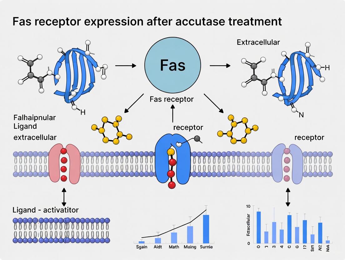
Accutase Compromises Fas Receptor Expression: A Critical Guide for Cell-Based Assays and Therapeutic Development
This article synthesizes current evidence demonstrating that Accutase, a commonly used enzymatic cell detachment solution, significantly cleaves and reduces the cell surface expression of the Fas receptor (CD95) and its...
Accutase Compromises Fas Receptor Expression: A Critical Guide for Cell-Based Assays and Therapeutic Development
Abstract
This article synthesizes current evidence demonstrating that Accutase, a commonly used enzymatic cell detachment solution, significantly cleaves and reduces the cell surface expression of the Fas receptor (CD95) and its ligand (FasL). Targeted at researchers and drug development professionals, this review covers the foundational mechanism of Accutase-induced protein cleavage, provides methodological guidance for detecting this artifact, outlines troubleshooting strategies to restore authentic protein expression, and validates non-enzymatic detachment methods. Understanding this pitfall is crucial for ensuring the accuracy of apoptosis, immunology, and cytotoxicity assays in biomedical research.
The Accutase Artifact: Uncovering Its Direct Impact on Fas Receptor Integrity
In cell culture, the process of detaching adherent cells is a fundamental step required for passaging, experimentation, and therapeutic application. The choice of detachment method, however, is far from trivial, as it can significantly influence cell viability, phenotype, and the integrity of critical surface markers. While trypsinization remains a widely used enzymatic method, its tendency to damage cell surface proteins has driven the adoption of gentler alternatives like accutase. Nevertheless, even accutase may not be suitable for all experimental endpoints. Emerging research specifically within the context of Fas receptor (Fas, CD95) and Fas ligand (FasL) biology indicates that certain enzymatic detachment methods can cleave these proteins, thereby compromising experimental results and potentially altering cellular function. This guide objectively compares the performance of common cell detachment methods, with a focused analysis on their impact on Fas receptor expression, to aid researchers in selecting the most appropriate technique for their work.
Cell detachment strategies generally fall into three categories: enzymatic, non-enzymatic, and mechanical. Each operates through a distinct mechanism to disrupt cell adhesion, with varying consequences for cell health and surface marker preservation.
Table 1: Overview of Common Cell Detachment Methods
| Method | Mechanism of Action | Key Advantages | Major Pitfalls | Ideal Use Cases |
|---|---|---|---|---|
| Trypsin [1] [2] | Proteolytic enzyme that cleaves peptides after lysine or arginine. | High efficiency; fast; cost-effective. | Degrades most cell surface proteins; can reduce cell viability; animal-derived. | Routine passaging of robust cell lines where surface marker integrity is not critical. |
| Accutase [1] [3] [4] | A blend of proteolytic and collagenolytic enzymes. | Gentle on cells; preserves many surface antigens; high viability; serum-free. | Can compromise specific surface proteins (e.g., Fas, FasL) [1]; requires recovery time. | Sensitive cell types (stem cells, primary cells); flow cytometry for most markers. |
| EDTA-based Solutions [1] [2] | Calcium chelator that disrupts integrin-mediated adhesion. | Non-enzymatic; avoids protein cleavage. | Ineffective for strongly adherent cells; may require mechanical assistance. | Lightly adherent cell lines; when complete preservation of surface proteins is essential. |
| Mechanical Scraping [1] | Physical dislodgement of cells. | Preserves surface proteins intact; no chemical exposure. | Can cause significant cell damage and death; not suitable for sensitive applications. | As a last resort for extremely adherent cells when protein integrity is the sole priority. |
The Impact of Detachment on Fas Receptor and Ligand: Experimental Evidence
The Fas/FasL pathway is a critical mediator of apoptosis and immune function. Recent studies demonstrate that the choice of detachment method can directly artifact experimental outcomes related to this pathway.
Key Experimental Findings
A 2022 study provides direct experimental evidence that accutase significantly decreases the surface levels of both Fas ligand and Fas receptor on murine macrophages (RAW264.7 and J774A.1 cells) compared to EDTA-based detachment or scraping [1]. The mean fluorescence intensity (MFI) of FasL was profoundly reduced after just 10 minutes of accutase treatment, while surface levels of another macrophage marker (F4/80) remained unchanged, indicating a specific effect on Fas/FasL [1].
Mechanism of Cleavage: Immunoblotting analysis revealed that accutase cleaves the extracellular portion of FasL, releasing small fragments under 20 kD into the supernatant. This cleavage was not observed in EDTA-treated cells [1]. Furthermore, immunofluorescence staining confirmed that FasL was no longer localized to the cell membrane after accutase treatment [1].
Reversibility and Recovery Time: The effects of accutase on Fas/FasL are reversible, but require a significant recovery period. The surface expression of Fas and FasL on macrophages took up to 20 hours in complete medium to return to normal levels after accutase detachment [1]. This finding is critical for planning experiments, as immediate analysis post-detachment will yield inaccurate data.
Table 2: Quantitative Impact of Accutase on Fas/FasL Expression (from [1])
| Experimental Metric | Accutase Treatment | EDTA-based Treatment | Mechanical Scraping | Notes |
|---|---|---|---|---|
| FasL Mean Fluorescence Intensity (MFI) | Significantly decreased (p<0.001) | Moderately decreased | Highest preservation | Incubation time (10-30 min) increased effect. |
| Fas Receptor MFI | Significantly decreased | Preserved | Not reported | Trend similar to FasL. |
| Cell Viability | High (maintained after 60-90 min) | Lower than Accutase | Not quantitatively reported | Accutase superior for maintaining viability. |
| Time for Full Surface Protein Recovery | ~20 hours | Not required | Not required | Critical for post-detachment assay timing. |
Detailed Experimental Protocol for Evaluating Detachment Methods
The following methodology, adapted from the cited research, can be used to validate the impact of detachment on specific cell types and surface markers of interest [1].
Objective: To compare the effect of different cell detachment methods on the surface expression of Fas and FasL via flow cytometry.
Materials:
- Adherent cells (e.g., RAW264.7 macrophages)
- Cell detachment solutions: Accutase, EDTA-based solution (e.g., Versene), Trypsin
- Complete cell culture medium
- Phosphate Buffered Saline (PBS)
- Flow cytometry antibodies: Anti-FasL, Anti-Fas, and a non-related surface marker antibody (e.g., Anti-F4/80 for macrophages)
- Flow cytometer
Procedure:
- Cell Culture: Grow adherent cells to 80-90% confluency in standard conditions.
- Detachment: For each detachment reagent, wash cells with PBS and then add enough solution to cover the monolayer.
- Accutase & Trypsin: Incubate at 37°C for 5-10 minutes. Monitor under microscope until cells round up.
- EDTA-based solution: Incubate at 37°C for 10-30 minutes. Tapping may be required.
- Neutralization: Gently collect the cell suspension.
- For trypsin, neutralize with serum-containing medium.
- For accutase and EDTA, dilution with PBS or serum-free medium is sufficient [4].
- Cell Processing: Centrifuge the harvested cells, wash with PBS, and resuspend in flow cytometry buffer.
- Staining: Aliquot cells and stain with fluorescently-labeled antibodies against FasL, Fas, and a control surface marker. Include an unstained control.
- Analysis: Analyze samples on a flow cytometer. Collect data for at least 10,000 events per sample. Compare the Mean Fluorescence Intensity (MFI) of each marker across the different detachment groups.
Diagram: Experimental Workflow for Detachment Method Comparison
The Fas Signaling Pathway and Its Critical Functions
The Fas receptor and its ligand (FasL) are members of the tumor necrosis factor (TNF) superfamily and play a pivotal role in regulating programmed cell death (apoptosis) and immune homeostasis [5]. Fas is widely expressed on many cell types, while FasL is primarily expressed on activated T-cells and Natural Killer (NK) cells [5].
Diagram: Simplified Fas-Mediated Apoptotic Signaling Pathway
This pathway is not only essential for immune cell cytotoxicity and the elimination of infected or cancerous cells, but it also governs the persistence of engineered lymphocytes like CAR-T and CAR-NK cells in cancer immunotherapy [6]. Disruption of Fas signaling can enhance the survival and antitumor efficacy of these therapeutic cells [6]. Therefore, accurate assessment of Fas and FasL expression is crucial in both basic immunology research and the development of advanced cell therapies. The finding that accutase can cleave these proteins highlights a significant potential pitfall in pre-clinical workflow.
The Scientist's Toolkit: Essential Research Reagents
Table 3: Key Reagents for Cell Detachment and Fas Expression Studies
| Reagent | Function in Research | Application Notes |
|---|---|---|
| Accutase [3] [4] | Gentle enzymatic cell detachment. | Preferred for sensitive cells (stem cells, primary cells). Caution: Can cleave Fas/FasL [1]. |
| EDTA-based Solution [1] | Non-enzymatic cell detachment via calcium chelation. | Ideal for preserving surface proteins like Fas/FasL; may be less efficient for strongly adherent cells. |
| Recombinant FasL Protein | To stimulate the Fas apoptotic pathway in functional assays. | Used to test Fas receptor functionality after detachment. |
| Anti-Fas / Anti-FasL Antibodies | Detection of surface receptor and ligand expression via flow cytometry or immunofluorescence. | Critical for quantifying the impact of detachment methods. |
| Flow Cytometry Assay Kits | (e.g., Caspase-3/7 activation, viability stains). | For assessing functional apoptosis and cell health post-detachment. |
| HEPES Buffered Saline | Maintaining stable pH during detachment outside a CO₂ incubator. | Ensures consistent enzymatic activity and cell health. |
The selection of a cell detachment method is a critical experimental variable that directly influences data integrity. While accutase is a superior gentle reagent for maintaining cell viability and preserving many surface antigens, evidence shows it specifically cleaves Fas and FasL, requiring a ~20-hour recovery period for re-expression [1]. For studies where the Fas/FasL pathway is a primary endpoint, non-enzymatic methods like EDTA or mechanical scraping, despite their own limitations, provide more accurate representation of native surface expression. Researchers must therefore align their detachment strategy with their specific experimental goals, validating the impact on key markers for their model system to avoid analytical pitfalls.
Cell detachment is a fundamental step in the culture of adherent cells, yet the method chosen can profoundly influence experimental outcomes, particularly in the study of sensitive cell surface proteins. This guide provides a mechanistic comparison of common cell detachment agents, with a focused examination of how the enzymatic solution Accutase specifically cleaves and alters the Fas Ligand (FasL) and its receptor. We present experimental data demonstrating that while Accutase offers excellent cell viability, its proteolytic activity can compromise the extracellular domains of key signaling proteins, an effect that is often reversible but requires substantial recovery time. Researchers must weigh these factors carefully when designing experiments involving flow cytometry, apoptosis assays, or immunotherapies reliant on intact Fas-FasL signaling.
In the cell culture environment, adherent cells require detachment for subculturing or analysis. Traditional methods range from harsh proteolytic enzymes to gentler non-enzymatic alternatives. Trypsin, a widely used enzymatic agent, is known to degrade most surface proteins and the extracellular matrix. Accutase, often marketed as a milder enzymatic replacement for trypsin, is believed to preserve cell surface markers better while providing efficient detachment. However, emerging evidence indicates that Accutase may selectively target certain surface proteins, notably the Fas receptor (Fas, CD95) and its ligand (FasL, CD95L), which are crucial mediators of apoptosis and immune function [7].
The Fas-FasL system is not only critical for immune homeostasis and cytotoxic T-cell function but is also gaining attention in cancer immunotherapy, particularly for its role in bystander killing of antigen-negative tumor cells by CAR-T cells [8]. Therefore, understanding how cell detachment methods affect this signaling axis is of paramount importance for experimental integrity and therapeutic development.
Mechanistic Action of Accutase on Fas Ligand
Proteolytic Cleavage of the Extracellular Domain
Accutase cleaves the extracellular domain of FasL into small fragments. Western blot analysis has revealed the presence of FasL fragments under 20 kD in the supernatant of Accutase-treated cells, which are absent in samples treated with EDTA-based solutions. This indicates that Accutase actively proteolyzes the receptor-binding portion of the membrane-anchored FasL [7].
Immunofluorescence staining corroborates these findings, showing that after Accutase treatment, FasL proteins are largely absent from the cell membrane, unlike in EDTA-treated cells where FasL remains properly localized on the membrane. This cleavage effectively impairs FasL-mediated signaling pathways by removing its capacity to bind the Fas receptor [7].
Specificity and Reversibility
The effect of Accutase is specific; it significantly reduces surface levels of FasL and Fas receptor but does not alter the expression of other markers like the murine macrophage-specific protein F4/80. This specificity suggests that the cleavage is not a result of general protein degradation but rather a targeted proteolytic event [7].
Furthermore, this effect is reversible. After removing Accutase and allowing cells to recover in complete medium, the surface expression of FasL and Fas receptor gradually returns. The recovery process is slow, requiring up to 20 hours to return to pre-treatment expression levels, indicating a significant cellular investment in repairing the damage caused by the enzyme [7].
Comparative Analysis of Cell Detachment Methods
The following data, synthesized from controlled studies, quantitatively compares the performance of Accutase against other common detachment methods, with particular focus on their impact on Fas pathway components.
Table 1: Impact of Detachment Methods on Fas Pathway and Cell Viability
| Detachment Method | Effect on Surface FasL | Effect on Surface Fas Receptor | Cell Viability | Key Advantages | Key Limitations |
|---|---|---|---|---|---|
| Accutase | Significant decrease (Reversible after ~20h) [7] | Significant decrease (Reversible after ~20h) [7] | Excellent (Significantly higher than EDTA after 60-90 min) [7] | Gentle; high viability; effective for strongly adherent cells [7] | Cleaves FasL extracellular domain; requires long recovery time for surface protein studies [7] |
| EDTA-Based Solutions | Minimal decrease [7] | Minimal decrease [7] | Moderate | Non-enzymatic; preserves most surface proteins [7] | Less potent; may require mechanical assistance (scraping) for strongly adherent cells [7] |
| Mechanical Scraping | Best preservation (Highest levels of surface FasL) [7] | Data not explicitly stated | Lower risk of proteolytic damage | Preserves surface protein integrity [7] | May cause cellular damage and reduce viability due to mechanical shear [7] |
| Trypsin | Presumed significant decrease (based on general proteolytic activity) [7] | Presumed significant decrease (based on general proteolytic activity) [7] | Variable (often lower than Accutase) [7] | Fast and potent dissociation [7] | Degrades most surface proteins and extracellular matrix components [7] |
Table 2: Quantitative Flow Cytometry Data (Mean Fluorescence Intensity - MFI) of Surface Markers Post-Detachment
| Detachment Method | FasL MFI | Fas Receptor MFI | F4/80 MFI (Control Marker) |
|---|---|---|---|
| Scraping | 100% ± 5% (Baseline) [7] | Data not explicitly stated | 100% ± 5% (Baseline) [7] |
| EDTA-Based Solution | ~90% ± 6% (Slight decrease) [7] | ~85% ± 7% (Slight decrease) [7] | ~98% ± 4% (Unaffected) [7] |
| Accutase (10 min) | ~40% ± 8% (Significant decrease) [7] | ~45% ± 9% (Significant decrease) [7] | ~99% ± 5% (Unaffected) [7] |
| Accutase (30 min) | ~35% ± 7% (Significant decrease) [7] | ~40% ± 8% (Significant decrease) [7] | ~97% ± 4% (Unaffected) [7] |
Experimental Protocols for Key Findings
Protocol: Assessing FasL Expression Post-Detachment
This protocol is adapted from the methodology used to generate the comparative data in Table 2 [7].
- Cell Culture: Culture adherent cells (e.g., RAW264.7 or J774A.1 murine macrophage cell lines) in standard conditions until they reach 80-90% confluency.
- Detachment Treatments:
- Accutase Group: Incubate cells with Accutase solution for 10-30 minutes at 37°C.
- EDTA Group: Incubate cells with a commercial, non-enzymatic EDTA-based solution (e.g., Versene) for 30 minutes at 37°C.
- Scraping Group: Use a cell scraper to mechanically dislodge cells in DPBS.
- Flow Cytometry Staining: Neutralize the detachment agents with complete medium. Wash cells and stain with fluorescently-labeled antibodies against FasL, Fas receptor, and a control surface protein (e.g., F4/80 for macrophages). Use isotype controls to set baselines.
- Analysis: Analyze cells using a flow cytometer. Calculate the Mean Fluorescence Intensity (MFI) for each marker and normalize to the scraping group to determine the percentage of surface protein remaining.
Protocol: Western Blot Analysis of FasL Cleavage
This protocol details the steps to confirm the mechanistic cleavage of FasL by Accutase, as shown in Figure 3 of the primary source [7].
- Cell Treatment and Sample Collection:
- Detach cells using Accutase and EDTA-based solutions as in Protocol 4.1.
- Collect cell supernatants by centrifugation to capture shed proteins.
- Harvest cell membranes to prepare lysates.
- Gel Electrophoresis and Transfer: Separate proteins from supernatants and lysates using SDS-PAGE. Transfer the separated proteins onto a nitrocellulose or PVDF membrane.
- Immunoblotting: Probe the membrane with an antibody specific for the extracellular portion of FasL.
- Expected Result: The blot will show small FasL fragments (<20 kD) in the supernatant and lysate of Accutase-treated cells. In contrast, the EDTA-treated sample will show full-length FasL (~40 kD) in the lysate and possibly some in the supernatant (from damaged cells during mechanical detachment).
Protocol: Determining Recovery Time of Surface FasL
This protocol assesses the reversibility of Accutase's effect [7].
- Detachment and Recovery: Treat cells with Accutase for 30 minutes. After detachment, seed the cells into new culture vessels with complete growth medium.
- Time-Course Harvesting: Harvest cells at various time points post-seeding (e.g., 0 h, 2 h, 6 h, 20 h) using a non-enzymatic method like scraping or a brief EDTA treatment to avoid repeated enzymatic damage.
- Analysis: Analyze surface FasL expression at each time point via flow cytometry as described in Protocol 4.1.
- Expected Outcome: Surface FasL levels will be lowest immediately after Accutase treatment (0 h) and will gradually increase, reaching near-baseline levels approximately 20 hours after recovery.
Visualization of Mechanisms and Workflows
FasL Cleavage and Recovery Pathway
FasL Cleavage and Recovery Process: This diagram illustrates the mechanistic pathway of Accutase-induced FasL cleavage and subsequent cellular recovery, showing the progression from intact protein to cleaved fragments and eventual restoration after a 20-hour recovery period.
Experimental Workflow for Detachment Comparison
Detachment Method Comparison Workflow: This diagram outlines the key steps for comparing cell detachment methods, from initial cell culture through different detachment treatments to multiple analysis techniques that assess surface protein expression, cleavage, and cell viability.
The Scientist's Toolkit: Essential Research Reagents
Table 3: Key Reagents for Studying Cell Detachment Effects
| Reagent / Material | Function / Application | Example Use Case |
|---|---|---|
| Accutase | Enzymatic cell detachment solution; a blend of proteolytic and collagenolytic enzymes. | Gentle dissociation of strongly adherent cells while maintaining high viability. [7] |
| EDTA-Based Solution (e.g., Versene) | Non-enzymatic chelating agent that binds calcium, disrupting cell-adhesion protein interactions. | Detaching cells while preserving sensitive surface markers like FasL for flow cytometry. [7] |
| Fluorochrome-Labeled Anti-FasL Antibody | Primary antibody for detecting and quantifying surface FasL expression. | Staining for flow cytometric analysis of FasL levels post-detachment. [7] |
| Fluorochrome-Labeled Anti-Fas Receptor Antibody | Primary antibody for detecting and quantifying surface Fas receptor expression. | Staining for flow cytometric analysis of Fas receptor levels post-detachment. [7] |
| Anti-FasL Antibody for Western Blot | Polyclonal or monoclonal antibody for detecting FasL and its cleavage fragments in immunoblotting. | Confirming proteolytic cleavage of FasL in cell lysates and supernatants. [7] |
| Cell Scraper | Mechanical tool for physically dislodging adherent cells from culture surfaces. | Harvesting cells without using chemical or enzymatic agents, preserving surface protein integrity. [7] |
| CCK-8 Assay Kit | Cell counting kit for assessing cell viability and proliferation. | Comparing the viability of cells after treatment with different detachment methods. [7] |
The choice of cell detachment method is a critical experimental parameter, especially for studies focused on the Fas-FasL signaling axis. While Accutase provides a gentle and effective means of cell dissociation that maximizes viability, its specific cleaving action on the extracellular domains of FasL and the Fas receptor presents a significant caveat.
Researchers must align their choice of detachment agent with their experimental endpoints. For immediate phenotypic analysis of surface proteins like FasL, non-enzymatic methods or scraping are superior. If Accutase is used for its other benefits, a recovery period of approximately 20 hours in complete culture medium is necessary to restore native surface expression of these proteins. This insight is indispensable for ensuring accurate data in flow cytometry, apoptosis research, and the development of immunotherapies that harness the Fas-FasL pathway.
Evidence of Decreased Surface Fas and FasL Expression Post-Accutase Treatment
Cell detachment reagents are essential tools in cell culture, yet their impact on critical cell surface proteins is often overlooked. This comparison guide evaluates the effect of various cell dissociation methods, with a particular focus on Accutase, a commonly used enzymatic solution, and its significant impact on the surface expression of Fas receptor (Fas) and Fas ligand (FasL). Experimental data demonstrate that Accutase treatment substantially reduces surface levels of these functionally linked proteins through proteolytic cleavage, unlike EDTA-based nonenzymatic solutions or mechanical scraping. The implications for immunological research and cancer therapy development are substantial, as the Fas/FasL pathway is a crucial regulator of apoptosis, immune homeostasis, and the efficacy of adoptive cell therapies. This analysis provides researchers with comprehensive experimental data and methodologies to inform appropriate cell detachment protocol selection for studies involving death receptor signaling pathways.
The Fas/FasL pathway represents a fundamental signaling system with widespread implications in immunology, cancer biology, and therapeutic development. Fas (CD95/APO-1), a member of the tumor necrosis factor receptor family, is ubiquitously expressed on most cells and tissues, particularly on immune cells such as activated macrophages and T cells [9] [5]. Its cognate ligand, FasL, is primarily expressed on activated T cells and natural killer cells and exists in both transmembrane (mFASL) and soluble (sFASL) forms [9] [5]. Upon interaction, Fas and FasL initiate critical biological processes including apoptotic signaling, immune regulation, and maintenance of cellular homeostasis [9] [5].
The functional integrity of this receptor-ligand pair is especially crucial in contemporary immunotherapy research. Recent investigations reveal that Fas/FasL interactions govern lymphocyte persistence through an autoregulatory circuit, directly impacting the efficacy of chimeric antigen receptor (CAR)-T and CAR-NK cell therapies [6]. Additionally, pathogens including bacteria and viruses have evolved mechanisms to modulate this pathway, further underscoring its biological significance [9] [5]. Given these critical functions, maintaining native conformation and surface expression of Fas and FasL during experimental procedures is paramount for obtaining biologically relevant data.
In standard cell culture practices, adherent cells require detachment methods for subculturing or analysis. While Accutase is frequently promoted as a gentle enzymatic alternative to trypsin, emerging evidence suggests it may compromise certain surface proteins [7]. This guide systematically evaluates the impact of Accutase treatment on Fas/FasL expression through direct comparison with alternative detachment methods, providing researchers with experimental data to inform protocol selection for death receptor studies.
Comparative Analysis of Cell Detachment Methods on Fas/FasL Surface Expression
Quantitative Comparison of Surface Protein Preservation
Table 1: Impact of Cell Detachment Methods on Surface Fas and FasL Expression in Macrophages
| Detachment Method | Mechanism of Action | Surface FasL MFI | Surface Fas MFI | F4/80 MFI (Control) | Cell Viability |
|---|---|---|---|---|---|
| Scraping (Mechanical) | Physical dislodgement | Highest preservation | Not reported | Not reported | Moderate |
| EDTA-based Solution | Calcium chelation | Moderate decrease | Preserved | No significant change | Moderate |
| Accutase (10 min) | Enzymatic cleavage | Significant decrease | Significant decrease | No significant change | High |
| Accutase (30 min) | Enzymatic cleavage | Maximum decrease | Maximum decrease | No significant change | High |
MFI: Mean Fluorescence Intensity; Data compiled from Scientific Reports [7]
The experimental data reveal striking differences in how detachment methods affect Fas/FasL surface expression. Mechanical scraping, while preserving the highest levels of surface FasL, often compromises cell viability due to physical shear stress [7]. EDTA-based nonenzymatic solutions, which function through calcium chelation to disrupt integrin-mediated adhesion, demonstrate moderate effects on FasL with preserved Fas expression and maintained viability [7].
Most significantly, Accutase treatment resulted in substantial reduction of both Fas and FasL surface expression in a time-dependent manner. In RAW264.7 macrophages, Accutase treatment for 10 minutes significantly decreased surface FasL levels compared to both scraping and EDTA-based solutions, with extended treatment (30 minutes) causing maximal reduction [7]. This effect was specific to certain surface proteins, as the macrophage-specific marker F4/80 remained unchanged under the same conditions [7]. Parallel experiments using J774A.1 cells confirmed similar trends, validating the consistency of this phenomenon across different cellular contexts [7].
Cell Viability and Functional Recovery Post-Treatment
Table 2: Functional Recovery and Viability Metrics Following Detachment
| Parameter | Scraping | EDTA-based Solution | Accutase |
|---|---|---|---|
| Viability after 60 min treatment | Moderate | Moderate | Highest (p < 0.01) |
| Viability after 90 min treatment | Low | Low | Highest (p < 0.001) |
| FasL Recovery Time | Not applicable | Not applicable | 20 hours |
| Fas Recovery Time | Not applicable | Not applicable | 20 hours |
| Mechanism of Impact | Physical disruption | Minimal direct effect | Proteolytic cleavage |
Data compiled from Scientific Reports [7]
Despite its detrimental effect on specific surface markers, Accutase demonstrated superior performance in maintaining cell viability. Even after extended incubation periods (60-90 minutes), Accutase-treated cells maintained significantly higher viability compared to those treated with EDTA solutions or phosphate-buffered saline buffer [7]. This apparent paradox—excellent viability despite protein cleavage—highlights the method's cell membrane-sparing properties while actively modifying specific surface proteins.
The Accutase-induced reduction in Fas/FasL surface expression proved reversible. After Accutase removal and incubation in complete medium, surface levels of both FasL and Fas required approximately 20 hours to recover, while F4/80 surface expression remained stable throughout the recovery period [7]. This recovery timeline has crucial implications for experimental planning, particularly for flow cytometry analyses or functional assays requiring intact death receptor signaling.
Molecular Mechanisms: Accutase-Induced Proteolytic Cleavage of Fas/FasL
Experimental Evidence for Direct Proteolysis
Western blot analysis of Accutase-treated RAW264.7 macrophages provided direct evidence of FasL cleavage. Immunoblotting revealed several small FasL fragments under 20 kD in size in the supernatant after Accutase treatment, which were absent in EDTA-treated cellular supernatants [7]. Furthermore, membrane lysates from Accutase-treated macrophages showed nearly complete cleavage of FasL to 20 kD fragments, whereas FasL in EDTA-treated macrophage lysates remained at the expected approximately 40 kD size [7].
Immunofluorescence staining corroborated these findings, demonstrating that most FasL proteins in Accutase-treated cells were not localized to the cell membrane, in stark contrast to the distinct membrane localization observed in EDTA-treated cells [7]. This redistribution from membrane to cytoplasmic compartments explains the reduced surface detection via flow cytometry.
The specificity of this cleavage is noteworthy, as Accutase did not affect all surface proteins equally. The preservation of F4/80 expression indicates that the enzyme mixture in Accutase has particular specificity for certain membrane protein conformations or cleavage sites present in Fas and FasL [7].
Regulatory Context of Fas/FasL Proteolytic Processing
The Accutase-mediated cleavage of FasL mirrors physiological regulatory mechanisms. Matrix metalloproteinases (MMPs) naturally cleave the extracellular region of FasL, generating soluble fragments [7]. This process represents a known mechanism for regulating Fas/FasL signaling intensity and duration in physiological contexts.
Other proteases also participate in Fas/FasL modulation. Recent research has identified that a human-specific amino acid substitution (Pro153Ser) in FasL renders it particularly susceptible to cleavage by plasmin, a protease frequently elevated in solid tumors [10]. This evolutionary adaptation may contribute to differential outcomes of T-cell-based immunotherapies and highlights the broader significance of proteolytic regulation in death receptor signaling.
Beyond extracellular cleavage, intracellular regulatory mechanisms also control Fas surface expression. Fas-associated phosphatase 1 (FAP-1) binds to the C-terminal region of Fas and inhibits its export to the cell surface, increasing intracellular retention within the cytoskeleton network [11]. Conversely, inhibition of FAP-1 expression enhances surface Fas expression, demonstrating another layer of regulation that could potentially interact with detachment-induced effects [11].
Experimental Protocols for Assessing Detachment Method Impacts
Standardized Cell Detachment and Analysis Workflow
Figure 1: Experimental workflow for evaluating detachment method effects on surface protein expression.
Detailed Methodological Protocols
Cell Culture and Detachment Protocol
- Cell Lines: RAW264.7 murine macrophages or J774A.1 cells maintained in Dulbecco's Modified Eagle Medium supplemented with 10% fetal bovine serum [7].
- Detachment Conditions:
- Accutase: Incubate for 10-30 minutes at 37°C according to manufacturer's instructions [7].
- EDTA-based solution: Use commercial Versene solution (Thermo Fisher Scientific) or 2-5mM EDTA in PBS, incubate for 30 minutes at 37°C [7].
- Scraping: Use mechanical dislodgement with cell scrapers without enzymatic or chelating agents [7].
- Post-detachment processing: Neutralize enzymatic activity with complete medium, centrifuge at 300 × g for 5 minutes, and resuspend in appropriate buffer for subsequent analysis [7].
Flow Cytometry Analysis of Surface Markers
- Staining procedure: Harvest 1×10^5 cells and stain with primary antibodies (anti-FasL [MFL3-biotin] or anti-Fas) for 20 minutes at 4°C [7] [12].
- Antibody concentrations: Use manufacturer-recommended dilutions (typically 1:100-1:200) in PBS with 1% BSA.
- Secondary detection: For biotinylated primary antibodies, use R-phycoerythrin-labeled streptavidin at 1:300 dilution for 20 minutes at 4°C [12].
- Analysis: Perform on FACScan instrument with appropriate analysis software (e.g., CellQuest), collecting a minimum of 10,000 events per sample [7] [12].
- Control markers: Include staining for non-affected surface markers (e.g., F4/80 for macrophages) as internal controls for method specificity [7].
Western Blot Detection of Cleavage Fragments
- Sample preparation: Prepare total cell extracts using RIPA buffer (PBS, 1% NP-40, 0.5% sodium deoxycholate, 0.1% SDS) with protease inhibitor cocktail [11].
- Membrane fractionation: Alternatively, prepare Triton X-100-soluble and -insoluble fractions to distinguish membrane-associated proteins [11].
- Electrophoresis: Resolve 50-100μg of protein by SDS-8% or 10% PAGE under reducing conditions [11].
- Transfer and detection: Electro-transfer to PVDF membrane, block with 5% non-fat milk, and incubate with primary antibodies (anti-FasL for extracellular domain detection) [7].
- Fragment identification: Detect cleavage fragments using enhanced chemiluminescence, expecting full-length FasL at ~40kD and cleavage fragments under 20kD with Accutase treatment [7].
The Scientist's Toolkit: Essential Research Reagents
Table 3: Key Research Reagents for Fas/FasL Detachment Studies
| Reagent/Cell Line | Specific Function | Experimental Role |
|---|---|---|
| RAW264.7 Cells | Murine macrophage cell line | Primary model system for detachment studies |
| J774A.1 Cells | Murine macrophage cell line | Validation cell line |
| Accutase | Enzymatic detachment solution | Test detachment method |
| EDTA-based Versene | Nonenzymatic detachment solution | Comparative control method |
| Anti-FasL (MFL3) | Fas ligand detection antibody | Flow cytometry and western blot |
| Anti-Fas Antibody | Fas receptor detection antibody | Flow cytometry analysis |
| Anti-F4/80 Antibody | Macrophage marker antibody | Specificity control for non-affected proteins |
| RIPA Buffer | Protein extraction solution | Total cell lysate preparation |
| CHO-K Cells | Chinese hamster ovary cells | Recombinant protein expression [10] |
Implications for Research and Therapeutic Development
Consequences for Immunotherapy Research
The Accutase-induced reduction in Fas/FasL expression has profound implications for CAR-T cell research and development. Recent findings demonstrate that Fas/FasL interactions govern CAR-engineered lymphocyte persistence through an autoregulatory circuit, with FasLG expression primarily limited to endogenous T cells, natural killer cells, and CAR-T cells themselves [6]. Disruption of Fas signaling through dominant-negative receptors enhances antitumor efficacy in multiple mouse models, highlighting the therapeutic potential of modulating this pathway [6].
The discovery that Accutase cleaves FasL suggests that cell manufacturing processes could inadvertently modify this critical therapeutic molecule. As FasL-mediated bystander killing is essential for CAR-T efficacy against antigen-heterogeneous tumors [10], preserving its integrity during ex vivo manipulation is crucial. These findings underscore the need for careful protocol selection in cell therapy manufacturing to maintain native death receptor function.
Considerations for Pathogen-Host Interaction Studies
Pathogens frequently manipulate the Fas/FasL pathway to facilitate infection and immune evasion. For instance, Yersinia pestis degrades cell surface FasL via its Pla protease, inhibiting FAS-mediated apoptosis and creating an immunosuppressive microenvironment [9] [5]. Similarly, influenza A virus (H1N1) upregulates FasL to enhance extrinsic apoptosis, potentially maintaining viral replicative niches [9] [5].
The use of Accutase during experimental investigation of these pathogens could confound results by artificially modifying the very pathway under study. Researchers examining pathogen-induced modulation of death receptor signaling should select detachment methods that preserve native Fas/FasL surface expression or account for potential artifacts introduced by enzymatic treatment.
This comparative analysis demonstrates that Accutase treatment significantly compromises surface expression of Fas and FasL through proteolytic cleavage, unlike EDTA-based solutions or mechanical scraping. While Accutase offers excellent cell viability preservation, its detrimental effects on these critical death receptors necessitates careful methodological consideration.
For studies focusing on death receptor signaling, immunotherapy development, or pathogen-host interactions, EDTA-based nonenzymatic detachment methods provide superior preservation of Fas/FasL surface expression. When Accutase must be used for practical reasons, researchers should incorporate appropriate recovery periods (approximately 20 hours) before assaying Fas/FasL-dependent functions or implement experimental controls to account for protein cleavage effects.
These findings highlight the broader principle that cell detachment methods are not universally interchangeable but represent strategic decisions that can significantly influence experimental outcomes in immunology and cancer research.
In cell-based research, the method used to detach adherent cells is a critical pre-analytical step that can significantly influence experimental outcomes. Certain enzymatic detachment reagents are known to cleave cell surface proteins, potentially compromising the study of specific markers. This guide objectively compares the effects of different cell detachment methods, focusing on the specific reduction of Fas receptor (Fas) and Fas ligand (FasL) expression, while confirming the stability of the macrophage marker F4/80. The data presented provides a framework for researchers to select appropriate dissociation protocols when evaluating Fas expression, ensuring the integrity of flow cytometry and functional apoptosis assays.
Comparative Data on Surface Marker Expression Post-Detachment
The table below summarizes quantitative flow cytometry data from studies investigating the impact of different detachment methods on surface marker expression.
Table 1: Impact of Cell Detachment Method on Surface Marker Expression in Murine Macrophages
| Surface Marker | Cell Type | Detachment Method | Effect on Expression (Mean Fluorescence Intensity) | Key Finding |
|---|---|---|---|---|
| Fas Receptor (Fas) | RAW264.7 macrophages | Accutase (10-30 min) | Significant decrease [7] | Expression is compromised by enzymatic cleavage. |
| EDTA-based solution | No significant decrease [7] | Recommended for preserving Fas. | ||
| Scraping | No significant decrease [7] | Recommended for preserving Fas. | ||
| Fas Ligand (FasL) | RAW264.7 macrophages | Accutase (10-30 min) | Significant decrease [7] | Expression is compromised; ligand is cleaved into fragments. |
| EDTA-based solution | Slight decrease [7] | Preferable over accutase. | ||
| Scraping | Highest level preserved [7] | Optimal method for preserving FasL. | ||
| F4/80 | RAW264.7 macrophages | Accutase (10-30 min) | No significant change [7] | Expression remains stable. |
| EDTA-based solution | No significant change [7] | Expression remains stable. | ||
| F4/80 | J774A.1 macrophages | Accutase | No significant change [7] | Expression remains stable in another macrophage line. |
| EDTA-based solution | No significant change [7] | Expression remains stable in another macrophage line. |
Mechanistic Insights: Why Fas is Affected and F4/80 is Not
The differential effect of Accutase on Fas/FasL versus F4/80 is rooted in the distinct structural and biochemical properties of these proteins.
Fas/FasL Sensitivity to Proteolytic Cleavage
The Fas/FasL pathway is a canonical mediator of apoptosis and inflammation [13]. The susceptibility of Fas and FasL to Accutase is likely due to the presence of protease-accessible sites on their extracellular domains.
- Experimental Evidence: Western blot analysis of macrophages detached with Accutase revealed the presence of small FasL fragments (under 20 kD) in the supernatant, while the full-length protein (approximately 40 kD) was detected in cell lysates from EDTA-treated samples. This indicates that Accutase actively cleaves the extracellular portion of FasL [7].
- Functional Consequence: Immunofluorescence staining confirmed that after Accutase treatment, FasL proteins were no longer localized to the cell membrane, providing a visual confirmation of the loss of surface expression [7]. This cleavage can directly interfere with Fas-FasL-mediated apoptosis assays.
F4/80 Resistance to Enzymatic Digestion
F4/80 (ADGRE1) is a well-established, highly specific surface marker for murine macrophages [14] [15]. Its resilience to Accutase treatment can be attributed to its unique molecular structure.
- Structural Robustness: F4/80 is a member of the adhesion G protein-coupled receptor (GPCR) family. It features a hybrid structure comprising multiple epidermal growth factor (EGF)-like domains at its extracellular N-terminus and a seven-span transmembrane domain at its C-terminus [14]. This complex EGF-TM7 configuration likely lacks cleavage sites susceptible to the proteolytic enzymes in Accutase or protects them within its tertiary structure.
- Functional Role: The F4/80 molecule itself plays a critical role in the generation of antigen-specific regulatory T cells (T-regs) and the induction of peripheral immune tolerance [14]. Its stable expression on the macrophage surface, even after Accutase treatment, makes it a reliable marker for identifying and isolating macrophages in experiments where other surface proteins like Fas are being studied [7].
Experimental Protocols for Key Findings
Protocol: Assessing Surface Marker Expression by Flow Cytometry
The following methodology was used to generate the comparative data in Table 1 [7].
- Cell Culture: Use the murine macrophage cell line RAW264.7 or J774A.1. Culture cells in standard DMEM or RPMI-1640 media supplemented with 10% Fetal Calf Serum (FCS) and 1% penicillin/streptomycin at 37°C in a 5% CO₂ atmosphere.
- Cell Detachment: At approximately 80% confluence, detach cells using the methods under investigation:
- Accutase: Incubate for 10-30 minutes at 37°C according to manufacturer instructions.
- EDTA-based solution: Incubate for 10-30 minutes at 37°C.
- Scraping: Use a cell scraper for mechanical detachment.
- Cell Staining & Analysis:
- Wash detached cells with PBS containing 0.5% Bovine Serum Albumin (BSA).
- Stain cells with fluorochrome-conjugated antibodies against Fas, FasL, and F4/80 for 30 minutes at 4°C.
- Analyze stained cells using a flow cytometer (e.g., FACSCalibur). Use forward/side scatter to gate on live cells.
- Quantify the Mean Fluorescence Intensity (MFI) for each marker and normalize to the control (scraping) group.
Protocol: Verifying Proteolytic Cleavage by Western Blot
This protocol confirms the direct cleavage of FasL by Accutase [7].
- Sample Preparation:
- Detach RAW264.7 macrophages using Accutase and an EDTA-based solution.
- Collect the cell culture supernatant and concentrate it to analyze released proteins.
- Prepare cell lysates from the detached pellets to analyze remaining cellular proteins.
- Gel Electrophoresis and Blotting:
- Separate proteins from supernatants and lysates by SDS-PAGE.
- Transfer proteins to a nitrocellulose or PVDF membrane.
- Immunodetection:
- Probe the membrane with an antibody specific to the extracellular portion of FasL.
- Use a horseradish peroxidase (HRP)-conjugated secondary antibody and develop with a chemiluminescent substrate.
- The expected result is the presence of low molecular weight FasL fragments (under 20 kD) in the supernatant of Accutase-treated cells, but not in EDTA-treated samples.
Signaling Pathways and Experimental Workflow
The following diagrams illustrate the Fas signaling pathway and the experimental workflow for evaluating detachment methods.
Diagram 1: The Fas-mediated apoptotic signaling pathway. Activation by FasL triggers a caspase cascade, leading to programmed cell death. Accutase cleaves FasL, disrupting this pathway initiation [13] [5].
Diagram 2: Experimental workflow for evaluating cell detachment methods. Key steps involve subjecting cells to different detachment protocols followed by analysis via flow cytometry and western blot to quantify and characterize surface marker expression [7].
The Scientist's Toolkit: Key Research Reagents
The table below lists essential materials and reagents used in the cited experiments for studying Fas and macrophage biology.
Table 2: Essential Reagents for Fas and Macrophage Surface Marker Research
| Reagent / Material | Function / Specificity | Example from Research |
|---|---|---|
| Anti-Fas Receptor Antibody | Flow cytometry and immunofluorescence detection of Fas. | Used to quantify surface Fas loss after Accutase treatment [7]. |
| Anti-Fas Ligand (FasL) Antibody | Detection of membrane-bound FasL (Western Blot, IF). | Antibody against the extracellular portion detected cleavage fragments [7]. |
| Anti-F4/80 Antibody | Specific marker for identifying murine macrophages. | Clone CI:A3-1 used to show F4/80 resistance to Accutase [7] [16]. |
| Accutase Detachment Solution | Proteolytic and collagenolytic enzyme mixture for cell dissociation. | The key reagent shown to cleave Fas/FasL but not F4/80 [7]. |
| EDTA-based Detachment Solution | Non-enzymatic chelating agent (e.g., Versene). | Used as a control method that better preserves Fas/FasL [7]. |
| RAW264.7 Cells | An immortalized mouse macrophage cell line. | Primary model system used for the detachment studies [7]. |
The data unequivocally demonstrates that the effect of Accutase on surface markers is highly specific. While it significantly compromises the detection of Fas and FasL, it leaves the expression of F4/80 unaffected. This specificity underscores the critical importance of selecting a cell detachment method that is tailored to the target proteins of interest.
For researchers evaluating Fas receptor expression:
- Avoid Accutase: Do not use Accutase for detachment immediately prior to analyzing Fas or FasL, as it causes rapid and significant cleavage.
- Opt for Milder Methods: Use EDTA-based solutions or mechanical scraping to preserve the integrity of the Fas-FasL axis.
- Allow for Recovery: If Accutase must be used for other reasons, note that surface levels of Fas/FasL require up to 20 hours of recovery culture post-detachment to return to baseline [7].
The stability of F4/80 across all detachment methods confirms its reliability as a robust macrophage marker, even in samples where other surface proteins have been degraded. By incorporating these findings into experimental design, scientists can ensure more accurate and reproducible data in immunology and apoptosis research.
Best Practices for Accurate Fas Detection in Accutase-Detached Cells
Recommended Protocols for Flow Cytometry After Enzymatic Detachment
For researchers studying specific cell surface receptors, such as Fas (CD95), the method used to detach adherent cells for flow cytometry analysis is not merely a procedural step but a critical experimental variable. The Fas receptor and its ligand (FasL) form a pivotal pathway governing extrinsic apoptosis, playing essential roles in immune homeostasis, cancer biology, and the efficacy of immunotherapies, including CAR-T cell bystander killing [17]. Consequently, preserving the native conformation and surface expression of these proteins during cell preparation is paramount for generating accurate, biologically relevant data. While enzymatic detachment solutions like trypsin and Accutase are widely used for their efficiency, emerging evidence indicates that they can profoundly compromise the detection of certain cell surface markers, with Fas and FasL being particularly susceptible [7]. This guide objectively compares the performance of different cell detachment methods, with a focused evaluation of their impact on Fas receptor expression, to aid researchers in selecting the most appropriate protocol for their experimental objectives.
Comparative Analysis of Cell Detachment Methods
The selection of a detachment method involves balancing cell viability, yield, and, crucially, the integrity of cell surface epitopes. The table below summarizes the key characteristics of common approaches.
Table 1: Comparison of Common Cell Detachment Methods for Flow Cytometry
| Detachment Method | Mechanism of Action | Impact on Fas/FasL | Cell Viability | Key Advantages | Major Limitations |
|---|---|---|---|---|---|
| Scraping | Mechanical dislodgement | Minimal impact; preserves highest surface levels [7] | Variable; can be low due to shear stress | Preserves surface antigen integrity fully | Can cause cell rupture and clumping; not suitable for all cell types |
| EDTA-based Solutions | Chelates calcium, disrupting integrin-mediated adhesion | Mild decrease possible; significantly preserves expression compared to enzymes [7] | Good | Non-enzymatic; gentle on most surface proteins | May be insufficient for strongly adherent cells; often requires mechanical aid |
| Accutase | Proteolytic, collagenolytic, and DNase activity [18] | Significant decrease in surface Fas and FasL; cleaves extracellular portion [7] | Excellent; maintained better than EDTA after prolonged incubation [7] | Effective for difficult-to-detach cells; high post-detachment viability | Compromises sensitive surface proteins like Fas/FasL; requires recovery time |
| Trypsin | Proteolytic; cleaves after lysine/arginine | Expected severe degradation (most surface proteins are affected) [7] [18] | Good, but can be over-digested | Rapid and highly effective | Degrades most surface proteins and extracellular matrix; harsh |
Quantitative Data on Fas Receptor Expression
A direct comparison of mean fluorescence intensity (MFI) from flow cytometry analysis quantifies the dramatic effect of Accutase on Fas receptor and ligand expression.
Table 2: Experimental Data on Detachment Method Impact on Surface Marker MFI
| Detachment Method | Fas Ligand (FasL) MFI | Fas Receptor MFI | Control Marker (F4/80) MFI |
|---|---|---|---|
| Scraping | Highest preservation [7] | Data not specifically provided | Unaffected [7] |
| EDTA-based Solution | Moderate decrease | Moderate decrease | Unaffected [7] |
| Accutase (10-min incubation) | Significant decrease [7] | Significant decrease [7] | Unaffected [7] |
| Accutase (30-min incubation) | Significant decrease (no major change from 10-min) [7] | Significant decrease (no major change from 10-min) [7] | Unaffected [7] |
Key Findings:
- Accutase specifically targets Fas/FasL: The surface expression of Fas and FasL is significantly reduced post-Accutase treatment, while the expression of other surface markers, such as the macrophage marker F4/80, remains unaltered. This indicates a specific susceptibility of the Fas-FasL complex to the enzyme mixture in Accutase [7].
- The effect is reversible but slow: The cleavage of Fas/FasL by Accutase is not permanent. However, cells require approximately 20 hours of recovery in complete culture medium post-detachment to fully restore surface expression levels [7].
- Mechanism of action: Western blot and immunofluorescence analyses confirm that Accutase cleaves the extracellular portion of FasL, releasing it from the cell membrane and thereby abolishing its detection by flow cytometry and its functional capacity [7].
Detailed Experimental Protocols
Below are standardized protocols for detaching adherent cells using the methods discussed, optimized for subsequent flow cytometry analysis.
Non-Enzymatic Detachment using EDTA
This protocol is recommended for studies where preserving sensitive epitopes like Fas is critical [7] [19].
Materials:
- EDTA-based non-enzymatic cell dissociation solution (e.g., Versene, 2-5 mM in PBS)
- Phosphate-Buffered Saline (PBS), without calcium and magnesium
- Flow Cytometry Staining Buffer (PBS with 1-2% FBS and optional sodium azide)
- Centrifuge tubes
- Cell culture-grade scraper (if needed)
Procedure:
- Aspirate the culture medium from the adherent cells and wash the monolayer gently with PBS to remove residual serum and divalent cations.
- Add enough EDTA solution to cover the cell monolayer (e.g., 3 mL for a T75 flask).
- Incubate at 37°C for 5-15 minutes. Observe cells under a microscope for rounding and detachment. For strongly adherent cells, gentle tapping or scraping may be required to aid detachment [7].
- Neutralize the EDTA by adding a sufficient volume of complete culture medium (containing serum) or staining buffer.
- Transfer the cell suspension to a centrifuge tube. Gently pipette to dissociate any clumps.
- Centrifuge at 300-400 x g for 5 minutes. Discard the supernatant.
- Resuspend the cell pellet in flow cytometry staining buffer and proceed with antibody staining [20].
Enzymatic Detachment using Accutase
Use this protocol with the understanding that a recovery period is essential for restoring sensitive proteins like Fas.
Materials:
- Accutase enzyme cell detachment medium
- Complete cell culture medium
- Flow Cytometry Staining Buffer
- Centrifuge tubes
Procedure:
- Aspirate and wash the cell layer with PBS as described in the EDTA protocol.
- Add enough Accutase to cover the monolayer.
- Incubate at 37°C for 10-30 minutes, or until most cells have detached. Accutase is gentle and often does not require mechanical dislodgement [7].
- Neutralize by adding a double volume of complete culture medium.
- Transfer, centrifuge, and resuspend as in steps 5-7 of the EDTA protocol.
- Critical Recovery Step: If Fas or other compromised proteins are the target of analysis, seed the detached cells in a culture flask with fresh complete medium and allow them to recover for 20-24 hours before re-harvesting for flow cytometry (using a gentle method like EDTA for the second harvest) [7].
"No-Touch" Staining Protocol for Adherent Cells in Microplates
This advanced protocol minimizes cell loss and avoids detachment-associated antigen damage entirely, ideal for high-throughput screening [19].
Materials:
- Tissue culture-treated 384-well flat-bottom microplates
- EDTA solution (15 mM)
- Antibodies diluted in serum-free medium
- PBS with 2% FCS and 2 μM EDTA
Procedure:
- Seed and Culture: Plate adherent cells directly into a 384-well microplate and culture until the desired confluency is reached after compound treatment.
- Detach with EDTA: Add 5 μL of 15 mM EDTA directly to each well containing 15 μL of culture medium (final EDTA concentration ~3 mM). Shake the plate orbially and incubate at 37°C for 45 minutes to allow detachment.
- Stain In-Situ: Directly add 5 μL of pre-diluted antibody cocktail to each well. Shake the plate at 300 RPM and incubate for 20 minutes at 4°C.
- Dilute and Analyze: Add 50 μL of PBS with 2% FCS and 2 μM EDTA to each well to dilute the sample. The plate is now ready for acquisition on a high-throughput flow cytometer without any washing, centrifugation, or transfer steps [19].
The Fas Signaling Pathway and Experimental Impact
Understanding the biological context of Fas reinforces why detachment method choice is critical. The Fas receptor is a key death receptor in the TNF superfamily. Upon engagement by its ligand (FasL), it trimerizes and recruits adapter proteins to form the Death-Inducing Signaling Complex (DISC), initiating a caspase cascade that leads to apoptosis [17]. This pathway is vital for immune cell regulation, tumor suppression, and the bystander killing effect of CAR-T cells in solid tumors [17].
Accutase directly cleaves the extracellular domain of FasL, rendering it undetectable by flow cytometry and non-functional [7]. This artifact can lead to falsely negative results and misinterpretation of the physiological state of the cells. The following diagram illustrates the structure of Fas and the critical epitope affected by enzymatic detachment.
The Scientist's Toolkit: Essential Research Reagents
Table 3: Key Reagents for Cell Preparation and Fas Analysis
| Reagent / Solution | Primary Function | Key Considerations for Fas Studies |
|---|---|---|
| EDTA-based Solution | Non-enzymatic cell detachment via calcium chelation | First choice for preserving Fas/FasL; may be less effective for very adherent cell lines [7]. |
| Accutase | Enzymatic cell detachment mixture | Provides high viability but cleaves Fas/FasL; requires a 20-hour recovery period for analysis [7]. |
| Trypsin | Proteolytic enzyme for rapid cell detachment | Not recommended; causes widespread degradation of surface proteins [7] [18]. |
| Flow Cytometry Staining Buffer | Buffer for washing, resuspending, and antibody staining | Contains FBS to block non-specific binding and azide to prevent internalization; essential for clean staining [20]. |
| Agonistic Anti-Fas Antibody | Antibody that activates Fas receptor signaling | Used in functional apoptosis assays to test Fas pathway competence [21] [17]. |
The data unequivocally demonstrates that the choice of cell detachment method is a decisive factor in the successful detection of the Fas receptor and ligand via flow cytometry. While Accutase offers superior cell viability, its specific cleavage of Fas/FasL makes it a poor choice for experiments focused on these proteins without a sufficient recovery period.
For researchers in immunology and drug development, the following evidence-based recommendations are proposed:
- For Direct Analysis: Use EDTA-based non-enzymatic detachment or mechanical scraping to obtain the most accurate snapshot of basal Fas/FasL surface expression.
- When Using Accutase is Unavoidable: Plan for a minimum 20-hour recovery culture period after detachment before performing flow cytometry to allow for protein re-synthesis and membrane localization.
- For High-Throughput Screening: Adopt the "no-touch" EDTA staining method in microplates to completely bypass the artifacts introduced by traditional detachment and washing steps, thereby maximizing data integrity and throughput.
Western Blot Techniques to Detect Cleaved FasL Fragments
The detection of cleaved Fas Ligand (FasL) fragments via Western blotting presents unique technical challenges that require specialized methodologies and careful experimental design. As a type II transmembrane protein belonging to the tumor necrosis factor (TNF) family, FasL undergoes proteolytic processing at specific sites that generate distinct biological activities and cleavage fragments. This technical guide comprehensively compares Western blot techniques for detecting these fragments, with particular emphasis on methodologies relevant to researchers evaluating Fas receptor expression in the context of accutase treatment and other experimental conditions that may alter FasL processing. The accurate detection of FasL cleavage products is crucial for understanding its role in apoptotic signaling, immune regulation, and cancer biology, requiring researchers to navigate issues of antibody specificity, fragment stability, and appropriate control selection.
FasL Biology and Cleavage Context
FasL (CD95L) is a critical death-inducing cytokine that binds to its receptor Fas (CD95), initiating caspase-dependent apoptosis through formation of the death-inducing signaling complex (DISC) [22] [23]. The membrane-bound form of FasL (approximately 40 kDa) can be proteolytically cleaved by various enzymes to generate soluble fragments with distinct signaling properties. Understanding these cleavage events is essential for interpreting Western blot results and understanding FasL biology.
Multiple proteases target FasL at specific cleavage sites:
- Matrix metalloproteinases (MMPs) cleave the extracellular region, releasing soluble FasL [7]
- ADAM10 processes membrane-bound FasL, producing an N-terminal fragment that lacks the receptor-binding extracellular domain [8] [24]
- SPPL2A subsequently liberates the FasL intracellular domain through intramembrane cleavage [24]
- Plasmin specifically cleaves human FasL at the 144RK145 site, a processing event unique to humans due to an evolutionary Pro153Ser substitution not found in non-human primates [8]
- Accutase, a cell detachment solution, cleaves the extracellular portion of FasL into fragments smaller than 20 kDa [7]
The molecular weights of detected fragments provide crucial information about the specific cleavage events occurring in experimental systems, making Western blotting an indispensable tool for FasL research.
Critical Methodological Considerations
Sample Preparation Protocols
Proper sample preparation is fundamental for accurate detection of FasL cleavage fragments. The choice of cell detachment method significantly impacts results, as enzymatic treatments can artificially cleave surface proteins including FasL.
Cell Detachment Methods: Comparative studies demonstrate that accutase treatment cleaves FasL extracellular domains into fragments under 20 kDa, while EDTA-based nonenzymatic detachment better preserves full-length FasL (approximately 40 kDa) [7]. When studying FasL processing, mechanical detachment methods such as scraping may provide the most reliable preservation of native FasL expression, though viability may be compromised.
Recovery Time Optimization: Research indicates that cells detached with accutase require approximately 20 hours of recovery to restore surface expression of FasL and Fas receptor to normal levels [7]. Experimental timelines should incorporate this recovery period when accutase treatment is unavoidable.
Lysis Conditions: Utilize modified RIPA buffer containing protease inhibitors (including metalloproteinase inhibitors) to prevent post-lysis cleavage. Immediate processing or storage at -80°C is recommended to preserve cleavage patterns.
Electrophoresis and Detection Techniques
Standard Western blot protocols require modification for optimal FasL fragment detection:
Gel Selection: 12-15% SDS-PAGE gels provide optimal resolution for fragments in the 15-40 kDa range. Gradient gels (4-20%) offer superior separation when analyzing multiple cleavage products simultaneously.
Transfer Conditions: Semi-dry transfer at constant current (2.5 mA/cm² for 60 minutes) using PVDF membranes enhances retention of lower molecular weight fragments. Nitrocellulose membranes may provide superior signal for some antibodies.
Antibody Selection: Choose antibodies targeting specific FasL epitopes based on experimental goals. Antibodies against the extracellular domain (e.g., AF0157) detect most cleavage fragments, while those against intracellular domains specifically identify the FasL intracellular domain generated by SPPL2A cleavage [24].
Table 1: FasL Cleavage Events and Detection Parameters
| Cleavage Enzyme | Cleavage Site | Fragment Sizes | Biological consequence | Detection Tips |
|---|---|---|---|---|
| MMPs | Extracellular domain | ~30 kDa soluble form | Altered signaling capacity | Extracellular domain antibodies |
| ADAM10 | Stalk region | N-terminal fragment | Loss of receptor binding | Reduced full-length signal |
| Plasmin | 144RK145 (human specific) | ~40 aa short fragment | Abolished cell death function [8] | Epitope mapping critical |
| Accutase | Multiple extracellular sites | <20 kDa fragments | Experimental artifact [7] | Avoid or allow recovery |
| SPPL2A | Intramembrane | Intracellular domain | Potential nuclear signaling [24] | Intracellular domain antibodies |
Controls and Validation
Robust experimental design necessitates appropriate controls for accurate interpretation:
Positive Controls: Commercially available Jurkat Apoptosis Cell Extracts (etoposide-treated) or Caspase-3 Control Cell Extracts provide validated positive controls for apoptosis-related proteins [25]. These extracts contain detectable levels of multiple caspases and cleaved substrates.
Specificity Validation: Include FasL knockout cells or siRNA-mediated knockdown to confirm antibody specificity. Pre-absorption with immunizing peptide validates signal identity when using polyclonal antibodies.
Loading Controls: GAPDH or other stable housekeeping proteins ensure equal loading, though their stability under apoptotic conditions should be verified.
Experimental Workflow for FasL Cleavage Detection
The following diagram illustrates the comprehensive experimental workflow for detecting FasL cleavage fragments, integrating critical decision points and methodological considerations:
FasL Cleavage Signaling Pathways
The biological context of FasL cleavage encompasses multiple signaling pathways that influence experimental outcomes:
Comparative Analysis of Detection Approaches
Table 2: Antibody-Based Detection Strategies for FasL Fragments
| Detection Target | Antibody Example | Recommended Applications | Advantages | Limitations |
|---|---|---|---|---|
| Extracellular domain | AF0157 (Affinity Biosciences) [24] | General FasL detection, most cleavage fragments | Broad reactivity to multiple forms | Cannot distinguish specific cleavage events |
| Intracellular domain | Custom antibodies | SPPL2A cleavage products | Specific for intracellular domain | Misses extracellular fragments |
| Specific cleavage sites | Cleavage-specific antibodies (limited availability) | Detection of specific proteolytic events | High specificity for cleavage event | Limited commercial availability |
| Tagged constructs | Anti-His, Anti-Fc | Recombinant FasL studies | Controlled experimental system | May not reflect endogenous regulation |
Table 3: Key Research Reagents for FasL Cleavage Studies
| Reagent/Category | Specific Examples | Function/Application | Considerations |
|---|---|---|---|
| Cell Detachment Reagents | EDTA-based solutions (Versene) [7] | Preserves surface FasL expression | Mild but may require scraping |
| Positive Controls | Jurkat Apoptosis Cell Extracts (etoposide) [25] | Apoptosis signaling validation | Contains multiple caspase cleavages |
| Protease Inhibitors | Metalloproteinase inhibitors | Prevents post-lysis cleavage | Critical for accurate fragment patterns |
| FasL Antibodies | AF0157 (extracellular domain) [24] | Detects most FasL forms | Reactivity to human and mouse |
| Plasmin Inhibitors | Aprotinin, tranexamic acid | Studying plasmin-mediated cleavage [8] | Human-specific relevance |
| Caspase Inhibitors | Z-VAD-FMK [23] | Distinguishing direct and indirect effects | Blocks caspase-mediated Grx1 degradation |
Technical Challenges and Troubleshooting
Researchers face several specific challenges when detecting FasL cleavage fragments:
Fragment Stability: Cleaved FasL fragments, particularly those generated by accutase treatment, may be rapidly degraded. Inclusion of complete protease inhibitor cocktails (including metalloproteinase inhibitors) during sample preparation is essential.
Antibody Epitope Accessibility: Some cleavage events may compromise antibody binding sites. For example, plasmin cleavage at 144RK145 in human FasL completely eliminates detection with certain antibodies due to epitope destruction [8]. Using antibodies targeting different epitopes or Fc-tagged FasL constructs can circumvent this issue.
Multiple Cleavage Events: Simultaneous cleavage by different proteases can generate complex fragment patterns. Inhibition experiments using specific protease inhibitors (e.g., GM6001 for MMPs, GI254023X for ADAM10) help decipher these patterns.
Human-Specific Regulation: The Pro153Ser substitution in human FasL renders it uniquely susceptible to plasmin cleavage compared to non-human primates [8]. This species specificity must be considered when selecting experimental models and interpreting results.
Western blot detection of cleaved FasL fragments demands meticulous methodology and understanding of FasL biology. The integration of appropriate controls, validated detection reagents, and standardized protocols enables accurate interpretation of FasL processing in diverse experimental contexts. Researchers must remain vigilant about technical artifacts, particularly those introduced by cell preparation methods like accutase treatment, while leveraging the growing toolkit of reagents and methodologies to advance our understanding of FasL regulation in health and disease.
Immunofluorescence Staining to Confirm Membrane Localization Loss
The Fas receptor (also known as CD95 or APO-1) is a critical cell surface death receptor belonging to the tumor necrosis factor receptor family. Its proper membrane localization is fundamental to executing programmed cell death and regulating immune responses [5]. Upon activation by its homologous ligand FasL, Fas receptor initiates the formation of the death-inducing signaling complex (DISC), which triggers caspase-8 activation and ultimately leads to apoptosis through both extrinsic and intrinsic pathways [5]. The receptor's position on the plasma membrane enables it to interact effectively with extracellular signals and initiate these crucial intracellular processes.
Recent research has revealed that certain laboratory reagents can unexpectedly compromise the membrane integrity of surface proteins. Specifically, accutase, a proteolytic enzyme blend commonly used for cell detachment, has been shown to significantly alter the membrane localization of Fas receptor and Fas ligand [7]. This discovery has profound implications for experimental accuracy in cell biology and immunology research, particularly for studies investigating death receptor signaling pathways. This guide provides a comprehensive comparison of methodologies to accurately assess Fas receptor membrane localization, with specific attention to the confounding effects of cell detachment agents.
Fas Receptor Signaling Pathways and Detection Challenges
Fas Receptor Biology and Technical Detection Challenges
The Fas receptor is a 48 kDa transmembrane protein composed of an extracellular domain containing cysteine-rich subdomains, a transmembrane structural domain, and a cytoplasmic region that includes a death domain [5]. When Fas ligand binds to the receptor, it promotes aggregation and conformational changes that trigger the assembly of intracellular signaling complexes. The receptor activates not only apoptotic pathways but also non-apoptotic signaling including NF-κB, MAPK, and PI3K/AKT pathways, which regulate immune responses, cell proliferation, migration, and invasion [5].
A significant methodological challenge in studying this receptor lies in the cell detachment process required for many analytical techniques. Research demonstrates that accutase, frequently used for dissociating adherent cells, cleaves the extracellular portion of Fas ligand and compromises Fas receptor membrane localization [7]. This proteolytic effect reversibly decreases surface expression levels, potentially confounding experimental results and leading to inaccurate conclusions about receptor expression and function.
Table 1: Comparison of Cell Detachment Methods for Fas Receptor Studies
| Detachment Method | Effect on Fas Receptor/Ligand | Recovery Time | Cell Viability | Best Use Cases |
|---|---|---|---|---|
| Scraping | Preserves highest surface levels of FasL | Immediate analysis possible | Moderate, potential for mechanical damage | Flow cytometry when enzymatic digestion must be avoided |
| EDTA-based Solutions | Minimal impact on surface expression | Immediate analysis possible | Good, maintains cell integrity | Routine Fas receptor studies requiring cell detachment |
| Trypsin | Extensive degradation of surface proteins | 20+ hours for full recovery | Variable, can damage surface receptors | Generally not recommended for Fas studies |
| Accutase | Significant decrease in surface Fas and FasL | 20 hours for full recovery | Excellent, even after extended treatment | Studies where receptor localization is not being analyzed |
Fas Receptor Signaling Pathway
The following diagram illustrates the key signaling pathways activated by the Fas receptor, highlighting its central role in controlling cell fate:
Comparative Analysis of Immunofluorescence Approaches
Traditional Immunofluorescence vs. Advanced Multiplexing Techniques
Immunofluorescence staining has evolved significantly from basic single-target detection to sophisticated multiplexed approaches that provide comprehensive spatial and molecular information. Each method offers distinct advantages and limitations for detecting subtle changes in membrane localization:
Standard Immunofluorescence typically relies on 1-3 marker panels and is widely accessible but offers limited molecular context. This approach can detect gross changes in Fas receptor localization but may miss subtle redistributions within complex cellular environments. The methodology involves sample fixation, permeabilization (for intracellular targets), primary antibody incubation, fluorophore-conjugated secondary antibody application, and imaging with standard fluorescence microscopy [26].
Multiplexed Immunofluorescence (MxIF) represents a technological advancement that enables simultaneous detection of numerous markers (10-40+) on a single sample. Techniques such as cyclic immunofluorescence (CyCIF) and one-shot multiplexing allow researchers to contextualize Fas receptor localization within complex cellular architectures and signaling environments [27] [28]. The Orion platform, for example, utilizes 16-18 parallel fluorescent channels with carefully selected ArgoFluor dyes coupled to antibodies against lineage markers, enabling precise subcellular localization assessment with minimal spectral overlap [28].
Experimental Workflow for Membrane Localization Assessment
The following diagram outlines a comprehensive experimental workflow for evaluating Fas receptor membrane localization using immunofluorescence:
Quantitative Comparison of Immunofluorescence Modalities
Table 2: Immunofluorescence Modalities for Membrane Localization Studies
| Technique | Plex Capacity | Spatial Resolution | Membrane Specificity | Equipment Requirements | Best Applications for Fas Studies |
|---|---|---|---|---|---|
| Standard IF | 1-3 markers | ~250 nm | Moderate | Standard fluorescence microscope | Initial screening of Fas receptor expression and gross localization changes |
| Confocal Microscopy | 3-5 markers | ~180 nm | High | Laser scanning confocal microscope | Detailed membrane vs. cytoplasmic distribution analysis |
| Multiplexed IF (MxIF) | 10-40+ markers | ~250 nm | High | Specialized imaging systems with spectral unmixing | Contextualizing Fas within complex cellular environments and signaling networks |
| One-shot MxIF (Orion) | 16-18 markers | ~220 nm | High | Custom platform with 7 lasers and tunable filters | Comprehensive spatial phenotyping with preserved membrane topology |
Experimental Protocols for Validating Fas Receptor Membrane Localization
Critical Protocol for Accutase-Treated Cell Analysis
When studying Fas receptor membrane localization in cells requiring detachment, follow this optimized protocol:
Cell Detachment and Recovery:
- For accutase-treated cells: Use gentle, EDTA-based nonenzymatic dissociation buffers instead of accutase to preserve surface Fas receptor integrity [7].
- If accutase must be used: Allow a 20-hour recovery period in complete culture medium after detachment and replating to enable surface protein reexpression [7].
- Include appropriate controls: Compare accutase-treated samples with EDTA-detached and scraped cells to establish baseline membrane localization patterns.
Immunofluorescence Staining:
- Fixation: Use 4% paraformaldehyde for 15 minutes at room temperature to preserve membrane architecture.
- Permeabilization: For membrane-specific staining, avoid permeabilization or use mild detergents (0.1% Triton X-100) for 5 minutes. Omit permeabilization step when analyzing surface receptors only.
- Blocking: Incubate with 5% BSA and 10% normal serum of secondary antibody host for 1 hour.
- Primary Antibody: Incubate with anti-Fas receptor antibody (e.g., CD95) diluted in blocking buffer overnight at 4°C. Include appropriate isotype controls.
- Secondary Antibody: Apply fluorophore-conjugated secondary antibody for 1 hour at room temperature.
- Counterstaining: Use wheat germ agglutinin (WGA) conjugates to outline cell membranes and DAPI for nuclei.
- Mounting: Use anti-fade mounting media to preserve fluorescence signal.
Imaging and Analysis:
- Acquire images using high-resolution microscopy (63x or 100x oil immersion objectives).
- For membrane localization assessment, use line-scan analysis across cell membranes to quantify receptor distribution.
- Calculate membrane-to-cytoplasmic ratio using fluorescence intensity measurements.
Advanced Multiplexed Panel Design for Contextual Analysis
For comprehensive assessment of Fas receptor in its functional context, develop multiplexed panels that include:
- Membrane integrity markers: NaKATPase, β-catenin, PanCK [27]
- Death receptor signaling components: FADD, Caspase-8
- Spatial context markers: Cytokeratin (epithelial cells), CD31 (endothelial cells)
- Immune context markers: CD4, CD8, CD68 [28]
- Cell state indicators: Ki-67 (proliferation)
This approach enables researchers to determine whether altered Fas receptor localization correlates with specific cellular states or microenvironments, providing mechanistic insights beyond simple expression patterns.
The Scientist's Toolkit: Essential Reagents and Materials
Table 3: Essential Research Reagents for Fas Membrane Localization Studies
| Reagent Category | Specific Examples | Function & Importance | Considerations for Fas Studies |
|---|---|---|---|
| Cell Detachment Reagents | EDTA-based solutions (e.g., Versene), Mechanical scraping | Release cells while preserving surface proteins | Avoid accutase; EDTA solutions preserve Fas receptor integrity [7] |
| Fixation Reagents | 4% paraformaldehyde, Methanol | Preserve cellular architecture and protein localization | Aldehyde-based fixatives better preserve membrane structure |
| Permeabilization Agents | Triton X-100, Saponin, Tween-20 | Enable antibody access to intracellular epitopes | Omit for surface-only staining; use mild concentrations for full localization |
| Primary Antibodies | Anti-Fas receptor (CD95), Isotype controls | Specifically bind target of interest | Validate for immunofluorescence; check species cross-reactivity |
| Fluorophore-Conjugated Secondaries | Alexa Fluor series, ArgoFluors | Enable target visualization | Choose bright, photostable fluorophores; consider spectral overlap in multiplexing |
| Membrane Markers | Wheat Germ Agglutinin (WGA) conjugates, Lipid membrane dyes | Define cell boundaries for localization assessment | Use different channels from Fas detection fluorophores |
| Mounting Media | Anti-fade reagents with DAPI | Preserve fluorescence and counterstain nuclei | Choose compatible media for intended imaging duration |
The accurate assessment of Fas receptor membrane localization requires meticulous methodological consideration, particularly regarding cell detachment methods. The evidence clearly demonstrates that accutase significantly compromises surface expression of both Fas receptor and Fas ligand, potentially leading to erroneous conclusions in experimental studies [7]. Researchers should implement EDTA-based detachment or mechanical scraping to preserve membrane integrity, and when accutase use is unavoidable, incorporate appropriate recovery periods and controls.
Advanced multiplexed immunofluorescence approaches provide unprecedented capability to contextualize Fas receptor localization within complex cellular environments and signaling networks. By implementing the optimized protocols and comparative frameworks outlined in this guide, researchers can generate more reliable, reproducible data on Fas receptor membrane dynamics, ultimately advancing our understanding of death receptor biology in health and disease.
Cell Viability and Functional Assays (e.g., CCK-8) Post-Detachment
In cell-based research, the processes of harvesting adherent cells and subsequently evaluating their health are inextricably linked, yet traditional detachment methods can profoundly influence experimental outcomes. The evaluation of cell viability and functional capacity following enzymatic detachment represents a critical juncture in experimental workflows, particularly for studies investigating sensitive cell surface markers like the Fas receptor (CD95). Research has demonstrated that specific detachment methodologies can significantly compromise cell surface architecture, with accutase treatment shown to substantially decrease surface levels of Fas ligand and Fas receptor on macrophages—effects that required up to 20 hours for recovery [7]. This intersection of detachment methodology and subsequent assay performance underscores the necessity for carefully validated protocols when studying receptor-mediated phenomena.
The Fas receptor, a member of the tumor necrosis factor receptor superfamily, activates caspase-dependent apoptosis upon engagement with its ligand (FasL) and plays pivotal roles in immune regulation, cell migration, and carcinogenesis [5] [29]. Accurate assessment of cellular responses involving this pathway requires preservation of receptor integrity throughout the experimental process. Within this context, the Cell Counting Kit-8 (CCK-8) assay has emerged as a valuable tool for quantifying cell viability and metabolic activity post-detachment due to its sensitivity, minimal cellular toxicity, and compatibility with various cell types [30]. This guide provides a comprehensive comparison of viability assessment methods with detailed protocols optimized for the challenging scenario of evaluating cells following detachment, particularly within Fas receptor research.
Comparative Analysis of Cell Viability Assays
Selecting an appropriate viability assay requires careful consideration of multiple parameters, especially when working with freshly detached cells that may have altered metabolic states or surface marker expression. The table below provides a systematic comparison of commonly used viability assays:
Table 1: Comprehensive Comparison of Cell Viability Assays
| Assay Method | Principle of Detection | Solubilization Step Required | Cellular Toxicity | Sensitivity | Key Advantages | Primary Limitations |
|---|---|---|---|---|---|---|
| CCK-8 (WST-8) | Measures dehydrogenase activity via reduction of WST-8 to water-soluble formazan | No | Non-toxic | Higher than other tetrazolium salts [30] | Simple protocol; suitable for long-term incubation; compatible with phenol red [30] | Cost may be higher than MTT; requires optimization of incubation time |
| MTT | Assesses mitochondrial activity via reduction to insoluble formazan crystals | Yes | Highly toxic [30] | Moderate | Robust and widely established; cost-effective | Solubilization step introduces variability; insoluble crystals; not suitable for prolonged monitoring |
| XTT | Reduces XTT to water-soluble formazan | No | Non-toxic | More sensitive than MTT but less than CCK-8 [30] | Water-soluble product; straightforward protocol | Requires electron coupling reagent; less stable than WST-8 |
| MTS | Analogous to MTT; reduces to water-soluble formazan | No | Non-toxic (but formazan product can be toxic) [30] | Comparable to XTT | Ready-to-use solution; no solubilization required | Formazan product toxicity may affect long-term assays |
| ATP-based Assays | Measures intracellular ATP as marker of viability | No | Non-toxic | Very high sensitivity and reliability [30] | Highly sensitive; rapid detection | Higher cost; requires specialized equipment for luminescence |
| LDH Assay | Detects lactate dehydrogenase released from damaged cells | No | Non-toxic | High sensitivity to membrane damage [30] | Measures cytotoxicity directly; no cell processing required | Susceptible to interference; serum and certain compounds with inherent LDH activity can affect results [30] |
For post-detachment applications, the CCK-8 assay offers distinct advantages, particularly its non-toxic nature which allows for extended monitoring of cell recovery—a critical factor when cells need time to regenerate surface markers compromised during detachment [30] [7]. The water-soluble formazan product eliminates the need for additional solubilization steps that could introduce variability, while the assay's compatibility with phenol red-containing media simplifies experimental workflow [30].
The Impact of Detachment Methods on Cell Surface Receptors
The method employed for detaching adherent cells significantly influences experimental outcomes, particularly for flow cytometry and functional studies investigating surface receptors. Research directly relevant to Fas receptor studies has demonstrated that accutase, often considered a mild enzymatic detachment solution, can substantially cleave and reduce surface expression of both Fas ligand and Fas receptor on macrophages [7]. Compared to EDTA-based nonenzymatic dissociation or mechanical scraping, accutase treatment resulted in significant decreases in the mean fluorescence intensity of surface FasL and Fas, while the macrophage-specific marker F4/80 remained unaffected [7].
Table 2: Effects of Cell Detachment Methods on Surface Marker Integrity
| Detachment Method | Mechanism of Action | Impact on Fas/FasL | Recovery Time | Overall Cell Viability | Recommended Applications |
|---|---|---|---|---|---|
| Accutase | Enzymatic cleavage of adhesion proteins | Significant decrease in surface expression; cleaves extracellular region of FasL [7] | ~20 hours for full recovery [7] | High viability maintained even after 90 min treatment [7] | General subculturing; studies of robust surface markers |
| Trypsin | Proteolytic digestion of surface proteins | Expected degradation based on protein sequence | Variable; typically 24+ hours | Moderate; time-dependent cytotoxicity | Routine passaging of resistant cell lines |
| EDTA-based Solutions | Calcium chelation disrupting integrin binding | Minimal impact on Fas/FasL expression [7] | Minimal required | Good for short treatments; may require scraping for strongly adherent cells | Flow cytometry for sensitive surface markers; Fas receptor studies |
| Mechanical Scraping | Physical dislodgement | Best preservation of surface Fas/FasL [7] | Immediate use possible | Variable; may cause membrane damage | When maximum surface protein preservation is critical |
These findings have profound implications for experimental design in Fas receptor research. The study demonstrated that the effects of accutase on FasL and Fas receptor were reversible, but required approximately 20 hours of recovery in complete medium for surface expression to return to normal levels [7]. This recovery timeline must be factored into experimental planning when using enzymatic detachment methods prior to functional assays investigating the Fas pathway.
Detailed CCK-8 Protocol for Post-Detachment Viability Assessment
Reagent Preparation and Material Requirements
The CCK-8 assay requires specific reagents and equipment to ensure reproducible results:
- CCK-8 Kit Components: Contains WST-8 tetrazolium salt (2-(2-methoxy-4-nitrophenyl)-3-(4-nitrophenyl)-5-(2,4-disulfophenyl)-2H-tetrazolium monosodium salt) and an electron mediator (typically 1-methoxy PMS) [30]. The kit should be stored at 0-5°C protected from light, where it remains stable for up to one year [30].
- Essential Laboratory Equipment: Microplate reader capable of measuring absorbance at 450 nm with a reference wavelength above 600 nm; CO₂ incubator maintaining 37°C and 5% CO₂; 96-well microplates with flat-bottom wells for optimal light transmission; multi-channel pipettes for reproducible reagent distribution; sterile pipette tips [30].
- Cell Culture Media: Standard culture media appropriate for the cell type being studied. The CCK-8 assay is compatible with phenol red-containing media, as the absorbance can be subtracted as a blank without interfering with results [30].
Step-by-Step Experimental Procedure
The following protocol is optimized for assessing viability of cells recently detached using enzymatic or non-enzymatic methods:
Cell Seeding Following Detachment:
- After detachment using the chosen method (e.g., accutase, EDTA, or trypsin), collect cells by centrifugation and resuspend in fresh complete medium.
- Count cells using a hemocytometer or automated cell counter and adjust concentration to 100,000-200,000 cells/mL [31].
- Seed 100 μL of cell suspension (10,000-20,000 cells) per well in a 96-well plate. Include control wells: blank (medium only, no cells), control (cells without treatment), and experimental wells (cells with test compounds) [31].
- For accurate statistical analysis, prepare at least 3-5 replicates per condition [31].
Incubation and Recovery Period:
- Allow freshly detached cells to recover and adhere (if adherent) by incubating the plate at 37°C with 5% CO₂ for 1-4 hours before adding experimental treatments [32].
- For Fas receptor studies where surface expression may be compromised by detachment methods, consider extending the recovery period to 20 hours based on recent research findings [7].
CCK-8 Reagent Addition and Incubation:
- Add 10 μL of CCK-8 solution directly to each well containing 100 μL of culture medium [30] [32].
- Carefully pipette to avoid introducing bubbles, which can interfere with absorbance readings.
- Return the plate to the 37°C incubator and incubate for 1-4 hours. The optimal incubation time varies by cell type and density and should be determined empirically by monitoring color development.
Absorbance Measurement:
- Measure the absorbance at 450 nm using a microplate reader, with a reference wavelength set above 600 nm (typically 600-650 nm) to correct for background interference [30] [31].
- If a plate reader with reference wavelength capability is unavailable, subtract the absorbance of a blank well (medium with CCK-8, no cells) from all measurements.
Data Analysis and Interpretation
Calculate cell viability using the following formula: Cell Viability (%) = [(As - Ab) / (Ac - Ab)] × 100% [32]
Where:
- As = Absorbance of experimental wells (cells + treatment + CCK-8)
- Ac = Absorbance of control wells (cells + CCK-8, no treatment)
- Ab = Absorbance of blank wells (medium + CCK-8, no cells or treatment)
For cytotoxicity assessment, the inhibition rate can be calculated as: Inhibition Rate (%) = [(Ac - As) / (Ac - Ab)] × 100% [32]
When interpreting results from freshly detached cells, consider that metabolic activity may be temporarily depressed immediately following detachment, particularly with enzymatic methods. For time-course studies, researchers may observe an initial decrease in metabolic activity followed by recovery over 24-48 hours.
Fas Signaling Pathway in Cell Death Regulation
The Fas receptor (CD95/APO-1) initiates programmed cell death through well-defined molecular mechanisms. Understanding this pathway provides crucial context for interpreting viability data in Fas-focused research.
Diagram 1: Fas-Mediated Signaling Pathways. Fas receptor activation can trigger both apoptotic pathways (extrinsic and intrinsic) and non-apoptotic signaling through NF-κB, influencing cell survival, proliferation, and inflammatory responses [5].
The Fas receptor activates apoptotic signaling through formation of the Death-Inducing Signaling Complex (DISC), where Fas-associated death domain (FADD) recruits and activates caspase-8 [5]. This initiates a caspase cascade culminating in apoptosis execution. In some cell types, caspase-8 cleaves Bid to tBid, connecting to the mitochondrial apoptotic pathway through Bax/Bak activation and cytochrome c release [5]. Recent single-cell transcriptomic analyses have revealed that FASLG expression in cancer patients is primarily restricted to T and NK cells, with CAR-T cells expressing significantly higher FASLG levels compared to endogenous T cells [6]. This autoregulatory circuit significantly impacts persistence of engineered lymphocytes.
Experimental Workflow for Integrated Detachment and Viability Assessment
A robust experimental design that integrates both cell detachment and subsequent viability assessment is essential for reliable results in Fas receptor studies.
Diagram 2: Integrated Workflow for Cell Detachment and Viability Assessment. The experimental timeline highlights the critical recovery period needed for Fas receptor regeneration post-detachment, based on research showing approximately 20 hours required for full recovery of surface expression following accutase treatment [7].
Essential Research Reagent Solutions
The following reagents and tools are fundamental for conducting robust post-detachment viability and functional assays:
Table 3: Essential Research Reagents for Viability and Fas Studies
| Reagent/Category | Specific Examples | Function and Application | Key Considerations |
|---|---|---|---|
| Cell Detachment Reagents | Accutase, EDTA-based solutions (e.g., Versene), Trypsin-EDTA | Release adherent cells from culture surfaces | Choice significantly impacts surface marker integrity; EDTA-based solutions better preserve Fas receptor expression [7] |
| Viability Assay Kits | CCK-8 Kit, MTT Assay Kit, ATP-based Luminescence Kits | Quantify metabolic activity or cell number | CCK-8 offers optimal balance of sensitivity, convenience, and low toxicity for post-detachment applications [30] |
| Fas Pathway Research Tools | Anti-Fas agonistic antibodies, Recombinant FasL, Caspase inhibitors (Z-VAD-FMK) | Modulate Fas signaling pathway for mechanistic studies | Fas receptor activation triggers both apoptotic and non-apoptotic signaling pathways [5] |
| Cell Culture Consumables | 96-well plates (flat-bottom), Sterile pipette tips, Cell culture media | Maintain cells during recovery and assessment | Edge wells should be filled with PBS to prevent evaporation during extended incubations [31] |
| Analysis Instruments | Microplate reader with 450nm capability, Hemocytometer or automated cell counter, Flow cytometer | Quantify assay endpoints and characterize cells | Absorbance measurement at 450nm with reference above 600nm for CCK-8 [30] |
The integration of appropriate cell detachment methods with carefully selected viability assays is paramount for generating reliable data in Fas receptor research. The demonstrated impact of accutase on Fas receptor integrity [7] highlights the necessity of methodological considerations that extend beyond simple cell harvesting to encompass downstream applications. The CCK-8 assay emerges as a particularly valuable tool in this context, offering the sensitivity, minimal toxicity, and procedural simplicity needed to accurately assess viability following detachment while allowing cells time to recover surface marker expression.
When designing experiments investigating Fas-mediated responses, researchers should incorporate adequate recovery periods post-detachment—potentially up to 20 hours based on recent findings [7]—and select detachment methods that preserve the biological features most relevant to their research questions. By adopting these optimized protocols and recognizing the interplay between cell harvesting techniques and subsequent analytical procedures, scientists can significantly enhance the reliability and translational value of their findings in cell viability and functional assessment.
Solving the Recovery Puzzle: Strategies to Mitigate Accutase Effects
In the cell culture environment, the process of harvesting adherent cells for passage or analysis is a fundamental procedure. However, this necessary step can significantly compromise cellular integrity, particularly affecting delicate surface proteins critical for experimental accuracy. Trypsinization, a frequently used method for cellular dissociation and detachment, is known to degrade most surface proteins and the extracellular matrix through enzymatic digestion. As a recommended replacement for trypsin, accutase is often promoted as a mild cell detachment buffer that dissociates adherent cells while avoiding cellular damage. Despite its perceived gentleness, emerging research demonstrates that accutase treatment specifically compromises the expression of Fas ligands (FasL) and Fas receptors on the cell surface [1] [33]. This finding has profound implications for researchers studying Fas-FasL-mediated apoptosis assays and related signaling pathways, necessitating a careful evaluation of detachment methods and an understanding of the critical recovery period required for surface protein re-expression.
The Fas-FasL pathway serves as an important mediator of cell cytotoxicity in the immune system and facilitates apoptosis-related cell death. Maintaining the integrity of these surface proteins during experimental procedures is therefore paramount for obtaining biologically relevant data. This comparison guide objectively evaluates the impact of different cell detachment methods on surface protein preservation, with particular focus on the documented recovery timeline for Fas receptor and Fas ligand expression following accutase treatment, providing researchers with essential data for methodological decision-making [1].
Comparative Analysis of Cell Detachment Methods
Mechanical and Enzymatic Dissociation Techniques
Various cell harvesting techniques are employed to detach cultured adherent cells in vitro for functional and phenotypic analyses, each with distinct advantages and limitations. EDTA-based solutions function through calcium chelation, removing calcium ions required for integrins to maintain cell adhesion. This method is generally considered mild but is often insufficient for strongly adherent cells, frequently requiring supplementary mechanical dislodgement by scraping, which may inadvertently tear cells and cause damage [1]. In contrast, enzymatic methods like trypsin and accutase facilitate detachment through proteolytic cleavage of adhesion proteins. Trypsin aggressively cleaves peptides after lysine or arginine residues, resulting in substantial degradation of most cell surface proteins depending on incubation time [1]. Accutase is described as a mild-acting enzyme mixture that supposedly does not affect most cell surface markers, though emerging evidence challenges this assumption for specific proteins including FasL and Fas receptor [1].
Impact on Surface Protein Expression
Quantitative flow cytometry analyses reveal significant differences in how detachment methods affect surface protein preservation. Research conducted on RAW264.7 macrophage cells demonstrates that accutase dissociation solutions significantly decrease the mean fluorescence intensity (MFI) of surface Fas ligand proteins compared to EDTA-based detachment solutions (p < 0.001) [1]. The same detrimental effect was observed for Fas receptors, with accutase treatment resulting in substantially reduced surface expression [1]. Interestingly, the surface level of the murine macrophage-specific marker F4/80 remained unaffected by accutase, indicating the specificity of this effect toward particular surface proteins [1].
Notably, the simple mechanical method of scraping tended to preserve the highest levels of surface FasL, though this approach presents other challenges including potential cell tearing and reduced viability [1]. The duration of exposure to detachment solutions also significantly influences protein integrity. While macrophages treated with EDTA-based solutions for 30 minutes exhibited only slightly decreased surface FasL expression levels, those treated with accutase for just 10 minutes showed significant reduction in surface FasL compared to both scraping and EDTA-based methods [1].
Table 1: Comparison of Cell Detachment Methods on Surface Protein Preservation
| Detachment Method | Mechanism of Action | Effect on FasL/Fas | Effect on F4/80 | Cell Viability |
|---|---|---|---|---|
| Cell Scraping | Mechanical disruption | Minimal effect; best preservation | Preserved | Lower due to tearing |
| EDTA-based Solutions | Calcium chelation | Mild reduction | Preserved | Moderate |
| Trypsin | Proteolytic cleavage | Severe degradation | Likely degraded | Variable |
| Accutase | Enzymatic mixture | Significant decrease | Preserved | Highest |
Temporal Dynamics of Protein Recovery
The compromised surface expression of FasL and Fas receptor following accutase treatment is not permanent but requires a substantial recovery period. Experimental data tracking protein re-expression over time demonstrates that surface levels of both FasL and Fas receptor gradually increase during post-treatment incubation in complete medium [1]. The recovery process is progressive, with signal intensities showing substantial improvement over 2 to 20 hours of recovery time [1]. Importantly, the surface levels of F4/80 remain stable throughout this recovery period, confirming that the observed effects are specific to the compromised proteins rather than general cell distress [1]. This documented recovery timeline has critical implications for experimental planning, particularly regarding the timing of assays following cell detachment.
Table 2: Recovery Timeline of Surface Proteins Post-Accutase Treatment
| Recovery Time | FasL Expression | Fas Receptor Expression | F4/80 Expression | Experimental Readiness |
|---|---|---|---|---|
| Immediate (0 h) | Severely compromised | Severely compromised | Unaffected | Unsuitable for Fas studies |
| 2 h | Partial recovery | Partial recovery | Unaffected | Limited suitability |
| 20 h | Substantial recovery | Substantial recovery | Unaffected | Recommended for assays |
Molecular Mechanisms and Experimental Evidence
Proteolytic Cleavage of Surface Proteins
Investigations into the mechanism behind accutase-induced reduction of FasL reveal that the enzyme mixture cleaves the extracellular region of FasL into smaller fragments. Western blot analysis of supernatants from accutase-treated RAW264.7 macrophages shows several small FasL fragments under 20 kD in size, which are absent in EDTA-treated cellular supernatants [1]. Conversely, full-length FasL (approximately 40 kD) is detected in EDTA-treated supernatants but not in accutase-treated samples [1]. Membrane lysates from accutase-treated macrophages show FasL almost completely cleaved to 20 kD fragments, while FasL in EDTA-treated macrophage lysates remains at approximately 40 kD [1]. This molecular evidence confirms that accutase actively proteolytically processes FasL, explaining the diminished surface detection.
Immunofluorescence staining further corroborates these findings, demonstrating that most FasL proteins in accutase-treated cells are not localized to the cell membrane, in stark contrast to EDTA-treated macrophages where FasL remains properly membrane-associated [1]. This mislocalization has functional implications for FasL-mediated signaling pathways and represents a critical consideration for researchers studying this apoptotic pathway.
Diagram 1: Molecular pathway of FasL cleavage and recovery following accutase treatment.
Experimental Protocols for Detachment and Analysis
Cell Culture and Detachment Methodology
The foundational research comparing detachment methods utilized murine macrophage-like cell lines RAW264.7 and J774A.1, maintained in appropriate culture conditions [1] [34]. For detachment procedures, researchers applied several cell detachment solutions including accutase and a commercial EDTA-based nonenzymatic detachment solution (Versene, Thermo Fisher Scientific) following manufacturer instructions [1]. The experimental workflow involved carefully timed incubations with detachment solutions (ranging from 10 minutes to a maximum of 1 hour), sufficient for cell detachment and passage without additional washes [1]. For scraping methods, cells were mechanically dislodged using appropriate cell scrapers. Following detachment, cells were processed for various analyses including western blot, FACS analysis, immunofluorescence, and cell proliferation assays [1] [34].
Flow Cytometry and Protein Detection
For assessment of surface protein expression, researchers employed standardized flow cytometry protocols. Cells were harvested using the different detachment methods and analyzed for surface levels of FasL, Fas receptor, and F4/80 using specific antibodies and appropriate isotype controls [1]. Mean fluorescence intensity (MFI) values were quantified and compared across experimental groups, with statistical analysis performed using ANOVA [34]. To investigate the recovery time course, macrophages were treated with accutase for 30 minutes and subsequently incubated in complete medium, with harvesting and flow cytometry analysis performed at indicated time points over 20 hours [1].
Western Blot and Immunofluorescence
For molecular analysis of FasL cleavage, researchers treated RAW264.7 macrophages with accutase or EDTA solutions for 30 minutes and collected cell lysates and supernatants for western blot analysis [1]. They utilized an antibody specifically recognizing the extracellular portion of FasL to detect potential cleavage fragments. For immunofluorescence staining, cells were processed following detachment treatments and stained with an antibody targeting the extracellular portion of FasL along with F-actin labeling for membrane localization assessment [1].
The Researcher's Toolkit: Essential Reagents and Materials
Table 3: Key Research Reagent Solutions for Cell Detachment Studies
| Reagent/Material | Function/Purpose | Example Products |
|---|---|---|
| Accutase | Enzymatic cell detachment solution | Accutase (Innovative Cell Technologies) |
| EDTA-based Solution | Non-enzymatic calcium chelation | Versene (Thermo Fisher Scientific) |
| Cell Scrapers | Mechanical detachment | Various manufacturers |
| Flow Cytometry Antibodies | Surface protein detection | Anti-FasL, Anti-Fas Receptor, Anti-F4/80 |
| Western Blot Materials | Protein cleavage analysis | FasL extracellular domain antibodies |
Implications for Experimental Design and Data Interpretation
The documented effects of accutase on FasL and Fas receptor expression, along with the 20-hour recovery period, necessitate methodological adjustments in experimental design. Researchers studying apoptosis pathways involving Fas signaling should carefully select detachment methods that preserve these surface proteins or build adequate recovery time into their protocols. The data presented suggests that for immediate analysis of Fas-related pathways, EDTA-based solutions or scraping may be preferable, despite potential trade-offs in cell viability or detachment efficiency [1].
For experiments requiring accutase detachment, such as those prioritizing maximum cell viability (where accutase demonstrates clear superiority, maintaining significantly better viability even after 60-90 minutes of treatment compared to EDTA solutions or DPBS buffer [1]), researchers should incorporate a minimum 20-hour recovery period before assessing Fas-related endpoints. This approach ensures that surface protein expression has substantially recuperated before experimental analysis, preventing artifactual results due to methodological rather than biological factors.
These findings also highlight the broader principle that cell detachment methods represent a critical variable in experimental design that can significantly influence phenotypic analyses and functional assays. Researchers should empirically validate detachment strategies for their specific cell types and research questions, particularly when investigating surface markers and receptors susceptible to enzymatic cleavage or chemical modulation.
Optimizing Post-Detachment Culture Conditions for Phenotypic Restoration
Cell detachment is a fundamental step in the routine culture and analysis of adherent cells. However, this process can significantly compromise cell surface integrity, potentially altering phenotypic and functional analyses. This guide objectively compares the performance of common cell detachment methods—specifically focusing on the enzymatic solution accutase versus non-enzymatic ethylenediaminetetraacetic acid (EDTA)-based solutions—in the context of Fas receptor and Fas ligand (FasL) surface expression. Fas signaling is critical for apoptosis, immune regulation, and cell proliferation, making its accurate assessment vital for research and drug development. We provide a synthesized comparison of experimental data, detailed methodologies for assessing detachment effects, and evidence-based protocols for phenotypic recovery, serving as an essential resource for researchers aiming to preserve authentic cell surface marker expression.
In the cell culture environment, adherent cells must be routinely detached for subculturing or experimental analysis. The method chosen for this detachment is not a mere technicality; it is a critical variable that can directly influence experimental outcomes by altering the cell's surface proteome. While trypsinization is frequently used, it is known to degrade most surface proteins and the extracellular matrix. Consequently, milder detachment buffers like accutase are often recommended as replacements to avoid cellular damage [7].
However, emerging evidence indicates that even accutase may compromise specific surface proteins. A pivotal 2022 study demonstrated that accutase significantly reduces the surface expression of Fas receptor (Fas, CD95) and its ligand (FasL) on macrophages compared to EDTA-based nonenzymatic cell dissociation buffers [7]. The Fas/FasL pathway is an essential mediator of apoptosis, inflammation, and immune cytotoxicity [5]. Its accurate quantification is therefore paramount in immunology, cancer research, and stem cell biology. For instance, FasL has been shown to promote the stem cell potency of adipose-derived mesenchymal cells, and Fas can direct T helper 17 cell differentiation, highlighting its diverse functional roles [35] [36].
This guide provides a direct comparison of detachment methods, centering on the challenge of preserving Fas/FasL expression. We summarize quantitative data on the effects of accutase and outline proven experimental conditions that allow cells to recover their native surface phenotype post-detachment.
Comparative Analysis of Cell Detachment Methods
Quantitative Comparison of Detachment Methods on Fas/FasL Expression
The following table summarizes key experimental findings from a systematic investigation into how different detachment methods affect the surface expression of Fas and FasL on murine macrophage cell lines (RAW264.7 and J774A.1) [7].
Table 1: Impact of Cell Detachment Methods on Fas/FasL Surface Expression and Cell Viability
| Detachment Method | Effect on FasL MFI | Effect on Fas Receptor MFI | Effect on F4/80 MFI | Cleavage of FasL | Cell Viability (after 60 min) | Key Characteristics |
|---|---|---|---|---|---|---|
| Accutase | Significant decrease (~40-60%) [7] | Significant decrease [7] | No significant change [7] | Yes (cleaves into <20 kD fragments) [7] | Highest [7] | Mild enzymatic blend; requires 10-30 min incubation. |
| EDTA-based Solution | Moderate decrease (less than accutase) [7] | Moderate decrease (less than accutase) [7] | No significant change [7] | No [7] | Lower than accutase [7] | Non-enzymatic, calcium chelator; often requires scraping. |
| Scraping | Minimal decrease; preserves highest levels [7] | Data not explicitly shown | Data not explicitly shown | No [7] | Lowest (mechanical damage) [7] | Purely mechanical; can tear cells. |
MFI: Mean Fluorescence Intensity, a measure of surface protein expression level via flow cytometry.
Key Findings from the Comparison
- Accutase Specifically Cleaves Fas/FasL: While accutase is often marketed as a gentle alternative, it directly cleaves the extracellular portion of FasL, releasing small fragments under 20 kD and reducing its surface presence. This effect is specific, as the surface levels of other markers like the macrophage-specific F4/80 remain unaltered [7].
- The Trade-off: Viability vs. Surface Marker Integrity: Accutase provides the highest cell viability post-detachment, even after extended incubation (60-90 minutes). In contrast, scraping, which best preserves FasL surface levels, inflicts mechanical damage that results in lower viability. EDTA-based solutions offer a middle ground but are often insufficient for strongly adherent cells without supplemental mechanical dislodgement [7].
- Post-Detachment Recovery is Feasible and Necessary: The accutase-induced reduction in Fas and FasL is reversible. Surface expression levels recover after cells are incubated in complete growth medium for a period of 20 hours post-detachment. This finding is crucial for planning experimental timelines [7].
Experimental Protocols for Assessing Detachment Effects
To generate the comparative data above, a standard set of methodologies was employed. The following protocol details the steps for evaluating the impact of detachment solutions on surface markers.
Protocol: Evaluating Detachment-Induced Changes via Flow Cytometry
This protocol is adapted from methods used to demonstrate the effects of accutase on Fas/FasL [7].
1. Cell Culture and Preparation
- Culture adherent cells (e.g., RAW264.7 macrophages) to approximately 80% confluence.
- Include appropriate control groups (e.g., scraping, EDTA-based solution, accutase).
2. Cell Detachment
- Accutase Group: Incubate cells with accutase at 37°C for 10-30 minutes, as per manufacturer instructions. Do not use additional mechanical force.
- EDTA-based Group: Incubate cells with a commercial, non-enzymatic EDTA-based solution (e.g., Versene) for 30 minutes. Gentle tapping or scraping may be required.
- Scraping Group: Use a cell scraper to mechanically detach cells in phosphate-buffered saline (PBS).
- After detachment, in all cases, neutralize the process with complete medium, collect cells, and wash with PBS.
3. Flow Cytometry Analysis
- Aliquot the detached cells for immediate analysis or for recovery time-course studies.
- For recovery studies, seed the detached cells in complete medium and re-harvest after 2, 6, and 20 hours.
- Stain cells with fluorescently-labeled antibodies against the target surface proteins (e.g., FasL, Fas receptor, and a control marker like F4/80).
- Analyze stained cells using a flow cytometer. Quantify the Mean Fluorescence Intensity (MFI) for each marker and compare across detachment groups and time points.
Protocol: Detecting FasL Cleavage via Western Blot
To confirm the proteolytic cleavage of FasL by accutase, the following supplemental protocol can be used [7].
1. Sample Preparation
- Detach cells using accutase and EDTA-based solutions as described above.
- Following detachment, separate cells from the supernatant by centrifugation.
- Collect cell lysates from the pellet and concentrate the supernatant to analyze released proteins.
- Use an antibody specific to the extracellular portion of FasL for detection.
2. Immunoblotting
- Separate proteins from cell lysates and concentrated supernatants using SDS-PAGE.
- Transfer to a membrane and probe with the anti-FasL antibody.
- The presence of small FasL fragments (<20 kD) in the supernatant and cell lysate of the accutase-treated group, but not the EDTA-treated group, indicates specific cleavage.
The Fas Signaling Pathway and Its Cellular Roles
To understand why preserving authentic Fas surface expression is critical, it is important to recognize its central signaling roles. The Fas receptor can trigger both apoptotic and non-apoptotic signaling pathways, and cell fate is often determined by qualitative and quantitative differences in signal intensity [37].
Diagram 1: Fas-mediated apoptotic and non-apoptotic signaling pathways. The binding of FasL to Fas can trigger apoptosis via the caspase cascade (red). Alternatively, it can activate non-apoptotic pathways like NF-κB, leading to proliferation and inflammation (blue). The modulator c-FLIP can influence the outcome [5] [37].
Optimizing Recovery: A Post-Detachment Culture Guide
Based on the empirical evidence, the following workflow is recommended for researchers who must use accutase but require accurate Fas/FasL data.
Diagram 2: Recommended workflow for phenotypic restoration after accutase detachment. A 20-hour recovery period in complete medium is critical for restoring surface Fas/FasL expression [7].
Key Considerations for Recovery:
- Recovery Time: Plan experiments to allow for a 20-hour recovery window after accutase detachment before assessing Fas/FasL. Shorter periods (e.g., 2-6 hours) are insufficient for full recovery [7].
- Culture Conditions: Be aware that broader culture conditions (e.g., cell confluence) can independently influence cellular phenotype and response to stimuli, as demonstrated in THP-1 monocyte models [38]. Maintain consistent and documented culture practices.
The Scientist's Toolkit: Essential Research Reagents
Table 2: Key Reagents for Studying Detachment Effects and Fas Signaling
| Reagent / Material | Function / Application | Example from Research |
|---|---|---|
| Accutase | Enzymatic cell detachment solution. A blend of proteolytic and collagenolytic enzymes. | Used to gently detach adherent cells; shown to cleave FasL [7]. |
| EDTA-based Solution | Non-enzymatic cell dissociation solution. Chelates calcium required for cell adhesion. | Used as a milder detachment control; better preserves Fas/FasL but may require scraping [7]. |
| Anti-FasL Antibody | Detects Fas ligand expression. Critical for flow cytometry and western blot. | Antibody against the extracellular domain used to show cleavage and surface loss [7]. |
| Anti-Fas Receptor Antibody | Detects Fas (CD95) receptor expression. | Used in flow cytometry to demonstrate accutase-induced reduction [7]. |
| c-FLIP Inhibitors | Modulates the Fas signaling pathway. | c-FLIP is a key regulator that can inhibit Fas-mediated apoptosis [5]. |
| Caspase Inhibitors (e.g., Z-VAD-FMK) | Pan-caspase inhibitor. | Used to dissect Fas signaling, confirming caspase-dependent apoptosis is separate from proliferation pathways [35]. |
The choice of cell detachment method is a critical determinant in the fidelity of subsequent surface marker analyses. While accutase offers superior cell viability, this guide demonstrates it comes with a significant caveat: the specific cleavage and reduction of Fas and FasL surface expression. For experiments where these specific markers are not of interest, accutase remains an excellent option. However, for research directly involving the Fas pathway, non-enzymatic methods like EDTA-based solutions coupled with gentle scraping are preferable to preserve the native phenotype. When the use of accutase is unavoidable, incorporating a mandatory 20-hour recovery period in complete culture medium is an essential step for phenotypic restoration, ensuring that experimental results reflect the true biology of the cell.
In cell biology research, accurately measuring cell surface biomarkers is paramount for drawing correct biological conclusions. However, the very methods used to prepare cells for analysis can artificially alter the expression of these markers, creating artifacts that may be misinterpreted as genuine biological phenomena. This is particularly critical when studying receptors involved in crucial cellular processes like apoptosis. The Fas receptor (CD95) and its ligand (FasL) represent a biologically significant pair where such methodological artifacts can profoundly impact data interpretation. The Fas/FasL system is not only a key mediator of apoptosis in immune regulation and tumor surveillance but also engages in multiple non-apoptotic functions, including promoting cancer stemness and inflammation [39] [40] [41]. This guide objectively compares how different cell detachment methods, specifically Accutase and EDTA-based solutions, affect the apparent expression of Fas and FasL, providing researchers with a framework to distinguish artifactual downregulation from true biological expression.
The Fas/FasL System: Biological Significance and Analytical Vulnerability
The Fas receptor (CD95) is a member of the tumor necrosis factor receptor superfamily and functions as a critical regulator of immune homeostasis. Its primary biological role involves transducing apoptotic signals upon binding with its cognate ligand (FasL), thereby eliminating infected, damaged, or obsolete cells [40] [41]. Beyond this classical death-inducing function, Fas signaling activates non-apoptotic pathways that influence inflammation, cell migration, and cancer progression [39] [40]. In cancer biology, Fas expression patterns can serve as markers of tumor aggressiveness, with some studies showing suppressed Fas in highly aggressive carcinomas compared to well-differentiated tumors [42].
The vulnerability of this system to analytical artifacts stems from the structural properties of these proteins. FasL exists as a type II transmembrane protein that can be cleaved by metalloproteases to release a soluble form [40]. As demonstrated in recent research, certain proteolytic enzymes used in cell dissociation can similarly cleave the extracellular domains of these proteins, leading to inaccurate measurement of their true cell surface expression [7] [1].
Comparative Experimental Data: Accutase vs. Alternative Detachment Methods
A systematic investigation comparing cell detachment methods revealed significant differences in their effects on Fas and FasL surface expression. The following table summarizes the key quantitative findings from flow cytometry analysis of murine macrophages (RAW264.7 cells):
| Detachment Method | Effect on FasL MFI | Effect on Fas Receptor MFI | Effect on F4/80 MFI | Cell Viability |
|---|---|---|---|---|
| Scraping (Mechanical) | Preserved highest levels | Not reported | Not reported | Lower due to mechanical damage |
| EDTA-Based Solution | Moderately decreased | Minimal effect | No significant change | Moderate maintenance |
| Accutase (10 min) | Significantly decreased | Significantly decreased | No significant change | Better maintenance |
| Accutase (30 min) | Most significantly decreased | Significantly decreased | No significant change | Best maintenance among methods |
MFI: Mean Fluorescence Intensity [7] [1]
The data demonstrate that Accutase treatment causes a substantial, time-dependent reduction in the surface detection of both FasL and Fas receptor, while the control macrophage marker F4/80 remains unaffected. This specific reduction suggests a targeted effect rather than general protein degradation. Importantly, cells detached with non-enzymatic EDTA-based solutions or mechanical scraping showed significantly better preservation of Fas and FasL surface expression.
Detailed Experimental Methodology
Cell Culture and Detachment Protocols
The comparative studies utilized mouse macrophage cell lines (RAW264.7 and J774A.1) maintained under standard culture conditions. The experimental detachment protocols were as follows:
Accutase Treatment: Cells were incubated with Accutase solution according to manufacturer's instructions for 10-30 minutes at 37°C. Once cells detached, the enzyme activity was neutralized with complete medium [7] [1].
EDTA-Based Treatment: Cells were treated with a commercial EDTA-based non-enzymatic dissociation solution (Versene) for 30 minutes. Mechanical dislodgement was occasionally required for strongly adherent cells [7].
Scraping Method: Cells were mechanically dislodged using a cell scraper without enzymatic or chelating agents, representing the control method with minimal chemical impact on surface proteins [7].
Assessment Techniques
Flow Cytometry: Detached cells were stained with fluorescently-labeled antibodies against FasL, Fas receptor, and F4/80 (macrophage marker control). Mean Fluorescence Intensity (MFI) was quantified and compared across detachment methods [7] [1].
Western Blot Analysis: Cell lysates and supernatants from Accutase- and EDTA-treated cells were immunoblotted with antibodies targeting the extracellular portion of FasL to detect cleavage fragments [7] [1].
Immunofluorescence Staining: Treated cells were stained with anti-FasL antibodies and F-actin labels to visualize membrane localization of FasL following different detachment methods [7].
Cell Viability Assay: Viability after detachment was assessed using CCK-8 assay, measuring viable cell counts at 60 and 90 minutes post-treatment [7].
Recovery Time Course: Accutase-treated cells were reseeded and analyzed at 2, 6, and 20 hours post-detachment to monitor surface protein re-expression [7] [1].
Molecular Mechanism of Artifactual Downregulation
The researchers conducted mechanistic studies to understand how Accutase causes reduced detection of Fas/FasL. Western blot analysis of supernatants from Accutase-treated cells revealed small FasL fragments (<20 kD), suggesting proteolytic cleavage of the extracellular domain. In contrast, EDTA-treated cells showed only full-length FasL (≈40 kD) in supernatants, likely released through mechanical damage during detachment. Immunofluorescence staining confirmed the loss of membrane-associated FasL in Accutase-treated cells, with most FasL protein no longer localized to the cell membrane [7] [1].
The following diagram illustrates the molecular mechanism of Accutase-induced artifactual downregulation of Fas/FasL:
Recovery Kinetics and Experimental Implications
The Accutase-induced reduction in Fas/FasL surface expression is reversible. Time-course experiments demonstrated that surface levels of both proteins gradually recover over 20 hours after Accutase removal and reseeding. This recovery timeline has crucial implications for experimental design:
- Immediate Analysis: Cells analyzed immediately after Accutase detachment will show artificially depressed Fas/FasL levels.
- Extended Recovery Required: Full restoration of surface expression requires approximately 20 hours, necessitating appropriate recovery periods before functional assays [7] [1].
The following workflow diagram illustrates the experimental process and critical decision points for accurate Fas/FasL analysis:
The Scientist's Toolkit: Essential Research Reagents
| Reagent / Solution | Function in Fas/FasL Research | Key Considerations |
|---|---|---|
| Accutase | Enzyme-based cell detachment solution; mixture of proteolytic and collagenolytic enzymes | Cleaves Fas/FasL extracellular domains; optimal for viability but causes transient surface protein loss [7] [43] |
| EDTA-Based Solutions | Non-enzymatic detachment via calcium chelation; disrupts integrin-mediated adhesion | Better preserves surface proteins but may require mechanical assistance for strongly adherent cells [7] |
| Trypsin | Proteolytic enzyme traditionally used for cell detachment | Degrades most surface proteins; generally unsuitable for Fas/FasL studies due to excessive proteolysis [7] |
| FasL-Specific Antibodies | Detect surface FasL expression in flow cytometry and immunofluorescence | Must target epitopes resistant to detachment method-induced cleavage for accurate quantification [7] |
| Fas Receptor-Specific Antibodies | Detect surface Fas receptor expression | Similarly vulnerable to epitope loss from enzymatic cleavage; requires validation with detachment method [7] |
| CCK-8 Assay Kit | Measure cell viability post-detachment | Accutase demonstrates superior viability preservation despite its effect on surface proteins [7] |
The choice of cell detachment method significantly influences the experimental interpretation of Fas and FasL expression data. Accutase, while excellent for maintaining cell viability, introduces substantial artifact through cleavage of Fas and FasL extracellular domains. EDTA-based solutions and mechanical scraping better preserve these surface markers but may compromise viability or require additional processing.
To distinguish true biological phenomena from methodological artifacts in Fas/FasL research:
- Select detachment methods based on experimental endpoints - prioritize non-enzymatic methods for surface protein analysis
- Implement recovery periods - allow 20 hours for surface protein re-expression if using Accutase
- Include method controls - validate findings across multiple detachment approaches
- Correlate with functional assays - connect surface expression data with functional outcomes like apoptosis sensitivity
These practices ensure accurate interpretation of Fas/FasL biology and prevent erroneous conclusions about their regulation in physiological and pathological processes.
In cell culture-based research, the method used to detach adherent cells is routinely considered a simple preparatory step. However, mounting scientific evidence demonstrates that detachment reagents can profoundly influence experimental outcomes, particularly in apoptosis and immune function studies. Among common enzymatic detachment solutions, Accutase is frequently promoted as a gentle, ready-to-use alternative to trypsin that minimizes damage to cell surface proteins. Nevertheless, a growing body of research reveals that Accutase significantly compromises key apoptotic receptors, including Fas (CD95/APO-1) and its ligand (FasL), which are crucial for death receptor-mediated apoptosis signaling [1].
This guide provides a comprehensive comparison of cell detachment methods, focusing specifically on their impact on apoptosis and immune function research. We present experimental data demonstrating why Accutase should be avoided in studies investigating the Fas-FasL pathway and provide methodological recommendations for preserving these critical apoptotic markers. Within the broader context of evaluating Fas receptor expression, understanding these methodological nuances becomes paramount for generating reliable, reproducible data in immunology and cancer research.
Comparative Analysis of Cell Detachment Methods
Quantitative Comparison of Detachment Methods on Apoptosis Markers
Table 1: Impact of Cell Detachment Methods on Apoptosis-Related Surface Markers
| Detachment Method | Mechanism of Action | Effect on FasL | Effect on Fas Receptor | Effect on F4/80 | Cell Viability | Recommended Applications |
|---|---|---|---|---|---|---|
| Accutase | Enzymatic (protease, collagenase, DNase) | Significant decrease (MFI reduced ~70-80%) [1] | Significant decrease (MFI reduced) [1] | Unaffected [1] | Excellent (maintained >90% even after 60-90 min) [1] | General cell culture, viability-critical applications |
| Trypsin | Enzymatic (serine protease) | Moderate to severe decrease (surface protein degradation) [1] | Moderate to severe decrease (surface protein degradation) [1] | Variable effects | Moderate (decreases with prolonged exposure) | Routine subculturing of robust cell lines |
| EDTA-based Solutions | Non-enzymatic (calcium chelation) | Mild decrease (preserves most expression) [1] | Mild decrease (preserves most expression) [1] | Unaffected [1] | Good (mechanical stress during harvesting) | Apoptosis studies, immune function assays [1] |
| Scraping | Mechanical dislodgement | Best preservation (highest MFI levels) [1] | Best preservation (highest MFI levels) [1] | Unaffected [1] | Variable (lower due to shear stress) | Critical apoptosis receptor studies [1] |
Table 2: Recovery Timeline of Surface Markers Post-Detachment
| Time Point | FasL Recovery | Fas Receptor Recovery | Research Implications |
|---|---|---|---|
| Immediately post-detachment | Severely compromised (20-30% of original signal) [1] | Severely compromised (20-30% of original signal) [1] | Data completely unreliable for quantitative assessment |
| 2 hours post-recovery | Minimal recovery | Minimal recovery | Measurements still significantly compromised |
| 20 hours post-recovery | Near-complete recovery [1] | Near-complete recovery [1] | Minimum recovery period before apoptosis assays |
Mechanistic Insights: How Accutase Compromises Apoptosis Receptors
The detrimental effects of Accutase on Fas and FasL are not merely due to receptor internalization, but involve direct proteolytic cleavage. Research demonstrates that Accutase cleaves the extracellular region of FasL into fragments smaller than 20 kD, effectively releasing soluble FasL from the membrane surface and destroying its signaling capacity [1]. Immunofluorescence studies confirm that after Accutase treatment, FasL proteins are no longer properly localized to the cell membrane, instead displaying diffuse intracellular staining patterns [1].
This cleavage mechanism has profound implications for apoptosis research. The Fas-FasL system is a critical pathway for immune-mediated cytotoxicity and cellular homeostasis. When FasL on effector cells (such as T lymphocytes or natural killer cells) binds to Fas receptors on target cells, it initiates caspase-dependent apoptosis [5]. By compromising both components of this system, Accutase detachment can yield falsely negative results in apoptosis assays and fundamentally alter the apparent functionality of immune cells.
Diagram: Mechanism of Accutase-Induced Compromise of Fas/FasL Apoptosis Pathway. Accutase cleaves the extracellular domain of FasL, impairing formation of the Death-Inducing Signaling Complex (DISC) and subsequent caspase activation, ultimately leading to underestimated apoptosis in experimental assays.
Experimental Evidence and Methodological Guidelines
Key Experimental Findings Supporting Accutase Avoidance
The case against using Accutase in apoptosis research is supported by multiple lines of experimental evidence:
Flow Cytometry Validation: In direct comparisons using murine macrophage cell lines (RAW264.7 and J774A.1), cells detached with Accutase showed significantly decreased mean fluorescence intensity (MFI) for both FasL and Fas receptor compared to EDTA-based detachment or scraping. The surface level of control marker F4/80 remained unchanged, confirming the specific detrimental effect on apoptosis-related markers [1].
Immunoblotting Confirmation: Western blot analysis revealed small FasL fragments (<20 kD) in supernatants from Accutase-treated cells, confirming proteolytic cleavage. In contrast, EDTA-treated cells maintained full-length FasL (approximately 40 kD) in cellular lysates [1].
Functional Recovery Timeline: Researchers treated macrophages with Accutase for 30 minutes, then monitored surface marker recovery over time. The signal intensities of surface FasL and Fas receptor required approximately 20 hours to return to pre-treatment levels, while F4/80 levels remained stable throughout the recovery period [1].
CAR-T Cell Research Implications: Recent studies highlight the critical importance of intact Fas-FasL signaling in CAR-engineered lymphocyte persistence, where this pathway operates as an autoregulatory circuit. Compromising this system through harsh detachment methods could fundamentally alter experimental outcomes in immunotherapy research [6].
Detailed Protocol for Apoptosis-Relevant Cell Detachment
Table 3: Research Reagent Solutions for Apoptosis Studies
| Reagent/Category | Specific Examples | Function/Application | Considerations for Apoptosis Research |
|---|---|---|---|
| Non-Enzymatic Detachment Solutions | Versene (EDTA-based), PBS-EDTA | Calcium chelation disrupts integrin-mediated adhesion | Preferred method for Fas/FasL studies; minimal surface protein damage [1] |
| Enzymatic Detachment to Avoid | Accutase, Trypsin-EDTA | Proteolytic cleavage of adhesion molecules | Significantly compromises Fas and FasL; requires 20-hour recovery [1] |
| Mechanical Detachment Tools | Cell scrapers, rubber policemen | Physical dislodgement of adherent cells | Best preservation of surface markers but may reduce viability [1] |
| Apoptosis Induction Reagents | Soluble FasL (sFasL), Anti-Fas antibodies | Direct activation of death receptor pathways | Use in combination with fasudil to enhance cancer cell sensitivity [44] |
| Membrane Tension Modulators | Fasudil, Blebbistatin, Dyngo-4a | Inhibit endocytosis to increase Fas surface density | Enhances Fas microaggregate formation and apoptosis sensitivity [44] |
Recommended Protocol for Fas-Preserving Cell Detachment:
EDTA-Based Detachment Method:
- Aspirate culture medium and rinse cells with phosphate-buffered saline (PBS) without calcium or magnesium.
- Add sufficient EDTA-based detachment solution (e.g., 2-5 mM EDTA in PBS) to cover the cell layer.
- Incubate at 37°C for 10-15 minutes until cells begin to round up and detach.
- Gently tap the vessel to dislodge remaining adherent cells.
- Neutralize with complete culture medium and collect by centrifugation.
- Note: Mechanical scraping may be combined with EDTA for strongly adherent cells when maximal Fas preservation is critical [1].
Post-Detachment Recovery Considerations:
- If Accutase must be used for viability reasons, allow cells to recover for at least 20 hours in complete culture medium before apoptosis assays.
- Validate surface marker expression by flow cytometry using internal controls (e.g., F4/80 for macrophages) before proceeding with experiments.
- Consider using membrane tension modulators like fasudil (40 µM for 2 hours) to enhance Fas surface expression in cancer cells when studying Fas-induced apoptosis [44].
Diagram: Experimental Workflow for Cell Detachment in Apoptosis Research. The flowchart illustrates the decision process for selecting detachment methods in Fas signaling studies, highlighting the extended recovery period required when Accutase is used.
The selection of cell detachment methodology represents a critical experimental variable that can significantly influence outcomes in apoptosis and immune function research. While Accutase provides excellent viability and gentle detachment for general cell culture applications, its proteolytic activity specifically targets and compromises the Fas-FasL system, a cornerstone of death receptor-mediated apoptosis signaling.
Based on current evidence, we recommend:
- Avoid Accutase in studies focusing on death receptors, particularly Fas (CD95) and FasL.
- Implement EDTA-based solutions or scraping methods when evaluating apoptosis-related surface markers.
- Allow 20-hour recovery if Accutase detachment is unavoidable before conducting apoptosis assays.
- Consider membrane tension modulation with reagents like fasudil to enhance Fas surface expression in cancer cell studies.
These methodological considerations are particularly relevant within the broader context of evaluating Fas receptor expression, where maintaining receptor integrity through appropriate detachment protocols ensures the generation of reliable, physiologically relevant data for both basic research and drug development applications.
Beyond Accutase: Validating Fas Expression with Alternative Detachment Methods
The process of harvesting adherent cells is a fundamental step in cell biology research, flow cytometry, and single-cell analysis. The choice of dissociation method is critical, as it can significantly influence cell viability, yield, and the integrity of cell surface markers, thereby affecting the reliability and interpretation of experimental data. Among the available techniques, enzymatic and non-enzymatic approaches represent two principal philosophies. Accutase, a blend of proteolytic and collagenolytic enzymes, is widely promoted as a gentle, ready-to-use detachment solution that requires no mammalian-derived components and no inactivation step [45]. In contrast, EDTA (Ethylenediaminetetraacetic acid)-based solutions are non-enzymatic and function by chelating calcium and magnesium ions, which are essential for cell adhesion proteins like integrins, thereby loosening the attachment of cells to the substrate [7] [45].
This guide provides an objective, data-driven comparison of these two methods, with a particular emphasis on their impact on the cell surface expression of the Fas receptor (Fas) and its Fas ligand (FasL). The Fas/FasL pathway is a crucial mediator of apoptosis and immune cytotoxicity, making its accurate analysis vital in immunology and cell death research [7] [46]. We summarize comparative experimental data on cell yield, viability, and surface marker preservation to aid researchers in selecting the most appropriate dissociation strategy for their specific applications.
At a Glance: Comparative Profile of Accutase and EDTA
The table below summarizes the core characteristics and general performance of Accutase and EDTA-based solutions across key parameters.
Table 1: Core Characteristics and General Performance of Accutase and EDTA
| Parameter | Accutase | EDTA-Based Solutions |
|---|---|---|
| Mechanism of Action | Enzymatic hydrolysis of adhesion proteins and collagen [45]. | Non-enzymatic chelation of divalent cations (Ca²⁺, Mg²⁺), disrupting integrin-mediated adhesion [7] [45]. |
| Typical Composition | A mixture of proteolytic and collagenolytic enzymes [45]. | EDTA in a balanced salt solution, sometimes referred to as "Versene" [7]. |
| Key Handling Notes | Ready-to-use; requires no serum for inactivation; stable at 4°C for up to two months [45]. | Mild; often requires mechanical assistance (e.g., scraping, pipetting) for strongly adherent cells [7] [47]. |
| General Cell Viability | Excellent; maintains high viability even with prolonged exposure (e.g., 60-90 minutes) [7]. | Good; but mechanical assistance (scraping) can increase risk of cell damage [7]. |
| General Cell Yield | High, especially for strongly adherent cells like macrophages [48] [47]. | Variable; can be insufficient for strongly adherent cells without mechanical force [7] [47]. |
Experimental Data: Direct Comparison of Key Metrics
Cell Yield, Viability, and Surface Marker Integrity
Independent studies have systematically compared the performance of Accutase and EDTA-based solutions, providing quantitative data for researcher consideration.
Table 2: Experimental Comparison of Cell Yield, Viability, and Surface Marker Effects
| Experimental Metric | Findings: Accutase | Findings: EDTA-Based Solutions | Experimental Context |
|---|---|---|---|
| Viable Cell Recovery | Superior recovery for tightly adherent GM-CSF BMDMs on standard TC dishes [47]. | Lower recovery for strongly adherent cells without mechanical assistance [47]. | Mouse GM-CSF and M-CSF differentiated Bone Marrow-Derived Macrophages (BMDMs) [47]. |
| Cell Viability (Prolonged Exposure) | Viable cell counts significantly higher after 60 and 90 minutes of treatment [7]. | Lower viable cell counts after prolonged exposure [7]. | Murine macrophage cell line RAW264.7 [7]. |
| Impact on FasL & Fas Receptor | Significantly decreases surface expression of FasL and Fas receptor by cleaving the extracellular portion [7]. | Preserves surface levels of FasL and Fas receptor significantly better than Accutase [7]. | RAW264.7 and J774A.1 murine macrophage cells, analyzed by flow cytometry and Western blot [7]. |
| Impact on Macrophage Markers | Can selectively cleave M2 markers (CD206, CD163); effect varies by donor [48]. Relatively higher F4/80 detection than trypsin [47]. | Better preserves surface levels of CD206 and CD163 compared to enzymatic methods [48]. | Human monocyte-derived macrophages [48] and mouse BMDMs [47]. |
| Recovery Time for Surface Proteins | Surface expression of FasL and Fas recovers after approximately 20 hours of post-detachment incubation [7]. | Not required, as surface proteins are largely preserved during detachment. | RAW264.7 cells analyzed by flow cytometry post-recovery [7]. |
Experimental Workflow for Comparison
The following diagram illustrates a generalized experimental workflow used in the cited studies to generate the comparative data on cell detachment methods.
Detailed Experimental Protocols
To ensure reproducibility, below are the detailed methodologies for key experiments cited in this comparison.
Protocol: Assessing Impact on Fas/FasL Surface Expression
This protocol is adapted from the comprehensive study published in Scientific Reports [7].
- Cell Lines: Mouse macrophage-like cell line RAW264.7 and/or J774A.1 cells.
- Detachment Solutions:
- Accutase: Use ready-to-use solution.
- EDTA-based solution: e.g., Versene solution (Thermo Fisher Scientific).
- Scraping: Cell scrapers (for a positive control of maximum preservation).
- Procedure:
- Culture cells to ~80% confluency in standard conditions.
- Aspirate growth medium and wash the cell layer once with PBS.
- Add sufficient Accutase or EDTA-based solution to cover the monolayer.
- Incubate at room temperature for the determined optimal time (e.g., 10-30 minutes). Monitor detachment microscopically.
- For Accutase: Once cells are rounded and detached, add complete growth medium to dilute and stop the reaction. For EDTA: Once detached, add medium containing serum or centrifuge to remove the chelator.
- Collect the cell suspension and centrifuge to pellet cells.
- Resuspend the pellet in FACS buffer (PBS with BSA/Serum) for immediate staining and flow cytometry analysis.
- Analysis:
- Stain cells with antibodies against FasL (CD178), Fas (CD95), and a control marker (e.g., F4/80 for macrophages).
- Analyze by flow cytometry. Compare the Mean Fluorescence Intensity (MFI) of FasL and Fas between Accutase- and EDTA-treated groups.
- Recovery Assay:
- After detachment with Accutase, re-seed cells in complete medium and culture for up to 24 hours.
- Harvest cells at intervals (e.g., 2h, 8h, 20h) using a gentle method like scraping or a brief EDTA treatment and re-analyze FasL/Fas expression by flow cytometry.
Protocol: Detaching Bone Marrow-Derived Macrophages (BMDMs)
This protocol is adapted from Frontiers in Immunology, which compared dissociation for different macrophage populations [47].
- Cell Preparation: Differentiate mouse bone marrow cells into macrophages using M-CSF (M-BMDMs) or GM-CSF (GM-BMDMs) for 7 days on TC-treated or non-TC-treated culture dishes.
- Detachment Solutions: Trypsin, Accutase, PBS, or EDTA.
- Procedure for M-BMDMs and GM-BMDMs on noTC dishes:
- Wash cells with PBS.
- Incubate with trypsin or accutase at room temperature for the pre-optimized time (see table in source).
- Gently pipette the cells without scraping and harvest the suspension.
- Procedure for tightly adherent GM-BMDMs on TC dishes:
- Wash cells with PBS.
- Incubate with trypsin or accutase.
- An additional scraping step is required to dislodge the cells effectively.
- Downstream Processing:
- For TC-cultured GM-BMDMs dissociated with Accutase, a gradient centrifugation-based dead cell removal step can be added to increase the proportion of viable cells.
- Proceed to flow cytometry for surface markers (e.g., F4/80, CD11b) or single-cell RNA sequencing.
The Fas Signaling Pathway and the Critical Impact of Detachment
The Fas pathway is a key apoptotic signaling cascade. An accurate assessment of its components is essential for studies in immunology and programmed cell death.
The Fas/FasL Apoptosis Signaling Pathway
The diagram below illustrates the canonical Fas-mediated apoptosis signaling pathway, highlighting components that can be affected by cell harvesting methods.
How Detachment Method Influences Experimental Outcomes
As illustrated, the initial step of Fas-mediated apoptosis is the interaction between the Fas receptor and its ligand. Research demonstrates that Accutase specifically cleaves the extracellular domains of both FasL and the Fas receptor, effectively removing them from the cell surface [7]. This leads to a severe underestimation of their expression levels in immediate downstream assays like flow cytometry. In contrast, the non-enzymatic mechanism of EDTA-based solutions preserves these critical surface proteins.
This effect is not merely inhibitory; the cleavage is physical. Western blot analysis has confirmed the presence of small FasL fragments in the supernatant of Accutase-treated cells, which are absent in EDTA-treated samples [7]. Consequently, experiments investigating Fas-FasL interactions, such as apoptosis assays or immunophenotyping of immune cells, can yield vastly different and potentially erroneous results depending on the detachment method chosen.
The Scientist's Toolkit: Essential Research Reagents
Table 3: Key Reagents for Cell Detachment and Fas Pathway Analysis
| Reagent / Solution | Primary Function / Description |
|---|---|
| Accutase | A gentle, enzymatic blend used for detaching adherent cells and dissociating cell clumps, with reported advantages for sensitive cell types like stem cells and macrophages [7] [47] [45]. |
| EDTA-Based Cell Dissociation Buffer | A non-enzymatic solution that chelates cations to disrupt cell adhesion. Recommended for preserving sensitive surface epitopes like Fas and FasL [7]. |
| Trypsin-EDTA | A traditional, potent enzymatic method for cell detachment. Known for its efficiency but also for potentially damaging cell surface proteins and requiring serum inhibition [7] [45]. |
| Fas Ligand (FasL) Antibody | An antibody used to detect the surface expression of FasL via flow cytometry or immunofluorescence. Its signal is highly dependent on the detachment method used [7]. |
| Fas Receptor (CD95) Antibody | An antibody used to detect the surface expression of the Fas receptor. Its signal is also compromised by Accutase treatment [7]. |
| F4/80 Antibody | A common antibody for identifying murine macrophages. Its surface expression is reportedly not affected by Accutase, serving as a useful internal control [7]. |
| Dead Cell Removal Kit | A gradient centrifugation-based solution that can be used after dissociation (e.g., for tightly adherent GM-BMDMs) to enrich for viable cells before downstream applications [47]. |
The choice between Accutase and EDTA-based dissociation solutions involves a critical trade-off. Accutase excels in efficiently harvesting robust, strongly adherent cells like certain macrophages and stem cells while maintaining exceptional cell viability. However, this guide demonstrates that its enzymatic activity comes with a significant caveat: the cleavage of specific surface markers, most notably Fas and FasL.
Therefore, the selection criteria must be driven by the experimental endpoint:
- For applications where maximum cell yield and viability from difficult-to-detach cultures are the primary goals, and where the surface markers of interest are known to be resistant to its enzymes (e.g., F4/80), Accutase is an excellent choice.
- For studies focused on the accurate quantification of surface receptors, especially the Fas/FasL complex, or for broad immunophenotyping where the sensitivity of many markers is unknown, EDTA-based solutions are unequivocally superior. If Accutase must be used for practical reasons, allowing cells to recover for approximately 20 hours in culture post-detachment is necessary for the surface re-expression of cleaved proteins [7].
Researchers must integrate these findings into their experimental design, as the detachment method is not merely a preparatory step but a pivotal variable that can directly determine the success and validity of the scientific data generated.
In the study of cellular processes such as apoptosis and immune signaling, the accurate measurement of cell surface receptors is paramount. The Fas receptor and its ligand (FasL) pathway are critical mediators of programmed cell death and immune function. However, a common but often overlooked laboratory practice—the method used to detach adherent cells for analysis—can profoundly compromise the integrity of these key surface proteins. Research demonstrates that while enzymatic detachment methods like accutase are popular for their efficiency, they can significantly cleave and decrease the surface expression of Fas and FasL. This guide provides a objective comparison of cell detachment methods, presenting evidence that mechanical scraping best preserves the authentic cellular phenotype for Fas-sensitive assays.
Key Research Reagent Solutions
The following table details essential reagents and materials used in the cited studies on cell detachment, providing researchers with a quick reference for key experimental components.
| Item | Function/Description | Key Considerations |
|---|---|---|
| Accutase | A mild enzymatic blend of proteases and collagenases for cell detachment [7]. | Can cleave specific surface proteins like Fas/FasL; requires recovery time for expression to normalize [7]. |
| EDTA-Based Solution (e.g., Versene) | Non-enzymatic, calcium-chelating solution that disrupts integrin-mediated adhesion [7]. | Gentler on surface proteins than enzymes, but may be insufficient for strongly adherent cells [7]. |
| Cell Scraper | A sterile, flexible plastic or rubber blade for mechanical cell dislodgement [7]. | Preserves surface protein integrity but may risk cell damage if performed harshly [7]. |
| Flow Cytometry Antibodies | Antibodies targeting surface markers (e.g., FasL, Fas receptor) for fluorescence detection [7]. | Critical for quantifying protein expression changes post-detachment. |
| RAW 264.7 Cells | A murine macrophage-like cell line frequently used in immunology research [7]. | Used as a model to demonstrate the impact of detachment methods on surface markers [7]. |
Comparative Analysis of Detachment Methods
The choice of detachment method directly impacts the viability, recovery, and most importantly, the surface phenotype of cells. The data below summarize the key performance differences.
Table 1: Method Comparison for Macrophage Detachment
| Method | Mechanism | Impact on Fas/FasL | Cell Viability & Recovery | Speed of Detachment |
|---|---|---|---|---|
| Scraping | Mechanical dislodgement | Preserves the highest levels of surface FasL expression [7]. | Risk of lower viability due to shear stress; requires careful technique [7]. | Immediate |
| EDTA-Based Solutions | Chemical chelation of calcium ions | Mild; results in a slight decrease in surface expression [7]. | Good viability; effective for weakly adherent cells [7]. | Slower (e.g., ~30 minutes) [7] |
| Accutase | Proteolytic enzyme blend | Significantly decreases surface Fas and FasL via cleavage; expression recovers after ~20 hours [7]. | Superior viability after extended incubation compared to EDTA [7]. | Fast (e.g., 10-30 minutes) [7] |
| Trypsin | Proteolytic enzyme (cleaves after Lys/Arg) | Known to degrade most surface proteins; not specifically studied for Fas here but expected to be highly damaging. | Varies with incubation time; typically harsher than accutase [49]. | Fast (e.g., ~20 minutes) [49] |
Table 2: Quantitative Impact on Surface Marker Expression (Mean Fluorescence Intensity)
| Detachment Method | Surface FasL | Surface Fas Receptor | Macrophage Marker F4/80 |
|---|---|---|---|
| Scraping | Highest (Baseline) [7] | Data not quantified | Data not quantified |
| EDTA (30 min) | Moderate Decrease [7] | Minimal Change [7] | No Significant Change [7] |
| Accutase (10 min) | Significant Decrease [7] | Significant Decrease [7] | No Significant Change [7] |
| Accutase (30 min) | Severe Decrease [7] | Significant Decrease [7] | No Significant Change [7] |
Experimental Protocols for Key Findings
To ensure reproducibility, the following details the core methodologies from the research comparing detachment techniques.
Protocol 1: Flow Cytometry Analysis of Surface Proteins
This protocol was used to generate the quantitative data on Fas/FasL expression presented in this guide [7].
- Cell Culture: Culture adherent cells (e.g., RAW 264.7 or J774A.1 murine macrophages) in standard conditions.
- Detachment: For a direct comparison, treat cells with:
- Scraping: Use a sterile cell scraper to dislodge cells.
- Non-enzymatic Solution: Incubate with an EDTA-based solution (e.g., Versene) for up to 30 minutes.
- Enzymatic Solution: Incubate with accutase for 10-30 minutes.
- Staining: Collect detached cells, wash, and stain with fluorescently-labeled antibodies against Fas receptor, FasL, and a control marker (e.g., F4/80).
- Analysis: Analyze cells using a flow cytometer. Compare the Mean Fluorescence Intensity (MFI) of each marker across the different detachment groups.
Protocol 2: Assessing Protein Recovery Post-Detachment
This protocol determines how long cells need to recover their surface phenotype after enzymatic treatment [7].
- Detachment and Re-plating: Detach cells using accutase as described in Protocol 1.
- Recovery Incubation: Re-seed the detached cells into new culture plates and allow them to adhere. Incubate them in complete culture medium for recovery periods of 0, 2, 10, and 20 hours.
- Harvest and Stain: At each time point, harvest cells using a gentle, non-enzymatic method (e.g., a brief EDTA incubation) to avoid re-cleaving proteins.
- Analysis: Perform flow cytometry (as in Protocol 1) to track the recovery of Fas and FasL MFI over time.
Visualizing the Experimental Workflow
The diagram below outlines the logical flow of the key experiments used to evaluate cell detachment methods.
Mechanistic Insight: How Accutase Affects FasL
Beyond merely reducing surface detection, research provides a clear mechanism for accutase's effect. Western blot analysis using an antibody against the extracellular portion of FasL revealed that accutase cleaves the full-length membrane-bound FasL (approximately 40 kD) into smaller fragments under 20 kD, which are released into the supernatant [7]. This proteolytic cleavage explains the severe reduction in surface signal detected by flow cytometry. Furthermore, immunofluorescence staining confirmed that after accutase treatment, FasL is largely removed from the cell membrane [7].
The evidence clearly indicates that the choice of cell detachment method is a critical determinant in the outcome of Fas-sensitive assays. While accutase offers excellent cell viability and is suitable for many applications, its propensity to cleave and remove the Fas receptor and ligand makes it unsuitable for experiments where the authentic, membrane-bound expression of these proteins is under investigation. For such studies, mechanical scraping, despite its potential risks to cell viability, remains the superior choice as it most faithfully preserves the native cell surface phenotype. Researchers must therefore align their detachment protocol with their experimental goals to ensure biologically relevant results.
Comparative Analysis of Cell Viability and Surface Protein Preservation Across Methods
In cell culture-based research, the detachment of adherent cells is a fundamental step, yet the method chosen can significantly influence experimental outcomes by altering cell viability and the integrity of cell surface proteins. While trypsinization has been a traditional standard, its proteolytic activity often damages sensitive surface markers, prompting the search for gentler alternatives [7] [45]. Among these, Accutase, a blend of proteolytic and collagenolytic enzymes, is frequently promoted for its gentle dissociation and superior preservation of cell viability [45] [50]. However, a critical and often overlooked consideration is its specific impact on biologically important surface receptors.
This guide objectively compares common cell detachment methods—focusing on Accutase, EDTA-based solutions, and mechanical scraping—within the specific context of evaluating Fas receptor (CD95) and Fas ligand (FasL) expression. The Fas/FasL pathway is crucial for mediating apoptosis and immune response [40], and its accurate quantification is essential in immunology, cancer research, and drug development. We summarize experimental data on how these detachment methods affect both cell viability and the preservation of Fas and FasL, providing detailed methodologies to support rigorous experimental design.
Key Comparative Data
The following tables synthesize quantitative findings from comparative studies, primarily using mouse macrophage cell lines (e.g., RAW264.7), to highlight the performance differences between cell detachment methods.
Table 1: Impact of Detachment Method on Surface Protein Expression and Cell Viability
| Detachment Method | Effect on Fas Receptor (MFI) | Effect on Fas Ligand (MFI) | Effect on F4/80 (MFI) | Impact on Cell Viability |
|---|---|---|---|---|
| Accutase | Significant decrease [7] [34] | Significant decrease [7] [34] | No significant change [7] [34] | Maintains high viability, even with prolonged incubation (up to 90 min) [7] |
| EDTA-based Solution | Minimal decrease [7] | Minimal decrease [7] | No significant change [7] | Lower viability compared to Accutase with prolonged incubation [7] |
| Mechanical Scraping | Best preservation [7] [34] | Best preservation [7] [34] | Data not specified | Can cause physical damage and cell tearing [7] |
| Trypsin | Data not specified | Data not specified | Data not specified | Can damage surface proteins and reduce viability; requires inactivation [45] |
Table 2: Key Characteristics of Cell Detachment Reagents
| Reagent | Mechanism of Action | Typical Incubation | Inactivation Required? | Key Advantages | Key Limitations |
|---|---|---|---|---|---|
| Accutase | Proteolytic & collagenolytic enzyme blend | 5-60 minutes, room temperature or 37°C [45] | No [45] | Gentle; high cell viability; ready-to-use; non-mammalian origin [45] | Cleaves specific surface proteins like Fas/FasL [7] |
| EDTA-based Solution | Calcium chelation disrupting cell adhesion | Varies (e.g., 30 minutes [7]) | No | Mild, non-enzymatic; better for surface marker preservation [7] | Less effective for strongly adherent cells; may require scraping [7] |
| Trypsin | Proteolytic cleavage of adhesion proteins | ~5 minutes, 37°C [45] | Yes (e.g., serum) [45] | Fast-acting; inexpensive [45] | Harsh; can damage many surface proteins and reduce viability [7] [45] |
Experimental Protocols for Method Comparison
To ensure reproducibility, here are detailed protocols for key experiments comparing detachment methods in the context of Fas expression.
Protocol for Comparing Detachment Methods via Flow Cytometry
This protocol is adapted from methods used to generate the data in Table 1 [7].
- Step 1: Cell Culture. Grow adherent cells (e.g., RAW264.7 macrophages) to ~80% confluency in standard culture flasks.
- Step 2: Cell Detachment.
- Accutase Group: Aspirate medium, add sufficient Accutase to cover the monolayer (e.g., 10 mL for a 75 cm² flask). Incubate for 10 minutes at room temperature [45]. Observe under a microscope until cells round up and detach. Gently tap the flask to dislodge remaining cells.
- EDTA-based Solution Group: Aspirate medium, add EDTA-based solution (e.g., Versene). Incubate for 30 minutes [7]. Since EDTA may be less potent, gentle scraping may be required for strongly adherent cells [7].
- Scraping Group: Aspirate medium and use a sterile cell scraper to mechanically dislodge cells into a suspension using a buffer like DPBS.
- Step 3: Cell Processing. For Accutase and EDTA groups, no inactivation is needed. Dilute the cell suspension with fresh medium. Centrifuge all samples and resuspend in flow cytometry buffer.
- Step 4: Staining and Analysis. Aliquot cells and stain with fluorescently-labeled antibodies against Fas (CD95), FasL, and a control surface marker (e.g., F4/80 for macrophages). Include isotype controls. Analyze using a flow cytometer and compare the Mean Fluorescence Intensity (MFI) between groups.
Protocol for Assessing Surface Protein Recovery Post-Detachment
This protocol determines the time required for cleaved surface proteins to regenerate after Accutase treatment [7].
- Step 1: Detachment and Seeding. Detach cells using Accutase as described in Protocol 3.1. Seed the harvested cells into new culture flasks with complete medium.
- Step 2: Recovery and Harvest. Allow cells to recover for various time points (e.g., 0 h, 2 h, 8 h, 20 h, 24 h). At each time point, harvest a sample of cells using a non-enzymatic method like a gentle EDTA-based solution or scraping to avoid re-cleaving the proteins.
- Step 3: Analysis. Analyze the surface expression of Fas/FasL on the harvested cells via flow cytometry as in Protocol 3.1. The time point at which MFI returns to the level seen in the scraping control group indicates the recovery period.
The Fas Signaling Pathway and Experimental Implications
The Fas receptor (CD95) and its ligand (FasL) are members of the Tumor Necrosis Factor (TNF) superfamily. Their primary interaction triggers an extrinsic apoptotic pathway, which is vital for immune system regulation and eliminating damaged cells [40]. Accutase has been shown to cleave the extracellular portion of FasL, releasing soluble fragments and reducing its membrane-bound form, which can compromise the integrity of this critical signaling pathway [7].
The diagram below illustrates the Fas signaling pathway and highlights where Accutase interference occurs.
The Scientist's Toolkit: Essential Research Reagents
The following table lists key reagents and materials used in the experiments cited in this guide.
Table 3: Key Research Reagents and Materials
| Reagent / Material | Function in Experiment | Example Specification |
|---|---|---|
| Accutase | Gentle enzymatic cell detachment solution | Ready-to-use solution containing proteolytic and collagenolytic enzymes [45] |
| EDTA-based Solution | Non-enzymatic cell detachment via calcium chelation | Versene solution or PBS-based buffer with low-concentration EDTA [7] |
| Fas (CD95) Antibody | Flow cytometry detection of Fas receptor expression | Fluorochrome-conjugated anti-Fas antibody [7] |
| Fas Ligand (FasL) Antibody | Flow cytometry detection of Fas ligand expression | Fluorochrome-conjugated anti-FasL antibody [7] |
| Flow Cytometer | Quantification of surface protein expression levels | Instrument capable of detecting relevant fluorochromes [7] |
| Cell Culture Vessels | Surface for adherent cell growth | Treated polystyrene flasks/plates (e.g., 75 cm²) [7] |
| Macrophage Cell Line | Model system for studying Fas/FasL expression | RAW264.7 or J774A.1 murine macrophage lines [7] |
Selecting an appropriate cell detachment method is a critical step that directly impacts data reliability, especially in studies focusing on surface receptors like Fas and FasL. While Accutase excels in preserving overall cell viability and is gentler than trypsin, evidence shows it can specifically cleave and reduce the surface expression of Fas and FasL, requiring a ~20-hour recovery period for re-expression [7]. In contrast, non-enzymatic methods like EDTA-based solutions and scraping better preserve these specific markers, though they may present challenges with cell yield or viability for certain cell types.
Therefore, the optimal choice is application-dependent. For general subculturing or assays where high viability is paramount and Fas/FasL are not of interest, Accutase remains an excellent choice. However, for flow cytometry or functional assays directly investigating the Fas/FasL pathway, a non-enzymatic method is strongly recommended to ensure the accurate representation of these biologically critical proteins.
Cell detachment is a fundamental step in preparing adherent cells for functional assays, yet the choice of dissociation method can significantly influence experimental outcomes. This case study investigates the impact of Accutase, a commonly used enzymatic detachment solution, on the integrity of key cell surface proteins, with a specific focus on the Fas receptor and its implications for T-cell cytotoxicity and CAR-T functional assays. Experimental data demonstrate that Accutase cleaves the extracellular portion of Fas receptor and Fas ligand, substantially reducing their cell surface expression. This effect is reversible but requires a 20-hour recovery period. These findings underscore the critical importance of selecting appropriate cell detachment protocols and allowing adequate recovery time to preserve the biological relevance of cytotoxicity and immune function assays.
In vitro cell culture and subsequent functional analysis form the cornerstone of immunology and drug development research. A critical, yet often overlooked, step in these workflows is the detachment of adherent cells for use in downstream applications. The method chosen to dissociate cells from culture surfaces can inadvertently alter the very cellular properties under investigation. While enzymatic reagents like trypsin are known to degrade surface proteins, milder alternatives such as Accutase are frequently presumed to be gentler and less disruptive.
This case study examines the specific effects of Accutase on the Fas/FasL signaling axis—a critical pathway mediating cytotoxic cell death—and its downstream consequences on assays designed to measure T-cell and CAR-T cell functionality. The Fas receptor (CD95) and its ligand (FasL) are type I and type II transmembrane proteins, respectively, belonging to the tumor necrosis factor (TNF) family. Their interaction initiates a caspase cascade leading to apoptosis, serving as a key mechanism for immune-mediated killing and immune regulation [51]. Compromising this pathway during cell harvest can fundamentally skew the results of cytotoxicity assays. We present comparative data to guide researchers in selecting optimal detachment methods that preserve cellular integrity and ensure the reliability of their functional data.
Comparative Analysis of Cell Detachment Methods
Quantitative Effects on Key Surface Receptors
The integrity of surface proteins post-detachment is paramount for accurate functional assays. A systematic comparison of common detachment methods reveals significant differences in their impact on the Fas receptor (Fas) and Fas ligand (FasL).
Table 1: Impact of Detachment Methods on Surface Receptor Expression and Cell Viability
| Detachment Method | Mechanism of Action | Effect on FasL & Fas MFI | Effect on F4/80 MFI | Cell Viability | Typical Detachment Time |
|---|---|---|---|---|---|
| Cell Scraping | Mechanical dislodgement | Minimal reduction (Gold Standard) | No significant change | Moderate (risk of physical damage) | Immediate |
| EDTA-based Solution | Calcium chelation | Slight decrease | No significant change | Good | ~30 minutes |
| Accutase | Enzymatic (mild protease/ collagenase) | Significant decrease (~50-70%) [52] | No significant change [52] | Excellent (best maintained even after 90 min) [52] | 10-30 minutes |
| Trypsin | Enzymatic (proteolytic) | Severe degradation (assumed) | Significant degradation (assumed) | Moderate (declines with prolonged exposure) | 5-15 minutes |
Mean Fluorescence Intensity (MFI) is a flow cytometry metric reflecting the density of a specific protein on the cell surface. The data indicate that while Accutase offers superior cell viability, it comes at the cost of specific surface protein integrity. Notably, the surface levels of other markers, such as the murine macrophage marker F4/80, remained unaltered by Accutase treatment, highlighting that its effect is target-specific and not a global downregulation of all membrane proteins [52]. This specificity suggests a cleavage-based mechanism rather than generalized internalization.
Temporal Dynamics of Protein Recovery
The damage inflicted by enzymatic detachment is not necessarily permanent. Research demonstrates that the decreased surface expression of FasL and Fas receptor on Accutase-treated cells is reversible.
Table 2: Recovery Timeline of Surface Proteins Post-Accutase Treatment
| Time Post-Accutase Detachment | FasL Surface Expression | Fas Receptor Surface Expression | Recommended for Functional Assays? |
|---|---|---|---|
| Immediately After | Severely Compromised | Severely Compromised | No |
| 2 Hours Recovery | Minimal Recovery | Minimal Recovery | No |
| 20 Hours Recovery | Fully Recovered | Fully Recovered | Yes |
Cells allowed to recover in complete culture medium for 20 hours post-detachment showed signal intensities of FasL and Fas receptor comparable to those in control cells (detached with non-enzymatic methods), whereas surface levels of F4/80 remained stable throughout the recovery period [52]. This recovery timeline provides a critical guideline for planning experimental workflows.
Experimental Protocols for Assessing Detachment Impact
Protocol 1: Flow Cytometry-Based Evaluation of Surface Protein Integrity
This protocol is designed to quantitatively assess the impact of any detachment method on surface receptors of interest.
- Cell Culture: Culture adherent cells (e.g., RAW264.7 macrophages or other relevant cell lines) to ~80% confluency.
- Experimental Detachment: Divide cells into treatment groups.
- Group 1 (Control): Detach using a non-enzymatic EDTA-based solution (e.g., Versene) for 30 minutes.
- Group 2 (Test): Detach using Accutase for 10-30 minutes.
- Group 3 (Mechanical Control): Detach by gentle scraping.
- Neutralization & Washing: Neutralize enzymatic activity and EDTA using complete medium (containing serum). Wash all cells twice with cold PBS.
- Staining for Flow Cytometry: Resuspend cell pellets in flow cytometry buffer.
- Stain with fluorochrome-conjugated antibodies against target proteins (e.g., anti-FasL, anti-Fas receptor) and a viability dye.
- Include isotype controls for each condition.
- Incubate for 30 minutes on ice in the dark, then wash twice.
- Data Acquisition and Analysis: Acquire data on a flow cytometer. Gate on live, single cells. Compare the Mean Fluorescence Intensity (MFI) of the target proteins across the different detachment groups to quantify the relative impact on surface expression [52] [53].
Protocol 2: Functional Cytotoxicity Co-culture Assay
This protocol evaluates the functional consequence of altered surface proteins in a T-cell mediated killing assay.
- Preparation of Target Cells:
- Culture adherent tumor cell lines to confluence.
- Detach one set using Accutase and another using an EDTA-based solution.
- Allow a subset of the Accutase-detached cells to recover in complete medium for 20 hours.
- Label the target cells from all groups with a fluorescent cell tracker dye (e.g., DiO).
- Preparation of Effector Cells:
- Isate CD8+ T cells from human peripheral blood or a relevant source.
- Activate and expand them using anti-CD3/CD28 dynabeads for 2-3 days. A second round of restimulation 5 days prior to the assay can enhance effector function [54].
- Co-culture:
- Co-culture the labeled target cells with effector T cells at varying Effector:Target (E:T) ratios (e.g., 1:1, 5:1, 10:1) in a 96-well plate.
- Include target cells alone as a control for spontaneous death.
- Viability Assessment and Analysis:
- After 4-18 hours of co-culture, stain the cells with a viability dye (e.g., LIVE/DEAD Violet).
- Analyze by flow cytometry. The percentage of specific killing is calculated as:
(1 - (% Viable Target Cells in Co-culture / % Viable Target Cells Alone)) × 100[54]. - Compare the cytotoxicity curves between target cells detached with Accutase (with and without recovery) and those detached with EDTA.
Visualizing the Workflow and Signaling Pathway
Experimental Workflow for Assessing Detachment Impact
The following diagram illustrates the logical flow of experiments designed to evaluate the impact of cell detachment methods:
The Fas Receptor Signaling Pathway
Understanding the pathway compromised by Accutase is key to interpreting the experimental results. The diagram below illustrates the Fas-mediated apoptosis pathway:
The Scientist's Toolkit: Essential Research Reagents
Table 3: Key Reagents for Cell Detachment and Functional Assays
| Reagent / Solution | Primary Function | Key Considerations |
|---|---|---|
| Accutase | Enzymatic cell detachment solution; a blend of proteolytic and collagenolytic enzymes. | Excellent for cell viability; can cleave specific surface proteins like Fas/FasL. Requires recovery time for functional assays. |
| EDTA-based Solution (e.g., Versene) | Non-enzymatic detachment via calcium chelation, disrupting cell-adhesion protein interactions. | Milder on surface proteins; may be insufficient for strongly adherent cells; often requires mechanical assistance. |
| CD19 CAR Detection Reagent | Flow cytometry-based detection of CD19-targeting CARs on T cells using biotinylated CD19 antigen. | Essential for monitoring CAR-T cell persistence and kinetics in patient samples [53]. |
| Recombinant Human IL-2 | T-cell growth factor used to expand and maintain T-cells in culture during activation. | Critical for in vitro T-cell and CAR-T cell expansion protocols to achieve sufficient effector cell numbers [54]. |
| Anti-CD3/CD28 Dynabeads | Magnetic beads for T-cell activation and expansion by providing Signal 1 (CD3) and Signal 2 (CD28). | Used to generate robust, activated effector T cells for cytotoxicity co-culture assays [54]. |
| LIVE/DEAD Viability Dyes | Fluorescent dyes that distinguish live from dead cells based on membrane integrity. | Crucial for flow cytometry-based cytotoxicity assays to quantify target cell killing accurately [54]. |
Discussion and Best Practice Recommendations
The experimental data presented clearly demonstrate that Accutase, despite its advantages in cell viability and efficiency, is not an inert detachment agent. Its ability to cleave the extracellular domains of Fas and FasL [52] poses a direct threat to the validity of assays measuring apoptosis-mediated cytotoxicity, a key mechanism of action for T-cells and CAR-T cells.
For researchers conducting functional immune assays, the following recommendations are proposed:
- Select a Detachment Method Based on Assay Readout: For assays where the integrity of specific surface proteins like Fas is crucial, non-enzymatic methods (EDTA, scraping) should be prioritized. If Accutase must be used for viability reasons, include a proper recovery period.
- Incorporate a Recovery Period: When using enzymatic detachment methods, allow cells to recover for at least 20 hours in complete culture medium before utilizing them in functional assays. This permits the re-synthesis and membrane re-localization of cleaved surface proteins.
- Validate Your Detachment Protocol: Prior to running critical experiments, conduct a pilot study using flow cytometry to assess the surface expression of key proteins post-detachment and post-recovery. This validation is essential for ensuring that the biological systems under study are intact.
- Contextualize CAR-T Monitoring Data: In clinical monitoring of CAR-T cells using flow cytometry, be aware that sample stability is a concern. CAR-T cell values can diminish significantly just one day after sample collection if not processed promptly, underscoring the need for standardized and rapid processing protocols [53].
In conclusion, the choice of cell detachment method is a critical variable in experimental design. By understanding the specific effects of reagents like Accutase and implementing protocols that mitigate their impacts, researchers can significantly enhance the reliability and biological relevance of their data in T-cell cytotoxicity and CAR-T functional assays.
Conclusion
The use of Accutase for cell detachment presents a significant and often overlooked methodological confounder, directly cleaving the Fas receptor and FasL and leading to potentially erroneous conclusions in apoptosis and immune function studies. The key takeaway is the necessity of a recovery period of up to 20 hours or the preferential use of non-enzymatic methods like EDTA or scraping for Fas-related investigations. For the field to advance, particularly in immunotherapy and drug development where the Fas pathway is a critical therapeutic target, researchers must adopt validated detachment and recovery protocols. Future work should focus on developing next-generation dissociation reagents that maintain cell viability without compromising the integrity of key immunomodulatory surface proteins.
References
- 1. Different methods of detaching adherent cells and their ... [https://pmc.ncbi.nlm.nih.gov/articles/PMC8983651/]
- 2. Cell detachment: A review of techniques, challenges, and ... [https://www.sciencedirect.com/science/article/abs/pii/S0079610725000173]
- 3. Accutase Cell Detachment Solution - Cell Culture Academy [https://www.procellsystem.com/resources/cell-culture-academy/accutase-cell-detachment-solution-gentle-on-cells-powerful-in-performance-2138]
- 4. Accutase - The Perfect Solution for Dissociating Your Cells [https://captivatebio.com/advantages-of-dissociating-your-cells-with-accutase/]
- 5. FAS mediates apoptosis, inflammation, and treatment ... - PMC [https://pmc.ncbi.nlm.nih.gov/articles/PMC12052831/]
- 6. CAR-engineered lymphocyte persistence is governed by a ... [https://www.nature.com/articles/s43018-025-01009-x]
- 7. Different methods of detaching adherent cells and their ... [https://www.nature.com/articles/s41598-022-09605-y]
- 8. Evolutionary regulation of human Fas ligand (CD95L) by ... [https://www.nature.com/articles/s41467-025-60990-0]
- 9. Frontiers | FAS mediates apoptosis, inflammation, and treatment of pathogen infection [https://www.frontiersin.org/journals/cellular-and-infection-microbiology/articles/10.3389/fcimb.2025.1561102/full]
- 10. Evolutionary regulation of human Fas ligand (CD95L) by ... [https://pmc.ncbi.nlm.nih.gov/articles/PMC12217004/]
- 11. FAP-1 Association with Fas (Apo-1) Inhibits Fas Expression on the Cell Surface - PMC [https://www.ncbi.nlm.nih.gov/pmc/articles/PMC164761/]
- 12. The regulation of FasL expression during activation-induced cell death (AICD) - PMC [https://pmc.ncbi.nlm.nih.gov/articles/PMC1783263/]
- 13. Fas/Fas-Ligand Interaction As a Mechanism of Immune ... [https://pmc.ncbi.nlm.nih.gov/articles/PMC5366321/]
- 14. The macrophage F4/80 receptor is required for the induction ... [https://pmc.ncbi.nlm.nih.gov/articles/PMC2212925/]
- 15. Using F4/80 as a marker for macrophage subsets ... [https://sysy-histosure.com/F480]
- 16. 111In-anti-F4/80-A3-1 antibody: a novel tracer to image ... [https://pmc.ncbi.nlm.nih.gov/articles/PMC4502320/]
- 17. Characterizing the regulatory Fas (CD95) epitope critical ... [https://www.nature.com/articles/s41418-023-01229-7]
- 18. Best Practices for Preparing a Single Cell Suspension from ... [https://pmc.ncbi.nlm.nih.gov/articles/PMC6375754/]
- 19. A “No‐Touch” Antibody‐Staining Method of Adherent Cells ... [https://pmc.ncbi.nlm.nih.gov/articles/PMC7496799/]
- 20. BestProtocols: Cell Preparation for Flow Cytometry Protocols [https://www.thermofisher.com/us/en/home/references/protocols/cell-and-tissue-analysis/protocols/cell-preparation-flow-cytometery.html]
- 21. A rapid ex vivo clinical diagnostic assay for Fas receptor ... [https://pmc.ncbi.nlm.nih.gov/articles/PMC3567298/]
- 22. The extracellular domains of FasL and Fas are sufficient for ... [https://pmc.ncbi.nlm.nih.gov/articles/PMC2171833/]
- 23. Redox amplification of apoptosis by caspase-dependent ... [https://pmc.ncbi.nlm.nih.gov/articles/PMC2654302/]
- 24. Fas Ligand Antibody [https://affbiotech.com/goods-79-AF0157-Fas_Ligand_Antibody.html]
- 25. How to Control Your Apoptosis, Autophagy & Mitophagy ... [https://blog.cellsignal.com/control-extracts-for-western-blot]
- 26. Immunofluorescence Detection Kit Strategic Insights [https://www.archivemarketresearch.com/reports/immunofluorescence-detection-kit-299168]
- 27. Topological-Preserving Membrane Skeleton Segmentation ... [https://pmc.ncbi.nlm.nih.gov/articles/PMC10545297/]
- 28. High-plex immunofluorescence imaging and traditional ... [https://www.nature.com/articles/s43018-023-00576-1]
- 29. FAS gene expression, prognostic significance and ... [https://www.frontiersin.org/journals/oncology/articles/10.3389/fonc.2024.1473515/full]
- 30. CCK-8 Assay: A sensitive tool for cell viability [https://www.abcam.com/en-us/knowledge-center/cell-biology/cck8-assay]
- 31. Protocol for Cell Viability Assays: CCK-8 and MTT [https://www.creative-biogene.com/support/protocol-for-cell-viability-assays-cck-8-and-mtt.html]
- 32. Detailed Protocol for CCK-8 Assay [https://www.linkedin.com/pulse/detailed-protocol-cck-8-assay-medchemexpress-llc-pwd6c]
- 33. Different methods of detaching adherent cells and their ... [https://pubmed.ncbi.nlm.nih.gov/35383242/]
- 34. Different methods of detaching adherent cells and their ... [https://www.bohrium.com/paper-details/different-methods-of-detaching-adherent-cells-and-their-effects-on-the-cell-surface-expression-of-fas-receptor-and-fas-ligand/812390983130939394-9354]
- 35. Fas-L promotes the stem cell potency of adipose-derived ... [https://www.nature.com/articles/s41419-018-0702-y]
- 36. Fas Promotes T Helper 17 Cell Differentiation and Inhibits ... [https://www.sciencedirect.com/science/article/pii/S1074761318300840]
- 37. Qualitative and quantitative differences in the intensity of ... [https://yonsei.elsevierpure.com/en/publications/qualitative-and-quantitative-differences-in-the-intensity-of-fas-/]
- 38. Effect of culture conditions on the phenotype of THP-1 ... [https://pmc.ncbi.nlm.nih.gov/articles/PMC3703650/]
- 39. The mechanism of how CD95/Fas activates the Type I IFN/ ... [https://www.nature.com/articles/s41598-020-58211-3]
- 40. CD95/Fas stoichiometry in future precision medicine [https://www.nature.com/articles/s41418-025-01493-9]
- 41. The Many Roles of FAS Receptor Signaling in the Immune ... [https://www.sciencedirect.com/science/article/pii/S1074761309000697]
- 42. Suppression of Fas Expression and Down-Regulation ... [https://www.sciencedirect.com/science/article/pii/S0023683722020943]
- 43. Scientific Papers | Accutase® & Accumax Publications | ICT [https://www.accutase.com/scientific-papers.html]
- 44. Mechano-inhibition of endocytosis sensitizes cancer cells ... [https://www.nature.com/articles/s41419-024-06822-3]
- 45. Accutase™ - Capricorn's gentle Detachment Solution [https://www.capricorn-scientific.com/knowledge-center/accutase-trypsin-alternative]
- 46. Increased activity of caspase 3 and caspase 8 in anti-Fas ... [https://pmc.ncbi.nlm.nih.gov/articles/PMC1905329/]
- 47. The Culture Dish Surface Influences the Phenotype and ... [https://www.frontiersin.org/journals/immunology/articles/10.3389/fimmu.2022.920232/full]
- 48. A detailed comparison of common dissociation methods [https://www.sciencedirect.com/science/article/pii/S2468498820300093]
- 49. Impact of Detachment Methods on M2 Macrophage ... [https://www.sciencedirect.com/science/article/abs/pii/S0022175915300284]
- 50. Accutase Solution Market Investment-Oriented, Growth & ... [https://www.linkedin.com/pulse/accutase-solution-market-investment-oriented-growth-outlook-re2gc/]
- 51. sciencedirect.com/topics/neuroscience/ fas - receptor [https://www.sciencedirect.com/topics/neuroscience/fas-receptor]
- 52. Different methods of detaching adherent cells and their effects on the... [https://www.nature.com/articles/s41598-022-09605-y?error=cookies_not_supported&code=b6d2c73c-bd46-44ae-a095-7a98a00165cf]
- 53. Monitoring of Circulating CAR T Cells: Validation of a Flow ... [https://www.frontiersin.org/journals/immunology/articles/10.3389/fimmu.2022.830773/full]
- 54. A cell-engineered system to assess tumor cell sensitivity ... [https://pmc.ncbi.nlm.nih.gov/articles/PMC6682348/]