Developing Robust MoA-Aligned Potency Assays: A Comprehensive Guide for Biomarker and Matrix Strategy
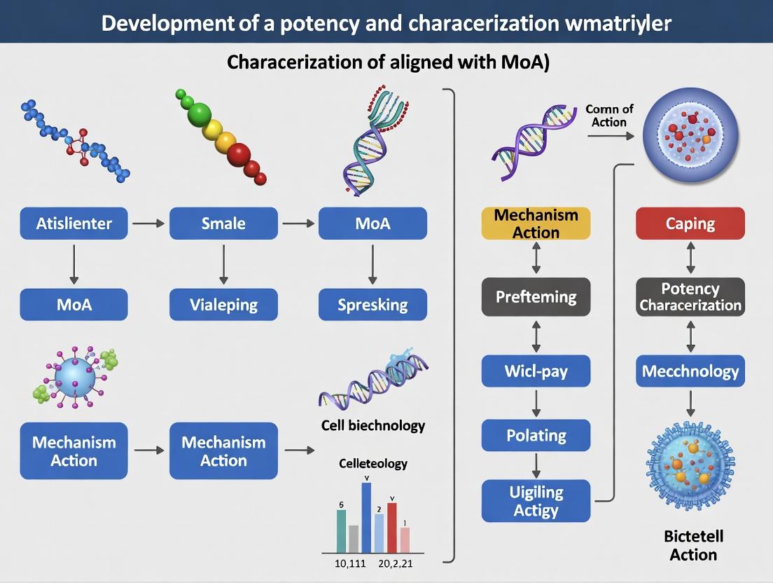
This article provides a comprehensive framework for researchers and drug development professionals to establish robust, mechanism-of-action (MoA)-aligned potency and characterization matrices.
Developing Robust MoA-Aligned Potency Assays: A Comprehensive Guide for Biomarker and Matrix Strategy
Abstract
This article provides a comprehensive framework for researchers and drug development professionals to establish robust, mechanism-of-action (MoA)-aligned potency and characterization matrices. It progresses from foundational principles of biomarker identification through methodological implementation of orthogonal assays, addresses common troubleshooting and optimization challenges, and culminates in validation strategies and comparative analysis. The content is designed to guide the development of biologically relevant potency assays critical for demonstrating product consistency, stability, and efficacy from early development through regulatory submission.
Laying the Groundwork: Defining MoA and Identifying Critical Quality Attributes for Potency
In the evolving landscape of biotherapeutics development, the potency assay is the critical quality attribute (CQA) that bridges the physical product to its biological function. A mechanism of action (MoA)-aligned potency assay is non-negotiable because it alone confirms the drug product elicits the specific biological effect intended, ensuring patient safety and efficacy. This document, framed within broader research on developing a comprehensive MoA-aligned potency and characterization matrix, details the application notes and protocols essential for modern researchers.
Core Principles: The Case for MoA-Alignment
Traditional impurity-focused quality control is insufficient for complex modalities like monoclonal antibodies, bispecifics, gene therapies, and cell therapies. The therapeutic activity of these products is defined by a specific, often multi-step, biological mechanism. An assay measuring a non-mechanistic attribute (e.g., general cytotoxicity for an immune cell engager) is irrelevant and fails as a meaningful release test.
Key Justifications:
- Regulatory Expectation: ICH Q6B and recent FDA/EMA guidance emphasize the need for biological assays reflecting the product's MoA.
- Risk Mitigation: Detects subtle changes in product quality (e.g., post-translational modifications, aggregation) that directly impact clinical function.
- Product Understanding: Forms the cornerstone of the quality target product profile (QTPP) and informs manufacturing process controls.
Quantitative Data: Comparative Analysis of Potency Assay Formats
Table 1: Comparison of Potency Assay Methodologies Aligned with Different MoAs
| Therapeutic Modality | Primary MoA | Suboptimal Assay (Non-Aligned) | MoA-Aligned Assay Format | Key Advantage of Aligned Assay |
|---|---|---|---|---|
| TNF-α Inhibitor (mAb) | Neutralization of soluble TNF-α | ELISA for antigen binding | Cell-based assay: Inhibition of TNF-α-induced cytotoxicity in L929 cells. | Measures functional neutralization, not just binding. Detects loss of function from denaturation. |
| Immune Checkpoint Inhibitor (Anti-PD-1) | Blockade of PD-1/PD-L1 interaction, restoring T-cell function | PD-1 binding ELISA | Cell-based reporter assay: PD-1/NFAT reporter cell co-cultured with PD-L1 aAPC; measurement of luciferase activation. | Quantifies the functional consequence of receptor blockade in a cellular context. |
| CAR-T Cell Product | Target cell recognition, T-cell activation, and cytotoxic killing | Flow cytometry for CD3+ viability | Multiparametric cytotoxicity assay: Co-culture with target cells expressing specific antigen; measurement of target cell lysis (e.g., impedance, luciferase) AND cytokine secretion (IFN-γ, IL-2). | Directly measures the integrated, multi-step potency outcome. |
| AAV Gene Therapy | Transduction of target cells and expression of therapeutic transgene | qPCR for viral genome titer | Transduction efficiency assay: Infection of permissive cell line; quantification of transgene protein expression via ELISA or functional enzymatic activity. | Measures the key biological outcome—functional protein production. |
| Bispecific T-cell Engager | Simultaneous binding to tumor antigen and CD3, leading to T-cell-mediated cytolysis | Two separate binding ELISAs | Potency cytotoxicity assay: Co-culture of primary human T-cells, target tumor cells, and the bispecific; measurement of specific tumor cell lysis. | Recapitulates the complex, cell-dependent bridging function. |
Detailed Experimental Protocols
Protocol 4.1: MoA-Aligned Cell-Based Potency Assay for an Immune Checkpoint Inhibitor (Anti-PD-1)
Objective: To quantify the potency of an anti-PD-1 monoclonal antibody by measuring its ability to block PD-1 signaling and activate a downstream transcriptional response.
Principle: Engineered Jurkat T-cells stably express human PD-1 and an NFAT-response element driving firefly luciferase. Upon engagement of PD-1 by its ligand PD-L1 presented on artificial antigen-presenting cells (aAPCs), signaling suppresses luciferase expression. The test antibody blocks this interaction, relieving suppression and inducing luciferase activity in a dose-dependent manner.
Materials (Scientist's Toolkit):
Table 2: Research Reagent Solutions for PD-1 Reporter Assay
| Item | Function/Description | Example Vendor/Cat. No. |
|---|---|---|
| PD-1/NFAT Reporter Jurkat Cells | Engineered effector cells; cornerstone reagent for MoA-specific readout. | Promega (J1621) |
| CHO-K1 PD-L1 aAPC Cells | Engineered cells presenting the PD-1 ligand to initiate suppression. | Promega (J1613) |
| Reference Standard Anti-PD-1 mAb | Qualified, well-characterized control for assay calibration and relative potency. | In-house or commercially sourced standard. |
| Test Article (Anti-PD-1 mAb) | Sample for potency determination. | In-house production. |
| Assay Medium (RPMI-1640 + FBS) | Culture medium for cell maintenance and assay performance. | Various (e.g., Gibco). |
| ONE-Glo Luciferase Assay Substrate | Detection reagent for quantifying NFAT-driven luciferase activity. | Promega (E6110) |
| White, Flat-Bottom 96-Well Plates | Optimal plate for luminescence signal detection. | Corning (3917) |
| Plate Reader (Luminometer) | Instrument for detecting luminescence signal. | Various (e.g., PerkinElmer EnVision). |
Procedure:
- Cell Preparation: Harvest PD-1/NFAT Reporter Jurkat cells and CHO-K1 PD-L1 aAPC cells. Centrifuge, resuspend in assay medium, and count. Adjust Jurkat cell density to 1.0 x 10^6 cells/mL and aAPC density to 0.5 x 10^6 cells/mL.
- Sample/Standard Dilution: Prepare a 10-point, 3-fold serial dilution of the Reference Standard and Test Article in assay medium. Use a concentration range spanning the expected full dose-response (e.g., 100 µg/mL to 0.05 µg/mL).
- Assay Plate Setup: In a white 96-well plate, add 50 µL of diluted Standard or Test Article per well (in triplicate). Include a negative control (assay medium only) and a positive control (maximum signal, e.g., a known potent blocker).
- Cell Addition: Add 50 µL of the Jurkat reporter cell suspension (50,000 cells) and 50 µL of the aAPC suspension (25,000 cells) to each well. Gently mix. Final assay volume is 150 µL/well.
- Incubation: Incubate the plate at 37°C, 5% CO2 for 6 hours.
- Signal Detection: Equilibrate plate to room temperature for 10 min. Add 75 µL of ONE-Glo Luciferase Reagent to each well. Shake plate for 2 minutes, then incubate in the dark for 10 minutes.
- Measurement: Read luminescence on a plate reader with an integration time of 0.5-1 second/well.
- Data Analysis: Plot Relative Light Units (RLU) vs. log10 concentration for Standard and Test Article. Fit data using a 4-parameter logistic (4PL) curve. Calculate the relative potency of the Test Article as the ratio of the EC50 values (Standard/Test).
Diagram 1: PD-1 Inhibitor Potency Assay MoA Workflow
Protocol 4.2: Potency Assay for a CAR-T Cell Product
Objective: To determine the integrated potency of a CAR-T cell product by measuring its antigen-specific cytotoxic activity and cytokine secretion.
Principle: CAR-T cells are co-cultured with target cells expressing the tumor-associated antigen. Effective CAR engagement triggers T-cell activation, leading to target cell killing and cytokine release. Potency is quantified by measuring both endpoints.
Materials (Scientist's Toolkit):
- Effector Cells: Final formulated CAR-T cell product.
- Target Cells: Tumor cell line engineered to stably express the target antigen (e.g., NALM-6 expressing CD19).
- Control Cells: Parental tumor cell line lacking the antigen (for specificity confirmation).
- Cytotoxicity Detection Reagent: Real-time cell analysis (RTCA) system (e.g., xCELLigence) OR luciferase-based kit (e.g., CytoTox-Glo).
- Cytokine Detection Kit: MSD or Luminex multiplex assay for IFN-γ, IL-2, etc.
- Assay Medium: Appropriate for both cell types, typically RPMI-1640 + 10% FBS.
Procedure (Luminescence-Based Cytotoxicity + MSD):
- Target Cell Preparation: Seed target cells (and control cells) in a white 96-well plate at 10,000 cells/well in 75 µL medium. Incubate overnight.
- Effector Cell Preparation: Thaw and wash CAR-T cells. Count and resuspend in assay medium.
- Co-culture Setup: Add CAR-T cells to target cells at multiple Effector:Target (E:T) ratios (e.g., 10:1, 3:1, 1:1). Include target cells alone (spontaneous death control) and target cells with lysis reagent (maximum death control). Final volume 150 µL/well. Incubate for 24-48 hours at 37°C, 5% CO2.
- Cytotoxicity Measurement: Add 75 µL of CytoTox-Glo reagent to each well. Mix, incubate 15 min, read luminescence (dead cell protease activity). Add 75 µL of Lysis Reagent, incubate 15 min, read luminescence again (total cell protease activity). Calculate % Cytotoxicity.
- Cytokine Measurement: Transfer 50 µL of supernatant from co-culture wells to an MSD cytokine plate. Follow kit protocol for detection of IFN-γ and IL-2.
- Data Analysis: Plot % Cytotoxicity and cytokine concentration vs. E:T ratio. Calculate the lytic unit (LU) or EC50 for cytotoxicity. Integrate both parameters for a comprehensive potency profile.
Diagram 2: Multi-Step Potency Assessment for CAR-T Cells
The protocols outlined exemplify the core principle of MoA-alignment. These functional potency assays are not standalone tests but are integral components of a comprehensive Characterization Matrix. This matrix cross-references multiple orthogonal methods (e.g., binding assays by SPR, structural analyses by LC-MS, functional potency assays) against each critical quality attribute. Within this research framework, the MoA-aligned potency assay serves as the biological anchor, ensuring that all other analytical data is interpreted in the context of the product's fundamental therapeutic function. Its development is non-negotiable for modern, robust, and clinically relevant biotherapeutic quality control.
Within the framework of MoA-aligned potency and characterization matrix development research, deconstructing the Mechanism of Action (MoA) is a critical, multi-layered analytical process. This involves systematically moving from the initial biochemical interaction of a drug candidate with its primary target, through a cascade of intracellular events, to the ultimate phenotypic response in a relevant biological system. A comprehensive MoA understanding is essential for predicting efficacy, safety, and patient stratification strategies. This document provides detailed application notes and protocols to standardize this deconstruction.
Core MoA Deconstruction Framework & Quantitative Data Landscape
The deconstruction follows a hierarchical logic, with key quantitative parameters measured at each tier. The data below, synthesized from recent literature and industry standards, should be integrated into a characterization matrix.
Table 1: Tiered Quantitative Parameters for MoA Characterization Matrix
| Analysis Tier | Key Measured Parameters | Typical Assay Formats | Reported Values / Benchmarks |
|---|---|---|---|
| Target Engagement | Binding Affinity (Kd), Kinetics (kon, koff), Occupancy (IC50) | SPR, ITC, CETSA, NanoBRET | Kd: pM to nM range; Residence Time: 0.1 to >10 hours |
| Primary Signaling | Phosphorylation State, Second Messenger (cAMP, Ca2+), Conformational Change | Phospho-ELISA/MS, HTRF/AlphaLISA, FRET/BRET | EC50 for pathway modulation; Max % Inhibition/Activation vs. control |
| Cellular Phenotype | Proliferation (IC50), Apoptosis (Caspase activity), Morphology, Migration | IncuCyte, High-Content Imaging, Boyden Chamber | IC50/GI50; Fold-change vs. vehicle; Z'-factor >0.5 |
| Functional Response | Gene Expression (RNA-seq), Protein Signature (CyTOF), Organoid Viability | Transcriptomics, Multiplexed Proteomics, 3D Cell Viability | Differential Gene Counts; Pathway Enrichment Scores (NES); IC50 in 3D |
Table 2: Key Reagent Solutions for MoA Deconstruction
| Reagent/Tool Category | Specific Example | Function in MoA Analysis |
|---|---|---|
| Target Engagement Probes | NanoBRET Target Engagement (TE) Tracers, CETSA Kits | Live-cell, quantitative measurement of drug-target binding affinity and occupancy. |
| Phospho-Specific Antibodies | Luminex xMAP Phospho-Kinase Panels, Total/Phospho Antibody Pairs | Multiplexed measurement of pathway node activation/inhibition downstream of target binding. |
| Biosensors | cAMP GloSensor, Ca2+ indicators (Fluo-4), FRET-based Kinase Reporters | Real-time, dynamic readout of second messenger levels or conformational changes in living cells. |
| Phenotypic Dye Kits | Caspase-Glo 3/7, RealTime-Glo MT Cell Viability, CellTracker Dyes | Quantification of apoptosis, proliferation, and cell health in endpoint or live-cell formats. |
| Advanced Model Systems | Patient-Derived Organoids (PDOs), Co-culture Spheroids, iPSC-Derived Cells | Contextual, physiologically relevant systems for measuring integrated functional responses. |
Detailed Experimental Protocols
Protocol 1: Cellular Target Engagement via NanoBRET
Purpose: To quantify drug-target binding kinetics and affinity in live cells. Key Reagents: NanoLuc-tagged target protein construct, cell-permeable NanoBRET tracer (competitive), NanoBRET Nano-Glo Substrate. Procedure:
- Cell Preparation: Seed cells (e.g., HEK293) in a white-walled 96-well plate. Transfect with the NanoLuc-tagged target construct.
- Tracer Equilibrium: 24h post-transfection, add the appropriate NanoBRET tracer to cells and incubate for 2h at 37°C to reach equilibrium.
- Compound Treatment: Add a serial dilution of the test compound. Include controls (vehicle, maximum inhibition control).
- Signal Detection: After compound incubation (e.g., 1-2h), add the Nano-Glo Substrate. Immediately measure BRET ratio (460nm acceptor / 610nm donor emission) using a plate reader with dual emission filters.
- Data Analysis: Plot % tracer displacement vs. log[compound]. Fit data to a 4-parameter logistic model to determine IC50. Convert to Ki using the Cheng-Prusoff equation.
Protocol 2: High-Content Analysis of Phenotypic Response
Purpose: To capture multi-parametric phenotypic changes (morphology, proliferation, death) in a single assay. Key Reagents: Cell line of interest, test compounds, nuclear stain (Hoechst 33342), viability/cytotoxicity dyes (e.g., Cytotox Green), fixation/permeabilization reagents. Procedure:
- Assay Setup: Seed cells in a collagen-coated 96-well imaging plate. Allow to adhere overnight.
- Dosing: Treat cells with a 10-point, 1:3 serial dilution of test compound. Include positive (e.g., staurosporine) and vehicle controls.
- Staining & Fixation: At assay endpoint (e.g., 72h), add live-cell dyes per manufacturer's instructions. Subsequently, fix cells with 4% PFA for 15 min.
- Image Acquisition: Use an automated high-content imager (e.g., ImageXpress) with a 20x objective. Acquire 9 fields per well in appropriate channels (DAPI, FITC).
- Image Analysis: Use onboard software (e.g., MetaXpress) to segment nuclei and cytoplasm. Quantify parameters: Cell Count (proliferation), Cytotox Green+ objects (death), mean cell area (morphology).
- Data Integration: Generate dose-response curves for each parameter. Calculate IC50 for proliferation, EC50 for cell death, etc.
Pathway & Workflow Visualizations
Title: MoA Deconstruction Tiers & Assay Flow
Title: RTK Inhibitor MoA from Binding to Phenotype
Identifying Key Biomarkers and Critical Quality Attributes (CQAs) Linked to Potency
Within the framework of MoA-aligned potency and characterization matrix development research, establishing a direct link between measurable product quality attributes and biological function is paramount. This application note details a systematic approach to identify and validate key biomarkers and CQAs that are predictive of a biotherapeutic’s potency. The strategy integrates multi-omic profiling, targeted binding/functional assays, and advanced data analytics to construct a predictive matrix, ensuring product quality is inherently tied to the mechanism of action (MoA).
The following tables summarize quantitative data from recent studies (2023-2024) linking specific attributes to potency outcomes for different therapeutic modalities.
Table 1: Key CQAs Linked to Potency for Monoclonal Antibodies
| Critical Quality Attribute (CQA) | Target Range/Value | Assay Method | Correlation with Potency (R²) | Impact on MoA |
|---|---|---|---|---|
| N-Glycan Profile (Afucosylation %) | >60% (enhanced ADCC) | HILIC-UPLC | 0.92 | Directly modulates FcγRIIIa binding & ADCC |
| High-Molecular-Weight (HMW) Aggregates | <1.0% | SEC-MALS | -0.89 | Reduces effective bioactive concentration; may induce immunogenicity |
| Charge Variants (Acidic/Basic) | Main peak ±15% | CEX-HPLC | 0.75 (for main peak) | Can affect target binding affinity & pharmacokinetics |
| Antigen Binding Affinity (KD) | < 2 nM | SPR (Biacore) | 0.96 | Direct determinant of target engagement |
| Fab Glycation Level | < 5% | LC-MS/MS | -0.82 | Can sterically hinder antigen binding |
Table 2: Functional Biomarkers for Cell Therapy Potency (CAR-T)
| Biomarker / Cellular Attribute | Target Phenotype | Measurement Method | Correlation with In Vivo Expansion (r value) | Link to Potency |
|---|---|---|---|---|
| Naïve/TSCM (CCR7+ CD45RA+) % | > 30% of CD8+ | Flow Cytometry | 0.85 | Predicts sustained persistence & long-term efficacy |
| Mitochondrial Mass (MTG MFI) | High (Quintile 5) | Flow Cytometry (MTG dye) | 0.78 | Indicates metabolic fitness & expansion potential |
| Activation Marker (CD25) Post-Stimulation | Dynamic range >10-fold | Flow Cytometry | 0.70 (peak level) | Indicates robust functional activation |
| Secreted IFN-γ (Post-Antigen Stimulus) | > 5000 pg/mL/10⁶ cells | ELISA / MSD | 0.91 | Direct functional output of effector activity |
| Chromatin Accessibility (ATAC-seq Peaks) | Specific regulatory regions | NGS (ATAC-seq) | N/A (Predictive) | Epigenetic signature of potency |
Experimental Protocols
Protocol 3.1: Integrated Multi-Omic Profiling for Biomarker Discovery
Objective: To identify predictive potency biomarkers from transcriptomic, proteomic, and metabolomic datasets correlated with functional potency assays.
Materials:
- Viable cells (therapy product) or target cell lines (for biologics).
- RNA/DNA/protein extraction kits (e.g., Qiagen, Thermo Fisher).
- LC-MS/MS system (e.g., Thermo Orbitrap).
- NGS platform (e.g., Illumina NovaSeq).
- Potency assay reagents (e.g., cytotoxicity, cytokine secretion).
Procedure:
- Sample Preparation: Generate a panel of product batches with defined potency variations (e.g., via stress conditions or different manufacturing runs). Aliquot for multi-omic and potency analysis.
- Parallel Multi-Omic Analysis: a. Transcriptomics: Isolate total RNA, prepare libraries (poly-A selection), and perform RNA-seq. Quantify gene expression (FPKM/TPM). b. Proteomics: Lyse cells, digest proteins with trypsin, label with TMT reagents (if multiplexing). Analyze by LC-MS/MS. Identify/quantify proteins and post-translational modifications (PTMs). c. Metabolomics: Quench metabolism, extract metabolites. Analyze polar/non-polar fractions via HILIC and C18 LC-MS/MS in positive/negative ion modes.
- Potency Assay Execution: Perform the primary MoA-aligned potency assay (e.g., in vitro cytotoxicity, ligand inhibition, receptor activation) on parallel samples.
- Data Integration & Biomarker Identification: Use bioinformatics pipelines (e.g., in R/Python:
limma,DESeq2for RNA;MaxQuantfor proteomics). Perform multi-variate analysis (PLS-Regression, Random Forest) to identify omic features (genes, proteins, metabolites) whose abundance strongly correlates (p<0.01, |r|>0.8) with potency readouts. Validate candidates via orthogonal methods (e.g., qPCR, Western Blot).
Protocol 3.2: Orthogonal Validation of Glycosylation CQA Impact on ADCC
Objective: To quantitatively link specific glycoform ratios (Afucosylation) to FcγRIIIa binding and effector function.
Materials:
- Purified mAb variants with controlled glycoforms (produced via engineered cell lines or enzymatic modulation).
- Recombinant human FcγRIIIa (V158 variant).
- Biolayer Interferometry (BLI) system (e.g., ForteBio Octet) or SPR.
- Peripheral Blood Mononuclear Cells (PBMCs) or engineered effector cells (e.g., ADCC Reporter Bioassay cells, Promega).
- Target cells expressing target antigen.
Procedure:
- Glycoform Characterization: Quantify glycan distribution (especially afucosylated G0/G1/G2) on each mAb variant using HILIC-UPLC with fluorescence detection. Normalize percentages.
- Binding Kinetics Assay: a. Load anti-His biosensors with His-tagged FcγRIIIa. b. Dip sensors into wells containing serially diluted mAb variants. c. Measure association/dissociation. Analyze data to calculate KD for each variant.
- Functional ADCC Assay: a. Label target cells with a fluorescent dye (e.g., BATDA). b. Co-culture labeled target cells with effector cells (PBMCs or reporter cells) at an appropriate E:T ratio in the presence of mAb variants (serial dilution). c. Incubate (e.g., 2-4 hours for PBMCs, 6-24h for reporter assay). d. Measure cytotoxicity: For BATDA, measure released dye; for reporter assay, measure luminescence.
- Correlation Analysis: Plot % afucosylation vs. FcγRIIIa KD and vs. ADCC EC50. Perform linear regression to establish correlation coefficients (R²).
Diagrams
Title: Integrated Biomarker & CQA Discovery Workflow
Title: Link Between CQA, MoA, and Potency
The Scientist's Toolkit: Key Research Reagent Solutions
| Research Reagent / Material | Primary Function in Potency & CQA Research |
|---|---|
| Surface Plasmon Resonance (SPR) Platform (e.g., Cytiva Biacore) | Gold-standard for label-free, real-time quantification of binding kinetics (KA, KD) between drug and target or effector molecules. Critical for affinity-based CQAs. |
| Multiplex Cytokine Analysis (e.g., Meso Scale Discovery (MSD) U-PLEX) | Simultaneously quantifies numerous secreted cytokines/chemokines from functional cell-based assays, providing a rich, MoA-relevant biomarker signature. |
| Flow Cytometry Panels for Immunophenotyping | Enables deep profiling of cell therapy products for critical potency biomarkers (e.g., TSCM subsets, activation markers, intracellular signaling proteins). |
| LC-MS/MS Systems with High-Resolution Mass Spectrometers | Enables precise characterization of CQAs like amino acid sequence, PTMs (oxidation, deamidation), and glycosylation patterns at the molecular level. |
| Glycan Analysis Kits (e.g., Waters GlycoWorks RapiFluor-MS) | Streamlines preparation and UPLC/MS analysis of released N-glycans for quantifying critical glycoforms linked to potency (e.g., afucosylation). |
| ADCC Reporter Bioassay Kits (e.g., Promega) | Provides a standardized, reproducible cell-based assay to measure Fc effector function without primary immune cells, linking CQAs to functional output. |
| Next-Generation Sequencing (NGS) for ATAC-seq/RNA-seq | Uncovers epigenetic (chromatin accessibility) and transcriptomic biomarkers predictive of cell therapy potency and manufacturing consistency. |
| Stable Isotope Labeling Reagents (e.g., TMT, SILAC) | Allows multiplexed, quantitative comparative proteomics to identify protein-level CQAs and biomarkers across many product samples. |
Within a broader thesis on Mechanism-of-Action (MoA)-aligned potency and characterization matrix development, the strategic selection of exploratory assays is a critical first step. Assays are broadly categorized by their readout type: Biochemical (molecular interactions), Cellular (intracellular signaling or reporter events), and Functional (phenotypic or physiological outcomes). The choice dictates the relevance, throughput, and informational value of early data, directly impacting the ability to construct a robust efficacy and safety profile. This application note provides protocols and frameworks for their deployment.
Assay Categories & Comparative Analysis
Table 1: Comparative Summary of Exploratory Assay Readouts
| Parameter | Biochemical Assay | Cellular Assay | Functional Assay |
|---|---|---|---|
| Complexity | Low (Purified components) | Medium (Live cells, engineered) | High (Primary cells, tissues, organisms) |
| Throughput | Very High (96/384/1536-well) | High (96/384-well) | Low to Medium (6-96 well, low automation) |
| Biological Relevance | Low (Decontextualized) | Medium (Cellular context intact) | High (Integrated system physiology) |
| Primary Information | Binding affinity, enzyme kinetics | Pathway modulation, toxicity, uptake | Phenotypic change, viability, contraction, beating |
| Cost per Data Point | $ | $$ | $$$ |
| Key Artifact Risks | Non-physiological conditions, compound interference (fluorescence, aggregation) | Off-target pathway activation, overexpression artifacts, cytotoxicity masking | Multicellular compensatory mechanisms, high variance |
| MoA Alignment | Target engagement confirmation | Pathway perturbation | Integrated biological outcome |
| Example Protocols | FP, TR-FRET, SPR | Reporter gene, HTRF phospho-antibody, FLIPR Ca2+ flux | Cardiomyocyte beating (MEA), neurite outgrowth, phagocytosis |
Detailed Protocols
Protocol 3.1: Biochemical Assay – Time-Resolved FRET (TR-FRET) Kinase Activity
Objective: Quantify inhibition of a purified kinase enzyme via competitive displacement of a fluorescent tracer. Reagent Solutions:
- Kinase Domain: Recombinant, purified human kinase.
- TR-FRET Tracer: A fluorescently-labeled ATP- or substrate-competitive probe.
- Anti-Tag Antibodies: Eu³⁺- or Tb³⁺-cryptate conjugated antibody (e.g., anti-GST) and a d2 or Alexa Fluor 647 acceptor antibody recognizing the tracer.
- Assay Buffer: Optimized for kinase activity (e.g., 50 mM HEPES, 10 mM MgCl₂, 1 mM DTT, 0.01% BSA). Procedure:
- In a low-volume 384-well plate, dispense 2 µL of compound in DMSO (or control) using an acoustic dispenser.
- Add 4 µL of kinase/tracer mixture in assay buffer. Incubate for 15 minutes at RT.
- Add 4 µL of detection antibody mixture in detection buffer. Incubate for 60+ minutes at RT protected from light.
- Read TR-FRET signal on a compatible plate reader (e.g., PerkinElmer EnVision). Excitation ~340 nm, measure emissions at ~615 nm (donor) and ~665 nm (acceptor).
- Data Analysis: Calculate ratio (665 nm / 615 nm) * 10,000. Fit dose-response curves to determine IC₅₀.
Protocol 3.2: Cellular Assay – β-Arrestin Recruitment (PathHunter)
Objective: Measure GPCR activation or inhibition via enzyme fragment complementation upon β-arrestin recruitment. Reagent Solutions:
- Engineered Cell Line: CHO-K1 cells stably expressing the target GPCR fused to a small enzyme fragment (EA) and β-arrestin fused to a larger complementary fragment (ED).
- Detection Reagent: PathHunter detection mix (lysis and chemiluminescent substrate).
- Assay Buffer: Cell plating medium (e.g., DMEM/F-12, 1% FBS) and stimulation buffer (HBSS, 20 mM HEPES). Procedure:
- Plate 5,000 cells/well in 20 µL into a white, tissue-culture treated 384-well plate. Culture overnight.
- Dispense 20 nL of test compound. Add 5 µL of reference agonist for antagonist mode (or buffer for agonist mode). Incubate for 90-180 min at 37°C.
- Add 12 µL of detection reagent. Incubate for 60 min at RT in the dark.
- Measure chemiluminescence (integration 0.5-1 sec/well).
- Data Analysis: Normalize to basal (0%) and maximal agonist control (100%). Calculate EC₅₀/IC₅₀.
Protocol 3.3: Functional Assay – iPSC-Derived Cardiomyocyte Beating Analysis
Objective: Profile compound effects on cardiomyocyte contraction frequency, amplitude, and morphology using impedance (label-free). Reagent Solutions:
- iPSC-Cardiomyocytes: Human induced pluripotent stem cell-derived cardiomyocytes, certified for electrophysiology.
- Recording Medium: Serum-free, cardiomyocyte maintenance medium.
- Microelectrode Array (MEA) Plate or Impedance Plate: 48- or 96-well plates with integrated electrodes. Procedure:
- Cell Preparation: Thaw cardiomyocytes and plate onto 0.1% gelatin-coated MEA/impedance plates at 50,000 cells/well in maintenance medium. Culture for 7-10 days, changing medium every 2 days, until stable, synchronous beating is observed.
- Baseline Recording: Replace medium with fresh recording medium. Record baseline beating for 5-10 minutes using the plate reader system (e.g., Axion Biosystems Maestro, or xCELLigence RTCA Cardio).
- Compound Addition: Add compound diluted in recording medium (final DMSO ≤0.3%). Record continuously for 10-30 minutes post-addition.
- Data Analysis: Software-derived parameters: Beat Rate (BPM), Beat Amplitude (impedance change, Ω), Field Potential Duration (for MEA), and irregularity indices.
Visualization of Assay Logic & Pathways
Diagram Title: Assay Readout Hierarchy & Information Flow
Diagram Title: Cellular GPCR β-Arrestin Recruitment Assay Pathway
The Scientist's Toolkit: Key Research Reagent Solutions
Table 2: Essential Reagents for Exploratory Assays
| Reagent / Solution | Category | Function / Explanation |
|---|---|---|
| HTRF (Cisbio) | Biochemical/Cellular | Homogeneous Time-Resolved FRET technology for kinase, GPCR, and biomarker assays. Minimizes autofluorescence. |
| PathHunter (Revvity) | Cellular | Enzyme fragment complementation (EFC) platform for GPCR, kinase, and cytochrome P450 assays. No wash, high sensitivity. |
| Tag-lite (Cisbio) | Biochemical (Cellular-surface) | SNAP/CLIP-tag based TR-FRET for ligand binding on live cells. Measures membrane protein interactions. |
| AlphaLISA/AlphaScreen (Revvity) | Biochemical | Bead-based proximity assay for biomolecular interactions. Amplified, no-wash signal. |
| iPSC-Derived Cardiomyocytes (e.g., Fujifilm CDI, Ncardia) | Functional | Physiologically relevant human cells for cardiotoxicity and efficacy screening. |
| FLIPR Tetra (Molecular Devices) | Cellular | High-throughput fluorescence imager for real-time kinetic measurements of ion flux (Ca2+, K+). |
| Matrigel (Corning) | Cellular/Functional | Basement membrane matrix for 3D cell culture and organoid assays, improving physiological relevance. |
| Cryopreserved Primary Hepatocytes (e.g., BioIVT) | Functional | Gold standard for assessing hepatic metabolism, toxicity, and transporter effects. |
| Recombinant Purified Proteins (e.g., Thermo Fisher, Sino Biological) | Biochemical | Essential components for constructing defined, minimal system binding or enzymatic assays. |
| NanoBRET (Promega) | Cellular | Bioluminescence resonance energy transfer for studying protein-protein interactions and target engagement in live cells. |
Literature and Competitor Analysis for MoA-Aligned Benchmarking
Within the broader thesis on Mechanism of Action (MoA)-aligned potency and characterization matrix development, this document details the application notes and protocols for conducting a systematic literature and competitor analysis. This foundational step is critical to establish the current therapeutic landscape, identify validated and novel biomarkers, and define the key assays required for precise, MoA-aligned benchmarking of novel drug candidates against established standard-of-care agents.
Literature Analysis Protocol
2.1 Objective To systematically identify, collate, and analyze published scientific data on the target biology, signaling pathways, existing therapeutic agents (including their MoAs), and relevant in vitro and in vivo biomarker endpoints.
2.2 Methodology: PRISMA-Informed Screening Workflow
2.3 Data Extraction Template & Key Fields Quantitative and qualitative data are extracted into a structured database. Core fields include:
- Drug/Target: Compound name, target(s), therapeutic modality.
- MoA Classification: e.g., Competitive inhibitor, allosteric modulator, degrader, antibody-dependent cellular cytotoxicity (ADCC).
- Key Biomarkers: Phosphoproteins, gene expression signatures, cell viability IC50, etc.
- Experimental Models: Cell line(s), animal model(s).
- Potency/Efficacy Metrics: IC50, EC50, GI50, maximal inhibition/response (%).
2.4 Key Research Reagent Solutions
| Reagent / Solution | Function in Analysis |
|---|---|
| Bioinformatics Databases (e.g., GEO, TCGA) | Provide transcriptomic/proteomic datasets to identify disease-relevant biomarkers and pathway activity. |
| Pathway Analysis Software (e.g., Ingenuity IPA, Metascape) | Enables systems biology analysis of extracted gene/protein lists to map MoA-aligned networks. |
| Reference Ligands (Competitor Compounds) | Critical positive controls for assay development; used to benchmark novel compound activity. |
| Validated Antibodies (Phospho-Specific) | Essential reagents for quantifying target engagement and downstream pathway modulation via Western blot or immunofluorescence. |
| Engineered Reporter Cell Lines | Stable cell lines with luciferase or GFP under pathway-responsive elements (e.g., NF-κB, STAT) for functional MoA readouts. |
Competitor Analysis Protocol
3.1 Objective To profile the biochemical, cellular, and pharmacological characteristics of competitor therapeutics, enabling direct, MoA-aligned comparison with internal development candidates.
3.2 Experimental Protocol: Cellular Target Engagement & Pathway Modulation Assay
- Aim: Quantify and compare the potency and temporal dynamics of pathway inhibition/activation by competitor Drug A (reference inhibitor) and novel Drug B.
- Cell Line: Disease-relevant cell line endogenously expressing the target.
- Procedure:
- Seed cells in 6-well plates and culture until 70-80% confluent.
- Serum-starve cells (if required for pathway basal state) for 12-16 hours.
- Pre-treat with titrated doses of Drug A, Drug B, or vehicle (DMSO) for 1 hour.
- Stimulate with relevant pathway agonist (e.g., cytokine, growth factor) for 15 minutes.
- Lyse cells using RIPA buffer supplemented with phosphatase/protease inhibitors.
- Analyze lysates via Western Blot for:
- Phosphorylation of direct target (if antibody available).
- Phosphorylation of immediate downstream node (key biomarker).
- Total protein levels for loading control.
- Quantify band intensity; normalize p-protein to total protein. Plot dose-response curves to calculate IC50 values.
3.3 Data Synthesis: MoA-Aligned Benchmarking Table Table: Comparative Profiling of PI3Kα Inhibitors in XYZ Cancer Cell Line
| Parameter | Competitor A (Alpelisib) | Competitor B (Copanlisib) | Novel Candidate X | Assay Format |
|---|---|---|---|---|
| Biochemical IC50 (nM) | 5.2 | 0.6 | 1.8 | Recombinant enzyme TR-FRET |
| Cellular p-AKT IC50 (nM) | 32 | 8.5 | 12 | Phospho-ELISA |
| Proliferation GI50 (nM) | 250 | 45 | 60 | CellTiter-Glo (96h) |
| Selectivity Index (vs PI3Kβ) | 50-fold | 7-fold | >200-fold | Panel kinase assay |
| MoA Classification | ATP-competitive, orthosteric | ATP-competitive, pan-PI3K | ATP-competitive, mutant-selective | N/A |
Integration into Characterization Matrix
4.1 Signaling Pathway Mapping for Assay Selection The literature and competitor data inform the selection of critical nodes for assay development within the relevant pathway.
4.2 Protocol: High-Content Imaging for Multi-Parameter MoA Profiling
- Aim: Simultaneously quantify multiple MoA-relevant biomarkers at single-cell resolution to create a phenotypic fingerprint for benchmarking.
- Cell Line: U2OS or HEK293T cells transfected with target of interest, or endogenous cell line.
- Procedure:
- Seed cells in 96-well imaging plates. Treat with compound titrations for 2-24 hours (time-course).
- Fix, permeabilize, and block cells.
- Perform multiplex immunofluorescence staining:
- Primary antibodies: Anti-p-target, Anti-cytoplasmic marker, Anti-nuclear marker (e.g., phospho-histone H3).
- Secondary antibodies: Conjugated to distinct fluorophores (Alexa 488, 555, 647).
- Stain nuclei with Hoechst 33342.
- Image using a high-content microscope (e.g., ImageXpress Micro) with a 20x objective. Acquire 9 fields/well.
- Analysis: Use onboard software (e.g., MetaXpress) to:
- Segment nuclei and cytoplasm.
- Measure mean intensity of p-target in cytoplasm.
- Measure nuclear/cytoplasmic ratio of transcription factors (e.g., FOXO1).
- Quantify cell count, mitosis index.
- Generate multi-parameter dose-response curves to create a comprehensive MoA signature.
Building the Matrix: Implementing Orthogonal and Redundant Assay Strategies
Application Notes
Within a thesis on Mechanism of Action (MoA)-aligned potency and characterization matrix development, the implementation of a tiered assay strategy is critical for linking biological activity to product quality. This structure provides a risk-based framework for quality control, process development, and comparability assessments. The matrix ensures that the potency assays are not just statistical measures but are biologically relevant reflections of the product's MoA.
Primary Potency Assays are quantitative, stability-indicating, and directly reflective of the product's primary MoA. They are validated per ICH Q2(R2) guidelines and serve as the lot release and stability testing cornerstone.
Secondary Potency Assays support and extend the understanding gained from the primary assay. They may measure different facets of the same MoA or a key downstream event. These assays are essential for investigating assay discordance and providing orthogonal data for comprehensive characterization.
Characterization Assays are used during development, extended characterization, and to support investigations. They are not validated for lot release but provide deep biological insight into aspects like signaling bias, kinetics, and pathway engagement, building the foundational science for the potency matrix.
Data Presentation: Comparative Overview of Tiered Potency Assay Strategy
| Assay Tier | Primary Objective | Validation Level | Typical Format | Critical Quality Attribute (CQA) Link |
|---|---|---|---|---|
| Primary Assay | Lot release & stability; direct MoA quantification | Full ICH Validation (Specificity, Accuracy, Precision, Linearity, Range, Robustness) | Cell-based functional (e.g., cytotoxicity, reporter gene) or binding (SPR/BLI) | Potency |
| Secondary Assay | Orthogonal confirmation; extended MoA insight | Qualification (Precision, Specificity, Linearity) | Cell-based signaling (phospho-protein, 2nd messenger) or competitive binding | Biological Activity, Consistency |
| Characterization Assay | Deep mechanistic profiling; investigation support | Research-grade or Fit-for-Purpose | Multi-parameter (e.g., high-content imaging, phospho-proteomics, cytokine multiplex) | Mechanism Understanding, Variant Assessment |
Experimental Protocols
Protocol 1: Primary Potency Assay – Cytotoxic T Cell Activation (Reporter Gene Assay)
Principle: Measures the ability of a bispecific T cell engager (BiTE) to induce NFAT-driven luciferase expression in a engineered Jurkat T cell line upon engagement with target tumor cells. Materials: See Scientist's Toolkit. Procedure:
- Seed target tumor cells (e.g., NCI-H929 myeloma cells) in a white-walled 96-well plate at 5,000 cells/well in 50 µL assay medium. Incubate overnight.
- Prepare a 3-fold serial dilution of the BiTE reference standard and test samples in assay medium across 10 concentrations.
- Add 25 µL of each dilution to the target cell plate in triplicate. Include a "No Antibody" control (medium only) and a "Max Signal" control (with a saturating concentration of reference).
- Resuspend NFAT-luciferase/GFP Jurkat effector cells, add 25 µL/well (25,000 cells) for an Effector:Target (E:T) ratio of 5:1. Final well volume is 100 µL.
- Incubate plate for 6 hours at 37°C, 5% CO₂.
- Equilibrate room temperature. Add 100 µL/well of ONE-Glo EX Luciferase Reagent.
- Shake plate for 5 minutes, protect from light, then incubate for 10 minutes.
- Measure luminescence on a plate reader. Fit data using a 4-parameter logistic (4PL) model to determine relative potency (EC₅₀).
Protocol 2: Secondary Potency Assay – Intracellular Phospho-STAT5 Quantification (Flow Cytometry)
Principle: Quantifies phosphorylation of STAT5 in target cells following engagement by a cytokine-based therapeutic, providing an early signaling readout. Procedure:
- Starve cytokine-dependent TF-1 cells in RPMI with 0.5% FBS for 18-24 hours.
- Prepare cytokine dilutions in starvation medium.
- Aliquot 100 µL of starved cells (1x10⁶ cells/mL) into a 96-well V-bottom plate. Add 100 µL of cytokine dilution per well. Incubate for 15 minutes at 37°C.
- Immediately fix cells by adding 200 µL of pre-warmed (37°C) 2x Phosflow Fix Buffer I. Mix and incubate 10 minutes at 37°C.
- Centrifuge at 500 x g for 5 min. Decant supernatant.
- Permeabilize by adding 200 µL of ice-cold 90% methanol. Vortex gently and incubate on ice for 30 minutes.
- Wash cells twice with 200 µL FACS buffer (PBS + 1% BSA). Centrifuge at 500 x g for 5 min.
- Resuspend cell pellet in 50 µL FACS buffer containing anti-phospho-STAT5 (pY694)-PE antibody (1:50 dilution). Incubate for 60 minutes at room temperature in the dark.
- Wash twice with FACS buffer, resuspend in 200 µL, and analyze on a flow cytometer. Determine Median Fluorescence Intensity (MFI) of the PE channel.
Protocol 3: Characterization Assay – High-Content Imaging for Pathway Profiling
Principle: Uses multiplexed immunofluorescence and automated imaging to simultaneously quantify nuclear translocation of multiple transcription factors (e.g., NF-κB, IRF3) in response to an innate immune modulator. Procedure:
- Seed THP-1-derived macrophages in a collagen-coated 96-well imaging plate at 15,000 cells/well. Differentiate with PMA for 48 hours, then rest for 24 hours.
- Treat cells with serial dilutions of TLR agonist (test article) and controls for 1 hour.
- Fix with 4% PFA for 15 min, permeabilize with 0.1% Triton X-100 for 10 min, and block with 3% BSA for 1 hour.
- Incubate with primary antibody cocktail (anti-NF-κB p65, anti-IRF3) overnight at 4°C.
- Wash 3x with PBS, then incubate with secondary antibody cocktail (Alexa Fluor 488 anti-rabbit, Alexa Fluor 594 anti-mouse) and Hoechst 33342 for 1 hour at RT in the dark.
- Wash 3x with PBS, leaving 100 µL/well for imaging.
- Image on a high-content imager (e.g., ImageXpress) using a 20x objective. Acquire 9 fields/well.
- Analyze using onboard software: identify nuclei (Hoechst), create a cytoplasmic ring expansion, and measure the mean fluorescence intensity (MFI) of each marker in the cytoplasm vs. nucleus. Calculate a nuclear:cytoplasmic ratio for each transcription factor per cell.
Visualizations
Tiered Assay Logic Flow
T Cell Engager MoA & Assay Alignment
The Scientist's Toolkit: Key Research Reagent Solutions
| Reagent / Material | Function in Potency Matrix | Example Vendor/Catalog |
|---|---|---|
| Engineered Reporter Cell Line | Provides a quantifiable, MoA-aligned readout (e.g., luminescence) for primary potency assays. | Promega (ONE-Glo Systems), Invitrogen (GeneBLAzer) |
| Phospho-Specific Antibodies | Enable detection of phosphorylated signaling proteins (e.g., pSTAT5) in secondary cell-based assays. | Cell Signaling Technology, BD Phosflow |
| Multiplex Cytokine Array Kits | Allow simultaneous quantification of multiple secreted analytes for deep characterization. | Meso Scale Discovery (MSD), Luminex |
| High-Content Imaging System | Automates acquisition and analysis of multi-parameter cell imaging data for characterization assays. | Molecular Devices (ImageXpress), PerkinElmer (Opera) |
| Surface Plasmon Resonance (SPR) Chip | Measures real-time binding kinetics (ka, kd, KD) for primary or characterization-level binding assays. | Cytiva (Biacore CM5 Chip) |
| Reference Standard | Qualified material serving as the benchmark for calculating relative potency in all assay tiers. | In-house or NIBSC derived |
| Cell Culture Media (Serum-free) | Provides consistent, defined conditions for bioassays, reducing variability. | Gibco AIM-V, ThermoFisher |
1. Introduction
Within the framework of a comprehensive thesis on mechanism-of-action (MoA)-aligned characterization, the development of robust cell-based potency assays (CBAs) is paramount. Unlike analytical methods that measure physical attributes, a CBA quantifies the biological activity of a therapeutic (e.g., monoclonal antibodies, gene therapies, cytokines) in a cellular system that mirrors its intended physiological effect. This application note provides a detailed protocol for developing a CBA that is explicitly aligned with the drug's primary MoA, ensuring the assay is not only a regulatory requirement but also a meaningful predictor of clinical efficacy.
2. MoA Deconstruction and Assay Target Selection
The initial phase involves a systematic deconstruction of the therapeutic's MoA to identify the most relevant and quantifiable biological endpoint.
- Step 1: Map the Signaling Pathway: Identify key molecular events from target engagement to final biological response.
- Step 2: Select a Quantifiable Endpoint: Choose an endpoint that is directly downstream of the target, specific, and has a dynamic range suitable for potency measurement. Common endpoints include:
- Cell Viability/Proliferation (for cytotoxics or growth factors)
- Phosphorylation/Protein Translocation (for kinase inhibitors or agonists)
- Gene Reporter Activity (e.g., Luciferase, SEAP)
- Surface Marker Expression (for immunomodulators)
- Cytokine Secretion (for agonists/antagonists)
3. Key Experimental Protocols
Protocol 1: Development of a Gene Reporter Assay for a Pathway Agonist
Objective: To measure the potency of a therapeutic that activates a specific intracellular signaling pathway (e.g., JAK/STAT, NF-κB) using a luciferase reporter gene system.
Materials (Research Reagent Solutions):
| Reagent/Material | Function & Explanation |
|---|---|
| Engineed Reporter Cell Line | Stably transfected cells containing a luciferase gene under the control of a responsive element (e.g., ISRE, NF-κB RE). Fundamental for converting pathway activation into a luminescent signal. |
| Reference Standard | A fully characterized batch of the therapeutic with assigned potency. Essential for assay calibration and relative potency calculation. |
| Luciferase Assay Substrate | Cell-permeable pro-luciferin (e.g., D-luciferin) or a "one-step" lysis/detection reagent. Provides the enzyme substrate for light generation. |
| Cell Culture Media (Serum-Free) | Optimized media for maintaining cell health during assay execution without serum interference. |
| White, Flat-Bottom 96- or 384-Well Plates | Plates designed to minimize light cross-talk for optimal luminescence signal detection. |
| Multimode Microplate Reader | Equipped with luminescence detection capabilities. |
Methodology:
- Cell Seeding: Harvest reporter cells in log growth phase. Seed cells in white assay plates at an optimized density (e.g., 20,000 cells/well in 100 µL serum-free media). Incubate overnight (37°C, 5% CO₂) for adherence and stabilization.
- Sample & Standard Dilution: Prepare a 3-fold serial dilution series of the test sample and reference standard in assay media. Typically, use 8 concentrations in duplicate or triplicate.
- Dosing: Add 50 µL of each dilution to the appropriate wells. Include a blank (media only) and a vehicle control (0% activity) and a maximal stimulus control (100% activity, if available).
- Incubation: Incubate plate for a predetermined time (e.g., 6-24 hours) to allow for pathway activation, transcription, and translation.
- Signal Detection: Equilibrate plate to room temperature. Add 50 µL of ONE-Glo Luciferase Assay Reagent (or equivalent). Shake gently for 5 minutes, then incubate for 10 minutes in the dark.
- Measurement: Read luminescence (RLU) on a microplate reader with an integration time of 0.5-1 second/well.
- Data Analysis: Fit the dose-response curves (RLU vs. log10[concentration]) using a 4-parameter logistic (4PL) model. Calculate the relative potency of the test sample against the reference standard by comparing the half-maximal effective concentration (EC₅₀) values or parallel-line analysis.
Protocol 2: Flow Cytometry-Based Potency Assay for an Immune Cell Activator
Objective: To measure the potency of a therapeutic (e.g., an immune checkpoint inhibitor or co-stimulatory agonist) by quantifying cell surface activation marker expression (e.g., CD69, CD25) on primary immune cells.
Methodology:
- Cell Preparation: Isolate primary human PBMCs or specific immune cell subsets (e.g., CD8+ T cells) using density gradient centrifugation and/or magnetic bead separation.
- Co-culture & Stimulation: Seed stimulator cells (e.g., antigen-presenting cells) or coat plates with relevant antigens/antibodies. Add the isolated effector cells and the serially diluted therapeutic. Incubate for 24-72 hours.
- Cell Staining: Harvest cells, wash with PBS, and stain with a fluorescent antibody panel: viability dye, lineage markers (e.g., CD3, CD8), and the activation marker of interest (e.g., CD69-APC).
- Flow Cytometry Acquisition: Resuspend cells in buffer and acquire data on a flow cytometer. Collect a minimum of 10,000 events in the live, target cell population gate.
- Data Analysis: Calculate the percentage of target cells positive for the activation marker (%CD69+) for each concentration. Generate a dose-response curve and determine the EC₅₀ via 4PL fitting.
4. Data Presentation and Analysis
Table 1: Assay Performance Parameters for a Model Reporter Gene Potency Assay
| Parameter | Target Value | Results (Example) |
|---|---|---|
| Linear Range (RLU) | >2 logs | 5,000 - 500,000 RLU |
| EC₅₀ of Reference (ng/mL) | Consistent within run | 1.5 ± 0.3 |
| Relative Potency Range | 50-150% | 80-125% |
| Intra-assay Precision (%CV) | ≤15% | 8% |
| Inter-assay Precision (%CV) | ≤20% | 12% |
| Specificity (Signal Inhibition) | >80% with neutralizing Ab | 95% |
| Dose-Response Fit (R²) | >0.98 | 0.99 |
Table 2: Comparison of MoA-Aligned CBA Platforms
| Assay Platform | Measured Endpoint | Typical Throughput | Key Advantage | Key Consideration |
|---|---|---|---|---|
| Gene Reporter | Pathway Activation | High | Excellent sensitivity & dynamic range | Requires engineered cells; may be artificial. |
| Flow Cytometry | Surface Marker Expression | Medium | Single-cell resolution; multiplexing | Technically complex; lower throughput. |
| Phospho-Specific ELISA/HTRF | Protein Phosphorylation | High | Direct proximal signal | May require cell lysis; epitope dependent. |
| Cytokine Secretion (MSD/ELISA) | Secreted Protein | High | Measures functional output | Can be influenced by non-mechanism factors. |
5. Visualization of Concepts and Workflows
Diagram 1: MoA-Aligned CBA Target Selection Strategy (84 characters)
Diagram 2: CBA Development Workflow (42 characters)
Incorporating Biochemical and Biophysical Assays (e.g., SPR, ELISA) for Binding Potency
Application Notes: Role in MoA-Aligned Potency Matrix Development
A Mechanism of Action (MoA)-aligned potency matrix is a multi-attribute framework designed to quantify a therapeutic molecule's biological activity through assays that mirror its defined mechanism. For biologics where target binding is the primary and rate-limiting step (e.g., monoclonal antibodies, fusion proteins), biochemical and biophysical binding assays are critical Tier 1 potency methods. They provide direct, quantitative, and high-resolution measurements of the key interaction, complementing or preceding functional cellular assays.
Key Advantages:
- Quantitative Precision: Provide definitive kinetic (kₐ, k_d) and equilibrium (K_D, IC₅₀) constants.
- Mechanistic Insight: Distinguish binding affinity from avidity, map epitopes, and assess binding competition.
- Stability-Indicating: Highly sensitive to conformational changes induced by degradation (aggregation, fragmentation, oxidation).
- High-Throughput Suitability: Formats like ELISA enable rapid screening of manufacturing batches.
Integration into the Potency Matrix: Binding potency assays are not standalone. Their data must be correlated with in vitro functional activity and in vivo outcomes. For instance, a confirmed loss in SPR-binding affinity should correlate with a reduction in neutralizing activity in a cell-based assay, confirming the MoA alignment of the stability profile.
Protocol 1: Surface Plasmon Resonance (SPR) for Kinetic Analysis
Objective: Determine the real-time association (kₐ) and dissociation (k_d) rate constants, and the equilibrium dissociation constant (K_D) of a therapeutic antibody binding to its immobilized target antigen.
Principle: A ligand (antigen) is immobilized on a sensor chip. Analyte (antibody) flows over the surface. Binding-induced changes in the refractive index at the chip surface are measured in Resonance Units (RU) versus time.
Detailed Protocol:
Sensor Chip Preparation:
- Immobilization: Using a CMS Series S chip, activate carboxylated dextran matrix with a 7-minute injection of a 1:1 mix of 0.4 M EDC and 0.1 M NHS.
- Ligand Coupling: Dilute antigen to 10-50 µg/mL in 10 mM sodium acetate buffer (pH 4.0-5.0). Inject until desired immobilization level (typically 50-100 RU for kinetic analysis) is reached.
- Blocking: Deactivate remaining esters with a 7-minute injection of 1 M ethanolamine-HCl, pH 8.5.
Kinetic Binding Experiment:
- Prepare a 3-fold dilution series of the antibody (e.g., 100 nM, 33 nM, 11 nM, 3.7 nM, 1.2 nM) in HBS-EP+ running buffer (10 mM HEPES, 150 mM NaCl, 3 mM EDTA, 0.05% v/v Surfactant P20, pH 7.4).
- Set instrument temperature to 25°C.
- Program cycle: 60-second baseline, 180-second association phase (sample injection), 600-second dissociation phase (buffer flow).
- Include a zero-concentration sample (buffer only) for double-referencing.
Data Analysis:
- Subtract reference flow cell and buffer blank sensorgrams.
- Fit processed data to a 1:1 Langmuir binding model using the instrument's evaluation software (e.g., Biacore Evaluation Software).
- Report kₐ (1/Ms), k_d (1/s), and K_D (M = k_d/kₐ).
Table 1: Representative SPR Kinetic Data for Anti-IL-17A mAb Batch Analysis
| Batch ID | kₐ (10⁵ 1/Ms) | k_d (10⁻⁴ 1/s) | K_D (nM) | % Relative Potency vs. Reference |
|---|---|---|---|---|
| Reference Std | 3.21 ± 0.15 | 9.87 ± 0.41 | 3.07 ± 0.12 | 100 |
| GMP Batch A | 3.18 ± 0.12 | 9.91 ± 0.38 | 3.12 ± 0.14 | 98.4 |
| Stability (40°C, 1M) | 2.95 ± 0.20 | 15.60 ± 1.10 | 5.29 ± 0.45 | 58.0 |
SPR Assay Workflow and Output
Protocol 2: Competitive ELISA for Relative Binding Potency
Objective: Determine the relative binding potency of test samples compared to a reference standard by measuring their ability to compete with a labeled ligand for target binding.
Principle: A target protein is coated on a plate. A pre-incubated mixture of a constant concentration of biotinylated therapeutic and a serial dilution of an unlabeled competitor (sample/standard) is added. Signal is inversely proportional to competitor binding potency.
Detailed Protocol:
Plate Coating:
- Coat high-binding 96-well plate with 100 µL/well of target antigen at 2 µg/mL in PBS. Seal and incubate overnight at 4°C.
- Aspirate and block with 300 µL/well of PBS + 1% BSA for 2 hours at room temperature (RT). Wash plate 3x with PBS + 0.05% Tween-20 (PBST).
Sample/Standard and Tracer Preparation:
- Prepare a 4-fold dilution series of the reference standard and test samples in assay buffer (PBS + 0.1% BSA). Use a top concentration ~10x expected IC₅₀.
- Prepare biotinylated therapeutic (tracer) at 2x the final desired concentration (typically near its EC₈₀).
Competition Reaction:
- Mix equal volumes (e.g., 75 µL) of each sample dilution with the 2x tracer solution in a separate plate. Pre-incubate for 1 hour at RT.
- Transfer 100 µL of each mixture to the coated, washed plate. Incubate for 1 hour at RT with shaking. Wash plate 5x with PBST.
Detection:
- Add 100 µL/well of streptavidin-HRP diluted 1:5000 in assay buffer. Incubate 30 min at RT. Wash 5x with PBST.
- Add 100 µL/well of TMB substrate. Develop for 5-15 minutes.
- Stop reaction with 100 µL/well of 1 M H₂SO₄. Read absorbance at 450 nm with 570 nm reference.
Data Analysis:
- Fit absorbance (log10[inhibitor] vs. response -- variable slope four-parameter logistic (4PL) curve.
- Calculate IC₅₀ for standard and samples. Relative Potency = (IC₅₀ Standard / IC₅₀ Sample) * 100%.
Table 2: Competitive ELISA Results for Biosimilar Candidate Assessment
| Sample Description | IC₅₀ (ng/mL) | 95% CI | Relative Binding Potency (%) | Conclusion |
|---|---|---|---|---|
| Innovator Reference | 15.3 | 14.1 - 16.6 | 100 | -- |
| Biosimilar Batch 1 | 16.1 | 14.8 - 17.5 | 95.0 | Within Acceptance Range (80-125%) |
| Biosimilar Batch 2 | 14.8 | 13.6 - 16.1 | 103.4 | Within Acceptance Range (80-125%) |
| Negative Control (Isotype) | >10,000 | N/A | N/A | No specific binding |
Competitive ELISA Steps and Data Analysis
The Scientist's Toolkit: Essential Research Reagents & Materials
Table 3: Key Reagents for Binding Potency Assays
| Item | Function | Example Product/Criteria |
|---|---|---|
| Biacore/Cytiva Series S Sensor Chip CMS | Gold surface with carboxymethylated dextran for ligand immobilization. | Cytiva, Cat # BR100530 |
| HBS-EP+ Buffer | Standard SPR running buffer, provides consistent ionic strength and reduces non-specific binding. | Cytiva, Cat # BR100669 |
| Amine Coupling Kit | Contains EDC, NHS, and ethanolamine for covalent immobilization of ligands. | Cytiva, Cat # BR100050 |
| High-Binding ELISA Plates | Polystyrene plates optimized for protein adsorption. | Corning Costar 9018 |
| Recombinant Target Antigen | High-purity (>95%), endotoxin-low protein for coating/trapping. | R&D Systems, Sino Biological |
| Biotinylation Kit | Enzymatic or chemical kit for consistent, site-specific biotin labeling of the therapeutic. | Thermo Fisher, EZ-Link NHS-PEG4-Biotin |
| Streptavidin-HRP Conjugate | High-sensitivity detection reagent for biotinylated tracers. | Jackson ImmunoResearch, 016-030-084 |
| TMB Substrate | Sensitive, colorimetric HRP substrate for ELISA detection. | Thermo Fisher, Cat # 34021 |
| Reference Standard | Well-characterized drug substance with assigned potency for assay calibration. | In-house qualified material. |
Objective: To quantify compound-induced phenotypic changes in a human osteosarcoma (U2OS) cell line, linking multiparametric readouts to putative Mechanisms of Action (MoA) within a potency characterization matrix.
Protocol: High-Content Imaging for Nuclear and Cytoskeletal Morphology
- Cell Seeding: Seed U2OS cells in a 96-well imaging microplate at 5,000 cells/well in McCoy's 5A medium supplemented with 10% FBS. Incubate for 24 hours at 37°C, 5% CO₂.
- Compound Treatment: Prepare serial dilutions of test compounds (e.g., kinase inhibitors, cytoskeletal disruptors) in DMSO. Treat cells with a 10-point dilution series (1 nM – 10 µM final concentration), including DMSO (0.1% v/v) as vehicle control. Incubate for 48 hours.
- Staining: Aspirate medium, wash with 1X PBS, and fix with 4% paraformaldehyde for 15 minutes. Permeabilize with 0.1% Triton X-100 for 10 minutes. Stain with Hoechst 33342 (1 µg/mL, nuclei), Phalloidin-Alexa Fluor 488 (1:1000, F-actin), and anti-α-tubulin antibody (1:500) followed by a secondary Alexa Fluor 555 antibody (1:1000). Incubate for 1 hour each at room temperature, with washes between steps.
- Image Acquisition: Acquire images using a high-content imaging system (e.g., ImageXpress Pico) with a 20x objective. Capture 9 fields per well across FITC, TRITC, and DAPI channels.
- Image Analysis: Use onboard software (e.g., CellReporterXpress) to segment nuclei and cytoplasm. Extract >50 morphological features per cell (e.g., nuclear area, intensity, texture; cytoskeletal fiber alignment, cell shape descriptors). Analyze a minimum of 1,000 cells per condition.
Data Presentation: Table 1: Quantified High-Content Features for Reference Compounds (48h Treatment)
| Compound (MoA) | Nuclear Area (px²) | Nuclear Roundness | F-Actin Intensity (a.u.) | Texture (Haralick) | Phenotypic Cluster |
|---|---|---|---|---|---|
| DMSO (Control) | 185 ± 12 | 0.92 ± 0.03 | 1550 ± 210 | 0.45 ± 0.05 | Baseline |
| Cytochalasin D (Actin disruptor) | 210 ± 25 | 0.87 ± 0.07 | 3200 ± 450 | 0.67 ± 0.08 | Cytoskeletal Aggregation |
| Nocodazole (Microtubule disruptor) | 165 ± 18 | 0.95 ± 0.02 | 1400 ± 180 | 0.31 ± 0.04 | Rounded Cell |
| Doxorubicin (DNA intercalator) | 255 ± 30 | 0.75 ± 0.09 | 1605 ± 195 | 0.52 ± 0.06 | Nuclear Enlargement |
High-Content Imaging and Analysis Workflow
The Scientist's Toolkit:
- U2OS Cell Line: Robust, adherent cell line with clear cytoskeletal and nuclear morphology.
- Hoechst 33342: Cell-permeant DNA stain for nuclear segmentation and quantification.
- Phalloidin-Alexa Fluor 488: High-affinity probe for filamentous actin (F-actin).
- Anti-α-Tubulin Antibody: Labels microtubule network; used with fluorescent secondary.
- High-Content Imaging System: Automated microscope with environmental control and automated image analysis software.
Application Note: Mass Cytometry (CyTOF) for Deep Immune Profiling
Objective: To characterize the dose-dependent effects of an immunomodulatory drug candidate on primary human peripheral blood mononuclear cell (PBMC) subsets simultaneously, quantifying >30 functional and phenotypic markers.
Protocol: Mass Cytometry for Phospho-Signaling and Phenotyping
- PBMC Stimulation: Isolate PBMCs from healthy donor buffy coats using Ficoll density gradient. Seed 2x10^6 cells/mL in RPMI-1640 + 10% FBS. Pre-treat cells with compound or vehicle for 1 hour, followed by stimulation with 10 ng/mL PMA/Ionomycin or CD3/CD28 beads for 15 minutes (signaling) or 24 hours (phenotype/cytokine).
- Cell Staining (Live/Dead & Surface): Wash cells, stain with Cell-ID Cisplatin (5 µM, 5 min) for viability. Block with Human TruStain FcX. Stain with preconjugated metal-tagged antibody cocktail for surface markers (e.g., CD45, CD3, CD4, CD8, CD19, CD56, CD14, CD16, CCR7, CD45RA) for 30 minutes at room temperature.
- Cell Fixation, Permeabilization & Intracellular Staining: Fix cells with 1.6% formaldehyde (20 min). Permeabilize with ice-cold 100% methanol (10 min, -80°C). Stain with metal-tagged intracellular antibodies (e.g., pSTAT1, pSTAT3, pS6, pERK1/2, IFN-γ, IL-2, TNF-α, Ki-67) for 30 minutes at room temperature.
- Cell Barcoding & Acquisition: Label cells with Cell-ID 20-Plex Pd Barcoding Kit per manufacturer's protocol. Pool samples, wash, and resuspend in Cell Acquisition Solution with EQ Four Element Calibration Beads. Acquire data on a Helios mass cytometer.
- Data Analysis: Normalize data using bead standards. Debarcode files. Use dimensionality reduction (viSNE, UMAP) and clustering (PhenoGraph) to identify cell populations. Analyze marker expression and signaling intensity across doses.
Data Presentation: Table 2: Mass Cytometry-Defined Immune Cell Frequency Shifts Post-Treatment (24h)
| Cell Population (Defining Markers) | Vehicle Frequency (%) | 100 nM Drug Frequency (%) | 1 µM Drug Frequency (%) | Key Functional Change (MFI) |
|---|---|---|---|---|
| CD4+ Naïve T Cells (CD3+CD4+CCR7+CD45RA+) | 25.2 | 22.1 | 15.4 | pS6: +205% |
| Regulatory T Cells (CD3+CD4+CD25+FoxP3+) | 4.5 | 6.8 | 10.2 | pSTAT3: +320% |
| Classical Monocytes (CD14+CD16-) | 8.1 | 9.5 | 12.3 | HLA-DR: -40% |
| NK Cells (CD3-CD56+) | 10.3 | 12.6 | 8.1 | IFN-γ (upon stim): -60% |
Mass Cytometry Experimental Pipeline
The Scientist's Toolkit:
- Cell-ID Cisplatin: Metal-conjugated viability dye for dead cell exclusion.
- Metal-Conjugated Antibodies: Antibodies tagged with rare earth isotopes, minimal spectral overlap.
- Cell-ID 20-Plex Pd Barcoding Kit: Allows sample multiplexing to reduce technical variability.
- EQ Four Element Calibration Beads: For signal normalization during acquisition.
- Helios Mass Cytometer: Inductively coupled plasma time-of-flight (ICP-TOF) mass spectrometer for single-cell analysis.
Application Note: Integrated Transcriptomics & Proteomics for MoA Deconvolution
Objective: To perform an orthogonal multi-omics analysis on compound-treated cancer cell lines, correlating transcriptional changes with proteomic shifts to infer pathway-level MoA and identify biomarker signatures for the potency matrix.
Protocol: Bulk RNA-Seq and LC-MS/MS Proteomics Part A: RNA Sequencing
- Treatment & Lysis: Treat adherent cells (e.g., A549) in triplicate with IC50/IC80 concentrations of compound or DMSO for 6h and 24h. Lyse cells directly in the plate using TRIzol reagent.
- RNA Extraction & QC: Isolate total RNA following phase separation. Assess RNA integrity (RIN > 9.0) using a Bioanalyzer.
- Library Prep & Sequencing: Prepare stranded mRNA-seq libraries using the Illumina TruSeq kit. Pool libraries and sequence on a NovaSeq 6000 platform for 50M paired-end 150 bp reads per sample. Part B: Quantitative Proteomics (TMT-LC/MS-MS)
- Protein Extraction & Digestion: Harvest parallel treated cell pellets. Lyse in RIPA buffer, reduce with DTT, alkylate with IAA, and digest with trypsin overnight.
- TMT Labeling & Fractionation: Label digested peptides from each sample with a unique TMTpro 16-plex isobaric tag. Pool labeled peptides and fractionate using high-pH reverse-phase chromatography.
- LC-MS/MS Analysis: Analyze fractions on an Orbitrap Eclipse tribrid mass spectrometer coupled to a nanoLC. Use MS3 method for TMT quantification to reduce ratio compression.
- Bioinformatics: Align RNA-seq reads (STAR), quantify gene expression (DESeq2). Identify differential proteins (Proteome Discoverer, limma). Perform integrative pathway analysis (Ingenuity Pathway Analysis, GSEA).
Data Presentation: Table 3: Integrated Omics Data for Compound X at 24h (IC80)
| Pathway (KEGG) | RNA-seq: Log2FC | RNA-seq: p.adj | Proteomics: Log2FC | Proteomics: p.value | Concordance |
|---|---|---|---|---|---|
| Cell Cycle | -1.85 | 3.2E-10 | -0.92 | 1.5E-04 | Yes |
| p53 Signaling | +2.10 | 5.8E-12 | +1.15 | 2.1E-05 | Yes |
| mTOR Signaling | -0.95 | 1.1E-03 | -0.45 | 0.023 | Partial |
| Xenobiotic Metabolism | +3.25 | 8.4E-15 | +0.55 | 0.15 | No |
Integrated Multi-Omics Analysis Pathway
The Scientist's Toolkit:
- TRIzol Reagent: For simultaneous RNA/protein extraction from single samples.
- TMTpro 16-plex Isobaric Labels: Enables multiplexed quantitative comparison of up to 16 samples in one MS run.
- Orbitrap Eclipse Tribrid Mass Spectrometer: High-resolution, sensitive instrument for deep proteome coverage.
- STAR Aligner: For fast, accurate alignment of RNA-seq reads to the genome.
- DESeq2 / limma: Statistical packages for robust identification of differentially expressed genes/proteins.
Application Note: MoA-Aligned Potency for a PD-1 Inhibiting Monoclonal Antibody (Biologic)
Thesis Context: Potency assays must measure the specific biological activity defined by the therapeutic's mechanism of action (MoA). For a PD-1 inhibitor, this involves blocking the PD-1/PD-L1 interaction, thereby restoring T-cell effector functions.
Key Quantitative Data: Table 1: Summary of Potency Assay Results for Anti-PD-1 mAB (Lot Comparison)
| Parameter | Lot A | Lot B | Reference Standard | Acceptance Criteria |
|---|---|---|---|---|
| IC50 (Blocking PD-1/PD-L1 Binding) | 0.8 nM | 0.85 nM | 0.82 nM | 0.5 - 1.5 nM |
| EC50 (T-cell IL-2 Secretion) | 1.2 nM | 1.3 nM | 1.25 nM | 0.7 - 2.0 nM |
| Relative Potency (%) | 102% | 98% | 100% | 80-125% |
| Binding Affinity (KD, SPR) | 0.9 nM | 1.0 nM | 0.95 nM | Report |
Experimental Protocol: MoA-Aligned T-cell Activation Assay
- Cell Preparation: Culture PD-L1+ antigen-presenting cells (e.g., CHO cells engineered to express human PD-L1) and isolate human primary CD4+ T-cells from leukopaks using immunomagnetic separation.
- Stimulation: Coat a 96-well plate with anti-CD3 antibody (1 µg/mL) overnight at 4°C. Wash plates.
- Treatment: Add PD-L1+ cells and T-cells at a 1:2 ratio. Add a serial dilution of the anti-PD-1 mAb (e.g., 10 nM to 0.01 nM).
- Incubation: Incubate co-culture for 48 hours at 37°C, 5% CO2.
- Readout: Quantify IL-2 secretion in supernatant using a validated ELISA kit.
- Analysis: Fit dose-response data using a 4-parameter logistic (4PL) model to calculate EC50 and assign relative potency versus the reference standard.
Diagram Title: PD-1 Inhibition MoA and Potency Assay Principle
The Scientist's Toolkit:
- Recombinant Human PD-L1 Protein: Coating antigen for ligand-binding assays.
- Anti-Human CD3 (OKT3) Antibody: For T-cell receptor stimulation.
- Human IL-2 ELISA Kit: Quantifies functional T-cell response.
- Biacore/SPR System: For kinetic analysis of binding affinity (KD).
- Primary Human CD4+ T-Cells: Biologically relevant effector cells.
Application Note: Characterization of a CAR-T Cell Therapy (Cell & Gene Therapy)
Thesis Context: The potency matrix must integrate multiple attributes reflecting CAR-T cell biological activity: CAR density (transduction efficiency), target cell killing (cytotoxicity), and cytokine release (serial killing potential).
Key Quantitative Data: Table 2: Characterization Matrix for an Anti-CD19 CAR-T Product
| Attribute | Method | Release Spec (Example) | Stability Indicator |
|---|---|---|---|
| Transduction Efficiency (% CAR+) | Flow Cytometry | ≥ 30% | Yes |
| CAR Copy Number (Vector Copies/Cell) | ddPCR | ≤ 5 | Yes |
| In Vitro Cytotoxicity (EC50, E:T Ratio) | Real-time impedance (e.g., xCELLigence) | ≤ 1:2 (vs. NALM-6 cells) | Yes |
| Cytokine Secretion (IFN-γ pg/mL) | ELISA after antigen stimulation | ≥ 1000 | Yes |
| Phenotype (Naïve/Memory %) | Flow (CD45RO, CD62L) | Report | No |
Experimental Protocol: Real-Time Cytotoxicity Assay
- Target Cell Seeding: Seed CD19+ target cells (NALM-6) at 5,000-10,000 cells/well in an E-plate with media. Incubate for 30 min at room temperature to allow cell settling, then place in the xCELLigence RTCA analyzer to establish a baseline impedance.
- Effector Cell Addition: After 24 hours, prepare CAR-T effector cells at varying Effector:Target (E:T) ratios (e.g., 5:1, 1:1, 1:5). Gently add CAR-T cells to the appropriate wells.
- Continuous Monitoring: Place the plate back in the analyzer. Monitor cell index every 15 minutes for 72-96 hours.
- Data Analysis: Normalize cell index to the timepoint just before effector addition. Cytotoxicity is indicated by a decrease in normalized cell index relative to target-only controls. Calculate EC50 or specific lysis at defined timepoints.
Diagram Title: Multi-Attribute Potency Matrix for CAR-T Therapies
The Scientist's Toolkit:
- Anti-Idiotype Antibody (for CAR detection): Essential for flow cytometry quantification of CAR expression.
- ddPCR Kits (for Vector Copy Number): Provides absolute quantification of transgene integration.
- Real-Time Cell Analysis (RTCA) Instrument: Enables label-free, kinetic measurement of cytotoxicity.
- CD19+ Target Cell Line (e.g., NALM-6): Consistent antigen-positive target for potency assays.
- Cytokine Multiplex Panels: For profiling secretory footprint (IFN-γ, IL-2, IL-6, etc.).
Application Note: Target Engagement & Pathway Inhibition for a BTK Inhibitor (Targeted Small Molecule)
Thesis Context: Characterization must confirm direct binding to the target (BTK) and subsequent inhibition of its downstream signaling pathway (e.g., BCR signaling), linking biochemical activity to cellular phenotype.
Key Quantitative Data: Table 3: Characterization Data for a Covalent BTK Inhibitor
| Assay Tier | Assay Type | Parameter | Result |
|---|---|---|---|
| Biochemical | Kinase Binding (SPR) | Kon, Koff, KD (Covalent) | Irreversible |
| Biochemical | Enzymatic Activity (ADP-Glo) | IC50 (BTK) | 0.5 nM |
| Cellular | Phospho-BTK (Flow Cytometry) | % Inhibition @ 100 nM | >95% |
| Cellular | Phospho-PLCγ2 (MSD ELISA) | IC50 | 5 nM |
| Functional | Ramos Cell Viability (ATP-Lite) | GI50 | 25 nM |
Experimental Protocol: Cellular Target Engagement via Flow Cytometry
- Cell Stimulation: Use a B-cell line (e.g., Ramos) or primary human B-cells. Pre-treat cells with a serial dilution of the BTK inhibitor (e.g., 1 µM to 0.1 nM) in serum-free media for 1-2 hours.
- Pathway Activation: Stimulate the B-cell receptor by adding anti-human IgM antibody (10 µg/mL) for 15 minutes at 37°C.
- Fixation & Permeabilization: Immediately transfer cells to a deep-well plate containing pre-warmed 4% PFA. Fix for 10 min, then pellet. Permeabilize with ice-cold 90% methanol for 30 min on ice.
- Staining: Wash cells and stain with conjugated antibodies specific for phosphorylated BTK (Tyr223) and a viability dye. Include unstimulated and stimulated/untreated controls.
- Acquisition & Analysis: Acquire data on a flow cytometer. Gate on live, single cells. Report geometric mean fluorescence intensity (gMFI) of pBTK. Calculate % inhibition of pBTK formation relative to the stimulated control.
Diagram Title: BTK Inhibitor Mechanism in BCR Signaling
The Scientist's Toolkit:
- Active Recombinant BTK Kinase: For biochemical inhibition assays.
- Phospho-Specific Antibodies (pBTK, pPLCγ2): Critical for measuring cellular pathway modulation.
- ADP-Glo Kinase Assay Kit: Homogeneous method for measuring BTK enzymatic activity.
- MSD/Meso Scale Discovery Phospho-ELISA Plates: For sensitive, multiplexed phospho-protein detection from cell lysates.
- Cell Viability Assay (ATP-based): Measures functional anti-proliferative outcome.
Navigating Challenges: Optimization, Signal-to-Noise, and Assay Robustness
Common Pitfalls in MoA-Aligned Assay Development and How to Avoid Them
Within the broader thesis on Mechanism of Action (MoA)-aligned potency and characterization matrix development, robust assay design is paramount. Misalignment between an assay's readout and the therapeutic's true biological mechanism leads to misleading potency estimates, flawed stability assessments, and ultimately, clinical-stage failures. This document outlines common pitfalls and provides detailed protocols to ensure assay relevance and reliability.
Pitfall 1: Using Surrogate Endpoints Disconnected from the Primary MoA
A prevalent error is measuring an easy-to-quantify but mechanistically distant endpoint. For example, for a T-cell engager, using only target cell binding (a simple ELISA) instead of primary T-cell activation and subsequent tumor cell killing.
Protocol 1.1: Primary Human T-Cell Activation and Cytotoxicity Assay (for a CD3xTAA Bispecific)
- Objective: Quantify MoA-aligned potency through target cell killing mediated by primary immune effector cells.
- Materials: Purified human CD3+ T-cells (negative selection), target tumor cell line expressing Target Antigen A (TAA), serial dilutions of the bispecific antibody, RPMI-1640 complete media, flow cytometer with viability stain (e.g., propidium iodide).
- Method:
- Isolate CD3+ T-cells from healthy donor PBMCs using a negative selection kit. Rest overnight.
- Label target tumor cells with a fluorescent membrane dye (e.g., CellTrace Violet).
- Co-culture T-cells and labeled target cells at an optimized E:T ratio (e.g., 5:1) in a 96-well plate with titrated bispecific antibody.
- Incubate for 24-48 hours at 37°C, 5% CO2.
- Harvest cells, stain with propidium iodide, and analyze by flow cytometry.
- Calculate Potency: % Specific Lysis = [(% Viable Target Cells in No Antibody Control) - (% Viable Target Cells in Test Well)] / (% Viable Target Cells in No Antibody Control) * 100. Fit dose-response curve to determine EC50.
Pitfall 2: Ignoring Critical Kinetics and Assay Timing
Measuring a transient signaling event (e.g., phosphorylation) too early or too late misses the peak response, distorting potency values.
Protocol 2.1: Time-Course Phosphoflow Cytometry for a Kinase Inhibitor
- Objective: Determine the optimal time point for measuring maximal pathway inhibition.
- Materials: Target cell line, kinase inhibitor, phospho-specific antibodies (e.g., p-ERK, p-AKT), fixation/permeabilization buffer, flow cytometer.
- Method:
- Serum-starve cells for 4-6 hours to reduce basal signaling.
- Pre-treat cells with a single concentration of inhibitor (near expected IC80) for 15, 30, 60, 120 minutes. Include a vehicle control and a stimulated control (e.g., growth factor).
- At each time point, stimulate all wells (including vehicle control) with agonist for 5 minutes.
- Immediately fix cells with pre-warmed paraformaldehyde (final 1.6%), permeabilize with ice-cold methanol, and stain with phospho-specific antibody.
- Analyze median fluorescence intensity (MFI) by flow cytometry.
- Plot % Inhibition vs. Time to identify the window of maximal inhibitory effect for subsequent potency assays.
Pitfall 3: Overlooking the Biological Matrix and Physiologically Relevant Conditions
Running assays in simple buffers, rather than in human serum or whole blood, can overestimate potency by ignoring factors like complement, protein binding, or soluble targets.
Protocol 3.1: Potency Assay in Human Serum
- Objective: Evaluate the impact of serum matrix on biologic activity.
- Materials: Test therapeutic (e.g., an antibody), matched normal human serum, heat-inactivated (HI) serum as control, relevant cell-based bioassay components.
- Method:
- Prepare two-fold serial dilutions of the therapeutic in both normal human serum and HI serum (diluted in assay buffer as needed).
- Pre-incubate the dilutions for 1 hour at 37°C to allow for matrix interactions.
- Transfer a fixed volume of this mixture into the primary bioassay system (e.g., a reporter gene assay or primary cell co-culture from Protocol 1.1).
- Run the bioassay as standard. Include a standard curve of the therapeutic in assay buffer.
- Compare the EC50 values derived in serum vs. HI serum vs. buffer to assess matrix effects.
Table 1: Common Pitfalls and MoA-Aligned Solutions
| Pitfall Category | Non-Aligned Example | MoA-Aligned Correction | Key Assay Parameter to Validate |
|---|---|---|---|
| Endpoint | Binding ELISA for a cytolytic biologic | Primary cell cytotoxicity/activation assay (Protocol 1.1) | Specific lysis EC50; cytokine release profile |
| Kinetics | Single time-point pSTAT assay | Time-course phosphoflow (Protocol 2.1) | Time to maximal inhibition/stimulation |
| Matrix | Assay in buffer-only system | Assay in human serum/plasma (Protocol 3.1) | Shift in EC50 in relevant biological fluid |
| Cell Model | Overexpressed receptor cell line | Primary cells or endogenous expression lines | Receptor density (ABC - antibodies bound per cell) |
| Specificity | Lack of target-negative control | Isogenic target-knockout cell line control | >10x window between WT and KO response |
Table 2: Example Potency Data Comparison: Aligned vs. Non-Aligned Assay
| Therapeutic (Example) | Non-Aligned Assay (EC50) | MoA-Aligned Assay (EC50) | Fold Difference | Implication |
|---|---|---|---|---|
| CD3xTAA Bispecific | Binding ELISA: 0.5 nM | T-cell Cytotoxicity (Protocol 1.1): 2.1 nM | 4.2x | Binding overestimates functional potency |
| Kinase Inhibitor X | Biochemical ATPase: 10 nM | Cellular pTarget (Peak Time): 85 nM | 8.5x | Cell permeability/off-target binding affects activity |
| TNF-α Antagonist | ELISA (Buffer): 0.05 µg/mL | Reporter Gene (10% Serum): 0.18 µg/mL | 3.6x | Serum factors reduce apparent potency |
Mandatory Visualizations
Title: MoA-Aligned vs. Non-Aligned Assay Logic
Title: Decision Flow for MoA-Aligned Assay Development
The Scientist's Toolkit: Research Reagent Solutions
Table 3: Essential Reagents for MoA-Aligned Assay Development
| Reagent Category | Specific Example(s) | Function in MoA-Aligned Development |
|---|---|---|
| Primary Cells | Human PBMCs, CD3+ T-cells, Primary Tumor Cells (if available) | Provide physiologically relevant signaling contexts and effector functions. Avoid artifacts from immortalized lines. |
| Isogenic Cell Pairs | CRISPR-edited Target Knockout vs. Wildtype control cell line | Critical for demonstrating on-target specificity of the therapeutic and assay. |
| Relevant Biological Matrix | Normal Human Serum, Disease-State Patient Serum, Whole Blood | Evaluates impact of soluble factors, complement, and protein binding on potency. |
| Validated Phospho-/Activation State Antibodies | Flow-validated pSTAT5, pERK, CD69, etc. | Enables measurement of proximal signaling events with correct timing (see Protocol 2.1). |
| Reporter Gene Assay Systems | Lentiviral NF-κB, STAT, or other pathway-specific reporters in relevant cells | Provides a sensitive, quantitative, and dynamic readout of pathway modulation. |
| Recombinant Soluble Targets/Ligands | His- or Fc-tagged soluble receptor, purified ligand (e.g., TNF-α) | Used for specificity controls, competition assays, and understanding binding-activity relationships. |
Within the framework of Mechanism of Action (MoA)-aligned potency and characterization matrix development, robust and reproducible bioassays are non-negotiable. The reliability of these assays hinges on the stringent optimization of three foundational pillars: the biological sensor (cell line), its physiological state (passage number), and the consistency of critical inputs (reagent stability). This application note provides detailed protocols and data-driven insights to anchor assay performance, ensuring that potency results are an accurate reflection of biological activity aligned with the therapeutic's MoA.
The Scientist's Toolkit: Research Reagent Solutions
| Item | Function in Assay Optimization |
|---|---|
| Authenticated Cell Lines | Provides a genetically defined, contaminant-free biological system, ensuring assay specificity and reproducibility. Critical for MoA alignment. |
| Mycoplasma Detection Kit | Essential for routine screening to prevent metabolic and phenotypic drift in cell cultures, which can invalidate potency data. |
| CRISPR-Cas9 Gene Editing Systems | Enables generation of isogenic cell lines with knockouts or reporters (e.g., luciferase under a pathway-specific promoter) for highly specific MoA assays. |
| Stable, Recombinant Ligands/Proteins | High-quality, activity-tested reagents (e.g., cytokines, growth factors) are required for stimulation in functional assays. Lot-to-lot consistency is key. |
| Cell Banking & Cryopreservation Media | Ensures long-term stability of the master cell bank (MCB) and working cell bank (WCB), preserving critical passage windows. |
| Defined, Serum-Free Cell Culture Media | Eliminates batch variability associated with fetal bovine serum (FBS), improving assay precision and reagent stability. |
| Cell Viability/Proliferation Assay Kits | For monitoring cell health across passages and determining optimal seeding density for assay linearity. |
| Short Tandem Repeat (STR) Profiling Service | Confirms cell line identity and detects cross-contamination, a mandatory step for assay standardization. |
Core Pillar Optimization: Data & Protocols
Cell Line Selection: Aligning with the Therapeutic MoA
Selection must be driven by the drug's intended biological target and signaling pathway.
Table 1: MoA-Aligned Cell Line Selection Criteria
| MoA Category | Example Therapeutic | Recommended Cell Line | Key Selection Rationale | Critical Assay Readout |
|---|---|---|---|---|
| Receptor Agonist/Antagonist | Anti-PD-1 mAb | Jurkat T-cells (PD-1 engineered) or Primary Human T-cells | Engineered to express PD-1 and a NFAT-response element driving luciferase. Primary cells reflect native physiology. | Luminescence (NFAT pathway activation/inhibition) |
| Ligand Trap | TNF-α Inhibitor | L929 Mouse Fibroblasts | Highly sensitive to TNF-α mediated cytotoxicity. Classical, robust assay system. | Cell Viability (MTT, CellTiter-Glo) |
| Signal Transduction Inhibitor | BTK Inhibitor | Ramos B-Cell Lymphoma | Endogenously expresses high levels of BTK; proliferation is dependent on BCR signaling. | Cellular Proliferation (³H-thymidine uptake) |
| ADC/ Cytotoxic | HER2-targeting ADC | NCI-N87 (Gastric Carcinoma) | High, homogeneous HER2 expression enables specific payload delivery and kill. | Cytotoxicity (Caspase-3/7 activation) |
| Gene Therapy (AAV) | AAV Vector | HEK293T | Provides necessary adenoviral E1 genes for AAV replication; standard for vector potency (transduction). | Transgene Expression (qPCR, ELISA) |
Protocol 3.1.A: Functional Qualification of a New Cell Line for a Signaling Assay Objective: To confirm the selected cell line responds appropriately to the relevant pathway stimulation/inhibition.
- Culture: Maintain candidate cell line under standard conditions for ≥3 passages.
- Stimulation: Seed cells in 96-well plates at optimal density. Treat with a titration of the native ligand (e.g., IL-2 for a T-cell assay) and/or a known pathway inhibitor.
- Readout: Measure the relevant endpoint (e.g., luminescence, phospho-protein via ELISA/MSD) at predetermined timepoints.
- Validation Criteria: The dose-response curve must have a sufficient signal-to-background ratio (>5:1) and a stimulatory (EC50) or inhibitory (IC50) value within the expected physiological range. The system must be inhibited by a specific MoA-relevant inhibitor.
Passage Number: Controlling Cellular Drift
Cumulative population doublings can lead to genetic, epigenetic, and phenotypic changes that alter assay responsiveness.
Table 2: Impact of Passage Number on Key Assay Parameters
| Cell Line | Recommended Passage Range (from WCB) | Parameter Measured | Passage 10 Value | Passage 25 Value | % Change | Acceptable Limit |
|---|---|---|---|---|---|---|
| HEK293 (for Transfection) | 15 - 25 | Transient Transfection Efficiency (%) | 75% ± 5% | 60% ± 8% | -20% | ≤ 15% drop |
| CHO-K1 (for Production) | 20 - 30 | Specific Productivity (pg/cell/day) | 25 ± 2 | 20 ± 3 | -20% | ≤ 10% drop |
| U937 (Differentiation Assay) | 5 - 15 | PMA-Induced CD11b Expression (MFI) | 950 ± 50 | 650 ± 70 | -32% | ≤ 20% drop |
| HepG2 (CYP450 Induction) | 10 - 20 | CYP3A4 Basal Activity (RLU) | 10,000 ± 500 | 6,500 ± 800 | -35% | ≤ 25% drop |
Protocol 3.2.A: Establishing a Passage Number Qualification Range Objective: To define the usable passage window for a specific functional assay.
- Banking: Create a large WCB at a low passage (e.g., p5). Aliquot and cryopreserve.
- Longitudinal Study: Thaw one vial and passage cells consistently (e.g., 1:4 split every 3 days). At every 5th passage (p5, p10, p15...p30), perform the key potency assay (Protocol 3.1.A).
- Data Analysis: Plot the calculated potency (EC50/IC50) and maximal response (Emax) against passage number.
- Define Window: The acceptable passage range is where both EC50/IC50 and Emax show no statistically significant trend (p>0.05 by linear regression) and remain within ±20% of the p5 reference values.
Reagent Stability: Ensuring Assay Consistency
The stability of critical reagents (e.g., ligands, detection antibodies, enzyme substrates) defines assay shelf-life and inter-run precision.
Table 3: Real-Time Stability Monitoring of Critical Liquid Reagents
| Reagent | Storage Condition | Assay Context | Stability Endpoint | Initial Titer/Activity | 6-Month Data | 12-Month Data |
|---|---|---|---|---|---|---|
| Recombinant Human VEGF | -80°C in 0.1% BSA/PBS | HUVEC Proliferation Assay | EC50 (pg/mL) | 45.2 ± 3.1 | 46.8 ± 4.0 | 52.1 ± 5.5* |
| Luciferase Cell Lysis Buffer | 4°C, protected from light | Reporter Gene Assay | Signal-to-Background (S/B) Ratio | 12.5 ± 0.8 | 11.9 ± 1.0 | 9.5 ± 1.2* |
| Phospho-ERK1/2 Detection Antibody | -20°C in 50% Glycerol | Phospho-Cell-Based ELISA | Mean Fluorescence Intensity (MFI) at Mid-Point | 8,250 ± 400 | 7,950 ± 450 | 7,100 ± 500* |
| Lyophilized ATP Standard | -20°C, desiccated | Cell Viability Assay (ATP standard curve) | Luminescence (RLU) for 1µM Standard | 1,000,000 ± 50,000 | 990,000 ± 55,000 | 975,000 ± 60,000 |
Indicates value outside pre-set specification (EC50 ±15%; S/B ratio ≥10; MFI ≥7,500).
Protocol 3.3.A: Establishing In-Use Stability for a Prepared Detection Substrate Objective: To determine how long a freshly reconstituted or diluted reagent can be used within a single assay run.
- Preparation: Reconstitute or dilute the critical reagent (e.g., a chromogenic TMB substrate) according to the manufacturer's instructions. This is Time Zero (T0).
- Aliquoting & Storage: Divide the reagent into single-use aliquots. Store under the intended in-use condition (e.g., room temperature in the dark, or at 4°C).
- Time-Point Testing: At T0, T+1hr, T+2hr, T+4hr, T+8hr, test the reagent in the core assay using a Mid-Point Control (a sample generating ~80% of maximal signal).
- Analysis: Plot the assay signal from the Mid-Point Control versus time. The in-use stability period is the longest duration where the signal remains within ±10% of the T0 value.
Integrated Experimental Workflow & Pathway Diagrams
Diagram 1: Integrated Assay Optimization Workflow
Diagram 2: MoA-Aligned Reporter Gene Assay Pathway
Concluding Protocol: The Potency Assay Qualification Run
This final integrated protocol validates the three optimized pillars in unison.
Title: Final Qualification of an Optimized MoA-Aligned Bioassay
Objective: To demonstrate that the assay, with defined cell line, passage range, and reagents, is suitable for measuring the potency of the therapeutic in development.
Materials:
- Cells from WCB within qualified passage range.
- Reference Standard Therapeutic (cold-chain managed, stability assured).
- All reagents from qualified stability lots.
- Appropriate cell culture plates and instrumentation.
Procedure:
- Pre-Assay: Thaw cells and culture for a minimum of two passages to recover log-phase growth. Confirm cell viability >95%.
- Day 1: Cell Seeding. Harvest cells and seed at the previously optimized density in assay plates. Incubate overnight.
- Day 2: Dose-Response. a. Prepare a 3-fold serial dilution of the Reference Standard (at least 8 points) in assay medium. b. Apply dilutions to cells in triplicate. Include maximum signal (stimulated control) and background (unstimulated control) wells. c. Incubate for the optimized duration (e.g., 6h for signaling, 72h for proliferation).
- Day 2/5: Readout. Develop the assay according to the optimized protocol (e.g., add luciferase substrate, read luminescence).
- Analysis: a. Fit the dose-response data using a 4-parameter logistic (4PL) model. b. Calculate the relative potency (EC50/IC50), relative activity (Emax), and statistical confidence intervals. c. Assess parallelism between the reference standard and test articles.
Acceptance Criteria:
- Signal-to-Background: ≥ 5.
- Signal-to-Noise: ≥ 10.
- Relative Potency 95% CI: Within 70-130% of labeled potency.
- Coefficient of Variation (CV): < 20% for mid-point replicates.
- R² of 4PL fit: > 0.99.
By adhering to these optimized conditions and protocols, the resulting potency data forms a reliable pillar within the comprehensive MoA-aligned characterization matrix, directly linking drug attribute to biological function.
Addressing High Variability and Improving Signal-to-Noise Ratio
In the development of a Mechanism of Action (MoA)-aligned potency and characterization matrix, high data variability is a primary obstacle to deriving robust biological insights. Variability obscures true dose-response relationships, confounds biomarker identification, and reduces confidence in conclusions regarding compound efficacy and selectivity. This document provides application notes and protocols to systematically identify sources of variability—biological, technical, and analytical—and implement strategies to enhance the signal-to-noise ratio (SNR) in key assays, thereby strengthening the overall characterization matrix.
Quantifying and Decomposing Variability
A critical first step is to quantify total variability and decompose it into its constituent sources. This is typically achieved through designed experiments and statistical analysis.
Protocol: Nested ANOVA for Variability Source Identification
Objective: To partition total variance into contributions from different experimental layers (e.g., between cell passages, between assay plates within a passage, and within-plate replicates).
Materials:
- Cell line of interest.
- Test compound (reference inhibitor/agonist).
- Key assay reagents (substrate, detection antibody, lysis buffer).
- 96-well or 384-well microplates.
- Plate reader or imaging system.
Procedure:
- Experimental Design: Over three separate days (representing different cell passages), prepare cells and seed them into 3 assay plates per day. On each plate, include a full dose-response curve of the test compound (8 concentrations) with 4 technical replicates per concentration.
- Assay Execution: Run the target assay (e.g., pERK ELISA, caspase-3 activity) according to standard protocol.
- Data Analysis: For a single informative concentration (e.g., IC80 or EC50), perform a nested ANOVA using statistical software (R, JMP, GraphPad Prism).
- Model:
Response = Day + Plate(Day) + Well(Plate) - Extract variance components (
σ²_day,σ²_plate,σ²_residual).
- Model:
Expected Outcome & Interpretation:
- A large
σ²_daysuggests biological variability (passage number, culture conditions). - A large
σ²_plateindicates technical variability in liquid handling or plate effects. - The
σ²_residualrepresents intra-plate (well-to-well) noise.
Table: Example Variance Component Analysis for a pERK HTRF Assay
| Variance Component | Estimated Variance (σ²) | % Contribution to Total Variance | Implied Source |
|---|---|---|---|
| Between Days (Passages) | 145.2 | 58% | Biological: Cell state, passage number, serum batch. |
| Between Plates (within Day) | 62.1 | 25% | Technical: Plate coating uniformity, reagent dispensing. |
| Within Plates (Residual) | 42.7 | 17% | Stochastic noise & pipetting error. |
| Total Variance | 250.0 | 100% |
Strategic Approaches to Improve SNR
Based on the variability decomposition, targeted strategies can be implemented.
Protocol: Normalization Using Internal Controls
Objective: Mitigate plate-to-plate and well-to-well technical variability.
Methodology:
- High/Low Controls: Include maximum signal (e.g., stimulated, no inhibitor) and minimum signal (e.g., unstimulated or full inhibition) controls on every plate.
- Neutral Controls: Include vehicle-only controls.
- Normalization Formula:
% Response = 100 * (Sample – Median Low Control) / (Median High Control – Median Low Control) - Advanced – Cell Number Normalization: Co-stain nuclei with DAPI or use a cytosolic dye (CellTracker). Use the fluorescence signal to normalize the primary assay signal on a per-well basis.
Protocol: Implementation of Robust Z'-Factor and SSMD QC Metrics
Objective: Quantitatively monitor and assure assay quality daily.
Methodology:
- For each assay plate, calculate:
- Z'-Factor:
1 – [3*(SD_high + SD_low) / |Mean_high – Mean_low|]. Aim for Z' > 0.5 for robust assays. - Strictly Standardized Mean Difference (SSMD):
(Mean_high – Mean_low) / sqrt(SD_high² + SD_low²). Aim for |SSMD| > 3 for strong differentiation.
- Z'-Factor:
- Log values in a run chart. Flag any plate where Z' < 0.4 or |SSMD| < 2 for investigation.
Protocol: Cell Banking & Standardization for Biological Replicability
Objective: Minimize inter-passage and inter-experiment biological variability.
Procedure:
- Create a large, homogeneous Master Cell Bank (MCB) of the relevant cell line under controlled conditions.
- From the MCB, create smaller Working Cell Banks (WCBs) for routine use.
- Standardized Thaw & Culture: Define and adhere to a strict protocol: vial thawing medium, seeding density after thaw, passaging ratio (never exceed confluency >90%), maximum passage number from WCB (e.g., 15), and media formulation (use single large lot of serum/FBS).
- Passage Tracking: Maintain a log for every experiment detailing the WCB vial number and cell passage number used.
The Scientist's Toolkit: Key Reagent Solutions
| Reagent / Material | Function in SNR Improvement | Key Consideration |
|---|---|---|
| Cell Viability Dye (e.g., DAPI, Cytotox Green) | Live/dead cell discrimination and cell number normalization. Reduces noise from variable seeding or compound toxicity. | Use a spectrally distinct channel from primary assay. |
| Barcoded/Lot-Tracked FBS | Reduces biological variability induced by serum composition differences. | Use a single, large lot for an entire project; pre-test for performance. |
| Assay-Ready, Acoustically Dispensed Compound Plates | Minimizes technical variability in compound dispensing (DMSO concentration, volume accuracy). | Enables direct addition of cells/lysis buffer, improving reproducibility. |
| HTRF or AlphaLISA Reagents | Homogeneous, no-wash assay formats reduce variability associated with washing steps. | High Stokes shift reduces autofluorescence background, improving SNR. |
| ECL / Chemiluminescent Substrates | Amplified signal output with very low background vs. colorimetric assays. | Dynamic range is critical; ensure linear range covers expected signals. |
| Automated Liquid Handlers (e.g., Bravo, Echo) | Enables highly precise and reproducible nanoliter-scale dispensing of compounds and reagents. | Regular calibration and tip quality checks are mandatory. |
Visualization of Key Concepts
Title: SNR Improvement Strategy Workflow
Title: Assay Plate Normalization Scheme
Strategies for Potency Assay Trending and Stability-Indicating Method Development
Within the broader thesis on Mechanism of Action (MoA)-aligned potency and characterization matrix development, the establishment of robust potency assay trending strategies and stability-indicating methods is paramount. This framework ensures that a biotherapeutic's biological activity, intrinsically linked to its complex structure, is monitored throughout its lifecycle—from development through commercial stability. A well-defined potency and characterization matrix, aligned with the product's MoA, provides a holistic quality attribute profile, enabling sensitive detection of degradants and variants that impact efficacy.
Foundational Principles of Stability-Indicating Potency Assays
A stability-indicating potency assay (SIPA) must demonstrate the ability to detect changes in the biological activity of a product that are specific to degradation resulting from stability studies. It is a critical component of the control strategy, linking product quality to clinical performance.
Core Attributes:
- MoA-Alignment: The assay must measure the specific biological activity defined by the therapeutic's primary mechanism of action.
- Specificity/Discriminatory Power: Must distinguish the active drug from its degraded forms and process-related impurities. This often requires orthogonal methods.
- Precision and Robustness: Must provide reliable and reproducible data suitable for formal stability trending and shelf-life determination.
- Quantitative Response: The dose-response curve should allow for accurate relative potency calculation (e.g., parallel-line analysis).
Strategies for Potency Assay Trending
Effective trending transforms raw potency data into predictive intelligence for product quality.
3.1. Data Management and Control Strategy Potency data should be tracked using statistical process control (SPC) principles. Establishment of meaningful action and alert limits is critical.
Table 1: Key Components of a Potency Trending Control Strategy
| Component | Description | Typical Threshold |
|---|---|---|
| Reported Potency | The calculated potency relative to a reference standard. | Expressed as % of reference (e.g., 80-120%). |
| Assay Suitability Criteria | System suitability parameters (e.g., positive control response, curve fit (R²), EC₅₀). | Defined per validated method (e.g., R² > 0.98). |
| Alert Limit | A threshold indicating potential drift; triggers investigation. | Often set at ±2-3 standard deviations from historical mean. |
| Action Limit | A threshold indicating a significant change; mandates root cause analysis and corrective action. | Often aligned with specification limits or ±3-4 standard deviations. |
| Trend | The direction and rate of change over multiple time points (e.g., stability intervals). | Monitored via control charts and regression analysis. |
3.2. Statistical Tools for Trending
- Control Charts (Individuals, Moving Range): For monitoring sequential potency results from lot release or stability.
- Linear Regression Analysis: Applied to stability data to estimate degradation rate and predict shelf-life.
- Analysis of Covariance (ANCOVA): Used in accelerated stability studies to compare degradation rates across different conditions (e.g., temperatures).
Protocol 1: Establishing a Potency Control Chart for Lot Release Trending Objective: To implement an Individuals (I) and Moving Range (MR) control chart for commercial lot release potency. Materials: Historical potency data for a minimum of 20-25 representative lots. Procedure:
- Calculate the overall mean (X̄) of all historical potency values.
- Calculate the moving ranges (absolute difference between consecutive lots).
- Calculate the mean moving range (MR̄).
- Calculate control limits:
- Upper Control Limit (UCL) for Individuals = X̄ + (2.66 * MR̄)
- Lower Control Limit (LCL) for Individuals = X̄ - (2.66 * MR̄)
- Upper Control Limit for Moving Range = 3.27 * MR̄
- Plot new lot potency values on the I-chart and the moving range on the MR-chart.
- Investigate any point outside control limits or non-random patterns (e.g., 7-point trend).
Development of Stability-Indicating Methods: Protocols
The development integrates forced degradation studies with method optimization.
Protocol 2: Forced Degradation Study for SIPA Development Objective: To generate relevant degradants and assess the discriminatory power of candidate potency assays. Reagent Solutions: Drug substance/product, relevant buffers (e.g., phosphate, acetate), oxidative agent (e.g., 0.01-0.1% H₂O₂), acid/base (e.g., 0.1N HCl/NaOH). Procedure:
- Sample Preparation: Aliquot the drug under defined conditions.
- Stress Conditions (Typical):
- Thermal: 5-70°C for 1-4 weeks.
- pH: Incubate at low (e.g., pH 3) and high (e.g., pH 10) for 1-24 hrs at 25-40°C.
- Oxidation: Treat with 0.01-0.1% H₂O₂ for 1-6 hrs at 25°C.
- Light: Expose to ICH Q1B conditions.
- Mechanical Stress: Agitation, freeze-thaw.
- Quenching: Neutralize pH stresses; dilute oxidative stresses.
- Analysis: Test stressed samples and unstressed controls in parallel in the candidate potency assay(s) and orthogonal physicochemical methods (e.g., SE-HPLC, CE-SDS).
- Data Analysis: A significant loss of potency (>10-20%) with corresponding appearance of new peaks/bands in orthogonal methods indicates the assay is stability-indicating.
Protocol 3: Development of a Cell-Based Bioassay for a Receptor-Agonist Therapeutic Objective: To establish a quantitative, stability-indicating bioassay based on cellular response. Research Reagent Solutions: Table 2: Key Reagents for Cell-Based Potency Assay
| Reagent | Function | Example |
|---|---|---|
| Engineered Cell Line | Reporter cells expressing the target receptor and a responsive reporter gene (e.g., luciferase, SEAP). | CHO-K1 cells with luciferase under NF-κB control. |
| Reference Standard | Well-characterized drug material for calibration and relative potency calculation. | Clinical trial material, primary reference. |
| Assay Medium | Serum-free or low-serum medium optimized for cell health and signal-to-noise. | RPMI-1640 + 0.5% FBS. |
| Detection Reagent | Quantifies reporter gene output. | Luciferase assay substrate, luminometer. |
| Positive Control | Confirms system responsiveness (may be reference standard). | A mid-point concentration of reference. |
| Negative Control | Measures background signal (cells only, vehicle). | Assay medium + cells. |
Procedure:
- Cell Preparation: Harvest log-phase reporter cells, count, and suspend in assay medium to optimal density (e.g., 1x10⁵ cells/mL).
- Sample Dilution: Prepare a 3-5 fold serial dilution series of reference standard and test samples in assay medium. Use a range spanning the expected EC₅₀ (e.g., 8 concentrations).
- Dispensing: Plate cells in a white-walled 96-well plate. Add sample dilutions. Include standard curve, positive control, and negative control in triplicate.
- Incubation: Incubate at 37°C, 5% CO₂ for the determined optimal period (e.g., 4-6 hrs for luciferase).
- Signal Detection: Add luciferase substrate per manufacturer's instructions. Measure luminescence immediately.
- Data Analysis: Fit the standard curve using a 4-parameter logistic (4PL) model. Calculate the relative potency of test samples via parallel-line analysis software.
Title: Stability-Indicating Potency Method Development Workflow
Title: Cell-Based Bioassay Principle and Potency Calculation
Integration into the Characterization Matrix
The SIPA and its trending data feed into the holistic MoA-aligned characterization matrix, which correlates biological function with specific critical quality attributes (CQAs). This matrix is essential for defining the control strategy, justifying specifications, and supporting comparability assessments post-manufacturing changes.
Table 3: Simplified MoA-Aligned Characterization Matrix Excerpt
| Critical Quality Attribute (CQA) | Analytical Method | Link to MoA / Stability-Indication | Trending Output |
|---|---|---|---|
| Biological Potency | Cell-based reporter gene bioassay | Directly measures primary pharmacological activity. SIPA qualified. | Relative potency (%) over time; degradation rate. |
| Binding Affinity | Surface Plasmon Resonance (SPR) | Measures target engagement kinetics. | Dissociation constant (KD), may detect subtle changes. |
| Aggregation | Size-Exclusion HPLC (SE-HPLC) | Aggregates may alter potency/immunogenicity. | % Monomer vs. % High Molecular Weight Species. |
| Charge Variants | Cation-Exchange HPLC (CEX-HPLC) | Acidic/basic variants may indicate degradation (deamidation, oxidation). | % Main peak, % Acidic/Basic peaks. |
| Fragment Content | Reduced CE-SDS | Cleavage may directly impact potency. | % Intact molecule. |
Managing Assray Interference from Matrix Components and Product Formulations
Within the framework of MoA-aligned potency and characterization matrix development, managing assay interference is a critical determinant of bioanalytical validity. Matrix components from complex biologics (e.g., monoclonal antibodies, cell therapies) and their formulated product buffers (e.g., excipients, stabilizers) can introduce significant analytical variance, leading to inaccurate potency estimates. This document provides application notes and protocols to systematically identify, characterize, and mitigate such interference, ensuring data integrity throughout drug development.
Core Interference Mechanisms and Quantitative Impact
Matrix and formulation-derived interference manifests through multiple mechanisms. The following table summarizes common interferents and their quantified impact on key assay parameters, based on recent literature and internal data.
Table 1: Common Interferents and Their Impact on Bioassays
| Interferent Category | Example Components | Primary Mechanism | Typical Impact on Assay (Potency Readout) | Suggested Mitigation Strategy |
|---|---|---|---|---|
| Protein/High Concentration Drug | Therapeutic mAb (≥10 mg/mL), Serum Albumin | Target or reagent depletion; Non-specific binding | Overestimation (Hook effect) or underestimation by up to 50-70% | Sample dilution linearity checks; Immunodepletion |
| Excipients & Stabilizers | Polysorbate 80, Methionine, Sucrose, Histidine | Surfactant quenching of luminescence; Chemical quenching or scavenging | Signal suppression up to 30-40%; Altered EC50 | Control matching; Dialysis/buffer exchange |
| Reducing Agents | Cysteine, Glutathione, DTT | Disruption of critical disulfide bonds in proteins or assay reagents | Complete loss of activity | Removal via spin columns; Alkylation |
| Viscosity Agents | Glycerol, Sucrose at high concentration | Altered diffusion kinetics; Pipetting inaccuracy | Potency shift of 20-35% | Calibration with matched viscosity; Automated liquid handlers |
| Cellular Debris & Lysate Components | Host cell proteins, DNA, Lipids | Non-specific activation/inhibition of reporter cells; Increased background | High CV (>20%); Loss of dose-response | Clarification (centrifugation/filtration); Use of engineered reporter cells |
Key Experimental Protocols
Protocol 3.1: Systematic Interference Screening (Spike/Recovery with Matrix Matching)
Objective: To quantify the interference of product formulation components on a specific bioassay (e.g., cell-based potency assay). Materials: Reference Standard, Test Article, Formulation Buffer (without API), Complete Assay Reagents, Appropriate Cell Line. Procedure:
- Prepare a dilution series of the Reference Standard in the assay medium (control curve).
- Prepare an identical dilution series of the Reference Standard in neat product formulation buffer. Then, further dilute this series into assay medium to the final testing concentration, maintaining the same standard levels as in step 1. This introduces a constant, low level of matrix.
- Prepare a dilution series of the Test Article per standard protocol.
- Run all samples in the same assay plate using the established bioassay method (e.g., cell-based reporter assay).
- Analysis: Calculate the potency (e.g., relative EC50) for the Reference Standard in assay medium (Control) and the Reference Standard spiked into formulation (Test Matrix). Calculate the %Recovery:
(Potency in Test Matrix / Potency in Control) * 100. Recovery outside 80-120% indicates significant interference. - Plot both dose-response curves. Parallelism (similar curve shape) confirms the analyte is being measured similarly; non-parallelism suggests interference with the mechanism of response.
Protocol 3.2: Immunodepletion of High-Abundance Interfering Proteins
Objective: To remove the therapeutic protein or abundant host cell protein from a sample to allow accurate measurement of low-level analytes (e.g., cytokines in PK samples). Materials: Protein A/G/L beads (choice depends on Ig isotype), anti-HCP antibody-conjugated beads, magnetic separation rack, formulation buffer. Procedure:
- Bead Preparation: Wash appropriate magnetic beads 3x with PBS/0.1% BSA.
- Sample Incubation: Incubate 100 µL of test sample with 50 µL of bead slurry for 60 minutes at 4°C with end-over-end mixing.
- Depletion: Place the tube in a magnetic rack for 2 minutes. Carefully transfer the supernatant (depleted sample) to a new tube.
- Control: Process a sample spiked with analyte into formulation buffer alongside the test sample to calculate recovery post-depletion.
- Assay: Proceed with the analysis of the supernatant using the target bioassay. Compare results to a non-depleted sample (diluted appropriately) to assess the impact of the interfering protein.
Protocol 3.3: Assessment of Surfactant Interference in Luminescence Assays
Objective: To quantify the signal quenching effect of polysorbates or other surfactants common in formulations. Materials: Luciferase assay kit, formulation buffers with varying polysorbate 80 concentrations (e.g., 0.001% to 0.1%), plate reader. Procedure:
- Prepare a constant, sub-saturating concentration of luciferase reagent in assay buffer.
- Add an equal volume of formulation buffers containing increasing percentages of polysorbate 80 to the reagent. Use assay buffer as a 0% control.
- Measure luminescence immediately.
- Analysis: Plot relative light units (RLU) vs. % polysorbate. Determine the concentration that causes a 20% signal reduction (IC20). Ensure the final concentration of surfactant in the assay (after sample dilution) is below this threshold.
Visualization of Workflows and Pathways
Diagram Title: Assay Interference Investigation Workflow
Diagram Title: Cell-Based Assay Pathway with Interference Points
The Scientist's Toolkit: Research Reagent Solutions
Table 2: Essential Reagents for Managing Assay Interference
| Reagent / Material | Primary Function | Application in Interference Mitigation |
|---|---|---|
| Matrix-Matched Calibrators & QCs | Provides calibration curve in the presence of identical matrix components as the sample. | Corrects for matrix effects by ensuring the standard curve experiences the same interference as the unknown sample. |
| Stable, Engineered Reporter Cell Lines | Provides consistent, specific response to the drug's MoA with low background. | Reduces non-specific signal from sample components (e.g., HCPs, cytokines) compared to primary cells. |
| Magnetic Beads (Protein A/G/L, Anti-HCP) | Selective immunocapture of interfering proteins. | Depletes high-abundance interfering proteins (e.g., drug, host cell proteins) from samples prior to analysis. |
| Rapid Spin Desalting Columns | Fast buffer exchange to remove small molecule interferents. | Removes excipients like reducing agents, histidine, or excess salts that may quench signals or affect cell health. |
| Alternative Luciferase Substrates (e.g., Nano-Glo, Cypridina) | Different luciferase enzymes with varying chemical susceptibility. | Bypasses interference from substances that quench firefly luminescence (e.g., certain surfactants, colored compounds). |
| Anti-Drug Antibody (ADA) Blocking Reagents | Blocks ADA present in samples from bridging assays. | Prevents false signal in immunogenicity or PK assays by inhibiting ADA-mediated interference. |
| High-Clarity, Low-Binding Microplates | Minimizes non-specific adsorption of proteins and reagents. | Reduces loss of analyte and reagents to plate surfaces, especially critical for low-concentration samples and viscous matrices. |
From Development to Deployment: Validation, Comparability, and Regulatory Strategy
Context: This application note is framed within a broader thesis on Mechanism-of-Action (MoA)-aligned potency and characterization matrix development. The strategic progression of analytical method validation from fit-for-purpose to full ICH compliance is critical for supporting biologics development from early research through marketing authorization.
The validation of analytical methods, particularly those for potency and critical quality attributes (CQAs), must evolve in parallel with product development. An MoA-aligned strategy requires methods that not only measure concentration but also biologically relevant functionality. The lifecycle begins with fit-for-purpose methods in early research, ensuring scientific relevance, and culminates in fully ICH Q2(R2)/Q14 compliant methods for commercial control, ensuring regulatory robustness.
Key Validation Parameters Across the Lifecycle
The following table summarizes the evolution of validation criteria from early-phase fit-for-purpose to full ICH compliance, aligned with stage-appropriate requirements.
Table 1: Validation Parameter Expectations Across the Development Lifecycle
| Validation Parameter | Fit-for-Purpose (Preclinical/Early Phase) | Enhanced (Late Phase) | Full ICH Q2(R2)/Q14 Compliance (Commercial) |
|---|---|---|---|
| Accuracy/Recovery | Demonstration of relevant signal response vs. biological expectation. | Established using spiked samples or reference standards; target: ±20-30%. | Formal assessment per ICH Q2(R2); target: ±15-20% depending on analyte. |
| Precision (Repeatability) | Reasonable consistency within a lab session (e.g., %CV <25%). | Intermediate precision initiated; %CV <15-20%. | Full repeatability and intermediate precision per guideline; %CV <10-15%. |
| Specificity/Selectivity | Demonstration against a limited set of potential interferents (e.g., matrix). | Assessed against degraded samples and known product-related variants. | Rigorously demonstrated against placebo, impurities, and degraded samples. |
| Linearity & Range | Evidence of proportional response over intended range. | Defined range suitable for process variability and stability trends. | Statistically justified range covering specification limits. |
| Robustness | Not formally required; noted observations. | Deliberate variation of key parameters (e.g., pH, temp). | Formal DoE to define method operational design space (per ICH Q14). |
| System Suitability | Ad-hoc checks to ensure day-to-day performance. | Defined set of criteria to be met before analysis. | Established, justified, and monitored system suitability tests (SST). |
| Documentation | Lab notebook or technical memo. | Method protocol or draft analytical procedure. | Fully detailed, approved analytical procedure with validation report. |
Application Note: Transitioning a Cell-Based Potency Assay
Background: A monoclonal antibody’s MoA involves receptor binding and inhibition of a downstream signaling pathway (e.g., JAK/STAT). A fit-for-purpose reporter gene assay was developed in Research. This note outlines the protocol for enhancing this assay for late-phase validation.
Experimental Protocol: Enhanced Specificity Assessment
Objective: To demonstrate assay specificity by showing response is due to the intended MoA and is insensitive to irrelevant cytokines or non-functional antibody variants.
Materials:
- Test Article: Drug substance (DS)
- Controls: Positive control (reference standard), negative isotype control.
- Interferents: Recombinant cytokines from alternative pathways (e.g., TNF-α, IL-1β).
- Stressed Samples: DS subjected to forced degradation (heat, light, oxidation).
- Cell Line: Engineered cell line with JAK/STAT-responsive reporter (e.g., luciferase).
- Reagents: Cell culture media, assay substrate, detection instrument.
Procedure:
- Cell Preparation: Seed cells in a 96-well plate at optimal density. Incubate overnight.
- Sample Preparation:
- Prepare a dilution series of the reference standard for the dose-response curve.
- Prepare test samples (DS) at the EC~80~ concentration.
- Spike Groups: Co-incubate the EC~80~ DS with a high concentration of each potential interferent cytokine.
- Stressed Sample Group: Dilute heat-aggregated and oxidized DS to the EC~80~ nominal concentration.
- Assay Execution: Aspirate media from cells. Add 100 µL of each prepared sample to designated wells (n=3 replicates). Include cell-only (blank) and positive/negative controls.
- Incubation & Detection: Incubate for the defined period (e.g., 6 hrs). Add detection reagent (e.g., luciferase substrate). Measure luminescence.
- Data Analysis:
- Fit the reference standard curve to a 4-parameter logistic model.
- Report the relative potency of test samples as a percentage of reference.
- Acceptance Criterion: The measured potency of DS spiked with interferents or containing stressed material should show a significant change only if the stress/modification impacts the MoA. Non-MoA interferents should not alter the signal.
Diagram 1: MoA-Aligned Potency Assay Specificity Check
Protocol: Robustness Evaluation via Design of Experiments (DoE)
Objective: To identify critical method parameters and define a method operational design space prior to formal validation.
Materials: As above, with predefined variations in key parameters.
Procedure:
- Define Factors & Ranges: Select 4-5 factors (e.g., cell passage number, incubation time, serum concentration, reagent lot, assay temperature). Set a high (+) and low (-) level for each based on preliminary data.
- Design Experiment: Use a fractional factorial design (e.g., Resolution V) to minimize runs while estimating main effects and two-way interactions. Include center points.
- Execute Runs: Perform the potency assay according to the randomized run order provided by the DoE software. Use a single batch of reference standard and DS.
- Analyze Data: Model the results (potency % and assay window). Identify factors with statistically significant (p<0.05) effects on the results.
- Define Design Space: The region where variations in factors do not critically impact assay performance. This informs the method's control strategy.
Diagram 2: DoE Workflow for Robustness Assessment
The Scientist's Toolkit: Key Research Reagent Solutions
Table 2: Essential Materials for MoA-Aligned Potency Assay Development & Validation
| Reagent / Material | Function in Validation Lifecycle | Key Considerations |
|---|---|---|
| Mechanistically Relevant Cell Line | Provides the biological context for the assay (e.g., reporter, primary cells). Essential for specificity. | Stability, passage limit, banking under GMP-like conditions for late phase. |
| Primary Reference Standard | The benchmark for calculating relative potency. Critical for accuracy and precision. | Well-characterized, stored under controlled conditions. A tiered system (primary, working) is established. |
| Relevant Impurities/Interferents | Used to challenge assay specificity (e.g., cytokines, stressed product, host cell proteins). | Should be scientifically justified based on product and process knowledge. |
| Qualified Critical Reagents | Key assay components (e.g., detection antibodies, substrates, growth factors). | Require qualification testing (titer, specificity) and change control procedures. |
| Process-Representative Samples | Samples from non-clinical and clinical manufacturing runs. | Used to demonstrate method suitability across real-world sample variability. |
| Statistical Software (DoE, ANOVA) | For designing robustness studies and analyzing validation data (linearity, precision). | Enables science- and risk-based justification of method conditions and acceptance criteria. |
Adopting a validation lifecycle approach that transitions from fit-for-purpose to ICH Q2(R2)/Q14 compliance is non-negotiable for modern biopharmaceutical development. This progression must be guided by the product's MoA to ensure that analytical methods, especially potency assays, remain biologically relevant while achieving regulatory rigor. The protocols and frameworks described herein provide a actionable pathway within the overarching thesis of developing a comprehensive, MoA-aligned characterization and control strategy.
Statistical Approaches for Establishing Potency Assay Criteria and Specifications
Within the framework of developing a Mechanism of Action (MoA)-aligned potency and characterization matrix, establishing statistically rigorous criteria and specifications for potency assays is paramount. This ensures that the biological activity of a biotherapeutic, which is intrinsically linked to its clinical efficacy, is consistently and accurately measured throughout development and manufacturing. This document provides application notes and detailed protocols for the statistical methodologies used to define potency assay acceptance criteria and product specifications, aligning them with the product's specific MoA.
Key Statistical Concepts and Data Analysis
Determination of Assay Suitability Criteria (System Suitability)
Assay Suitability Criteria (ASC) are run-time controls that verify the assay system is performing adequately before sample data can be accepted. Statistical analysis of historical qualification and validation data is used to set these limits.
Protocol 2.1.1: Establishing Positive Control Range (PCR)
- Objective: To define the acceptable range for the response of a qualified positive control (e.g., reference standard, control antibody) within a potency assay.
- Materials: Historical data from a minimum of 20 independent assay runs performed during assay qualification/validation under intermediate precision conditions (different days, analysts, equipment).
- Procedure:
- For each historical run, record the mean response (e.g., EC50, % Neutralization) of the positive control replicates (n≥2).
- Calculate the overall mean (µ) and standard deviation (σ) of these run means.
- Set the PCR as µ ± kσ, where k is a multiplier based on the desired confidence level and tolerance for false rejection. A common approach is using tolerance intervals.
- Alternatively, apply a prediction interval for a future observation. For a 95% prediction interval with n historical runs, use: µ ± t(0.975, n-1) * σ * √(1 + 1/n), where t is the critical value from the t-distribution.
- Data Presentation:
Table 1: Example Calculation of Positive Control Range for a Cell-Based Potency Assay
| Statistical Parameter | Value (EC50 in ng/mL) | Notes |
|---|---|---|
| Number of Historical Runs (n) | 24 | From validation study |
| Overall Mean (µ) | 10.5 | Mean of 24 run means |
| Standard Deviation (σ) | 1.2 | SD of 24 run means |
| 95% Prediction Interval Multiplier (t * √(1+1/n)) | 2.09 | t(0.975, 23) = 2.069 |
| Proposed PCR (µ ± kσ) | 7.9 – 13.1 ng/mL | 10.5 ± (2.09 * 1.2) |
Establishment of Product Potency Specification
Product specifications are binding acceptance criteria for the lot release of a drug substance or product. For potency, this is typically expressed as a range relative to a reference standard (e.g., 70%-130%).
Protocol 2.2.1: Setting Specifications Using Process Capability and Stability Data
- Objective: To derive a statistically justified potency specification range that encompasses expected manufacturing variability and product stability decline.
- Materials:
- Potency data from a minimum of 10-15 engineering or clinical/commercial lots representing the manufacturing process.
- Real-time stability data from multiple lots across the proposed shelf-life.
- Procedure (Combined Approach):
- Process Capability Analysis: Calculate the mean (µp) and standard deviation (σp) of potency values (expressed as % of reference) from manufactured lots. Calculate a provisional range, e.g., µp ± 3σp.
- Stability Analysis: Perform linear regression on potency (% label claim) vs. time for each lot. Pool data to estimate the worst-case degradation slope and the 95% confidence limit for degradation at the end of shelf-life.
- Synthesis: Widen the provisional range from Step 1 to ensure the specification encompasses the lower confidence bound of potency at release (e.g., 95% one-sided) and the lower confidence bound of potency at the end of shelf-life. Justify the final range (e.g., 70%-130%) based on the totality of data, clinical experience, and regulatory guidelines (e.g., ICH Q6B).
Table 2: Example Data Synthesis for Potency Specification Setting
| Data Source | Mean (% Label Claim) | Standard Deviation | Lower Bound (95% Confidence) | Upper Bound (95% Confidence) |
|---|---|---|---|---|
| Manufacturing History (15 lots) | 102% | 8.5% | 86% (approx. µ - 2σ) | 118% (approx. µ + 2σ) |
| Stability Projection (EOSL) | N/A | N/A | 78% (Worst-case 95% lower bound) | N/A |
| Proposed Commercial Specification | N/A | N/A | 70% (Includes safety margin) | 130% (Symmetric, based on variability) |
Signaling Pathway & Statistical Workflow Visualization
Diagram 1: MoA-aligned Statistical Framework for Potency Criteria
Diagram 2: Experimental & Statistical Workflow for Setting Criteria
The Scientist's Toolkit: Key Research Reagent Solutions
Table 3: Essential Materials for MoA-Aligned Potency Assay Development & Analysis
| Item | Function in Context |
|---|---|
| WHO International Standard or In-House Reference Standard | Calibrates the assay system; serves as the benchmark for assigning relative potency to test samples. Critical for longitudinal consistency. |
| Relevant Cell Line (Engineered or Primary) | Provides the biological system that replicates the therapeutic's MoA (e.g., ligand binding, receptor activation, reporter gene expression). |
| Recombinant Target Protein / Ligand | Used as a critical reagent in cell-based or binding assays to stimulate the pathway inhibited or activated by the biotherapeutic. |
| Reference (Control) Article | A well-characterized sample (e.g., clinical material) used as a system suitability control to monitor inter-assay performance. |
| Statistical Analysis Software (e.g., JMP, R, PLA 3.0) | Performs complex statistical analyses (parallelism testing, nonlinear regression for EC50, tolerance intervals, linear regression) with high precision and appropriate algorithms. |
| Automated Liquid Handling System | Improves the precision and reproducibility of assay setup, a key factor in reducing assay variability and generating robust data for statistical analysis. |
| Stable, Qualified Assay Reagents (e.g., FBS, media, detection kits) | Minimizes background variability introduced by reagent lot changes, ensuring that observed variance is attributable to the product or control. |
Demonstrating Assay Comparability for Process Changes and Manufacturing Site Transfers
Within the broader research thesis on Mechanism of Action (MoA)-aligned potency and characterization matrix development, establishing robust assay comparability is a critical pillar. This framework ensures that any measured change in a biological product's critical quality attributes (CQAs) is due to an actual alteration in the product itself—from a process change or site transfer—and not an artifact of assay variability. This document provides application notes and detailed protocols for designing and executing such comparability exercises, with a focus on bioassays central to the potency matrix.
Key Principles & Regulatory Framework
Assay comparability bridges pre- and post-change product profiles. For a successful study, the analytical procedures must be fit-for-purpose, stability-indicating, and MoA-relevant. Key guidance documents include ICH Q5E, USP <1033>, and recent FDA/EMA guidelines on analytical procedures. The goal is not to re-validate the assay fully but to demonstrate that the modified conditions (new site, slightly changed reagent) do not alter the assay's ability to measure the product's potency accurately and precisely.
Table 1: Typical Pre-Study Assay Performance Metrics (Example: Cell-Based Potency Assay)
| Performance Characteristic | Target Acceptance Criterion | Pre-Change Data (Mean ± SD) | Justification |
|---|---|---|---|
| Relative Potency (RP) | Report result | 98% ± 12% | Historical control range (70-130%) |
| Intermediate Precision (%CV) | ≤ 20% | 15% | Aligned with biologics complexity |
| Specificity/Signal-to-Noise | ≥ 3 | 5.2 | Sufficient window for detection |
| Dose-Response Curve Fit (R²) | ≥ 0.95 | 0.98 | Indicates robust model fit |
| Sample Stability (48h, RT) | RP within 80-125% of T0 | 105% | Supports assay duration |
Application Note 1: Comparability for Critical Reagent Change (e.g., New Cell Bank)
Hypothesis
A new working cell bank (WCB) yields equivalent assay performance (sensitivity, precision, specificity) as the original WCB, ensuring no impact on reported relative potency for legacy and new samples.
Experimental Protocol
Protocol 1.1: Side-by-Side Assay Characterization
- Cell Preparation: Thaw vials from the original (WCB-A) and new (WCB-B) cell banks. Culture for a minimum of three passages to ensure comparable metabolic state.
- Plating: Plate cells from both banks in parallel on the same day, using the same media batch, into 96-well plates. Target density must be optimized for linear growth phase (e.g., 1x10⁴ cells/well).
- Dose-Response Curve: Prepare a 10-point, 1:2 serial dilution of the Reference Standard (Ref Std). Add dilutions to plated cells (n=3 wells per dilution per cell bank).
- Incubation & Detection: Incubate per validated method (e.g., 48h ± 2h). Develop assay using validated detection reagent (e.g., luminescent cell viability substrate).
- Data Analysis:
- Fit data to a 4-parameter logistic (4PL) model for each cell bank curve.
- Calculate Relative Potency (RP) of the Ref Std vs itself (theoretical 100%).
- Compare Key Curve Parameters: EC₅₀, Hill Slope, Max Signal (Top), Min Signal (Bottom).
- Assess Assay Window: Calculate Z'-factor for both plates.
Z' = 1 - [3*(SD_sample + SD_control) / |Mean_sample - Mean_control|]. A Z' > 0.5 is acceptable.
- Acceptance Criteria: The 95% confidence interval (CI) for the RP (WCB-B vs. WCB-A) must fall within 80-125%. EC₅₀ values should not differ by more than 2-fold. Z'-factor for both must be >0.5.
Table 2: Example Results for Cell Bank Comparability
| Parameter | WCB-A (Original) | WCB-B (New) | Difference | Meets Criteria? |
|---|---|---|---|---|
| Relative Potency (%) | 100 (CI: 88-115) | 108 (CI: 95-122) | +8% | Yes (CI overlap within 80-125%) |
| EC₅₀ (ng/mL) | 5.2 | 4.7 | -0.5 | Yes (<2-fold difference) |
| Hill Slope | -1.05 | -1.12 | -0.07 | Comparable |
| Z'-factor | 0.72 | 0.68 | -0.04 | Yes (both >0.5) |
Application Note 2: Comparability for Manufacturing Site Transfer
Hypothesis
The potency assay, when performed at the receiving site (Site B) using transferred equipment, analysts, and procedures, generates data statistically comparable to that from the originating site (Site A) for the same set of blinded samples.
Experimental Protocol
Protocol 2.1: Inter-Site Method Transfer & Testing
- Pre-Transfer Alignment:
- Document and harmonize all critical assay parameters (CAPs): reagent sourcing, equipment calibration, software analysis settings.
- Site A analysts train Site B analysts using a hands-on, "demonstrate-do" approach.
- Blinded Sample Study Design:
- Prepare a panel of 12-15 blinded samples: Ref Std (3 concentrations), pre-change drug substance (DS) lots (3), post-change DS lots (3), and system controls (high/low).
- Randomize and blind the sample identities using a third party.
- Ship aliquots to Site B under qualified conditions.
- Parallel Testing: Both sites run the full panel of samples in a minimum of 3 independent runs over different days by different analysts.
- Statistical Analysis for Comparability:
- Equivalence Testing: Perform linear regression of mean RP results from Site B (y-axis) vs. Site A (x-axis). The slope's 90% CI should fall within 0.80-1.25.
- Precision Comparison: Compare the pooled intermediate precision (%CV) from both sites using an F-test (α=0.05).
- Acceptance Criteria: 1) Equivalence test passed (slope CI within 0.80-1.25, R² > 0.90). 2) No significant difference in intermediate precision (p > 0.05 in F-test). 3) All sample results within validated specification limits at both sites.
Visualizing the Comparability Workflow
Diagram Title: Assay Comparability Study Decision Workflow
The Scientist's Toolkit: Research Reagent Solutions
Table 3: Essential Materials for MoA-Aligned Comparability Studies
| Item | Function in Comparability Studies | Example/Justification |
|---|---|---|
| Mechanistically-Relevant Cell Line | The primary biosensor; must express the drug target and downstream signaling components accurately. | Engineered reporter cell line (e.g., NF-κB/SEAP). New WCB must be fully characterized. |
| Reference Standard (Biologics) | The unchanging benchmark for all Relative Potency calculations. Must be well-characterized and stable. | A GMP-grade, site A-qualified lot reserved for analytical use. |
| Critical Assay Reagents | Ligands, detection antibodies, substrates. Changes in source/lot are a major comparability variable. | e.g., Luciferase assay kit. Document vendor, catalog #, and lot # meticulously. |
| Blinded Sample Panel | Unbiased test articles representing process changes, sites, and controls. | Includes pre/post-change DS, Ref Std at potencies spanning the curve, and placebo. |
| Statistical Analysis Software | To perform equivalence testing, compute confidence intervals, and compare variances. | JMP, PLA, or R with validated scripts for 4PL fitting and linear regression. |
Benchmarking Against Reference Standards and Clinical Lot Data
Within the broader thesis on Mechanism of Action (MoA)-aligned potency and characterization matrix development, benchmarking is the critical bridge between analytical data and biological relevance. This process ensures that the potency assays and characterization methods used to assess a biotherapeutic's quality are not merely precise but are unequivocally aligned with its therapeutic mechanism. Benchmarking against qualified reference standards and historical clinical lot data establishes a "gold standard" correlation, anchoring product quality attributes (PQAs) to clinical performance. This document provides detailed application notes and protocols for executing this essential comparability exercise.
Application Notes: Strategic Framework
Objective: To establish a scientifically rigorous and compliant framework for comparing test articles (e.g., process validation, stability, or biosimilar lots) against a well-characterized reference standard and a historical database of clinical lot data.
Core Principles:
- MoA Alignment: All assays used in the benchmark must be justified as relevant to the drug's biological mechanism.
- Tiered Approach: Data is evaluated at multiple levels: individual assay, orthogonal method correlation, and holistic quality attribute assessment.
- Statistical Rigor: Pre-defined acceptance criteria (e.g., equivalence margins, statistical power) are required for objective judgment.
- Context of Use: The benchmarking outcome (e.g., for lot release, process change, or stability indication) dictates the required depth of analysis.
Experimental Protocols
Protocol 3.1: Comprehensive Potency & Characterization Panel
Aim: To generate a multi-attribute profile for benchmarking.
Materials: See "The Scientist's Toolkit" (Section 6).
Procedure:
- Sample Reconstitution: Reconstitute the Reference Standard (RS) and Test Articles (TA) per prescribed protocols. Allow equilibration.
- Potency Assays (Bioactivity):
- Cell-Based Assay: Seed reporter or primary cells in 96-well plates. Prepare a 4-parameter logistic (4PL) curve using the RS (e.g., 8-point dilution, triplicate). Run TA samples at a single concentration (in triplicate) or in full dilution. Incubate per method (e.g., 24-72h). Measure response (luminescence, fluorescence, absorbance).
- Binding Assay (e.g., SPR/BLI): Immobilize target ligand. Perform kinetic analysis for both RS and TA to determine association rate (ka), dissociation rate (kd), and affinity (KD).
- Physicochemical Characterization:
- SEC-HPLC: Inject 10-20 µg of RS and TA. Integrate peak areas for monomer, aggregates, and fragments.
- CE-SDS (reduced & non-reduced): Perform capillary electrophoresis. Quantify main peak and variant percentages.
- LC-MS (Intact/Middle-Up): Deconvolute mass spectra to determine molecular weight and post-translational modification (PTM) levels (e.g., glycation, oxidation).
- Data Analysis: For potency, calculate relative potency (%) of TA vs. RS using parallel line analysis (PLA) or comparable methods. For physicochemical data, compute % differences.
Protocol 3.2: Statistical Benchmarking Against Clinical Lot History
Aim: To statistically compare a test article's profile to the historical distribution of clinical lots.
Procedure:
- Historical Database Curation: Compile data from N (≥15 recommended) clinical lots for all key attributes (e.g., potency, aggregate level, charge variants).
- Calculate Distribution Parameters: For each attribute, calculate the historical mean (µhist) and standard deviation (σhist).
- Set Equivalence Margins: Define the acceptable difference (∆), often as a multiple of σhist (e.g., 3σ) or a pre-defined biological relevance threshold.
- Perform Equivalence Test (TOST): For the TA result (XTA), perform a two one-sided t-test.
- Hypothesis: H0: |µhist - XTA| ≥ ∆ vs. H1: |µhist - XTA| < ∆.
- If both one-sided tests reject H0, conclude equivalence.
- Control Chart Visualization: Plot the TA result on an Individual Moving Range (I-MR) or Xbar-S chart of the historical data. Assess if it falls within control limits (e.g., ±3σ).
Table 1: Benchmarking Results for Test Article XYZ-001 vs. Reference Standard
| Quality Attribute | Method | Reference Standard Result | Test Article Result | % Difference vs. RS | Pre-set Criteria | Outcome |
|---|---|---|---|---|---|---|
| Relative Potency | Cell-based reporter assay | 100% (by definition) | 98% | -2.0% | 70-130% | Pass |
| Binding Affinity (KD) | SPR | 5.2 nM | 5.5 nM | +5.8% | ≤ 20% diff | Pass |
| High Molecular Weight | SEC-HPLC | 1.2% | 1.5% | +0.3% abs | ≤ 2.0% | Pass |
| Main Peak Purity | CE-SDS (NR) | 94.5% | 93.8% | -0.7% abs | ≥ 92.0% | Pass |
| Acidic Variants | CEX-HPLC | 18.5% | 20.1% | +1.6% abs | ≤ +5.0% abs | Pass |
Table 2: Statistical Comparison to Clinical Lot History (N=20 Lots)
| Attribute | Historical Mean (µhist) | Historical Std Dev (σhist) | Test Article Result | Equivalence Margin (∆ = 3σ) | 90% CI for Difference | Equivalence Met? |
|---|---|---|---|---|---|---|
| Potency (%) | 99.5% | 8.2% | 98.0% | ±24.6% | (-9.1%, 6.1%) | Yes |
| Aggregates (%) | 1.4% | 0.3% | 1.5% | ±0.9% | (-0.2%, 0.4%) | Yes |
| Acidic Variants (%) | 19.2% | 1.5% | 20.1% | ±4.5% | (0.1%, 1.7%) | Yes |
Mandatory Visualizations
Diagram Title: MoA-Aligned Benchmarking Logic Flow
Diagram Title: Benchmarking Experimental Workflow
The Scientist's Toolkit: Research Reagent Solutions
| Item / Reagent | Function in Benchmarking | Key Consideration |
|---|---|---|
| Qualified Reference Standard | Serves as the primary benchmark for all analytical comparisons. Ensures data continuity over time. | Must be extensively characterized, representative of clinical material, and stored under controlled conditions. |
| Relevant Cell Line | Used in bioassay to measure biological potency aligned with the drug's MoA (e.g., reporter gene, proliferation). | Must demonstrate appropriate sensitivity, specificity, and robustness. Passage number and culture conditions must be controlled. |
| Recombinant Target Protein | Used in binding affinity assays (SPR, BLI) and potentially as a capture reagent in ELISA. | High purity and confirmed activity are critical. Batch-to-batch consistency must be monitored. |
| Characterized Clinical Lots | Provides the historical dataset for statistical comparison. Forms the "voice of the process." | Should span multiple manufacturing runs to capture natural process variation. |
| Stable, Multi-Attribute Assay Kits | For physicochemical testing (e.g., SEC columns, CE-SDS reagents, MS calibration standards). | Vendor qualification and method robustness are essential for reproducible data. |
| Statistical Software (e.g., JMP, R) | To perform parallel line analysis, equivalence testing (TOST), and control chart generation. | Protocols must be pre-defined and validated to ensure consistent, unbiased analysis. |
Within the framework of Mechanism of Action (MoA)-aligned product characterization, integrating robust potency data into the Chemistry, Manufacturing, and Controls (CMC) package is a critical regulatory requirement. This document provides detailed application notes and protocols for generating, analyzing, and presenting potency data that directly reflects the biological activity defined by the product's MoA. Alignment with the ICH Q6B and relevant regional guidelines (e.g., FDA, EMA) is paramount.
Application Note: Developing an MoA-Aligned Potency Assay Matrix
A single assay is often insufficient to fully capture a biologic's complex biological activity. An MoA-aligned potency assay matrix employs multiple, orthogonal assays to measure different aspects of biological function, creating a comprehensive "characterization signature." This approach is essential for demonstrating consistent product quality and for investigating any observed variations.
Key Principles:
- Primary Assay: Directly measures the activity tied to the primary therapeutic MoA (e.g., cell-based cytotoxicity for an antibody-drug conjugate, receptor activation for an agonist).
- Secondary/Binding Assays: Quantify key molecular interactions (e.g., target antigen binding, FcγRIIIa binding for ADCC-competent mAbs).
- Orthogonality: Assays should be based on different biological or biochemical principles to avoid common interference.
Table 1: Example MoA-Aligned Potency Matrix for a Monoclonal Antibody Agonist
| Assay Type | Specific Assay | Measured Parameter | Relevance to MoA | Acceptable Range (Relative Potency) | Assay Format |
|---|---|---|---|---|---|
| Primary (Cell-based) | Reporter Gene Assay | Receptor Activation & Downstream Signaling | Direct measure of intended agonistic activity | 70-130% | Stable cell line with luciferase reporter |
| Secondary (Binding) | ELISA / SPR | Target Antigen Binding Affinity (KD) | Ensures correct target engagement | 80-125% | In vitro biochemical |
| Secondary (Cell-based) | Phospho-ERK1/2 Flow Cytometry | Early Signaling Pathway Activation | Confirms proximal signaling event | 60-140% | Primary cells or cell lines |
| Characterization (QC) | HPLC-SEC | Monomer & Aggregate Percentage | Links purity to functional consistency | ≥95% monomer | Analytical |
Diagram Title: MoA-Aligned Potency Assay Matrix Development Flow
Detailed Experimental Protocols
Protocol 3.1: Reporter Gene Assay for Receptor Agonist Activity
Objective: Quantify biological potency by measuring activation of a downstream luciferase reporter gene upon receptor engagement.
Materials & Reagents:
- Stable reporter cell line expressing target receptor and luciferase gene under response element control.
- Reference Standard (well-characterized drug substance).
- Test articles (drug product lots).
- Luciferase assay substrate (e.g., One-Glo, Steady-Glo).
- Cell culture medium, assay plates (white, tissue-culture treated).
- Microplate luminometer.
Procedure:
- Cell Preparation: Harvest reporter cells in log growth phase. Count and suspend in assay medium to ( 2.5 \times 10^5 ) cells/mL.
- Plate Cells: Dispense 80 µL of cell suspension (( 20,000 ) cells/well) into a 96-well plate. Incubate (37°C, 5% CO₂) for 4-6 hours.
- Sample Dilution: Prepare a 3-fold serial dilution series of Reference Standard and each test article (e.g., 8 concentrations in duplicate). Use assay medium as diluent.
- Dosing: Add 20 µL of each dilution to assigned wells. Include medium-only control wells (background) and a system suitability control (e.g., a known active control).
- Incubation: Incubate plate for 18-20 hours (37°C, 5% CO₂).
- Signal Detection: Equilibrate plate to room temperature. Add 100 µL of luciferase substrate per well. Protect from light, incubate 5-10 minutes, then read luminescence on a plate reader.
- Data Analysis: Subtract average background luminescence. Fit dose-response data using a 4-parameter logistic (4PL) model. Calculate relative potency of test articles vs. Reference Standard.
Protocol 3.2: Binding Affinity Determination by Surface Plasmon Resonance (SPR)
Objective: Measure the kinetic rate constants (( ka ), ( kd )) and equilibrium dissociation constant (( K_D )) of the drug-target interaction.
Procedure:
- Sensor Chip Preparation: Immobilize the target antigen onto a CM5 chip via amine coupling to achieve a target density of ~50-100 Response Units (RU).
- Running Conditions: Use HBS-EP+ (10 mM HEPES, 150 mM NaCl, 3 mM EDTA, 0.05% P20, pH 7.4) as running buffer. Set flow rate to 30 µL/min.
- Sample Analysis: Dilute Reference Standard and test articles in running buffer. Inject a 2-fold dilution series (e.g., 5 concentrations) over the antigen surface for 180s (association), followed by buffer flow for 600s (dissociation).
- Regeneration: Regenerate the surface with 10 mM Glycine, pH 2.0 (2 x 30s pulses).
- Data Processing: Subtract reference flow cell and blank buffer injection data. Fit the resulting sensorgrams globally to a 1:1 Langmuir binding model to determine ( ka ), ( kd ), and ( K_D ).
Table 2: Example SPR Binding Data for Stability Study
| Sample (Lot/Stress Condition) | ka (1/Ms) | kd (1/s) | KD (nM) | Relative Binding (%) vs. Ref. Std. |
|---|---|---|---|---|
| Reference Standard | ( 2.1 \times 10^5 ) | ( 8.5 \times 10^{-4} ) | 4.0 | 100 |
| DP Lot A | ( 2.0 \times 10^5 ) | ( 8.7 \times 10^{-4} ) | 4.3 | 93 |
| DP Lot B | ( 2.2 \times 10^5 ) | ( 8.2 \times 10^{-4} ) | 3.7 | 108 |
| DP Lot C (Thermal Stress) | ( 1.8 \times 10^5 ) | ( 1.5 \times 10^{-3} ) | 8.3 | 48 |
The Scientist's Toolkit: Key Reagent Solutions
Table 3: Essential Research Reagents for MoA-Aligned Potency
| Item | Function in Potency/Characterization | Critical Quality Attribute |
|---|---|---|
| Primary Reference Standard | Serves as the benchmark for all relative potency calculations. Must be extensively characterized. | Well-defined potency, purity, and stability. Traceable to recognized standards. |
| Cell-based Assay Reagents | Reporter cell lines, primary cells, cytokines, detection substrates. Enable functional activity measurement. | Consistent performance (passage number, viability), specificity, low background. |
| Binding Assay Reagents | Biotinylated/immobilizable antigens, purified receptors, SPR chips, ELISA capture antibodies. Quantify molecular interactions. | High purity, correct conjugation/labeling, maintained native conformation. |
| Critical Process-Related Impurities | Host cell proteins, DNA, leached Protein A, product-related variants (aggregates, fragments). Used in spike-recovery studies. | Defined identity and concentration. Used to demonstrate assay robustness. |
Integration into the CMC Package & Regulatory Submission
A well-integrated CMC potency section tells a cohesive story. Data should flow logically from method development and validation to routine lot release and stability.
Diagram Title: Potency Assay Data Flow into CMC Submission
Key Submission Elements:
- Rationale: Justify the chosen potency assay matrix based on the MoA.
- Validation Reports: Include full validation data demonstrating accuracy, precision, linearity, range, and robustness for each GxP assay.
- Specifications: Justify proposed acceptance criteria (e.g., relative potency range) based on process capability and stability data.
- Stability Data: Present potency trends over real-time and accelerated stability studies, correlating with other quality attributes where possible.
- Comparability: Use potency matrix data as the cornerstone of any manufacturing change comparability protocol.
Conclusion
Developing a robust MoA-aligned potency and characterization matrix is a strategic imperative that bridges basic science and clinical application. By first establishing a deep understanding of the mechanism of action, researchers can design a tiered, orthogonal assay framework that truly reflects biological function. Methodical implementation, coupled with proactive troubleshooting, ensures assay robustness and reliability. Ultimately, rigorous validation and comparative analysis transform these assays from development tools into essential components of the regulatory dossier, providing critical evidence of product quality and consistency. Future directions will involve greater integration of AI for biomarker discovery, real-time potency monitoring via biosensors, and more nuanced matrices for complex modalities like multi-specifics and engineered cell therapies, further personalizing and de-risking the therapeutic development pipeline.