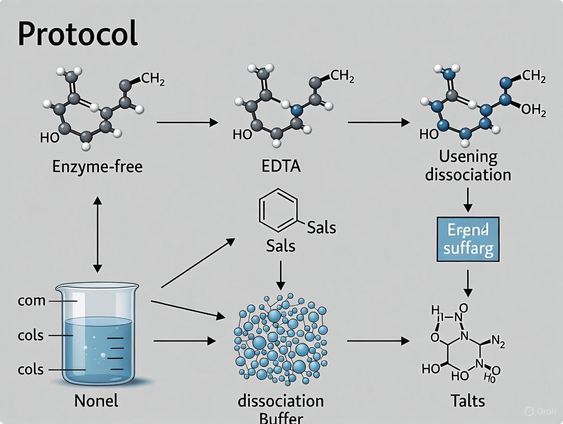
Enzyme-Free Cell Dissociation Buffer: A Complete Protocol Guide for Researchers
This article provides a comprehensive guide to enzyme-free cell dissociation, a gentle technique crucial for applications requiring intact cell surface proteins and high viability, such as single-cell analysis, stem cell...
Enzyme-Free Cell Dissociation Buffer: A Complete Protocol Guide for Researchers
Abstract
This article provides a comprehensive guide to enzyme-free cell dissociation, a gentle technique crucial for applications requiring intact cell surface proteins and high viability, such as single-cell analysis, stem cell research, and drug development. It covers the foundational principles of how non-enzymatic buffers work, detailed step-by-step protocols for adherent cell lines and sensitive pluripotent stem cells, common troubleshooting and optimization strategies, and a comparative analysis with traditional enzymatic methods. The content is tailored for researchers, scientists, and drug development professionals seeking to implement robust, reproducible, and gentle cell dissociation in their workflows.
Understanding Enzyme-Free Dissociation: Principles and Advantages for Modern Cell Biology
What is Enzyme-Free Cell Dissociation and How Does It Work?
In the realm of cell biology and regenerative medicine, the initial step of creating a single-cell suspension from tissues or cultured monolayers is critical for downstream analysis and therapeutic application. Enzyme-free cell dissociation represents a class of techniques that achieve this cellular separation without employing proteolytic enzymes like trypsin, collagenase, or accutase. Unlike enzymatic methods that digest extracellular matrix proteins and cell adhesion molecules, enzyme-free approaches rely on chemical chelation and/or physical forces to disrupt cell-cell and cell-substrate connections, thereby preserving the structural and functional integrity of cell surface proteins [1] [2].
This methodology is gaining significant traction within the scientific community, particularly for applications where cell surface marker integrity is paramount. The global cell dissociation market reflects this trend, with the non-enzymatic segment projected to expand at a compound annual growth rate (CAGR) of 17.67% through 2030, significantly outpacing the overall market growth [3]. This shift is driven by the escalating demand for robust, reproducible cell processing in cell and gene therapy production, single-cell omics, and personalized medicine pipelines, where preserving native cell states is essential for accurate diagnostics and effective treatments [3] [4].
Mechanisms of Action: How Enzyme-Free Methods Work
Enzyme-free dissociation operates primarily through two fundamental mechanisms: chemical chelation and physical disruption. Often, advanced systems integrate both to enhance efficiency.
Chemical Chelation-Based Mechanisms
The most common chemical approach utilizes cell dissociation buffers containing chelating agents such as Ethylenediaminetetraacetic acid (EDTA). These buffers are isotonic, membrane-filtered solutions formulated in calcium-free and magnesium-free phosphate-buffered saline (PBS) [2].
- Principle of Operation: Cell adhesion is heavily dependent on calcium and magnesium ions to stabilize integrin-mediated attachments to the extracellular matrix and cadherin-mediated cell-cell junctions. Chelating agents like EDTA bind these divalent cations, sequestering them from the cellular environment. This binding disrupts the ionic bridges essential for maintaining adhesion complexes, leading to a weakening of cell attachments and eventual cell dissociation [2] [5].
- Key Characteristics: This method is considered exceptionally gentle, as it avoids the proteolytic cleavage of surface proteins. This makes it ideal for studies requiring intact cell surface receptors, such as ligand binding assays, flow cytometry, and immunohistochemistry [2].
Physical and Mechanical Mechanisms
For solid tissues, purely mechanical or innovative physical methods are employed to generate single-cell suspensions without chemical or enzymatic intervention.
- Automated Mechanical Disaggregation: Systems like the Medimachine II process minced tissue fragments within a disposable cartridge containing a fine mesh and microblades. The automated spinning action forces tissue pieces against the mesh, physically shearing them into single cells while filtering out debris [6]. This standardizes a process that was traditionally operator-dependent, enhancing reproducibility.
- Hypersonic Levitation and Spinning (HLS): A groundbreaking, non-contact method uses a triple-acoustic resonator probe generating GHz-frequency acoustic waves. This creates microscale "liquid jets" within a confined field, producing precise hydrodynamic forces. The target tissue levitates and executes a rapid 'press-and-rotate' operation, which applies shear stress to the tissue surface, effectively disrupting cell-cell connections while safeguarding cell integrity [7].
- Electric Field Facilitation: Some novel platforms use controlled electric fields to dissociate tissues into viable single cells rapidly, achieving dissociation in as little as five minutes while maintaining high cell viability [1] [8].
Table 1: Core Mechanisms in Enzyme-Free Cell Dissociation
| Mechanism Type | Representative Examples | Core Principle | Key Action |
|---|---|---|---|
| Chemical Chelation | EDTA-based Buffer [2] | Ion Sequestration | Binds Ca²⁺ and Mg²⁺ ions, destabilizing adhesion complexes. |
| Automated Mechanical | Medimachine II [6] | Physical Shearing | Minces tissue against a micro-mesh using automated spinning. |
| Acoustic Energy | Hypersonic Levitation (HLS) [7] | Hydrodynamic Shear | Uses acoustic streaming to levitate and spin tissue, generating dissociating forces. |
| Electrical Energy | Electric Field Dissociation [1] | Electroporation/Field Effects | Applies electric fields to disrupt tissue architecture. |
Advantages, Limitations, and Comparative Analysis
The adoption of enzyme-free dissociation is driven by distinct advantages, but it is crucial to understand its limitations to apply it appropriately.
Key Advantages
- Preservation of Cell Surface Markers: This is the most significant advantage. By avoiding proteolytic digestion, cell surface proteins, receptors, and antigens remain intact. This is critical for flow cytometry, cell sorting, and functional immunology studies where accurate phenotyping is required [1] [2]. Research shows that even enzymes considered "gentle," like accutase, can cleave specific surface proteins such as Fas ligands, requiring a 20-hour recovery period for re-expression [5].
- Enhanced Cell Viability and Function: Enzyme-free processes minimize cellular stress and damage. Studies comparing automated mechanical (Medimachine II) and enzymatic dissociation found that mechanically derived cells showed better preservation of lysosome and mitochondrial labeling, indicating superior organelle function post-harvest [6].
- Reduced Artifacts in Downstream Analysis: The absence of enzymatic activity prevents the alteration of gene expression profiles or the induction of stress responses that can distort single-cell RNA sequencing data and other sensitive assays [1] [7].
- Regulatory and Safety Benefits: Non-enzymatic reagents are often animal-origin free, eliminating the risk of zoonotic pathogen introduction and simplifying regulatory approval for therapeutic manufacturing [3] [4].
Inherent Limitations
- Efficacy on Complex Tissues: Enzyme-free chemical buffers are generally unsuitable for dissociating dense or fibrous native tissues (e.g., connective tissue). They are most effective for lightly adherent cell lines (e.g., HeLa, NIH 3T3) in culture or as a component of integrated systems for tissues [2].
- Lower Yield in Some Contexts: For firmly established cell cultures or complex tissues, a purely enzyme-free approach may result in lower cell yields compared to robust enzymatic protocols, sometimes necessitating mechanical assistance like scraping [5].
- Technology Cost: Advanced non-enzymatic platforms, such as automated dissociators or acoustic systems, can involve significant capital investment, potentially limiting access for smaller laboratories [3] [4].
Table 2: Comparison of Dissociation Methods Based on Key Performance Metrics
| Performance Metric | Enzymatic Dissociation | Enzyme-Free Chemical (e.g., EDTA) | Enzyme-Free Physical (e.g., HLS, Automated) |
|---|---|---|---|
| Cell Surface Protein Integrity | Compromised [1] [5] | Excellent [2] | Excellent [7] |
| Typical Viability | Variable, can be low with over-digestion [1] | High [2] | High (e.g., >92% with HLS) [7] |
| Processing Speed | Slow (often >1 hour) [1] | Fast for cultured cells (min) [2] | Very Fast (e.g., 15 min for HLS) [7] |
| Suitability for Complex Tissues | Excellent | Poor [2] | Good to Excellent [7] [6] |
| Operator-induced Variability | High | Moderate | Low (Automated) [6] |
Detailed Experimental Protocols
Protocol 1: Enzyme-Free Dissociation of Adherent Cells Using Chelation
This protocol is adapted for common adherent cell lines and is based on the use of commercial buffers like Gibco Cell Dissociation Buffer [2].
The Scientist's Toolkit: Reagents and Materials
- Cell Culture: Sub-confluent (60-80%) monolayer of adherent cells (e.g., HeLa, NIH 3T3).
- Dissociation Reagent: Gibco Cell Dissociation Buffer, enzyme-free, PBS (Catalog #13151014) or equivalent.
- Other Reagents: Dulbecco's Phosphate-Buffered Saline (DPBS), without Ca²⁺ and Mg²⁺; complete growth medium containing serum.
- Labware: Tissue culture flasks/plates, serological pipettes, centrifuge tubes, cell strainer (optional).
- Instrumentation: Water bath, biological safety cabinet, centrifuge.
Step-by-Step Workflow:
- Preparation: Pre-warm the enzyme-free dissociation buffer and DPBS to 37°C. Ensure the growth medium is ready to neutralize the process.
- Aspiration: Remove and discard the complete growth medium from the culture vessel.
- Rinsing: Gently rinse the cell monolayer with pre-warmed DPBS to remove any residual serum and calcium/magnesium ions, which can inhibit dissociation. Aspirate and discard the DPBS wash.
- Application: Add sufficient pre-warmed enzyme-free dissociation buffer to completely cover the cell monolayer (e.g., 2 mL for a T-75 flask).
- Incubation: Incubate the culture vessel at 37°C for 5-15 minutes. Observe cells periodically under a microscope. Cells are ready when they appear rounded and begin to detach. Avoid prolonged incubation, as it can be counterproductive.
- Detachment: Gently tap the side of the vessel to dislodge the cells. For stubborn cells, a stream of complete growth medium can be pipetted across the monolayer to aid detachment. Do not scrape, as this can cause mechanical damage.
- Neutralization & Collection: Transfer the cell suspension to a centrifuge tube containing a volume of complete growth medium that is at least twice the volume of the dissociation buffer used. The serum in the medium effectively neutralizes the process.
- Centrifugation: Pellet the cells by centrifugation at approximately 200-300 × g for 5 minutes.
- Resuspension: Aspirate and discard the supernatant. Gently resuspend the cell pellet in fresh, pre-warmed complete growth medium.
- Counting and Seeding: Count cells using a hemocytometer or automated counter and seed at the desired density for subsequent experiments.
Diagram 1: Enzyme-free cell dissociation workflow for adherent cells.
Protocol 2: Automated Mechanical Dissociation of Solid Tissue
This protocol utilizes the Medimachine II system for processing small solid tissue samples (e.g., tumor biopsies, spleen) into single-cell suspensions [6].
The Scientist's Toolkit: Reagents and Materials
- Tissue Sample: Freshly harvested tissue (e.g., spleen, tumor), ideally kept in cold PBS or transport medium.
- Dissociation System: Medimachine II instrument and disposable Medicons with a 50µm or 100µm mesh.
- Reagents: Cold RPMI 1640 medium or other appropriate buffer; complete growth medium with serum; 70% ethanol for decontamination.
- Labware: Petri dishes, scalpels, forceps, 5 mL syringes, cell strainer (optional), centrifuge tubes.
- Instrumentation: Biological safety cabinet, centrifuge.
Step-by-Step Workflow:
- Preparation: Sterilize the workspace. Place the Medimachine II in the biosafety cabinet. Fill a Medicon with 1 mL of cold RPMI 1640 medium.
- Tissue Mincing: Transfer the tissue to a Petri dish containing cold PBS. Using scalpels and forceps, mince the tissue into fragments of approximately 1 mm³.
- Loading: Using forceps, transfer 3-5 minced tissue fragments into the pre-filled Medicon.
- Assembly and Run: Close the Medicon and insert it into the Medimachine II. Select the appropriate disaggregation program (e.g., run for 2-10 minutes). The instrument will automatically spin the Medicon, forcing tissue fragments against the mesh.
- Collection: After the run, remove the Medicon. Using a 5 mL syringe, pipette the cell suspension from the Medicon into a centrifuge tube through a possible cell strainer to remove any remaining large aggregates.
- Washing: Rinse the Medicon with an additional 1-2 mL of cold buffer to collect any remaining cells and add to the centrifuge tube.
- Centrifugation: Pellet the cells by centrifugation at 300-400 × g for 5 minutes.
- Red Blood Cell Lysis (if needed): For tissues like spleen, resuspend the pellet in an appropriate red blood cell lysis buffer, incubate as per protocol, then centrifuge again.
- Resuspension and Counting: Aspirate the supernatant and resuspend the final cell pellet in complete growth medium or an appropriate buffer for downstream applications. Perform a cell count and viability assay (e.g., Trypan Blue exclusion).
Diagram 2: Automated mechanical dissociation workflow for solid tissues.
Applications in Research and Therapy
Enzyme-free dissociation is critical in fields where cell integrity and function are non-negotiable.
- Cell Therapy Development: In manufacturing therapies like CAR-T cells, enzyme-free methods are used to isolate peripheral blood mononuclear cells or dissociate tumor tissues, ensuring that the therapeutic cells' surface receptors remain unaltered for efficient engineering and function. Automated systems provide the consistency required for Good Manufacturing Practice (GMP) [9].
- Single-Cell Multi-Omics Analysis: For single-cell RNA sequencing (scRNA-seq), preserving the native transcriptome is essential. Enzymatic digestion can induce stress-related gene expression artifacts. Enzyme-free techniques, including the novel HLS method, have been shown to preserve rare cell populations and provide a more accurate representation of cellular heterogeneity in tissues like human renal cancer [1] [7].
- Cancer Research and Diagnostics: Dissociating tumor tissues without enzymes allows for the accurate profiling of cell surface biomarkers used for diagnosis and patient stratification. It also enables the study of tumor heterogeneity with minimal introduction of technical artifacts [9].
- Regenerative Medicine and Organoid Culture: When creating organoids or tissue-engineered constructs, the dissociation of stem cell aggregates must maintain cell viability and differentiation potential. Gentle, non-enzymatic methods are preferred to avoid damaging these sensitive cells [9] [4].
- Fundamental Biological Research: Any study focusing on cell adhesion, migration, or surface receptor signaling benefits from dissociation techniques that do not themselves alter the proteins being investigated [5].
The trajectory of enzyme-free cell dissociation is firmly aligned with the evolving needs of precision medicine and advanced therapeutics. The market shift towards non-enzymatic products, projected to grow at a CAGR of 17.67%, underscores a fundamental transition in laboratory and clinical practice [3]. Future advancements will likely focus on increased automation, integration with AI for process control, and the development of even gentler, high-throughput systems such as advanced microfluidic and acoustic platforms [4] [7].
In conclusion, enzyme-free cell dissociation is not merely an alternative but often a superior approach for modern cell-based applications. Its ability to deliver viable, functional cells with unaltered surface phenotypes makes it indispensable for critical research and clinical workflows. As the technology continues to evolve, becoming more accessible and integrated, it will play a pivotal role in ensuring that the first step in cell analysis—the creation of a single-cell suspension—does not compromise the integrity of the data or the safety and efficacy of resulting therapies.
Cell adhesion is a fundamental process in maintaining tissue integrity and cellular function. In laboratory research, the controlled disruption of this adhesion is critical for harvesting and studying cells. Enzyme-free cell dissociation buffers offer a gentle, specific method for disrupting cell adhesion without damaging sensitive cell surface proteins, making them indispensable for applications like flow cytometry, ligand binding assays, and drug discovery research [10] [11]. This application note details the core mechanisms by which these buffers, specifically their chelating agents and salt solutions, function to disassemble cell-matrix and cell-cell adhesions. We provide a quantitative comparison of cell line responses, detailed experimental protocols for functional adhesion assessment, and visualization of the underlying processes to support researchers in implementing these techniques effectively.
Core Mechanisms of Action
Enzyme-free cell dissociation buffers disrupt adhesion through a combination of chemical chelation and physiological buffering, avoiding the proteolytic degradation associated with enzymatic methods like trypsin.
The Role of Chelating Agents
The primary active components in these buffers are chelating agents, most commonly Ethylenediaminetetraacetic acid (EDTA) [10] [12]. EDTA functions by sequestering divalent cations such as calcium (Ca²⁺) and magnesium (Mg²⁺) from the cellular environment [13] [14]. These ions are essential co-factors for cadherins and integrins, the key transmembrane proteins that mediate cell-cell and cell-matrix adhesion, respectively. By binding to these cations, EDTA causes conformational changes in cadherins and integrins, weakening their hold on adjacent cells and the extracellular matrix (ECM) [14]. This process is purely physicochemical and does not digest the proteins, thereby preserving their structural and functional integrity for subsequent analysis.
The Role of Salt Solutions
The chelating agents are formulated in a balanced salt solution, typically Calcium- and Magnesium-Free Hank's Balanced Salt Solution (HBSS) [10] [15]. This base solution serves multiple critical functions:
- Isotonic Environment: HBSS provides an isotonic medium that prevents osmotic shock to the cells during the dissociation process, maintaining high cell viability (typically >90%) [11] [16].
- Ionic Control: The deliberate omission of Ca²⁺ and Mg²⁺ synergizes with the chelating agent, ensuring these ions are not reintroduced to the system, which would counteract the dissociation process.
- Physiological Support: Other salts in the solution (e.g., KCl, KH₂PO₄, NaHCO₃, NaCl) help maintain a physiologically compatible pH and ionic strength, conditioning the cells throughout the procedure [10].
The following diagram illustrates the sequential mechanism of action for enzyme-free cell dissociation buffers.
Quantitative Data on Cell Dissociation and Adhesion
The effectiveness of enzyme-free dissociation is cell line-dependent. The following table summarizes functional metrics of adhesion and migration for selected cancer cell lines, providing a context for understanding dissociation requirements [17].
Table 1: Functional Metrics of Cell Adhesion and Migration in Paired Cancer Cell Lines
| Cell Line | Tissue Origin | Metastatic Potential | Predominant Aggression Metric | Relative Adhesion Strength |
|---|---|---|---|---|
| MCF-7 | Breast | Low | Wound Closure Migration | High |
| MDA-MB-231 | Breast | High | Loss of Cell Adhesion | Low |
| Ishikawa | Endometrium | Low | Wound Closure Migration | High |
| KLE | Endometrium | High | Loss of Cell Adhesion | Low |
| Cal-27 | Tongue | Low | Wound Closure Migration | High |
| SCC-25 | Tongue | High | Loss of Cell Adhesion | Low |
Cell lines with low metastatic potential, such as MCF-7 and Ishikawa, generally exhibit stronger cell-matrix adhesion, making them potentially more resistant to gentle, enzyme-free dissociation. In contrast, highly metastatic lines like MDA-MB-231 and KLE have weaker adhesion and detach more readily, a functional reflection of their in vivo potential for detachment from primary tumors [17].
Experimental Protocols
Standard Protocol for Enzymatic and Non-Enzymatic Cell Dissociation
The table below outlines the primary methods for dissociating adherent cells, highlighting the role of non-enzymatic buffers [11] [16].
Table 2: Overview of Cell Dissociation Methods and Applications
| Procedure | Dissociation Agent | Typical Applications | Key Considerations |
|---|---|---|---|
| Scraping | Cell Scraper | Cell lines sensitive to proteases; may damage some cells. | Physical method; can compromise cell viability. |
| Enzymatic Dissociation | Trypsin, TrypLE, Accutase | Strongly adherent cells; routine passaging. | Can cleave cell surface receptors; requires inhibitor. |
| Enzymatic Dissociation | Trypsin + EDTA | Enhances enzyme activity on robust cell lines. | Combined chemical and enzymatic action. |
| Non-Enzymatic Dissociation | Cell Dissociation Buffer (EDTA-based salts) | Lightly adherent cells; studies requiring intact cell surface proteins (e.g., flow cytometry, ligand binding). | Gentle; preserves antigenicity. Not for strongly adherent cells. |
Detailed Protocol: Using Enzyme-Free Cell Dissociation Buffer
This protocol is adapted for a T75 culture flask [11] [15] [16].
Research Reagent Solutions & Essential Materials:
- Gibco Cell Dissociation Buffer (enzyme-free, with EDTA in HBSS) or equivalent [10] [12].
- ZymeFree Enzyme Free Cell Dissociation Reagent (alternative) [15].
- DPBS (without calcium and magnesium)
- Complete growth medium
- T75 culture flask with adherent cells at 70-90% confluency
- 37°C water bath or incubator
- Centrifuge and conical tubes
- Automated or manual cell counter
Methodology:
- Preparation: Warm the cell dissociation buffer and complete growth medium to 37°C. Pre-warming is critical to avoid thermal shock and ensure efficient dissociation [16].
- Remove Medium: Aspirate and discard the spent cell culture medium from the flask.
- Rinse Monolayer: Wash the cell monolayer with 5-10 mL of pre-warmed, calcium- and magnesium-free DPBS. Gently rock the flask for 30-60 seconds to rinse away any residual media containing divalent cations. Aspirate and discard the rinse solution. Note: This step is crucial for maximizing the efficiency of the chelating agent.
- Apply Dissociation Buffer: Add 5 mL of pre-warmed enzyme-free Cell Dissociation Buffer to the flask, ensuring it completely covers the cell sheet. Rock the flask gently to distribute the solution [15] [16].
- Incubate and Monitor: Incubate the flask at room temperature for 1-2 minutes. Firmly tap the vessel against the palm of your hand to dislodge the cells. Observe the cells under an inverted microscope. If cells remain adherent, allow the flask to sit at room temperature for an additional 2-5 minutes and tap again. Note: The exact time varies by cell line; monitor closely to avoid over-exposure, which can still be cytotoxic.
- Neutralize and Harvest: Once the majority of cells have rounded up and detached (they will appear phase-bright), add at least 5 mL of complete growth medium to the flask. The serum and components in the medium will neutralize the dissociation process.
- Resuspend Cells: Pipette the cell suspension repeatedly over the surface of the flask to disperse them into a single-cell suspension. Transfer the suspension to a 15 mL conical tube.
- Centrifuge and Count: Centrifuge the tube at approximately 100 × g for 5-10 minutes. Discard the supernatant and resuspend the cell pellet in fresh, pre-warmed complete medium. Determine viable cell density and percent viability (should be >90%) using an automated cell counter or hemocytometer [11].
Protocol for Functional Assessment of Cell Adhesion
This protocol describes a quantitative method for assessing cellular adhesive strength, which can be used to optimize dissociation parameters or study metastatic potential [17].
Materials:
- Parallel plate flow chamber or microfluidic device
- Peristaltic pump or syringe driver
- DPBS or cell culture medium (for shear flow)
- Inverted microscope with camera for time-lapse imaging
Methodology:
- Culture Cells: Seed the cells of interest (e.g., MCF-7 vs. MDA-MB-231) onto culture dishes or coverslips that fit the flow chamber and allow them to adhere and form a monolayer.
- Assemble Flow Chamber: Place the cell-coated substrate into the parallel plate flow chamber apparatus.
- Apply Shear Stress: Initiate a unidirectional, controlled flow of DPBS or medium over the cells using a pump. The flow rate is gradually increased to apply defined levels of wall shear stress, mimicking physiological forces.
- Image and Quantify: Under the microscope, record time-lapse images of the cells during flow application. The point at which the shear force exceeds the adhesive force of the cells to the culture surface, causing them to detach, is recorded.
- Analyze Data: Quantify the percentage of cells detached at each shear stress level. As demonstrated in research, cell lines with high metastatic potential (e.g., MDA-MB-231) will typically show greater detachment at lower shear stresses compared to their low metastatic potential counterparts (e.g., MCF-7) [17].
The workflow for this functional adhesion assay is visualized below.
The Scientist's Toolkit
Table 3: Essential Research Reagent Solutions for Cell Adhesion and Dissociation Studies
| Item | Function/Application | Example Products |
|---|---|---|
| Enzyme-Free Cell Dissociation Buffer | Gentle detachment of lightly adherent cells while preserving surface protein integrity. Essential for flow cytometry, ligand binding. | Gibco Cell Dissociation Buffer [10], ZymeFree [15] |
| Calcium- and Magnesium-Free DPBS | Washing solution to remove residual divalent cations, enhancing the efficiency of chelating agents. | Standard DPBS (-Ca²⁺, -Mg²⁺) |
| Trypsin-EDTA Combination | For robust dissociation of strongly adherent cell lines via enzymatic proteolysis and cation chelation. | 0.25% Trypsin-EDTA |
| TrypLE Express Enzyme | A recombinant fungal trypsin substitute for cell dissociation; animal origin-free. | Gibco TrypLE Select [11] |
| Parallel Plate Flow Chamber | Microfluidic device for applying controlled shear stress to quantitatively measure cell adhesion strength. | Custom or commercial systems [17] |
| Automated Cell Counter | Accurately determine viable cell density and percent viability post-dissociation. | Countess Automated Cell Counters [11] |
In the fields of cell biology, immunology, and drug development, the preparation of high-quality single-cell suspensions is a critical prerequisite for downstream applications such as flow cytometry, single-cell RNA sequencing, and cell-based therapies. Traditional enzymatic dissociation methods using trypsin, collagenase, or other proteases present significant limitations for these sensitive applications, primarily due to their proteolytic activity that can damage cell surface markers and compromise cell viability [18] [19]. Enzyme-free cell dissociation techniques have emerged as vital alternatives that effectively address these challenges through gentler, non-proteolytic mechanisms of action.
Enzyme-free dissociation buffers typically consist of isotonic, pH-balanced solutions containing chelating agents such as EDTA or EGTA that work by sequestering divalent cations like calcium and magnesium [2]. These cations are essential for cell adhesion through cadherin-mediated junctions and integrin binding to extracellular matrix components. By removing these cations, enzyme-free buffers disrupt cell-cell and cell-substrate interactions while preserving the structural and functional integrity of cell surface proteins [16] [2]. This fundamental difference in mechanism underlies the key advantages of enzyme-free methods for applications requiring intact cell surface markers and maximal cell viability.
Key Advantages and Comparative Performance
Superior Preservation of Cell Surface Markers
The preservation of cell surface markers is perhaps the most significant advantage of enzyme-free dissociation methods. Unlike enzymatic approaches that cleave peptide bonds in proteins, enzyme-free buffers maintain the structural integrity of cell surface epitopes, which is crucial for accurate immunophenotyping, cell sorting, and receptor-ligand binding studies.
Recent research demonstrates that enzymatic dissociation can significantly alter cell surface marker expression. One systematic comparison of dissociation techniques for analyzing the mouse colon immune system found that "both enzymatic approaches were associated with a marked decrease of several cell-surface markers," whereas mechanical (enzyme-free) dissociation preserved marker integrity [20]. Similarly, studies on neural tissues have shown that enzymatic dissociation induces artifactual expression of immediate early genes (IEGs) such as Fos, Arc, and Egr1, making inactive neurons appear activated—a critical concern for single-cell transcriptomic studies [19].
Advanced enzyme-free technologies like Bulk Lateral Ultrasonic (BLU) energy have demonstrated superior preservation of specific cell surface markers across multiple tissue types. Research shows that BLU dissociation resulted in significantly improved recovery of critical immune markers including CD86, Ly6G, and various lymphocyte and macrophage markers compared to enzymatic methods [19].
Enhanced Cell Viability and Function
Enzyme-free dissociation methods consistently demonstrate advantages in maintaining cell viability and functionality:
- Reduced Cellular Stress: Enzyme- and heat-free methods avoid the cellular stress responses triggered by prolonged enzymatic digestion at elevated temperatures. Studies indicate that "long dissociation protocol times" correlate strongly with "the stress index experienced by the cell" [19].
- Preserved Functional Integrity: Research on mesenchymal stem cells (MSCs) highlights that cells dissociated with enzyme-free buffers maintain better functional characteristics, though viability immediately after dissociation may be lower than with enzymatic methods [21].
- Improved Recovery of Sensitive Cell Types: Enzyme-free methods demonstrate particular advantages for fragile cell populations, including certain immune cells and stem cells, which are more vulnerable to proteolytic damage [7].
Table 1: Comparative Analysis of Cell Dissociation Methods
| Parameter | Enzymatic Methods | Enzyme-Free Buffers | Advanced Non-Enzymatic Technologies |
|---|---|---|---|
| Cell Surface Marker Integrity | Potential cleavage of surface proteins and epitopes [18] | Preserves structural integrity of membrane proteins [2] | Superior preservation of sensitive markers [19] |
| Immediate Cell Viability | Generally high immediately post-dissociation [21] | May be lower initially but better preservation of function [21] | High viability maintained (e.g., 92.3% with HLS) [7] |
| Cellular Stress Response | Induces stress genes and artificial activation [19] | Minimal stress induction | Minimal stress response and artifact generation |
| Processing Time | Variable (minutes to hours) [1] | Typically 15-30 minutes [21] | Rapid processing (e.g., 15 minutes for HLS) [7] |
| Application Flexibility | Broad but with marker damage risk | Ideal for surface protein studies [2] | Versatile across tissue types with high quality |
Special Considerations for Different Cell Types
The performance of enzyme-free dissociation methods varies across different cell and tissue types:
- Immune Cells: Enzyme-free mechanical dissociation has proven particularly advantageous for comprehensive immune profiling in complex tissues. Research on mouse colon demonstrated that mechanical dissociation was "more suitable and efficient than enzymatic methods for recovering immune cells from all colon layers at once" [20].
- Stem Cells: While enzyme-free methods preserve surface markers, studies on mesenchymal stem cells indicated that "the proportion of viable cells that reattached was significantly lower for cells obtained by dissociation with enzyme-free dissociation buffer compared to trypsin" [21]. This suggests that the optimal dissociation method must be determined based on the specific downstream application.
- Primary Tissues: For complex primary tissues, emerging technologies such as hypersonic levitation and spinning (HLS) show remarkable performance, achieving 92.3% cell viability while preserving rare cell populations that are often lost with conventional methods [7].
Table 2: Cell Type-Specific Recommendations for Enzyme-Free Dissociation
| Cell/Tissue Type | Recommended Enzyme-Free Approach | Key Benefits | Considerations |
|---|---|---|---|
| Immune Cells | Mechanical dissociation (gentle crushing) [20] | Superior preservation of cell surface markers | More effective for comprehensive immune profiling than enzymatic methods |
| Stem Cells | Chelation-based buffers [21] | Maintains differentiation potential | Reattachment rates may be lower than enzymatic methods |
| Epithelial Cells | EDTA-based dissociation buffers [2] | Preserves junctional proteins | May require optimization for strongly adherent lines |
| Complex Tissues | Automated systems (e.g., BLU, HLS) [7] [19] | Maintains tissue heterogeneity and rare cells | Higher initial equipment investment |
| Neural Tissues | Cold mechanical or acoustic methods [19] | Avoids artifactual neural activation | Essential for accurate transcriptomic studies |
Experimental Protocols and Methodologies
Basic Protocol for Enzyme-Free Buffer Dissociation
The following protocol adapts established methodologies for enzyme-free dissociation of adherent cell cultures [16] [2]:
Reagents and Equipment:
- Enzyme-free cell dissociation buffer (e.g., Gibco Cat. No. 13151014 or equivalent)
- Calcium- and magnesium-free phosphate-buffered saline (PBS)
- Complete growth medium with serum
- Tissue culture vessels with adherent cells
- Sterile pipettes and centrifuge tubes
- Inverted microscope
- Automated cell counter or hemocytometer
Procedure:
- Preparation: Warm the enzyme-free dissociation buffer and calcium-/magnesium-free PBS to 37°C prior to use. Ensure all reagents are sterile.
- Removal of Growth Medium: Aspirate and discard the complete growth medium from the culture vessel.
- Rinsing Step: Gently rinse the cell monolayer with 5 mL of calcium-/magnesium-free PBS per T75 flask or 100 mm dish. Rock the vessel for 30-60 seconds at room temperature, then aspirate and discard the rinse solution. Repeat this rinsing step once more.
- Dissociation Buffer Application: Add approximately 5 mL of enzyme-free cell dissociation buffer to the vessel, ensuring complete coverage of the cell monolayer.
- Incubation: Gently rock the vessel at room temperature and monitor dissociation progress under a microscope. Lightly tap the flask against the palm to facilitate detachment.
- Completion of Detachment: Once approximately 60-80% of cells have detached (typically 5-15 minutes), add at least 5 mL of complete growth medium containing serum to neutralize the dissociation buffer.
- Cell Collection: Gently pipette the cell suspension to ensure single-cell dispersion. Transfer to a sterile centrifuge tube.
- Centrifugation and Washing: Centrifuge at 300 × g for 5 minutes. Discard the supernatant and resuspend the cell pellet in fresh complete medium.
- Viability Assessment: Count cells using an automated cell counter or hemocytometer with trypan blue exclusion. Cell viability should exceed 90% for most applications [16].
Troubleshooting Notes:
- For strongly adherent cell lines, a pre-wash with 1-5 mM EDTA in PBS may enhance dissociation.
- Optimal cell confluence for dissociation is 60-80%. Overly confluent cultures may resist gentle dissociation.
- If dissociation is incomplete after 15 minutes, gently pipette the solution across the monolayer or extend incubation time in 5-minute increments.
Mechanical Dissociation Protocol for Solid Tissues
For solid tissues requiring single-cell suspension preparation while preserving surface markers, mechanical dissociation offers a robust enzyme-free alternative [20]:
Reagents and Equipment:
- Cold isotonic buffer (e.g., RPMI 1640)
- Cell strainers (70 μm and 40 μm)
- Petri dishes
- Syringe plunger or cell scraper
- Centrifuge tubes
Procedure:
- Tissue Collection and Preparation: Place dissected tissue samples in a sterile Petri dish containing cold RPMI 1640 medium on ice.
- Mechanical Disruption: For tissues such as spleen or colon, gently grind with the flat head of a syringe plunger on a 70 μm cell strainer. Apply slightly stronger manual pressure for denser tissues, positioning the tissue mucosal side on the cell strainer when applicable.
- Filtration and Collection: Filter the resulting cell suspension through a 40 μm cell strainer to remove debris and clumps.
- Recovery Incubation: Incubate cells in RPMI medium supplemented with 10% FBS for 2 hours at 37°C to promote the re-appearance of any temporarily internalized cell surface markers.
- Viability Assessment: Count viable cells using a hemocytometer with trypan blue exclusion.
Advanced Non-Enzymatic Technologies
Emerging technologies offer sophisticated alternatives to traditional enzyme-free methods:
Hypersonic Levitation and Spinning (HLS): This contact-free approach utilizes a triple-acoustic resonator probe to levitate and spin tissue samples, generating microscale "liquid jets" that exert precise hydrodynamic forces to dissociate tissues without mechanical contact [7]. The method achieves 92.3% viability with 90% tissue utilization in just 15 minutes, significantly outperforming conventional methods.
Bulk Lateral Ultrasonic (BLU) Energy: This enzyme- and heat-free technology uses ultrasonic energy to dissociate tissues while preserving cell surface markers. Studies demonstrate BLU dissociation results in improved recovery of specific immune cell populations compared to enzymatic methods [19].
The Scientist's Toolkit: Essential Research Reagents
Table 3: Essential Reagents for Enzyme-Free Cell Dissociation
| Reagent/Equipment | Function | Example Products/Specifications |
|---|---|---|
| EDTA-Based Dissociation Buffer | Chelates divalent cations to disrupt cell adhesions | Gibco Cell Dissociation Buffer (Catalog #13151014) [2] |
| Calcium-/Magnesium-Free PBS | Removes cations prior to dissociation; prevents reaggregation | Standard PBS formulation without Ca²⁺/Mg²⁺ [16] |
| Cell Strainers | Removes cell clumps and debris post-dissociation | 40 μm and 70 μm mesh sizes [20] |
| Automated Cell Counter | Assesses cell viability and concentration post-dissociation | Trypan blue exclusion method [21] |
| Serum-Containing Medium | Neutralizes dissociation buffer; supports cell recovery | Standard growth medium with 10% FBS [16] |
| Advanced Dissociation Systems | Provides standardized, automated dissociation | Singleron PythoN systems [22]; HLS technology [7] |
Workflow and Decision Framework
The following diagram illustrates the decision-making process for selecting appropriate dissociation methods based on research objectives and sample characteristics:
Enzyme-free cell dissociation methods offer significant advantages for applications requiring preservation of cell surface markers and high cell viability. Through non-proteolytic mechanisms involving chelation of divalent cations or advanced physical methods, these techniques maintain the structural integrity of membrane proteins while minimizing cellular stress responses. While enzymatic methods may still be preferable for certain applications where complete dissociation efficiency is paramount, the growing emphasis on accurate cell surface marker analysis and minimal manipulation in cell-based therapies positions enzyme-free methods as essential tools in modern biological research and drug development.
The continued development of advanced non-enzymatic technologies such as hypersonic levitation and acoustic methods promises to further enhance our ability to obtain high-quality single-cell suspensions from even the most challenging tissue types while preserving their native molecular signatures. As single-cell analysis technologies continue to advance, the importance of gentle, artifact-free dissociation methods will only increase, solidifying the role of enzyme-free approaches in generating biologically relevant data.
Enzyme-free cell dissociation technologies represent a paradigm shift in the preparation of cellular samples for advanced research and diagnostic applications. Traditional enzymatic methods, while effective at degrading extracellular matrices, often introduce significant biases by damaging cell surface epitopes, altering transcriptomic profiles, and selectively eliminating sensitive cell populations. The emerging enzyme-free approaches leverage physical, chemical, and acoustic mechanisms to gently dissociate tissues and cell cultures while preserving cellular integrity and biological relevance. This document provides detailed application notes and standardized protocols for implementing these methods across three critical applications: stem cell passaging, single-cell sequencing, and flow cytometry, supported by quantitative performance data and step-by-step methodologies.
Performance Comparison of Enzyme-Free Dissociation Methods
The table below summarizes key performance metrics for various enzyme-free dissociation methods, highlighting their advantages for specific research applications.
Table 1: Quantitative Performance Metrics of Enzyme-Free Dissociation Methods
| Method | Principle | Processing Time | Cell Viability | Single-Cell Yield | Ideal Applications |
|---|---|---|---|---|---|
| Chemical (ReLeSR/GCDR) [23] [24] | Selective ionic modulation (Ca²⁺/Mg²⁺) | 10-15 minutes | >90% (as aggregates) | N/A (Passaged as clumps) | Stem cell passaging |
| Electric Field [25] | Oscillating electric field (100 V/cm) | ~5 minutes | High (Specific for single-cell) | 96% recovery rate | Single-cell RNA sequencing |
| Hypersonic Levitation (HLS) [7] | Acoustic streaming & hydrodynamic shear | 15 minutes | 92.3% | 90% tissue utilization | Flow cytometry, rare cell population studies |
| Acoustic (SimpleFlow) [26] | Acoustic waves | 4 minutes | Higher than enzymatic | Higher than enzymatic | Preserving heterogeneous cell populations |
Application-Specific Protocols
Stem Cell Passaging Using Chemical Methods
Background: Maintaining the pluripotency and genomic integrity of human pluripotent stem cells (hPSCs), including embryonic (ES) and induced pluripotent (iPS) cells, requires gentle passaging that minimizes cell damage. Enzyme-free reagents like ReLeSR and Gentle Cell Dissociation Reagent (GCDR) work by modulating divalent cations, selectively disrupting cell-substrate adhesion while preserving cell-cell connections, allowing passaging as intact aggregates [23] [24] [27].
Table 2: Reagent Kit for Enzyme-Free Stem Cell Passaging
| Reagent/Material | Function | Example Product |
|---|---|---|
| Vitronectin XF | Defined substrate for coating cultureware to support hPSC attachment and growth. | STEMCELL Technologies Cat #07180 |
| mTeSR Plus Medium | Serum-free, defined culture medium optimized for hPSC maintenance. | STEMCELL Technologies Cat #05825 |
| ReLeSR or GCDR | Enzyme-free dissociation buffer for detaching cells as aggregates. | STEMCELL Technologies Cat #05872 / #07174 |
| D-PBS (Without Ca⁺⁺/Mg⁺⁺) | Washing solution to remove residual calcium and magnesium prior to dissociation. | STEMCELL Technologies Cat #37350 |
| 6-Well Plate | Non-tissue culture-treated plate required for use with Vitronectin XF. | Falcon Cat #38040 |
Protocol for Passaging with ReLeSR (for one well of a 6-well plate):
- Coating: At least one hour before passaging, coat a new non-tissue culture-treated plate with Vitronectin XF or Corning Matrigel [23].
- Preparation: Warm mTeSR Plus medium to room temperature (15-25°C). Do not use a 37°C water bath [23].
- Wash: Aspirate the spent medium from the culture and wash the cells with 1 mL of D-PBS (without Ca⁺⁺ and Mg⁺⁺). Aspirate the wash [23].
- Dissociation: Add 1 mL of ReLeSR to the well. Ensure the liquid covers the colonies, then aspirate the reagent within 1 minute, leaving a thin film [23].
- Incubation: Incub the plate at 37°C for 6-8 minutes. The optimal time may vary by cell line. Monitor until the colonies begin to pull away from the edges and appear slightly contracted [23].
- Detachment: Add 1 mL of mTeSR Plus to the well. Detach the cells by placing the plate on a plate vortexer at 1200 rpm for 2-3 minutes or by firmly tapping the side of the plate for 30-60 seconds [23].
- Transfer & Plate: Transfer the cell aggregate suspension (target size 50-200 µm) to a conical tube or plate directly onto the pre-coated wells containing fresh mTeSR Plus. A typical split ratio is between 1:10 and 1:50 [23].
- Distribution: Place the plate in a 37°C incubator and move it in several quick, short, back-and-forth and side-to-side motions to evenly distribute the aggregates. Do not disturb for 24 hours to allow for attachment [23].
Diagram 1: Stem Cell Passaging with ReLeSR
Single-Cell Sequencing Using Physical Methods
Background: For single-cell RNA sequencing (scRNA-seq), the integrity of the transcriptome is paramount. Enzymatic digestion at 37°C can activate cellular stress responses, altering gene expression profiles. Automated, enzyme-free physical methods like electric field dissociation and Hypersonic Levitation and Spinning (HLS) rapidly generate high-viability single-cell suspensions with minimal transcriptional artifacts [25] [7].
Protocol for Automated Electric Field Dissociation:
- Apparatus: A fully automated system integrating electric field dissociation with purification and centrifugation [25].
- Sample Preparation: Transfer the tissue sample (e.g., glioblastoma spheroid, mouse spleen) to the dissociation chamber.
- Dissociation Parameters: Apply a square wave oscillating electric field at 100 V/cm for 5 minutes or less [25].
- Cell Recovery: The automated system performs fluid replacement and filtration, outputting a purified single-cell suspension ready for counting and library preparation.
- Quality Control: The resulting cells show minimal transcriptomic changes (R² = 0.997 compared to untreated controls) and high recovery rates (96% for spheroids) [25].
Key Considerations for scRNA-seq:
- Starting Material: The choice between single cells or single nuclei depends on the research question and tissue type. Nuclei are more robust for archived or difficult-to-dissociate tissues like neurons [28].
- Minimizing Bias: Enzyme-free methods like acoustic dissociation (SimpleFlow) have been shown to better preserve sensitive cell types, such as astrocytes and neurons, which are often underrepresented in enzymatically dissociated brain samples [26].
Diagram 2: Single-Cell Sequencing Workflow
Flow Cytometry Using Enzyme-Free Dissociation
Background: Flow cytometry and FACS rely on the integrity of cell surface antigens for accurate cell identification and sorting. Enzymes like trypsin and collagenase can cleave these surface proteins, leading to false-negative results and loss of critical biological information. Mechanical and acoustic enzyme-free methods preserve these epitopes, ensuring data accuracy [29] [26].
Protocol for Mechanical Dissociation with a Tissue Grinder:
- Apparatus: Pre-cool a bead mill homogenizer (e.g., Bullet Blender) or a mechanical tissue grinder. Using larger beads and lower speeds helps preserve viability [29].
- Sample Preparation: Place a finely minced tissue sample (up to 100 mg) in a tube with pre-chilled buffer.
- Dissociation: For a bead homogenizer, process the sample at a low speed for a short duration (e.g., 30-60 seconds). Monitor efficiency to avoid over-processing. For a tissue grinder, use a gentle, controlled grinding motion.
- Filtration and Washing: Pass the resulting cell suspension through a sterile cell strainer (e.g., 40-70 µm) to remove debris and large aggregates. Centrifuge the filtrate at a low g-force (e.g., 300-400 x g for 5 minutes) to pellet cells.
- Staining and Analysis: Resuspend the cell pellet in an appropriate staining buffer and proceed with antibody labeling for flow cytometry.
Advantages of Enzyme-Free for Flow Cytometry:
- Epitope Preservation: Avoids cleavage of surface receptors and antigens, crucial for immunophenotyping [29].
- Improved Viability: Gentle mechanical or acoustic methods reduce cell mortality compared to harsh enzymatic treatments [7] [29].
- Representative Populations: Technologies like HLS and SimpleFlow achieve higher yields and better preserve rare and fragile cell populations [7] [26].
The Scientist's Toolkit: Key Research Reagent Solutions
Table 3: Essential Reagents and Equipment for Enzyme-Free Protocols
| Item | Function | Application Notes |
|---|---|---|
| ReLeSR | Enzyme-free chemical dissociation reagent for hPSCs. | Passages cells as aggregates; no scraping required [23]. |
| Gentle Cell Dissociation Reagent (GCDR) | Enzyme-free chemical dissociation reagent for hPSCs. | Allows for manual scraping and selection of differentiated areas [24]. |
| D-PBS (Without Ca²⁺ & Mg²⁺) | Washing buffer to remove ions critical for cell adhesion. | Essential pre-treatment step for chemical dissociation methods [23]. |
| mTeSR Plus Medium | Defined, serum-free culture medium for hPSCs. | Supports robust growth and maintenance of pluripotency [23] [24]. |
| Vitronectin XF | Defined, recombinant matrix for coating cultureware. | Used with mTeSR Plus for a fully defined hPSC culture system [23]. |
| Electric Field / HLS Automate... | Automated instrument for physical dissociation. | Provides standardized, high-throughput dissociation for single-cell apps [25] [7]. |
| Bead Mill Homogenizer | Mechanical dissociation device using beads. | Must have low minimum speed settings to maintain cell viability [29]. |
In the evolving landscape of cell-based research and therapy development, enzyme-free cell dissociation methods have gained significant attention for their gentleness on cell surface proteins and reduced cellular stress. These techniques, including chemical, physical, and novel approaches like hypersonic levitation and electrochemical detachment, offer substantial benefits for specific applications [30] [7] [31]. However, it is crucial for researchers and drug development professionals to recognize that these methods are not universally applicable. This application note delineates the specific scenarios where enzyme-free dissociation buffers and protocols demonstrate significant limitations, providing critical considerations for protocol selection within therapeutic development workflows.
Key Limitations of Enzyme-Free Dissociation Methods
Despite their advantages in preserving cell surface markers and viability, enzyme-free methods face several inherent constraints that restrict their utility across all tissue types and research applications.
Tissue-Specific Efficacy Challenges
Dense and Fibrous Tissues: Enzyme-free methods, particularly mechanical approaches and chemical buffers, often prove insufficient for dissociating tissues with extensive extracellular matrix (ECM) components. Tissues rich in collagen, such as tendon, dense tumor masses, and fibrotic organs, require specialized enzymatic digestion (e.g., collagenase) to effectively break down the structural matrix [1] [11] [29]. Non-enzymatic methods may yield low cell counts and poor viability when applied to these challenging tissues.
- Evidence: Studies comparing dissociation efficacy across tissue types demonstrate that mechanical-only methods for dense tissues like heart and kidney yield significantly lower cell viability (50-60% for mouse heart) compared to enzymatic or combined approaches (often >80-90%) [1].
- Practical Implication: Protocols for solid tumors often require optimized enzymatic cocktails; substituting with enzyme-free alternatives without validation can drastically reduce representative cell yield.
Application-Specific Limitations
Downstream Functional Assays: While enzyme-free methods preserve membrane integrity, some physical methods (e.g., scraping, vigorous pipetting) can induce unintended cellular stress responses that confound downstream functional analyses [31] [29].
- Mechanical Stress Impact: Scraping and bead mill homogenization can activate cellular stress pathways, potentially altering transcriptomic profiles and metabolic states critical for drug response assays [29].
- Surface Protein Requirements: Although enzyme-free methods generally preserve surface epitopes better than trypsin, some non-enzymatic buffers containing chelating agents (e.g., EDTA) can affect integrin-mediated signaling, potentially impacting subsequent adhesion-based assays [11] [31].
Methodological Constraints
Throughput and Scalability: Many innovative enzyme-free technologies face practical implementation barriers in industrial-scale biomanufacturing contexts.
- Microfluidic Limitations: Enzyme-free microfluidic devices offer precise dissociation but are often constrained by channel clogging with tissue fragments and limited processing capacity, making them unsuitable for large-scale applications [1] [7].
- Process Validation: Regulatory frameworks for cell-based therapies often require rigorous validation of dissociation processes. Established enzymatic methods have extensive validation data, while newer enzyme-free approaches lack comparable historical data for regulatory submissions [4] [31].
Table 1: Comparative Performance of Dissociation Methods Across Tissue Types
| Tissue Type | Enzymatic Method Efficacy | Enzyme-Free Method Efficacy | Key Limitations of Enzyme-Free |
|---|---|---|---|
| Solid Tumors | High yield (e.g., ~400,000 cells/mg kidney tissue) [1] | Variable efficacy (53%±8% for sonication alone in liver) [1] | Incomplete dissociation of complex stroma |
| Epithelial Tissues | Efficient with trypsin/collagenase [11] | Moderate with buffer-only [11] | Compromised sheet integrity, lower yield |
| Connective Tissues | Effective with collagenase-optimized protocols [29] | Generally poor efficacy [29] | Insufficient ECM disruption |
| Neural Tissues | Standard for primary culture [31] | Challenging for viable neurons [29] | Underrepresentation in single-cell data |
Experimental Evidence: Quantitative Performance Gaps
Recent comparative studies provide quantitative evidence highlighting specific scenarios where enzyme-free dissociation underperforms relative to enzymatic approaches.
Efficiency and Yield Disparities
Primary Tissue Dissociation: Comprehensive testing across tissue types reveals significant yield reductions with enzyme-free methods:
- Liver Tissue: Sonication-alone dissociation achieved only 53%±8% efficiency compared to 72%±10% with combined sonication-enzymatic approach [1].
- Complex Tissue Analysis: Single-cell RNA sequencing workflows for heterogeneous tissues often require optimized enzymatic protocols to ensure representative capture of all cell populations, particularly rare cell types that may be lost with gentler enzyme-free methods [1] [29].
Time and Processing Considerations
Protocol Duration: While some novel enzyme-free methods offer rapid processing (e.g., electric field dissociation in 5 minutes), many mechanical approaches require extended processing times that may compromise cell health [1] [29].
- Extended Processing Impact: Murine lung tissue dissociation protocols demonstrated that extending processing time beyond optimal duration to improve yield with gentler methods significantly compromised cell viability [29].
Table 2: Quantitative Comparison of Dissociation Method Performance
| Performance Metric | Traditional Enzymatic | Advanced Enzyme-Free | Clinical Relevance |
|---|---|---|---|
| Processing Time | 30 minutes to 18 hours [11] | 5 minutes to 2 hours [1] [7] | Longer processing affects cell therapy manufacturing efficiency |
| Cell Viability | >90% with optimization [11] | 70%-98% (method-dependent) [1] [30] | Critical for regenerative medicine applications |
| Rare Cell Population Preservation | Variable (enzyme-dependent) [1] | Enhanced with novel methods (e.g., HLS) [7] | Essential for cancer stem cell research |
| Surface Protein Integrity | Often compromised [1] [31] | Better preservation [30] [31] | Crucial for immunophenotyping and CAR-T therapy |
Decision Framework and Alternative Protocols
Method Selection Algorithm
The following workflow provides a systematic approach for determining when enzyme-free methods are appropriate versus when enzymatic approaches should be prioritized:
Alternative Protocol: Modified Enzymatic Dissociation for Sensitive Applications
For scenarios where standard enzymatic methods are too harsh but enzyme-free methods are insufficient, this modified enzymatic protocol balances yield and preservation:
Step 1: Tissue Preparation
- Mince tissue into 2-4 mm fragments using sterile scalpel or scissors [11].
- Wash tissue pieces 2-3 times with cold HBSS containing calcium and magnesium to preserve cell junctions during preparation [11] [29].
Step 2: Gentle Enzymatic Cocktail
- Prepare digestion medium: Collagenase D (50-100 U/mL) + low-concentration dispase (0.6-1.2 U/mL) in HBSS with calcium and magnesium [11] [29].
- Use 3-4 mL digestion medium per 100 mg tissue to ensure adequate coverage without excessive dilution [29].
Step 3: Controlled Digestion
- Incubate at 37°C with gentle orbital shaking (50-100 rpm) for 30-45 minutes [29].
- Monitor dissociation every 15 minutes by visual inspection and pipette agitation to avoid over-digestion [11].
Step 4: Enzymatic Neutralization and Cell Recovery
- Add complete culture medium (with serum) or specific enzyme inhibitors to terminate digestion [11].
- Filter through 70-100 μm cell strainer to remove undissociated tissue [11] [29].
- Centrifuge at 100-300 × g for 5 minutes and resuspend in appropriate buffer [11].
Step 5: Quality Assessment
- Determine viability via trypan blue exclusion or automated cell counting (>85% acceptable) [11].
- Validate surface marker preservation via flow cytometry for critical antigens [31] [29].
The Scientist's Toolkit: Research Reagent Solutions
Table 3: Essential Reagents and Equipment for Dissociation Method Evaluation
| Item | Function | Application Context |
|---|---|---|
| Collagenase D [29] | Degrades native collagen in ECM | Essential for fibrous tissues; gentler than trypsin on surface proteins |
| Cell Dissociation Buffer [11] | Chelating agent-based, enzyme-free | Lightly adherent cells; surface protein-sensitive applications |
| Hypersonic Levitation System [7] | Contactless dissociation via acoustic waves | Delicate tissues; rare cell population preservation |
| TrypLE Express Enzyme [11] | Recombinant fungal-derived protease | Animal-origin-free applications; consistent activity |
| Orbital Shaker Incubator [29] | Provides consistent agitation during digestion | Standardized mechanical force application |
| Automated Cell Counter [11] | Quantifies yield and viability | Essential for method comparison and optimization |
| Stainless Steel Mesh Filters [11] | Removes tissue aggregates post-digestion | Standardizing single-cell suspension preparation |
Enzyme-free dissociation methods represent valuable tools in specific research and therapeutic contexts, particularly for surface protein-sensitive applications and delicate cell types. However, their limitations in processing dense tissues, achieving representative yields from complex samples, and scaling for industrial applications necessitate careful method selection. Researchers should validate dissociation outcomes against their specific experimental endpoints—whether for single-cell analysis, primary culture, or cell therapy manufacturing—and consider hybrid approaches that balance the gentle processing of enzyme-free methods with the efficacy of enzymatic digestion where needed. As the field evolves with technologies like hypersonic levitation and electrochemical detachment, the application-specific limitations of enzyme-free methods will likely diminish, but the fundamental requirement for method-validation against experimental objectives will remain constant.
Step-by-Step Protocol: From Routine Passaging to Specialized Stem Cell Work
Within cell culture research, the subculturing of adherent cell lines is a fundamental procedure. For applications demanding intact cell surface proteins, such as flow cytometry or transmembrane receptor studies, or when working with sensitive cell types like pluripotent stem cells, enzyme-free dissociation buffers present a superior alternative to traditional proteolytic enzymes like trypsin [11] [32]. These buffers work by chelating calcium and magnesium ions, disrupting cell-to-cell and cell-to-substrate interactions, thereby enabling gentle detachment with minimal impact on membrane integrity [11] [16]. This application note provides a detailed, step-by-step protocol for the warming, rinsing, and detachment of general adherent mammalian cell lines using enzyme-free methods, ensuring the preservation of cell viability and critical surface markers.
Research Reagent Solutions
The following table lists key reagents and materials essential for executing the enzyme-free dissociation protocol effectively.
| Reagent/Material | Function/Description |
|---|---|
| Enzyme-Free Cell Dissociation Buffer | A calcium- and magnesium-free solution containing chelating agents (e.g., EDTA) to disrupt integrin-mediated adhesion without proteolytic activity [11] [16]. |
| Balanced Salt Solution (without Ca2+ & Mg2+) | Used to rinse the cell monolayer, removing residual calcium, magnesium, and serum that would inhibit the action of the dissociation buffer [11] [33]. |
| Complete Growth Medium | Used to neutralize the dissociation process after cells have detached and to resuspend the cell pellet. Contains serum which helps inactivate the chelation action [11] [34]. |
| Pre-warmed PBS (without Ca2+ & Mg2+) | Phosphate Buffered Saline used for rinsing; the lack of divalent cations enhances the efficacy of the subsequent dissociation step [33] [16]. |
Quantitative Comparison of Cell Dissociation Techniques
Selecting an appropriate dissociation method is critical and depends on the cell line and experimental endpoint. The table below summarizes the primary techniques, highlighting the central role of enzyme-free buffers for specific applications.
Table 1: Overview of Cell Dissociation Techniques and Their Applications
| Procedure | Dissociation Agent | Typical Applications | Key Considerations |
|---|---|---|---|
| Mechanical | Shake-off or Vigorous Pipetting | Loosely adherent cells or mitotic cells [11]. | Gentle but may yield low cell numbers and is not suitable for all cell types [11]. |
| Mechanical | Cell Scraper | Cell lines sensitive to proteases [11] [16]. | Can cause physical damage to some cells [11]. |
| Enzymatic | Trypsin | Strongly adherent cells; widely used standard [11] [16]. | Can damage cell surface proteins; requires serum or inhibitor to neutralize [11] [34]. |
| Enzymatic | Trypsin + Collagenase | High-density cultures, multi-layered cultures (e.g., fibroblasts) [11] [16]. | More aggressive digestion for complex cultures. |
| Enzymatic | Dispase | Detaching cells as intact sheets (e.g., epidermal cells) [11] [16]. | Useful for maintaining cell-to-cell junctions. |
| Enzymatic | TrypLE | Strongly adherent cells; animal origin-free direct substitute for trypsin [11]. | Recombinant enzyme, offers consistency. |
| Non-Enzymatic | Enzyme-Free Cell Dissociation Buffer | Lightly adherent cells; applications requiring intact cell surface proteins; sensitive stem cells [11] [16] [32] | Gentle; preserves membrane integrity; not recommended for strongly adherent cells [11]. |
Detailed Experimental Protocol
Pre-Warming and Preparation
- Warm Reagents: Pre-warm the enzyme-free cell dissociation buffer, balanced salt solution (e.g., DPBS without calcium and magnesium), and complete growth medium to 37°C before starting the procedure. This prevents thermal shock to the cells, which helps maintain high viability [16].
- Aseptic Technique: Perform all subsequent steps under sterile conditions in a laminar flow hood using proper aseptic technique [33].
Rinsing Step
- Remove Medium: Aspirate and discard the spent cell culture media from the culture vessel [11] [33].
- Wash Cell Monolayer: Gently add a balanced salt solution without calcium and magnesium (approximately 2 mL per 10 cm² [33] or 5 mL for a T75 flask [16]) to the side of the vessel opposite the attached cell layer to avoid disturbing the monolayer.
- Rock and Discard: Gently rock the vessel back and forth for 30 to 60 seconds to rinse the cells thoroughly [16]. This step is critical for removing any traces of serum, calcium, and magnesium that would inhibit the action of the dissociation buffer [33].
- Aspirate the wash solution completely and discard it [33]. Some protocols recommend repeating this rinse step once more to ensure complete removal of inhibitors [16].
Detachment Step
- Apply Dissociation Buffer: Add an appropriate volume of the pre-warmed, enzyme-free cell dissociation buffer to the vessel (e.g., 2-3 mL per 25 cm² [11] or 5 mL for a T75 flask [16]). Ensure the solution covers the entire cell sheet [11].
- Incubate: Incubate the vessel at room temperature. The incubation time is typically short (1-5 minutes) and must be determined empirically [11] [16].
- Monitor Detachment: Observe the cells under an inverted microscope every minute. Cells will appear rounded and begin to detach from the surface.
- Dislodge Cells: When the majority of cells appear rounded, firmly tap the flask or dish against the palm of your hand to dislodge the cells. If cells do not detach readily, allow them to sit at room temperature for another 2 to 5 minutes and tap again [16]. The process should not take more than 15 minutes for most cell lines [11].
- Neutralize: Once cells are fully detached (≥90%), add at least 5 mL of pre-warmed complete growth medium to the flask. The serum in the medium helps neutralize the dissociation process. Gently pipette the medium over the surface of the vessel to ensure all cells are collected and to create a single-cell suspension [11] [16]. Note: Unlike enzymatic reactions, enzyme-free buffers do not strictly require neutralization with serum, but adding complete medium is standard practice to halt the chelation process and provide nutrients [32].
Post-Detachment Processing
- Transfer and Centrifuge: Transfer the cell suspension to a 15 mL conical tube and centrifuge at 100-200 × g for 5–10 minutes to form a cell pellet [11] [33].
- Resuspend: Carefully decant the supernatant and resuspend the cell pellet in 2–5 mL of fresh, pre-warmed complete growth medium [11].
- Count and Seed: Determine the viable cell density and percent viability using an automated cell counter or a hemocytometer with Trypan Blue exclusion. A viability greater than 90% should be achieved [11] [33] [34]. Finally, dilute the cell suspension to the recommended seeding density and pipette the appropriate volume into new culture vessels [33].
Diagram 1: Enzyme-Free Cell Dissociation Workflow. This flowchart outlines the key steps for detaching adherent cells using a non-enzymatic buffer, highlighting the cyclical monitoring process until detachment is achieved.
Troubleshooting and Technical Notes
- Incomplete Detachment: If cells do not detach after a reasonable incubation period, the cell line may be too strongly adherent for a standard enzyme-free buffer. Consider optimizing the incubation time or temperature, or using a different dissociation reagent like a gentle enzyme (e.g., Accutase) for stubborn cell lines [16]. Strongly adherent insect cells, for example, may require a quick, firm shake to dislodge [33].
- Cell Viability: Cell viability should be routinely monitored at the time of subculturing and must remain greater than 90% [11] [16]. A significant drop in viability could indicate over-incubation in the dissociation buffer or mechanical damage from overly vigorous pipetting.
- Cell-Specific Optimization: This protocol is a general guide. The optimal conditions (e.g., incubation time, volume of dissociation buffer) for individual cell lines should be determined empirically. For instance, human ES and iPS cells dissociated with enzyme-free reagents may require precise incubation times of 6-12 minutes at room temperature, depending on the substrate [32].
Specialized Protocol for Human ES/iPS Cells Using Gentle Cell Dissociation Reagent (GCDR)
Within the rapidly advancing field of regenerative medicine, the maintenance of high-quality human embryonic stem (ES) and induced pluripotent stem (iPS) cells is paramount. A critical, yet often challenging, aspect of this maintenance is the passaging process. Traditional enzymatic methods, while effective at dissociation, can compromise cell viability, surface markers, and pluripotency due to their aggressive action on cell-cell and cell-matrix interactions [1]. The research community is increasingly moving towards enzyme-free dissociation buffers to overcome these limitations, as they offer a gentler alternative that preserves cellular integrity [1] [16]. This application note details a specialized protocol using Gentle Cell Dissociation Reagent (GCDR), an enzyme-free, animal component-free solution, for the effective passaging of human ES and iPS cells, contributing robust methodology to enzyme-free cell dissociation buffer research [35] [32].
Materials and Reagents
Research Reagent Solutions
The following table lists the essential materials required for the successful execution of this protocol.
Table 1: Essential Research Reagents and Materials
| Item | Function/Description | Example Catalog Number |
|---|---|---|
| Gentle Cell Dissociation Reagent (GCDR) | Enzyme-free, chemically defined solution for detaching cells as aggregates without enzymatic damage. | #07174 [24] |
| mTeSR Plus Medium | Specialized, feeder-free culture medium for optimal growth of human ES/iPS cells. | #05825 [24] |
| Vitronectin XF | Defined, recombinant cell culture matrix used to coat plates for cell adhesion. | #07180 [24] |
| ROCK Inhibitor (Y-27632) | Small molecule that increases survival of single pluripotent stem cells; used when generating single-cell suspensions. | N/A [32] |
| Cell Scraper | Tool for gently dislodging cell colonies from the culture vessel surface after GCDR incubation. | #200-0592 [24] |
| Non-Tissue Culture-Treated Plates | Required for use with defined matrices like Vitronectin XF to prevent unwanted cell adhesion to the plastic. | e.g., Falcon #38016 [24] |
Methodology
Protocol Workflow
The following diagram outlines the complete workflow for passaging human ES/iPS cells using GCDR, from preparation to final plating.
Detailed Experimental Protocol
Before You Begin:
- Pre-coat new culture plates with an appropriate matrix (e.g., Vitronectin XF for at least 1 hour at room temperature) [24].
- Warm sufficient mTeSR Plus to room temperature. Do not use a 37°C water bath [24].
Step-by-Step Procedure:
- Assess Cell Culture: Visually inspect cells under a microscope. Identify and mark regions of spontaneous differentiation using a lens marker on the bottom of the plate [24].
- Remove Differentiated Areas: Using a pipette tip or aspiration, carefully scrape off and remove the marked areas of differentiation. Avoid having the culture plate outside the incubator for more than 15 minutes at a time [24].
- GCDR Incubation: Aspirate the culture medium from the well. Add 1 mL of GCDR to a single well of a 6-well plate and incubate at room temperature [24].
- Refer to the table below for precise incubation times based on your culture matrix.
- Aspirate GCDR and Detach Cells: After incubation, carefully aspirate the GCDR. Add 1 mL of fresh mTeSR Plus to the well. Gently detach the cell colonies by scraping the entire surface of the well with a serological pipette or cell scraper. The goal is to lift the colonies while minimizing their breakup into single cells [24].
- Collect and Break Aggregates: Transfer the detached cell aggregates into a 15 mL conical tube. There is no need for centrifugation at this step. Gently pipette the mixture up and down according to the guidance in Table 2 to achieve a uniform suspension of aggregates approximately 50-200 µm in size. Do not create a single-cell suspension for routine passaging [24].
- Plate Cells: Plate the cell aggregate mixture at the desired density onto the pre-coated plates containing mTeSR Plus. A typical split ratio for cultures at optimal density is between 1:10 to 1:50, to be passaged every 4-7 days [24].
- Final Distribution: Place the plate in a 37°C incubator. To ensure even distribution of aggregates, quickly move the plate in several short, back-and-forth and side-to-side motions. Do not disturb the plate for the next 24 hours to facilitate attachment [24].
Generation of Single Cells for Differentiation
For downstream applications like directed differentiation that require a single-cell suspension, the GCDR protocol can be modified.
- Procedure: Incubate cells in GCDR for 8-10 minutes at 37°C instead of at room temperature [32].
- Critical Note: When preparing single-cell suspensions, it is highly recommended to include a ROCK inhibitor (Y-27632) in the culture medium to enhance cell survival and prevent apoptosis [32].
Discussion
Advantages in the Context of Enzyme-Free Research
The adoption of enzyme-free dissociation buffers like GCDR addresses significant bottlenecks in stem cell research and manufacturing. Traditional enzymatic methods (e.g., using collagenase, trypsin, or dispase) are associated with long processing times, potential damage to cell surface markers, reduced viability, and introduction of experimental artifacts due to residual enzymatic activity [1]. GCDR mitigates these issues by providing a chemically defined, gentle alternative that operates without proteolytic activity, thereby better preserving cell integrity and function.
This protocol aligns with the broader industry trend towards developing rigorous, standardized, and validated systems for tissue and cell dissociation, which is a current bottleneck in manufacturing cell-based regenerative therapies [1]. The move to enzyme-free reagents enhances experimental reproducibility and is a critical step toward clinical applications where defined, animal component-free processes are mandatory.
Technical Considerations and Troubleshooting
- Incubation Time is Key: Optimal incubation time is cell line-dependent and must be determined empirically. Under-incubation will result in poor detachment, while over-incubation can lead to excessive breakdown of aggregates into single cells, reducing post-passaging viability [24] [32].
- Handling of Aggregates: The scraping and pipetting steps must be performed gently. The goal for routine passaging is to generate small clumps of cells (aggregates), not a single-cell suspension, as aggregates exhibit higher survival rates post-plating [24].
- Scalability: This protocol is described for a 6-well plate format but can be scaled to other culture vessels by adjusting reagent volumes proportionally to the growth surface area.
This application note provides a detailed, reliable protocol for using Gentle Cell Dissociation Reagent to passage human ES and iPS cells. By enabling efficient, enzyme-free dissociation, the GCDR method supports the cultivation of high-quality pluripotent stem cells with improved viability and maintained pluripotency. This contributes significantly to the standardization of enzyme-free workflows, facilitating more reproducible and translatable research in drug development, disease modeling, and the advancement of regenerative medicine.
Within the broader scope of enzyme-free cell dissociation buffer protocol research, optimizing incubation parameters is not merely a procedural step but a critical determinant of experimental success. Enzyme-free cell dissociation buffers provide a gentle, non-proteolytic alternative to traditional enzymatic methods, preserving the integrity of cell surface proteins crucial for downstream applications like flow cytometry, ligand binding studies, and immunotherapy development [2]. These isotonic, membrane-filtered solutions, often containing salts and chelating agents like EDTA in a calcium-free and magnesium-free PBS base, rely on chemical chelation to disrupt cell-substrate attachments without digesting surface proteins [2]. The efficacy of this process is profoundly influenced by two key physical parameters: incubation time and temperature. This application note details the critical parameters and provides a standardized protocol for optimizing these factors to maximize cell yield, viability, and the functional integrity of surface markers.
Critical Parameters for Optimization
The successful dissociation of cells using enzyme-free buffers hinges on fine-tuning incubation conditions. The optimal settings balance complete cell detachment with the preservation of cell health and function. The following parameters are interdependent and should be determined empirically for each cell line and experimental condition.
Incubation Time
Incubation time is a primary variable influencing dissociation efficiency. Under-incubation results in incomplete detachment, while over-incubation can compromise cell viability and surface protein integrity.
- Typical Range: Dissociation with enzyme-free buffers is typically rapid. The process can take between 1 to 10 minutes at room temperature, though this is highly cell line-dependent [2] [16].
- Monitoring: It is crucial to monitor detachment microscopically every 1-2 minutes during incubation. The endpoint is reached when ≥90% of cells appear rounded up and are detached from the growth surface [33] [16].
- Cell Confluency: The buffer should be used with cells that are sub-confluent (60-80%). If cells are more than 80% confluent, it becomes significantly harder to detach them, potentially requiring prolonged incubation, which increases stress on the cells [2].
Incubation Temperature
Temperature directly influences the kinetics of the chelation reaction and cell metabolism, thereby affecting the speed and gentleness of dissociation.
- Standard Recommendation: For many standard cell lines, incubation at room temperature (typically 15°C to 30°C) is sufficient and recommended to maintain a gentle process [2] [16].
- Pre-warming Reagents: Despite room temperature incubation, all reagents, including the dissociation buffer and the growth medium used for neutralization, should be warmed to 37°C before use to avoid thermal shock to the cells once they are in suspension [16].
- Accelerated Detachment: For more stubborn or strongly adherent cell lines, incubation at 37°C may be used to accelerate dissociation. However, this should be approached with caution, as higher temperatures can increase the risk of damaging sensitive surface epitopes.
Table 1: Optimization Guidelines for Incubation Time and Temperature
| Parameter | Recommended Range | Key Considerations | Impact on Outcome |
|---|---|---|---|
| Incubation Time | 1 - 10 minutes [2] [16] | Monitor microscopically for ≥90% detachment; depends on cell confluency (ideal: 60-80%) [2] [33]. | Too short: Incomplete detachment.Too long: Reduced viability & potential damage to surface proteins. |
| Incubation Temperature | Room Temperature (15°C - 30°C) [2] OR 37°C for accelerated detachment. | Pre-warm all reagents to 37°C to avoid cell shock, regardless of incubation temperature [16]. | Room Temp: Gentler, preserves surface markers.37°C: Faster but may increase risk to sensitive cells. |
Recommended Protocol for Enzyme-Free Cell Dissociation
The following protocol is adapted from standard cell culture techniques and vendor recommendations for using enzyme-free dissociation buffers [33] [16]. It is designed to be a robust starting point that can be customized based on optimization data.
Materials and Reagents
Table 2: Research Reagent Solutions for Enzyme-Free Dissociation
| Item | Function / Description | Example / Specification |
|---|---|---|
| Enzyme-Free Cell Dissociation Buffer | Chelating agent solution for non-proteolytic detachment. | Gibco Cell Dissociation Buffer, isotonic, PBS-based with EDTA [2]. |
| Phosphate-Buffered Saline (PBS) | Wash solution to remove inhibitory ions. | Calcium-free and magnesium-free formulation [33]. |
| Complete Growth Medium | Neutralizes dissociation buffer & provides nutrients. | Contains serum which inhibits chelating agents. |
| Cell Culture Vessel | Surface for cell growth. | T-flask, dish, or plate. |
Step-by-Step Procedure
- Preparation: Ensure all reagents, including PBS, dissociation buffer, and complete growth medium, are pre-warmed to 37°C [16].
- Wash Cells: Aspirate and discard the spent growth medium from the culture vessel. Gently rinse the cell monolayer with a sufficient volume of pre-warmed, calcium- and magnesium-free PBS (e.g., 2 mL per 10 cm² surface area) to remove any residual serum, calcium, and magnesium that inhibit dissociation. Aspirate and discard the wash solution [33].
- Apply Dissociation Buffer: Add enough pre-warmed enzyme-free dissociation buffer to completely cover the cell layer (e.g., 0.5 mL per 10 cm²) [33]. Gently rock the vessel to ensure even coverage.
- Incubate and Monitor: Incubate the vessel at the chosen temperature (room temperature or 37°C). Observe the cells under a microscope every 1-2 minutes. Cells should appear rounded and begin to detach.
- Tap and Confirm Detachment: When most cells are rounded, gently tap the side of the vessel against the palm of your hand to dislodge the cells. Confirm that ≥90% of cells are detached [33]. Do not exceed 10 minutes of total incubation without re-evaluating the protocol for the specific cell line.
- Neutralize: Once detached, promptly add a volume of pre-warmed complete growth medium that is at least twice the volume of dissociation buffer used. Gently pipette the medium over the growth surface to ensure a homogeneous cell suspension and to neutralize the action of the chelating agents [16].
- Harvest and Count: Transfer the cell suspension to a centrifuge tube. Centrifuge at approximately 200 x g for 5-10 minutes to pellet the cells. Resuspend the cell pellet in fresh, pre-warmed growth medium and perform a cell count and viability assessment (e.g., using Trypan Blue exclusion) [33]. Cell viability should routinely be >90% at the time of subculturing [33].
The following workflow diagram summarizes the key decision points in the protocol:
Troubleshooting Common Issues
Even with an optimized protocol, challenges may arise. The table below outlines common problems, their potential causes, and recommended solutions.
Table 3: Troubleshooting Guide for Enzyme-Free Cell Dissociation
| Problem | Potential Cause | Recommended Solution |
|---|---|---|
| Incomplete Detachment | Incubation time too short; Cell confluency >80%; Insufficient buffer volume. | Increase incubation time in 1-min increments; Use cells at 60-80% confluency; Ensure buffer covers monolayer [2]. |
| Poor Cell Viability Post-Dissociation | Over-incubation; Temperature too high; Vigorous pipetting. | Reduce incubation time; Use room temperature incubation; Use wider-bore pipettes for gentle resuspension. |
| Clumping of Cells | Overly vigorous pipetting; Insufficient neutralization. | Gently pipette; Ensure neutralization medium volume is ≥2x buffer volume [16]. |
| Damage to Cell Surface Receptors | Over-incubation; Non-optimal temperature. | Although enzyme-free buffer is gentle, optimize for the shortest effective time and lowest effective temperature [2]. |
Within the broader scope of enzyme-free cell dissociation buffer protocol research, the steps following cellular detachment from the substratum are critical for ensuring experimental reproducibility and cell viability. Post-dissociation handling—encompassing neutralization, centrifugation, and resuspension—directly influences the health, functionality, and reliability of subsequent assays in drug development. This application note provides a detailed, standardized protocol for these key steps, consolidating best practices for researchers and scientists.
The following chart outlines the complete post-dissociation workflow, from initial reagent preparation to final cell counting and seeding.
Quantitative Parameters for Post-Dissociation Steps
Optimal cell handling requires specific parameters tailored to maintain viability and ensure successful subculturing. The following table summarizes key quantitative data for major steps in the protocol.
Table 1: Key Quantitative Parameters for Post-Dissociation Steps
| Protocol Step | Parameter | Typical Value or Range | Application Note |
|---|---|---|---|
| Neutralization | Volume of Complete Medium | 5-10 mL (for T75 flask) [11] | Sufficient volume is critical to neutralize the dissociation buffer effectively. |
| Centrifugation | Relative Centrifugal Force (RCF) | 100 × g [11] | A gentle spin pellets cells while minimizing mechanical damage. |
| Centrifugation | Duration | 5-10 minutes [11] | Optimized to ensure cell recovery without promoting excessive aggregation. |
| Resuspension | Volume of Fresh Medium | 2-5 mL [11] | Can be adjusted based on initial culture scale and desired final cell density. |
| Quality Control | Cell Viability | >90% [11] [16] | A minimum viability threshold for healthy cultures post-dissociation. |
Detailed Experimental Protocol
Neutralization
The neutralization step halts the action of the dissociation reagent and provides nutrients to stabilize the cells.
- Preparation: Pre-warm a sufficient volume of complete growth medium (containing serum or other appropriate supplements) to 37°C before starting the dissociation process [16]. Using cold medium can shock the cells and reduce viability.
- Addition of Medium: Once cells are fully detached (as verified under a microscope), add the pre-warmed complete growth medium directly to the culture vessel. For a T75 flask or a 100 mm dish, a volume of 5-10 mL is typical [11]. Gently tilt and rock the vessel to ensure the medium thoroughly mixes with and dilutes the dissociation buffer.
- Cell Collection: Pipette the neutralized cell suspension repeatedly over the growth surface to ensure all cells are collected. Transfer the entire suspension to a sterile 15 mL or 50 mL conical tube, appropriate for the volume [11].
Centrifugation
Centrifugation pellets the cells, allowing for the removal of the spent medium and neutralized dissociation reagent.
- Parameter Setting: Balance the conical tubes in a centrifuge and spin at a low speed of 100 × g for 5 to 10 minutes [11]. This gentle centrifugation is sufficient to form a loose cell pellet without subjecting the cells to damaging gravitational forces.
- Post-Centrifugation Handling: After the spin, carefully remove the tubes from the centrifuge. Avoid vigorous shaking or jarring, which can disrupt the pellet.
Resuspension
Resuspension prepares the cells for counting and subsequent culture by placing them in a fresh, nutrient-rich environment.
- Supernatant Removal: Aspirate and discard the supernatant from the conical tube without disturbing the cell pellet. If using an automated pipette, point the tip away from the pellet.
- Pellet Loosening: Gently tap the bottom of the tube to loosen the cell pellet. This makes resuspension easier and more uniform.
- Medium Addition: Add 2-5 mL of fresh, pre-warmed complete growth medium to the tube [11]. The exact volume can be adjusted depending on the initial culture scale and the desired concentration for counting.
- Suspension of Cells: Resuspend the cells gently by pipetting the medium up and down. Use a pipette with a tip of wide bore if available, and avoid generating bubbles, as shearing forces can damage cells. Continue until a single-cell suspension is achieved with no visible clumps.
Cell Assessment and Seeding
Determining cell concentration and viability is a critical quality control step before proceeding with experiments.
- Viability and Counting: Use an automated cell counter (e.g., Countess) or manual methods (e.g., hemocytometer with Trypan Blue exclusion) to determine the viable cell density (cells/mL) and percent viability [11]. Cell viability should be routinely monitored and exceed 90% at the time of subculturing [11] [16].
- Seeding Cells: Seed, incubate, and subculture the cells according to the normal protocols established for your specific cell type [11].
The Scientist's Toolkit
Table 2: Essential Research Reagent Solutions for Post-Dissociation Handling
| Item | Function/Application |
|---|---|
| Complete Growth Medium | Used for neutralization and resuspension; provides essential nutrients and growth factors to support cell health after the stressful dissociation process [11]. |
| Enzyme-Free Cell Dissociation Buffer | A gentle, non-enzymatic reagent for detaching lightly adherent cells; ideal for applications requiring intact cell surface proteins [11]. |
| Gentle Cell Dissociation Reagent (GCDR) | An enzyme-free, animal component-free solution specifically formulated for the passaging of sensitive stem cells, such as human ES and iPS cells [32]. |
| Balanced Salt Solution (without Ca2+/Mg2+) | Used to wash the cell monolayer prior to dissociation; the absence of calcium and magnesium facilitates cell detachment [11]. |
| ROCK Inhibitor (Y-27632) | A small molecule inhibitor used to significantly improve the survival of single human pluripotent stem cells after dissociation and during subculturing [32]. |
| Soybean Trypsin Inhibitor | Used to neutralize trypsin or other proteolytic enzymes in serum-free conditions where the serum proteins in complete medium are not present to inhibit the enzyme [11]. |
Within the broader scope of enzyme-free cell dissociation buffer research, a paramount goal is the preservation of cell health throughout experimental procedures. Achieving and maintaining a cell viability exceeding 90% is not merely a best practice but a critical benchmark that ensures the reliability and reproducibility of downstream applications, from basic research to drug development [11] [36]. This document outlines detailed application notes and protocols designed to guide researchers in effectively using enzyme-free dissociation buffers while rigorously assessing and securing optimal cell viability.
Enzyme-free cell dissociation buffers offer a gentle alternative to proteolytic enzymes like trypsin. They work by chelating calcium and magnesium ions, which are essential for cell adhesion, thereby allowing cells to detach without digesting critical surface proteins [11] [16]. This method is particularly advantageous for applications requiring intact cell surface receptors, for sensitive cell types, or for any workflow where the introduction of enzyme activity is undesirable.
Key Principles for High Viability
The following principles are fundamental to achieving high cell viability with enzyme-free dissociation buffers:
- Gentle Mechanism of Action: Unlike enzymatic methods that degrade adhesion proteins, non-enzymatic buffers use chelating agents to disrupt cell-cell and cell-substrate interactions. This non-destructive approach is key to preserving membrane integrity and function [16].
- Application-Specific Suitability: These buffers are ideally suited for lightly adherent cells and are not recommended for strongly adherent cell lines. They are the preferred choice when the experimental goal requires the preservation of intact cell surface proteins [11].
- Routine Viability Monitoring: Cell viability should be routinely checked at the time of subculturing. Consistent monitoring is essential for detecting any deviations from the health benchmark and for troubleshooting protocols effectively [11] [33].
Detailed Experimental Protocol for Enzyme-Free Cell Dissociation
This protocol provides a step-by-step guide for detaching adherent cells using an enzyme-free dissociation buffer, with steps specifically designed to ensure viability remains above 90%.
Materials and Equipment
- Cell Dissociation Buffer, non-enzymatic (e.g., Gibco Cell Dissociation Buffer or Gentle Cell Dissociation Reagent)
- Balanced Salt Solution without calcium and magnesium (e.g., DPBS)
- Complete Growth Medium pre-warmed to 37°C
- Standard cell culture consumables: Serological pipettes, pipette tips, centrifuge tubes, culture vessels
- Equipment: Laminar flow hood, CO₂ incubator, centrifuge, light microscope, automated cell counter or hemocytometer
Step-by-Step Procedure
- Preparation: Warm all reagents, including the cell dissociation buffer, balanced salt solution, and complete growth medium, to 37°C prior to use to avoid thermal shock to the cells [16] [36].
- Removal of Growth Medium: Aseptically remove and discard the spent cell culture media from the culture vessel [11] [33].
- Rinsing (Critical Step): Thoroughly rinse the cell monolayer with a balanced salt solution without calcium and magnesium (e.g., 5 mL per T75 flask). Gently rock the vessel for 30-60 seconds at room temperature, then aspirate and discard the rinse solution. Repeat this wash step once more [33] [16]. This step is crucial for removing any trace serum, calcium, or magnesium that would inhibit the action of the dissociation buffer.
- Application of Dissociation Buffer: Add a sufficient volume of pre-warmed enzyme-free cell dissociation buffer to the vessel (e.g., 5 mL for a T75 flask) to completely cover the cell layer. Gently rock the vessel at room temperature for 1-2 minutes, then check for initial signs of dissociation under a microscope. Aspirate the solution and discard it [16].
- Cell Detachment: Firmly tap the flask against the palm of your hand to dislodge the cells. If cells do not detach readily, allow the vessel to sit at room temperature for another 2-5 minutes and tap again. For more stubborn cells, this step may be repeated with an additional 5 mL of dissociation buffer. Once the majority of cells are visibly detached (typically within 5-15 minutes total), add at least 5 mL of complete growth medium to neutralize the dissociation process [11] [16].
- Cell Collection and Washing: Transfer the cell suspension to a centrifuge tube and centrifuge at approximately 200-250 x g for 5-10 minutes. Carefully discard the supernatant without disturbing the cell pellet [33] [36].
- Resuspension and Viability Assessment: Resuspend the cell pellet in a minimal volume of fresh, pre-warmed complete growth medium. Remove a sample to determine total cell count and percent viability using an automated cell counter (e.g., Countess) or a hemocytometer with the Trypan blue exclusion method [11] [33]. Cell viability should be greater than 90% at this stage.
- Subculturing or Seeding: Dilute the cell suspension to the recommended seeding density and pipet the appropriate volume into new culture vessels. Return the cells to the incubator under standard conditions [33].
The Scientist's Toolkit: Research Reagent Solutions
The table below details key reagents and materials essential for executing the enzyme-free dissociation protocol successfully.
Table 1: Essential Research Reagents and Materials
| Item | Function & Application Notes |
|---|---|
| Enzyme-Free Cell Dissociation Buffer | A gentle, non-proteolytic solution used to detach cells by chelating divalent cations; ideal for preserving surface protein integrity [11] [32]. |
| Calcium/Magnesium-Free PBS (DPBS) | Used to wash cells pre-dissociation, removing inhibitory ions and serum residues to ensure efficient buffer action [11] [33]. |
| Complete Growth Medium | Used to neutralize the dissociation process and provide nutrients for cell health post-detachment; must be pre-warmed [33] [36]. |
| Gentle Cell Dissociation Reagent (GCDR) | A specific, enzyme-free, animal component-free reagent designed for passaging human ES and iPS cells with high viability and expansion rates [32]. |
| ROCK Inhibitor (Y-27632) | A small molecule used to improve the survival and attachment of single cells, particularly pluripotent stem cells, after dissociation [32]. |
| Soybean Trypsin Inhibitor | Used to inactivate trypsin in protocols requiring it; not needed for enzyme-free dissociation but a key reagent in related workflows [11]. |
Viability Assessment Methodologies
Accurately determining cell viability is a non-negotiable part of the protocol. The following methods are commonly employed:
- Trypan Blue Exclusion: A widely used dye exclusion method where non-viable cells with compromised membranes take up the blue dye, while viable cells exclude it. This allows for manual counting with a hemocytometer [36] [37].
- Automated Cell Counters: Instruments like the Invitrogen Countess Automated Cell Counter provide a rapid, automated assessment of both cell concentration and viability, often using principles similar to Trypan blue exclusion [11] [33].
- Fluorescence-Based Viability Assays: Reagents like PrestoBlue HS, which contain resazurin, are reduced by metabolically active live cells to a fluorescent product. The resulting fluorescence signal is directly proportional to the number of viable cells, making it suitable for high-throughput screening [37].
- Flow Cytometry with 7-AAD: For a more advanced analysis, flow cytometry can be used with a viability dye such as 7-Aminoactinomycin D (7-AAD), which is excluded by viable cells but penetrates the compromised membranes of non-viable cells. This method is highly accurate and can be combined with cell surface marker staining [38].
Troubleshooting and Optimization
Even with a standardized protocol, achieving consistent results requires attention to detail. The table below outlines common challenges and evidence-based solutions.
Table 2: Troubleshooting Common Issues in Enzyme-Free Dissociation
| Problem | Potential Cause | Recommended Solution |
|---|---|---|
| Low Post-Dissociation Viability (<90%) | Over-exposure to dissociation buffer; excessive mechanical force. | Optimize incubation time empirically for your cell line. Minimize harsh tapping or pipetting. Always pre-warm reagents [11] [16]. |
| Incomplete Cell Detachment | Buffer incubation time too short; cell line is too strongly adherent. | Increase incubation time in 2-minute increments, monitoring under a microscope. For strongly adherent lines, an enzymatic method may be necessary [11]. |
| Poor Cell Attachment Post-Seeding | Residual dissociation buffer carried over; cells were over-digested. | Ensure a complete medium change after centrifugation to remove all traces of the buffer. For sensitive cells, consider using a ROCK inhibitor [32]. |
| Clumpy Cell Suspension | Inadequate dispersion after detachment. | Gently but thoroughly pipette the cell suspension after adding complete medium to break up cell clumps [33]. |
Experimental Workflow Diagram
The following diagram visualizes the key decision points and steps in the enzyme-free cell dissociation and viability assessment protocol.
Troubleshooting Common Issues and Optimizing Your Dissociation Workflow
Adherent cell culture is a cornerstone of biological research and biomanufacturing. The process of detaching these cells from their growth surface is a critical step for subculturing, experimentation, and production. Within the context of advanced enzyme-free cell dissociation buffer research, the failure of cells to detach presents a significant operational bottleneck. This challenge can compromise cellular integrity, reduce experimental yield, and introduce variability, thereby impacting the reproducibility and reliability of downstream applications in drug development [11] [16].
Enzyme-free dissociation buffers, typically containing chelating agents like EDTA, work by sequestering divalent cations such as calcium and magnesium. These ions are essential for the function of cell adhesion molecules like cadherins and integrins. By removing them, the buffers disrupt cell-to-cell and cell-to-substrate interactions, promoting detachment [11] [5]. When this process fails, it is imperative for researchers to have a systematic, evidence-based troubleshooting framework to identify the root cause and implement an effective solution, ensuring the success of their enzyme-free protocols.
Troubleshooting: A Systematic Approach to Cell Detachment Failure
When facing issues with cell detachment using enzyme-free buffers, a structured investigation into the most common variables is crucial. The following table outlines primary failure points and their respective solutions.
Table 1: Common Causes and Solutions for Failed Cell Detachment with Enzyme-Free Buffers
| Problem Category | Specific Issue | Recommended Solution | Supporting Evidence |
|---|---|---|---|
| Protocol Execution | Inadequate rinsing with Ca²⁺/Mg²⁺-free PBS before dissociation [11] [16]. | Rinse cell monolayer thoroughly 1-2 times with a balanced salt solution without calcium and magnesium to remove residual media ions [11] [16]. | Contaminating calcium and magnesium can counteract the chelating action of EDTA, preventing effective detachment [11]. |
| Protocol Execution | Insufficient incubation time with dissociation buffer [16]. | Increase incubation time at room temperature (e.g., from 2 to 5-10 minutes) and monitor detachment under a microscope. Gently tap the flask to aid dislodgement [16]. | Some strongly adherent cell lines require longer exposure for the chelation mechanism to effectively loosen cell adhesion [11]. |
| Cell Line & Culture Conditions | Use of a strongly adherent cell line unsuitable for gentle, non-enzymatic methods [11]. | Switch to a enzymatic dissociation reagent (e.g., TrypLE, Accutase) or a combination of trypsin and collagenase for multi-layered cultures [11] [5]. | Non-enzymatic buffers are not recommended for strongly adherent cells; enzymatic cleavage of adhesion proteins is required [11]. |
| Cell Line & Culture Conditions | Prolonged culture period leading to over-confluence and secretion of excessive extracellular matrix [39]. | Passage cells at a lower density before they reach over-confluence. For existing over-confluent cultures, consider an enzymatic approach [39]. | Aging and over-confluent cultures can have strengthened adhesion, making them resistant to gentle chelation [39]. |
| Reagents & Environment | Incorrect storage or use of expired dissociation buffer [39]. | Use fresh, properly stored reagents. Pre-warm all reagents to 37°C before use to avoid thermal shock that can cause cell contraction [16] [39]. | Cold shock can induce cells to detach. Pre-warming ensures optimal buffer activity and prevents stress-induced artifacts [39]. |
| Reagents & Environment | Use of non-TC (Tissue Culture-treated) vessels [39]. | Always use TC-treated cultureware, which provides a hydrophilic surface that is optimized for cell attachment and, consequently, for controlled detachment [39]. | Non-TC treated surfaces do not support proper adhesion/detachment cycles and can lead to unpredictable results [39]. |
The following diagram illustrates the logical workflow for diagnosing and resolving cell detachment failures, guiding researchers through the decision-making process.
Quantitative Comparison of Cell Detachment Methods
Selecting the appropriate detachment method is a critical determinant of success. The choice depends on the cell type, the required downstream application, and the need to preserve specific cell surface markers. The following table provides a quantitative and qualitative comparison of common methods, highlighting the trade-offs between viability, protein preservation, and efficiency.
Table 2: Performance Comparison of Common Cell Detachment Methods
| Detachment Method | Typical Viability | Impact on Surface Proteins | Typical Incubation Time | Key Applications |
|---|---|---|---|---|
| Enzyme-Free Buffer (e.g., EDTA) | >90% [11] | Minimal cleavage; best for preserving sensitive epitopes [5] | 5-15 min [11] | Lightly adherent cells; flow cytometry; applications requiring intact cell surface proteins [11] |
| Accutase | High (often >90%) [5] | Can cleave specific proteins (e.g., FasL, FasR); requires 20h recovery [5] | 10-30 min [5] | Gentle alternative to trypsin; stem cells; primary cells [16] [5] |
| Trypsin | >90% (with optimization) [11] | Broad-spectrum proteolytic activity; degrades many surface proteins [5] | 5-15 min [11] | Strongly adherent cell lines; standard subculturing where protein damage is not a concern [11] |
| TrypLE | >90% [11] | Similar to trypsin but gentler; animal origin-free [11] | 5-15 min [11] | Direct trypsin replacement; applications requiring animal origin-free reagents [11] |
| Scraping | Variable (can be lower) | Physical shearing; can damage membranes but preserves all proteins [5] | Immediate | Protease-sensitive cells where any enzymatic damage is unacceptable [11] [5] |
Detailed Protocol: Standardized Enzyme-Free Cell Dissociation
This protocol provides a detailed, step-by-step guide for using enzyme-free cell dissociation buffers, incorporating critical steps to prevent common failures [11] [16].
Objective: To reliably detach adherent cells using an enzyme-free dissociation buffer while maintaining high viability and cellular integrity.
Materials:
- Reagents: Pre-warmed complete growth medium; pre-warmed Ca²⁺/Mg²⁺-free Phosphate Buffered Saline (PBS); pre-warmed enzyme-free Cell Dissociation Buffer (e.g., Gibco Cell Dissociation Buffer, STEMCELL Gentle Cell Dissociation Reagent) [16] [32].
- Equipment: Tissue culture flask/dishes; 37°C incubator; cell scraper (for backup); centrifuge and conical tubes; automated cell counter or hemocytometer [11].
Workflow:
Procedure:
Preparation & Rinsing: Warm all reagents (PBS, dissociation buffer, complete medium) to 37°C prior to use. Aspirate and discard the spent cell culture media from the flask. Rinse the cell monolayer thoroughly with 5 mL of pre-warmed, calcium- and magnesium-free PBS per T75 flask. Gently rock the vessel for 30-60 seconds to ensure complete rinsing. Aspirate and discard the rinse solution. Repeat this rinse step once more. This double rinse is critical for removing all traces of divalent cations that would inhibit the dissociation buffer [11] [16].
Buffer Application & Incubation: Add the pre-warmed enzyme-free Cell Dissociation Buffer to the culture vessel (approximately 5 mL for a T75 flask). Ensure the solution completely covers the cell sheet. Gently rock the vessel at room temperature for 1-2 minutes. Check for cell detachment under an inverted microscope. Cells will appear rounded and may begin to float. If cells remain adherent, firmly tap the flask against the palm of your hand to dislodge them. For stubbornly adherent cells, allow the flask to sit at room temperature for an additional 2-5 minutes and tap again [16].
Cell Harvesting: Once the majority of cells are visibly detached (rounded and floating), stand the flask upright to allow cells to drain to the bottom. Add at least 5-10 mL of pre-warmed complete growth medium to the flask. The serum or other components in the complete medium will neutralize the dissociation buffer. Gently pipette the cell suspension up and down to disperse any remaining cell clumps into a single-cell suspension [11] [16].
Assessment & Downstream Use: Transfer the cell suspension to a 15 mL conical tube and centrifuge at 100-300 x g for 5-10 minutes. Discard the supernatant and resuspend the cell pellet in an appropriate volume of fresh, pre-warmed complete medium. Determine the viable cell density and percent viability using an automated cell counter or manual method with Trypan Blue exclusion. Cell viability should be greater than 90%. The cells are now ready for subculturing or downstream experiments [11].
Advanced and Emerging Solutions
For cell lines that persistently resist standard enzyme-free protocols, advanced reagents and novel technologies offer promising solutions.
Advanced Reagent: Gentle Cell Dissociation Reagent (GCDR) This enzyme-free, animal component-free reagent is specifically designed for sensitive applications like the passaging of human embryonic stem (ES) and induced pluripotent stem (iPS) cells. Its key advantage is that it does not require washing or centrifugation steps after dissociation, simplifying the workflow and reducing mechanical stress. For passaging as clumps, incubate cells in GCDR for 6-12 minutes at room temperature. To generate a single-cell suspension, incubate for 8-10 minutes at 37°C, and include a ROCK inhibitor in the medium to enhance survival post-dissociation [32].
Emerging Technology: Electrochemical Cell Detachment MIT engineers have pioneered a novel, enzyme-free method for on-demand cell detachment using electrochemically generated bubbles. The system employs a thin gold electrode deposited on a glass surface. When a voltage is applied, water splits to form hydrogen and oxygen bubbles directly at the cell-surface interface. The detachment force comes from the fluid shear stress created as these bubbles form and detach. A critical innovation was the separation of the anode to prevent the formation of bleach from sodium chloride in the culture medium, making it safe for cells. This method has demonstrated high detachment efficiency (up to 95%) for various cells, including algae, osteosarcoma, and ovarian cancer cells, while maintaining viability over 90%. This scalable, physical approach holds potential for revolutionizing bioreactor operations and automated biomanufacturing by eliminating fouling and reducing downtime [40] [41].
The Scientist's Toolkit: Key Reagent Solutions
Table 3: Essential Reagents for Enzyme-Free Cell Dissociation Research
| Reagent / Kit | Composition / Type | Primary Function & Application |
|---|---|---|
| Cell Dissociation Buffer [11] [16] | EDTA-based solution, non-enzymatic | Chelates calcium and magnesium to disrupt integrin-mediated cell adhesion. Ideal for lightly adherent cells and applications requiring intact surface proteins. |
| Gentle Cell Dissociation Reagent (GCDR) [32] | Chemically defined, enzyme-free solution | Provides a gentle, animal origin-free method for passaging human ES/iPS cells as clumps or generating single-cell suspensions without centrifugation. |
| Precellys Multi-Tissue Dissociation Kit [42] | Bead-based homogenization kit | Uses proprietary beads for gentle mechanical shearing of tissues in combination with a homogenizer. Optimized for high-yield single-cell recovery from complex primary tissues (e.g., heart, lung, liver). |
| Dispase [11] | Enzymatic (neutral protease) | Detaches cells as intact sheets by cleaving fibronectin and collagen IV, useful for epidermal cells or in combination with collagenase for tissue dissociation. |
| Accutase [16] [5] | Enzymatic blend of trypsin and collagenase | A gentler enzymatic alternative to trypsin for dissociating a wide variety of adherent cells, including some stem cells and primary cells. |
Resolving the problem of cells not detaching requires a methodical approach that begins with optimizing the fundamental steps of the enzyme-free protocol itself—namely, thorough rinsing and sufficient incubation. Understanding the inherent properties of your cell line is paramount, as strongly adherent or over-confluent cultures will inevitably require a switch to enzymatic or advanced dissociation strategies. By leveraging the detailed protocols, quantitative comparisons, and emerging solutions outlined in this application note, researchers and drug development professionals can effectively troubleshoot detachment failures, enhance cell viability and yield, and ensure the consistency and success of their critical cell-based workflows.
Within the broader research on enzyme-free cell dissociation buffers, a significant and recurrent challenge is the problem of low cell viability following the dissociation process. Enzyme-free dissociation buffers, which typically work by chelating calcium and magnesium ions to disrupt cell-cell and cell-matrix adhesions, are valued for their gentle action and ability to preserve cell surface markers [21]. However, their application can sometimes result in a significantly lower proportion of viable cells and a reduced capacity for reattachment post-dissociation compared to enzymatic methods like trypsin [21]. This application note details the primary causes of low cell viability in enzyme-free protocols and provides evidence-based corrective actions to optimize outcomes for researchers and drug development professionals.
The following tables summarize key quantitative findings from the literature regarding cell viability under different dissociation conditions and the performance of various viability assessment methods.
Table 1: Comparative Cell Viability Following Different Dissociation Methods
| Dissociation Method | Cell Type | Viability (%) | Reattachment/Post-Dissociation Function | Citation |
|---|---|---|---|---|
| Trypsin | Mesenchymal Stem Cells (MSC) | 93.2% ± 3.2 | Significantly higher reattachment rate | [21] |
| Enzyme-Free Buffer | Mesenchymal Stem Cells (MSC) | 68.7% ± 5.0 | Significantly lower reattachment rate | [21] |
| Trypsin | Frozen-Thawed MSC | 90.8% ± 2.8 | Consistently higher viability post-thaw | [21] |
| Enzyme-Free Buffer | Frozen-Thawed MSC | 68.7% ± 7.1 | Lower viability post-thaw | [21] |
| Hypersonic Levitation & Spinning (HLS) | Human Renal Cancer Tissue | 92.3% | High preservation of rare cell populations | [7] |
Table 2: Common Cell Viability Assays and Their Characteristics
| Assay Type | Principle / Marker | Key Advantage | Key Disadvantage | Citation |
|---|---|---|---|---|
| Trypan Blue Exclusion | Membrane integrity (inward dye penetration) | Cost-effective; various automated cell counters available | Short incubation required; may underestimate dead cells | [43] |
| ATP Detection (e.g., CellTiter-Glo) | Presence of ATP | Excellent sensitivity, broad linearity, fast (<10 min) | Requires cell lysis (endpoint) | [44] |
| Tetrazolium Reduction (e.g., MTT, MTS) | Metabolic activity (reductase enzymes) | Well-established | Long incubation (1-4 hours); signal can be artifactual | [21] [43] [44] |
| Resazurin Reduction (e.g., CellTiter-Blue) | Metabolic activity | More sensitive than tetrazolium assays | Fluorescence from test compounds may interfere | [44] |
| Protease Activity (Live-Cell, e.g., CellTiter-Fluor) | Live-cell protease activity | Short incubation (30-60 min); multiplexable with other assays | Requires fluorometer | [44] |
| LDH Release | Membrane integrity (outward enzyme leakage) | Measures cytotoxicity directly | High background in some sera; can leak from stressed cells | [43] [44] |
| Fixable Viability Dyes (FVD) | Membrane integrity (covalent protein binding) | Compatible with fixation/permeabilization and flow cytometry | Requires flow cytometer | [45] |
Experimental Protocols
Detailed Protocol: Assessing Dissociation Method Impact on Viability and Reattachment
This protocol is adapted from a study comparing enzymatic and non-enzymatic dissociation of Mesenchymal Stem Cells (MSCs) [21].
Key Resources
- Cell Line: Human Bone Marrow-derived MSCs (e.g., Lonza).
- Reagents: 0.05% (w/v) Trypsin-EDTA; Enzyme-free, PBS-based Cell Dissociation Buffer; Cell culture medium (e.g., MSCGM BulletKit); Phosphate-Buffered Saline (PBS), with and without Ca²⁺/Mg²⁺; MTT reagent (3-(4,5-dimethylthiazol-2-yl)-2,5-diphenyltetrazolium bromide); Dimethyl Sulfoxide (DMSO).
- Equipment: Cell culture incubator (37°C, 5% CO₂), water bath, automated cell counter (e.g., Vi-CELL XR) or hemocytometer, centrifuge, microplate reader.
Procedure
- Cell Culture: Grow confluent monolayers of MSCs in 12-well plates.
- Preparation: Pre-warm trypsin-EDTA, enzyme-free dissociation buffer, and PBS (without Ca²⁺/Mg²⁺) to 37°C.
- Washing: Aspirate the growth medium from the wells and wash the cell monolayers twice with Ca²⁺/Mg²⁺-free PBS.
- Dissociation:
- Trypsin Group: Add 1 mL of pre-warmed 0.05% trypsin-EDTA to each well. Incubate at 37°C, with gentle pipetting every 2-3 min. Monitor until cells detach (~5-6 min).
- Enzyme-Free Buffer Group: Add 1 mL of pre-warmed enzyme-free dissociation buffer to each well. Incubate at 37°C, with gentle pipetting every 2-3 min. Monitor until cells detach (~15-16 min).
- Cell Collection: Collect the cell suspension from each well and centrifuge at 500 × g for 5 min. Discard the supernatant.
- Viability Assessment (Trypan Blue Exclusion):
- Reattachment Assessment (MTT Assay):
- After dissociation and centrifugation, resuspend the cell pellet in 1.0 mL of complete culture medium.
- Re-seed the cells onto fresh 12-well plates.
- After 24 hours of culture, wash the wells with PBS to remove unattached cells.
- Add 1.0 mL of 1 mg/mL MTT solution in culture media to each well. Incubate for 3 hours at 37°C in the dark.
- Remove the MTT solution and air-dry the cells.
- Solubilize the formed formazan crystals by adding 0.25 mL of DMSO to each well and incubating in the dark at room temperature.
- Transfer 100 µL aliquots to a 96-well plate and measure the absorbance at 570 nm using a microplate reader.
- Calculate the percentage of reattached viable cells by comparing the absorbance of dissociated samples to a nondissociated control [21].
Workflow Diagram: Evaluating Dissociation Efficiency
The following diagram illustrates the logical workflow for evaluating a cell dissociation protocol, from preparation to data analysis.
Diagram 1: Workflow for evaluating cell dissociation protocols, highlighting key steps from preparation to reattachment assessment.
The Scientist's Toolkit: Research Reagent Solutions
Table 3: Key Reagents for Enzyme-Free Dissociation and Viability Analysis
| Reagent / Kit | Function / Principle | Key Application Notes | Citation |
|---|---|---|---|
| Enzyme-Free Cell Dissociation Buffer | Chelates Ca²⁺/Mg²⁺ ions to disrupt cell adhesions without proteolytic activity. | Ideal for preserving surface markers; may require longer incubation than trypsin. | [21] [11] [16] |
| Gentle Cell Dissociation Reagent (GCDR) | Enzyme-free, animal component-free solution for passaging pluripotent stem cells. | Incubation time and temperature (RT vs. 37°C) critical for clump vs. single-cell generation. | [32] |
| TrypLE Express | Recombinant bacterial enzyme, animal-origin free, functions like trypsin. | Direct substitute for trypsin in many protocols; gentler on some cell types. | [11] |
| Collagenase IV | Enzymatic digestion of collagen in extracellular matrix. | Essential for dissociating tough tissues (e.g., adrenal tumors); concentration and time must be optimized. | [46] |
| Trypan Blue | Viability dye excluded by intact membranes of live cells. | Quick, cost-effective; use short incubation to avoid false positives from aggregate dissociation. | [43] |
| CellTiter-Glo Assay | Measures ATP content as a marker of metabolically active cells. | Highly sensitive, luminescent, suitable for high-throughput screening. | [44] |
| Fixable Viability Dyes (FVD) | Amine-reactive dyes covalently label dead cells; compatible with fixation. | Crucial for flow cytometry experiments to exclude dead cells from analysis. | [45] |
Causes and Corrective Actions for Low Viability
A systematic approach is required to diagnose and resolve the issue of low cell viability. The cause-and-effect relationships and corresponding solutions are mapped in the following diagram.
Diagram 2: Causes of low cell viability and corresponding corrective actions. Key issues include over-dissociation and mechanical stress, with solutions focusing on protocol optimization.
Cause 1: Over-Dissociation and Prolonged Exposure. Leaving cells in enzyme-free buffer for too long can compromise membrane integrity and cell health, even without proteolytic activity [21] [46].
Cause 2: Excessive Mechanical Force. Vigorous or frequent pipetting during and after dissociation can physically damage cells, leading to lysis and reduced viability [21] [7].
- Corrective Action: Use gentle pipetting with wide-bore tips. For complex tissues, consider emerging non-contact technologies like Hypersonic Levitation and Spinning (HLS), which uses acoustic streaming to generate gentle, controlled hydrodynamic shear forces, significantly improving viability compared to traditional methods [7].
Cause 3: Inadequate Post-Dissociation Handling. The process of centrifugation, washing, and re-seeding can be highly stressful to newly dissociated cells, especially in single-cell suspension.
- Corrective Action: Use lower centrifugation forces (e.g., 100-500 × g) and minimize processing time. For sensitive cells like pluripotent stem cells, include a Rho-associated kinase (ROCK) inhibitor in the plating medium to improve single-cell survival and reattachment [32]. Note that for some enzyme-free reagents used for passaging stem cells as clumps, centrifugation steps are not required post-dissociation [32].
Cause 4: Intrinsic Cell Type Sensitivity. Some cell types, such as primary cells, stem cells, and non-adherent cells, are inherently more fragile and may not tolerate standard enzyme-free buffer protocols.
- Corrective Action: Validate the dissociation protocol for each specific cell type. If an enzyme-free buffer consistently yields poor results, consider switching to a gentler enzymatic alternative such as TrypLE or Accutase, which can be more effective for strongly adherent cells while remaining animal-origin-free [11] [16].
Cause 5: Suboptimal Reagent for Complex Tissues. When working with primary tissues, enzyme-free buffers alone may be insufficient to break down the dense extracellular matrix (ECM) rich in collagen and other proteins, leading to low cell yield and viability from the mechanical force required to dissociate the tissue [46].
- Corrective Action: For tissues, use a tailored enzymatic cocktail. Research has shown that for endocrine tumors, the use of Collagenase IV or commercial multi-tissue dissociation kits (MTDK) significantly improves cell viability compared to generic protocols [46]. The dissociation time is also critical and should be optimized for each tissue type (e.g., 20 min for adrenal medullary tumors) [46].
Tissue dissociation into single-cell suspensions is a critical first step in cell therapy manufacturing, single-cell analysis, and downstream processing. While traditional methods often rely on enzymatic digestion, growing evidence highlights significant drawbacks including potential damage to cell surface proteins, reduced cell viability, and the introduction of analytical artifacts. Enzyme-free dissociation methods present a compelling alternative, offering gentler processing that maintains cellular integrity and function. This application note provides a structured framework for the empirical determination of optimal enzyme-free dissociation conditions across diverse cell lines, enabling researchers to maximize yield, viability, and phenotype preservation for their specific experimental needs.
Quantitative Comparison of Dissociation Modalities
The selection of a dissociation method involves critical trade-offs between cell yield, viability, and the preservation of surface markers. The following table synthesizes performance data from recent studies comparing enzymatic and enzyme-free approaches across various tissue types.
Table 1: Comparative Performance of Tissue Dissociation Methods
| Technology | Dissociation Type | Tissue/Cell Type | Key Efficacy Findings | Viability | Time |
|---|---|---|---|---|---|
| Chemical-Mechanical Workflow [1] | Enzymatic & Mechanical | Bovine Liver Tissue | 92% ± 8% (vs. 37%-42% enzymatic only) | >90% (MDA-MB-231 cells) | 15 min |
| Optimized Enzymatic Protocol [1] | Mechanical & Enzymatic | Human Breast Cancer | 2.4 × 10^6 viable cells | 83.5% ± 4.4% | >1 h |
| Mechanical Dissociation (Colon) [20] | Mechanical (Crushing) | Mouse Colon | Superior preservation of cell-surface markers vs. enzymatic methods | Not Specified | Not Specified |
| Explant Method (ADSCs) [47] | Mechanical (Explant) | Human Adipose Tissue | Lower initial yield, but high purity and expansion potential | High (Clinically relevant) | >1 week |
| Electric Field Dissociation [1] | Electrical (Non-enzymatic) | Bovine Liver, Glioblastoma | 95% ± 4%; >5x higher than traditional method (GBM) | 90% ± 8% (MDA-MB-231); ~80% (GBM) | 5 min |
Experimental Protocols for Empirical Condition Determination
A systematic, empirical approach is essential for optimizing enzyme-free dissociation for a specific cell line. The protocols below outline a general optimization strategy and a specific mechanical method for robust tissues.
Protocol: Systematic Optimization of Enzyme-Free Dissociation Buffer
This protocol provides a framework for testing and refining enzyme-free dissociation parameters for adherent cell cultures.
I. Materials
- Cell Dissociation Buffer (commercial, e.g., Gibco) or custom formulation (e.g., based on salts and EDTA/EGTA) [11].
- Adherent cell culture of interest.
- Balanced salt solution without calcium and magnesium (e.g., DPBS).
- Complete growth medium.
- Centrifuge, timer, hemocytometer or automated cell counter.
II. Methodology [11]
- Preparation: Warm all reagents to 37°C prior to use.
- Cell Washing: Remove and discard the growth medium. Rinse the cell monolayer thoroughly with 5 mL of a balanced salt solution without calcium and magnesium (e.g., DPBS) per T75 flask. Gently rock the flask for 30-60 seconds, then aspirate and discard the rinse solution. Repeat this wash step once.
- Buffer Application & Incubation:
- Add a pre-determined volume of Cell Dissociation Buffer (e.g., 5 mL for a T75 flask), ensuring it completely covers the cell layer.
- Rock the vessel gently and incubate at room temperature. This is the primary variable to test (e.g., 1, 2, 5, 10, 15 minutes).
- Monitor the dissociation process under a microscope.
- Cell Harvesting: When the majority of cells appear rounded and are beginning to detach, firmly tap the flask against the palm of your hand to dislodge the cells.
- For strongly adherent cells that do not detach readily, allow the flask to sit for an additional 2-5 minutes and tap again. Alternatively, a fresh aliquot of dissociation buffer can be applied.
- Reaction Termination & Cell Collection: Once cells are detached, add at least 5 mL of complete growth medium to neutralize the dissociation buffer. Gently pipette the medium across the surface to resuspend the cells and break up any clumps.
- Cell Counting and Viability Assessment:
- Transfer the cell suspension to a conical tube and centrifuge at 100–300 × g for 5–10 minutes [47] [11].
- Discard the supernatant and resuspend the cell pellet in fresh, pre-warmed complete medium.
- Determine viable cell density and percent viability using an automated cell counter or hemocytometer with trypan blue exclusion. Cell viability should be greater than 90% [11].
III. Empirical Optimization Steps
- Variable Testing: Systematically vary key parameters such as incubation time, temperature (room temp vs. 37°C), and the chemical composition of the buffer (e.g., chelator concentration).
- Analysis: For each condition, quantify cell yield, viability, and the presence of critical surface markers via flow cytometry. This data will identify the optimal set of conditions for your cell line.
Protocol: Mechanical Dissociation for Complex Tissues
This method is particularly suitable for tissues like the colon, where enzymatic cocktails can degrade sensitive surface markers [20].
I. Materials
- Fresh tissue sample (e.g., mouse colon).
- Cold RPMI 1640 medium or other appropriate transport buffer.
- 70 µm cell strainer.
- Syringe plunger.
- Petri dish.
- DNase I (optional, to prevent clumping).
II. Methodology [20]
- Tissue Preparation: Place the dissected tissue sample in a sterile Petri dish containing cold RPMI medium on ice. For colon tissue, remove mesenteric fat and open the colon longitudinally.
- Mechanical Disruption: Transfer the tissue onto a 70 µm cell strainer placed over a collection tube. Use the flat head of a syringe plunger to apply gentle but firm manual pressure, grinding the tissue through the strainer.
- Marker Recovery (Optional but Recommended): Filter the resulting single-cell suspension through a 40 µm cell strainer. To promote the re-appearance of cell-surface markers that may have been temporarily internalized or disturbed during processing, incubate the cells in RPMI medium supplemented with 10% FBS for 2 hours at 37°C [20].
- Cell Collection and Assessment: Centrifuge the suspension gently (e.g., 1500 RPM for 5 minutes), resuspend the pellet in an appropriate buffer, and count viable cells using a hemocytometer with trypan blue.
Workflow Visualization
The following diagram illustrates the logical decision-making process for optimizing an enzyme-free dissociation protocol, integrating both buffer-based and mechanical methods.
The Scientist's Toolkit: Research Reagent Solutions
Successful empirical determination relies on a core set of materials and reagents. The following table details essential items for implementing enzyme-free dissociation protocols.
Table 2: Essential Reagents and Materials for Enzyme-Free Dissociation
| Item | Function/Description | Application Notes |
|---|---|---|
| Cell Dissociation Buffer | A ready-to-use, non-enzymatic solution typically containing salts and chelating agents (e.g., EDTA) to sequester calcium and disrupt cell-adhesion proteins [11]. | Ideal for lightly adherent cells. Preserves sensitive cell surface epitopes. Not recommended for strongly adherent cell lines [11]. |
| Chelating Agents (EDTA/EGTA) | Key components of dissociation buffers that bind divalent cations (Ca²⁺, Mg²⁺), which are essential for integrin-mediated cell adhesion and cell-cell junctions [11]. | Concentration can be empirically adjusted for optimal dissociation of specific cell lines. |
| Calcium/Magnesium-Free Buffer (e.g., DPBS) | Used to wash cells prior to dissociation, removing divalent cations that would otherwise inhibit the action of the chelating agent [11]. | A critical preparatory step to ensure consistent and efficient dissociation. |
| Cell Strainers (40µm & 70µm) | Nylon or steel meshes used to filter single-cell suspensions post-dissociation to remove undissociated tissue clumps and generate a uniform suspension [20]. | Essential for processing tissues after mechanical dissociation. Sequential filtering through 70µm then 40µm may improve sample quality. |
| Serum-Containing Medium | Used to neutralize the dissociation process and provide nutrients for cells. Serum can also aid in the recovery of cell surface markers after mechanical stress [20]. | A post-dissociation incubation step in medium with 10% FBS can help restore the surface proteome for flow cytometry [20]. |
The Role of ROCK Inhibitor in Enhancing Single-Cell Survival
Within the context of advanced cell culture techniques, particularly those focusing on enzyme-free dissociation protocols, the challenge of low cell survival following single-cell separation remains a significant bottleneck. This is especially critical for sensitive cell types like human pluripotent stem cells (hPSCs), which undergo dissociation-induced apoptosis—a form of cell death triggered when cell-cell and cell-extracellular matrix contacts are disrupted. The Rho-associated protein kinase (ROCK) inhibitor Y-27632 has emerged as a critical tool to mitigate this effect. This application note details the role of ROCK inhibition in enhancing single-cell survival, providing quantitative data, detailed protocols, and visual workflows to facilitate its integration into enzyme-free cell dissociation buffer research.
The Mechanism of ROCK Inhibition
Dissociation of adherent cells, especially via enzyme-free methods that preserve cell surface proteins, generates cellular stress that activates the ROCK signaling pathway. ROCK is a serine-threonine kinase that, when overactivated, leads to hyperactivation of actin-myosin contraction, forcing cells into apoptosis [48]. This process, known as anoids, is particularly prevalent in epithelial-like cells, including stem cells.
ROCK inhibitor Y-27632 specifically blocks this pathway by competing with ATP for binding to the ROCK enzyme. This inhibition prevents the downstream cytoskeletal contractions that lead to apoptosis, thereby dramatically increasing cell survival post-dissociation. Research on salivary gland stem cells (SGSCs) demonstrates that Y-27632 upregulates the expression of the anti-apoptotic protein BCL-2 and significantly reduces populations of early apoptotic, late apoptotic, and necrotic cells [48]. In human embryonic stem cells (hESCs), this protective effect is achieved without immediately altering the core pluripotency phenotype, although metabolic adaptations occur as early as 12 hours post-exposure [49].
The following diagram illustrates the signaling pathway targeted by Y-27632:
Diagram Title: ROCK Inhibition Prevents Dissociation-Induced Cell Death
Quantitative Evidence of Efficacy
The protective effect of ROCK inhibitor Y-27632 has been quantitatively demonstrated across multiple cell types and experimental conditions. The data consistently show significant improvements in cell survival and recovery, which is crucial for applications requiring single-cell suspensions, such as fluorescence-activated cell sorting (FACS) and single-cell cloning.
Table 1: Quantitative Effects of ROCK Inhibitor Y-27632 on Cell Survival
| Cell Type | Experimental Context | Key Improvement with Y-27632 | Reference |
|---|---|---|---|
| Human Embryonic Stem Cells (hESCs) | Post-FACS recovery with multiple cell surface markers | Up to 4-fold improvement in cell recovery [50] | |
| Salivary Gland Stem Cells (SGSCs) | In vitro culture after isolation | Significantly reduced apoptosis: -Early apoptosis: 0.32% vs 1.86% (control)-Late apoptosis: 0.72% vs 4.43% (control)-Necrosis: 2.43% vs 10.43% (control) [48] | |
| Human Pluripotent Stem Cells (hPSCs) | Single-cell culture | Maintained pluripotency markers (TRA-1-81, SSEA3, OCT4, NANOG, SOX2) for up to 48 hours [49] | |
| Various hPSCs | Single-cell cloning workflow | Enabled viable single-cell expansion while retaining pluripotency and normal karyotype [51] |
Beyond immediate survival metrics, research indicates that hPSCs maintain their pluripotency phenotype after Y-27632 exposure for at least 48 hours, although their metabolism begins to adapt much earlier. One study found that glycolysis, glutaminolysis, and the TCA cycle were downregulated within 12-24 hours of exposure, with metabolic activity increasing again by 48 hours [49]. This suggests that while ROCK inhibition preserves survival and stemness, it induces a transient metabolic adaptation that researchers should consider in experimental timing.
Experimental Protocols
Protocol 1: Enzyme-Free Single-Cell Dissociation with ROCK Inhibitor for hPSCs
This protocol is adapted for use with enzyme-free dissociation reagents like Gentle Cell Dissociation Reagent (GCDR), aligning with research into enzyme-free buffer systems [32].
Materials:
- Pre-warmed, appropriate cell culture medium
- Pre-warmed Dulbecco's Phosphate Buffered Saline (DPBS), without calcium and magnesium
- Gentle Cell Dissociation Reagent (GCDR) or similar enzyme-free buffer [32]
- ROCK inhibitor Y-27632 (prepared as a stock solution, typically used at 10 µM final concentration)
- Coated culture vessel (e.g., with Matrigel or Vitronectin)
Method:
- Preparation: Pre-warm all reagents to 37°C before starting the procedure.
- ROCK Inhibitor Addition: Add Y-27632 to the fresh culture medium to achieve a final concentration of 10 µM. This medium will be used for replating later.
- Washing: Aspirate and discard the spent cell culture medium. Gently rinse the cell layer with 5 mL of DPBS (without calcium and magnesium) per T75 flask. Rock the vessel for 30-60 seconds, then aspirate and discard the wash solution [11] [16].
- Dissociation: Add enough GCDR to completely cover the cell sheet (approximately 5 mL for a T75 flask).
- For clump passaging: Incubate for 6-8 minutes at room temperature.
- For single-cell suspension: Incubate for 8-10 minutes at 37°C [32].
- Cell Detachment: Monitor dissociation under a microscope. Gently tap the side of the vessel to encourage detachment. Once the majority of cells are detached (typically within 10 minutes), add 5-10 mL of the prepared culture medium containing Y-27632 to neutralize the dissociation reagent.
- Cell Collection: Pipette the cell suspension to break clusters into single cells if needed. Transfer the suspension to a conical tube.
- Note: With enzyme-free reagents like GCDR, centrifugation is often not required post-detachment, which helps maintain cell viability [32].
- Cell Counting and Seeding: Count cells using an automated cell counter or hemocytometer. Seed cells at the desired density into new culture vessels containing the medium with 10 µM Y-27632.
- Post-Sorting Culture: After 24 hours, replace the medium with fresh standard culture medium without Y-27632. Continue with normal culture protocols.
Protocol 2: Enhancing Post-FACS Recovery of hESCs
This protocol is specifically designed to improve cell survival after fluorescence-activated cell sorting.
Materials:
- Standard hESC culture medium (feeder-dependent or feeder-independent)
- ROCK inhibitor Y-27632
- FACS buffer (e.g., DPBS with 1% BSA or FBS)
Method:
- Preparation: Prepare post-sort culture medium by supplementing standard hESC medium with 10 µM Y-27632.
- Cell Sorting: Perform FACS on hESCs per standard protocol, using relevant cell surface markers (e.g., SSEA-3, TRA-1-81) [50].
- Post-Sort Plating: Collect sorted cells and pellet them via gentle centrifugation (100 × g for 5 minutes). Resuspend the cell pellet in the prepared medium with Y-27632.
- Culture: Seed cells into culture plates. Maintain the cells in the presence of Y-27632 for the first 24 hours post-sorting.
- Medium Change: After 24 hours, replace the medium with standard hESC medium without ROCK inhibitor.
- Validation: Monitor cells for normal morphology, pluripotency marker expression, and karyotype stability over subsequent passages [50].
The following workflow diagram integrates ROCK inhibition into a single-cell cloning process:
Diagram Title: Single-Cell Cloning Workflow with ROCK Inhibition
The Scientist's Toolkit: Essential Reagents
Successful implementation of single-cell protocols with enhanced survival requires specific reagents. The following table catalogues key solutions for integrating ROCK inhibition with enzyme-free dissociation methods.
Table 2: Essential Research Reagents for Single-Cell Survival Experiments
| Reagent | Function | Application Notes |
|---|---|---|
| ROCK Inhibitor Y-27632 | A selective, cell-permeable ROCK inhibitor that reduces dissociation-induced apoptosis. | - Used at 10 µM concentration [50] [49]- Add to culture medium during and after dissociation for 24 hours [32]. |
| Gentle Cell Dissociation Reagent (GCDR) | An enzyme-free, animal component-free solution for detaching cells [32]. | - Ideal for preserving cell surface proteins.- Incubation: 6-12 minutes at room temperature or 37°C, depending on matrix [32]. |
| TrypLE Express Enzymes | A recombinant enzyme blend that functions as a direct substitute for trypsin. | - Useful when enzymatic dissociation is permissible. |
| Cell Dissociation Buffer | A non-enzymatic, ready-to-use solution containing salts and chelating agents. | - Gently detaches lightly adherent cells.- Maintains integrity of cell surface receptors [11] [16]. |
| RevitaCell Supplement | A commercial supplement containing a ROCK inhibitor and other components. | - Used in single-cell cloning workflows to enhance cell survival [51].- Often combined with matrices like Geltrex or Vitronectin. |
The integration of ROCK inhibitor Y-27632 into single-cell workflows, particularly those employing enzyme-free dissociation buffers, represents a fundamental advancement in cell culture methodology. By specifically inhibiting dissociation-induced apoptosis, this small molecule enables the high survival rates essential for modern applications like single-cell cloning, FACS, and genome editing. The protocols and data presented herein provide a framework for researchers to robustly apply this technology, ensuring the reliable and consistent results required for both basic research and drug development.
Integrating with Automated Systems for High-Throughput Applications
The integration of enzyme-free cell dissociation buffers with automated high-throughput screening (HTS) systems represents a significant advancement in drug discovery and biomedical research. This combination addresses a critical need for gentle, reproducible cell processing that maintains cellular integrity while meeting the demanding throughput requirements of modern screening pipelines. Enzyme-free dissociation buffers work by chelating divalent cations like calcium and magnesium, which are essential for cell-to-cell and cell-to-substrate adhesion, thereby allowing for gentle detachment without proteolytic damage to cell surface proteins [11] [16]. This preservation is crucial for assays investigating immunology, receptor signaling, and stem cell biology where surface marker integrity directly impacts experimental outcomes.
Automation in HTS brings unmatched precision, reproducibility, and efficiency to laboratory workflows [52]. When applied to cell dissociation, automated liquid handling systems can process hundreds of samples with exact timing and reagent volumes, significantly reducing human error and inter-assay variability. This synergy enables researchers to perform large-scale, complex experiments that would be impractical manually, accelerating the pace of discovery while ensuring data quality and reliability for critical decision-making in drug development.
Key Research Reagent Solutions
The successful implementation of automated enzyme-free dissociation requires careful selection of reagents and materials tailored to both biological and automation requirements. The table below outlines essential components for establishing these workflows:
Table 1: Essential Reagents and Materials for Automated Enzyme-Free Dissociation Workflows
| Item | Primary Function | Application Notes |
|---|---|---|
| Enzyme-Free Cell Dissociation Buffer | Chelates calcium and magnesium to disrupt cell adhesion without enzymatic activity [11]. | Ideal for sensitive cells; preserves surface markers [16]. |
| Dulbecco's Phosphate Buffered Saline (DPBS), without Ca²⁺ and Mg²⁺ | Washes cells pre-dissociation; removes divalent cations that inhibit dissociation [11]. | Critical for buffer effectiveness. |
| Complete Growth Medium | Neutralizes dissociation process; provides nutrients after dissociation [11] [16]. | Contains serum which halts EDTA-based chelation. |
| T75 Culture Flasks / 100 mm Dishes | Standard vessels for adherent cell culture. | Compatible with automated plate handlers and robotic arms [53]. |
| Microplates (96-, 384-, 1536-well) | Standard format for HTS assays following dissociation [53]. | Enables miniaturization of assays. |
Quantitative Comparison of Cell Dissociation Methods
Selecting the appropriate dissociation method requires careful consideration of cell type, application needs, and compatibility with automation. The following table provides a comparative analysis of common techniques:
Table 2: Quantitative Comparison of Cell Dissociation Techniques for High-Throughput Applications
| Method | Typical Dissociation Time | Cell Viability Post-Treatment | Key Applications in HTS | Automation Compatibility |
|---|---|---|---|---|
| Enzyme-Free (Chemical) Buffer | 5-15 minutes [11] | >90% [11] [16] | Assays requiring intact surface proteins (e.g., FACS, phospho-flow) [54]. | High (consistent liquid handling) |
| Trypsin (Enzymatic) | 5-15 minutes [11] | >90% [11] | General subculturing of robust, adherent lines (e.g., HEK293, HeLa). | High (consistent liquid handling) |
| Trypsin + EDTA (Enzymatic + Chemical) | 5-15 minutes [16] | >90% | Strongly adherent cells; combination enhances efficiency [16]. | High (consistent liquid handling) |
| TrypLE (Enzymatic) | 5-15 minutes [11] | >90% [11] | Animal origin-free workflows; direct trypsin substitute [11]. | High (consistent liquid handling) |
| Accutase (Enzymatic Blend) | Variable (often 10-20 min) | >90% | Gentle dissociation of sensitive cells (e.g., stem cells, primary cells) [16]. | High (consistent liquid handling) |
| Mechanical Scraping | Immediate | Variable (can be lower due to damage) | Cells highly sensitive to all chemical/enzymatic methods [11] [16]. | Low (difficult to standardize) |
Detailed Experimental Protocol for Automated Systems
Automated Workflow for Enzyme-Free Cell Dissociation in HTS
The following diagram illustrates the core automated workflow for dissociating and seeding cells in a high-throughput format:
Step-by-Step Protocol for Integration
Title: Automated Enzyme-Free Cell Dissociation Protocol for High-Throughput Screening
Principle: This protocol utilizes a chemically defined, enzyme-free buffer to gently dissociate adherent cells, preserving surface protein integrity for downstream high-throughput applications. Automation ensures reproducibility and scalability [11] [16].
Materials:
- Adherent cell cultures (e.g., in T75 flasks or 96-well plates)
- Pre-warmed (37°C) enzyme-free cell dissociation buffer
- Pre-warmed Dulbecco's Phosphate Buffered Saline (DPBS) without calcium and magnesium
- Pre-warmed complete growth medium
- Automated liquid handling system (e.g., Agilent Bravo, Tecan Fluent) [53]
- Automated microplate centrifuge [53]
- Automated plate hotel and robotic arm [53]
Procedure:
- Reagent Preparation: Pre-warm all reagents (enzyme-free dissociation buffer, DPBS, complete growth medium) to 37°C using the automated system's integrated heating stations or pre-warmed reservoirs [11] [16].
- Medium Aspiration: Using the automated liquid handler, aspirate and discard the spent cell culture growth medium from the culture vessel.
- Cell Washing: Dispense 5 mL of pre-warmed Ca²⁺/Mg²⁺-free DPBS per T75 flask (or proportional volume for other vessels) to rinse the cell monolayer. Gently rock the vessel via the robotic arm for 30-60 seconds. Aspirate and discard the wash solution. Repeat this rinsing step once more [11] [16].
- Dissociation Buffer Application: Add a pre-defined volume of enzyme-free cell dissociation buffer (e.g., 5 mL for a T75 flask) to ensure complete coverage of the cell monolayer.
- Incubation: Incubate the vessels at room temperature for 5-15 minutes. The robotic system can hold the vessels during this time. For integrated systems, use an onboard shaker for gentle agitation.
- Detachment Monitoring: Monitor cell detachment using an integrated automated microscope. Gently tap the flask with the robotic arm if necessary to expedite removal. Critical Step: Avoid prolonged exposure to the dissociation buffer after cells have detached to maintain viability >90% [11] [16].
- Neutralization: Once ≥90% of cells are detached, add at least an equal volume of pre-warmed complete growth medium to neutralize the dissociation buffer. The serum in the medium stops the chelation action.
- Cell Harvesting: Pipette the cell suspension repeatedly using the liquid handler to ensure a single-cell suspension. Transfer the suspension to a microplate or conical tube.
- Centrifugation and Resuspension: Centrifuge the microplate or tube at 100 × g for 5-10 minutes in an automated centrifuge. Aspirate the supernatant and resuspend the cell pellet in an appropriate volume of pre-warmed complete medium [11].
- Seeding: Dispense the homogenous cell suspension into assay-ready microplates (96-, 384-, or 1536-well) using the non-contact liquid handler to ensure accuracy and sterility [52] [53].
Notes:
- Optimal incubation time with the dissociation buffer varies by cell line and should be determined empirically.
- Cell viability should be routinely checked using an automated cell counter and must be greater than 90% at subculturing [11].
Automation System Configuration
Successful integration requires a purpose-built automated work cell. Key components include:
Core Liquid Handling: An automated liquid handler (e.g., Agilent Bravo, Tecan Fluent) is central to the workflow for precise reagent dispensing and aspiration [53].
Robotic Arm and Transport: A collaborative robotic arm (e.g., ACell) manages the movement of labware (flasks, microplates) between different stations within the work cell [53].
Supporting Modules:
- Microplate Hotel: Provides intermediate parking positions with random access to prevent workflow errors and deadlock [53].
- Microplate Centrifuge: Centrifuges plates post-resuspension to pellet cells [53].
- Automated Incubator: Maintains cells at optimal temperature and CO₂ levels when needed [53].
- Lid Valet System: Rapidly removes and replaces microplate lids to facilitate open-plate liquid handling steps [53].
This configuration, managed by scheduling software like Cellario, allows for a seamless, walk-away automation of the entire dissociation and seeding process, dramatically increasing throughput and consistency [53].
Validation and Comparison: How Enzyme-Free Methods Stack Up Against Enzymatic and Emerging Techniques
Within the context of enzyme-free cell dissociation buffer protocol research, selecting the appropriate cell dissociation method is a critical strategic decision that directly impacts cell viability, phenotype, and subsequent experimental outcomes. The choice between traditional enzymatic methods and non-enzymatic alternatives involves balancing efficiency against the preservation of delicate cell surface markers. This application note provides a structured, data-driven comparison of four common dissociation methods—Enzyme-Free Buffer, Trypsin, TrypLE, and Accutase—to guide researchers and drug development professionals in protocol optimization and reagent selection. The quantitative data and standardized protocols presented herein are designed to enhance reproducibility and support informed decision-making for various downstream applications, from flow cytometry to cell-based therapies.
Quantitative Comparison of Dissociation Reagents
The following tables summarize key performance metrics for the four dissociation reagents, based on aggregated experimental data.
Table 1: Cell Viability and Yield Performance
| Dissociation Reagent | Cell Viability (%) | Reattachment Rate (%) (24h post-dissociation) | Approximate Dissociation Time (minutes) | Key Observations |
|---|---|---|---|---|
| Enzyme-Free Buffer | 68.7 ± 5.0 [21] | Significantly lower than trypsin [21] | ~15-16 [21] | Lower cell yield; gentle on surface markers [21] [55] |
| Trypsin (0.25%) | 93.2 ± 3.2 [21] | High [21] | ~5-6 [21] | Can degrade surface receptors (e.g., PDGF-R, CD29) [55] |
| TrypLE | ~97-100 (varies by cell line) [56] | High, with maintained morphology [56] | ~2-28 (cell line dependent) [56] | Excellent surface epitope preservation; high viability post-transfection [56] [57] |
| Accutase | Data varies by cell line | Data varies by cell line | Data varies by cell line | Gentler than trypsin; maintains chemotactic function in some MSCs [55] |
Table 2: Functional Impact on Downstream Applications
| Dissociation Reagent | Impact on Surface Epitopes | Effect on Chemotaxis/Adhesion | Variability (Coefficient of Variation) | Recommended Applications |
|---|---|---|---|---|
| Enzyme-Free Buffer | Minimal damage [21] | Preferred for migration assays [55] | Higher | Flow cytometry, receptor studies [21] [55] |
| Trypsin | Significant degradation (e.g., CD2) [56] [55] | Diminished migration and adhesion [55] | Moderate | Routine sub-culturing where surface markers are not critical |
| TrypLE | Superior preservation (e.g., CD24, CD2) [56] | Maintains function; high reproducibility post-transfection [57] | Lowest (1.13%) [57] | Transfection studies, flow cytometry, bioproduction [56] [57] |
| Accutase | Moderate preservation | Can maintain chemotaxis [55] | Moderate | General purpose, gentle dissociation |
Detailed Experimental Protocols
To ensure reproducibility, below are standardized protocols for assessing dissociation reagents.
Protocol 1: Basic Cell Dissociation and Viability Assessment
This protocol is adapted from a study comparing trypsin and enzyme-free buffer for mesenchymal stem cell dissociation [21].
Key Materials:
- Confluent monolayer of cells (e.g., Mesenchymal Stem Cells in a 12-well plate)
- Pre-warmed (37°C) dissociation reagents: Trypsin-EDTA (0.05% w/v) and Enzyme-Free PBS-based Cell Dissociation Buffer
- Pre-warmed Ca²⁺-free Phosphate Buffered Saline (PBS)
- Complete cell culture medium (e.g., containing serum to inactivate trypsin)
- Automated cell counter or hemocytometer
- Trypan blue solution (0.4%)
Methodology:
- Preparation: Wash the cell monolayer twice with pre-warmed Ca²⁺-free PBS.
- Dissociation: Add 1 ml of pre-warmed dissociation reagent to each well. Place the plate in a 37°C cell culture incubator.
- Monitoring: Gently pipette the solution every 2-3 minutes. Monitor under a microscope until cell detachment is complete. Note the time required for each reagent.
- Neutralization: Collect the cell suspension and add an equal volume of complete medium to neutralize the enzyme activity. For enzyme-free buffer, dilution alone may suffice.
- Centrifugation: Centrifuge the cell suspension at 500×g for 5 minutes. Discard the supernatant and resuspend the cell pellet in 0.5 ml of PBS (with Ca²⁺).
- Viability Assessment:
- Mix the cell suspension with an equal volume of 0.4% trypan blue solution.
- Count the viable (unstained) and non-viable (blue) cells using an automated cell counter or hemocytometer.
- Calculate cell viability:
(Number of viable cells / Total number of cells) × 100%.
Protocol 2: Post-Dissociation Reattachment and Metabolic Activity (MTT) Assay
This protocol assesses the ability of dissociated cells to reattach and remain metabolically active, a critical factor for sub-culturing and seeding for experiments [21].
Key Materials:
- Cells dissociated as described in Protocol 1.
- Fresh cell culture medium.
- MTT (3-(4,5-dimethylthiazol-2-yl)-2,5-diphenyltetrazolium bromide) solution (1 mg/ml in culture medium).
- Dimethyl sulfoxide (DMSO).
- 96-well flat-bottomed cell culture plate.
- Microplate reader.
Methodology:
- Re-seeding: After dissociation and centrifugation, resuspend the cell pellet in a known volume of complete culture medium. Seed a consistent number of cells into a new multi-well plate.
- Incubation: Allow cells to reattach for 24 hours in a 37°C incubator.
- MTT Assay:
- After 24 hours, remove the medium and add the MTT solution (e.g., 1 ml per well of a 12-well plate).
- Incubate for 3 hours at 37°C in the dark.
- Carefully remove the MTT solution.
- Solubilize the formed purple formazan crystals by adding DMSO (e.g., 0.25 ml per well). Agitate gently in the dark at room temperature for 15-20 minutes.
- Transfer 100 µl aliquots to a 96-well plate and measure the absorbance at 570 nm using a microplate reader.
- Analysis: The absorbance value is proportional to the number of viable, metabolically active cells that have successfully reattached. Compare results across dissociation methods.
Protocol 3: Flow Cytometric Analysis of Apoptosis After Transfection
This protocol is crucial for evaluating how dissociation enzymes affect sensitive assays like apoptosis detection, especially after transfection [57].
Key Materials:
- Transfected cells (e.g., 24 hours post-transfection with miRNA).
- Pre-warmed dissociation reagents: Trypsin, TrypLE, Accutase, Accumax.
- Annexin V binding buffer.
- Pacific Blue-conjugated Annexin V and 7-AAD viability staining dye.
- Flow cytometry tubes and a flow cytometer.
Methodology:
- Harvesting: 24 hours post-transfection, wash cells with PBS. Add 500 µl of each dissociation reagent to triplicate wells and incubate at 37°C for exactly 5 minutes [57].
- Collection: Collect cells in the reverse order of enzyme addition to standardize incubation time. Transfer to FACS tubes containing an equal volume of cold PBS.
- Washing: Centrifuge tubes at 300 rcf for 5 min at 4°C. Aspirate supernatant and repeat wash twice.
- Staining: Resuspend the cell pellet in ~150 µl of remaining volume. Add 100 µl of Annexin binding buffer. Add 5 µl of Pacific Blue Annexin V and 5 µl of 7-AAD to each tube. Incubate for 30 minutes in the dark at room temperature.
- Acquisition and Analysis: Analyze samples on a flow cytometer within 1 hour. Use unstained and single-stained controls for compensation. Compare the percentages of viable (Annexin V-/7-AAD-), early apoptotic (Annexin V+/7-AAD-), and late apoptotic/necrotic (Annexin V+/7-AAD+) cells between different dissociation enzymes.
Workflow and Decision Pathway
The following diagram illustrates the logical decision-making process for selecting an appropriate cell dissociation method based on key experimental requirements.
The Scientist's Toolkit: Essential Research Reagent Solutions
This table details key reagents and their functions as discussed in the featured experiments and broader context.
Table 3: Key Reagents and Materials for Cell Dissociation Studies
| Item | Function/Application in Dissociation Protocols | Example Source / Note |
|---|---|---|
| Trypsin-EDTA (0.05%-0.25%) | Gold standard proteolytic enzyme for rapid cell detachment. EDTA chelates Ca²⁺/Mg²⁺, disrupting cell adhesions [58]. | Various suppliers (e.g., Gibco, Corning). |
| Enzyme-Free Cell Dissociation Buffer | Isotonic, PBS-based solution containing chelating agents. Disrupts cell attachments without proteolytic activity, preserving surface proteins [21]. | e.g., Gibco PBS-based CDB [21] [55]. |
| TrypLE Express/Select | Recombinant, animal-origin-free fungal-derived trypsin-like enzyme. Gentle on cells, requires no inhibition, and preserves surface epitopes [56] [59]. | Thermo Fisher Scientific. TrypLE Select is for bioproduction [56]. |
| Accutase | A mixture of proteolytic and collagenolytic enzymes. Often considered a gentler alternative to trypsin for sensitive cells [57] [55]. | Innovative Cell Technologies, etc. |
| Annexin V / 7-AAD Apoptosis Kit | Used with flow cytometry to distinguish viable, early apoptotic, and late apoptotic/necrotic cells after dissociation [57]. | Various suppliers (e.g., Invitrogen) [57]. |
| MTT Reagent | A tetrazolium salt used to assess metabolic activity of reattached cells post-dissociation, indicating cell health [21]. | Sigma-Aldrich, etc. |
| Trypan Blue Solution (0.4%) | A vital dye used to exclude non-viable cells in a count, providing a quick viability assessment post-dissociation [21]. | Various suppliers. |
Within cell-based research and therapy development, the initial step of creating high-quality single-cell suspensions is a critical determinant of downstream success. This application note focuses on the pivotal dissociation phase, with particular emphasis on the growing role of enzyme-free methods in advancing research reproducibility and cellular integrity. As the field moves toward more physiologically relevant models, such as three-dimensional cultures [60], the demand for gentle dissociation techniques that preserve native cell states has intensified. We present a comprehensive comparative analysis of dissociation technologies, detailed experimental protocols, and essential reagent solutions to guide researchers in selecting optimal methodologies for their specific applications.
Comparative Performance Metrics of Dissociation Methods
The selection of a tissue dissociation method involves balancing multiple performance metrics, including cell viability, yield, processing time, and suitability for specific downstream applications. The table below provides a quantitative comparison of contemporary dissociation technologies, highlighting the trade-offs inherent to each approach.
Table 1: Performance comparison of different cell dissociation methods.
| Technology / Method | Viability (%) | Yield / Efficacy | Processing Time | Primary Applications & Notes |
|---|---|---|---|---|
| Enzymatic + Mechanical (Standard Protocol) | >90% [1]83.5% ± 4.4% [1] | 37%-42% (Enzymatic only)92% ± 8% (with Mechanical) [1] | 15 min to >3 hours [1] | A versatile and widely used combination for various tissues; requires optimization to avoid cell surface antigen damage [1] [61]. |
| Multi-tissue Dissociation Kits | >80% [42] | High, across multiple tissue types (e.g., heart, lung, liver) [42] | ~50 minutes [42] | Designed to streamline workflows across different fresh tissues; offers a standardized balance of enzymatic and mechanical action [42]. |
| Hypersonic Levitation & Spinning (HLS) | 92.3% [7] | 90% tissue utilization [7] | 15 minutes [7] | Enzyme-free. Contactless method ideal for preserving rare cell populations and fragile cells for single-cell sequencing [7]. |
| Ultrasound Dissociation | 91%-98% (Sonication only) [1] | 53% ± 8% (sonication alone)72% ± 10% (with enzymatic) [1] | 30 minutes [1] | Can be used as a standalone enzyme-free method or to enhance enzymatic digestion; effective for bovine liver and cancer cell lines [1]. |
| Cold-Process Acoustic (Enzyme-Free) | 36.7% - 92% (varies by tissue) [1] | 1.4 x 10⁴ to 2.0 x 10⁵ live cells/mg [1] | Not Specified | Enzyme-free. Effective for mouse heart, lung, brain, and melanoma; viability is highly tissue-dependent [1]. |
| Electric Field Dissociation | ~80% [1] | >5x higher than traditional methods (for Glioblastoma) [1] | 5 minutes [1] | Enzyme-free. Rapid process suitable for dense tissues like glioblastoma and bovine liver [1]. |
| Chemical Dissociation (e.g., EDTA, EGTA) | High viability post-expansion [61] | High cell number [61] | Can be time-consuming [61] | Gentle, non-enzymatic process that chelates cations to disrupt cell adhesion. Does not alter cell surface proteins [16] [61]. Best for lightly adherent cultures and embryonic cells [11] [61]. |
Experimental Protocols for Key Dissociation Methods
Protocol: Enzyme-Free Cell Dissociation Buffer for Monolayer Cultures
This protocol is designed for gently detaching lightly adherent cells without using proteolytic enzymes, thereby preserving cell surface markers [11] [16].
- Step 1: Preparation → Warm all reagents, including the enzyme-free cell dissociation buffer and complete growth medium, to 37°C before starting [16].
- Step 2: Rinsing → Aspirate and discard the spent growth medium. Rinse the cell monolayer thoroughly with a balanced salt solution without calcium and magnesium (e.g., DPBS) to remove residual media and divalent cations [33].
- Step 3: Dissociation → Add a sufficient volume of pre-warmed enzyme-free Cell Dissociation Buffer to cover the cell layer. Gently rock the vessel for 1-2 minutes at room temperature [16].
- Step 4: Monitoring and Harvesting → Observe the cells under a microscope. If cells are not detached, allow the vessel to sit for an additional 2-5 minutes. Firmly tap the flask against the palm of your hand to dislodge the cells [16].
- Step 5: Neutralization and Collection → Once ≥90% of cells are detached, add at least an equal volume of complete growth medium to neutralize the dissociation buffer. Gently pipette the suspension to disperse the cells and transfer them to a centrifuge tube [11] [16].
- Step 6: Washing and Counting → Centrifuge the cell suspension at approximately 100-200 × g for 5-10 minutes. Discard the supernatant, resuspend the cell pellet in fresh pre-warmed medium, and determine viable cell density and percent viability (aim for >90%) using an automated cell counter or hemocytometer [11] [33].
Protocol: Multi-Tissue Dissociation for Single-Cell RNA Sequencing
This optimized protocol combines enzymatic and mechanical dissociation for complex tissues to achieve high yields of viable single cells suitable for scRNA-seq [1] [42] [46].
- Step 1: Tissue Preparation → Obtain fresh tissue and store it in a cold preservation solution until processing. Mince the tissue into 3-4 mm fragments using a sterile scalpel or scissors in a petri dish on ice [11] [46]. Wash the tissue fragments several times with a cold balanced salt solution to remove blood and debris [11].
- Step 2: Enzymatic Digestion → Transfer the minced tissue to a tube containing a pre-warmed multi-tissue enzyme cocktail (e.g., Collagenase IV or a commercial Multi-Tissue Dissociation kit). Use approximately 1 mL of enzyme solution per 100 mg of tissue [11] [46].
- Step 3: Incubation and Mechanical Agitation → Incubate the tube at 37°C for a defined period (e.g., 20-50 minutes, tissue-dependent) with continuous gentle agitation. This can be achieved using a rocker platform or by gentle pipetting every 5 minutes [1] [46]. The combination of enzymatic activity and mild mechanical force is key to efficient dissociation.
- Step 4: Reaction Termination and Filtration → Add cold complete growth medium or a buffer containing serum to inhibit the enzymes. Pass the cell homogenate through a sterile cell strainer (e.g., 70 µm or 100 µm nylon mesh) to remove undigested tissue fragments and large aggregates [11].
- Step 5: Post-Dissociation Purification → Centrifuge the filtered cell suspension at 300-400 × g for 5 minutes. Discard the supernatant. To improve sample quality, resuspend the pellet and perform debris removal and/or red blood cell lysis according to kit specifications [46].
- Step 6: Quality Control → Resuspend the final cell pellet in an appropriate buffer. Determine cell count, viability (should be >80%), and ensure a high proportion of single cells for downstream scRNA-seq analysis [42].
Diagram 1: Multi-tissue dissociation workflow.
The Scientist's Toolkit: Key Research Reagent Solutions
Successful cell dissociation relies on a suite of specialized reagents and tools. The following table catalogues essential solutions for setting up and executing the protocols described in this note.
Table 2: Essential reagents and tools for cell dissociation protocols.
| Item | Function & Application |
|---|---|
| Enzyme-Free Cell Dissociation Buffer | A gentle, non-proteolytic solution containing chelating agents (e.g., EDTA/EGTA) that sequester divalent cations critical for cell adhesion. Ideal for applications requiring intact cell surface proteins [11] [16] [61]. |
| Multi-Tissue Dissociation Kits | Pre-optimized blends of collagenases, neutral proteases, and other enzymes designed for standardized dissociation of a wide range of complex fresh tissues, streamlining protocol development [42] [46]. |
| TrypLE Express Enzyme | A recombinant fungal-derived protease that serves as a direct, animal-origin-free substitute for trypsin. Effective for dissociating strongly adherent cell lines [11] [16]. |
| Collagenase IV | A proteolytic enzyme that specifically degrades native collagen, a major component of the extracellular matrix. Crucial for isolating viable primary cells from dense and delicate tissues [46]. |
| Dispase | A neutral protease that cleaves fibronectin and collagen IV. Useful for dissociating epithelial cells as intact sheets, preserving cell-cell junctions [11] [16]. |
| Balanced Salt Solution (without Ca2+/Mg2+) | Used for washing cell layers before dissociation to remove inhibitory divalent cations, thereby enhancing the efficiency of enzymatic and non-enzymatic dissociation agents [11] [33]. |
| DNase I | An enzyme added during or after dissociation to degrade DNA released from lysed cells, thereby reducing cell clumping and viscosity for a cleaner single-cell suspension [46]. |
| Automated Homogenizer with Cooling | Instrumentation that provides controlled, high-energy agitation (e.g., with beads) for mechanical tissue disruption while maintaining low temperatures to preserve cell viability [42]. |
Technical Diagrams
Performance Comparison Logic
Diagram 2: Method selection decision logic.
Enzyme-Free Buffer Mechanism
Diagram 3: Enzyme-free dissociation mechanism.
The journey from pluripotent stem cells to functionally specialized cells is a complex process orchestrated by precise signaling cues and culture environments. Validating the success of stem cell differentiation and ensuring culture quality are therefore paramount across all applications—from basic developmental research to clinical cell therapy development. The foundation of any successful validation strategy rests on employing a comprehensive suite of functional assays that collectively assess cellular identity, purity, and physiological function. Within this framework, the choice of cell dissociation method, particularly the use of enzyme-free cell dissociation buffers, plays a crucial yet often overlooked role. These gentle dissociation protocols help preserve critical cell surface markers and receptors that are essential for subsequent functional characterization, ensuring that validation data reflects the true state of the cells rather than artifacts introduced by harsh enzymatic treatment [11] [16].
This application note provides a detailed guide for validating stem cell differentiation and culture, with protocols and best practices designed for researchers, scientists, and drug development professionals. The content is specifically framed within broader research on enzyme-free dissociation buffers, highlighting how gentle harvesting techniques support accurate assay outcomes by maintaining cellular integrity and function.
Core Functional Assays for Validated Outcomes
A robust validation strategy moves beyond simple marker expression to include assessments of metabolic activity, secretory function, and structural maturity. The following assays form a core panel for comprehensive validation.
Metabolic Activity Assessment via WST-1 Assay
The WST-1 assay provides a quantitative, colorimetric measure of cellular metabolic activity, which serves as a sensitive indicator of cell viability and proliferation during differentiation protocols and routine culture maintenance [62].
Principle: Metabolically active cells contain mitochondrial dehydrogenases that cleave the WST-1 tetrazolium salt, producing a water-soluble formazan dye. The amount of formazan produced, measured by absorbance, is directly proportional to the number of viable cells [62].
Step-by-Step Protocol:
- Cell Seeding: Seed cells into a 96-well plate at an optimized density. For differentiated cell types, determine the optimal density empirically beforehand.
- Incubation: Culture cells under standard conditions according to the experimental design.
- Reagent Addition: Add 10 µL of WST-1 reagent directly to each 100 µL of culture medium.
- Controls Setup: Include blank control wells (medium + WST-1 only), and untreated control wells (cells + medium only).
- Incubation with WST-1: Incubate the plate for 0.5 to 4 hours at 37°C, monitoring color development. The optimal incubation time must be determined for each cell type.
- Absorbance Measurement: Shake the plate gently and measure the absorbance at 440–450 nm, using a reference wavelength above 600 nm for background correction [62].
Advantages for Stem Cell Applications:
- Non-radioactive and safe, simplifying waste disposal.
- One-step procedure that is simple to perform.
- Higher sensitivity compared to other tetrazolium salts like MTT.
- The water-soluble formazan product eliminates the need for solubilization steps, allowing for multiple readings from the same well for time-course studies [62].
Hormonal Secretion Profiling
For differentiated endocrine cells, such as pancreatic alpha cells, the ability to secrete hormones in a regulated manner is the ultimate functional validation. This principle applies to any secretory cell type, including hepatocytes, neurons, and immune cells.
Principle: This assay measures the capacity of differentiated cells to release specific hormones (e.g., glucagon from alpha cells) in response to physiological stimuli, typically using enzyme-linked immunosorbent assays (ELISA) for quantification [63].
Step-by-Step Protocol (Example: SC-α Cell Glucagon Secretion):
- Cell Preparation: Differentiate human pluripotent stem cells into SC-α cells using a defined 3D suspension culture protocol [63]. At the endpoint, gently dissociate 3D aggregates using an enzyme-free dissociation buffer to preserve surface receptors critical for stimulus recognition.
- Stimulation: Wash the cells and pre-incubate in a low-glucose buffer. Challenge the cells by transferring them into secretion buffers containing low glucose (1 mM) for stimulatory conditions and high glucose (20 mM) for inhibitory conditions for 1-2 hours.
- Sample Collection: Centrifuge the plates to pellet cells and collect the supernatant.
- Hormone Quantification: Use a commercially available glucagon ELISA kit to measure the hormone concentration in the supernatant, following the manufacturer's instructions [63].
Key Insight: The use of a gentle, enzyme-free dissociation buffer for harvesting cells prior to the secretion assay is critical. It prevents the cleavage of surface proteins like receptors and ion channels, thereby ensuring the cells remain responsive to physiological stimuli and that the measured secretory profile is accurate [11] [16].
Stratification Potential in 3D Culture
For epithelial lineages like keratinocytes, the ability to form stratified tissues is a hallmark of functional maturity. This 3D model recapitulates the in vivo tissue architecture far more accurately than 2D culture.
Principle: Cells are cultured at an air-liquid interface (ALI) to promote oxygenation and stratification, mimicking the natural tissue environment. The resulting 3D structure is then assessed histologically [64].
- Step-by-Step Protocol:
- Cell Differentiation: Differentiate human embryonic stem cells (hESCs) into surface epithelium and subsequently into keratinocyte progenitors [64].
- Harvesting: Passage and expand progenitors using gentle dissociation methods.
- ALI Culture: Seed the cells on a permeable membrane support and, upon confluence, raise the medium level only in the basal chamber, exposing the apical cell surface to air.
- Calcium-Mediated Differentiation: Implement a calcium switch to induce terminal differentiation and stratification.
- Validation: After several weeks, fix the 3D structure and process for histological analysis (e.g., H&E staining) to visualize and quantify the number of cell layers formed [64].
The table below provides a comparative overview of key functional assays to guide researchers in selecting the appropriate validation methods for their specific needs.
Table 1: Summary of Key Functional Assays for Stem Cell Validation
| Assay Type | Measured Parameter | Typical Readout | Key Applications | Throughput |
|---|---|---|---|---|
| WST-1 Assay [62] | Cellular metabolic activity | Absorbance (440-450 nm) | Viability, proliferation, cytotoxicity screening | High |
| Hormonal Secretion [63] | Regulated hormone release | Concentration (e.g., ng/L via ELISA) | Functional maturity of endocrine cells (alpha, beta cells) | Medium |
| 3D Stratification [64] | Tissue-forming capacity | Histological layers (count) | Functional maturity of epithelial/keratinocyte lineages | Low |
| Model-Based Process Analytics [65] | Growth kinetics & confluency | Cell number, confluency (%) | Robust process development for MSC cultivation | High |
Visualizing the Validation Workflow
The following diagram illustrates the integrated workflow for the differentiation and functional validation of stem cells, highlighting critical steps where enzyme-free dissociation buffers are recommended to preserve cellular function.
The Scientist's Toolkit: Essential Research Reagent Solutions
Successful execution of the protocols outlined above depends on the use of specific, high-quality reagents. The following table details key solutions and their functions within the context of stem cell differentiation and validation.
Table 2: Key Research Reagent Solutions for Differentiation & Validation
| Reagent / Solution | Function / Application | Example in Protocol |
|---|---|---|
| Enzyme-Free Cell Dissociation Buffer [11] [16] | Gently detaches adherent cells by chelating calcium and magnesium, preserving surface proteins. | Harvesting cells for functional secretion assays to ensure receptor integrity. |
| Rho Kinase (ROCK) Inhibitor (Y-27632) [63] | Enhances survival of single stem cells and dissociated progenitors by inhibiting apoptosis. | Added to culture medium after passaging to improve cell viability. |
| WST-1 Assay Reagent [62] | Tetrazolium salt cleaved by mitochondrial dehydrogenases in viable cells to form a soluble formazan dye. | One-step addition to culture wells to quantify metabolic activity and viability. |
| CHIR99021 (GSK-3 Inhibitor) [63] | A small molecule that activates Wnt signaling, commonly used to direct differentiation. | Used in early stages of pancreatic alpha cell differentiation protocol. |
| Retinoic Acid (RA) [63] | A potent morphogen derived from Vitamin A, crucial for patterning and cell fate specification. | Key component in the mid-stage differentiation of pancreatic alpha cells. |
| LDN 193189 [63] | An inhibitor of BMP signaling, used to steer cells toward specific lineages. | Employed in differentiation protocols to suppress alternative cell fates. |
Application Notes: Integrating Gentle Dissociation into Validation Protocols
Practical Implications for Drug Screening
In high-throughput drug screening, consistency and predictability of cell response are paramount. The use of enzyme-free dissociation buffers for harvesting and subculturing stem cell-derived models ensures that cellular response to drug candidates is not confounded by variable damage to cell surface targets. This leads to more reliable and reproducible dose-response data, improving the predictive value of screens for cytotoxic compounds or receptor-acting drugs [11] [62].
Ensuring Ethical and Regulatory Compliance
Stem cell research operates within a strict ethical and regulatory framework. The International Society for Stem Cell Research (ISSCR) guidelines mandate rigor, oversight, and transparency in all research practices [66]. This includes:
- Institutional Oversight: Prior approval for using human pluripotent stem cells is typically required by an institutional committee (e.g., SCRO - Stem Cell Research Oversight) [63].
- Adherence to Guidelines: Following established guidelines, such as the ISSCR Guidelines for Stem Cell Research and Clinical Translation, ensures that research is conducted with scientific and ethical integrity [66].
- Fundamental Principles: Researchers must adhere to core principles including integrity of the research enterprise, primacy of participant welfare, and transparency [66]. Integrating validated, gentle dissociation methods aligns with the commitment to rigor by minimizing experimental variables and protecting cellular welfare.
Tissue dissociation into single-cell suspensions is a critical first step in numerous biological and medical research applications, including single-cell analysis, cell therapy manufacturing, and drug development [1]. For decades, this process has been dominated by enzymatic methods using reagents such as trypsin, collagenase, and dispase [11] [16]. While effective for detachment, these enzymes present significant drawbacks including compromised cell viability, destruction of cell surface markers, and the introduction of artifacts that can distort downstream analyses [1]. The biopharmaceutical industry's growing focus on complex cell therapies and sensitive primary cells has intensified the need for gentler, more precise dissociation techniques.
This Application Note explores emerging enzyme-free technologies that overcome these limitations by leveraging physical principles—acoustics and electric fields—for cell dissociation. These innovative approaches maintain higher cell viability, preserve surface protein integrity, and enable superior preservation of rare cell populations, making them particularly valuable for drug discovery, clinical diagnostics, and regenerative medicine applications [1] [7].
Acoustic-Based Dissociation Technologies
Acoustic technologies utilize controlled sound waves to dissociate tissues through precise mechanical forces without direct contact. Recent advancements have demonstrated remarkable efficacy across various tissue types.
Hypersonic Levitation and Spinning (HLS) represents a groundbreaking acoustic approach. This method employs a triple-acoustic resonator probe that levitates tissue samples and subjects them to a 'press-and-rotate' operation within a confined flow field, generating microscale 'liquid jets' that exert precise hydrodynamic forces [7]. The non-contact nature of this technology minimizes mechanical stress on cells while efficiently disrupting cell-cell and cell-matrix connections. In comprehensive experiments on human renal cancer tissue, HLS achieved 90% tissue utilization in just 15 minutes while maintaining 92.3% cell viability—significantly outperforming traditional methods that typically achieve 70% utilization in 60 minutes [7].
Enzyme-Free, Cold-Process Acoustic Methods using bulk lateral ultrasound have shown promising results across multiple mouse tissue types, achieving live cell yields of 3.6×10⁴ live cells/mg (heart), 1.4×10⁴ live cells/mg (lung), and 2.0×10⁵ live cells/mg (melanoma) while operating at lower temperatures that better preserve native cell states [1].
For single-cell processing after dissociation, acoustic microfluidic platforms enable large-scale single cell trapping and selective releasing using spherical air cavities embedded in a PDMS substrate. These systems can handle up to 20,000 individual cells across a 1 cm² area, providing exceptional precision for downstream single-cell analysis without compromising viability [67].
Electric Field-Based Dissociation Technologies
Electric field technologies leverage controlled electrical pulses to disrupt tissue integrity through electroporation and dielectric breakdown mechanisms.
Pulsed Electric Field (PEF) Technology applies short, high-voltage pulses (microseconds to milliseconds) to samples, creating an external electric field that induces transmembrane potential across cell membranes. When this potential exceeds a critical threshold (approximately 0.5–1 V), structural rearrangements occur in the lipid bilayer, resulting in pore formation (electroporation) [68]. At higher field strengths, dielectric breakdown of the membrane causes extensive pore formation and complete cell lysis [68]. These mechanisms act synergistically to dissociate tissues while offering precise control over the dissociation degree.
Recent research demonstrates that Electric Field Facilitated Rapid Dissociation can achieve remarkable results, with 95% ± 4% dissociation efficacy for bovine liver tissue and >5× higher cell recovery compared to traditional enzymatic-mechanical methods for human clinical glioblastoma tissue [1]. This approach maintains approximately 90% viability for cancer cell lines and 80% for challenging primary tissues, all within a dramatically reduced processing time of just 5 minutes [1].
Quantitative Performance Comparison
The table below summarizes key performance metrics for emerging enzyme-free technologies compared to optimized traditional methods:
Table 1: Performance Comparison of Emerging Enzyme-Free Dissociation Technologies
| Technology | Dissociation Type | Tissue Type | Dissociation Efficacy | Viability | Time |
|---|---|---|---|---|---|
| Hypersonic Levitation & Spinning (HLS) | Acoustic | Human renal cancer tissue | 90% tissue utilization | 92.3% | 15 min [7] |
| Electric Field Facilited Dissociation | Electrical | Bovine liver tissue | 95% ± 4% | 90% ± 8% (cell lines) | 5 min [1] |
| Ultrasound + Enzymatic | Ultrasound + Enzymatic | Bovine liver tissue | 72% ± 10% | 91%-98% (sonication only) | 30 min [1] |
| Enzyme-Free Cold Acoustic | Ultrasound | Mouse heart tissue | 3.6×10⁴ live cells/mg | 36.7% | Not specified [1] |
| Traditional Enzymatic-Mechanical | Enzymatic + Mechanical | Bovine liver tissue | 37%-42% (enzymatic only) | >90% | 60 min [1] |
Detailed Experimental Protocols
Hypersonic Levitation and Spinning (HLS) Protocol
Principle: Utilizes GHz-frequency acoustic waves to generate hypersonic streaming jets that levitate and spin tissue samples, creating precise hydrodynamic shear forces for dissociation while preserving cell integrity [7].
Materials:
- HLS automated tissue dissociation apparatus (with triple-acoustic resonator probe)
- Fresh tissue sample (≤100 mg)
- Sterile phosphate-buffered saline (PBS), calcium- and magnesium-free
- Appropriate cell culture medium
- 37°C water bath or incubator
- Sterile collection tubes
Procedure:
- Apparatus Setup: Power on the HLS system and pre-cool the collection chamber to 4°C. Sterilize the fluidic path with 70% ethanol followed by three rinses with sterile PBS.
- Tissue Preparation: Obtain fresh tissue sample (human renal cancer tissue used in original study) and mince into approximately 1-2 mm³ pieces using sterile scalpel or scissors.
- Sample Loading: Transfer tissue fragments to the digestion chamber of the HLS apparatus using wide-bore pipette tips to minimize mechanical stress.
- Buffer Addition: Add 10 mL of pre-warmed (37°C), calcium- and magnesium-free PBS supplemented with 10 mM EDTA to the digestion chamber.
- Acoustic Dissociation:
- Set acoustic resonator frequency to 1.2 GHz (optimized for renal tissue)
- Activate hypersonic levitation for 15 minutes at room temperature
- Monitor dissociation progress through viewing window; tissue should demonstrate stable levitation and rapid spinning
- Cell Collection: Upon completion, automated fluid replacement transfers dissociated cells through integrated filtration system (40 μm mesh) to collection chamber.
- Post-Processing: Centrifuge cell suspension at 300 × g for 5 minutes. Resuspend pellet in appropriate culture medium or analysis buffer.
- Viability Assessment: Determine cell count and viability using trypan blue exclusion or automated cell counter. Expect >90% viability and >85% single-cell yield [7].
Troubleshooting Tips:
- If levitation is unstable, check for air bubbles in the system and degas buffers before use
- For low viability, reduce acoustic exposure time in 2-minute increments
- For low yield, extend dissociation time by 5-minute increments up to 25 minutes total
Pulsed Electric Field Dissociation Protocol
Principle: Applies short, high-voltage pulses to induce reversible or irreversible electroporation and dielectric breakdown of cell membranes, effectively dissociating tissues while preserving intracellular components [1] [68].
Materials:
- Pulsed Electric Field generator (capable of 0.1-10 kV/cm, 1-100 Hz)
- Electroporation chamber with parallel plate electrodes
- Fresh tissue sample (≤500 mg)
- Sterile, low-conductivity buffer (e.g., 10 mM phosphate buffer, 1 mM MgCl₂, 250 mM sucrose)
- Temperature control system
- Sterile collection tubes and filters
Procedure:
- System Calibration: Calibrate PEF system according to manufacturer instructions. Ensure cooling system is functional to maintain temperature <30°C during pulsing.
- Tissue Preparation: Mince tissue into 2-3 mm³ pieces using sterile instruments. Wash tissue fragments three times with ice-cold low-conductivity buffer to remove blood and debris.
- Sample Loading: Transfer tissue pieces to electroporation chamber containing 5 mL of pre-cooled (4°C) low-conductivity buffer. Ensure even distribution between electrodes.
- PEF Treatment:
- Set electric field intensity to 0.5-1.2 kV/cm (tissue-dependent)
- Program pulse duration: 100 μs bipolar pulses
- Set pulse frequency: 1-10 Hz
- Total treatment time: 5-10 minutes (until tissue fragments dissociate)
- Maintain temperature at 15-25°C throughout using cooling system
- Cell Collection: Transfer cell suspension from chamber through 100 μm cell strainer to remove undissociated tissue. Centrifuge filtrate at 200 × g for 10 minutes.
- Cell Washing: Resuspend pellet in 10 mL of complete culture medium. Repeat centrifugation.
- Final Resuspension: Resuspend final cell pellet in appropriate buffer or medium for downstream applications.
- Quality Control: Assess cell yield and viability. Expect 80-95% viability depending on tissue type and electric field parameters [1].
Optimization Notes:
- For fragile cells (neurons, stem cells): Use lower field strengths (0.5-0.8 kV/cm) and shorter pulse durations (50 μs)
- For tough tissues (cartilage, tumor): Use higher field strengths (1-1.2 kV/cm) and add 1-2 mM CaCl₂ to buffer
- Always include viability controls with known samples when establishing protocol
Technology Workflow and Mechanism Diagrams
Diagram 1: Mechanism of Action for Enzyme-Free Dissociation Technologies. HLS (top) uses acoustic energy to generate hydrodynamic shear forces through tissue levitation and spinning. PEF (bottom) employs electrical pulses to induce membrane poration through electroporation and dielectric breakdown [1] [68] [7].
Research Reagent Solutions
Successful implementation of enzyme-free dissociation technologies requires specific reagents and equipment optimized for these methods. The table below outlines essential solutions for establishing these protocols in research settings:
Table 2: Essential Research Reagents and Equipment for Enzyme-Free Dissociation
| Product/Reagent | Type | Key Components | Function/Application | Example Source |
|---|---|---|---|---|
| Gentle Cell Dissociation Reagent | Enzyme-free buffer | Salts, chelating agents, cell-conditioning agents in Ca²⁺/Mg²⁺-free PBS | Gentle dissociation for flow cytometry, ligand binding studies; maintains surface protein integrity | STEMCELL Technologies [32] |
| Cell Dissociation Buffer | Enzyme-free, isotonic solution | EDTA in Ca²⁺/Mg²⁺-free PBS | Lightly adherent cell lines (HeLa, NIH/3T3); applications requiring intact surface receptors | Thermo Fisher Scientific [2] |
| Low-Conductivity PEF Buffer | Specialized electroporation buffer | 10 mM phosphate, 1 mM MgCl₂, 250 mM sucrose | Enhances electric field effects during PEF treatment; minimizes joule heating | Custom formulation [1] |
| Hypersonic Levitation System | Instrument platform | Triple-acoustic resonator, conical confinement, fluidics | Automated tissue dissociation via acoustic streaming; integrated filtration | Research-grade systems [7] |
| Pulsed Electric Field Generator | Electrical treatment system | High-voltage pulse generator, parallel plate electrodes, cooling | Controlled electroporation and dielectric breakdown for tissue dissociation | Commercial laboratory equipment [1] [68] |
| Acoustic Microfluidic Traps | Single-cell processing | PDMS substrate with spherical air cavities | Large-scale single cell trapping/releasing post-dissociation; maintains viability | Research platforms [67] |
Applications in Drug Development and Research
Emerging enzyme-free dissociation technologies offer particular advantages for pharmaceutical research and development workflows:
Primary Cell Isolation for Screening
Electric field and acoustic dissociation enable isolation of primary cells with intact surface receptors and signaling capabilities, providing more physiologically relevant models for compound screening. Preservation of cell surface proteins ensures accurate assessment of drug-target interactions [2].
Rare Cell Population Recovery
Hypersonic Levitation and Spinning demonstrates exceptional capability in preserving rare cell populations, including cancer stem cells and circulating tumor cells, which are critical targets for novel therapeutics but often lost in enzymatic dissociation [7].
Single-Cell Analysis for Biomarker Discovery
The high viability and minimal processing artifacts from enzyme-free methods ensure that single-cell RNA sequencing and proteomic analyses more accurately reflect in vivo states, enabling identification of novel biomarkers and drug targets [1] [7].
Cell Therapy Manufacturing
For regenerative medicine applications, enzyme-free technologies maintain cell potency and functionality during processing, addressing critical regulatory concerns about safety and efficacy in therapeutic cell production [1].
Enzyme-free cell dissociation technologies represent a significant advancement over traditional enzymatic methods, offering superior preservation of cell integrity, surface markers, and native states. Acoustic-based approaches like Hypersonic Levitation and Spinning provide gentle, non-contact dissociation with exceptional viability, while electric field methods enable rapid, controlled tissue disruption through precise electroporation.
These technologies align with the growing demands of modern drug development for more physiologically relevant cell models, rare cell population analysis, and cell therapy manufacturing. As the field evolves, integration of these enzyme-free methods with advanced microfluidics, real-time monitoring, and AI-driven optimization will further enhance their precision and reproducibility, establishing new standards for cell preparation in research and clinical applications.
The transition from bulk RNA sequencing to single-cell RNA sequencing (scRNA-seq) has fundamentally transformed biomedical research by enabling the resolution of cellular heterogeneity, the identification of rare cell populations, and the mapping of developmental trajectories at unprecedented resolution [69]. This shift has placed increased demands on the initial steps of sample preparation, particularly the dissociation of tissues into high-quality single-cell suspensions. Within this context, enzyme-free dissociation methods have emerged as a critical alternative to conventional enzymatic protocols, offering distinct advantages for preserving cell surface markers and minimizing transcriptional stress responses that can distort analytical outcomes [1] [21].
This case study examines the implementation of an enzyme-free tissue dissociation protocol within a complete scRNA-seq workflow, highlighting its successful application to human skin biopsies. We present comprehensive quantitative data on cell viability, yield, and transcriptomic integrity, alongside detailed methodologies that researchers can adapt for similar tissues. By framing this within the broader thesis of enzyme-free dissociation research, we demonstrate how mechanical dissociation strategies can overcome limitations associated with enzymatic approaches, particularly for delicate tissues and when preserving native transcriptional states is paramount [70] [6].
Results
Performance Metrics of Enzyme-Free Dissociation
The optimized enzyme-free dissociation protocol was evaluated against traditional enzymatic methods across multiple performance criteria. The results, summarized in Table 1, demonstrate its effectiveness in generating high-quality single-cell suspensions suitable for scRNA-seq applications.
Table 1: Performance comparison of dissociation methods
| Performance Metric | Enzyme-Free Mechanical Dissociation | Traditional Enzymatic Dissociation |
|---|---|---|
| Cell Viability | >90% [6] | 83.5% ± 4.4% [1] |
| Cell Yield (per 4mm skin biopsy) | ~24,000 cells [70] | Varies with protocol [70] |
| Processing Time | ~15-55 seconds [6] | 1 hour to overnight [1] |
| Impact on Cell Surface Markers | Preserved integrity [21] | Potential degradation [1] |
| Intracellular ROS Generation | Low to moderate [6] | Lower [6] |
| Lysosome & Mitochondria Labeling | Better preserved [6] | - |
The implementation of the enzyme-free protocol for human skin biopsies resulted in a consistently high yield of viable cells, enabling comprehensive scRNA-seq analysis. The resulting data successfully captured major skin cell populations—including keratinocytes, fibroblasts, and immune cells—as well as rare cell types such as mast cells, demonstrating the protocol's effectiveness in representing native tissue heterogeneity [70]. Cell viability remained exceptionally high, exceeding 90%, which is crucial for maximizing cell recovery in downstream droplet-based scRNA-seq platforms [70] [6].
Functional and Transcriptomic Outcomes
Beyond basic viability and yield, the enzyme-free method showed superior preservation of specific cell functions. Flow cytometric and confocal analyses revealed better maintained lysosomal and mitochondrial integrity compared to enzymatically dissociated cells [6]. However, it is noteworthy that enzymatic methods sometimes induced a lower amount of intracellular reactive oxygen species (ROS), which in certain contexts can function as a pro-survival signal [6].
In terms of transcriptomic output, scRNA-seq libraries generated from enzyme-free dissociated cells exhibited quality metrics comparable to, and in some cases surpassing, those from traditional methods. The data showed minimal stress-related gene expression signatures, which are often induced by prolonged exposure to proteolytic enzymes during extended digestion periods [1] [70].
Discussion
The successful implementation of enzyme-free dissociation in this case study underscores several advantages that align with the broader goals of robust single-cell research. The mechanical dissociation process, being automated and operator-independent, minimizes variability and enhances experimental reproducibility—a significant concern in enzymatic protocols where batch-to-batch enzyme activity and operator skill can introduce substantial bias [6].
From a practical standpoint, the dramatic reduction in processing time—from hours or even overnight incubations to mere minutes—not only increases laboratory throughput but also reduces the window for ex vivo transcriptional changes, leading to a more accurate snapshot of in vivo states [1] [6]. This speed is particularly valuable in clinical settings where sample stability is time-sensitive.
Perhaps most significantly, the preservation of cell surface markers through enzyme-free processing maintains antigenicity for subsequent cell sorting or surface protein detection in multimodal single-cell applications [21]. This advantage is crucial for immunology and stem cell research where specific surface markers are used to identify and isolate functionally distinct cell populations. The protocol's effectiveness with small skin biopsies also makes it particularly suitable for clinical research where sample size is often limited [70].
Methods
Sample Collection and Preparation
Human Skin Biopsies:
- Obtain 4 mm punch biopsies from human skin and place immediately in cold complete RPMI medium (supplemented with 10% FCS) [70].
- Ship samples at 4°C and process within 2 hours of collection to maintain optimal cell viability [70].
- Using a scalpel, mince the biopsy material into small fragments of approximately 1 mm³ in a Petri dish containing cold phosphate-buffered saline (PBS) [70] [6].
Enzyme-Free Mechanical Dissociation
Medimachine II System Protocol:
- Transfer the minced tissue fragments into a disposable Medicon capsule prefilled with 1 mL of RPMI 1640 medium [6].
- Insert the Medicon into the Medimachine II instrument and run for a duration of 15-55 seconds at a constant speed of 100 rpm [6].
- Following processing, collect the cell suspension from the Medicon and filter it through a 50 μm Filcon filter to remove any remaining tissue aggregates or large debris [6].
- Centrifuge the filtered cell suspension at 500 × g for 5 minutes, then resuspend the resulting cell pellet in an appropriate buffer for subsequent counting and processing [6].
Single-Cell RNA Sequencing Workflow
Cell Counting and Viability Assessment:
- Determine cell concentration and viability using an automated cell counter (e.g., Vi-Cell XR analyzer) with the trypan blue exclusion method [21]. Alternatively, a hemocytometer or the Invitrogen Countess Automated Cell Counter can be used [33].
- Target a cell viability of >90% prior to library preparation to ensure optimal performance in downstream microfluidic partitioning [33] [70].
Library Preparation and Sequencing:
- Utilize the Chromium Single Cell platform (10x Genomics) for single-cell partitioning and barcoding. The Universal 3' Gene Expression assay employs microfluidic chips to create Gel Beads-in-emulsion (GEMs), where single cells are lysed, and mRNA transcripts are barcoded during reverse transcription [71].
- According to the demonstrated protocol, target approximately 6,000 single cells for encapsulation to achieve optimal recovery [70].
- Construct sequencing libraries using the Chromium Next GEM Single Cell 3' Kit v3.1, following the manufacturer's instructions [72].
- Pool the completed libraries and sequence on an Illumina NovaSeq 6000 system, aiming for a minimum sequencing depth of 50,000 reads per cell [70].
Data Analysis Pipeline
Primary Analysis:
- Process the raw sequencing data (BCL files) through the Cell Ranger pipeline (version 7.2.0) for demultiplexing, alignment to the reference genome (GRCh38), and feature counting [72] [71].
Secondary Analysis with CytoAnalyst:
- Import the Cell Ranger output (in .h5 format) or AnnData objects (.h5ad) into the CytoAnalyst web platform for comprehensive analysis [73].
- Perform quality control filtering, removing cells with fewer than 200 or more than 2,500 detected genes, and those where >5% of transcripts originate from mitochondrial genes [72].
- Conduct downstream analyses including data normalization, integration (using Harmony or RPCA if multiple samples), clustering (Leiden algorithm), and differential expression analysis (Wilcoxon rank-sum test) within the CytoAnalyst environment [73].
- Annotate cell types based on established marker genes and visualize results using UMAP projections [70] [73].
The Scientist's Toolkit
Table 2: Essential research reagents and materials
| Item | Function/Description | Example/Reference |
|---|---|---|
| Medimachine II System | Automated mechanical disaggregation instrument for tissue dissociation without enzymes. | [6] |
| Medicon Capsules | Disposable dissociators containing a steel mesh with microblades for tissue processing. | [6] |
| RPMI 1640 Medium | Buffer medium used within the Medicon during mechanical dissociation. | [6] |
| Fetal Bovine Serum (FBS) | Serum supplement for culture media; helps stabilize cells after dissociation. | [70] |
| Chromium Single Cell Kit | Reagent kit for generating barcoded scRNA-seq libraries on the 10x Genomics platform. | [72] [71] |
| Cell Ranger Pipeline | Software for demultiplexing, aligning, and counting features from scRNA-seq data. | [71] |
| CytoAnalyst | Web-based platform for comprehensive scRNA-seq data analysis and visualization. | [73] |
Workflow and Pathway Diagrams
Figure 1: Experimental workflow for enzyme-free scRNA-seq. This diagram outlines the key steps from sample collection through data analysis, highlighting the mechanical dissociation core.
Figure 2: Mechanical dissociation mechanism and advantages. This diagram illustrates the principle of enzyme-free tissue processing and summarizes its key benefits for single-cell research.
Conclusion
Enzyme-free cell dissociation has established itself as an indispensable technique for research demanding maximal preservation of cellular integrity. By providing a gentle, animal component-free alternative to enzymatic digestion, it enables the reliable generation of high-viability single-cell suspensions essential for advanced applications like single-cell genomics, stem cell therapy, and precision medicine. The future of this field is bright, with ongoing innovations in automated, non-contact dissociation technologies such as acoustic and electric field-based methods promising even greater standardization, efficiency, and gentle handling. As these protocols continue to be refined and validated, they will undoubtedly accelerate discoveries in basic biology and the development of next-generation cell-based therapies.
References
- 1. Recent advancements in tissue dissociation techniques for ... [https://pmc.ncbi.nlm.nih.gov/articles/PMC12622302/]
- 2. Cell Dissociation Buffer, enzyme-free, PBS 100 mL [https://www.thermofisher.com/order/catalog/product/13151014]
- 3. Cell Dissociation Market Size & Share Analysis [https://www.mordorintelligence.com/industry-reports/cell-dissociation-market]
- 4. Cell Dissociation Market Demand 2025-2035 [https://www.futuremarketinsights.com/reports/cell-dissociation-market]
- 5. Different methods of detaching adherent cells and their ... [https://www.nature.com/articles/s41598-022-09605-y]
- 6. Automated—Mechanical Procedure Compared to Gentle ... [https://www.mdpi.com/2218-273X/12/5/701]
- 7. Hypersonic levitation and spinning: paving the way for ... [https://www.nature.com/articles/s44172-025-00497-0]
- 8. Innovative Method for Fully Automated, Enzyme-Free ... [https://www.semanticscholar.org/paper/Innovative-Method-for-Fully-Automated%2C-Enzyme-Free-Planchak-Welch/e6b6734497e4928c7a19b178685b0612b15a74b1]
- 9. Cell Dissociation in the Real World: 5 Uses You'll Actually ... [https://www.linkedin.com/pulse/cell-dissociation-real-world-5-uses-youll-actually-bazlf]
- 10. Cell Dissociation Buffer, enzyme-free, Hanks' Balanced ... [https://www.thermofisher.com/order/catalog/product/fr/en/13150016]
- 11. Enzymatic and Non-Enzymatic Cell Dissociation Protocols [https://www.thermofisher.com/us/en/home/references/gibco-cell-culture-basics/cell-culture-protocols/cell-dissociation-protocol.html]
- 12. Gibco™ Cell Dissociation Buffer, enzyme-free, Hanks' ... [https://www.fishersci.com/shop/products/gibco-cell-dissociation-buffer-enzyme-free-hank-s-balanced-salt-solution/13150016]
- 13. Update on chelating agents in endodontic treatment [https://pmc.ncbi.nlm.nih.gov/articles/PMC11078497/]
- 14. Chelating Agent - an overview [https://www.sciencedirect.com/topics/medicine-and-dentistry/chelating-agent]
- 15. ZymeFreeTM Enzyme Free Cell Dissociation Reagent in ... [https://www.diagnocine.com/Product/ZymeFreeTM-Enzyme-Free-Cell-Dissociation-Reagent-in-Hanks-Balanced-Salt-Solution-1X-Cell-culture-tested/65836?srsltid=AfmBOorSki8KaUMm2fhKr5qdItikkHtB5INguVWAuwTuM9mTZYeAldOJ]
- 16. Dissociation Protocols [https://www.cytion.com/us/Knowledge-Hub/Cell-Culture-Techniques/Dissociation-Protocols/]
- 17. Functional assessment of migration and adhesion to quantify ... [https://pubs.rsc.org/en/content/articlehtml/2025/sm/d4sm01351d]
- 18. Best Practices for Preparing a Single Cell Suspension from ... [https://pmc.ncbi.nlm.nih.gov/articles/PMC6375754/]
- 19. Eliminating artifacts in tissue dissociation methods: A Review [https://blog.cellsonics.com/blog/tissue-dissociation]
- 20. Efficient enzyme-free method to assess the development ... [https://www.nature.com/articles/s41598-024-61834-5]
- 21. Comparison of Enzymatic and Non- ... [https://pmc.ncbi.nlm.nih.gov/articles/PMC3055293/]
- 22. How to Dissociate Tissue [https://singleron.bio/what-is-tissue-dissociation/]
- 23. Protocol for Passaging Human Pluripotent Stem Cells Non ... [https://www.stemcell.com/enzyme-free-passaging-of-hpscs-using-relesr.html]
- 24. Protocol for Passaging Human Pluripotent Stem Cells Non- ... [https://www.stemcell.com/enzyme-free-passaging-of-hpscs-using-gcdr.html]
- 25. Innovative Method for Fully Automated, Enzyme-Free ... [https://pubmed.ncbi.nlm.nih.gov/40963713/]
- 26. Cellsonics: Home [https://cellsonics.com/]
- 27. Enzyme-free Passage of Human Pluripotent Stem Cells by ... [https://www.nature.com/articles/srep04646]
- 28. Establishing single cell RNA transcriptomics: a brief guide [https://pmc.ncbi.nlm.nih.gov/articles/PMC12403637/]
- 29. Enzymatic and Mechanical Approaches for Mammalian ... [https://orbitalshakers.net/blogs/blog/enzymatic-and-mechanical-approaches-for-mammalian-tissue-dissociation]
- 30. Researchers present novel, enzyme-free strategy for ... [https://meche.mit.edu/news-media/researchers-present-novel-enzyme-free-strategy-detaching-cells-culture-surfaces]
- 31. Cell detachment: A review of techniques, challenges, and ... [https://www.sciencedirect.com/science/article/abs/pii/S0079610725000173]
- 32. Enzyme-Free Passaging and Generation of Single Cells ... [https://www.stemcell.com/enzyme-free-passaging-and-generation-of-single-cells-from-human-es-and-ips-cells-with-gentle-cell-dissociation-reagent.html]
- 33. Adherent Cell Culture Protocol [https://www.thermofisher.com/us/en/home/references/gibco-cell-culture-basics/cell-culture-protocols/subculturing-adherent-cells.html]
- 34. Cell culture protocols [https://www.culturecollections.org.uk/training-and-support/technical-support/cell-culture-protocols/]
- 35. Gentle Cell Dissociation Reagent for Human ES/iPS Cells [https://www.stemcell.com/products/gentle-cell-dissociation-reagent.html]
- 36. Cell culture and maintenance protocol [https://www.abcam.com/en-us/technical-resources/protocols/cell-culture-and-maintenance-protocol]
- 37. Protocol for high-throughput viability screening and gene ... [https://pmc.ncbi.nlm.nih.gov/articles/PMC12302921/]
- 38. CONSISTENTLY ACHIEVING > 90% VIABILITY OF ... [https://www.sciencedirect.com/science/article/abs/pii/S1465324924003086]
- 39. Reasons and Solutions for Adherent Cell Detachment [https://www.linkedin.com/pulse/reasons-solutions-adherent-cell-detachment-pricella-np0me]
- 40. MIT engineers solve the sticky-cell problem in bioreactors ... [https://news.mit.edu/2025/mit-engineers-solve-sticky-cell-problem-bioreactors-and-other-industries-1015]
- 41. An improved way to detach cells from culture surfaces [https://news.mit.edu/2025/improved-way-detach-cells-culture-surfaces-1118]
- 42. Optimizing Tissue Dissociation for High-Yield Single Cell ... [https://www.the-scientist.com/optimizing-tissue-dissociation-for-high-yield-single-cell-recovery-72399]
- 43. An Overview of the Current State of Cell Viability ... [https://pmc.ncbi.nlm.nih.gov/articles/PMC11719996/]
- 44. How to Measure Cell Viability [https://www.promega.com/resources/guides/cell-biology/cell-viability/]
- 45. BestProtocols: Viability Staining Protocol for Flow Cytometry [https://www.thermofisher.com/us/en/home/references/protocols/cell-and-tissue-analysis/protocols/viability-staining-flow-cytometry.html]
- 46. Factors Affecting Cell Viability during the Enzymatic ... [https://www.mdpi.com/2079-7737/13/9/665]
- 47. An Enzyme-free Method for Isolation and Expansion of ... [https://www.jove.com/t/59419/an-enzyme-free-method-for-isolation-expansion-human-adipose-derived]
- 48. A Rho Kinase (ROCK) Inhibitor, Y-27632, Inhibits the ... [https://pmc.ncbi.nlm.nih.gov/articles/PMC8124333/]
- 49. Human embryonic and induced pluripotent stem cells ... [https://www.nature.com/articles/srep42138]
- 50. The ROCK inhibitor Y-27632 improves recovery of human ... [https://pubmed.ncbi.nlm.nih.gov/20730054/]
- 51. Improving single-cell cloning workflow for gene editing in ... [https://www.sciencedirect.com/science/article/pii/S1873506118301958]
- 52. The Benefits of Automation in High Throughput Screening [https://dispendix.com/blog/benefits-of-automation-in-high-throughput-screening]
- 53. High Throughput Screening [https://highresbio.com/applications/ready-work-cells/high-throughput-automation]
- 54. Cell Dissociation Methods for Disaggregation of Tissue [https://www.akadeum.com/blog/cell-dissociation/?srsltid=AfmBOop5g5bLOnISVQOHUlRO3DoNmEBdeMeU-MNA34OMQej7KJQ7BX2q]
- 55. Mesenchymal stem cell detachment with trace trypsin is ... [https://www.sciencedirect.com/science/article/abs/pii/S0006291X17302346]
- 56. TrypLE Reagents for Cell detachment [https://www.thermofisher.com/us/en/home/life-science/cell-culture/mammalian-cell-culture/reagents/tryple-reagents.html]
- 57. Cell Dissociation Enzymes Affect Annexin V/Flow- ... [https://ar.iiarjournals.org/content/42/6/2819]
- 58. How Trypsin-EDTA Solution For Cell Dissociation Works [https://www.linkedin.com/pulse/how-trypsin-edta-solution-cell-dissociation-works-one-worff]
- 59. Exploring the Differences Between Trypsin and TrypLE [https://shop.cgenomix.com/archives/29789/decoding-cell-dissociation-exploring-the-differences-between-trypsin-and-tryple/?srsltid=AfmBOoo6_j95jB0Bs7knScLPateRlZtcyLcjcXTbaMu2XZx12N5eCP71]
- 60. Three-Dimensional Cell Cultures in Drug Discovery and ... [https://pmc.ncbi.nlm.nih.gov/articles/PMC5448717/]
- 61. Cell Dissociation Methods for Disaggregation of Tissue [https://www.akadeum.com/blog/cell-dissociation/?srsltid=AfmBOorUmFvnYGjNdcsb38GQOHdFMWwCmXuBHlGkNfKIq6L6nDll_o_F]
- 62. WST-1 Assay: principles, protocol & best practices for cell ... [https://www.abcam.com/en-us/knowledge-center/cell-biology/wst-1-assay-principles-protocols-and-best-practices-for-cell-viability]
- 63. Protocol to generate stem cell-derived alpha cells in 3D ... [https://pmc.ncbi.nlm.nih.gov/articles/PMC12491158/]
- 64. Protocol for differentiation of human embryonic stem cells ... [https://www.sciencedirect.com/science/article/pii/S2666166725003259]
- 65. Determination and validation of design space for ... [https://pubmed.ncbi.nlm.nih.gov/40341300/]
- 66. Guidelines — International Society for Stem Cell Research [https://www.isscr.org/guidelines]
- 67. Large-scale acoustic single cell trapping and selective ... [https://pubs.rsc.org/en/content/articlelanding/2025/lc/d4lc00736k]
- 68. Application of pulsed electric field technology in tomato ... [https://link.springer.com/article/10.1007/s44187-025-00663-5]
- 69. A practical guide to single-cell RNA-sequencing for ... [https://genomemedicine.biomedcentral.com/articles/10.1186/s13073-017-0467-4]
- 70. An Optimized Tissue Dissociation Protocol for Single-Cell ... [https://www.frontiersin.org/journals/cell-and-developmental-biology/articles/10.3389/fcell.2022.872688/full]
- 71. Single cell RNA-seq: An introductory overview and tools for ... [https://www.10xgenomics.com/blog/single-cell-rna-seq-an-introductory-overview-and-tools-for-getting-started]
- 72. Optimizing single cell RNA sequencing of stem ... [https://www.frontiersin.org/journals/cell-and-developmental-biology/articles/10.3389/fcell.2025.1590889/full]
- 73. CytoAnalyst web platform facilitates comprehensive single ... [https://www.nature.com/articles/s41598-025-14398-x]