Essential Quarantine Procedures for New Cell Lines: A Complete Guide to Preventing Contamination
This article provides a comprehensive guide for researchers, scientists, and drug development professionals on establishing and maintaining effective quarantine protocols for new cell lines.
Essential Quarantine Procedures for New Cell Lines: A Complete Guide to Preventing Contamination
Abstract
This article provides a comprehensive guide for researchers, scientists, and drug development professionals on establishing and maintaining effective quarantine protocols for new cell lines. It covers the foundational principles of why quarantine is critical for data integrity and reproducibility, details step-by-step methodological applications including a two-incubator system and mandatory testing. The guide also offers advanced troubleshooting strategies for handling contamination events and optimization techniques, and concludes with validation frameworks and comparative analyses of testing methodologies to ensure cell line authenticity and a secure transition out of quarantine.
Why Quarantine is Non-Negotiable: Protecting Your Research from Contamination
Cell culture contamination remains one of the most persistent and costly challenges in both academic research and biopharmaceutical manufacturing. In research settings, contamination compromises data integrity and experimental reproducibility, potentially leading to erroneous scientific conclusions and wasted resources. The stakes escalate dramatically in Good Manufacturing Practice (GMP) environments, where contamination can lead to batch failures, regulatory violations, and direct risks to patient safety [1]. Within the specific context of establishing new cell lines—a critical endeavor for advancing biomedical research—the implementation of robust quarantine procedures serves as the primary defense against these multifaceted risks. This application note delineates the impacts of contamination and provides detailed protocols for quarantining new cell lines to safeguard research integrity and therapeutic products.
Types and Impacts of Contamination
Classification of Common Contaminants
Contamination in cell culture can arise from various sources, including human handling, environmental exposure, consumables, and raw materials [1]. The table below summarizes the major contamination types, their characteristics, and primary impacts.
Table 1: Types of Cell Culture Contamination and Their Impacts
| Contaminant Type | Detection Methods | Visible Signs | Primary Impacts |
|---|---|---|---|
| Bacterial | Microscopy, cloudiness, pH shifts | Cloudy media, rapid pH change | High cell mortality, experimental failure [1] |
| Fungal/Yeast | Microscopy, turbidity | Visible filaments, turbidity | Slowed cell growth, metabolic interference [1] |
| Mycoplasma | PCR, fluorescence-based assays | None (covert) | Altered gene expression, misleading results [1] |
| Viral | PCR, ELISA, specialized assays | None (often covert) | Altered cellular metabolism, patient safety concerns [1] |
| Cross-Contamination | STR profiling, karyotyping | None (covert) | Cell line misidentification, invalid outcomes [1] |
Consequences Across Research and Clinical Contexts
The ramifications of contamination differ significantly between research and production environments, though both are severe:
- In Research Labs: Contamination primarily affects data integrity and reproducibility. The presence of undetected contaminants can introduce false-positive or false-negative findings, skewing scientific conclusions and invalidating studies. Mycoplasma contamination, for instance, alters cellular function without visible signs, leading to publication of misleading data [1].
- In GMP Manufacturing: Contamination presents financial, regulatory, and patient safety risks. A single contamination event can lead to the loss of an entire production batch, resulting in millions of dollars in losses and potential regulatory actions. Most critically, contaminated biologics pose direct threats to patient safety [1].
Quarantine Procedures for New Cell Lines
Rationale for a Structured Quarantine System
Introducing new cell lines without proper quarantine poses significant risk to existing cultures. A dedicated quarantine system prevents potential contaminants from spreading to established cell lines, thereby protecting valuable research assets and ensuring the integrity of master cell banks [2]. The core principle is to treat all newly acquired cell lines as potentially contaminated until proven otherwise through rigorous testing.
The Two-Incubator Transfer Protocol
A validated quarantine procedure, such as the one implemented by the Anderson Lab, employs a two-incubator system to methodically verify cell line status before integration into main culture spaces [2].
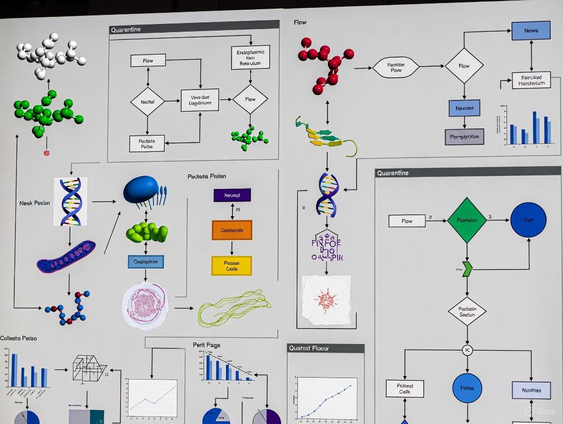
Diagram 1: Two-Incubator Quarantine Workflow
Detailed Quarantine Protocol
The following step-by-step protocol adapts established guidelines for receiving and validating new cell lines [2]:
- Designated Quarantine Space: Maintain a physically separate quarantine room or cabinet with dedicated equipment. Post clear signage indicating quarantine status and contact information [2].
- Initial Placement: Upon receipt, thaw and culture new cell lines only in the assigned "Receiving" Quarantine Incubator A [2].
- Initial Testing Phase: Immediately initiate testing for mycoplasma, followed by karyotyping and screening for human pathogens [2].
- Conditional Progression: Only after cells pass all initial tests should they be moved to "Derivation" Incubator B. In this second incubator, cells can be expanded and prepared for cryopreservation [2].
- Final Validation and Integration: Cells may only move into the main laboratory space after passing a second mycoplasma test and confirming absence of human pathogens with normal karyotype [2].
- Positive Contamination Finding: If contamination is detected at any stage, immediately dispose of the culture. Do not maintain mycoplasma-positive or otherwise contaminated cell lines. Notify the relevant facility manager and arrange for decontamination of all affected equipment and hoods [2].
Essential Testing Methodologies During Quarantine
Mycoplasma Detection
Mycoplasma contamination is particularly problematic because it is covert and alters cellular functions. Regular testing is crucial [1].
Table 2: Key Research Reagent Solutions for Contamination Control
| Reagent/Kit | Specific Function | Application Context |
|---|---|---|
| MycoProbe Mycoplasma Detection Kit | Detects mycoplasma contamination | Routine screening in quarantine; recommended monthly [2] |
| PCR-based Mycoplasma Tests | Molecular detection of mycoplasma DNA | Highly sensitive confirmation testing [1] |
| Bacdown Detergent (2%) | Surface decontamination | Cleaning biosafety cabinets and incubators [2] |
| 70% Ethanol | Surface disinfection | Routine decontamination of work surfaces and equipment [3] |
| DMSO (Cryoprotectant) | Prevents ice crystal formation | Creating secure master cell banks from validated lines [3] |
Diagram 2: Mycoplasma Testing Methods
Additional Identity and Safety Testing
- Karyotyping: Performed to confirm species identity and genetic stability. The Anderson Lab protocol recommends routine karyotyping every 1-4 months and at least 20 spreads should be counted [2].
- Human Pathogen Screening: Pool samples for human pathogen testing to ensure safety for researchers [2].
Contamination Prevention and Control Strategies
Aseptic Technique and Laboratory Design
Maintaining an aseptic workspace is the cornerstone of contamination prevention. This includes:
- Laboratory Design: Implementing separate areas for quarantine and material processing to minimize contamination risks [3].
- Aseptic Workspace: Maintaining laminar airflow in hoods, correctly positioning the sash, autoclaving tools, and using 70% ethanol for surface decontamination [3].
- Personal Practices: Proper gowning, glove use, and restricting access to cell culture areas [1] [2].
Comprehensive Prevention Strategies Across Environments
Table 3: Contamination Prevention Strategies in Research vs. GMP Contexts
| Prevention Area | Research Laboratory Strategies | GMP Manufacturing Strategies |
|---|---|---|
| Environmental Control | Biosafety cabinets, surface disinfection [1] | Classified HEPA-filtered cleanrooms, strict gowning [1] |
| Equipment & Consumables | Sterile single-use consumables [1] | Closed and Single-Use Systems (SUS) [1] |
| Process & Monitoring | Routine mycoplasma/microbial testing [1] | Real-time monitoring, sterility validation [1] |
| Documentation & Control | Cell bank validation [1] | Comprehensive batch tracking, documented root cause analysis [1] |
Contamination in cell culture presents unacceptably high stakes, jeopardizing scientific integrity, therapeutic product safety, and public health. The implementation of rigorous quarantine procedures for new cell lines is not optional but fundamental to responsible science. The protocols outlined herein—centered on a two-incubator transfer system, comprehensive mycoplasma and pathogen testing, and strict aseptic practices—provide a actionable framework for researchers. By adopting these disciplined approaches, the scientific community can significantly mitigate the risks of contamination, enhance the reproducibility of research, and ensure the safety of biologics developed for patients.
Cell culture is a foundational tool in biomedical research, but its reliability is perpetually threatened by biological contaminants. Within the critical context of establishing quarantine procedures for new cell lines, understanding and mitigating these contaminants is paramount to ensuring data integrity and reproducibility. The most insidious threats often come from contaminants that escape routine microscopic observation, namely mycoplasma and viruses, as well as from cross-contamination with other cell lines. These contaminants can alter virtually every aspect of cell physiology, from growth rate and metabolism to genetic stability and gene expression, leading to compromised and irreproducible experimental results [4] [5] [6]. This document provides detailed application notes and protocols for researchers, scientists, and drug development professionals to effectively identify, manage, and prevent these common contaminants, with a specific focus on safeguarding newly acquired cell lines during the mandatory quarantine period.
Mycoplasma Contamination
Impact and Characteristics
Mycoplasma are among the smallest and most insidious prokaryotes, lacking a cell wall and parasitizing the surface of host cells [6]. With contamination rates estimated between 30% and 60%, they represent a pervasive problem [6]. Their small size (0.1–0.3 µm) makes them invisible under standard light microscopy, and they are resistant to common antibiotics like penicillin [6]. Infection can cause a range of nonspecific but detrimental effects on cell cultures, including altered growth rates, morphological changes, chromosomal abnormalities, and disrupted metabolic pathways [6]. Because these changes can be subtle and undetected, mycoplasma can lead to the publication of false and irreproducible data [5] [7].
Detection Methods and Protocols
Routine testing is crucial, especially during the quarantine of new cell lines. Several methods are available, each with advantages and limitations, as summarized in Table 1.
Table 1: Comparison of Major Mycoplasma Detection Methods
| Method | Principle | Time to Result | Sensitivity | Key Advantage | Key Disadvantage |
|---|---|---|---|---|---|
| Culture Method | Growth on specialized agar | Up to 4 weeks | High (Gold Standard) | High reliability, considered a reference method | Time-consuming, requires expertise [6] |
| DNA Fluorochrome Staining | Stains extranuclear DNA with Hoechst 33258 | 1-2 days | Moderate | Relatively fast, visual result | Can yield equivocal results; host cell DNA can cause false positives [8] [6] |
| qPCR | Amplifies mycoplasma-specific DNA sequences | Hours | High | Very sensitive and specific, rapid | Requires specific equipment and reagents [6] |
| Enzyme Immunoassays | Detects mycoplasma-specific enzymes | 1-2 days | Moderate | Suitable for high-throughput screening | May have lower specificity than other methods [6] |
Protocol: Enhanced DNA Fluorochrome Staining for Accurate Detection A common challenge with Hoechst staining is interference from cytoplasmic DNA or apoptotic bodies, which can lead to false positives [8]. The following co-localization protocol improves accuracy.
- Sample Preparation: Seed cells onto a sterile coverslip in a culture dish and incubate until 60-70% confluent.
- Staining:
- Prepare a working solution containing a DNA-binding dye (e.g., Hoechst 33258) and a cell membrane-selective fluorescent dye (e.g., Wheat Germ Agglutinin, WGA, conjugated to a red fluorophore).
- Replace the culture medium with the staining solution and incubate for 15-20 minutes at 37°C, protected from light.
- Microscopy and Analysis:
- Wash the cells gently with PBS and fix with a mild fixative (e.g., 4% paraformaldehyde) for 10 minutes.
- Mount the coverslip and observe under a fluorescence microscope.
- Key Assessment: Genuine mycoplasma contamination is indicated by the co-localization of the blue Hoechst DNA signal with the red cell membrane stain (WGA) on the outer surface of the plasma membrane. Fluorescence not associated with the membrane is likely cellular debris or apoptotic bodies [8].
Prevention and Treatment
Prevention: The best strategy is rigorous prevention. Always quarantine new cell lines and test them for mycoplasma before integrating them into your main culture facility [7]. Maintain strict aseptic technique, use dedicated lab coats and gloves, and keep incubators clean [9] [7]. It is advisable to periodically culture cells without antibiotics to prevent the masking of low-level contaminations [9].
Treatment: If contamination is detected, the contaminated culture should be immediately isolated [7].
- Antibiotic Treatment: Commercial antibiotic mixtures like Plasmocin (25 µg/mL for 1-2 weeks) are commonly used [7]. Other effective antibiotics include ciprofloxacin, doxycycline, and tetracycline [6].
- Post-Treatment Validation: After treatment, cells must be cultured in antibiotic-free medium for 1-2 weeks and then re-tested for mycoplasma to confirm successful eradication [7]. For irreplaceable cell lines, a second round of treatment may be necessary. However, discarding the culture is often the safest course of action to protect other cell lines in the laboratory [4] [7].
Viral Contamination
Impact and Characteristics
Viral contamination is particularly challenging because viruses are difficult to detect without specialized methods and there are no effective treatments for infected cultures [10]. Viruses can be introduced through contaminated biological reagents (e.g., serum, trypsin) or the original donor tissue [4] [1]. Unlike bacteria, they often do not cause visible changes in the culture, but they can alter cellular metabolism, function, and genetic stability, leading to flawed results and wasted resources [4] [10]. Furthermore, they pose a potential hazard to laboratory personnel, especially when handling human-derived materials [4]. Common viruses of concern include Epstein-Barr Virus (EBV), human immunodeficiency virus (HIV), Hepatitis B, and Hepatitis C [4] [10].
Detection Methods and Protocols
Screening for viral contaminants is a critical component of quality control for master cell banks and is highly recommended during the quarantine of new cell lines, particularly those of human or animal origin.
Table 2: Susceptible Cell Lines and Detection Methods for Example Viruses
| Virus | Susceptible Cell Lines | Preferred Detection Method(s) |
|---|---|---|
| Epstein-Barr Virus (EBV) | B-lymphocytes, HEK293, various human and animal cell lines | qPCR, observation of latent or active infection cycles [10] |
| Ovine Herpesvirus 2 (OvHV-2) | A wide range of animal species cell lines (over 33 species) | qPCR, specific antigen detection assays [10] |
| General Panel (e.g., HIV, HBV, HCV) | Human-derived cell lines | qPCR, ELISA, immunofluorescence [4] |
Protocol: Detection via Cytopathic Effect (CPE) and qPCR
- Observation for Cytopathic Effect (CPE): Monitor cells daily for morphological changes under a light microscope. Virus-specific CPE can include cell rounding, detachment, syncytia (cell fusion) formation, and granulation [10]. For instance, uninfected A549 cells appear uniform, while HSV-2 infection causes significant rounding and detachment [10]. While not sufficient alone, CPE can be an important initial indicator.
- qPCR for Direct Detection:
- DNA Extraction: Extract total DNA from a sample of the cell culture using a standard kit.
- PCR Setup: Prepare a qPCR reaction mix using primers and probes specific for the virus of interest (e.g., EBV, OvHV-2, or common human pathogens).
- Amplification and Analysis: Run the qPCR and analyze the amplification curve. A positive signal indicates the presence of viral DNA sequences. qPCR is highly sensitive, relatively easy to establish, and should be the method of choice for routine screening [4]. Viral testing can also be outsourced to certified laboratories [4].
Prevention Strategies
Prevention is the only viable strategy, as viral contamination cannot be treated. All human and animal-derived materials should be treated as potentially infectious and handled at Biosafety Level 2 (BSL-2) or equivalent containment [4]. Ideally, donors should be pre-screened for viral pathogens. If this is not possible, the cell line should be tested at the earliest possible timepoint [4]. Investment in high-quality, well-characterized, and traceable biological reagents is critical [4].
Cross-Contamination and Chemical Contamination
Cross-Contamination
Cross-contamination, the misidentification or overgrowth of a cell line by another, is a widespread problem that renders experimental results useless [1] [5]. The ICLAC register lists hundreds of misidentified cell lines, and estimates suggest about 16.1% of published papers may have used problematic lines [5]. Highly proliferative lines like HeLa can overgrow slower-growing cultures, leading to false conclusions [1].
Prevention and Authentication:
- Handling: Handle only one cell line at a time and use dedicated media and reagents for each [9].
- Authentication: Regularly authenticate cell lines using Short Tandem Repeat (STR) profiling, which is considered the gold standard for human cells [10] [5]. This is a non-negotiable step before banking a new cell line and should be performed periodically thereafter.
Chemical Contamination
Chemical contamination can arise from endotoxins, residual detergents on glassware, or extractables from plastic consumables [1]. These contaminants can affect cell viability, differentiation, and experimental variability [1].
Prevention: Use high-quality, validated reagents and consumables. Ensure glassware is thoroughly rinsed after cleaning and adhere to strict sterilization protocols [1].
Integrated Quarantine Protocol for New Cell Lines
Implementing a robust quarantine procedure is the most effective defense against the introduction of contaminants into a established cell culture facility. The workflow below outlines the key stages and decision points.
Diagram 1: A workflow for the quarantine and validation of new cell lines, integrating critical checks for contaminants and identity.
The Scientist's Toolkit: Essential Reagents and Materials
Table 3: Key Research Reagent Solutions for Contamination Control
| Item | Function/Benefit |
|---|---|
| Mycoplasma Removal Reagents (e.g., Plasmocin) | Antibiotic-based formulations specifically designed to eliminate mycoplasma from contaminated cultures without excessive toxicity to host cells [6] [7]. |
| Mycoplasma Detection Kits (qPCR-based) | Provide high-sensitivity, specific, and rapid detection of mycoplasma nucleic acids, suitable for routine screening [6] [9]. |
| Viral Detection qPCR Kits | Pre-designed assays for the detection of specific viruses (e.g., EBV, HIV, HBV, HCV) to ensure safety and cell line integrity [4] [10]. |
| STR Profiling Kits/Services | Reagents or external services for DNA profiling to authenticate cell lines and prevent cross-contamination, a critical step for publication [5]. |
| Anti-Mycoplasma Antibiotics (e.g., Ciprofloxacin, Tetracycline) | Individual antibiotics used for treatment or prophylaxis, though their continuous use is discouraged as it can mask contaminants [6]. |
| DNA Fluorochrome Staining Kit (e.g., Hoechst) | Allows for visual detection of extranuclear DNA associated with mycoplasma contamination, though may require co-staining for accuracy [8] [6]. |
| Validated, High-Quality Fetal Bovine Serum (FBS) | Sourced from reputable suppliers to minimize the risk of introducing viral or mycoplasma contaminants from raw materials [4] [1]. |
Vigilance against contamination is not merely a technical task but a fundamental aspect of scientific rigor. Mycoplasma, viruses, and cross-contamination represent persistent threats that can invalidate years of research. By framing this challenge within the context of a strict quarantine protocol for new cell lines, researchers can establish a critical barrier. Adherence to the detailed detection protocols, preventive measures, and the integrated workflow outlined in this document will empower scientists to maintain the health and authenticity of their cell cultures, thereby ensuring the reliability and reproducibility of their research outcomes.
The introduction of new cell lines into a research facility presents a significant risk of contamination that can compromise experimental integrity, lead to erroneous data, and cause substantial financial losses [1]. A dedicated quarantine space is fundamental to biosecurity, designed to isolate new cell lines until their sterility and authenticity are confirmed. This protocol outlines the specific facility requirements and workflow separations necessary to prevent cross-contamination, aligning with broader thesis research on robust quarantine procedures for cell line acquisition [2].
Facility and Spatial Design Requirements
The quarantine facility must be a physically distinct area with controlled access and dedicated equipment to ensure absolute separation from main culture laboratories.
Table 1: Minimum Facility Requirements for a Dedicated Quarantine Space
| Requirement Category | Specific Specification | Rationale and Purpose |
|---|---|---|
| Access & Signage | • Controlled access; posted contact signage with dates of use.• "Quarantine in Progress" signs on doors. | Prevents unauthorized entry; alerts personnel to active containment procedures; ensures traceability [2]. |
| Primary Equipment | • Dedicated biosafety cabinet (BSC).• Dedicated CO₂ incubator(s).• Dedicated centrifuge, microscope, and vacuum system. | Eliminates the risk of cross-contamination via shared equipment surfaces or air circulation [2] [1]. |
| Environmental Control | • Use of biosafety cabinets for all procedures.• Surface disinfection protocols.• Restricted airflow zones. | Maintains an aseptic environment; contains potential contaminants within the quarantine zone [1]. |
| Personal Protective Equipment (PPE) | • Lab coats designated for the quarantine room only.• Gloves; closed-toe shoes; masks and hair covers as needed. | Prevents personnel from becoming a vector for contamination into or out of the quarantine space [2]. |
Workflow Separation and the Two-Incubator System
A core component of an effective quarantine protocol is the strict temporal and physical separation of workflows, most critically implemented through a two-incubator system for mycoplasma screening [2].
The Quarantine Workflow
The following diagram illustrates the sequential, gated workflow for introducing a new cell line, emphasizing the critical separation between quarantine and main laboratory spaces.
Key Procedural Gates
- Incubator A (Receiving): All newly received cell lines are initially cultured and tested in this incubator [2].
- Incubator B (Derivation): Cell lines that pass initial tests are moved to this second quarantine incubator for expansion and a subsequent mycoplasma test. No lines may leave the quarantine space until they have passed two mycoplasma tests and screenings for human pathogens and normal karyotype [2].
- Contamination Response: If contamination is detected at any stage, cell lines must be disposed of immediately, and all affected equipment (incubators, biosafety cabinets) must be decontaminated. New samples must be obtained for testing [2] [1].
Critical Validation Experiments and Protocols
Rigorous and routine testing is the cornerstone of the quarantine protocol. The following table and associated methodologies detail the essential validation experiments.
Table 2: Mandatory Validation Experiments for New Cell Lines
| Test | Frequency | Key Methodologies | Purpose & Acceptance Criteria |
|---|---|---|---|
| Mycoplasma Detection | Upon arrival; before transfer to new location; monthly routine. | PCR, Fluorescence staining (e.g., MycoProbe), ELISA. | Detect mycoplasma contamination. Criteria: Two consecutive negative results are required for release from quarantine [2] [1]. |
| Karyotyping | Upon arrival; every 10 passages; every 1-4 months. | G-banded metaphase spread analysis (e.g., via Cell Line Genetics). | Confirm genetic stability and authenticity of the cell line. Criteria: Normal, species-appropriate karyotype [2]. |
| Human Pathogen Screening | Upon arrival; before release from quarantine. | Pooled sample screening for a panel of human pathogens. | Ensure the cell line is free from infectious agents that pose a risk to handlers. Criteria: Negative for tested pathogens [2]. |
| Cell Line Authentication | Upon arrival; at the time of freezing master stocks. | Short Tandem Repeat (STR) profiling. | Verify cell line identity and avoid cross-contamination. Criteria: STR profile matches expected identity [1]. |
Detailed Experimental Protocol: Mycoplasma Testing by PCR
Principle: This protocol uses polymerase chain reaction (PCR) to amplify highly conserved DNA sequences specific to mycoplasma, providing a highly sensitive and rapid detection method [2] [1].
Materials:
- Mycoplasma Detection Kit: e.g., MycoProbe (R&D Systems, Cat No. CUL001B) [2].
- PCR Thermal Cycler
- Gel Electrophoresis Equipment
- DNA Ladder
- Sterile, nuclease-free water and pipette tips
Methodology:
- Sample Collection: Collect 100-200 µL of cell culture supernatant from the test culture. Avoid cell debris by brief centrifugation if necessary.
- DNA Extraction: Extract DNA from the supernatant according to the manufacturer's instructions provided with the mycoplasma detection kit.
- PCR Setup: Prepare the PCR master mix on ice. For each sample, combine:
- 12.5 µL of PCR Master Mix (from kit)
- 1 µL of Forward Primer (from kit)
- 1 µL of Reverse Primer (from kit)
- 9.5 µL of Nuclease-free Water
- 1 µL of Template DNA
- PCR Amplification: Place the tubes in a thermal cycler and run the following program:
- Initial Denaturation: 95°C for 2 minutes
- 35 Cycles of:
- Denaturation: 95°C for 30 seconds
- Annealing: 55°C for 30 seconds
- Extension: 72°C for 1 minute
- Final Extension: 72°C for 5 minutes
- Hold: 4°C ∞
- Analysis: Analyze the PCR products by agarose gel electrophoresis (e.g., 1.5% gel). Visualize the DNA bands under UV light.
- Interpretation: A positive control should show a band at the expected size. The test sample is considered negative if no band is present at that size. A band in the test sample lane indicates mycoplasma contamination.
The Scientist's Toolkit: Essential Research Reagents and Materials
Table 3: Essential Research Reagent Solutions for Quarantine Procedures
| Item | Function and Application in Quarantine |
|---|---|
| Bacdown Detergent (2%) | A specialized disinfectant for decontaminating surfaces, biosafety cabinets, and incubators. Used for cleaning spills and routine wiping [2]. |
| Ethanol (70%) | Used for rapid surface decontamination within the biosafety cabinet and for sterilizing smaller equipment before entry into the cabinet [2]. |
| Mycoplasma Detection Kit | Essential reagent for performing the mandatory mycoplasma screening tests, such as PCR or fluorescence-based assays [2] [1]. |
| Sterile, Single-Use Consumables | Pre-sterilized pipettes, tips, and culture flasks. Using single-use items eliminates the risk of cross-contamination from improper cleaning of reusable glassware [1]. |
| Validated Fetal Bovine Serum (FBS) | A critical growth medium component. Must be sourced from suppliers that provide validation for the absence of contaminants, including viruses and mycoplasma [1]. |
Implementation and Workflow Automation
The transition from manual, paper-based workflows to automated, electronic systems is critical for enhancing the accuracy and efficiency of quarantine procedures. Automated workflows guide users through standardized procedures, ensuring consistency and reducing the likelihood of human error in data entry and process execution [11]. This is particularly valuable for tracking complex quarantine timelines, test results, and maintaining a clear audit trail for regulatory compliance.
The following diagram outlines the logical relationship between the researcher, the automated system, and the key decision points within the quarantine workflow.
The introduction of new cell lines into a research laboratory presents a significant risk of contaminating existing cultures with microbial pathogens or cross-contaminating with other cell lines, potentially compromising years of research and development. Reproducible research in biomedicine and drug development is fundamentally dependent on the genetic integrity and authenticity of the biological models used [12]. Studies indicate that misidentified or contaminated cell lines have been used in thousands of published research papers, wasting an estimated billions of research dollars and threatening the validity of scientific conclusions [12]. Therefore, a rigorous quarantine protocol encompassing isolation, testing, and validation is not merely a best practice but a fundamental necessity for research integrity. This application note details a comprehensive and actionable protocol to serve as a standard for researchers and scientists, ensuring that all new cell lines, whether acquired externally or derived in-house, meet the highest standards of quality before being integrated into critical research workflows.
Core Principles of the Quarantine Protocol
The proposed quarantine protocol is built upon three interdependent pillars: physical and procedural isolation, a multi-tiered testing regime, and systematic validation prior to release. Adherence to these principles minimizes the risk of widespread contamination and ensures that only authenticated, high-quality cell lines are used for experimental work.
Principle 1: Isolation
The first and most critical step is the complete physical and procedural isolation of new cell lines. This involves designating a separate Quarantine Laboratory Space equipped with its own dedicated equipment, including biosafety cabinets, incubators, centrifuges, and microscopes [2] [3]. This space should have clear signage indicating its status and the contact information of the responsible personnel [2].
Within this space, a two-incubator system is recommended for managing incoming cell lines [2]. The process is as follows:
- Incubator A (Receiving Incubator): All newly received cell lines are initially thawed and cultured in this incubator. No cell lines are permitted to leave this incubator until they have passed initial microbiological contamination tests [2].
- Incubator B (Derivation Incubator): Once a cell line passes initial tests, it may be moved to this second quarantine incubator. Here, cells can be expanded to create working stocks and undergo further characterization. Cell lines must pass a second round of testing before they can leave the quarantine area entirely [2].
Personnel must employ strict aseptic techniques and use personal protective equipment (PPE) dedicated to the quarantine area. All laboratory waste from the quarantine space should be treated as potentially infectious and disposed of via established, safe routes [13] [2].
Principle 2: Testing
A multi-faceted testing strategy is essential to detect various forms of contamination and misidentification. The testing should be conducted at specific timepoints, as outlined in Table 1.
Table 1: Recommended Testing Schedule for New Cell Lines
| Test | Timing | Key Purpose | Reference Method |
|---|---|---|---|
| Mycoplasma Detection | Upon arrival, and again before final release from quarantine. Also performed monthly as routine monitoring. | Detect mycoplasma contamination, which can alter cell behavior and metabolism. | Fluorescent staining (Hoechst 33258) or PCR-based kits [2] [14]. |
| Cell Line Authentication | Upon acquisition, before banking, and periodically during routine culture (e.g., every 10 passages). | Verify species and unique identity of the cell line; detect cross-contamination. | Short Tandem Repeat (STR) profiling for human cell lines [15] [14] [12]. |
| Karyotyping / Cytogenetic Analysis | Upon arrival and after establishment in culture (e.g., every 10 passages or 1-4 months). | Assess genetic stability and detect large-scale chromosomal abnormalities. | G-banded metaphase spread analysis [2]. |
| Morphology & Growth Analysis | Daily observation during quarantine; formal growth curve analysis upon establishment. | Monitor cell health, confirm expected morphology, and establish baseline growth kinetics. | Phase-contrast microscopy and population doubling time calculation [13] [14]. |
Principle 3: Validation
Validation is the process of interpreting test results against defined criteria to make a data-driven decision on the cell line's release. The core of identity validation for human cell lines is the analysis of the STR profile. The obtained STR profile must be compared to reference databases, such as Cellosaurus, to confirm its identity and check for known misidentifications [12]. A match score of ≥80% is often used as a threshold for authentication, though results require careful interpretation by trained personnel [15].
Validation is also an ongoing process. It requires documenting the cell line's passage number, freezing a Master Cell Bank (MCB) at the earliest possible passage, and subsequently creating Working Cell Banks (WCB) from the characterized MCB [15]. This two-tiered biobanking strategy, illustrated in Figure 1, ensures a continuous supply of low-passage, quality-assured cells for research and guards against genetic drift and future contamination.
Experimental Protocols
Protocol: Short Tandem Repeat (STR) Profiling for Cell Line Authentication
Principle: STR profiling amplifies highly polymorphic regions of the genome via multiplex PCR to create a unique genetic fingerprint for each human cell line, allowing for identity verification and detection of cross-contamination [14] [12].
Materials:
- DNA extraction kit (e.g., DNeasy Blood & Tissue Kit, Qiagen).
- Commercial STR Multiplex PCR Kit (e.g., PowerPlex 18D System, Promega).
- Capillary Electrophoresis System (e.g., ABI 3500 Genetic Analyzer).
- GeneMapper or equivalent software for data analysis.
Methodology:
- DNA Extraction: Extract high-quality genomic DNA from the cell line following the manufacturer's protocol. Quantify DNA concentration and purity using a spectrophotometer.
- PCR Amplification: Set up the multiplex PCR reaction using the commercial STR kit. The reaction typically includes:
- 100 ng of genomic DNA
- Primer mix (containing fluorescently-labeled primers for core STR loci)
- PCR master mix Run the PCR in a thermal cycler using the cycling conditions specified by the kit manufacturer.
- Capillary Electrophoresis: Dilute the PCR product as recommended and denature it. Load the samples onto the capillary electrophoresis instrument alongside an internal size standard.
- Data Analysis: Use the analysis software to automatically call alleles at each STR locus. The software will generate an electrophoretogram and a table of allele calls.
- Interpretation: Compare the resulting STR profile to a reference profile from the original donor, if available. If not, compare the profile to the earliest available stock or to a database of known cell line profiles using a tool like the Cellosaurus STR similarity search tool (CLASTR) [12]. A match of 80% or higher generally indicates authentication.
Protocol: Mycoplasma Detection by Fluorescent Staining
Principle: The fluorescent dye Hoechst 33258 binds specifically to DNA. Because mycoplasmas adhere to the cell surface, staining reveals a characteristic pattern of particulate or filamentous fluorescence in the cytoplasm of infected cultures when viewed under a fluorescence microscope [14].
Materials:
- Hoechst 33258 stain solution.
- Fixed cell preparation (test cells and negative/positive controls grown on coverslips).
- Mounting medium.
- Fluorescence microscope with DAPI filter.
Methodology:
- Cell Culture: Grow the test cell line and controls (a known negative and a known positive mycoplasma-contaminated cell line) on sterile coverslips in culture dishes until subconfluent.
- Fixation: Remove the culture medium and rinse the cells gently with PBS. Fix the cells with a fresh mixture of acetic acid and methanol (1:3) for 5 minutes.
- Staining: Remove the fixative and add the Hoechst 33258 stain solution (diluted as per manufacturer's instructions). Incubate in the dark for 15-30 minutes.
- Washing and Mounting: Rinse the coverslips thoroughly with PBS to remove unbound stain. Mount the coverslips onto glass slides with mounting medium.
- Microscopy: Examine the slides under a fluorescence microscope at 500x magnification. In a negative sample, only the cell nuclei will be stained. In a positive sample, in addition to the nuclei, a fine, particulate or filamentous blue-fluorescence will be observed in the cytoplasm, indicating the presence of mycoplasma DNA [14].
The Scientist's Toolkit: Essential Research Reagents
The following table details key reagents and materials required for the effective implementation of the quarantine protocol.
Table 2: Essential Reagents and Materials for Cell Line Quarantine
| Item | Function / Application | Examples / Notes |
|---|---|---|
| Mycoplasma Detection Kit | Routine and sensitive detection of mycoplasma contamination. | MycoProbe (R&D Systems), MycoAlert (Lonza). Both biochemical and PCR-based kits are available [2]. |
| STR Profiling Kit | Authentication of human cell lines via DNA fingerprinting. | PowerPlex 18D System (Promega). Amplifies 17 STR loci plus amelogenin for gender determination [14]. |
| Cell Culture Media & Reagents | Expansion and maintenance of cell lines during the quarantine period. | Sourced from reputable suppliers (e.g., Thermo Fisher, Sigma). Record batch numbers for all reagents to ensure traceability [13]. |
| Cryopreservation Medium | Creation of secure Master and Working Cell Banks. | Typically contains a cryoprotectant like DMSO or glycerol in fetal bovine serum or culture medium [3]. |
| DNA Extraction Kit | Isolation of high-quality genomic DNA for STR profiling. | DNeasy Blood & Tissue Kit (Qiagen). Ensures pure DNA free of PCR inhibitors. |
| Hoechst 33258 Dye | Fluorescent staining for mycoplasma detection. | A bisbenzimide dye that binds preferentially to DNA [14]. |
| Bacdown Detergent / 70% Ethanol | Decontamination of biosafety cabinets, incubators, and work surfaces. | Essential for maintaining an aseptic environment and preventing cross-contamination [2]. |
Workflow and Data Interpretation
The entire quarantine process, from cell arrival to final release, follows a logical sequence designed to systematically mitigate risk. Figure 1 below illustrates this comprehensive workflow.
Data Interpretation and Decision Points: The workflow contains two critical decision points. The first depends on the results of initial mycoplasma testing. A positive result necessitates immediate disposal of the cell line and decontamination of all affected equipment [2]. The second decision point relies on the interpretation of STR profiling and other validation data. A successful STR match (typically ≥80%) against a reference profile confirms identity and allows for release. A failure or unexpected match to another cell line (e.g., HeLa) indicates misidentification, and the cell line must be discarded to prevent the propagation of erroneous data [12].
Implementing a Robust Quarantine Workflow: A Step-by-Step Protocol
The integrity of biological research and drug development hinges on the use of authentic and uncontaminated cell lines. The initial step of receiving and documenting new cell lines is therefore critical, as introduced contaminants or misidentified cells can compromise experimental data, leading to erroneous conclusions and costly delays. This application note details a robust quarantine procedure, framed within a broader thesis on cell line management, to safeguard cell repositories and ensure the generation of reliable, reproducible results for researchers, scientists, and drug development professionals.
Pre-Arrival Preparation and Documentation
Proper planning before a new cell line arrives is essential for a seamless and secure integration into your laboratory.
Administrative Documentation
- Approval and Registration: Obtain formal approval for the use of dedicated quarantine space [2]. Prior to the cell line's arrival, register it in your laboratory's cell line log, assigning a unique identifier.
- Signage: Post clear signage on the quarantine room door indicating the responsible personnel, their contact information, and the date range of the assigned usage [2].
- Safety Protocols: Ensure all involved personnel have completed required safety courses and that relevant biosafety and ethical approvals (e.g., hSCRO, IBC) are in place and list the quarantine room as a location [2].
Quarantine Laboratory Setup
A physically segregated quarantine area is the cornerstone of this protocol.
- Dedicated Space: Designate a separate laboratory room for quarantine purposes [16] [3]. If a separate room is not feasible, use a dedicated biosafety cabinet and incubator located away from main culture areas.
- Equipment: The quarantine space should contain its own biosafety hood, incubator(s), centrifuge, microscope, and cryostorage facility [2]. All equipment must be clearly labeled "For Quarantine Use Only."
- Reagents: Supply the area with dedicated media, sera, and other reagents to prevent cross-contamination.
Initial Processing and Quarantine Workflow
Upon receipt, the new cell line must be processed systematically within the designated quarantine area.
Receipt and Initial Inspection
- Documentation Check: Verify the accompanying documentation, including the cell line's name, source, passage number, and culture conditions.
- Vial Inspection: Visually examine the cryovial for damage or leakage. Note the volume and appearance of the frozen material.
- Inventory Logging: Record the cell line's unique identifier, date of receipt, source, and your assigned internal lot number in a master cell bank log.
The Quarantine Incubation System
A two-incubator transfer system is recommended to rigorously establish the cell line's sterility status [2].
- Incubator A (Receiving Incubator): Newly thawed cells are initially cultured here.
- Incubator B (Derivation Incubator): Cells that pass initial quality controls move to this second incubator for expansion and freezing.
- Movement Restriction: No cell lines may leave the quarantine space or be moved to general culture incubators until they have passed all quality tests described below [2].
The following workflow diagram outlines the key stages of this quarantine procedure.
Essential Quality Control Assays and Protocols
A comprehensive testing regime is non-negotiable for validating a new cell line. Key assays, their methodologies, and recommended schedules are summarized below.
Table 1: Schedule for Essential Quality Control Tests on a New Cell Line
| Test | First Test | Follow-up Frequency | Key Methodological Details |
|---|---|---|---|
| Mycoplasma Detection | Immediately upon thawing [2] | Before moving to a new location; monthly routine verification [2] | Use a commercial kit (e.g., MycoProbe, Mycoplasma Detection Kit). Follow manufacturer's protocol for culture medium supernatant or cell lysate testing [2]. |
| Cell Line Authentication | After initial expansion in Incubator B | Once per lineage, or if morphology changes | Perform Short Tandem Repeat (STR) profiling. Compare results with reference databases (e.g., ATCC, DSMZ). |
| Karyotyping | After initial expansion in Incubator B | Every 10 passages or every 1-4 months [2] | Send cells for G-banded metaphase spread analysis (e.g., to Cell Line Genetics). At least 20 spreads should be counted [2]. |
| Pathogen Screening | Pool samples after initial expansion | As required by the source or institutional policy | Pool cell culture samples and use PCR-based methods to screen for a panel of human pathogens [2]. |
The Scientist's Toolkit: Key Research Reagent Solutions
The following reagents and kits are essential for implementing the quality control protocols described.
Table 2: Essential Reagents for Cell Line Quarantine and Quality Control
| Reagent / Kit | Function / Application |
|---|---|
| Mycoplasma Detection Kit (e.g., MycoProbe, R&D Systems) | Detects the presence of mycoplasma contamination in cell culture supernatants via enzymatic or PCR-based methods [2]. |
| STR Profiling Kit | Genetically fingerprints cell lines using polymerase chain reaction (PCR) to amplify short tandem repeat loci, confirming unique identity and detecting cross-contamination. |
| Cell Culture Media & Sera | Dedicated, pre-tested lots of medium and fetal bovine serum (FBS) reserved for use in the quarantine facility to prevent external contamination. |
| Cryopreservation Medium | A ready-to-use solution containing a cryoprotectant like DMSO and serum/proteins to ensure high post-thaw viability during cell banking [3]. |
| Bacdown Detergent (2%) & 70% Ethanol | Used for decontaminating surfaces and equipment. Bacdown is specifically recommended for cleaning up spills within biosafety cabinets and incubators [2]. |
Implementation of the Quarantine Protocol
- Aseptic Technique: Strict aseptic technique is mandatory. Wear a lab coat designated for the quarantine room only, gloves, and closed-toe shoes [2]. Use 70% ethanol for surface decontamination.
- Incubator and Hood Maintenance: Clean the biosafety hood and incubator with 2% Bacdown detergent and 70% ethanol before and after use. For CO₂ incubators, follow the manufacturer's manual for decontamination cycles [2].
- Waste Disposal: All labware that comes into contact with human cultures must be disposed of in designated red biohazard containers. Liquid waste should be aspirated through a system where the receiver bottle is filled with Bacdown detergent and emptied frequently [2].
- Decontamination Protocol: If a cell line tests positive for mycoplasma, it must be disposed of immediately. The incubators and hoods used must then be thoroughly decontaminated, and the incident reported to the responsible personnel [2].
A meticulous and multi-stage approach to receiving and documenting new cell lines is a fundamental investment in research quality. By implementing this structured quarantine protocol—encompassing physical segregation, systematic documentation, and a rigorous panel of quality control assays—research facilities can significantly mitigate the risks of contamination and misidentification. This proactive stewardship of cell resources ensures the integrity of scientific data, enhances reproducibility, and ultimately accelerates progress in drug development and biological research.
The two-incubator system is a fundamental component of robust cell culture quarantine procedures, designed to prevent cross-contamination of established cell lines with pathogens from newly acquired cultures. This system operates on a simple yet critical principle: physical separation of incoming cell lines until their health and sterility status is confirmed. Implementing this system within a designated quarantine room, such as Core Facility Room 1201, is essential for maintaining the integrity of a research facility's cell repository [2]. The system utilizes two assigned incubators—Incubator A for the initial "Receiving" phase and Incubator B for the subsequent "Derivation" phase. No cell lines cultured in the quarantine space may be used outside this area until they have passed two separate mycoplasma tests. Furthermore, under no circumstances should incubators be used to maintain mycoplasma-positive cell lines; if an infection is detected, the lines must be disposed of immediately, and the incubators and biosafety hoods must be decontaminated [2].
Experimental Protocols for Cell Line Validation
During the quarantine period, a series of validation experiments must be performed to ensure the new cell lines are free of contaminants and genetically stable. The following table summarizes the key tests, their methodologies, and the acceptable results for a cell line to progress through the two-incubator system.
Table 1: Essential Validation Tests for New Cell Lines During Quarantine
| Test | Methodology / Protocol | Frequency / Timing | Acceptable Result for Progression |
|---|---|---|---|
| Mycoplasma Testing [2] | Use a commercial detection kit (e.g., MycoProbe Mycoplasma Detection Kit, R&D Systems, Cat No. CUL001B). Follow manufacturer's instructions for sample processing and analysis. | Upon arrival in Incubator A, and again before moving from Incubator B to main facility. Also performed monthly as routine monitoring. | Two consecutive negative results are required. The first test must be passed before moving to Incubator B, and a second test must be passed before leaving quarantine. |
| Karyotyping [2] | Send cell samples to a specialized service (e.g., Cell Line Genetics). Analysis typically involves G-banded metaphase spreads. Alternatively, schedule using institutional resources like a CytoVision Karyotyping Workstation. | Upon arrival in Incubator A. Routine testing should be performed every 10 passages or every 1-4 months thereafter. At least 20 metaphase spreads should be counted. | Normal karyotype for the cell line. |
| Human Pathogen Screening [2] | Pool cell samples and send for analysis to test for a panel of common human pathogens. | Upon arrival in Incubator A, before moving to the main facility. | Negative for all tested human pathogens. |
Workflow of the Two-Incubator Quarantine System
The following diagram illustrates the step-by-step workflow for processing a new cell line through the two-incubator quarantine system, from thawing to final transfer into the main laboratory facility.
The Scientist's Toolkit: Essential Materials and Reagents
Successful implementation of the quarantine protocol requires specific reagents and materials. The table below details the key items necessary for the validation experiments and routine culture maintenance within the two-incubator system.
Table 2: Essential Research Reagent Solutions for Cell Line Quarantine
| Item | Function / Application | Example / Notes |
|---|---|---|
| Mycoplasma Detection Kit | Detects the presence of Mycoplasma contamination in cell culture supernatants or lysates. | MycoProbe Mycoplasma Detection Kit (R&D Systems, CUL001B) [2]. |
| Cell Culture Media | Provides essential nutrients for cell growth and maintenance. Formulations are cell line-specific. | Use high-quality media and supplements; change media regularly to prevent nutrient depletion and pH fluctuation [17]. |
| Cryopreservation Medium | Allows for long-term storage of cell stocks in liquid nitrogen. Contains cryoprotectants to prevent ice crystal formation. | Typically contains a base medium with DMSO or glycerol as a cryoprotective agent [17]. |
| Decontamination Reagents | Used for surface sterilization and cleaning of equipment, biosafety cabinets, and incubators. | 70% Ethanol and 2% Bacdown detergent are specified for decontaminating surfaces and aspirating tubing [2]. |
| Sterile Labware | For all cell culture procedures to maintain aseptic conditions and prevent contamination. | Includes pipettes, tips, flasks, and plates. Use sterile equipment and practice aseptic technique rigorously [17]. |
The introduction of new cell lines into a research laboratory carries the inherent risk of contaminating existing cell cultures. Contaminants such as mycoplasma, pathogens, and cells with an abnormal karyotype can compromise experimental integrity, leading to unreliable data and wasted resources. Consequently, a rigorous quarantine procedure with a defined essential testing regimen is a critical component of good cell culture practice [18]. This application note details the protocols for mycoplasma detection, karyotyping, and pathogen screening, framed within a comprehensive quarantine strategy for new cell lines. Adherence to these procedures ensures the authenticity, genetic stability, and microbiological purity of cellular models, which is fundamental for high-quality research and drug development.
A systematic approach to testing new cell lines involves multiple assays to assess different types of contamination and genetic stability. The table below summarizes the core tests, their targets, and key methodological details.
Table 1: Essential Testing Regimen for New Cell Lines
| Test Type | Primary Target | Key Methodologies | Detection Capability | Typical Timing/Frequency |
|---|---|---|---|---|
| Mycoplasma Testing | Mycoplasma species (e.g., M. orale, M. hyorhinis) | PCR, enzymatic detection (MycoProbe), culture-based [2] [19] | Contamination by various mycoplasma species | Upon arrival, before moving from quarantine, and monthly as routine verification [2] |
| Karyotyping | Chromosomal number and structure (aneuploidy, translocations, etc.) | G-banded metaphase analysis [20] [21] | Gross numerical and structural abnormalities | Upon receipt, every 10 passages, or every 1-4 months to monitor genetic stability [2] |
| Pathogen Screening | Human pathogens (viral, bacterial) | PCR, RT-PCR, serological methods (e.g., ELISA) [2] [22] | Specific pathogenic agents as defined by the testing panel | Upon receipt, before moving from quarantine [2] |
Detailed Experimental Protocols
Protocol for Mycoplasma Detection by PCR
Mycoplasma contamination can alter cell behavior and metabolism without causing overt turbidity in the culture medium. PCR provides a highly sensitive and rapid method for detection [19].
I. Research Reagent Solutions
Table 2: Key Reagents for Mycoplasma PCR Testing
| Reagent / Equipment | Function / Specification |
|---|---|
| Mycoplasma Primer Mix | A pool of forward and reverse primers targeting conserved regions across multiple mycoplasma species [19]. |
| Taq Polymerase | Thermostable DNA polymerase for PCR amplification. |
| dNTPs | Deoxynucleotide triphosphates (building blocks for DNA synthesis). |
| PCR Buffer with MgCl₂ | Provides optimal chemical environment for polymerase activity. |
| Thermal Cycler | Instrument for programmed temperature cycling during PCR. |
| Agarose Gel Electrophoresis System | For visualizing PCR amplification products. |
II. Step-by-Step Methodology
- Primer Preparation: Obtain and resuspend the specified forward and reverse primers to a concentration of 100 µM. Create a working primer mix by combining all forward primers to a final concentration of 10 µM each, and do the same for all reverse primers [19].
- Sample Collection: Take a 100 µL sample of supernatant from a dense (80-100% confluent) cell culture. Avoid disturbing the cell monolayer.
- Sample Preparation: Heat the supernatant sample at 95°C for 5 minutes to denature proteins and release DNA. Centrifuge for 2 minutes at maximum speed in a microcentrifuge to pellet debris. The supernatant is used as the template in the PCR reaction [19].
- PCR Reaction Setup: Prepare a 25 µL PCR reaction mix as follows:
- 10x PCR Buffer: 2.5 µL
- 25 mM MgCl₂: 2.0 µL
- 10 mM dNTPs: 1.0 µL
- Forward Primer Mix: 1.0 µL
- Reverse Primer Mix: 1.0 µL
- Template Supernatant: 2.0 µL
- Taq Polymerase: 0.2 µL
- Water: 15.3 µL
- Total Volume: 25.0 µL
- Always include a negative control (water) and a positive control if available [19].
- PCR Amplification: Run the PCR using the following cycling program:
- Initial Denaturation: 95°C for 2 minutes.
- 5 Cycles of:
- Denaturation: 94°C for 30 seconds.
- Annealing: 50°C for 30 seconds.
- Extension: 72°C for 35 seconds.
- 30 Cycles of:
- Denaturation: 94°C for 15 seconds.
- Annealing: 56°C for 15 seconds.
- Extension: 72°C for 30 seconds.
- Final Hold: 4°C indefinitely [19].
- Analysis: Analyze the PCR products by agarose gel electrophoresis. A positive result, indicated by a band of approximately 500 base pairs, confirms mycoplasma contamination.
The workflow for this quarantine and testing procedure is outlined in the diagram below.
Protocol for Karyotype Analysis
Karyotyping provides a global view of the chromosome complement and is essential for identifying and monitoring gross genetic abnormalities in cell lines, such as aneuploidy or translocations, which can occur with passaging [20] [2].
I. Research Reagent Solutions
Table 3: Key Reagents for Karyotype Analysis
| Reagent / Equipment | Function / Specification |
|---|---|
| Cell Culture Flask | For growing cells to ~70% confluence. |
| Colcemid | A mitotic spindle inhibitor that arrests cells in metaphase. |
| Hypotonic Solution (e.g., Potassium Chloride) | Causes cells to swell, spreading the chromosomes. |
| Carnoy's Fixative (3:1 Methanol:Acetic Acid) | Preserves and fixes the chromosomal morphology. |
| Giemsa Stain (G-banding) | Creates a characteristic banding pattern for chromosome identification. |
| Microscope with Oil Immersion Objective | For visualizing and analyzing G-banded metaphase spreads. |
II. Step-by-Step Methodology
- Cell Culture and Mitotic Arrest: Grow cells to approximately 70% confluence. Add Colcemid to the culture medium to a final working concentration (e.g., 0.1 µg/mL) and incubate for 1-4 hours. This arrests actively dividing cells in metaphase, when chromosomes are most condensed [20].
- Cell Harvesting: Gently dislodge the cells (trypsinization may be used for adherent lines) and transfer them to a centrifuge tube. Pellet the cells by centrifugation.
- Hypotonic Treatment: Carefully resuspend the cell pellet in a pre-warmed hypotonic solution (e.g., 0.075 M KCl). Incubate at 37°C for 15-20 minutes. This step causes the cells to swell.
- Fixation: Pellet the cells again and carefully remove the hypotonic solution. Gently resuspend the pellet in freshly prepared Carnoy's fixative. Repeat this fixation step 2-3 times to ensure complete removal of water and cellular debris.
- Slide Preparation: Drop the fixed cell suspension onto clean, wet microscope slides and allow them to air dry. This creates metaphase spreads where chromosomes are separated.
- Staining and Banding (G-banding): Age the slides and then treat them with trypsin followed by Giemsa stain. This produces the characteristic light and dark banding pattern (G-bands) used for chromosome identification [20].
- Analysis: Analyze the slides under a microscope with an oil immersion objective. For a basic analysis, a minimum of 20 metaphase cells should be analyzed to rule out major abnormalities. If mosaicism is suspected, 30-50 metaphases should be analyzed [20]. Chromosomes are identified, counted, and arranged into a karyogram to assess for numerical and structural abnormalities.
The process of preparing and analyzing the karyotype is detailed in the following workflow.
The Scientist's Toolkit
Table 4: Essential Research Reagent Solutions for Cell Line Quarantine Testing
| Item | Function/Application |
|---|---|
| MycoProbe Mycoplasma Detection Kit | A commercial kit for rapid and sensitive detection of mycoplasma contamination in cell cultures [2]. |
| Primer Sets for Mycoplasma PCR | Custom oligonucleotide primers targeting multiple mycoplasma species for in-house PCR testing [19]. |
| Colcemid | A mitotic inhibitor used in karyotyping to arrest cells in metaphase for chromosome analysis. |
| Giemsa Stain | A cytogenetic stain used in G-banding to produce a unique banding pattern for each chromosome pair [20]. |
| Short Tandem Repeat (STR) Profiling Kits | Reagents for authenticating cell lines by DNA profiling, confirming their unique identity and ruling out cross-contamination [18]. |
| Enzyme-Linked Immunosorbent Assay (ELISA) Kits | Serological-based kits for detecting specific human pathogens (e.g., viruses) in cell culture samples [22]. |
| Pathogen-Specific PCR Primers | Primers designed to detect DNA or RNA from specific human pathogens that may be present in cell lines. |
Implementing a strict quarantine procedure with the essential testing regimen for mycoplasma, karyotype, and pathogens is not optional but a fundamental requirement for robust and reproducible biomedical research. The protocols detailed herein provide a clear framework for establishing this practice. By validating the microbiological and genetic integrity of new cell lines before their integration into the main culture system, researchers safeguard their experiments, enhance data reliability, and contribute to the overall quality and sustainability of the scientific enterprise.
Within the critical framework of cell line quarantine procedures, mastering aseptic technique is non-negotiable for preventing contamination and ensuring research integrity. Aseptic technique refers to the set of practices performed under controlled conditions to prevent contamination from microorganisms [23]. It is distinct from sterilization, which is an absolute state of being free from all microbial life; aseptic technique is the continuous process of maintaining that sterility during handling [23]. In the specific context of a quarantine laboratory, where new cell lines of unknown microbial status are handled, these practices form the primary defense against the introduction of contaminants into established culture collections. A single lapse can jeopardize weeks of work and compromise valuable research, making rigorous adherence to protocol essential for successful drug development and biomedical research [23].
Fundamentals of Aseptic Technique
Core Principles
The execution of aseptic technique is governed by several core principles:
- Distinguishing Sterile and Non-Sterile Zones: Clearly demarcate and maintain separate areas for sterile and non-sterile materials and operations [24].
- Proper Personal Protective Equipment (PPE): Wearing a clean lab coat, sterile gloves, and safety glasses is mandatory to prevent contamination from personnel [23] [24].
- Meticulous Disinfection: All work surfaces, particularly within the biosafety cabinet, must be thoroughly disinfected with a 70% ethanol or other appropriate disinfectant before and after all operations [23] [2].
- Minimizing Exposure: Sterile culture vessels, media, and reagents should be open to the environment for the shortest time possible to reduce the risk of airborne contamination [23].
Aseptic Environment and Workspace Management
A dedicated and properly maintained workspace is foundational to aseptic technique. The quarantine room itself should have strict access controls and be sanitized regularly [2]. Key environmental preparations include:
- Room Sterilization: Surfaces should be wiped with sodium hypochlorite or benzalkonium chloride solution, followed by UV sterilization for a minimum of 30 minutes before use [24].
- Minimizing Movement: Unnecessary movements and conversations should be avoided within the culture room, as they can disrupt airflow and mobilize contaminants [23] [24].
- Organized Work Area: Only essential items should be placed in the biosafety cabinet, and they should be arranged strategically to avoid clutter and disruption of the laminar airflow [23].
Biosafety Cabinets: The Primary Engineering Control
BSC Fundamentals and Classification
A Biosafety Cabinet (BSC) is a primary engineering control that uses laminar airflow and High-Efficiency Particulate Air (HEPA) filtration to provide a sterile work environment [25]. HEPA filters are capable of trapping 99.97% of particles 0.3 µm in diameter, effectively removing all known infectious agents from the air [26]. It is critical to distinguish BSCs from other similar-looking equipment. Laminar flow hoods or "clean benches" only provide product protection by blowing HEPA-filtered air outward towards the user and must not be used for work with biohazardous materials [26] [25]. Similarly, chemical fume hoods are designed to protect the user from chemical vapors but offer no product protection and are not equipped with HEPA filters for containment of biological agents [26] [25].
BSCs are classified based on the level of protection they provide. The following table outlines the common classes and their appropriate use cases, with Class II being the most prevalent in research laboratories.
Table 1: Classification of Biosafety Cabinets and Their Applications
| BSC Class/Type | Personnel Protection | Product Protection | Environmental Protection | Common Use Cases in Quarantine |
|---|---|---|---|---|
| Class I | Yes | No | Yes | Enclosing equipment (e.g., centrifuges) or procedures that generate aerosols; not suitable for sterile cell culture work [25]. |
| Class II (A2) | Yes | Yes | Yes | The most widely used type for general cell culture, including quarantine procedures; provides a sterile environment for handling new cell lines [26] [25]. |
| Class III | Yes (Maximum) | Yes | Yes | Used with high-risk biological agents; provides a total physical barrier (glove box) [25]. |
Standard Operating Procedure for BSC Use
A detailed Standard Operating Procedure (SOP) ensures the correct and consistent use of the BSC, which is vital in a quarantine setting.
Pre-Use Procedures:
- Preparation: Tie back long hair, remove jewelry, and don a clean lab coat, gloves, and safety glasses [23] [2].
- Activation: Turn on the BSC and allow the blower to run for at least 15 minutes to purge the work surface and stabilize airflow [23] [24].
- Disinfection: Thoroughly spray and wipe all interior surfaces of the BSC—including the work surface, side walls, and back panel—with 70% ethanol using a sterile lint-free wipe [23] [24]. Allow the ethanol to evaporate completely.
- Material Placement: Gather all necessary sterile materials (media, pipettes, reagents) and place them neatly inside the cabinet. Items should be kept at least six inches from the front grille to avoid disrupting the protective airflow curtain. Do not block the rear grille [23].
Procedures During Work:
- Workflow: Perform all operations over the designated work surface. Work from "clean to dirty" areas within the hood.
- Flaming: Flame the necks of bottles and flasks briefly before opening and after closing to create an upward convection current that prevents airborne contaminants from falling in [23] [24].
- Handling Lids: When removing caps or lids, hold them with the sterile inner surface facing downward. Place them on the sterile work surface, not on a non-sterile surface [23].
- Pipetting: Never let the sterile tip of a pipette touch a non-sterile surface. Avoid moving hands or materials over open containers [23].
- Minimize Aerosols: Perform all procedures slowly and deliberately to minimize the generation of aerosols [25].
Post-Use Procedures:
- Clearance: Remove all materials, equipment, and any liquid spills from the BSC.
- Final Disinfection: Wipe down all interior surfaces again with 70% ethanol [23] [24].
- UV Sterilization (if applicable): For cabinets equipped with a UV light, it may be activated for a set period (e.g., 5-30 minutes) after use as an additional decontamination measure [24].
- Shutdown: After the final disinfection, run the blower for a few minutes to purge contaminants before turning off the cabinet and closing the sash [24].
BSC Certification and Maintenance
Biosafety cabinets are complex instruments that require regular validation to ensure they are functioning correctly. BSCs must be inspected and certified by trained personnel when newly installed, after being moved or repaired, and on an annual basis thereafter [27]. This certification verifies the integrity of the HEPA filters, the integrity of the cabinet, and the correct airflow velocity and pattern, ensuring that the cabinet provides the intended level of protection.
Integrated Quarantine Workflow for New Cell Lines
The following diagram illustrates the logical workflow for processing a new cell line within a quarantine facility, integrating aseptic technique and BSC usage with critical quality control checks.
Diagram: Quarantine Workflow for New Cell Lines
This workflow, adapted from established university core facility guidelines [2], ensures that no cell line enters the main laboratory space without passing rigorous contamination checks. The process hinges on the two-incubator transfer system, where a cell line must pass at least two mycoplasma tests (one upon arrival and one after expansion) before being cleared for use outside the quarantine space [2].
Monitoring, Detection, and Troubleshooting
Identifying Contamination
Despite best efforts, contamination can occur. Recognizing the signs is the first step in troubleshooting.
- Bacterial Contamination: Often manifests as a cloudy or turbid appearance in the culture medium within 24-48 hours. Under a microscope, tiny, shimmering particles may be visible [23].
- Fungal Contamination: Yeast appears as small, refractile spheres, while mold may form fuzzy, off-white, or black structures on the culture surface [23].
- Mycoplasma Contamination: This is an insidious threat as it does not cause turbidity. It can subtly alter cell growth, metabolism, and gene expression. Detection requires specific testing, as it is not visible under a standard microscope [23] [28] [18].
Essential Quality Control Testing
The following table summarizes the key quality control tests that should be performed on new cell lines during the quarantine period.
Table 2: Essential Quality Control Tests for New Cell Lines in Quarantine
| Test Type | Methodology | Frequency in Quarantine | Purpose and Rationale |
|---|---|---|---|
| Mycoplasma Testing | PCR-based assays (e.g., MycoProbe), enzymatic, or culture methods [28] [2]. | Upon arrival and again after expansion/before release from quarantine [2]. | To detect the most common and hidden contaminant that can compromise research validity [28] [18]. |
| Sterility Testing | Culture medium in broth or on agar plates to screen for bacterial/fungal growth [28]. | Upon arrival and post-banking. | To confirm the absence of fast-growing bacterial and fungal contaminants. |
| Cell Line Authentication | Short Tandem Repeat (STR) profiling analyzing 15+ loci [28] [18]. | Before release from quarantine and periodically thereafter. | To provide unambiguous identification of the cell line and prevent consequences of misidentification, a major problem in research [28] [18]. |
| Viability & Growth Assessment | Cell counting (hemocytometer or automated) with dye exclusion (e.g., Trypan Blue) [28]. | Post-thaw and during expansion. | To ensure cell health and determine appropriate seeding densities for culture. |
If contamination is suspected, the affected culture must be immediately quarantined and all materials used with it should be disposed of or decontaminated. The entire procedure, from handwashing to reagent preparation, should be reviewed to identify the potential breach [23]. Incubators and hoods used for contaminated cultures require immediate decontamination [2].
The Scientist's Toolkit: Essential Materials for Quarantine Culture
Table 3: Essential Research Reagent Solutions for Aseptic Quarantine Work
| Item | Function and Importance in Quarantine |
|---|---|
| 70% Ethanol | The primary disinfectant for all work surfaces within the BSC and for wiping down materials before introduction [23] [24]. |
| Sterile Cell Culture Media | Formulated to support the growth of specific cell types. Should be aliquoted to minimize repeated exposure to air [23]. |
| Sterile Serological Pipettes and Tips | Disposable, sterile tools for transferring liquids. Essential for preventing cross-contamination between cultures [23]. |
| Sterile Gloves | Worn to prevent contamination from the user's hands. Should be changed frequently, especially after touching non-sterile surfaces [23]. |
| Mycoplasma Detection Kit | Specialized kits (e.g., PCR-based) are necessary to test for this common and invisible contaminant in new cell lines [28] [2]. |
| Cryopreservation Medium | Contains DMSO and serum to protect cells during the freezing process for creating secure master cell banks [28]. |
| Bacdown Detergent (or equivalent) | Used for cleaning up spills in the BSC and for routine cleaning of incubators and water baths [2]. |
| Aspirating System with Waste Collection | A sterile vacuum system with a collection flask filled with disinfectant for safe removal of spent media and washes [2]. |
Within the comprehensive framework of establishing quarantine procedures for new cell lines, the implementation of robust decontamination protocols is a critical defensive measure. The introduction of non-authenticated or contaminated cell lines into a research facility poses a significant threat to experimental integrity, the validity of scientific data, and the safety of personnel and the environment. Contamination can arise from various sources, including microbial pathogens (bacteria, fungi, mycoplasma), viruses, and cross-contamination with other cell lines [1]. Furthermore, shared equipment and liquid waste generated during the initial processing of new cell lines can act as vectors for these contaminants, potentially compromising all research activities within a laboratory. Therefore, precise and validated procedures for the decontamination of equipment and the treatment of liquid waste are indispensable for maintaining a secure quarantine environment, protecting valuable cell stocks, and ensuring the reproducibility of research outcomes for scientists and drug development professionals [2] [29].
Key Concepts and Definitions
- Decontamination: A broad process that eliminates or reduces microbial contamination to a safe level, encompassing both cleaning and disinfection. In the context of cell culture quarantine, it is the primary step for rendering equipment and surfaces safe for use or release from the contained area [30].
- Disinfection: The process of destroying or inhibiting the growth of pathogenic microorganisms, typically using chemical agents. It does not necessarily eliminate all microbial forms, such as bacterial spores [1].
- Sterilization: The complete elimination of all viable microorganisms, including bacteria, viruses, fungi, and spores. This is often achieved through methods like autoclaving, which is crucial for the final treatment of liquid waste and the preparation of some equipment [2].
- Mycoplasma: A class of bacteria lacking a cell wall that is a common and pernicious contaminant in cell cultures. They are difficult to detect without specialized testing (e.g., PCR) and can alter cell physiology and experimental results without causing turbidity in the culture medium [1] [29].
- Biosafety Cabinet (BSC): A ventilated enclosure that provides a clean workspace for cell culture procedures and protects the user from aerosols. Its surfaces are a primary focus for decontamination protocols to prevent cross-contamination [31] [32].
- Aseptic Technique: A set of practices and procedures used to prevent contamination from microorganisms, essential during all manipulations of cell cultures within and outside the quarantine zone [3].
Decontamination Protocols for Laboratory Equipment
Effective decontamination of reusable equipment is fundamental to breaking the chain of contamination. The following protocols detail evidence-based methods for critical items within a quarantine laboratory.
Biosafety Cabinet (BSC) Surface Decontamination
The BSC is the first line of defense against contamination during cell culture handling. A multi-step cleaning and disinfection protocol is required.
- Experimental Protocol: Wipe-Based Decontamination of BSC Work Surfaces
- Principle: To mechanically remove and chemically inactivate microbial and viral contaminants from the BSC work surface to prevent cross-contamination between cell lines, especially during the quarantine period.
- Materials:
- Disposable, lint-free wipes (e.g., Wypall X60) [31]
- Appropriate disinfectant solutions (see Table 1)
- 70% Ethanol
- Nitrile gloves
- Method:
- Pre-cleaning: Put on gloves. Before any disinfection, wipe the entire work surface with a wipe moistened with sterile distilled water to remove gross debris and soluble residues.
- Disinfection: Thoroughly saturate a fresh wipe with the selected disinfectant. Wipe the entire work surface methodically, ensuring contact with all areas, including the side and back walls and the interior of the glass sash. Use overlapping strokes, moving from the cleanest area (typically the back) towards the front grille.
- Contact Time: Allow the disinfectant to remain wet on the surface for the manufacturer's recommended contact time to ensure efficacy (e.g., 10 minutes for many formulations) [31].
- Final Rinse (if required): For disinfectants that leave a residue (e.g., sodium hypochlorite), a final wipe with sterile water or 70% ethanol may be necessary to prevent corrosion of stainless steel surfaces.
- Drying: Allow the surface to air dry completely before initiating cell culture work.
- Note: Spraying disinfectants directly onto surfaces is discouraged, as it can create aerosols and spread contamination. Wipes should be used to apply the solution [31].
Efficacy of Disinfectants Against Specific Contaminants
The choice of disinfectant should be informed by the nature of the potential contaminant. Research has quantified the efficacy of various agents against different classes of pathogens.
Table 1: Efficacy of Disinfectants and Physical Inactivation Methods Against Cell Culture Contaminants
| Contaminant / Pathogen | Agent / Method | Concentration / Condition | Efficacy / Key Finding | Source |
|---|---|---|---|---|
| Mpox Virus | Heat Treatment | 56°C for 10+ minutes | Completely inactivates virus (non-infectious) | [33] |
| Mpox Virus | Alcohol-based disinfectants | Recommended concentration | Completely inactivates virus | [33] |
| Mpox Virus | Sodium Hypochlorite (Bleach) | Recommended concentration | Completely inactivates virus | [33] |
| Mycoplasma | Benzalkonium Chloride (BKC) | Wiping protocol | Inhibits growth effectively | [32] |
| Mycoplasma | 70% Ethanol (ETH) | Wiping protocol | Ineffective; mycoplasma detected after cleaning | [32] |
| Virus (Feline Calicivirus) | Benzalkonium Chloride (BKC) | Wiping protocol | Reduced to below detection limit | [32] |
| Virus (Feline Calicivirus) | Distilled Water (DW) | Wiping protocol | Reduced to below detection limit | [32] |
| Virus (Feline Calicivirus) | 70% Ethanol (ETH) | Wiping protocol | Reduced to below detection limit | [32] |
| Endotoxins | 70% Ethanol (ETH) | Wiping protocol | Ineffective; did not significantly reduce endotoxins | [32] |
| Endotoxins | Benzalkonium Chloride (BKC) | Wiping protocol | More effective than ethanol for residue reduction | [32] |
| Cyclophosphamide (Chemical) | Sodium Hypochlorite | 2% | >99.997% reduction after multiple cleanings | [31] |
| Cyclophosphamide (Chemical) | Quaternary Ammonium | As recommended | >99.99% reduction after multiple cleanings | [31] |
Comprehensive Equipment Decontamination Workflow
A systematic approach ensures all critical equipment within the quarantine area is properly decontaminated. The workflow below outlines the logical sequence and decision points for this process.
Diagram 1: Workflow for comprehensive decontamination of equipment and waste in a cell culture quarantine laboratory.
Decontamination and Disposal of Liquid Waste
Liquid waste from quarantined cell cultures (e.g., spent media, trypsin, wash buffers) presents a high risk for disseminating contamination and must be handled with stringent protocols.
- Experimental Protocol: Chemical Inactivation of Liquid Waste
- Principle: To chemically inactivate viable microorganisms in liquid waste generated from quarantined cell cultures before final disposal, minimizing the risk of environmental release.
- Materials:
- Dedicated, leak-proof, and labeled waste collection container (e.g., carboy)
- Chemical disinfectant (e.g., sodium hypochlorite)
- Personal Protective Equipment (PPE): lab coat, gloves, safety glasses
- Method:
- Collection: Collect all liquid waste from procedures involving the quarantined cell line in a dedicated container containing a predetermined volume of disinfectant. A common practice is to make the final waste solution contain at least 1% sodium hypochlorite [2].
- Contact Time: Ensure the waste-disinfectant mixture is allowed to stand for a sufficient contact time, typically a minimum of 30 minutes to 1 hour, to ensure complete inactivation. The container should be clearly marked with the contents and the date/time of inactivation.
- Neutralization (if required): For disinfectants like sodium hypochlorite, the inactivated waste can be neutralized with sodium thiosulfate to reduce corrosivity before disposal, if local regulations require it [31].
- Disposal: After confirmed inactivation, the neutralized liquid can be safely disposed of via the laboratory sink with copious amounts of running water.
- Alternative Method: Autoclaving
- Liquid waste can also be collected in autoclave-safe containers and sterilized by autoclaving (e.g., 121°C for 30-60 minutes) [2]. After cooling, the sterilized liquid can be disposed of down the sink.
The Scientist's Toolkit: Reagents for Decontamination
Selecting the correct reagents is critical for effective decontamination. The following table details key solutions used in the protocols featured in this note.
Table 2: Essential Research Reagent Solutions for Decontamination Protocols
| Reagent / Solution | Function / Purpose | Key Considerations & Mechanisms |
|---|---|---|
| Sodium Hypochlorite (Bleach) | Broad-spectrum disinfectant for surfaces and liquid waste inactivation. | Effective against viruses [33], mycoplasma [32], and chemical contaminants [31]. Corrosive to metals; requires rinsing or neutralization. Concentration is critical (0.02% to 2%). |
| 70% Ethanol (ETH) | Surface decontamination and flamer sterilization of tools. | Effective against many enveloped viruses and bacteria [33] [32]. Evaporates quickly, limiting contact time. Ineffective against mycoplasma and endotoxins [32]. |
| Benzalkonium Chloride (BKC) | Disinfectant for surfaces. | Effective against viruses and mycoplasma in BSCs [32]. A quaternary ammonium compound; its efficacy can be reduced by organic matter. |
| Quaternary Ammonium Compounds | Detergent-disinfectant for surface decontamination. | Effective for decontaminating surfaces exposed to hazardous drugs like cyclophosphamide [31]. |
| Detergent (e.g., Bacdown) | Cleaning agent for general surface cleaning before disinfection. | Removes organic material, lipids, and salts, allowing subsequent disinfectants to make better contact with surfaces [2]. |
| Dimethyl Sulfoxide (DMSO) | Cryopreservative for cell banking. | Not a disinfectant. Used at 7-10% to protect cells during freezing. Must be used fresh to avoid oxidation [34]. |
The implementation of rigorous, evidence-based decontamination procedures for equipment and liquid waste is a non-negotiable component of a secure cell line quarantine system. By understanding the strengths and limitations of various disinfectants against different contaminants—such as the ineffectiveness of ethanol against mycoplasma and endotoxins, or the high efficacy of sodium hypochlorite against viruses and chemical residues—researchers can make informed decisions [33] [31] [32]. Adherence to the detailed protocols for BSC decontamination, liquid waste inactivation, and the systematic workflow outlined in this document will significantly mitigate the risk of cross-contamination. This safeguards not only the quarantined cell line but also the integrity of all other research within the facility, ensuring the generation of reliable and reproducible data essential for scientific advancement and drug development.
Troubleshooting Contamination and Optimizing Your Quarantine System
Mycoplasma contamination represents a pervasive and often invisible threat to the integrity of cell culture research, potentially compromising experimental results and jeopardizing years of scientific investigation. As a primary focus within comprehensive quarantine procedures for new cell lines, the identification and confirmation of mycoplasma presence is a critical competency for researchers, scientists, and drug development professionals. These diminutive bacteria, lacking cell walls and measuring a mere 0.15-0.3 μm, can achieve concentrations of 10^7–10^8 organisms/mL in culture without producing turbidity or other obvious signs under standard light microscopy [35] [36].
The insidious nature of mycoplasma contamination lies in its ability to persistently colonize cell cultures while evading casual detection, ultimately influencing virtually every parameter within the cell culture system [35]. Between 15-35% of continuous cell cultures worldwide are estimated to harbor mycoplasma contamination, with primary cell cultures exhibiting at least a 1% contamination rate [37]. Within the framework of quarantine procedures, establishing robust protocols for identifying this "invisible enemy" becomes paramount to maintaining research validity and ensuring the reliability of preclinical studies.
Understanding the Threat: Mycoplasma Biology and Impact
Characteristic Features of Mycoplasma
Mycoplasmas belong to the class Mollicutes, representing the smallest known free-living organisms capable of self-replication [35]. Their distinguishing biological characteristics include:
- Size and Filterability: With dimensions of 0.2–0.3 μm, mycoplasmas can potentially penetrate filter membranes with standard 0.2 μm pore sizes, especially under higher pressure differentials [35] [38].
- Absence of Cell Wall: The lack of a rigid cell wall makes mycoplasmas resistant to common antibiotics like penicillin that target cell wall synthesis and contributes to their plasticity and ability to assume various shapes [35] [38].
- Adherence Mechanisms: Mycoplasmas employ specialized tip organelles containing high concentrations of adhesins to attach to eukaryotic host cells, enabling close association and potential fusion with host cell membranes [35].
Consequences of Mycoplasma Contamination on Host Cells
The table below summarizes the documented effects of mycoplasma contamination on cultured cells:
Table 1: Documented Effects of Mycoplasma Contamination on Cultured Cells
| Affected Cellular Parameter | Specific Consequences | Research Implications |
|---|---|---|
| Cell Growth & Viability | Decreased rate of cell proliferation; Reduced saturation density; Cell death in advanced cases [36] [38] | Altered experimental timelines; Compromised assay endpoints |
| Metabolism | Nutrient deprivation (particularly arginine, sugars); Metabolic interference [39] [35] | Skewed metabolic studies; Unreliable biochemical data |
| Genetic Integrity | Chromosomal aberrations and instability; Inhibition of nucleic acid synthesis [39] [37] | Invalid genetic and transfection studies |
| Gene Expression & Signaling | Alterations in gene expression profiles; Interference with signal transduction pathways [39] [37] | Misleading conclusions in mechanistic studies |
| Membrane Properties | Changes in cell membrane antigenicity [36] [37] | Compromised immunodetection and surface marker studies |
| Experimental Techniques | Altered virus susceptibility; Decreased transfection rates [36] [37] | Failed or inconsistent virology and genetic manipulation experiments |
Signs of Contamination: From Subtle Clues to Overt Indicators
Despite their "invisible" nature, mycoplasma contamination can manifest through various indirect signs that alert experienced researchers to potential problems:
- Chronic Indicators: Cultures may exhibit a gradual decrease in cell proliferation rates and reduced saturation density over time. Adherent cultures might show morphological changes, while suspension cultures may display agglutination [36] [38].
- Metabolic Abnormalities: The culture medium may become unusually acidic despite regular changes, indicating metabolic activity from both mammalian cells and contaminating mycoplasmas competing for nutrients [36].
- Experimental Anomalies: Unexplained experimental inconsistencies, particularly in studies involving metabolism, gene expression, or transfection efficiency, can signal underlying mycoplasma contamination [37].
It is crucial to note that these signs are not pathognomonic for mycoplasma contamination and may have other causes. Consequently, verification through specific testing methods remains essential within any quarantine protocol.
Confirmation: Methodologies for Mycoplasma Detection
Multiple detection methods are available with varying sensitivities, specificities, and time requirements. The selection of an appropriate method depends on laboratory resources, required turnaround time, and the level of confirmation needed.
Comparison of Primary Detection Methods
Table 2: Comparison of Primary Mycoplasma Detection Methodologies
| Method | Principle | Duration | Sensitivity | Advantages | Limitations |
|---|---|---|---|---|---|
| Microbiological Culture [37] | Growth on specialized agar or broth | 4-5 weeks | Moderate (10⁴ - 10⁵ CFU/mL) | Considered historical "gold standard" | Lengthy incubation; Detects only cultivable species (∼1% of contaminants) |
| DNA Staining (Hoechst) [39] [37] | Fluorescent dye binding to extranuclear DNA | 1-2 days | Low to Moderate (10⁵ - 10⁶ CFU/mL) | Rapid; Visual confirmation | Subjective interpretation; Requires indicator cells |
| PCR-Based Methods [39] [37] | Amplification of mycoplasma-specific 16S rRNA genes | 1-2 days | High (10-100 genome copies) | Broad species detection; High sensitivity | Risk of false positives from contamination |
| Enzyme Immunoassays [40] | Detection of mycoplasma-specific enzymes | 1-2 days | Moderate | Species-specific detection | Limited to target species; Less comprehensive |
| mNGS [40] | Unbiased sequencing of all nucleic acids | 3-5 days | Very High | Comprehensive pathogen detection | High cost; Technically demanding |
Recommended Protocol: Universal PCR Detection
A recently developed PCR method provides a reliable, cost-effective approach for routine mycoplasma screening during cell line quarantine [39]. This protocol utilizes ultra-conserved eukaryotic and mycoplasma sequence primers covering approximately 92% of all species across the six orders of the class Mollicutes.
Experimental Workflow
The following diagram illustrates the comprehensive workflow for mycoplasma testing within cell line quarantine procedures:
Detailed Procedure
Reagents and Materials:
- Cell culture sample (supernatant or cell pellet)
- DNA extraction kit
- Primers:
- Mycoplasma-specific primers targeting conserved 16S rRNA regions
- Eukaryotic primers (Uc48) as internal control
- PCR master mix
- Thermocycler
- Agarose gel electrophoresis equipment
Methodology:
- Sample Preparation: Collect 1-2 mL of cell culture supernatant or 10^6 cells. Include a known mycoplasma-positive control and negative control.
- DNA Extraction: Extract DNA using a commercial kit according to manufacturer's instructions.
- PCR Setup:
- Prepare reaction mixture containing:
- 1X PCR buffer
- 2.5 mM MgCl₂
- 200 μM dNTPs
- 0.5 μM of each primer (mycoplasma and eukaryotic)
- 1.25 U DNA polymerase
- 5 μL template DNA
- Total reaction volume: 50 μL
- Prepare reaction mixture containing:
- Amplification Parameters:
- Initial denaturation: 95°C for 5 minutes
- 35 cycles of:
- Denaturation: 95°C for 30 seconds
- Annealing: 60°C for 30 seconds
- Extension: 72°C for 45 seconds
- Final extension: 72°C for 7 minutes
- Product Analysis:
- Separate PCR products on 2% agarose gel
- Visualize with UV transillumination
- Expected bands:
- Eukaryotic control: 105 bp
- Mycoplasma detection: 166-191 bp
Validation: This method has demonstrated a detection limit of 6.3 pg of mycoplasma DNA, equivalent to approximately 8.21×10^3 genomic copies, in the presence of background eukaryotic DNA [39].
The Researcher's Toolkit: Essential Reagents for Detection
Implementation of effective mycoplasma detection requires specific research reagents and materials. The following table outlines essential components for establishing a testing protocol:
Table 3: Essential Research Reagents for Mycoplasma Detection
| Reagent/Material | Function | Application Notes |
|---|---|---|
| Primer Sets [39] | Amplification of conserved 16S rRNA regions | Ultra-conserved primers cover >90% of Mycoplasmatota species |
| DNA Extraction Kit | Isolation of high-quality DNA from cell culture | Critical for PCR sensitivity; removes inhibitors |
| PCR Master Mix | Provides enzymes, buffers, nucleotides for amplification | Optimized for sensitivity and specificity |
| Agarose Gel System | Separation and visualization of PCR products | Confirmation of expected band sizes |
| Hoechst 33258 Dye [37] | Fluorescent DNA staining for microscopic detection | Requires fluorescence microscopy; use with indicator cells |
| Mycoplasma Control DNA | Positive control for assay validation | Verifies PCR efficiency and detection capability |
| Specialized Agar Plates [37] | Culture-based mycoplasma detection | Long incubation; limited to cultivable species |
Integration with Comprehensive Quarantine Procedures
Effective management of mycoplasma risk requires integration of detection methodologies within a systematic quarantine framework, as visualized below:
Key elements of an integrated quarantine system include:
- Physical Separation: Maintain dedicated quarantine space with separate incubators, biosafety cabinets, and equipment [2].
- Sequential Handling: Process new cell lines only after working with confirmed mycoplasma-free cultures, following a "clean-to-dirty" workflow [36].
- Comprehensive Testing Protocol: Implement a two-stage testing regimen where cell lines must pass two mycoplasma tests before transition from quarantine to main culture facilities [2].
- Documentation and Signage: Clearly label quarantine areas with contact information and dates of testing, maintaining meticulous records of all screening results [2].
Vigilance against mycoplasma contamination represents a fundamental aspect of quality assurance in cell culture-based research. Through understanding the subtle signs of contamination, implementing validated detection methodologies like the universal PCR protocol, and integrating these approaches within a comprehensive quarantine framework, research laboratories can effectively safeguard their experimental systems against this invisible enemy. The systematic application of these principles ensures the reliability of research outcomes and maintains the integrity of scientific investigations in both academic and drug development contexts.
Mycoplasma contamination represents one of the most serious challenges in cell culture, with estimated contamination rates of 15-35% in continuous cell lines and profound effects on virtually all aspects of cell physiology [35] [37]. Within the broader context of quarantine procedures for new cell lines, a positive mycoplasma test constitutes a critical emergency that demands immediate, systematic action. This Application Note provides a structured protocol for researchers, scientists, and drug development professionals to effectively respond to mycoplasma contamination, minimizing its impact on research integrity and biopharmaceutical production.
Immediate Actions Upon Positive Result
Containment and Isolation
- Immediately quarantine the contaminated culture and all materials that have been in contact with it [37]. Mycoplasmas spread rapidly in laboratory environments; studies demonstrate that a single contaminated culture can lead to widespread laboratory contamination within weeks [35].
- Restrict access to the quarantine area and use dedicated equipment for handling contaminated cultures to prevent cross-contamination.
- Alert all personnel working in the laboratory about the confirmed contamination incident. Emphasize that mycoplasmas are unaffected by commonly used antibiotics like penicillin and streptomycin, making containment crucial [37].
Verification and Assessment
- Confirm the positive result using an alternative detection method to rule out false positives, particularly if using PCR-based methods which can have higher false positive rates [41].
- Document all affected cultures and trace potential exposure to other cell lines through shared equipment, media, or personnel.
- Assess the value of contaminated cultures to determine whether to attempt elimination or proceed directly to destruction. Unique or irreplaceable cell lines may warrant decontamination efforts [35].
Table 1: Common Mycoplasma Species in Cell Culture and Their Sources
| Mycoplasma Species | Primary Source | Frequency in Contamination |
|---|---|---|
| M. orale | Human oropharyngeal tract | Among most common species |
| M. hyorhinis | Porcine trypsin solutions | Among most common species |
| M. arginini | Fetal Bovine Serum (FBS) | Between 5-35% of contaminated cultures |
| M. fermentans | Human origin | Accounts for ~95% of contaminations with 7 other species |
| Acholeplasma laidlawii | Bovine sera | Between 5-35% of contaminated cultures |
Mycoplasma Elimination Strategies
Decision Framework: Elimination vs. Destruction
The decision to eliminate mycoplasma or destroy contaminated cultures depends on several factors, as outlined in the following workflow:
Antibiotic Treatment Protocols
For valuable cell lines where elimination is attempted, several antibiotic approaches are available:
- Commercial Antibiotic Formulations: Use specific anti-mycoplasma antibiotics that target protein synthesis or DNA replication, as mycoplasmas lack cell walls and are resistant to penicillins [35] [37].
- Treatment Duration: Apply antibiotics for a minimum of 10-14 days, with medium changes and re-treatment every 2-3 days.
- Post-Treatment Validation: Maintain treated cells for at least 2-4 weeks without antibiotics before comprehensive retesting using multiple detection methods.
Table 2: Mycoplasma Detection Methods for Confirmation of Elimination
| Method | Time to Result | Sensitivity | Advantages | Disadvantages |
|---|---|---|---|---|
| Direct Culture | 4-5 weeks | High (~100 CFU/mL) | Gold standard, detects viable organisms | Extremely time-consuming [41] |
| Indirect Culture (Hoechst Staining) | 1-2 weeks | Moderate | Visual confirmation, faster than culture | Requires indicator cells, less sensitive [41] [37] |
| PCR-Based Methods | 2.5-5 hours | Very high (few genome copies) | Rapid, high sensitivity | Potential false positives, detects non-viable organisms [41] |
| ELISA Serological Tests | Several hours | Moderate | Detects active infection | Lower sensitivity than molecular methods [41] [42] |
Laboratory Decontamination Procedures
Equipment and Workspace Decontamination
- Thoroughly decontaminate all biosafety cabinets, incubators, and work surfaces with sporicidal agents effective against mycoplasma. Research shows live mycoplasma can persist on laminar flow hood surfaces for 4-6 days after contact with contaminated cultures [35].
- Autoclave all plasticware, glassware, and liquid wastes that contacted contaminated cultures.
- Decontaminate water baths with appropriate disinfectants and replace water regularly.
Prevention of Future Contamination
- Implement strict quarantine protocols for all new cell lines before introduction to main cell culture facility [3] [37].
- Establish routine mycoplasma testing schedules, typically every 1-3 months for working stocks and more frequently for master cell banks.
- Use validated mycoplasma-free reagents, particularly sera, which historically showed contamination rates of 18-40% [35].
- Consider using 0.1µm filters instead of standard 0.2µm filters for filtering critical solutions, as mycoplasmas can sometimes pass through larger pores due to their small size and plasticity [35].
The Scientist's Toolkit: Key Research Reagent Solutions
Table 3: Essential Reagents for Mycoplasma Management
| Reagent/Kit | Function | Application Context |
|---|---|---|
| MycoSENSOR RT-PCR Assay Kit (Agilent) | Rapid DNA-based detection | Screening laboratory cell cultures, results in <2 hours [41] |
| MycoSEQ Detection System (Life Technologies) | Quantitative PCR detection | Industrial process control, detects up to 90 species [41] |
| SERODIA MYCO-II (Fuji Rebio) | Particle agglutination for antibody detection | Serological testing for MP-IgM [42] |
| Hoechst 33258 DNA Stain | Fluorescent staining of mycoplasma DNA | Indirect detection method using DNA binding [41] [37] |
| ATCC Universal Mycoplasma Detection Kit | PCR-based detection | Research laboratory screening, detects >60 species [41] |
| Mycoplasma Elimination Antibiotics | Selective inhibition of mycoplasma growth | Treatment of contaminated valuable cell lines [35] |
Quality Control and Documentation
Post-Contamination Monitoring
- Maintain detailed records of the contamination event, including affected cell lines, detection methods used, elimination procedures applied, and verification testing results.
- Implement enhanced monitoring of previously contaminated areas and equipment for至少 3 months following the incident.
- Retest all cell lines in the facility within 2-4 weeks of containment to ensure complete eradication.
Regulatory Considerations
For biopharmaceutical applications, regulatory authorities including the FDA and European Medicines Agency require stringent mycoplasma testing following defined pharmacopeia methods, which may include both culture and indicator cell culture tests taking up to 5 weeks to complete [41]. Proper documentation of contamination events and responses is essential for regulatory compliance.
A positive mycoplasma test demands immediate, decisive action following the structured protocol outlined in this document. Through rapid containment, systematic decontamination, and evidence-based elimination strategies, research and production facilities can mitigate the substantial risks posed by mycoplasma contamination. Integration of these emergency response measures with robust preventive quarantine procedures for new cell lines represents the most effective strategy for safeguarding cell culture integrity and ensuring the reliability of research and bioproduction outcomes.
Optimizing Testing Frequency and Passage Number Tracking
Within the critical framework of cell line quarantine procedures, establishing robust protocols for testing frequency and passage number tracking is fundamental to ensuring research reproducibility and data integrity. Cell lines are dynamic entities susceptible to phenotypic drift, genotypic instability, and microbial contamination, especially when introduced into a new laboratory environment [29]. These problems are avoidable with the necessary foresight and systematic testing [29]. This application note provides detailed methodologies and evidence-based schedules for monitoring cell lines during the crucial quarantine phase, safeguarding the quality of downstream research and drug development processes.
Establishing a Testing Framework for New Cell Lines
Upon receipt of a new cell line, a multi-stage testing protocol should be initiated within a dedicated quarantine laboratory space [2]. The following table summarizes the recommended tests and their frequency during the initial quarantine period, which extends from cell line receipt until the establishment of a master cell bank.
Table 1: Recommended Testing Schedule During Cell Line Quarantine
| Testing Phase | Test Type | Recommended Frequency | Key Purpose |
|---|---|---|---|
| Upon Receipt (Phase 1) | Mycoplasma Detection [43] [2] | Immediately upon thawing | Establish a baseline of microbial sterility. |
| Species & Identity (e.g., STR) [43] [14] | Once, for human lines | Verify species and identity; cross-check with databases. | |
| Cellular Morphology [14] | Daily, via microscopy | Assess cell health and confirm expected phenotype. | |
| During Expansion (Phase 2) | Growth Curve Analysis [14] | At least once during expansion | Quantify population doubling time and establish growth baseline. |
| Mycoplasma Re-testing [2] | Before moving from "Receiving" to "Derivation" incubator | Confirm sterility after initial expansion. | |
| Pre-Banking (Phase 3) | Karyotyping/Pathogen Screening [2] | Once, before master bank creation | Assess genetic stability and screen for human pathogens. |
| Final Mycoplasma Test [2] | Once, after derivation and before release from quarantine | Final confirmation of sterility before integration into main cell culture space. |
The following workflow diagram outlines the logical progression of a new cell line through the quarantine and testing process, leading to either rejection or certification for experimental use.
Key Experimental Protocols for Quarantine Testing
Protocol: Short Tandem Repeat (STR) Profiling for Authentication
Principle: STR profiling uses multiplex PCR to amplify highly polymorphic short tandem repeat loci from genomic DNA, creating a unique genetic fingerprint for a human cell line [43]. This is the gold standard method for confirming cell line identity and detecting cross-contamination [43] [14].
Materials:
- Purified genomic DNA from the cell line.
- Commercial STR Kit (e.g., Applied Biosystems Identifiler Plus or GlobalFiler) [43].
- Thermal Cycler (e.g., VeritiPro or ProFlex PCR System) [43].
- Capillary Electrophoresis Instrument (e.g., Applied Biosystems SeqStudio Flex Series) [43].
- Genetic Analysis Software (e.g., GeneMapper or cloud-based MSA software) [43].
Method:
- DNA Extraction: Isolate high-quality genomic DNA from a freshly grown cell culture pellet using a validated method.
- PCR Amplification: Set up the PCR reaction according to the commercial kit's instructions. The kit contains primers to amplify the core CODIS loci and other highly variable STR regions.
- Capillary Electrophoresis: Load the amplified PCR products into the genetic analyzer for fragment size separation.
- Data Analysis: Use the analysis software to generate an allelic profile (electropherogram). Compare this profile to a reference database (e.g., ATCC, Cellosaurus) or the profile provided by the cell line supplier.
- Interpretation: A match of 80% or higher is generally considered acceptable, but a perfect match is ideal. Any significant discrepancies indicate misidentification or contamination [43].
Protocol: Mycoplasma Detection by Fluorescent Staining
Principle: This biochemical method uses the fluorescent DNA-binding dye Hoechst 33258 to stain DNA. Mycoplasma, which adheres to the cell surface, will appear as extracellular particulate or filamentous fluorescence, distinct from the host cell's nuclear DNA [14].
Materials:
- Cell culture grown on a sterile glass coverslip or in a well plate.
- Fixative (e.g., fresh Carnoy's solution: 3:1 methanol:glacial acetic acid).
- Hoechst 33258 stain solution.
- Fluorescence microscope with a DAPI filter set.
- Mounting medium.
Method:
- Culture Cells: Grow the test cell line to approximately 50-70% confluency on a sterile coverslip placed in a culture dish.
- Fix Cells: Aspirate the medium and carefully add the fixative to cover the cells. Incubate for 10-15 minutes at room temperature.
- Stain: Aspirate the fixative and add the Hoechst 33258 stain solution. Incubate in the dark for 15-30 minutes.
- Mount and Visualize: Rinse the coverslip gently with distilled water, mount on a glass slide, and visualize under a fluorescence microscope at 500X magnification or higher.
- Interpretation:
- Negative: Only the nuclei of the cultured cells are visible with smooth, round outlines.
- Positive: Characteristic patterns of tiny, extracellular particles or filaments are visible on the cell surface or in intercellular spaces [14].
Protocol: Growth Curve Analysis
Principle: Monitoring cell proliferation over time establishes a baseline for population doubling time and reveals inconsistencies that may indicate underlying problems with the culture, such as contamination or genetic drift [14].
Materials:
- Hemocytometer or automated cell counter.
- Tissue culture plates.
Method:
- Seed Cells: Seed cells at a low, defined density (e.g., 1 x 10⁴ cells/cm²) into multiple culture vessels.
- Daily Counts: Every 24 hours for 7-10 days, trypsinize and count triplicate samples of cells. Viability can be assessed simultaneously using Trypan Blue exclusion.
- Plot and Analyze: Plot the mean cell number (on a log₁₀ scale) against time (linear scale). Calculate the population doubling time during the exponential (log) phase of growth using the formula: Doubling Time = (T - T₀) * log(2) / log(N / N₀), where T is time, and N is cell number.
- Interpretation: Compare the growth curve and doubling time to established data for the cell line. Significant deviations warrant further investigation.
Tracking and Managing Passage Number
The Critical Role of Passage Number
The passage number—the number of times a cell line has been subcultured—is a critical parameter. It is recommended to limit subculturing to no more than 20 passages to avoid undesirable cellular changes that may arise from extended culture [43]. Cells that have been excessively subcultured may no longer reflect the phenotypic and genotypic characteristics of the original donor material due to genetic drift [43] [14]. It is good cell culture practice to start experiments with fresh, low-passage cells and to use the cells in a predetermined range of passage numbers for best results [14].
Protocol for Consistent Passage Number Tracking
Principle: Maintaining an accurate and consistent record of passage numbers is essential for experimental reproducibility and for knowing when to replace a culture with a new vial from the cell bank.
Materials:
- Electronic laboratory notebook or dedicated physical logbook.
- Cryogenic vials for cell banking.
Method:
- Define Passage Zero: For a newly acquired cell line, the first culture after thawing a vial is designated at a specific passage number (e.g., passage 3). The supplier's documentation should specify the passage number at cryopreservation.
- Incrementing Passage Number: The passage number is incremented each time cells are subcultured, typically at a specific split ratio, after they have been dissociated from the substrate [29].
- Record Keeping: Maintain a detailed log for each cell line that includes:
- Date of subculture.
- Passage number before splitting.
- Split ratio or seeding density.
- Passage number after splitting.
- Cumulative population doublings (if calculated).
- Morphological observations.
- Establish a Working Limit: Define a maximum passage number for experimental work (e.g., passage 15). Once a culture approaches this limit, terminate it and initiate a new culture from a low-passage working cell bank.
Table 2: The Scientist's Toolkit: Essential Reagents and Kits for Cell Line Authentication and Quality Control
| Item | Function/Application | Example Products / Notes |
|---|---|---|
| Commercial STR Kits | Validated for reliable human cell line authentication via PCR and capillary electrophoresis. | Applied Biosystems CLA Identifiler Plus (16 STR loci) or GlobalFiler (24 STR loci) [43]. |
| Fluorescent DNA Stain | Detection of mycoplasma contamination by staining extracellular microbial DNA. | Hoechst 33258 [14]. |
| Mycoplasma Detection Kits | Comprehensive kits for enzymatic or PCR-based mycoplasma testing. | MycoProbe Mycoplasma Detection Kit [2]. |
| Capillary Electrophoresis System | Instrumentation for fragment analysis of STR amplicons. | Applied Biosystems SeqStudio Flex Series Genetic Analyzer [43]. |
| Genetic Analysis Software | Software to analyze STR data and generate allelic profiles for comparison. | GeneMapper Software, Microsatellite Analysis (MSA) Software [43]. |
| Cell Bank Vials | For creating secure master and working cell banks of authenticated, low-passage cells. | Cryogenic vials compatible with liquid nitrogen storage. |
Integrating a disciplined schedule of testing frequency with meticulous passage number tracking is a non-negotiable component of modern cell culture quarantine procedures. By adhering to the application notes and protocols outlined herein, researchers can proactively combat the pervasive issues of misidentification, contamination, and genetic drift. This rigorous approach forms the foundation of reliable, reproducible scientific data in basic research and robust, efficient drug development pipelines.
Establishing a Master Cell Bank (MCB) from quarantined stock is a critical foundation for ensuring the integrity, reproducibility, and safety of biomedical research and biopharmaceutical production. This protocol details advanced strategies for the derivation, authentication, and preservation of cell lines following a rigorous quarantine phase, specifically designed to mitigate risks associated with microbial contamination, cross-contamination, and phenotypic drift. Adherence to these procedures within a structured quality control framework is essential for generating secure, well-characterized cell stocks that underpin reliable scientific and preclinical data.
The process of cell banking serves as the cornerstone for the long-term stability of research and drug development programs. A Master Cell Bank (MCB) is defined as a collection of cryopreserved cells of uniform composition derived from a single tissue or cell source, aliquoted from a single pool, and processed at the same time. Creating an MCB from a properly quarantined starting material is paramount to prevent the propagation of contaminated or misidentified cell lines, a problem that affects up to 36% of cell lines and jeopardizes data validity and resource allocation [44]. These guidelines are framed within a comprehensive thesis on quarantine procedures, emphasizing that the initial handling of new cell lines determines the long-term quality and reliability of the cellular reagents used in discovery and development.
Pre-Banking Quarantine Procedures
All newly acquired cell lines must be treated as potentially contaminated until proven otherwise. The quarantine phase is designed to isolate incoming cells and conduct essential screening before they are incorporated into the main culture facility or used to generate an MCB.
Physical Isolation and Access Control
- Dedicated Space: Cell lines under quarantine must be maintained in a physically separate tissue culture room or a designated biosafety cabinet and incubator [2].
- Signage: Post clear signage on the door listing the responsible personnel, contact information, and the date range of assigned usage [2].
- Strict Workflow: Perform all procedures involving quarantined cells at the end of the day to minimize cross-contamination risks. Use dedicated lab coats, equipment, and consumables for the quarantine area.
Initial Screening and the Two-Incubator Transfer System
A two-stage incubation and testing protocol is recommended to ensure culture purity before MCB creation [2].
Figure 1: Two-stage quarantine workflow for new cell lines.
- Incubator A (Receiving/Initial Quarantine): Immediately upon thawing, culture cells in a dedicated quarantine incubator. Perform the first battery of tests, including mycoplasma detection, karyotyping, and screening for human pathogens [2].
- Incubator B (Derivation/Expansion): Only after passing all initial tests should cells be moved to a second, clean quarantine incubator. In this space, cells can be expanded to generate sufficient biomass for creating the MCB.
- Final Clearance: A second mycoplasma test must be performed on cells from Incubator B immediately before harvesting cells for the MCB. No cell line may exit quarantine and enter main storage or general lab use until it has passed two mycoplasma tests and other relevant authentication checks [2].
Master Cell Bank Creation Protocol
Pre-Banking Validation and Expansion
Before initiating the MCB creation process, ensure cells have passed quarantine and are in an optimal state for cryopreservation.
- Cell Status: Use early-passage, logarithmically growing cells. It is recommended to freeze new cell lines at low passage numbers to preserve genetic stability [44].
- Pre-banking Assessment: Perform a final cell count and viability assessment using a trypan blue exclusion assay or an automated cell counter. A viability of >90% is typically recommended for banking.
- Authentication: Conduct definitive identity testing, such as Short Tandem Repeat (STR) profiling for human cell lines, on the cell population destined for the MCB [18] [28] [44].
Cryopreservation Methodology
This protocol is designed for adherent mammalian cells. Adjustments may be needed for suspension cells or other cell types.
Materials & Reagents:
- Cells from quarantine Incubator B (passage 3-8, >90% viability)
- Pre-warmed, appropriate cell culture medium
- Pre-warmed trypsin/EDTA or other dissociation reagent
- Cryopreservation medium (e.g., culture medium with 10% DMSO and 20% FBS, or a commercially available optimized freeze medium like Freeze Medium CM-1 [45])
- Cryogenic vials
- Controlled-rate freezer or isopropanol freezing chamber
- -80°C freezer and liquid nitrogen storage tank
Step-by-Step Procedure:
- Harvesting: Culture cells to 70-80% confluence. Dissociate cells using standard trypsinization techniques.
- Washing and Counting: Neutralize trypsin with complete medium, centrifuge the cell suspension, and resuspend the pellet in a small volume of fresh medium. Perform a cell count and viability assessment.
- Preparation for Freezing: Centrifuge again and resuspend the cell pellet in pre-chilled (4°C) cryopreservation medium at a pre-determined concentration. A typical concentration range is 1 x 10^6 to 5 x 10^6 cells/mL [45].
- Aliquoting: Quickly aliquot the cell suspension into cryogenic vials (e.g., 1 mL per vial). Label each vial with a unique MCB identifier, passage number, date, and cell concentration.
- Controlled-Rate Freezing: Place vials in a controlled-rate freezer, programmed to cool at -1°C per minute to -40°C, then rapidly cool to -100°C before transfer to liquid nitrogen vapor phase. If a controlled-rate freezer is unavailable, use an isopropanol chamber placed at -80°C for 24 hours.
- Long-Term Storage: Transfer the vials to the vapor phase (typically -150°C to -196°C) of a liquid nitrogen storage system for long-term preservation.
Recommended MCB Scale and Storage
It is recommended to create 10-20 ampoules for a Master Cell Bank to ensure a sufficient, uniform starting stock for all future work [45]. The MCB should be stored in at least two geographically separate liquid nitrogen storage locations to safeguard against catastrophic loss [45].
Quality Control and Authentication
A comprehensive quality control (QC) regimen is non-negotiable for MCB validation. It is recommended that 5-10% of the bank undergo rigorous testing to ensure the bank is contamination-free and phenotypically accurate [45]. The following table summarizes the essential QC tests and their recommended frequencies.
Table 1: Essential Quality Control Tests for Master Cell Bank Validation
| Test | Methodology | Purpose | Frequency | Acceptance Criteria |
|---|---|---|---|---|
| Sterility | Culture-based or automated systems (e.g., BacT/ALERT) | Detect bacterial/fungal contamination | 5-10% of bank [45] | No microbial growth |
| Mycoplasma | PCR, fluorescence staining, or ELISA | Detect occult mycoplasma contamination | 5-10% of bank; upon arrival & pre-banking [45] [2] | Negative result |
| Viability & Recovery | Post-thaw cell count and viability assay | Confirm successful cryopreservation | 1-2 vials | >80% post-thaw viability |
| Cell Line Authentication (Human) | Short Tandem Repeat (STR) Profiling [18] [44] | Verify species and donor identity, prevent misidentification | On the cell pool used for MCB | Match to reference profile |
| Cell Line Authentication (Other Species) | Isoenzyme Analysis [44] or SNP Analysis [44] | Verify species of origin | On the cell pool used for MCB | Match to expected species |
| Genetic Stability | Karyotyping or FISH [2] [44] | Detect gross chromosomal abnormalities | On the cell pool used for MCB | Consistent with baseline |
| Phenotypic Characterization | Growth curve analysis, morphology check [44] | Confirm expected phenotype and function | On a post-thaw vial | Consistent doubling time and morphology |
Documentation and Regulatory Compliance
Meticulous documentation is the backbone of a compliant and traceable MCB.
- Record Keeping: Maintain detailed records of the MCB creation process, including the cell line provenance, passage number, all QC testing results, and the exact storage location of each vial [45].
- Informed Consent and Ethics: For human-derived cell lines, ensure all original tissue was obtained with appropriate patient consent and ethical review approval, as governed by regulations like the Human Tissue Act [18].
- Standard Operating Procedures (SOPs): All procedures—from media preparation and cell culture to cryopreservation and equipment maintenance—must be governed by written SOPs to ensure consistency and reproducibility [44].
The Scientist's Toolkit: Essential Research Reagents and Materials
Table 2: Key Reagents and Materials for MCB Creation from Quarantined Stock
| Item | Function/Application | Example/Notes |
|---|---|---|
| Optimized Cryopreservation Medium | Protects cells from ice crystal damage during freeze-thaw cycles. | Contains DMSO and serum, or defined commercial formulations like Freeze Medium CM-1 [45]. |
| Mycoplasma Detection Kit | Sensitive detection of mycoplasma contamination. | PCR-based or fluorescence-based kits (e.g., MycoProbe) [2]. |
| STR Profiling Kit | Genetic authentication of human cell lines. | Multiplex PCR kits analyzing core STR loci [28] [44]. |
| Cell Dissociation Reagent | Detaches adherent cells for passaging and harvesting. | Trypsin-EDTA, or gentler enzyme-free alternatives. |
| Controlled-Rate Freezer | Ensures reproducible and optimal freezing rate for maximum cell viability. | Critical for process standardization; isopropanol chambers are an alternative. |
| Liquid Nitrogen Storage System | Provides long-term storage at temperatures below -150°C. | Ensure continuous monitoring and backup systems are in place. |
| Pre-Validated Fetal Bovine Serum (FBS) | Provides essential growth factors and nutrients for cell expansion. | Source from reputable suppliers; batch testing is critical. |
The creation of a Master Cell Bank from rigorously quarantined stock is a fundamental investment in research quality. By implementing the advanced strategies outlined herein—including strict physical isolation, a two-stage testing protocol, comprehensive QC, and meticulous documentation—researchers and drug developers can establish a secure and well-characterized foundation of cellular material. This proactive approach mitigates the profound risks of contamination and misidentification, thereby safeguarding the integrity, reproducibility, and regulatory compliance of all downstream scientific endeavors.
Validation, Authentication, and Comparative Analysis of Testing Methods
The introduction of a new cell line into a research laboratory is a common but critical event. Cell lines can be contaminated with microorganisms, most notably mycoplasma, or misidentified, leading to erroneous and irreproducible data [29] [18]. A robust quarantine procedure is the primary defense against these threats, safeguarding not only the new cell line but also existing cultures within the facility. The transition out of quarantine is a high-stakes decision that must be governed by objective, predefined release criteria. These criteria form a quality gateway, ensuring that only authenticated, contaminant-free, and well-characterized cell lines are integrated into core research activities. These Application Notes provide a detailed protocol for establishing and executing these essential release criteria.
The Critical Release Criteria
A cell line must satisfy all mandatory criteria in the following table before it can be released from quarantine. Failure to meet any single criterion necessitates corrective action or disposal of the cell line.
Table 1: Mandatory Release Criteria for New Cell Lines
| Criterion Category | Specific Requirement | Pass Condition | Key Method(s) |
|---|---|---|---|
| Microbiological Sterility | Mycoplasma Testing | Negative result on two consecutive tests, spaced a minimum of 1-2 weeks apart, post-thaw [2]. | PCR-based assays, MycoProbe, Mycoplasma Detection Kit [2]. |
| Sterility Testing | No evidence of bacterial or fungal contamination throughout the quarantine period. | Visual inspection of culture media; microbiological culture tests if indicated [29]. | |
| Cell Line Authentication | Species and Donor Verification | STR DNA profile matches the donor tissue or reported origin [29] [18]. | Short Tandem Repeat (STR) Profiling [29]. |
| Basic Characterization | Viability and Stability | Stable and robust growth kinetics over multiple passages in quarantine. | Generation of a growth curve, monitoring of doubling time [18]. |
| Phenotypic Consistency | Cell morphology is consistent with the expected cell type and published descriptions. | Phase-contrast microscopy, image documentation [29]. |
Experimental Protocols for Verification
Protocol: Mycoplasma Detection by PCR
Principle: This method utilizes polymerase chain reaction (PCR) to amplify highly conserved DNA sequences specific to mycoplasma, enabling sensitive detection of contamination.
- Reagents:
- Mycoplasma PCR Detection Kit (e.g., MycoProbe, R&D Systems CUL001B) [2].
- Nuclease-free water.
- Test cell line supernatant.
- Positive and negative control DNA.
- Procedure:
- Sample Collection: Aseptically collect ~100 µL of cell culture supernatant from a test cell line that has been cultured for at least 3-5 days without antibiotic treatment [29].
- DNA Preparation: Use the supernatant directly or extract DNA according to the detection kit's instructions.
- PCR Setup: Prepare the PCR master mix on ice. For each reaction, combine:
- 12.5 µL of PCR-ready mix
- 1.0 µL of each primer (from the kit)
- 5.0 µL of template DNA (sample, positive control, or nuclease-free water for negative control)
- Nuclease-free water to a total volume of 25 µL.
- PCR Amplification: Place the tubes in a thermal cycler and run the program as specified by the kit manufacturer. A typical program is:
- Initial Denaturation: 95°C for 2 minutes
- 35-40 cycles of:
- Denaturation: 95°C for 30 seconds
- Annealing: 55-60°C for 30 seconds
- Extension: 72°C for 1 minute
- Final Extension: 72°C for 5 minutes
- Analysis: Analyze the PCR products by agarose gel electrophoresis. The presence of a band at the expected size in the test sample, comigrating with the positive control, indicates mycoplasma contamination.
Protocol: Cell Line Authentication by STR Profiling
Principle: Authentication corroborates the identity of a cell line with reference to its origin by analyzing hypervariable regions of satellite DNA [18]. The frequency of Short Tandem Repeats (STRs) at multiple loci creates a unique DNA fingerprint.
- Reagents:
- Commercial STR Profiling Kit.
- DNA extraction kit.
- Cell pellet from the test cell line.
- Reference DNA from donor tissue (if available) [29].
- Procedure:
- DNA Extraction: Extract high-quality genomic DNA from a cell pellet of the test cell line following the manufacturer's instructions. The A260/A280 ratio should be ~1.8.
- PCR Amplification of STR Loci: Amplify the extracted DNA using a multiplex PCR assay that targets a standard panel of STR loci (e.g., 8-16 loci).
- Fragment Analysis: Separate the fluorescently labeled PCR products by capillary electrophoresis on a genetic analyzer.
- Data Interpretation: The analysis software will generate an allele profile for each locus. This profile for the test cell line must be compared against a database of known profiles or the original donor tissue sample. A match at all core loci confirms authenticity.
The Quarantine and Release Workflow
The following diagram illustrates the logical pathway and decision points from cell line receipt to final release, governed by the defined criteria.
Diagram 1: Quarantine release workflow.
Quarantine Testing Timeline
The following Gantt chart provides a projected timeline for the key activities in the quarantine process, from initial testing to final release.
Diagram 2: Testing timeline.
Research Reagent Solutions
Table 2: Essential Materials for Quarantine Verification
| Item | Function / Application |
|---|---|
| Mycoplasma Detection Kit (e.g., MycoProbe) | Sensitive and specific detection of mycoplasma contamination via PCR or other enzymatic methods [2]. |
| STR Profiling Kit | Standardized multiplex PCR for cell line authentication by analyzing short tandem repeat loci. |
| Cryopreservation Medium | Formulated medium (e.g., with DMSO) for creating secure backup stocks of the cell line once it passes initial checks [29]. |
| Defined Culture Media & Sera | High-quality, consistent reagents for maintaining cells during quarantine without inducing phenotypic drift. |
| Bacdown Detergent / 70% Ethanol | Decontamination and cleaning agents for maintaining an aseptic work environment in biosafety cabinets and incubators [2]. |
Cell line authentication is a critical quality control process in biomedical research, ensuring that the biological models used in experiments are accurately identified and free from contamination. Short Tandem Repeat (STR) profiling has emerged as the international gold standard method for authenticating human cell lines, providing a genetic fingerprint that can uniquely identify cell lines and detect cross-contamination [46] [18]. The persistence of cell line misidentification in research laboratories represents a significant threat to scientific integrity, with studies estimating that 18-36% of cell lines used in research are misidentified or contaminated [47]. These issues have led to the retraction of scientific publications, wasted resources, and the dissemination of erroneous data [46] [18].
Within the framework of comprehensive quarantine procedures for new cell lines, STR profiling serves as an essential technical validation step. This authentication process confirms that newly acquired cell lines match their claimed identity before they are introduced into mainstream research activities, thereby protecting experimental integrity and supporting reproducible science [2] [18]. Major funding agencies and scientific journals, including the National Institutes of Health (NIH) and Nature Publishing Group, now require cell line authentication as a prerequisite for publication and funding [48] [49] [47]. This application note provides detailed protocols and methodologies for implementing STR profiling within a cell line quarantine and management system.
The Critical Need for Authentication
Historical Context and Prevalence of Misidentification
The problem of cell line misidentification has persisted for decades. The first human cell line, HeLa, was established in 1951, and by 1967-1968, Stanley Gartler demonstrated that 18 extensively used cell lines had all been taken over by HeLa cells [46]. Despite early warnings, the scientific community continued to utilize misidentified lines, and current reports indicate that at least 209 cell lines in the Cellosaurus database are actually HeLa cells [46]. More recent analyses continue to reveal cross-contamination in cell lines purportedly representing various tissues, including breast cancer, prostate cancer, and esophageal cancer [46].
Data from academic core facilities highlights the ongoing nature of this problem. One university facility reported that in 2017, 28.8% of submitted cell line samples did not match their reference profiles at 100%, with 26.3% being completely misidentified [47]. While these numbers improved to 3.8% for both categories by 2019, they nevertheless demonstrate that cell line misidentification remains a persistent issue in contemporary research laboratories [47].
Consequences of Using Unauthenticated Cell Lines
The risks associated with using unauthenticated cell lines extend throughout the research ecosystem:
- Scientific Misinformation: Publications based on misidentified cell lines add false information to the scientific literature, potentially misleading entire research fields [48].
- Resource Waste: Millions of dollars in research funding are wasted annually on experiments using improperly identified cellular models [48] [50].
- Irreproducibility: The inability to reproduce research findings using different stocks of supposedly identical cell lines undermines scientific progress [48] [18].
- Ethical Concerns: When human tissue is used to establish new cell lines, misidentification represents an ethical breach regarding the proper use of donor materials [18].
STR Profiling Methodology
Principles of STR Profiling
Short Tandem Repeats (STRs) are repetitive DNA sequences 2-7 base pairs in length that are repeated in tandem arrays throughout the human genome [49]. These regions exhibit significant polymorphism between individuals due to variations in the number of repeat units, making them ideal for genetic identification [46]. STR profiling for cell line authentication adapts forensic DNA identification techniques to create a unique genetic signature for each human cell line [48] [51].
The STR profiling process involves several key steps. First, DNA is extracted from the cell line of interest. Next, multiple STR loci are co-amplified using fluorescently labeled primers in a multiplex PCR reaction. The amplified fragments are then separated by capillary electrophoresis to determine their sizes with approximately 0.5 nucleotide accuracy. Finally, the number of repeats at each locus is determined by comparison with internal size standards and allelic ladders, generating a complete STR profile for the cell line [46] [49].
Core STR Markers and Standards
The ANSI/ATCC ASN-0002-2022 standard establishes the current guidelines for human cell line authentication using STR profiling [50]. This standard specifies a core set of 13 autosomal STR loci (CSF1PO, D3S1358, D5S818, D7S820, D8S1179, D13S317, D16S539, D18S51, D21S11, FGA, TH01, TPOX, and vWA) plus the sex determinant marker Amelogenin [49] [50]. Some testing laboratories utilize expanded marker sets, including up to 23 STR loci, to increase discriminatory power, particularly when working with closely related cell lines [51].
Table 1: Core STR Loci for Human Cell Line Authentication
| STR Locus | Chromosomal Location | Repeat Motif | Key Characteristics |
|---|---|---|---|
| D3S1358 | 3p21.31 | [TCTA]ₙ [TCTG]ₙ | Tetra-nucleotide repeat |
| TH01 | 11p15.5 | [TCAT]ₙ | Tetra-nucleotide repeat |
| D21S11 | 21q21.1 | [TCTA]ₙ [TCTG]ₙ | Complex tetra-nucleotide |
| D18S51 | 18q21.33 | [AGAA]ₙ | Tetra-nucleotide repeat |
| Penta E | 15q26.2 | [AAAGA]ₙ | Penta-nucleotide repeat |
| D5S818 | 5q23.2 | [AGAT]ₙ | Tetra-nucleotide repeat |
| D13S317 | 13q31.1 | [TATC]ₙ | Tetra-nucleotide repeat |
| D7S820 | 7q21.11 | [GATA]ₙ | Tetra-nucleotide repeat |
| D16S539 | 16q24.1 | [GATA]ₙ | Tetra-nucleotide repeat |
| CSF1PO | 5q33.3-34 | [AGAT]ₙ | Tetra-nucleotide repeat |
| Penta D | 21q22.3 | [AAAGA]ₙ | Penta-nucleotide repeat |
| vWA | 12p13.31 | [TCTA]ₙ [TCTG]ₙ | Tetra-nucleotide repeat |
| D8S1179 | 8q24.13 | [TCTA]ₙ [TCTG]ₙ | Tetra-nucleotide repeat |
| TPOX | 2p25.3 | [AATG]ₙ | Tetra-nucleotide repeat |
| FGA | 4q28 | [TTTC]ₙ [TTTT]ₙ... | Complex tetra-nucleotide |
| Amelogenin | Xp22.1-22.3, Yp11.2 | Non-STR | Sex chromosome marker |
Sample Requirements and Preparation
Proper sample preparation is essential for successful STR profiling. The following requirements should be observed:
- DNA Samples: A minimum concentration of 10 ng/μL genomic DNA is required, though approximately 50 ng/μL is ideal. The minimum volume should be 20 μL regardless of concentration. DNA should be diluted in low TE buffer (with only 0.1 mM EDTA) as higher concentrations of EDTA can inhibit PCR reactions [48] [47].
- Cell Pellet Samples: When submitting cell pellets, approximately 2 million cells should be provided. Cells should be thoroughly washed and pelleted, with as much media or wash buffer removed as possible [47].
- Quality Assessment: DNA concentration measurement method (e.g., Nanodrop, Qubit) and 260/280 ratio should be documented, as this information helps determine sample quality [48].
Experimental Protocol: STR Profiling Workflow
DNA Extraction and Quantification
- Extract genomic DNA from approximately 5 × 10⁶ cells using a validated DNA extraction kit such as the QIAamp DNA Blood Mini Kit [51].
- Quantify DNA using a fluorometric method (e.g., Qubit fluorometer) for accurate concentration measurement [51].
- Assess DNA purity by measuring the A260/A280 ratio. Ideal ratios range from 1.8 to 2.0 [48].
- Adjust DNA concentration to approximately 10-50 ng/μL in low TE buffer for optimal PCR amplification [48] [47].
Multiplex PCR Amplification
- Prepare PCR reaction using a commercial STR amplification kit such as the AmpFLSTR Identifiler Plus PCR Amplification Kit, PowerPlex 16 HS System, or GenePrint 24 System [48] [49] [47].
- Add template DNA (typically 0.5-1.0 ng per reaction) to the PCR master mix containing STR-specific primers, DNA polymerase, nucleotides, and reaction buffers [49].
- Perform PCR amplification using manufacturer-recommended thermal cycling conditions. A typical protocol includes:
- Initial denaturation: 95°C for 2-11 minutes
- 25-30 cycles of: Denaturation at 94°C for 1 minute, Annealing at 59°C for 1 minute, Extension at 72°C for 1 minute
- Final extension: 60°C for 20-60 minutes [49]
- Store amplified products at 4°C until capillary electrophoresis analysis.
Capillary Electrophoresis and Fragment Analysis
- Prepare samples by mixing amplified PCR products with highly deionized formamide and internal size standards [49].
- Denature samples at 95°C for 3-5 minutes followed by immediate cooling on ice [49].
- Load samples onto a capillary electrophoresis instrument such as an ABI 3500xl Genetic Analyzer or Spectrum Compact CE System [49] [47].
- Perform electrophoresis using manufacturer-specified run conditions, typically with polymer type, run temperature, and voltage optimized for STR fragment separation [49].
- Analyze raw data using specialized software such as GeneMapper ID to determine fragment sizes and call alleles by comparison with allelic ladders [47].
The following workflow diagram illustrates the complete STR profiling process:
Data Interpretation and Analysis
Authentication Algorithms and Match Calculation
STR profile comparison utilizes specific algorithms to determine the degree of relatedness between test cell lines and reference profiles. Two primary algorithms are commonly employed:
- Tanabe Algorithm: Percent Match = (Number of Shared Alleles / Total Number of Alleles in Query Profile) × 100% [48]
- Masters Algorithm: Percent Match = (2 × Number of Shared Alleles) / (Total Alleles in Query Profile + Total Alleles in Reference Profile) × 100% [51]
The following table summarizes the interpretation guidelines for each algorithm:
Table 2: STR Profile Match Interpretation Guidelines
| Algorithm | Related (Same Origin) | Ambiguous/Mixed | Unrelated (Different Origin) | Key Characteristics |
|---|---|---|---|---|
| Tanabe | ≥90% match | 80-90% match | <80% match | More stringent, penalizes allele imbalances |
| Masters | ≥80% match | 60-80% match | <60% match | More lenient, accounts for genetic drift |
Genetic Alterations and Contamination Detection
During STR analysis, several types of genetic alterations may be observed in cell lines compared to their reference profiles:
- Stable (S): No alteration occurred between query and reference cell lines [51]
- Loss of Heterozygosity (L): An allele was lost in the query cell line sample compared to the reference alleles [51]
- Occurrence of Additional Allele (Aadd): An additional allele appeared in the query cell line sample (e.g., allele 16,17 > allele 16,17,19) [51]
- Occurrence of New Allele (Anew): Allele replacement occurred in the query cell line sample (e.g., allele 16,17 > allele 16,19) [51]
The presence of multiple alleles at three or more loci suggests potential cross-contamination with another cell line, which would require additional investigation [49].
The following diagram illustrates the decision process for interpreting STR profiling results:
Integration with Cell Line Quarantine Procedures
Comprehensive Quarantine Framework
STR profiling represents a critical technical component within a broader cell line quarantine framework. Effective quarantine procedures for newly acquired cell lines should incorporate multiple validation steps:
- Initial Quarantine Placement: Newly received cell lines should be immediately placed in a designated quarantine incubator (Incubator A) separate from established laboratory cell lines [2].
- Mycoplasma Testing: Test for mycoplasma contamination immediately upon cell line arrival and before transferring to a new location [2].
- STR Profiling: Perform initial authentication via STR profiling during the quarantine period [2] [18].
- Additional Characterization: Implement supplementary characterization such as karyotyping and pathogen screening as needed [2].
- Progressive Quarantine: Only after passing initial tests should cell lines move to a "Derivation Incubator" (Incubator B) for expansion and freezing [2].
- Final Release: Cell lines may only be released from quarantine after passing two mycoplasma tests, human pathogen screening, and STR authentication [2].
Recommended Authentication Frequency
Regular authentication throughout the cell line lifecycle is essential for maintaining experimental integrity:
- Upon receiving a cell line from another source to confirm identity and establish a baseline [47]
- Prior to freezing down new cell stocks [49] [47]
- Every other month while growing in continuous culture [47]
- Before starting a new series of experiments [49]
- When observing inconsistent cell behavior or unexpected results [49]
- Prior to publication of research findings [49] [47]
Research Reagent Solutions
Implementation of STR profiling for cell line authentication requires specific reagents and instrumentation. The following table details key research solutions:
Table 3: Essential Reagents and Instruments for STR Profiling
| Product Category | Example Products | Key Features | Application Notes |
|---|---|---|---|
| STR Amplification Kits | AmpFLSTR Identifiler Plus [48], PowerPlex 16 HS System [47], GenePrint 24 System [49], SiFaSTR 23-plex System [51] | Multiplex PCR capability, fluorescent dye labeling, compatibility with standard CE instruments | GenePrint 24 System amplifies all recommended ANSI/ATCC ASN-0002 loci with maximum power of discrimination [49] |
| DNA Extraction Kits | QIAamp DNA Blood Mini Kit [51], Maxwell 16 LEV Blood DNA Kit [47] | High-quality DNA extraction, automated options available, optimized for PCR | Automated systems like the Promega Maxwell 16 provide consistent DNA extraction quality [47] |
| Capillary Electrophoresis Systems | ABI 3500xl Genetic Analyzer [47], Spectrum Compact CE System [49] | Multi-color fluorescence detection, high fragment size resolution, internal size standards | Spectrum Compact CE System offers flexible run scheduling and benchtop design for core facilities [49] |
| Analysis Software | GeneMapper [47], GeneManager Software [51] | Automated allele calling, peak height analysis, mixture detection | Specialized software compares fragment sizes to allelic ladders for accurate repeat number determination [46] [47] |
STR profiling represents an essential, reliable, and standardized method for authenticating human cell lines within comprehensive quarantine procedures. By implementing the protocols and methodologies described in this application note, research facilities can significantly reduce the risk of using misidentified or cross-contaminated cell lines, thereby enhancing experimental reproducibility and data validity. The integration of STR profiling with other quality control measures, including mycoplasma testing and proper cell culture practices, creates a robust framework for maintaining cell line integrity throughout the research lifecycle.
As technological advances continue to emerge, including the potential application of next-generation sequencing for genetic stability assessment [52], the fundamental importance of cell line authentication remains constant. Regular STR profiling, following the standardized protocols and interpretation guidelines established by ANSI/ATCC ASN-0002-2022, provides researchers with confidence in their cellular models and ensures that scientific conclusions are based on properly identified biological reagents.
The introduction of new cell lines into a research facility presents a significant risk of contaminating existing cell stocks with microorganisms such as viruses, mycoplasma, and bacteria. Effective quarantine procedures are therefore essential in biomedical research and drug development to ensure the authenticity, genetic stability, and microbiological cleanliness of cell cultures [29]. Central to these procedures are robust diagnostic tools capable of detecting contaminants with high sensitivity and specificity. This application note provides a detailed comparison of three cornerstone diagnostic technologies—PCR, ELISA, and High-Throughput Sequencing (HTS)—within the context of cell line quarantine protocols. We present experimental methodologies, data comparison, and practical guidance for implementing these tools to safeguard cell line integrity and ensure research reproducibility.
Technology Comparison
The table below provides a quantitative comparison of the key characteristics of PCR, ELISA, and High-Throughput Sequencing for diagnostic applications in cell line quarantine.
Table 1: Comparative Analysis of PCR, ELISA, and High-Throughput Sequencing
| Parameter | PCR | ELISA | High-Throughput Sequencing (HTS) |
|---|---|---|---|
| Primary Target | Nucleic Acids (DNA/RNA) [53] | Proteins (Antigens/Antibodies) [54] | Nucleic Acids (DNA/RNA) [55] [56] |
| Sensitivity | Single-molecule sensitivity (Digital PCR) [57] | <1 fM (Simoa digital ELISA) [57] | Capable of detecting low-abundance viruses [55] |
| Throughput | High (e.g., 96- or 384-well plates) | High (e.g., 96-well plates) | Very High (Massively parallel) [56] |
| Multiplexing Capability | Medium (Multiplex PCR) [53] | Low to Medium (Luminex bead-based arrays) [54] | Very High (Unbiased detection of all genomic material) [55] |
| Key Advantage | High sensitivity and specificity for known targets | Direct detection of protein biomarkers and infections | Unbiased, comprehensive pathogen discovery [55] |
| Key Limitation | Requires prior knowledge of target sequence | Limited multiplexing in standard formats | Higher cost and complex data analysis [56] |
| Time to Result | ~2-4 hours (Standard PCR) | ~3-5 hours (including incubation steps) | Several days (including library prep and analysis) |
| Quantification | Excellent (qPCR, digital PCR) [57] | Excellent (Colorimetric/chemiluminescent signal) [54] | Semi-quantitative to quantitative |
| Best Application in Quarantine | Targeted testing for specific pathogens (e.g., mycoplasma) | Detection of viral protein components or host antibodies | Comprehensive, untargeted screening for unknown contaminants [55] |
Experimental Protocols for Cell Line Quarantine
Protocol 1: PCR-Based Mycoplasma Detection
Principle: This protocol uses reverse transcription-quantitative PCR (RT-qPCR) to detect mycoplasma ribosomal RNA with high sensitivity, a common contaminant in cell cultures [29].
Materials:
- Cell Culture Supernatant: From the quarantined cell line.
- Nucleic Acid Extraction Kit: For RNA/DNA purification.
- qPCR Master Mix: Contains DNA polymerase, dNTPs, and buffer.
- Mycoplasma-Specific Primers/Probes: Target conserved 16S rRNA regions.
- Real-Time PCR Instrument.
Procedure:
- Sample Collection: Centrifuge 1 mL of cell culture supernatant at 12,000 × g for 5 minutes to pellet any cells and debris. Transfer 500 µL of the clear supernatant to a new tube.
- Nucleic Acid Extraction: Extract total nucleic acids from the supernatant using a commercial kit according to the manufacturer's instructions. Elute in 50 µL of nuclease-free water.
- Reaction Setup: Prepare a 20 µL qPCR reaction mix containing 10 µL of 2x master mix, 1 µL of primer-probe mix, 4 µL of nuclease-free water, and 5 µL of the extracted nucleic acid template.
- Amplification: Run the reaction in a real-time PCR instrument with the following cycling conditions: 50°C for 10 minutes (reverse transcription), 95°C for 3 minutes (initial denaturation), followed by 45 cycles of 95°C for 15 seconds and 60°C for 1 minute (annealing/extension).
- Analysis: A sample is considered positive for mycoplasma if the cycle threshold (Ct) value is less than a predetermined cut-off (e.g., 35-40 cycles). Include positive and negative controls in each run.
Protocol 2: ELISA for Viral Antigen Detection
Principle: This sandwich ELISA protocol detects specific viral antigens that may be present in the cell culture, indicating infection [54].
Materials:
- Capture Antibody: Specific to the target viral antigen.
- Detection Antibody: Biotinylated antibody specific to the target viral antigen.
- Cell Lysate: From the quarantined cell line.
- Blocking Buffer: (e.g., 5% BSA in PBS).
- Streptavidin-Horseradish Peroxidase (HRP) Conjugate.
- HRP Substrate: (e.g., TMB).
- Stop Solution: (e.g., 1M H₂SO₄).
- Microplate Reader.
Procedure:
- Coating: Coat a 96-well plate with 100 µL per well of capture antibody diluted in coating buffer. Incubate overnight at 4°C.
- Blocking: Wash the plate three times with wash buffer (PBS with 0.05% Tween-20). Add 200 µL of blocking buffer to each well and incubate for 1-2 hours at room temperature.
- Sample Incubation: Wash the plate three times. Add 100 µL of cell lysate or culture supernatant to test wells and positive/negative control wells. Incubate for 2 hours at room temperature.
- Detection Antibody Incubation: Wash the plate three times. Add 100 µL of biotinylated detection antibody to each well. Incubate for 1 hour at room temperature.
- Enzyme Conjugate Incubation: Wash the plate three times. Add 100 µL of streptavidin-HRP conjugate to each well. Incubate for 30 minutes at room temperature in the dark.
- Signal Development: Wash the plate five times. Add 100 µL of TMB substrate to each well and incubate for 10-30 minutes until color develops.
- Reaction Stop & Reading: Add 50 µL of stop solution to each well. Read the absorbance immediately at 450 nm using a microplate reader. A signal above the mean of the negative controls plus a defined threshold (e.g., 3 standard deviations) indicates a positive result.
Protocol 3: High-Throughput Sequencing for Unbiased Pathogen Discovery
Principle: This protocol uses ribodepleted total RNA sequencing to conduct an unbiased survey of the entire transcriptome, enabling the detection of known and novel viral pathogens without prior target selection [55].
Materials:
- Cell Pellet: From the quarantined cell line.
- Total RNA Extraction Kit: (e.g., with DNase treatment).
- Ribodepletion Kit: To remove ribosomal RNA.
- RNA Library Prep Kit: For HTS.
- High-Throughput Sequencer: (e.g., Illumina, Oxford Nanopore).
- Bioinformatics Software: For sequence alignment and analysis.
Procedure:
- Nucleic Acid Extraction: Extract total RNA from the cell pellet using a commercial kit. Treat the RNA with DNase to remove genomic DNA contamination. Assess RNA quality and quantity using an instrument like a Bioanalyzer.
- Ribodepletion: Deplete ribosomal RNA from the total RNA sample using a ribodepletion kit to enrich for viral and other non-ribosomal RNA sequences.
- Library Preparation: Construct a sequencing library from the ribodepleted RNA using an HTS library preparation kit. This typically involves fragmentation, cDNA synthesis, adapter ligation, and PCR amplification.
- Sequencing: Pool the library and sequence on a high-throughput platform (e.g., Illumina NextSeq 2000) to generate millions of short reads [58].
- Bioinformatic Analysis:
- Quality Control: Filter raw sequencing reads for quality and remove adapter sequences.
- Host Depletion: Map reads to the host genome (e.g., human or mouse) and discard matching sequences.
- Pathogen Identification: Align the remaining non-host reads to comprehensive viral, bacterial, and fungal databases using tools like BLAST.
- Confirmation: Any putative viral identifications should be confirmed by alternative methods such as RT-PCR [55].
The Scientist's Toolkit: Key Research Reagent Solutions
The following table details essential materials and their functions for implementing the diagnostic protocols described in this note.
Table 2: Essential Research Reagents for Diagnostic Testing in Cell Line Quarantine
| Reagent / Material | Function / Application |
|---|---|
| Nucleic Acid Extraction Kit | Purifies high-quality DNA and/or RNA from cell culture samples for PCR and HTS. |
| Mycoplasma Detection Kit | A specialized PCR or qPCR kit containing pre-optimized primers and probes for sensitive mycoplasma screening [2]. |
| Pathogen-Specific Primers/Probes | Designed to target conserved regions of common viral or bacterial contaminants for specific PCR assays. |
| Capture and Detection Antibodies | Essential components of ELISA for specifically binding to and detecting target protein antigens. |
| HRP Conjugate and TMB Substrate | Enzyme and chromogenic substrate system that generates a measurable signal in ELISA. |
| Ribodepletion Kit | Selectively removes abundant ribosomal RNA to improve the detection of viral transcripts in HTS samples [55]. |
| HTS Library Preparation Kit | Contains all enzymes and reagents needed to convert purified nucleic acids into a sequencer-compatible library. |
| ReliaPrep Resin | Used in a rapid viral dsRNA enrichment method to improve virus detection sensitivity in HTS workflows [59]. |
Workflow and Decision Pathway
The following diagram illustrates the recommended diagnostic workflow for new cell lines entering quarantine, integrating the three technologies for comprehensive screening.
Diagram 1: Quarantine screening workflow for new cell lines.
A multi-layered diagnostic strategy is the most effective approach for securing cell line quarantine procedures. While PCR and ELISA provide rapid, sensitive, and cost-effective methods for routine screening of specific, known contaminants, High-Throughput Sequencing offers an unparalleled powerful tool for unbiased pathogen discovery and comprehensive health assessment of valuable or novel cell lines [55]. By integrating these complementary technologies into a standardized quarantine protocol, research and drug development laboratories can significantly mitigate the risk of contamination, ensure the integrity of their biological models, and uphold the highest standards of data reproducibility and experimental validity.
The introduction of a new cell line into a research setting is a critical juncture that carries significant risks for data integrity, experimental reproducibility, and regulatory compliance. Effective quarantine procedures serve as the primary defense against these threats, ensuring that cellular models are authentic, uncontaminated, and suitable for generating reliable scientific data. This application note establishes a comprehensive framework for quarantine procedures, emphasizing the documentation and compliance standards necessary for meeting stringent regulatory requirements and publication criteria. By implementing systematic quarantine protocols, researchers can mitigate the risks of contamination, misidentification, and phenotypic instability that frequently compromise cellular research [29].
The consequences of inadequate quarantine procedures are far-reaching, potentially leading to retraction of published findings, rejection of regulatory submissions, and irreproducible experimental results. Contemporary research guidelines underscore that proper cell culture practices are not merely technical exercises but fundamental components of research ethics and scientific integrity [29]. Within the broader context of a thesis on quarantine procedures, this document provides the specific methodological details and compliance frameworks needed to operationalize these principles effectively in both academic and industrial settings.
Regulatory and Ethical Framework
Governance Structures and Compliance Requirements
Cell culture research operates within a complex regulatory landscape designed to ensure safety, ethical compliance, and scientific validity. Principal Investigators must navigate multiple governance structures and comply with specific requirements:
Institutional Biosafety Committee (IBC) Oversight: Research involving human source materials, including cell lines, typically requires IBC protocol approval. This comprehensive risk assessment must detail how work with biohazards will minimize risk to personnel, the community, and the environment [60]. The protocol should specifically justify the use of all biological materials and demonstrate implementation of appropriate containment measures.
hSCRO and IBC Approvals: When working with human stem cells, approvals must specifically designate the quarantine facility (e.g., Core Room 1201) and list all authorized personnel. Copies of these approvals must be maintained as part of the study documentation [2].
NIH Guidelines Compliance: Research involving recombinant or synthetic nucleic acid molecules must comply with NIH Guidelines, which categorize experiments based on risk level. Even experiments classified as "exempt" require registration with the IBC to confirm their exempt status [60].
Material Transfer Agreements (MTAs): Legal frameworks governing the transfer of proprietary cell lines must be established before acquisition. These agreements often specify usage restrictions, intellectual property rights, and publication requirements [29].
Documentation and Record-Keeping Standards
Comprehensive documentation creates an audit trail that demonstrates regulatory compliance and supports data integrity. Essential records include:
Origin Documentation: For new cell lines derived from human tissue, records should include donor age and sex, site of origin, histopathology reports, evidence of informed consent, and waiver of commercial rights by the donor. Personally identifiable information must be stored separately with enhanced security measures [29].
Cell Line Designation: Each cell line must have an unambiguous, unique designation that maintains donor anonymity. The format should follow: Institution - Source or series - code or log number - clone number (e.g., MOG-G123-D4) [29].
Culture Manipulation Records: Complete records of all culture details from initiation through banking, including media types and batch numbers, split ratios, passage numbers, and all manipulations [29].
Testing Logs: Chronological records of all quality control tests, including dates, methods, results, and personnel involved. These logs must be readily available for regulatory inspections and manuscript review [2] [29].
Quarantine Procedure Workflow
Receipt and Initial Processing
The quarantine process begins immediately upon receipt of a new cell line with systematic documentation and initial assessment:
Verification of Shipment Contents: Confirm that all received vials match the accompanying manifest and any discrepancies are resolved with the supplier before proceeding [28].
Quarantine Laboratory Designation: Isolate new cell lines in a dedicated quarantine space with separate incubators, biosafety cabinets, and equipment. In facilities with limited space, temporal separation of procedures may be implemented [2] [16].
Initial Viability Assessment: Perform post-thaw cell counts and viability assessment using standardized methods such as trypan blue exclusion. Document total cell numbers, concentrations, and viability percentages [28].
Cryopreservation of Security Stock: Create a separate security stock of frozen vials stored independently from working stocks to preserve the original material in case of contamination during the quarantine process [3].
Comprehensive Testing Protocol
The core of the quarantine procedure involves systematic testing to exclude common contaminants and verify cell line identity:
Mycoplasma Testing
- Frequency: Test upon arrival, before transfer to clean areas, and monthly as routine verification [2].
- Method Selection: Utilize PCR-based assays (e.g., MycoProbe, Mycoplasma Detection Kit) for high sensitivity and rapid detection [2] [28].
- Two-Stage Protocol: Implement a two-incubator transfer system where cell lines cannot leave quarantine until passing two mycoplasma tests conducted at different time points [2].
- Documentation: Record test dates, methods, results, and lot numbers of testing kits. Maintain original data printouts or digital files.
Cell Line Authentication
- STR Profiling: Analyze 15 Short Tandem Repeats (STRs) and X/Y chromosome markers to generate a unique DNA profile [28] [29].
- Database Comparison: Cross-reference the generated profile against databases of known cell lines (e.g., ICLAC database) to exclude misidentification [16] [29].
- Frequency: Perform authentication upon initial receipt and every 6-12 months thereafter, or every 10 passages [2] [29].
- Reference Material: When developing new cell lines, preserve original donor tissue or DNA for future authentication [29].
Additional Characterization Tests
- Karyotyping: Perform G-banded metaphase spread analysis every 1-4 months or every 10 passages to monitor genetic stability [2].
- Pathogen Screening: Pool samples for human pathogen testing, particularly for viruses that may alter cellular functions [2] [61].
- Growth Characterization: Document population doubling times, confluence patterns, and morphological features through photomicrographs [28].
- Functional Assessment: Evaluate protein expression and localization through Western blot, immunocytochemistry, or flow cytometry to verify phenotypic characteristics [28].
Table 1: Testing Timeline and Documentation Requirements
| Test Type | Testing Frequency | Key Methodologies | Documentation Requirements |
|---|---|---|---|
| Mycoplasma Detection | Upon arrival, before transfer, monthly routine | PCR, fluorescence staining, ELISA | Test dates, results, method details, kit lot numbers |
| Cell Line Authentication | Upon receipt, every 6-12 months or 10 passages | STR profiling, DNA barcoding, isoenzyme analysis | STR profile, database comparison results |
| Sterility Testing | Upon arrival | Bacterial/fungal culture, qPCR | Culture results, contamination identification |
| Karyotyping | Every 1-4 months, every 10 passages | G-banded metaphase spreads | Karyotype images, spread counts, abnormality records |
| Pathogen Screening | Upon arrival, when indicated | Viral PCR panels, immunofluorescence | Pathogen-specific results, methodology |
Research Reagent Solutions
Successful quarantine implementation requires specific reagents and tools designed to ensure contamination control and identity verification. The following table details essential materials and their applications within the quarantine workflow:
Table 2: Essential Research Reagents for Quarantine Procedures
| Reagent/Category | Specific Function | Application Notes |
|---|---|---|
| Mycoplasma Detection Kits (e.g., MycoProbe, Mycoplasma Detection Kit) | Detection of mycoplasma contamination through PCR or fluorescence-based methods | Select kits with high sensitivity; use before moving cells from quarantine and regularly thereafter [2] [61] |
| STR Profiling Kits | Cell line authentication through short tandem repeat analysis | Analyze 15 STR loci plus X/Y chromosomes; compare to reference databases [28] [29] |
| Bacdown Detergent (2%) | Surface decontamination of equipment and biological safety cabinets | Effective against microbial contaminants; use for cleaning incubators, hoods, and surfaces [2] |
| Sterile Single-Use Consumables | Prevention of cross-contamination between cell lines | Use pre-sterilized flasks, pipettes; dedicate reagents to specific cell lines [3] [61] |
| Certified Mycoplasma-Free FBS | Cell culture supplement free of microbial contaminants | Source from reliable, tested suppliers; avoid lots with undefined contamination status [61] |
| qRT-PCR Reagents (e.g., VetMAX African Swine Fever Virus Detection Kit) | Pathogen-specific detection in susceptible cell lines | Example: Detection of ASFV p72 capsid gene; adapt to relevant pathogens [62] |
Data Management and Publication Standards
Documentation for Regulatory Compliance
Comprehensive documentation demonstrates adherence to regulatory requirements and provides essential material for manuscript preparation:
Testing Verification: Maintain records of all contamination screening results, including mycoplasma testing dates and outcomes. Journals increasingly require certification of negative mycoplasma status prior to publication [29].
Provenance Documentation: Preserve all information regarding cell line origin, including tissue source, derivation methods, and genetic modification techniques. For reprogrammed cells, document the specific reprogramming methods and vectors used [29].
Cryopreservation Records: Document freezing details including passage number, freeze medium composition, freezing protocol, and storage location. Maintain an updated inventory of all frozen vials [3].
Manuscript Preparation and Disclosure
Transparent reporting of cell culture practices is essential for publication credibility:
Methods Section Requirements: Include complete details of cell line origin, authentication method, passage number, mycoplasma testing method and date, and culture conditions [29].
Cell Line Designation: Use the full, unambiguous cell line designation in materials and methods sections. When using banked cells, include the accession number [29].
Data Availability: Some journals require cell line availability as a publication condition. Include information on cell bank deposits or contact information for obtaining cells [29].
Table 3: Quantitative Impact of Quarantine Procedures on Research Integrity
| Parameter | Without Proper Quarantine | With Systematic Quarantine | Evidence Source |
|---|---|---|---|
| Mycoplasma Contamination Rate | Up to 35% of cell lines in some studies | Reduced to near 0% with regular screening | [29] [61] |
| Cross-Contamination/Misidentification | Affects 18-36% of cell lines | Effectively prevented through authentication | [29] |
| Experimental Reproducibility | Significantly compromised | Protected through contamination control | [1] [29] |
| Data Retraction Risk | Substantially increased | Minimized through verification procedures | [29] |
| Financial Impact of Contamination | High (months of lost work) | Limited to cost of preventive screening | [1] [61] |
Implementation of systematic quarantine procedures with comprehensive documentation is not merely a technical requirement but a fundamental component of research integrity. By establishing rigorous protocols for contamination screening, cell line authentication, and detailed record-keeping, researchers can ensure the generation of reliable, reproducible data that meets both regulatory standards and publication requirements. The framework presented in this application note provides a actionable pathway for integrating these essential practices into routine cell culture workflows, thereby supporting the overall validity and impact of biomedical research.
Conclusion
Implementing stringent quarantine procedures for new cell lines is a fundamental pillar of responsible and reproducible scientific research. A systematic approach—combining physical isolation in a dedicated space, a rigorous multi-stage testing protocol, and clear release criteria—is essential to safeguard cell cultures against microbial contamination, cross-contamination, and misidentification. Adherence to these protocols not only protects valuable research from costly failures but also upholds data integrity, ensures compliance with publishing and regulatory standards, and ultimately contributes to the development of safer and more effective therapeutic products. The future of biomedical research relies on the foundational practice of clean and validated cell culture systems, making effective quarantine not just a procedure, but an ethical imperative.
References
- 1. Cell culture challenges: Contamination & prevention [https://greenelephantbiotech.com/blog/cell-culture-challenges-contamination-prevention/]
- 2. Quarantine Room Guidelines [https://stemcell.uci.edu/Resources/Equipment_training/quarantine.php]
- 3. Protocols for Culturing Human Cell Lines [https://www.cytion.com/us/Knowledge-Hub/Blog/Protocols-for-Culturing-Human-Cell-Lines/]
- 4. Appendix 3: Cell Culture Hygiene Practices [https://www.isscr.org/basic-research-standards/appendix-3]
- 5. A Beginner's Guide to Cell Culture: Practical Advice for ... [https://pmc.ncbi.nlm.nih.gov/articles/PMC10000895/]
- 6. The Guide to Mycoplasma Contamination Treatment [https://www.procellsystem.com/resources/cell-culture-academy/the-guide-to-mycoplasma-contamination-treatment-2100]
- 7. Mycoplasma Contamination: Prevention, Testing & Treatment [https://blog.cellsignal.com/mycoplasma-contamination]
- 8. A new method for detecting mycoplasma in cell cultures ... [https://www.sciencedirect.com/science/article/pii/S2405580825002201]
- 9. Prevention of Contaminations in Cell Culture [https://ibidi.com/content/436-prevention-of-contaminations]
- 10. Viral contamination in cell culture: analyzing the impact of ... [https://www.frontiersin.org/journals/microbiology/articles/10.3389/fmicb.2025.1442321/full]
- 11. The Shift to Automated Workflows and Real-Time Data [https://mccreadiegroup.com/empowering-clinical-research-the-shift-to-automated-workflows-and-real-time-data/]
- 12. Cell line authentication: a necessity for reproducible ... [https://pmc.ncbi.nlm.nih.gov/articles/PMC9289526/]
- 13. Steps to Validate a New Cell Line for Research Use [https://www.atlantisbioscience.com/blog/steps-to-validate-a-new-cell-line-for-research-use/?srsltid=AfmBOorJ8HB3Fp6B0StYXUFcBH7FRvHuI7esI_-SXBguSk8Ksc9yvNjZ]
- 14. Cell Line Authentication Test Recommendations [https://www.atcc.org/resources/technical-documents/cell-line-authentication-test-recommendations]
- 15. Section 1: Basic Characterization [https://www.isscr.org/basic-research-standards/basic-characterization]
- 16. Cell Culture FAQ: What to do with a new cell line in your lab? [https://www.eppendorf.com/dk-en/lab-academy/life-science/cell-biology/cell-culture-faq-what-to-do-with-a-new-cell-line-in-your-lab]
- 17. for Culturing Human Protocols Cell Lines [https://www.cytion.com/Knowledge-Hub/Blog/Protocols-for-Culturing-Human-Cell-Lines/]
- 18. Guidelines for the use of cell lines in biomedical research - PMC [https://pmc.ncbi.nlm.nih.gov/articles/PMC4453835/]
- 19. Homemade PCR Test for Mycoplasma Contamination [https://bitesizebio.com/23682/homemade-pcr-test-for-mycoplasma-contamination/]
- 20. Genetics, Cytogenetic Testing and Conventional Karyotype [https://www.ncbi.nlm.nih.gov/books/NBK563293/]
- 21. Fetal karyotyping in adolescent pregnancies: a population- ... [https://www.frontiersin.org/journals/genetics/articles/10.3389/fgene.2025.1581249/full]
- 22. Diagnostic tools for detection of pathogens and diseases in ... [https://pubmed.ncbi.nlm.nih.gov/41130576/]
- 23. Mastering Aseptic Technique: The Foundation of ... [https://www.labmanager.com/mastering-aseptic-technique-the-foundation-of-successful-cell-culture-34208]
- 24. Aseptic Technique in the Lab: Complete Step-by- ... [https://www.mantacc.com/blog/aseptic-technique?srsltid=AfmBOoqSrJvvyCo7Xtg-sy077vYtsNtc-kt-SLFRlxVuHJFvbGfxaHsk]
- 25. Biosafety Cabinets | Office of Research Safety [https://researchsafety.gwu.edu/biosafety-cabinets]
- 26. Biosafety Cabinets: Protection in Biological Labs [https://aspr.hhs.gov/S3/Pages/Biosafety-Cabinets.aspx]
- 27. Biological Safety Cabinets - Environment, Health & Safety [https://ehs.umich.edu/research-clinical/engineering-controls/biological-safety-cabinets/]
- 28. Essential Quality Control Considerations for Cell Culture [https://absbio.com/white-papers/essential-quality-control-considerations-for-cell-culture/]
- 29. Guidelines for the use of cell lines in biomedical research [https://www.nature.com/articles/bjc2014166]
- 30. Review of decontamination protocols for shared non- ... [https://www.sciencedirect.com/science/article/pii/S0195670121003856]
- 31. Comparison of Decontamination Efficacy of Cleaning ... [https://pmc.ncbi.nlm.nih.gov/articles/PMC5737182/]
- 32. Cleaning methods for biosafety cabinet to eliminate ... [https://www.sciencedirect.com/science/article/pii/S2352320424002128]
- 33. Evaluating the susceptibility of various common cell lines ... [https://pubmed.ncbi.nlm.nih.gov/41217186/]
- 34. Protocols [https://unclineberger.org/tissueculture/protocols/]
- 35. Prevention and Detection of Mycoplasma Contamination in ... [https://pmc.ncbi.nlm.nih.gov/articles/PMC3584481/]
- 36. Mycoplasma [https://unclineberger.org/tissueculture/contaminant/mycoplasmacontam/]
- 37. Mycoplasma Contamination [https://www.atcc.org/the-science/authentication/mycoplasma-contamination]
- 38. Mycoplasma Contamination | Thermo Fisher Scientific - AR [https://www.thermofisher.com/ar/en/home/references/gibco-cell-culture-basics/biological-contamination/mycoplasma-contamination.html]
- 39. A PCR protocol to establish standards for routine mycoplasma ... [https://pmc.ncbi.nlm.nih.gov/articles/PMC10192841/]
- 40. Evaluation of non-invasive diagnostic tests for Mycoplasma ... [https://pmc.ncbi.nlm.nih.gov/articles/PMC12518258/]
- 41. Mycoplasma Detection in Cell Cultures [https://www.rapidmicrobiology.com/test-method/rapid-detection-of-mycoplasmas]
- 42. Comparison of different detection methods for Mycoplasma ... [https://bmcpediatr.biomedcentral.com/articles/10.1186/s12887-021-02523-4]
- 43. Best Practices for Cell Line Authentication [https://cellculturedish.com/best-practices-for-cell-line-authentication/]
- 44. Steps to Validate a New for... - Atlantis Bioscience Pte Ltd Cell Line [https://www.atlantisbioscience.com/blog/steps-to-validate-a-new-cell-line-for-research-use/]
- 45. Essential Guidelines for Reliable and Compliant Cell ... [https://www.cytion.com/us/Knowledge-Hub/Blog/Essential-Guidelines-for-Reliable-and-Compliant-Cell-Banking/]
- 46. Authentication of Human and Mouse Cell Lines by Short ... [https://www.ncbi.nlm.nih.gov/books/NBK144066/]
- 47. Cell Line Authentication [https://research.pathology.wisc.edu/cell-line-authentication/]
- 48. Human Cell Line Authentication Through STR Profiling [https://www.cgm.northwestern.edu/cores/nuseq/services/cell-line-authentication.html]
- 49. Cell Line Authentication Resources [https://www.promega.com/resources/guides/cell-biology/cell-line-authentication/]
- 50. Authentication of Human Cell Lines Through STR Profiling [https://blog.ansi.org/ansi/authentication-human-cell-lines-str-atcc-asn-0002/]
- 51. Innovative use of forensic STRs for authentication ... [https://www.oaepublish.com/articles/jtgg.2025.08]
- 52. Genetic Stability Testing Using NGS and MAM [https://www.genedata.com/resources/learn/details/blog/cho-cell-lines-characterization-with-ngs]
- 53. Future Directions and Innovations in PCR Technology [https://www.mybiosource.com/learn/future-directions-and-innovations-in-pcr-technology/]
- 54. High-throughput proteomics: a methodological mini-review [https://www.nature.com/articles/s41374-022-00830-7]
- 55. The First High-Throughput Sequencing-Based Study of ... [https://pmc.ncbi.nlm.nih.gov/articles/PMC12073551/]
- 56. Next-Generation Sequencing Technologies and Their ... [https://www.mybiosource.com/learn/next-generation-sequencing-technologies-and-their-impact-on-genome-analysis/]
- 57. Analytical techniques for nucleic acid and protein detection ... [https://pmc.ncbi.nlm.nih.gov/articles/PMC12130535/]
- 58. Molecular Assay Market Size & Demand 2025-2035 [https://www.futuremarketinsights.com/reports/next-generation-molecular-assay-market]
- 59. Comparative analysis of three dsRNA enrichment methods ... [https://www.sciencedirect.com/science/article/pii/S0166093424001216]
- 60. Institutional Biosafety Committee (IBC) e-Protocol Guidance [https://ohiostateresearch.knowledgebase.co/article/institutional-biosafety-committee-40;ibc-41;-e-protocol-guidance-99.html]
- 61. Cell Culture Contamination: 5 Common Sources and How ... [https://www.capricorn-scientific.com/knowledge-center/cell-culture-contamination]
- 62. Highly suitable LFBK cells for African swine fever virus ... [https://veterinaryresearch.biomedcentral.com/articles/10.1186/s13567-025-01543-7]