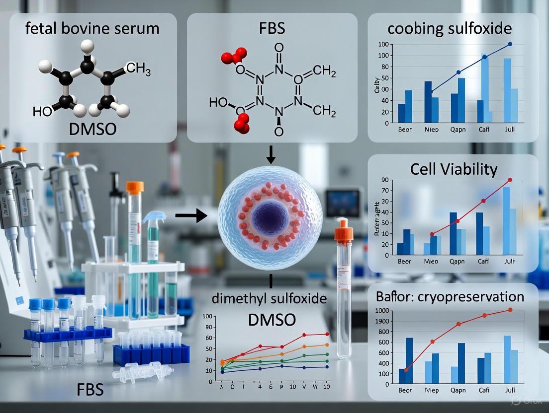
FBS with 10% DMSO Cryopreservation Media: A Complete Protocol and Research Review
This article provides a comprehensive guide to the preparation, application, and optimization of Fetal Bovine Serum (FBS) with 10% Dimethyl Sulfoxide (DMSO) cryopreservation media, a cornerstone technique for preserving cell...
FBS with 10% DMSO Cryopreservation Media: A Complete Protocol and Research Review
Abstract
This article provides a comprehensive guide to the preparation, application, and optimization of Fetal Bovine Serum (FBS) with 10% Dimethyl Sulfoxide (DMSO) cryopreservation media, a cornerstone technique for preserving cell viability in biomedical research and drug development. It covers foundational principles and established protocols for freezing and thawing cells, alongside targeted troubleshooting strategies to overcome common challenges. Furthermore, it presents a critical evaluation of the method, discussing its validation against long-term storage data and comparing its performance with emerging serum-free, animal-component-free alternatives to address ethical, safety, and reproducibility concerns. The content is tailored to enable researchers and scientists to implement robust and reliable cell cryopreservation practices.
Understanding FBS and DMSO: The Foundation of Cell Cryopreservation
The Critical Role of Cryopreservation in Biomedical Research and Biobanking
1. Introduction
Cryopreservation is an indispensable technique in biomedical research and biobanking, enabling the long-term storage of viable cells and tissues at ultra-low temperatures, typically below -135°C in liquid nitrogen vapor phase [1] [2]. By halting biological activity, this process preserves structural integrity and cellular function over indefinite periods, facilitating critical applications from drug discovery to cell-based therapies [1] [3]. The foundational method for cryopreservation involves the use of a cryoprotective medium, most commonly incorporating fetal bovine serum (FBS) and 10% dimethyl sulfoxide (DMSO) [2] [4]. This application note details the principles, protocols, and practical considerations for using FBS and DMSO-based media, providing researchers with a standardized framework for effective cell preservation within a broader thesis on cryopreservation media preparation.
2. Fundamental Principles of Cryopreservation
The success of cryopreservation hinges on mitigating the two primary causes of cell death during freezing: the formation of intracellular ice crystals, which mechanically disrupt membranes and organelles, and the rise in solute concentration to toxic levels as pure water freezes [2]. Cryoprotective Agents (CPAs) are employed to counteract these damaging processes.
- Permeating Agents: Small, amphiphilic molecules like DMSO (and glycerol) easily cross cell membranes. They depress the freezing point of water and promote vitrification—the formation of a non-crystalline, glassy state—thereby minimizing ice crystal formation [2]. DMSO, typically at a 10% concentration, also increases membrane permeability, allowing water to exit cells more readily during cooling [2].
- Non-Permeating Agents: Components like FBS in the freezing medium act extracellularly. FBS provides a protective environment, helps stabilize the cell membrane, and supplies proteins that buffer against osmotic shock and cold-induced damage [1] [5].
The standard slow-freezing protocol, with a cooling rate of approximately -1°C per minute, is critical. It allows sufficient time for water to exit the cell before intracellular freezing occurs, thereby minimizing osmotic stress and mechanical damage [1] [2] [5].
3. Standardized Protocols
3.1 Preparation of FBS with 10% DMSO Freezing Medium
- Materials:
- Fetal Bovine Serum (FBS)
- Dimethyl Sulfoxide (DMSO), cell culture grade
- Sterile glass pipette or DMSO-resistant pipette tips
- Sterile container (e.g., bottle, tube)
- Procedure:
- Chill the required volume of FBS to 2-8°C.
- Safety Note: DMSO is a potent solvent that can facilitate the transport of molecules through skin. Handle with gloves and appropriate personal protective equipment [1].
- Using a glass or DMSO-resistant pipette, slowly add DMSO to the cold FBS while gently swirling to mix. The final concentration should be 90% FBS and 10% DMSO (v/v). For example, to make 10 mL of freezing medium, combine 9 mL of FBS with 1 mL of DMSO.
- Keep the prepared freezing medium cold (2-8°C) and use it promptly.
3.2 Cell Freezing Procedure
- Materials:
- Procedure:
- Prepare Cells: For adherent cells, detach them using a standard subculture method (e.g., trypsin) and neutralize the enzyme with complete growth medium. For suspension cells, proceed directly from the culture vessel [1] [5].
- Count and Centrifuge: Determine the total cell count and viability. Centrifuge the cell suspension at 200-400 x g for 5-10 minutes to form a pellet. Carefully aspirate the supernatant [1] [6].
- Resuspend in Freezing Medium: Gently resuspend the cell pellet in cold freezing medium to achieve a final concentration specific to the cell type (e.g., 0.5 - 10 x 10^6 cells/mL for PBMCs) [6] [5].
- Aliquot: Rapidly transfer 1 mL aliquots of the cell suspension into pre-labeled cryogenic vials. Keep the vials on ice.
- Initiate Freezing: Immediately place the vials into a controlled-rate freezing device and transfer them to a -80°C freezer for 24 hours. This ensures a consistent cooling rate of approximately -1°C/minute [1] [6] [5].
- Long-Term Storage: After 24 hours, promptly transfer the vials to long-term storage in vapor-phase liquid nitrogen (below -135°C) [1] [6].
The following workflow summarizes the key stages of the cryopreservation process:
4. Comparative Performance Data
While FBS with 10% DMSO is a widely used and effective freezing medium, research into serum-free, defined alternatives is advancing. A 2025 study evaluated the long-term (2-year) performance of PBMCs cryopreserved in various media, providing quantitative data on viability and functionality [7].
Table 1: Viability and Recovery of PBMCs after 2-Year Cryostorage in Different Media [7]
| Freezing Medium | Key Composition | DMSO Concentration | Post-Thaw Viability (2 Years) | T-cell Functionality |
|---|---|---|---|---|
| FBS10 (Reference) | 90% FBS, 10% DMSO | 10% | High | Preserved |
| CryoStor CS10 | Serum-Free, Defined | 10% | High | Preserved (Comparable to FBS10) |
| NutriFreez D10 | Serum-Free, Defined | 10% | High | Preserved (Comparable to FBS10) |
| Bambanker D10 | Serum-Free, Defined | 10% | High | Slight Divergence from FBS10 |
| Media with <7.5% DMSO | Serum-Free, Various | <7.5% | Significant Loss | Not Assessed (Eliminated from study) |
Table 2: Impact of Cryopreservation on Human Bone Marrow-Derived Mesenchymal Stem Cells (hBM-MSCs) [4]
| Cell Attribute | 0-4 Hours Post-Thaw | 24 Hours Post-Thaw | Beyond 24 Hours Post-Thaw |
|---|---|---|---|
| Viability | Reduced | Recovered | Recovered |
| Apoptosis Level | Increased | Dropped | Variable |
| Metabolic Activity | Impaired | Remained Lower than Fresh | Variable |
| Adhesion Potential | Impaired | Remained Lower than Fresh | Variable |
| Proliferation Rate | - | - | No Significant Difference |
| Colony-Forming Ability | - | - | Reduced in some cell lines |
5. The Scientist's Toolkit: Essential Research Reagents
The following table lists key materials and their functions for successful cryopreservation using FBS and DMSO-based media.
Table 3: Essential Materials for Cryopreservation with FBS/DMSO Media
| Item | Function & Rationale |
|---|---|
| Fetal Bovine Serum (FBS) | Provides extracellular proteins, growth factors, and nutrients that stabilize the cell membrane and protect against cold shock and osmotic stress during freezing and thawing [6] [4]. |
| Dimethyl Sulfoxide (DMSO) | Serves as a permeating cryoprotectant. Lowers the freezing point, prevents intracellular ice crystal formation by promoting vitrification, and increases membrane permeability [2]. |
| Controlled-Rate Freezer (e.g., CoolCell) | Ensures a consistent, optimal cooling rate of -1°C/minute, which is critical for cell survival by allowing controlled dehydration and minimizing intracellular ice formation [1] [6]. |
| Cryogenic Vials | Specially designed tubes that withstand extreme thermal stresses of liquid nitrogen temperatures for secure long-term sample storage [1]. |
| Liquid Nitrogen Storage System | Provides stable long-term storage at temperatures below -135°C (typically in vapor phase to prevent explosion risks), effectively pausing all biochemical activity [1] [6]. |
6. Conclusion
The preparation and application of cryopreservation media with FBS and 10% DMSO remains a cornerstone technique for reliable long-term cell storage in biomedical research. Adherence to standardized protocols—utilizing log-phase cells, cold media, controlled slow freezing, and proper vapor-phase liquid nitrogen storage—is paramount for maximizing post-thaw viability and functionality. As the field progresses, the development and validation of serum-free, defined media offer a promising path forward, reducing variability and safety concerns while maintaining the critical role of biobanking in supporting drug development and advanced therapeutic applications.
Fetal Bovine Serum (FBS) is a critically important supplement in cell culture systems, valued for its complex composition of nutrients, growth factors, and protective elements [8]. As a natural medium additive, FBS provides the essential components required for cellular survival, proliferation, and maintenance in vitro [8] [9]. In the specific context of cryopreservation media preparation with 10% DMSO, FBS plays a vital role in protecting cells from the multiple stresses associated with the freezing and thawing processes [1] [6]. This application note details the core functions of FBS and provides standardized protocols for its use in cryopreservation media formulation, specifically addressing the needs of researchers, scientists, and drug development professionals working to preserve valuable cell lines for therapeutic and research applications.
Core Functions of FBS in Cell Culture and Cryopreservation
FBS serves three primary, interconnected functions that make it indispensable for cell culture and cryopreservation.
Nutritional Support
FBS provides a rich source of essential nutrients and energy substrates necessary for cell survival and metabolic activity, which is crucial for preparing cells for the cryopreservation process [8].
Table 1: Key Nutritional and Macromolecular Components of FBS
| Component Category | Specific Examples | Primary Function in Cell Culture |
|---|---|---|
| Nutrients & Energy Sources | Sugars, Vitamins, Lipids, Amino Acids [8] | Provides building blocks for biosynthesis and energy production. |
| Proteins | Albumin, Transferrin, other carrier proteins [8] [9] | Binds and transports lipids, hormones, and metals; provides buffering capacity. |
| Electron Carriers & Cofactors | -- | Supports essential metabolic pathways and redox reactions. |
Supply of Growth and Attachment Factors
FBS contains a wide array of biologically active components that directly promote cell growth and maintenance, helping to ensure cells are in a robust, log-phase state prior to cryopreservation [8].
Table 2: Growth-Promoting Factors in FBS
| Factor Type | Function in Cell Culture |
|---|---|
| Growth Factors | Stimulate cell proliferation and differentiation [8]. |
| Hormones | Regulate cellular metabolism and growth cycles [8]. |
| Attachment Factors | Facilitate cell adhesion to culture surfaces, promoting monolayer formation [8]. |
Cell Protection
A critical function of FBS, especially in cryopreservation, is its ability to protect cells from various stressors [8]. It provides buffering capacity to counteract pH shifts and contains factors that inactivate proteases and protect cells from toxic agents and shear forces [8]. In cryopreservation, the proteins in FBS work synergistically with cryoprotectants like DMSO to mitigate ice crystal formation and osmotic shock, thereby enhancing post-thaw viability and recovery [6] [5].
Essential Research Reagent Solutions
The following table catalogues key materials required for working with FBS in cell culture and cryopreservation protocols.
Table 3: Essential Reagents for Cell Culture and Cryopreservation with FBS
| Reagent/Material | Function & Application |
|---|---|
| Fetal Bovine Serum (FBS) | Universal supplement for cell culture media and a key component of cryopreservation media to support growth and viability [8] [1]. |
| Dimethyl Sulfoxide (DMSO) | A cryoprotective agent (CPA) that penetrates cells, reduces ice crystal formation, and prevents osmotic lysis during freezing [1] [6]. |
| Serum-Free Cryopreservation Media | Chemically defined, animal-component-free media (e.g., CryoStor CS10) used as an FBS alternative, often containing DMSO [6] [10]. |
| Basal Cell Culture Medium | The nutrient base (e.g., DMEM, RPMI-1640) to which FBS is typically added at 5-10% for routine cell culture [8]. |
| Controlled-Rate Freezer / Isopropanol Chamber | Device to ensure a consistent, slow freezing rate (approx. -1°C/min), which is critical for high cell viability post-thaw [1] [5]. |
| Cryogenic Storage Vials | Sterile vials designed to withstand ultra-low temperatures for long-term storage in liquid nitrogen [1]. |
FBS in Cryopreservation Media Preparation: Application Protocol
This protocol details the preparation of cryopreservation media using FBS and 10% DMSO for mammalian cells, based on established methodologies [1] [6] [5].
Background and Principle
Cryopreservation media supplemented with FBS and DMSO provides a protective environment for cells during the freeze-thaw cycle. FBS supplies macromolecules that buffer cells against pH shifts, detoxify harmful agents, and reduce mechanical stress from ice formation [8]. DMSO, a penetrating cryoprotectant, decreases the intracellular freezing point and minimizes the formation of lethal intracellular ice crystals [1] [6]. The combination is a gold standard for preserving a wide range of cell types.
Materials and Equipment
- Log-phase cells at high viability (>90%)
- Complete Growth Medium (Basal medium + FBS)
- Fetal Bovine Serum (FBS)
- Cell culture-grade Dimethyl Sulfoxide (DMSO)
- Sterile cryogenic vials
- Centrifuge and conical tubes
- Pipettes and pipette tips
- Hemocytometer or automated cell counter
- Trypan Blue solution
- Isopropanol freezing container (e.g., "Mr. Frosty") or controlled-rate freezer
- Liquid nitrogen storage tank
Step-by-Step Procedure
Preparation of Cryopreservation Medium:
Cell Harvest and Counting:
- For adherent cells, detach using a standard method (e.g., trypsin/EDTA), neutralize with complete medium, and transfer to a conical tube. For suspension cells, transfer directly to a conical tube [1] [5].
- Perform a cell count and viability assessment using Trypan Blue exclusion and a hemocytometer or automated cell counter [1].
Cell Pelletting and Resuspension:
- Centrifuge the cell suspension at approximately 300 × g for 5-10 minutes [1].
- Carefully aspirate the supernatant without disturbing the cell pellet.
- Resuspend the cell pellet in the cold cryopreservation medium to achieve a final concentration specific to the cell type. A common range is 0.5 - 10 x 10^6 cells/mL [1] [6].
Aliquoting and Freezing:
- Rapidly aliquot 1 mL of the cell suspension into each pre-labeled cryogenic vial.
- Immediately place the vials in an isopropanol freezing container and transfer it to a -80°C freezer for at least 24 hours (or overnight). This apparatus ensures a cooling rate of approximately -1°C/minute [1] [5].
- Alternatively, use a controlled-rate freezer if available.
Long-Term Storage:
Critical Considerations and Troubleshooting
- Cell State: Always use cells in the log phase of growth and at the lowest possible passage number for optimal post-thaw recovery [1].
- DMSO Handling: DMSO can facilitate the entry of other molecules through the skin; handle with appropriate personal protective equipment (PPE) [1]. Do not store pure DMSO on ice, as it may crystallize [6].
- Temperature Control: Keep cells and freezing medium cold after adding DMSO, and work quickly to minimize DMSO exposure time at room temperature, as it can be toxic to cells [6].
- Lot-to-Lot Variability: FBS is a natural product with inherent variability. For long-term projects, pre-test and reserve a large quantity of a single FBS lot to ensure experimental consistency [8] [11].
Workflow and Functional Pathways
The following diagram illustrates the logical workflow for cryopreserving cells using FBS and DMSO, integrating the key procedural steps and the functional roles of the reagents.
Cryopreservation is a cornerstone technology for the long-term storage of cells, playing an indispensable role in biomedical research, drug development, and cellular therapeutics [1] [12]. The fundamental challenge of cryopreservation lies in managing the phase change of water, as the formation and growth of intracellular and extracellular ice crystals can cause lethal mechanical damage to cellular structures [12]. Dimethyl Sulfoxide (DMSO) is one of the most widely used cryoprotective agents (CPAs) to mitigate this damage. When preparing cryopreservation media, a common and effective formulation combines 10% DMSO with Fetal Bovine Serum (FBS) [5] [13]. This application note details the mechanism by which DMSO prevents intracellular ice formation and provides standardized protocols for its use in a research setting, contextualized within the broader scope of cryopreservation media preparation.
The Mechanism of DMSO Action
DMSO, a small, polar, and amphipathic molecule, protects cells during freezing primarily through colligative properties and its ability to penetrate cell membranes [14] [15]. Its efficacy is a direct result of how it alters the physical behavior of water and interacts with the cellular environment during the freeze-thaw cycle.
Core Principles of Cryoinjury
During slow freezing, extracellular water freezes first. This removes pure water from the solution, increasing the concentration of solutes outside the cell and creating a hypertonic environment. Consequently, water osmotically flows out of the cell, leading to excessive dehydration and solute damage. During rapid cooling, water cannot exit the cell quickly enough, leading to the formation of lethal intracellular ice [12]. Ice recrystallization during the thawing process also causes significant mechanical damage [12].
How DMSO Modifies these Processes
DMSO acts on these processes through several interconnected mechanisms:
- Freezing Point Depression & Vitrification: DMSO disrupts the hydrogen bonding network between water molecules. This "colligative" effect lowers the freezing point of the solution and increases its viscosity, thereby slowing ice crystal growth and facilitating a transition to a glassy, vitrified state at ultra-low temperatures, rather than forming crystalline ice [15].
- Reducing Intracellular Ice Formation: As a permeating CPA, DMSO readily crosses the cell membrane. This allows it to exert its colligative effects both inside and outside the cell. By increasing the intracellular solute concentration, it reduces the amount of water available to form ice and lowers the intracellular freezing point, thereby minimizing the risk of lethal intracellular ice crystallization [1] [13].
- Mitigating Osmotic Stress: The presence of DMSO inside the cell reduces the osmotic differential across the membrane during freezing. This moderates the rate and extent of cellular dehydration, protecting the cell from shrinkage-induced damage [16].
The following diagram illustrates the protective pathway of DMSO during the cryopreservation process.
Quantitative Data on DMSO Efficacy
The protective effect of DMSO is concentration-dependent, balancing cryoprotection with inherent cytotoxicity. Recent research also explores strategies to reduce DMSO concentration.
Table 1: Impact of DMSO Concentration on Cell Viability Post-Cryopreservation
| DMSO Concentration (% v/v) | Reported Cell Viability | Key Observations | Source Model |
|---|---|---|---|
| 0% | Very Low | Extensive cell death due to ice crystal damage. | General Principle [12] |
| 2.5% | ~70% (Clinical threshold) | Viability meets minimum clinical requirement when combined with hydrogel microencapsulation. | Mesenchymal Stem Cells (MSCs) [17] |
| 10% | High (Optimal for many lines) | Considered the standard for many cell types; offers robust protection. | General Protocol [1] [13] |
Table 2: Advanced Strategies for DMSO Reduction/Replacement
| Strategy | Mechanism | Key Findings | Reference |
|---|---|---|---|
| Hydrogel Microencapsulation | Alginate hydrogel shields cells, reduces ice crystal damage. | Enabled reduction to 2.5% DMSO while maintaining >70% MSC viability. [17] | |
| Macromolecular Cryoprotectants | Ice recrystallization inhibition (IRI), membrane stabilization. | Biodegradable DNA frameworks show efficacy with minimal cytotoxicity. [14] | |
| DMSO-Free Commercial Media | Complex formulations using sugars, polymers, and other osmolytes. | Products like StemCell Keep and CryoStor CS10 are available, but require validation. [15] |
Detailed Experimental Protocols
Standard Protocol: Cryopreserving Cells with FBS and 10% DMSO
This is a generalized protocol for cryopreserving adherent mammalian cell lines. Always refer to cell-specific recommendations.
Research Reagent Solutions & Materials
- Cryopreservation Medium: 90% Fetal Bovine Serum (FBS) + 10% DMSO. Prepare fresh and keep cold (2-8°C). [5] [13]
- Growth Medium: Complete cell culture medium, pre-warmed to 37°C.
- Wash Solution: Phosphate-Buffered Saline (PBS), without calcium or magnesium.
- Dissociation Reagent: Trypsin-EDTA or other cell-specific detachment enzyme.
- Equipment: Cryogenic vials, controlled-rate freezing apparatus (e.g., CoolCell or "Mr. Frosty"), liquid nitrogen storage tank, centrifuge, hemocytometer or automated cell counter.
The workflow for the standard cryopreservation protocol is outlined below.
Step-by-Step Methodology:
- Cell Harvest: Use healthy, log-phase cells with high viability (>90%) and at a low passage number [1] [13]. For adherent cells, rinse with PBS and detach using a suitable dissociation reagent. Neutralize the enzyme with growth medium.
- Pellet and Count: Centrifuge the cell suspension at approximately 300 × g for 5 minutes [5] [6]. Aspirate the supernatant and resuspend the pellet in a small volume of growth medium to perform a cell count and determine viability via Trypan Blue exclusion.
- Prepare Freezing Suspension: Re-centrifuge the counted cell suspension. Aspirate the supernatant completely. Gently resuspend the cell pellet in cold cryopreservation medium (90% FBS / 10% DMSO) to a final concentration of 1-5 x 10^6 cells/mL [5] [13]. Mix gently but thoroughly to ensure a homogeneous suspension.
- Aliquot and Begin Freezing: Quickly aliquot 1 mL of the cell suspension into each pre-labeled cryovial. Place the vials immediately into a controlled-rate freezing device and transfer them to a -80°C freezer. The freezing device ensures a critical cooling rate of approximately -1°C per minute [1] [13].
- Long-Term Storage: After 24 hours, promptly transfer the frozen cryovials to a liquid nitrogen storage tank for long-term preservation in the vapor phase (below -135°C) [6] [13].
Advanced Protocol: Cryopreservation with Reduced DMSO Using Hydrogel Microencapsulation
This protocol is adapted from recent research for cryopreserving sensitive cells like MSCs with lower DMSO [17].
Workflow Overview:
- Encapsulate Cells: Fabricate cell-laden hydrogel microcapsules using a high-voltage electrostatic coaxial spraying device. The core solution contains cells suspended in a mixture of sodium alginate, while the shell is a cross-linking solution like calcium chloride.
- Culture Microcapsules: Transfer the formed microspheres into complete culture medium and maintain them in a 37°C, 5% CO₂ incubator to allow cell recovery.
- Cryopreserve with Low DMSO: Resuspend the microcapsules in cryopreservation medium containing a low concentration of DMSO (e.g., 2.5% v/v). The hydrogel matrix provides a physical barrier that mitigates cryoinjury.
- Freeze and Store: Follow a slow freezing process, similar to the standard protocol, using a controlled-rate freezer before transferring to liquid nitrogen.
The Scientist's Toolkit: Essential Research Reagents
Table 3: Key Reagents for Cryopreservation Media Preparation
| Reagent / Material | Function & Rationale |
|---|---|
| Dimethyl Sulfoxide (DMSO) | Permeating cryoprotectant; lowers freezing point, minimizes intracellular ice formation. [1] [15] |
| Fetal Bovine Serum (FBS) | Provides nutrients, growth factors, and proteins that stabilize cell membranes and support post-thaw recovery. [5] |
| Serum-Free Cryopreservation Media | Chemically defined, xeno-free alternative to FBS; essential for clinical applications. [1] [6] |
| Programmable Freezing Chamber | Ensures consistent, controlled cooling rate of ~1°C/min, critical for high viability. [1] [13] |
| Hydrogel Biomaterials (e.g., Alginate) | Used in advanced strategies to create a protective 3D environment, enabling DMSO reduction. [17] |
Critical Considerations and Limitations
While 10% DMSO is highly effective, researchers must be aware of its limitations. DMSO is not biologically inert and can induce cellular changes, including alterations in the epigenetic landscape and transcriptome, even at low concentrations [18]. Furthermore, DMSO can induce unwanted differentiation in stem cells and cause adverse reactions in patients receiving cell therapies [15] [18]. Therefore, the washing of DMSO post-thaw or the use of reduced concentrations or DMSO-free alternatives is critical for sensitive applications and clinical translation [17] [15]. The principle of "freeze slowly, thaw quickly" is paramount: slow freezing allows water to exit the cell, while rapid thawing in a 37°C water bath minimizes the damaging effects of ice recrystallization [16].
Advantages and Inherent Drawbacks of the FBS + 10% DMSO Formulation
The cryopreservation of cellular specimens is a fundamental practice in biomedical research and clinical applications, enabling long-term storage while maintaining viability and functionality for subsequent analysis. The formulation comprising Fetal Bovine Serum (FBS) and 10% Dimethyl Sulfoxide (DMSO) has emerged as a long-standing benchmark for the cryopreservation of diverse cell types, including peripheral blood mononuclear cells (PBMCs) and mesenchymal stem cells (MSCs) [7] [19]. This application note delineates the advantages and inherent drawbacks of this ubiquitous formulation, contextualized within a comprehensive thesis on cryopreservation media preparation. It is structured to provide researchers, scientists, and drug development professionals with a detailed, evidence-based assessment of the FBS + 10% DMSO protocol, encompassing its mechanistic basis, documented efficacy, and significant limitations, supplemented by structured experimental data and practical methodologies.
Core Formulation: Mechanisms, Advantages, and Drawbacks
Cryoprotective Mechanism of Action
The FBS + 10% DMSO formulation confers cytoprotection through a multimodal mechanism that mitigates the principal insults of the freezing process: intracellular ice crystal formation and osmotic stress.
- DMSO Function: As a penetrating cryoprotectant, DMSO freely crosses the cell membrane due to its low molecular weight and hydrophilicity [20]. It functions by disrupting ice crystal nucleation through hydrogen bonding with intracellular water molecules, thereby reducing the freezing point and minimizing the formation of damaging intracellular ice crystals during rapid cooling [20]. Furthermore, its membrane permeability helps stabilize the cell membrane and prevents severe osmotic shock by equilibrating intra- and extracellular solute concentrations [7] [21].
- FBS Function: Acting as a non-penetrating agent, FBS provides a complex mixture of nutrients, growth factors, and proteins. It contributes to membrane stabilization and provides an extracellular matrix that helps mitigate the detrimental effects of solute concentration and ice crystal growth outside the cell [20] [22]. The serum also offers undefined factors that support post-thaw recovery and viability.
The synergistic interaction between these components during a slow freeze protocol is crucial for optimal cell survival. The following diagram illustrates the protective workflow of this formulation during the cryopreservation process.
Documented Advantages and Efficacy
The widespread adoption of the FBS + 10% DMSO formulation is predicated on its proven performance across a spectrum of cell types and long-term storage durations. Quantitative data from recent studies underscore its efficacy.
Table 1: Documented Efficacy of FBS + 10% DMSO Cryopreservation Formulation
| Cell Type | Study Duration | Post-Thaw Viability | Key Functional Assays | Reference |
|---|---|---|---|---|
| Peripheral Blood Mononuclear Cells (PBMCs) | 2 years | High viability maintained | Cytokine secretion, T/B cell FluoroSpot, intracellular cytokine staining | [7] |
| Human Dermal Fibroblasts (HDFs) | 3 months | >80% viability | Cell attachment, Ki67 and Collagen-I expression | [21] |
| Adipose-Derived Stem Cells (ASCs) | 2 weeks | ~84% ± 8% viability | Adipogenic and osteogenic differentiation | [22] |
| Dental Pulp Stem Cells (DPSCs) | Not specified | Effective post-thaw recovery | Phenotype maintenance, multipotency retention | [23] |
A primary advantage is its consistent performance. A comprehensive 2-year study demonstrated that PBMCs cryopreserved in FBS + 10% DMSO maintained high viability and functionality, comparable to the best serum-free alternatives, across all evaluated time points [7]. This formulation effectively preserves the capacity for immune response, which is critical for immunological studies and vaccine clinical trials [7]. Furthermore, the protocol is well-established, straightforward to implement, and the components are readily available and affordable [21] [22].
Inherent Drawbacks and Limitations
Despite its efficacy, the use of FBS + 10% DMSO is associated with significant drawbacks that pose challenges for both research reproducibility and clinical applications.
Table 2: Inherent Drawbacks of the FBS + 10% DMSO Formulation
| Drawback Category | Specific Issue | Impact on Research/Clinical Use |
|---|---|---|
| DMSO Cytotoxicity | - Induction of cell differentiation & epigenetic changes [20].- Alters calcium signaling & gene expression [20].- Causes adverse reactions in patients (nausea, cardiac effects) [20]. | Compromises experimental validity and raises safety concerns for cell-based therapies. |
| FBS Batch Variability | - Inconsistent composition of hormones, growth factors, and other undefined components [7].- Risk of pathogen transmission (viruses, mycoplasma) [7]. | Leads to poor experimental reproducibility and requires rigorous batch qualification. |
| Ethical & Logistical Concerns | - Ethical issues regarding animal welfare in FBS production [7].- Import restrictions in certain countries [7]. | Limits the global standardization of protocols and conflicts with animal-free mandates. |
| Impact on Cell Function | - Can induce unwanted immunological responses in PBMC cultures [7].- High concentrations (5-10%) are cytotoxic to human apical papilla cells [24]. | Can skew experimental outcomes in sensitive assays and reduce recovery of specific cell types. |
The cytotoxicity of DMSO is a paramount concern. Beyond its direct toxic effects, which can reduce cell viability [24], DMSO has been shown to dysregulate gene expression and modify DNA methylation profiles, potentially inducing unwanted cell differentiation [20]. For clinical infusions, the presence of even residual DMSO is linked to adverse reactions, including gastrointestinal, cardiovascular, and respiratory symptoms [20].
The use of FBS introduces another layer of complexity. Its undefined and variable nature can impede experimental reproducibility [7]. Moreover, for clinical-grade cell products, the presence of xenogenic proteins carries a risk of immune reactions and pathogen transmission, necessitating a move toward serum-free and xeno-free alternatives [7] [22].
The Scientist's Toolkit: Essential Research Reagents & Materials
Successful implementation of the FBS + 10% DMSO cryopreservation protocol requires a set of essential reagents and laboratory equipment.
Table 3: Key Reagents and Materials for Cryopreservation Protocols
| Item Name | Function/Description | Application Note |
|---|---|---|
| Fetal Bovine Serum (FBS) | Provides extracellular cryoprotection, nutrients, and growth factors. | Qualification of each batch is critical for consistency. Sourced from commercial vendors [7]. |
| Dimethyl Sulfoxide (DMSO) | Penetrating cryoprotectant that prevents intracellular ice crystal formation. | Use high-grade, sterile-filtered DMSO. Handle with care due to cytotoxicity [20] [21]. |
| Cryogenic Vials | Specially designed tubes for safe storage in liquid nitrogen. | Ensure they are leak-proof and capable of withstanding extreme temperatures. |
| Controlled-Rate Freezer / CoolCell | Device to achieve the optimal cooling rate of approximately -1°C/min. | CoolCell is a non-programmable, isopropanol-based freezing container [7] [21]. |
| Liquid Nitrogen Storage System | Provides long-term storage at -135°C to -196°C. | Cells are typically stored in the vapor phase to minimize contamination risk [19] [21]. |
| Deoxyribonuclease I (DNase I) | Enzyme added during thawing to prevent cell clumping from DNA release. | Used in the thawing solution to improve cell recovery and viability [7]. |
Detailed Experimental Protocol: PBMC Cryopreservation and Thawing
The following section outlines a standardized protocol for the cryopreservation and thawing of PBMCs using FBS + 10% DMSO, as derived from the cited literature [7]. The accompanying workflow diagram maps the key procedural stages.
Cryopreservation Protocol
Methodology: PBMCs are isolated from whole blood using a lymphocyte density gradient medium (e.g., Lymphoprep) and washed in an appropriate buffer like Hanks' Balanced Salt Solution [7].
- Post-isolation: After the final centrifugation, resuspend the cell pellet in the pre-chilled cryopreservation medium (90% FBS + 10% DMSO) at a high concentration (e.g., 12 × 10^6 cells/mL) [7].
- Aliquoting: Dispense 1 mL of the cell suspension into pre-cooled cryogenic vials.
- Controlled-Rate Freezing: Immediately transfer the vials into a CoolCell or similar freezing container and place it in a -80°C freezer for a minimum of 4 hours and up to 7 days. This device ensures an optimal cooling rate of approximately -1°C/min, which is critical for cell survival [7] [21].
- Long-Term Storage: After the initial freezing period, transfer the vials to a long-term storage system, ideally a vapor-phase liquid nitrogen tank, where temperatures are maintained below -135°C [7].
Thawing and Viability Assessment Protocol
A careful thawing process is vital to ensure high cell recovery and minimize the osmotic stress associated with removing DMSO.
- Rapid Thawing: Retrieve a cryovial from storage and gently agitate it in a 37°C water bath until only a small ice crystal remains (typically 1-2 minutes) [7] [21].
- DNase Treatment: Immediately upon thawing, add a mixture of FBS and DNase I (e.g., 10 µg/mL) to the vial to prevent cell clumping caused by DNA released from damaged cells [7].
- Dilution and Washing: Transfer the entire cell suspension into a tube containing 10 mL of pre-warmed culture medium (e.g., RPMI 1640 + 10% FBS). There are two primary revival methods:
- Viability and Functionality Assessment:
- Viability/Yield: Assess cell viability and count post-thaw using Trypan Blue exclusion in a hemocytometer [21].
- Functionality: For PBMCs, evaluate immune functionality through assays such as cytokine secretion (ELISA/ELISpot), T and B cell FluoroSpot, or intracellular cytokine staining by flow cytometry [7]. For MSCs, differentiation potential (osteogenic, adipogenic, chondrogenic) and immunophenotype should be confirmed post-thaw [19] [23].
The FBS + 10% DMSO formulation remains a highly effective and widely used cryopreservation medium, delivering proven results in maintaining cell viability and functionality over extended periods, as evidenced by its performance in 2-year storage studies [7]. Its mechanism, leveraging the synergistic action of a penetrating cryoprotectant and a complex serum, is well-understood. However, significant inherent drawbacks, including the cytotoxicity and differential effects of DMSO, the batch-to-batch variability of FBS, and associated ethical concerns, cannot be overlooked [7] [20] [22]. These limitations are driving the field toward the development and adoption of serum-free, chemically defined alternatives and methods to reduce or eliminate DMSO. Therefore, while the FBS + 10% DMSO protocol is a robust and reliable method, its application must be carefully considered in the context of specific research objectives and clinical requirements.
Cryopreservation media formulations containing fetal bovine serum (FBS) and 10% dimethyl sulfoxide (DMSO) have long served as the traditional standard for preserving peripheral blood mononuclear cells (PBMCs) and other biologically valuable samples in biomedical research and clinical applications [7]. This combination effectively protects cells from freezing-induced damage; DMSO stabilizes the cell membrane and prevents osmotic shock, while FBS provides a rich mixture of proteins and growth factors [7] [25]. However, mounting evidence reveals significant limitations associated with FBS, including substantial batch-to-batch variability, considerable ethical concerns regarding animal welfare, and non-negligible risks of pathogen contamination [7] [10]. These challenges compromise experimental reproducibility, raise safety concerns for clinical applications, and conflict with ethical principles in research. This application note delineates these critical challenges and provides validated, detailed protocols for implementing serum-free alternatives, empowering researchers and drug development professionals to enhance the reliability and ethical standing of their cryopreservation practices.
Key Challenges of FBS-Based Cryopreservation Media
Batch-to-Batch Variability
FBS is a complex, biologically sourced material with an undefined and highly variable composition, leading to significant inconsistencies between production lots [10].
- Impact on Reproducibility: The variable nature of FBS can directly affect the quality and performance of experiments, contributing to the broader reproducibility crisis in scientific research [10]. Variations in growth factors, hormones, and other constituents can alter cellular phenotypes; for instance, serum origin has been shown to change the phenotype of engineered skeletal muscle and override the native gene expression of primary tendon cells [10].
- Practical Consequences: This variability necessitates rigorous and costly pre-testing of multiple FBS lots to identify a suitable batch for sensitive applications, creating substantial administrative, financial, and logistical burdens for laboratories [10].
Ethical Concerns
The production of FBS raises profound ethical questions, as it is obtained from bovine fetuses during the slaughter of pregnant cows [7] [10]. The procedures involved have prompted many academic and pharmaceutical groups to actively seek animal-protein-free alternatives for both cell culture and cryopreservation media [7] [10]. Furthermore, some countries have implemented import restrictions on FBS, complicating its acquisition and use [7].
Risk of Pathogen Contamination
The use of animal-derived components inherently carries a risk of introducing adventitious agents into cell cultures.
- Documented Contaminants: FBS batches can harbor various contaminants, including viruses, prions, bacteria, fungi, endotoxins, and exogenous extracellular vesicles [10]. Viral antibodies and viruses themselves have been detected in FBS for over half a century, and concerns persist regarding emerging pathogens [10].
- Clinical Implications: For cells destined for therapeutic applications, the presence of xenogenic substances from FBS can induce an immune response in patients, potentially compromising the efficacy and safety of cell transplantation therapies [10]. Regulatory bodies like the U.S. FDA and European Medicines Agency therefore impose stringent requirements on clinical-grade FBS, further increasing the cost and complexity of its use [10].
Quantitative Comparison of Cryopreservation Media
A comprehensive 2025 study evaluated the viability and functionality of PBMCs cryopreserved in various media over a two-year period, providing robust quantitative data for comparison [7]. The study assessed a reference FBS-based medium (90% FBS + 10% DMSO) against several commercially available, serum-free, animal-component-free media.
Table 1: Post-Thaw Viability of PBMCs Cryopreserved in Different Media Over Time (Viability %)
| Cryopreservation Medium | 3 Weeks (M0) | 3 Months (M3) | 6 Months (M6) | 1 Year (M12) | 2 Years (M24) |
|---|---|---|---|---|---|
| Reference: FBS + 10% DMSO | High | High | High | High | High |
| CryoStor CS10 (10% DMSO) | High | High | High | High | High |
| NutriFreez D10 (10% DMSO) | High | High | High | High | High |
| Bambanker D10 (10% DMSO) | High | High | High | High | High |
| Media with < 7.5% DMSO | Lower | N/A (Eliminated) | N/A | N/A | N/A |
Table 2: Functionality Assessment of PBMCs Post-Thaw (Based on Immunoassays)
| Cryopreservation Medium | Cytokine Secretion | T-cell Functionality (FluoroSpot) | B-cell Functionality (IgG Secretion) |
|---|---|---|---|
| Reference: FBS + 10% DMSO | Normal | Normal | Normal |
| CryoStor CS10 | Comparable to Reference | Comparable to Reference | Increased post-activation [26] |
| NutriFreez D10 | Comparable to Reference | Comparable to Reference | Not Specified |
| Bambanker D10 | Comparable to Reference | Tended to Diverge | Not Specified |
Key Findings: The study demonstrated that serum-free media containing 10% DMSO, specifically CryoStor CS10 and NutriFreez D10, effectively maintained PBMC viability, recovery, and immune functionality at levels comparable to the traditional FBS reference medium throughout the 2-year storage period [7]. In contrast, media formulations with DMSO concentrations below 7.5% showed significantly lower viability and were eliminated from the study after initial assessment [7]. These results underscore that while DMSO concentration is critical for success, the elimination of FBS is feasible without sacrificing cell quality.
Recommended Serum-Free Protocols
Protocol: Cryopreservation of PBMCs using Serum-Free Media
This protocol is adapted from the methods used in the cited study for freezing human PBMCs [7].
The Scientist's Toolkit: Essential Research Reagents
| Item | Function | Example Product(s) |
|---|---|---|
| Serum-Free Freezing Medium | Protects cells during freezing; defined, animal-component-free. | CryoStor CS10 [26], NutriFreez D10 [7] |
| Programmable Freezer or CoolCell | Ensures controlled, slow freezing rate (-1°C to -3°C/min). | CoolCell [7] |
| Liquid Nitrogen Storage Tank | Provides long-term storage at ≤ -135°C. | Vapor phase storage recommended [27] |
| Cryogenic Vials | Safely contains cell suspension for ultra-low temp storage. | N/A |
| Lymphocyte Separation Medium | Isolates PBMCs from whole blood. | Lymphoprep [7] |
Procedure:
- Cell Preparation: Isolate PBMCs from whole blood using a density gradient medium such as Lymphoprep. Ensure cells are healthy and in the log phase of growth [7] [27].
- Centrifugation and Counting: Wash the isolated PBMCs in a balanced salt solution (e.g., HBSS). Perform a final centrifugation and resuspend the cell pellet to a precise concentration for counting [7].
- Resuspension in Freezing Medium: Resuspend the cell pellet in the chosen serum-free, animal-component-free freezing medium (e.g., CryoStor CS10) to a target concentration of 12 × 10^6 cells/mL [7].
- Aliquoting: Dispense 1 mL of the cell suspension into each pre-cooled cryovial. Label vials with a liquid nitrogen-resistant marker, including cell identity, passage number, and date [7] [27].
- Controlled-Rate Freezing: Transfer the cryovials to a controlled-rate freezing device (e.g., a CoolCell container) and place them immediately in a -80°C freezer for 1-7 days. This step ensures a consistent freezing rate of approximately -1°C per minute, which is critical for high viability [7] [27].
- Long-Term Storage: After the initial freezing period, promptly transfer the cryovials to a long-term storage location, such as the vapor phase of a liquid nitrogen tank (≤ -135°C) [7] [27].
Protocol: Thawing and Assessing Cryopreserved PBMCs
Procedure:
- Rapid Thaw: Remove the cryovial from liquid nitrogen storage and immediately place it in a 37°C water bath. Gently agitate the vial until only a tiny ice crystal remains [16].
- Dilution and DNase Treatment: Immediately upon thawing, add a mixture of FBS and deoxyribonuclease I (DNase) at 10 µg/mL to the vial to dilute the DMSO and prevent cell clumping. Transfer the entire suspension into a larger volume (e.g., 10 mL) of pre-warmed culture medium [7] [16].
- Centrifugation and Plating: Centrifuge the cell suspension to remove the cryopreservation medium containing DMSO. Resuspend the cell pellet in fresh, pre-warmed complete culture medium and plate the cells for functional assays [7].
- Viability and Functionality Assessment:
- Viability Analysis: Assess cell viability post-thaw using a method like trypan blue exclusion or flow cytometry with propidium iodide staining. CryoStor CS10 has demonstrated post-thaw viabilities of 94-98% for human B cells [26].
- Functional Assays: Evaluate immune cell functionality using assays such as:
Workflow and Decision Pathway
Diagram 1: Pathway for Addressing FBS Challenges
Diagram 2: Serum-Free Cryopreservation Workflow
Transitioning from traditional FBS-based cryopreservation media to defined, serum-free, and animal-component-free alternatives is both scientifically justified and practically feasible. Robust, commercially available solutions like CryoStor CS10 and NutriFreez D10 effectively mitigate the critical challenges of batch-to-batch variability, ethical concerns, and pathogen risk without compromising post-thaw cell viability or functionality, even over long-term storage of up to two years [7]. By adopting the detailed application notes and protocols outlined herein, researchers and drug development professionals can significantly enhance the reproducibility, safety, and ethical compliance of their cryopreservation practices, thereby strengthening the overall integrity of biomedical research and clinical development.
Step-by-Step Protocol: Preparing and Using FBS with 10% DMSO Cryomedium
Within the critical field of biopreservation, the preparation of reliable cryopreservation media is a foundational technique for safeguarding the long-term viability of cell lines and primary cells in research and drug development. This application note provides a detailed framework for the preparation of cryopreservation media utilizing Fetal Bovine Serum (FBS) and 10% DMSO, framed within a broader thesis on optimizing cryopreservation protocols. The consistent functionality of biological reagents after thawing is paramount for the integrity of experimental data and the success of downstream applications. This document outlines the sourcing of critical materials, provides standardized protocols, and presents essential quality control data to ensure that researchers can prepare cryopreservation media with confidence, supporting robust and reproducible scientific outcomes.
The Scientist's Toolkit: Research Reagent Solutions
The following table details the essential materials and reagents required for the successful preparation of cryopreservation media.
Table 1: Essential Materials and Reagents for Cryopreservation Media Preparation
| Item | Function & Application Notes |
|---|---|
| Fetal Bovine Serum (FBS) | Serves as a source of essential nutrients, growth factors, hormones, and attachment factors that protect cells from the stresses of freezing and thawing [28] [29]. |
| Dimethyl Sulfoxide (DMSO) | A penetrating cryoprotectant that reduces ice crystal formation within cells, thereby mitigating mechanical damage and preserving cellular integrity during the freezing process. |
| Basal Growth Medium | A buffered salt solution (e.g., DMEM, RPMI-1640) used as the base to which FBS and DMSO are added, providing a physiological environment for the cells prior to freezing. |
| Controlled-Rate Freezer | Equipment designed to enforce a consistent, optimal freezing rate (typically -1°C/minute) to ensure high post-thaw cell viability [30]. |
| Liquid Nitrogen Storage System | Provides stable, long-term storage of cryopreserved samples at temperatures below -130°C, effectively halting all metabolic activity [30]. |
Sourcing and Qualifying Fetal Bovine Serum (FBS)
FBS Quality Tiers and Specifications
FBS is a complex mixture derived from bovine fetuses and is a critical, yet variable, component of cell culture systems. Its composition includes proteins, carbohydrates, growth factors, cytokines, fats, vitamins, minerals, and hormones [29]. When sourcing FBS for cryopreservation, it is crucial to select a grade that matches the sensitivity of the cell lines in use. Supplier quality tiers are defined by specific release specifications, which are more reliable indicators of performance than the geographical origin of the serum [28].
Table 2: FBS Quality Tier Specifications for Sourcing Decisions [28]
| Quality Profile | Value FBS | Premium FBS | Premium Plus FBS |
|---|---|---|---|
| Endotoxin | ≤20 EU/mL | ≤10 EU/mL | ≤5 EU/mL |
| Hemoglobin | ≤25 mg/dL | ≤25 mg/dL | ≤20 mg/dL |
| Sterility Testing | ✓ (Bacterial & Fungal) | ✓ (Bacterial & Fungal) | ✓ (Bacterial & Fungal) |
| Mycoplasma Testing | ✓ | ✓ | ✓ |
| Biochemical & Hormonal Profile | - | ✓ (Incl. Albumin, Glucose, Insulin, etc.) | ✓ (Incl. Albumin, Glucose, Insulin, etc.) |
| Virus Testing (9CFR & EMA) | 9CFR only | ✓ (9CFR & EMA) | ✓ (9CFR & EMA) |
| Growth Performance Testing | ✓ (RGP, RCE, RPE) | ✓ (RGP, RCE, RPE) | ✓ (RGP, RCE, RPE) |
| Typical Application | Robust cell lines in standard research | High-quality sera for most sensitive cell lines; the most popular grade [28] | Highest quality for the most fastidious cells (e.g., stem cells) |
Protocol: Qualification of a New FBS Lot for Cryopreservation
Before adopting a new lot of FBS for routine cryopreservation, it must be qualified to ensure it supports high post-thaw viability and cell growth.
Experimental Workflow:
Materials:
- Candidate FBS lots (e.g., Premium FBS for sensitive cell lines)
- Base medium appropriate for the test cell lines
- DMSO (cell culture grade)
- Representative cell lines (e.g., a robust line like HEK293 and a sensitive line like primary fibroblasts)
- Trypan blue or an automated cell counter
- Equipment for controlled-rate freezing and liquid nitrogen storage [30]
Methodology:
- Cell Preparation: Culture the representative cell lines to the mid-log phase of growth. Harvest and create a single-cell suspension, determining cell concentration and viability. The initial viability should be >95%.
- Media Preparation: For each candidate FBS lot, prepare a batch of cryopreservation medium with a final composition of 50-70% basal medium, 20-40% FBS, and 10% DMSO.
- Freezing: Aliquot the cell suspension into cryovials, add the pre-chilled cryopreservation medium drop-wise, and freeze using a controlled-rate freezer set to cool at -1°C/min to at least -80°C before transferring to liquid nitrogen for storage [30].
- Post-Thaw Analysis: After a minimum of 24-48 hours, rapidly thaw one vial from each test condition in a 37°C water bath.
- Immediately transfer the cell suspension to a pre-warmed culture medium containing 10% FBS to dilute the DMSO.
- Perform a cell count and viability assessment (e.g., via Trypan Blue exclusion) at 0 hours.
- Seed the cells at a known density and monitor growth and morphology over 3-5 days. Calculate population doubling time or use a metabolic activity assay (e.g., MTT) to assess functionality.
Acceptance Criteria: A qualifying FBS lot should support a post-thaw viability of >80% for robust cell lines and >70% for sensitive primary cells, with a return to normal logarithmic growth within 48-72 hours.
Cryopreservation Media Preparation Protocol
Protocol: Preparation of FBS/DMSO Cryopreservation Medium
This protocol describes the aseptic preparation of a standard 10% DMSO cryopreservation medium supplemented with FBS.
Materials:
- FBS (Qualified lot, e.g., Premium FBS [28])
- DMSO (Cell culture tested, sterile-filtered)
- Basal Medium (e.g., DMEM)
- Sterile serological pipettes
- Sterile centrifuge tubes (e.g., 50 mL conical tube)
- 0.22µm sterile filter unit (if components are not pre-sterilized)
Procedure:
- Aseptic Setup: Perform all steps under a laminar flow hood using sterile technique.
- Combine Base Components: Into a sterile 50 mL centrifuge tube, add 40 mL of the basal medium.
- Add FBS: Add 50 mL of the qualified FBS to the tube, resulting in a 50% FBS solution.
- Add DMSO: Carefully add 10 mL of DMSO drop-wise and with gentle agitation to the medium-FBS mixture. Note: The addition of DMSO is exothermic. Adding it slowly and with mixing prevents local heating and potential precipitation of components.
- Final Filtration (Optional): If there is any concern about sterility, filter the complete cryopreservation medium through a 0.22µm PES filter unit into a new sterile container.
- Quality Check: Label the medium with the date, composition, and FBS lot number. The medium can be stored at 2-8°C for up to 2 weeks, though preparation on the day of use is recommended.
Logical Workflow:
The meticulous sourcing and qualification of reagents, particularly FBS, is a critical determinant in the success of cell-based research and development. By implementing the detailed protocols and quality standards outlined in this application note—from selecting FBS based on performance-driven specifications to executing a rigorous lot qualification and media preparation workflow—researchers and drug development professionals can significantly enhance the reliability and reproducibility of their cryopreservation practices. This systematic approach ensures that valuable cellular models are preserved with maximum viability and functionality, thereby underpinning the integrity of long-term research programs and bioprocessing pipelines.
Within the broader scope of cryopreservation research, the preparation of consistent and effective freezing media is a foundational step for ensuring the long-term viability and functionality of biological specimens. The combination of 90% Fetal Bovine Serum (FBS) and 10% Dimethyl Sulfoxide (DMSO) remains a benchmark formulation in cryopreservation science, widely used for its proven effectiveness in protecting diverse cell types from the stresses of the freeze-thaw cycle [6]. This protocol details the precise preparation of this medium, a critical reagent for maintaining reproducible cell stocks in basic research and drug development workflows.
FBS serves as a rich source of nutrients and proteins, stabilizing the cell membrane and providing a protective environment during freezing. DMSO, a penetrating cryoprotectant, functions primarily by reducing ice crystal formation within cells, thereby minimizing physical damage and osmotic shock [13]. This application note provides a standardized methodology for preparing, qualifying, and applying the 90% FBS/10% DMSO freezing medium, ensuring reliability for banking mammalian cell lines and primary cells such as Peripheral Blood Mononuclear Cells (PBMCs).
Materials and Reagents
Research Reagent Solutions
The following table catalogues the essential materials required for the preparation and use of the freezing medium.
Table 1: Essential Materials and Reagents for Freezing Medium Preparation
| Item | Function/Description | Safety/Handling Notes |
|---|---|---|
| Fetal Bovine Serum (FBS) [5] [6] | Provides a protective, nutrient-rich matrix for cells. | Use a qualified lot with low endotoxin and appropriate for the cell type. |
| Dimethyl Sulfoxide (DMSO) [5] [6] | Penetrating cryoprotectant that prevents intracellular ice crystal formation. | Use tissue culture grade; handle with gloves as it readily penetrates skin [16]. |
| Base Culture Medium (e.g., DMEM, RPMI) [5] | Optional component for diluting FBS in some formulations. | Sterile-filtered. |
| Cryogenic Vials [1] [6] | For containing cell suspensions for long-term storage. | Use sterile vials designed for liquid nitrogen storage. |
| Serological Pipettes [6] | For accurate, sterile liquid handling. | Sterile. |
| Centrifuge Tubes (15 mL or 50 mL) [1] [5] | For concentrating and resuspending cells. | Sterile, conical-bottom. |
Preparation of 20% DMSO for PBMC Cryopreservation
A specific two-step dilution method is recommended for sensitive cells like PBMCs to minimize DMSO exposure shock [6]. This involves first creating a 20% DMSO intermediate solution.
Table 2: Formulation for 20% DMSO Intermediate Solution
| Component | Volume for 10 mL Final | Final Percentage |
|---|---|---|
| FBS | 8 mL | 80% |
| DMSO | 2 mL | 20% |
Safety Note: Do not place 100% DMSO on ice, as it may crystallize. Use a glass or plastic pipette dedicated to handling DMSO for accurate measurement [6].
Methodology
Freezing Medium Preparation Workflow
The following diagram illustrates the logical workflow for preparing the 90% FBS/10% DMSO freezing medium, highlighting two common methodological approaches.
Step-by-Step Protocol
The protocol must be performed under sterile conditions using aseptic technique.
- Preparation: Pre-cool the FBS and, if used, base medium to 2–8°C. Work in a laminar flow hood to maintain sterility [1] [6].
- Mixing: For the Direct Mixing Method, combine 90 mL of cold FBS with 10 mL of tissue culture grade DMSO in a sterile container. For the Two-Step Dilution Method, first prepare the 20% DMSO intermediate as described in Table 2.
- Final Formulation: The final formulation, whether prepared by direct mixing or the two-step method, will be 90% FBS and 10% DMSO. Gently mix the complete freezing medium to ensure homogeneity without creating foam.
- Storage: The prepared freezing medium can be stored at 2–8°C for immediate use. For longer-term stability, aliquot and store at -20°C or below, protecting from light.
Experimental Application & Validation
Cell Freezing Workflow Using Prepared Medium
The prepared 90% FBS/10% DMSO medium is integral to the standard cell freezing process. The following workflow diagrams its application from cell preparation to long-term storage.
Experimental Design for Medium Qualification
To validate the performance of a newly prepared batch of freezing medium, a qualification experiment is recommended. The following table outlines a standard experimental design.
Table 3: Experimental Design for Freezing Medium Qualification
| Experimental Parameter | Recommendation | Purpose |
|---|---|---|
| Cell Type | Use a well-characterized, relevant cell line (e.g., HEK-293, HeLa) or PBMCs. | Serves as a biosensor for medium performance. |
| Pre-freeze State | Cells should be in log-phase growth with >90% viability [1]. | Ensures freezing starts with a robust population. |
| Freezing Cell Density | 0.5 - 10 x 10^6 cells/mL for PBMCs [6]; ~1 x 10^6 cells/mL for mammalian cells [13]. | Prevents overcrowding and ensures sufficient recovery. |
| Freezing Rate | Controlled-rate freezing at approximately -1°C/minute [1] [13]. | Critical for slow dehydration and minimizing ice crystal damage. |
| Control | Compare against a pre-qualified commercial freezing medium (e.g., CryoStor CS10) [7] [6]. | Provides a benchmark for assessing performance. |
| Post-thaw Assessment | Viability: Measure via Trypan Blue exclusion immediately post-thaw [1].Functionality: Assess through growth curve analysis, specific assays (e.g., cytokine secretion for immune cells) after 24-72 hours in culture [7]. | Determines both survival and retention of key biological functions. |
Troubleshooting and Technical Notes
- DMSO Toxicity: DMSO is cytotoxic at room temperature. Once cells are resuspended in the freezing medium, they should be aliquoted and begin the freezing process within 10 minutes to minimize DMSO exposure [13] [16].
- Safety in Storage: For long-term storage, cryovials should be kept in the vapor phase of liquid nitrogen rather than submerged in the liquid phase. This reduces the risk of explosive vial rupture during handling [1].
- Serum Alternatives: While 90% FBS/10% DMSO is effective, researchers should be aware of the growing availability of serum-free, xeno-free commercial alternatives (e.g., CryoStor, NutriFreez D10) that can eliminate lot-to-lot variability and ethical concerns associated with FBS, while providing comparable viability and functionality for cells like PBMCs [7] [6].
The 90% FBS/10% DMSO formulation is a cornerstone reagent in cryopreservation, providing a robust and widely applicable solution for banking mammalian cells. This protocol provides a detailed guide for its precise preparation and application, underscoring the importance of technique and quality reagents in reproducible biobanking. Adherence to this standardized protocol, coupled with rigorous batch qualification, ensures the integrity of valuable cellular models and primary cells, thereby supporting the generation of reliable and reproducible data in scientific research and drug development.
Cell Preparation and Harvesting for Optimal Cryopreservation
Within the broader research on cryopreservation media preparation utilizing Fetal Bovine Serum (FBS) and 10% dimethyl sulfoxide (DMSO), the processes of cell preparation and harvesting represent critical foundational steps that significantly impact post-thaw viability and functionality. Cryopreservation serves as a pivotal technique for safeguarding valuable cellular resources, enabling long-term storage while maintaining genetic stability and preventing cellular aging [1] [31]. The successful implementation of this technology hinges upon meticulous attention to pre-freezing procedures, as suboptimal preparation can compromise even the most sophisticated freezing protocols. This application note provides detailed methodologies for cell preparation and harvesting, specifically framed within the context of cryopreservation research utilizing FBS and DMSO-based media, to ensure researchers can achieve consistent, reproducible results in drug development and basic research applications.
The integration of proper cell preparation with optimized cryopreservation media represents a synergistic approach to maintaining cellular integrity throughout the freeze-thaw cycle. By focusing on the critical phases before freezing—including cell selection, harvesting techniques, and quality assessment—researchers can significantly enhance recovery rates and experimental reproducibility. This protocol specifically addresses the technical requirements for working with FBS and 10% DMSO cryopreservation systems, which remain widely utilized in research settings due to their established efficacy and cost-effectiveness [31] [32].
The Scientist's Toolkit: Essential Research Reagents and Materials
The following table details key reagents and materials essential for implementing robust cell preparation and harvesting protocols for cryopreservation:
Table 1: Essential Research Reagents and Materials for Cell Preparation and Cryopreservation
| Item | Function/Application | Specific Examples/Notes |
|---|---|---|
| Log-phase Cultured Cells | Ensures cells are at optimal growth phase for maximum post-thaw viability [1] [33] | Typically at 80-90% confluency; high viability (>90%) recommended [1] |
| Complete Growth Medium | Provides nutrients and growth factors for maintaining cell health pre-freezing [1] | Basal medium + serum + supplements; pre-warmed to 37°C [1] |
| Cryoprotective Agents | Prevents ice crystal formation and protects cells during freezing [1] [20] | DMSO (10%) with FBS is common; commercial serum-free options available [1] [31] |
| Dissociation Reagents | Detaches adherent cells from culture surfaces [1] | Trypsin, TrypLE Express; without phenol red for sensitive applications [1] |
| Balanced Salt Solution | Washes cells without introducing calcium/magnesium interference [1] | DPBS (without calcium, magnesium, or phenol red) [1] |
| Viability Assessment Tools | Determines cell count and viability before freezing [1] [31] | Automated cell counters, hemocytometer, Trypan Blue exclusion [1] [31] |
| Centrifuge | Pelletizes cells for media exchange and cryoprotectant addition [1] | Soft pelleting recommended (100–400 × g for 5-10 min) [1] |
| Sterile Cryogenic Vials | Storage of cell suspension at cryogenic temperatures [1] [34] | Polypropylene screw-capped vials designed for low temperatures [34] |
Quantitative Assessment of Cryopreservation Media Performance
Recent comparative studies have quantitatively evaluated various cryopreservation media, providing evidence-based guidance for media selection. The following table summarizes key performance metrics from published research:
Table 2: Comparative Performance of Cryopreservation Media Formulations
| Cryopreservation Medium | Post-Thaw Viability (%) | Cell Recovery (%) | Key Findings/Applications |
|---|---|---|---|
| FBS + 10% DMSO | 71.5% [32] - >80% [31] | 80.9% [32] | Optimal for human dermal fibroblasts; higher Ki67 and Collagen-I expression post-thaw [31] |
| CryoStor CS10 | 70.1% (Trypan Blue) [32] - 94.7% (SytoxGreen) [32] | 78.0% [32] | Superior viability in mononuclear cells; minimal alteration to leukocyte distribution [32] |
| HPL + 10% DMSO | Not specified | Lower than FBS + 10% DMSO [31] | Human platelet lysate alternative to FBS; showed lower live cell numbers vs. FBS [31] |
| Synth-a-Freeze | 62.4% [32] | 68.4% [32] | Protein-free, chemically defined; suitable for stem and primary cells [1] |
| 70% RPMI/20% FBS/10% DMSO | 63.7% [32] | 72.5% [32] | Lower performance in mononuclear cell recovery vs. other media [32] |
| DMSO-Free Formulations | >90% (hiPSC-CMs) [35] | Not specified | Trehalose-glycerol-isoleucine cocktail; outperformed 10% DMSO for cardiomyocytes [35] |
Experimental Protocols for Cell Preparation and Harvesting
Pre-harvesting Cell Culture Conditions
Principle: Cells must be in optimal physiological condition prior to cryopreservation to withstand the stresses of freezing and thawing. Log-phase growth ensures maximum metabolic activity and membrane integrity, which correlates with improved post-thaw recovery [1] [33].
Detailed Methodology:
- Culture Monitoring: Maintain cells under standard culture conditions appropriate for the specific cell type. For adherent cells, monitor confluence daily and harvest when reaching 80-90% confluence [1] [34].
- Contamination Screening: Prior to freezing, characterize cells and check for microbial contamination through microscopic examination and direct culture testing for bacteria, fungi, and mycoplasmas [1] [34]. Using antibiotic-free medium for several passages before freezing can help identify latent contaminants.
- Passage Number Documentation: Record the passage number of cells being cryopreserved, as lower passage numbers generally yield better post-thaw outcomes due to reduced senescence [1].
- Media Formulation: Use complete growth medium consisting of basal medium supplemented with appropriate serum and growth factors, pre-warmed to 37°C [1]. Ensure consistent media formulation across replicates to minimize variability.
Cell Harvesting and Detachment Protocol
Principle: Gentle detachment and handling preserve membrane integrity and cellular function, minimizing pre-freeze stress that can compromise cryopreservation success [1] [34].
Detailed Methodology for Adherent Cells:
- Washing Step: Aspirate culture medium and gently wash the cell monolayer with a balanced salt solution (e.g., DPBS without calcium, magnesium, or phenol red) to remove residual serum and debris [1].
- Detachment: Add appropriate dissociation reagent (e.g., trypsin, TrypLE Express) sufficient to cover the monolayer. Incubate at 37°C for the minimum time required for detachment (typically 2-10 minutes depending on cell type) [1].
- Reaction Neutralization: Once cells detach (confirmed by microscopic examination), add complete growth medium containing serum to neutralize the dissociation enzyme. Use a volume ratio of at least 1:1 (medium:dissociation reagent).
- Suspension Preparation: Gently pipette the cell suspension to break up aggregates, avoiding vigorous pipetting that can damage cells. Transfer the suspension to a sterile conical tube.
Detailed Methodology for Suspension Cells:
- Direct Harvesting: Transfer cell suspension directly to sterile conical tubes without dissociation reagents.
- Clump Reduction: If necessary, gently pipette to disperse cell aggregates or pass through a cell strainer to ensure single-cell suspension.
Cell Counting, Viability Assessment, and Cryomedia Preparation
Principle: Accurate cell quantification and viability assessment ensure consistent freezing densities, while proper cryomedium preparation provides optimal cryoprotection [1] [31].
Detailed Methodology:
- Cell Counting:
- Mix cell suspension thoroughly by gentle pipetting.
- Combine cell suspension with Trypan Blue at appropriate dilution (typically 1:1).
- Load mixture onto hemocytometer or automated cell counter slide.
- Count cells and calculate concentration (cells/mL) and total cell yield.
- Record viability percentage based on Trypan Blue exclusion [1] [31].
Centrifugation:
- Centrifuge cell suspension at appropriate force (typically 100-400 × g for 5-10 minutes) to pellet cells [1].
- Adjust centrifugation speed and duration based on cell type sensitivity.
- Carefully aspirate supernatant without disturbing the soft cell pellet.
Cryomedium Preparation:
- Prepare freezing medium containing FBS and 10% DMSO immediately before use and store at 2°-8°C until needed [1] [31].
- For FBS + 10% DMSO formulation: Combine the appropriate volume of FBS with cell culture grade DMSO in basal medium. Note: DMSO solutions should be handled in a laminar flow hood using equipment appropriate for handling potentially hazardous materials [1].
- Gently resuspend cell pellet in cold freezing medium at the recommended viable cell density for the specific cell type (typically 1×10^6 to 1×10^7 cells/mL) [1].
Workflow Integration for Optimal Cryopreservation
The following diagram illustrates the complete integrated workflow from cell preparation through freezing, highlighting the critical relationships between each procedural phase:
Technical Considerations and Troubleshooting
Critical Parameter Optimization
Several technical parameters require careful optimization during cell preparation and harvesting:
Cell Density Optimization: Freezing cells at appropriate densities prevents both excessive cell death (at low densities) and cell clumping (at high densities). Different cell types require specific optimization, though a range of 1×10^6 to 1×10^7 cells/mL is commonly effective [1] [34].
Cryoprotectant Exposure Time: Limit the time cells are exposed to DMSO-containing media before freezing to less than 30 minutes, as prolonged exposure can increase toxicity [20]. Prepare cryomedium fresh and keep chilled to minimize adverse effects.
Temperature Control: Maintain cells at chilled temperatures after harvesting to slow metabolism and prevent clumping [34]. However, avoid extended cold exposure that could trigger cold shock responses in sensitive cell types.
Troubleshooting Common Issues
Table 3: Troubleshooting Guide for Cell Preparation and Harvesting
| Problem | Potential Causes | Solutions |
|---|---|---|
| Low Pre-freeze Viability | Over-confluence, enzymatic over-digestion, contamination | Harvest at 80-90% confluence; optimize enzyme exposure time; enhance contamination screening [1] [34] |
| Cell Clumping | Incomplete dissociation, excessive centrifugation force, high cell density | Use filtration; optimize enzyme concentration/time; reduce centrifugation force; adjust cell density [34] |
| Poor Post-thaw Recovery | Suboptimal pre-freeze health, improper cryomedium formulation, slow processing | Ensure >90% viability pre-freeze; use fresh cryomedium; minimize time between harvesting and freezing [1] [31] |
| Inconsistent Freezing Results | Variable cell counts, uneven cryomedium mixing, technician variability | Standardize counting protocols; mix cell suspension frequently during aliquoting; implement training protocols [1] |
Proper cell preparation and harvesting techniques establish the foundation for successful cryopreservation outcomes when using FBS and 10% DMSO cryopreservation media. Through meticulous attention to pre-freeze cell health, gentle harvesting methodologies, accurate quantification, and proper cryomedium integration, researchers can significantly enhance post-thaw viability, functionality, and experimental reproducibility. The protocols detailed in this application note provide a robust framework for implementing these critical techniques within drug development and basic research contexts, ultimately supporting the advancement of cryopreservation science and its applications in regenerative medicine and biobanking.
Cryopreservation is a vital technique for maintaining long-term viability and genetic stability of biological samples, including cell lines, primary cells, and stem cells [1] [13]. Within the broader context of cryopreservation media preparation research, particularly formulations involving Fetal Bovine Serum (FBS) and 10% dimethyl sulfoxide (DMSO), the controlled-rate freezing process emerges as a critical determinant of post-thaw success [21]. This application note provides a detailed comparison of two common controlled-rate freezing methods—traditional isopropanol containers and the alcohol-free CoolCell system—and outlines standardized protocols for their implementation in research and drug development settings.
The fundamental principle of controlled-rate freezing involves cooling cells at approximately -1°C per minute to minimize intracellular ice crystal formation, which can cause irreversible cellular damage and death [13] [21]. By managing the rate of heat removal, these systems protect cell membrane integrity and significantly enhance post-thaw viability and functionality, making them essential tools for reproducible cryopreservation outcomes.
Technology Comparison
Traditional Isopropanol Containers
Traditional isopropanol (IPA) freezing containers utilize the thermal properties of isopropanol to achieve a gradual cooling rate when placed at -80°C. The isopropanol solution acts as a buffer to slow the cooling process, approximating the -1°C/minute rate ideal for many cell types [1] [6]. However, these systems present several operational challenges, including the requirement for regular alcohol replacement (typically every 5 uses), potential for messy handling, and inconsistent freezing rates over time and between units [36].
CoolCell Alcohol-Free Freezing Containers
The CoolCell system represents an advanced approach to controlled-rate freezing through a patent-pending design featuring a thermo-conductive alloy core surrounded by highly insulative outer material [36]. This engineering achieves the optimal -1°C/minute cooling profile without requiring alcohol or any fluids, eliminating replacement needs and associated handling issues. Validation studies demonstrate identical cooling profiles over multiple consecutive freeze cycles, ensuring high reproducibility for diverse cell types including stem cells, primary cells, PBMCs, cell lines, insect cells, and yeast [36].
Comparative Performance Data
Table 1: Quantitative Comparison of Controlled-Rate Freezing Methods
| Parameter | Isopropanol Containers | CoolCell System |
|---|---|---|
| Cooling Rate | Approximately -1°C/minute [1] | Precisely -1°C/minute [36] |
| Replacement Interval | Every 5 uses [36] | Reusable with no consumables [36] |
| Consistency | Variable between cycles [36] | High reproducibility across cycles [36] |
| Cell Viability | Good with proper maintenance [1] | High post-thaw recovery and viability [36] |
| Handling | Potential for spills and evaporation [36] | Alcohol-free, clean operation [36] |
| Cost Profile | Lower initial cost, recurring replacement costs [36] | Higher initial investment, lower long-term cost [36] |
Experimental Protocols
Cryopreservation Media Preparation with FBS and 10% DMSO
The composition of cryopreservation media significantly impacts cell survival during freezing and thawing processes. The standard formulation for research applications involves:
- Base Medium: 90% FBS + 10% DMSO [5] [13] [21]
- Alternative Formulation: 70% growth medium + 20% FBS + 10% DMSO [5]
Safety Note: DMSO facilitates the entry of organic molecules into tissues. Handle reagents containing DMSO with appropriate equipment and practices, and dispose of them in compliance with local regulations [1]. DMSO should not be stored on ice as it may form crystals [6].
Cell Preparation Protocol
- Culture Conditions: Grow cells to optimal density, typically 85-95% confluence for adherent cells or log-phase growth for suspension cells [1] [5].
- Characterization: Assess cells for contamination and determine viability (should be at least 75-90%) using Trypan Blue exclusion with a hemocytometer or automated cell counter [1] [13].
- Harvesting:
- Centrifugation: Pellet cells at 100-400 × g for 5-10 minutes at room temperature [1]. Carefully aspirate supernatant without disturbing the pellet.
- Resuspension: Loosen cell pellet by gentle flicking and resuspend in cold freezing medium at recommended density:
- Aliquoting: Dispense 1 mL volumes into sterile cryogenic vials and secure lids [13].
Controlled-Rate Freezing Procedure
Using CoolCell System
- Transfer cryovials directly into CoolCell at room temperature [13].
- Place the entire unit into a -80°C freezer [36] [13].
- The CoolCell will maintain the optimal -1°C/minute cooling rate without requiring temperature adjustments [36].
- After approximately 24 hours, remove vials from CoolCell and transfer to long-term storage in liquid nitrogen [13].
Using Isopropanol Container
- Place cryovials into isopropanol freezing container (e.g., "Mr. Frosty") at room temperature [1] [6].
- Transfer the container to a -80°C freezer for overnight storage [1] [6].
- Ensure the isopropanol level is adequate and replaced according to manufacturer guidelines (typically every 5 uses) [36].
- After 18-24 hours, transfer frozen vials to long-term storage in vapor phase liquid nitrogen below -135°C [1] [6].
Table 2: Troubleshooting Controlled-Rate Freezing
| Issue | Potential Causes | Solutions |
|---|---|---|
| Low post-thaw viability | Suboptimal freezing rate; improper cryoprotectant concentration; cells not in log phase | Verify freezing container function; ensure proper media formulation; freeze at optimal cell density [13] |
| Inconsistent results between batches | Variable cooling rates; isopropanol depletion; improper vial placement | Replace isopropanol regularly; use CoolCell for consistent performance; ensure uniform vial arrangement [36] |
| Contamination | Non-sterile technique; compromised cryoprotectants | Maintain aseptic technique; use sterile reagents; wipe external containers with 70% ethanol [6] [5] |
| Poor cell recovery | Excessive time in freezing media at room temperature; slow thawing process | Limit time in freezing media to <10 minutes at room temperature; thaw rapidly in 37°C water bath [13] |
The Scientist's Toolkit
Table 3: Essential Research Reagent Solutions for Controlled-Rate Freezing
| Item | Function | Application Notes |
|---|---|---|
| CoolCell or Isopropanol Container | Provides controlled cooling rate (-1°C/minute) | CoolCell offers alcohol-free, reproducible freezing; isopropanol containers require regular maintenance [36] |
| Cryogenic Vials | Sample containment during freezing and storage | Use temperature-resistant polypropylene vials capable of withstanding -196°C; barcoded vials enhance sample tracking [36] |
| Fetal Bovine Serum (FBS) | Provides nutrients and protective factors in freezing media | Lot-to-lot variability may affect performance; not recommended for clinical applications [6] |
| Dimethyl Sulfoxide (DMSO) | Penetrating cryoprotectant reduces ice crystal formation | Use cell culture-grade; final concentration typically 10%; cytotoxic at room temperature—limit exposure [1] [13] |
| Serum-Free Cryopreservation Media | Chemically defined alternative to FBS-containing media | Preferred for clinical applications; eliminates serum variability; examples include CryoStor CS10 [6] |
| Programmable Freezer | Precision-controlled cooling with customizable rates | Alternative to passive containers; allows protocol optimization for sensitive cell types [1] |
Workflow Visualization
The following diagram illustrates the comparative workflows for controlled-rate freezing using both CoolCell and isopropanol container systems:
Controlled-rate freezing represents a critical step in the cryopreservation workflow, directly impacting cell viability and functionality upon revival. Within research focused on cryopreservation media preparation with FBS and 10% DMSO, the selection of appropriate freezing technology significantly influences experimental reproducibility and outcomes.
The CoolCell system offers distinct advantages for standardized operations through its alcohol-free design, consistent performance across multiple freeze cycles, and elimination of maintenance requirements. Traditional isopropanol containers remain a viable option with proper maintenance and replacement protocols. Implementation of the detailed methodologies presented in this application note will support researchers and drug development professionals in achieving reliable cryopreservation results, maintaining the integrity of valuable cell stocks, and ensuring experimental consistency across studies.
For research involving cryopreservation media based on Fetal Bovine Serum (FBS) and 10% Dimethyl Sulfoxide (DMSO), selecting an appropriate long-term storage strategy is paramount to maintaining cellular viability and functionality. Transfer to liquid nitrogen vapor phase has emerged as the preferred method for secure long-term biobanking of precious biological samples [37] [38]. This protocol outlines the best practices for this procedure, contextualized within a broader research framework on cryopreservation media optimization.
While liquid phase storage (direct submersion in liquid nitrogen at -196°C) has been the historical gold standard, vapor phase storage (holding samples in the cold nitrogen vapor above the liquid phase, typically between -150°C and -190°C) offers significant advantages [37] [39] [40]. The most critical benefits include the substantial reduction of cross-contamination risks between samples, as the vapor phase minimizes the potential for pathogen transmission via liquid nitrogen [37]. Furthermore, vapor phase storage eliminates the risk of vial explosion caused by liquid nitrogen seepage into cryovials, which upon warming, can rapidly expand and cause containers to rupture violently [37]. Modern vapor phase freezers also provide enhanced operator safety by reducing the risk of cryogenic splash-back during sample retrieval and can offer improved storage organization and access [37].
Key Data and Comparative Analysis
Viability of PBMCs Cryopreserved in FBS + 10% DMSO Over 2 Years
Research consistently supports the use of FBS with 10% DMSO as an effective cryopreservation medium for long-term storage. A comprehensive 2-year study on Peripheral Blood Mononuclear Cells (PBMCs) validated this formulation's performance in vapor phase conditions [7].
Table 1: Viability and functionality of PBMCs cryopreserved in FBS + 10% DMSO over a 2-year period.
| Time Point | Post-Thaw Viability | T Cell Functionality | B Cell Functionality |
|---|---|---|---|
| 3 Weeks (M0) | High | Preserved | Preserved |
| 3 Months (M3) | High | Preserved | Preserved |
| 6 Months (M6) | High | Preserved | Preserved |
| 1 Year (M12) | High | Preserved | Preserved |
| 2 Years (M24) | High | Preserved | Preserved |
The study concluded that serum-free media with 10% DMSO could also be viable alternatives, but the FBS + 10% DMSO reference medium consistently maintained high cell viability and functionality across the entire 24-month period, confirming its reliability for long-term studies [7].
Optimizing Post-Thaw Outcomes: The Impact of Revival Methods
The success of long-term storage is also determined by post-thaw protocols. An analysis of a primary cell bank highlighted the impact of different variables on the success of reviving cryopreserved cells, particularly dermal fibroblasts preserved in FBS + 10% DMSO [21].
Table 2: Impact of cryopreservation conditions on revived human dermal fibroblast attachment.
| Condition | Optimal Protocol | Observed Outcome |
|---|---|---|
| Cell Type | Fibroblasts | Highest attachment rate |
| Storage Duration | 0-6 months | Optimal cell attachment |
| Revival Method | Direct seeding | Superior live cell number & viability >80% |
| Storage Phase | Vapor Phase | Preferred for sample integrity |
Notably, while the direct revival method yielded higher initial cell numbers, the indirect method (involving centrifugation) resulted in significantly higher expression of the proliferation marker Ki67 (97.3%) after 3 months of storage, suggesting better retention of growth potential for fibroblasts [21]. This indicates that the optimal revival protocol may be cell-type specific.
Experimental Protocols for Vapor Phase Transfer
Sample Preparation and Freezing Protocol
This protocol assumes cells have already been suspended in the research cryopreservation medium (e.g., FBS + 10% DMSO).
Materials:
- Cryovials containing cell suspension in cryomedium
- CoolCell or Mr. Frosty freezing container
- Mechanical -80°C freezer
- Personal Protective Equipment (PPE): cryogenic gloves, face shield, lab coat, closed-toe shoes [39]
Procedure:
- Preparation: Label all cryovials with indelible ink. Ensure vials are properly sealed to prevent liquid nitrogen ingress during future storage [37].
- Controlled-Rate Freezing: Place filled cryovials into a CoolCell or Mr. Frosty freezing container. Transfer the container immediately to a -80°C freezer for a minimum of 4 hours (or overnight). This device ensures a consistent cooling rate of approximately -1°C/minute, which is critical for high cell viability [21].
- Temporary Holding: Samples can be held at -80°C for a few days if necessary, but prolonged storage at this temperature is not recommended for long-term biobanking.
Transfer to Long-Term Vapor Phase Storage
Materials:
- Pre-cooled cryovial forceps or tongs
- Vapor phase liquid nitrogen freezer
- PPE: Full face shield, insulated cryogenic gloves, long-sleeved coat, and apron [39]
Procedure:
- Safety Check: Don all required PPE. Ensure the work area is well-ventilated [39].
- Rapid Transfer: Quickly retrieve your samples from the -80°C freezer. Using pre-cooled forceps, swiftly transfer the cryovials from the CoolCell to their designated storage location within the vapor phase liquid nitrogen freezer.
- Storage Positioning: Place samples in the vapor phase zone, which is typically above the liquid nitrogen level. Modern freezers maintain this zone at temperatures between -150°C and -190°C, well below the glass transition point of water (-135°C), thus halting all biological activity [37] [39].
- Inventory Management: Immediately update your sample inventory/logbook with the precise storage location (e.g., Tank, Rack, Box, and Position) and the date of transfer.
Sample Thawing and Revival Protocol
Materials:
- Water bath (37°C)
- Pre-warmed complete culture medium
- Centrifuge (if using indirect method)
- Deoxyribonuclease I (DNase) [7]
Procedure:
- Retrieval: Retrieve the cryovial from the vapor phase storage using cryogenic gloves and forceps.
- Rapid Thaw: Gently agitate the vial in a 37°C water bath until only a small ice crystal remains [7] [21].
- Decontamination: Wipe the outside of the vial with 70% ethanol before opening in a sterile environment.
- Direct Revival Method:
- Transfer the cell suspension directly to a culture vessel containing a pre-warmed, appropriate volume of complete culture medium [21]. This method is faster and minimizes manipulation.
- Indirect Revival Method (Recommended for removing cryoprotectant):
- Gently transfer the thawed cell suspension to a centrifuge tube containing 10 mL of pre-warmed medium supplemented with DNase (10 µg/mL) to prevent cell clumping [7].
- Centrifuge at a moderate speed (e.g., 5000 rpm for 5 minutes) to pellet the cells [21].
- Carefully aspirate the supernatant, which contains the DMSO and other components of the freezing medium.
- Resuspend the cell pellet in fresh, pre-warmed culture medium and seed into culture vessels.
- Post-Thaw Analysis: Assess cell viability and functionality using assays like Trypan Blue exclusion, metabolic activity tests, or immunophenotyping, as required by your research objectives [7] [21].
The workflow for the complete process of preparing, storing, and reviving samples is summarized in the diagram below.
The Scientist's Toolkit: Essential Research Reagents and Materials
Table 3: Key reagents and materials for cryopreservation and storage protocols.
| Item | Function / Purpose | Example / Note |
|---|---|---|
| Fetal Bovine Serum (FBS) | Provides nutrients and proteins that protect cells during freezing and thawing. | Raises ethical concerns and batch variability; subject to import restrictions [7]. |
| Dimethyl Sulfoxide (DMSO) | Permeable cryoprotectant that prevents intracellular ice crystal formation. | Standard concentration is 10%; cytotoxic at room temperature [7] [21]. |
| Liquid Nitrogen Vapor Phase Freezer | Provides long-term storage at temperatures below -135°C, halting all biological activity. | Maintains samples at -150°C to -190°C, minimizing cross-contamination risks [37] [39]. |
| Programmable Freezer / CoolCell | Ensures a controlled, slow cooling rate (approx. -1°C/min) critical for cell survival. | Mr. Frosty or CoolCell containers are cost-effective alternatives to programmable units [21]. |
| Cryovials | Secure containers for frozen samples. | Must be properly sealed to prevent LN2 ingress and potential explosion during thawing [37]. |
| Deoxyribonuclease I (DNase) | Enzyme added during thawing to digest DNA released from lysed cells, preventing cell clumping. | Improves cell recovery and accuracy of post-thaw counts [7]. |
Within the broader context of optimizing cryopreservation media preparation with FBS and 10% DMSO, the post-thaw recovery phase represents a critical determinant of experimental success. This application note provides detailed protocols for rapid warming and dimethyl sulfoxide (DMSO) removal, techniques essential for maximizing cell viability and functionality upon thawing. While the combination of fetal bovine serum (FBS) and 10% DMSO remains a cornerstone cryopreservation strategy for many cell types due to its proven efficacy [7] [32], the cytotoxicity of DMSO at room temperature necessitates its rapid removal post-thaw to prevent damage to recovered cells [13]. The procedures outlined herein are designed to support researchers and drug development professionals in maintaining the integrity of precious cellular samples for downstream applications.
Principles of Rapid Thawing and DMSO Cytotoxicity
The Critical Nature of Rapid Warming
The principle of rapid thawing stands in direct opposition to the controlled, slow freezing process. Where slow freezing (typically at -1°C per minute) facilitates water efflux from cells to minimize lethal intracellular ice crystal formation [13] [41], rapid warming is necessary to minimize the time cells are exposed to cytotoxic DMSO and to limit damage from small ice crystals that may form during the freezing process [13]. The warming rate is therefore a critical parameter for ensuring high post-thaw viability.
DMSO Cytotoxicity Mechanisms
DMSO exerts its cytotoxic effects through multiple mechanisms. At room temperature, DMSO can rapidly disrupt cell membrane integrity and induce cellular stress responses [7]. Furthermore, DMSO has been associated with adverse reactions in clinical applications, including transient symptoms such as nausea, vomiting, and more serious cardiopulmonary or neurological events in patients with pre-existing conditions [42] [43]. The dose-dependent toxicity profile of DMSO necessitates either rapid dilution upon thawing or active removal procedures for sensitive cell types or clinical applications [42] [43].
Diagram: The critical workflow for thawing and DMSO removal highlights two parallel paths to mitigate DMSO cytotoxicity, both essential for obtaining viable cells.
Quantitative Assessment of Post-Thaw Recovery
The effectiveness of thawing and DMSO removal protocols can be quantitatively assessed through multiple cellular metrics. The following table summarizes recovery data from published studies investigating DMSO reduction techniques:
Table 1: Cell Recovery Metrics Following DMSO Reduction Protocols
| Cell Type/Product | Viable Nucleated Cell Recovery (%) | Viable CD34+ Cell Recovery (%) | Colony-Forming Unit (CFU) Recovery (%) | Reference |
|---|---|---|---|---|
| Hematopoietic Progenitor Cells (HPCs) | 120.85 (median) | 51.49 (median) | 93.37 (median) | [42] |
| PBMCs in CryoStor CS10 | >90% viability | - | Comparable to FBS10 reference | [7] |
| PBMCs in NutriFreez D10 | >90% viability | - | Comparable to FBS10 reference | [7] |
| MNCs in 90% FBS/10% DMSO | 80.9% | - | - | [32] |
| MNCs in CryoStor CS10 | 78.0% | - | - | [32] |
The data reveal considerable variability in recovery outcomes across different cell types and processing methods. Of particular note is the significant decrease in viable CD34+ cell recovery compared to nucleated cells following DMSO reduction, underscoring the differential sensitivity of cell populations to processing stress [42]. Media composition also significantly influences recovery, with serum-free commercial formulations like CryoStor CS10 demonstrating performance comparable to traditional FBS-containing media [7] [32].
Experimental Protocols
Standard Protocol: Rapid Thawing and Dilution
This fundamental protocol is suitable for most research applications where the DMSO concentration can be reduced through simple dilution.
Materials and Reagents
Table 2: Essential Reagents and Materials for Thawing and Recovery
| Item | Function/Application | Examples/Notes |
|---|---|---|
| Water Bath | Provides consistent 37°C environment for rapid thawing | Calibrated, clean to prevent contamination |
| Culture Medium | Dilutes cryoprotectant and provides nutrients | Pre-warmed to 37°C |
| Centrifuge | Pellet cells after dilution | Standard benchtop model, 300-400 × g |
| Cryovials | Contain frozen cells | Internal or external thread designs |
| Dextran-40/Albumin | Supplements washing solutions | Reduces osmotic stress during processing |
| Cell Counter & Viability Assay | Quantify recovery success | Hemocytometer, automated cell counter, Trypan Blue |
Step-by-Step Procedure
- Preparation: Pre-warm a sufficient volume of complete culture medium to 37°C. Prepare a 50 mL conical tube containing 10 mL of warm medium [13].
- Thawing: Remove the cryovial from liquid nitrogen storage and immediately place it in a 37°C water bath. Gently agitate the vial until only a small ice crystal remains (approximately 60-90 seconds). Critical: Do not submerge the vial cap to maintain sterility [13] [41].
- Decontamination: Wipe the exterior of the vial with 70% ethanol before transferring to a biological safety cabinet.
- Dilution: Using a pipette, gently transfer the thawed cell suspension dropwise into the prepared 15 mL tube containing 10 mL of warm medium. This gradual dilution minimizes osmotic shock [41].
- Centrifugation: Pellet the cells by centrifugation at 300 × g for 5 minutes at room temperature [13] [44].
- Resuspension: Carefully aspirate the supernatant, which contains the diluted DMSO. Gently resuspend the cell pellet in fresh, pre-warmed complete culture medium.
- Assessment: Count cells and assess viability using Trypan Blue exclusion or an equivalent method [44].
- Culture: Plate cells at the recommended density and transfer to a 37°C, 5% CO₂ incubator.
Advanced Protocol: Controlled DMSO Removal for Sensitive Applications
For clinical applications or sensitive cell types, a more controlled DMSO removal process is necessary. This protocol is adapted from clinical-scale procedures for hematopoietic progenitor cells [42].
Additional Specialized Materials
- Automated Cell Processor (e.g., COBE 2991, Sepax S-100) or laboratory centrifuge
- Washing Solution: Normosol-R, Plasma-Lyte 148, or 0.9% NaCl supplemented with 1-5% human serum albumin or 5-10% dextran-40 [42]
- Acid Citrate Dextrose (ACD-A) Solution: Prevents cell clumping during processing
Step-by-Step Procedure
- Thawing: Rapidly thaw the cryobag or cryovial in a 37°C water bath as described in the standard protocol.
- Dilution: Transfer the thawed cell product to a washing bag or sterile conical tube. Add a volume of washing solution (e.g., HES with ACD-A) that is approximately three times the volume of the thawed product [42].
- Centrifugation: Centrifuge the diluted suspension at 400 × g for 20 minutes at 4°C to pellet the cells [42].
- Supernatant Removal: Carefully remove approximately 300 mL of supernatant from a standard 100 mL bag (adjust proportionally for smaller volumes), which contains the majority of the DMSO [42].
- Resuspension: Resuspend the cell pellet in an appropriate volume of clinical-grade solution (e.g., saline with 1% human albumin) or culture medium.
- Quality Control: Sample the product for cell count, viability, and sterility testing as required.
- Administration/Culture: The processed cells are now ready for immediate administration to patients or for downstream experimental use.
Table 3: Troubleshooting Guide for Thawing and DMSO Removal
| Problem | Potential Cause | Solution |
|---|---|---|
| Low Post-Thaw Viability | Slow thawing; prolonged DMSO exposure | Ensure rapid thawing in 37°C water bath; dilute immediately post-thaw [13] |
| Low Cell Recovery After Washing | Excessive centrifugal force; aggressive resuspension | Optimize centrifugation speed and time; use gentle resuspension techniques [42] |
| Poor Cell Functionality | Osmotic shock during DMSO removal | Supplement washing solutions with dextran-40 or human serum albumin [42] |
| Cell Clumping After Thawing | Release of DNA from damaged cells | Include DNase (10 µg/mL) in the dilution medium [7] |
The Scientist's Toolkit: Research Reagent Solutions
Table 4: Essential Research Reagents and Equipment for Thawing and Recovery
| Category | Specific Product/Equipment | Function in Thawing/Recovery |
|---|---|---|
| Cryopreservation Media | FBS + 10% DMSO | Standard freezing medium; provides cryoprotection but requires removal [32] [13] |
| Serum-Free Media | CryoStor CS10, NutriFreez D10 | Commercial, defined alternatives to FBS; show high viability recovery [7] [32] |
| Washing Solutions | Dextran-40, Human Serum Albumin | Additives to reduce osmotic stress and cell loss during DMSO removal [42] |
| Thawing Equipment | 37°C Water Bath, Calibrated | Ensures consistent, rapid warming of frozen samples [13] |
| Cell Processing | COBE 2991 Cell Processor, Sepax S-100 | Automated systems for controlled, sterile DMSO reduction [42] |
| Viability Assessment | Trypan Blue, Automated Cell Counters | Quantify post-thaw recovery and protocol success [44] |
The successful recovery of cryopreserved cells hinges on the meticulous execution of rapid thawing and appropriate DMSO removal techniques. The protocols detailed in this application note provide a framework for researchers to optimize these critical post-thaw processes. When implementing these methods, careful consideration of cell type-specific sensitivities and the intended application (research versus clinical) is paramount. By adhering to these standardized protocols, scientists can ensure the reliable recovery of viable, functional cells, thereby supporting robust and reproducible research outcomes in drug development and basic science.
Troubleshooting Low Viability and Optimizing Cryopreservation Outcomes
Cryopreservation is a cornerstone technique in biomedical research and drug development, enabling long-term storage of primary cells and immortalized lines for subsequent analysis. The conventional use of freezing media containing Fetal Bovine Serum (FBS) and 10% Dimethyl Sulfoxide (DMSO) remains widespread due to its proven efficacy across diverse cell types [7] [21]. However, this method presents significant challenges, including notoriously variable post-thaw viability and functionality, which can compromise experimental reproducibility and clinical application reliability [10].
This application note delineates the most prevalent pitfalls encountered during the cryopreservation workflow utilizing FBS-based media with 10% DMSO. It provides a detailed, actionable framework of protocols and solutions designed to help researchers mitigate these issues, thereby ensuring the consistent recovery of high-quality, functional cells.
Key Pitfalls and Quantitative Impact on Cell Viability
Understanding the specific factors that detrimentally affect post-thaw outcomes is the first step toward optimization. The following table summarizes the primary pitfalls and their demonstrated quantitative impact on cell recovery, as evidenced by recent studies.
Table 1: Common Pitfalls in Cryopreservation with FBS/10% DMSO and Their Impact on Viability
| Pitfall Category | Specific Issue | Quantitative Impact on Viability/Recovery | Supporting Evidence |
|---|---|---|---|
| Cryoprotectant & Media | Use of serum-free media with <7.5% DMSO | Significant viability loss; media eliminated from long-term study [7] | |
| Use of suboptimal commercial serum-free media | Viability maintained, but T-cell functionality diverged from FBS10 reference [7] | ||
| Storage Conditions | Storage duration exceeding 6 months | Progressive decline in the percentage of cell vials showing optimal attachment [21] | |
| Sub-optimal storage location (e.g., liquid vs. vapor phase) | Variable outcomes; vapor phase storage generally preferred [45] [21] | ||
| Cell Handling & Protocol | Poor pre-freeze cell health & confluency | >20-40% reduction in post-thaw recovery [46] | |
| Inconsistent or rapid thawing process | Increased risk of osmotic shock and membrane damage [33] [45] | ||
| Refreezing previously thawed cells | "Very low viability compared to cells...thawed once" [45] |
Detailed Experimental Protocols for Mitigation
To address the pitfalls identified above, the following standardized protocols are recommended. Adherence to these procedures is critical for achieving consistent and high post-thaw viability.
Pre-Freeze Cell Preparation and Cryomedia Formulation
The foundation of successful cryopreservation is laid with healthy, log-phase cells and properly prepared media.
- Cell Culture Pre-Conditioning: Ensure cells are in a healthy, logarithmic growth phase before harvesting. For adherent cells, this typically means 70-80% confluency. Feed cells daily prior to cryopreservation and confirm the absence of microbial contamination, particularly mycoplasma [33] [45].
- Harvesting with Care: Handle cells gently during passaging and harvesting. Avoid over-exposure to proteolytic enzymes like trypsin. Upon resuspension, centrifuge at 200-300 x g for 2-5 minutes to prevent unnecessary mechanical stress [45] [21].
- Cryomedium Preparation: The standard research-grade cryomedium is 90% FBS + 10% DMSO. For clinical or translational work, consider validated serum-free alternatives like CryoStor CS10 or NutriFreez D10, which have demonstrated comparable viability and functionality to FBS-based media over 2-year storage periods [7].
- Note: Prepare the cryomedium fresh on the day of the experiment and keep it chilled (4°C) before use to minimize DMSO cytotoxicity [45].
- Cell Suspension and Aliquotting: Resuspend the final cell pellet in the cold cryomedium at a standard density of 1-2 x 10^6 cells/mL [45]. Aliquot 1 mL of the cell suspension into pre-chilled cryovials. Work efficiently to minimize the time cells are exposed to the cryomedium at room temperature.
Optimized Freezing, Storage, and Thawing Workflow
A controlled, slow freeze and a rapid thaw are non-negotiable for high cell recovery. The entire workflow is summarized in the diagram below.
Controlled-Rate Freezing and Storage
- Freezing Container: Immediately after aliquotting, place the cryovials into an isopropyl alcohol-based freezing container (e.g., Corning CoolCell) that has been pre-equilibrated to room temperature.
- Controlled Freezing: Transfer the entire container directly to a -80°C freezer for a minimum of 4 hours, preferably overnight. This system ensures an optimal, consistent cooling rate of approximately -1°C/minute, which is critical for preventing lethal intracellular ice crystal formation [33] [45] [21].
- Long-Term Storage: After 24 hours, promptly transfer the cryovials to long-term storage. For optimal viability, store in the vapor phase of liquid nitrogen (typically between -150°C and -180°C) or in an ultra-low temperature freezer set to ≤ -150°C. This prevents temperature fluctuations above the critical glass transition point, which can cause cryo-injury [33] [45].
Rapid Thawing and Post-Thaw Processing
- Rapid Thaw: Retrieve the cryovial from storage and immediately place it in a 37°C water bath. Gently agitate the vial until only a small ice crystal remains (usually under 2 minutes). It is critical to thaw quickly to minimize the toxic effects of DMSO and prevent devitrification [45] [47].
- Immediate Dilution: Upon thawing, immediately and gently transfer the 1 mL cell suspension into a tube containing 10 mL of pre-warmed complete culture medium. Add the cell suspension dropwise while gently swirling the tube. This gradual dilution is essential to prevent osmotic shock [7] [45]. For sensitive cells like PBMCs, supplement this dilution medium with DNase I (10 µg/mL) to mitigate clumping caused by free DNA from dead cells [7].
- Centrifugation and Seeding: Centrifuge the cell suspension at 200-300 x g for 5 minutes to remove the cryomedium containing DMSO. Gently decant the supernatant and resuspend the cell pellet in a small volume of fresh, pre-warmed complete medium. Perform a cell count and viability assessment (e.g., Trypan Blue exclusion) before seeding at the desired density for culture or assay [21] [47].
The Scientist's Toolkit: Essential Research Reagents
The following table lists key reagents and tools referenced in the protocols above that are crucial for successful cryopreservation.
Table 2: Essential Research Reagents and Materials for Cryopreservation
| Reagent / Material | Function / Application | Example & Notes |
|---|---|---|
| DMSO (Dimethyl Sulfoxide) | Penetrating cryoprotectant agent (CPA) that enters cells, reduces ice crystal formation, and prevents electrolyte concentration. | Sigma-Aldrich #D2650; Use high-grade, sterile-filtered. Cytotoxic at room temperature; always use pre-chilled [7] [45]. |
| Fetal Bovine Serum (FBS) | Base component of traditional cryomedium; provides nutrients, proteins, and undefined factors that support cell stability during freezing. | Hyclone #SH30084; Subject to batch-to-batch variability and ethical concerns [7] [10]. |
| Serum-Free Cryomedium | Chemically defined, animal-component-free alternative to FBS-based media. Reduces variability and safety risks. | CryoStor CS10 (STEMCELL Technologies) or NutriFreez D10 (Tebu Bio) are validated alternatives [7]. |
| Controlled-Rate Freezing Container | Device that ensures a consistent, optimal cooling rate of -1°C/min when placed in a -80°C freezer. | Corning CoolCell; Essential for labs without programmable freezing equipment [45] [21]. |
| Deoxyribonuclease I (DNase) | Enzyme added during thawing to digest free DNA released by dead cells, preventing cell clumping and improving recovery. | Roche #11284932001; Particularly important for sensitive cells like PBMCs [7]. |
Achieving consistently high post-thaw viability and cell recovery with FBS and 10% DMSO cryopreservation is contingent upon a meticulous and well-understood protocol. The major pitfalls—ranging from poor pre-freeze cell health and inadequate DMSO concentration to uncontrolled freezing rates and improper thawing techniques—are readily avoidable. By implementing the detailed application notes and standardized protocols outlined in this document, researchers and drug development professionals can significantly enhance the reliability and reproducibility of their work involving cryopreserved cells.
Optimizing Cell Concentration and Health Before Freezing
Within the broader research on cryopreservation media preparation using Fetal Bovine Serum (FBS) and 10% Dimethyl Sulfoxide (DMSO), the initial health and concentration of the cell culture are critical determinants of post-thaw viability and functionality. Cryopreservation exposes cells to substantial stress, and cells not in optimal condition prior to freezing demonstrate significantly reduced recovery rates [1]. This application note details standardized protocols and quantitative benchmarks for preparing cells to ensure the reliable and reproducible cryopreservation of mammalian cells, with a specific focus on methodologies applicable to research and drug development.
The Critical Role of Pre-Freeze Cell Health
The success of cryopreservation is intrinsically linked to the physiological state of the cells at the moment of freezing. Suboptimal conditions lead to poor post-thaw outcomes, compromising experimental data and the value of preserved cell stocks.
- Log-Phase Growth: Cells should be harvested during their maximum growth phase (log phase), typically at greater than 80% confluency for adherent cells [48]. Cells in this active state are most resilient and better withstand the rigors of the freezing process.
- High Viability: Cells must be characterized and show at least 90% viability prior to cryopreservation [1]. Freezing a population with low viability will result in a correspondingly low post-thaw recovery.
- Low Passage Number: To minimize the risks of genetic drift, senescence, and phenotypic change, cells should be cryopreserved at as low a passage number as possible [1]. This practice ensures that working cell banks are derived from cells closest to their original characterization.
Table 1: Pre-Freeze Cell Quality Benchmarks for Different Cell Types
| Cell Type | Recommended Confluency | Minimum Viability | Key Viability Assay |
|---|---|---|---|
| Adherent Cell Lines [48] | >80% (Log Phase) | ≥90% [1] | Trypan Blue Exclusion [1] |
| Suspension Cell Lines [1] | High Density, Log Phase | ≥90% [1] | Trypan Blue Exclusion [1] |
| PBMCs [7] | N/A | ≥90% | Trypan Blue Exclusion |
| Pluripotent Stem Cells | N/A | ≥90% [1] | N/A |
Quantitative Guidelines for Cell Concentration
Determining the correct cell concentration for freezing is a balance between ensuring sufficient cell numbers for recovery and avoiding the negative effects of overcrowding, such as nutrient depletion and cell clumping.
- General Concentration Range: A widely applicable concentration for cryopreserving many cell types is between 1x10^6 to 1x10^7 cells/mL [1] [48]. However, the optimal concentration is often cell line-specific.
- PBMC-Specific Protocol: For Peripheral Blood Mononuclear Cells (PBMCs), a standard concentration range is 0.5 - 10 x 10^6 cells/mL [6]. It is recommended to empirically determine the best concentration for a specific application by testing multiple concentrations and assessing post-thaw viability and functionality [6].
- Validation is Key: The optimal cell concentration should be validated for each cell type and specific experimental need, as a concentration that is too high can lead to cell clumping, while one that is too low can result in poor survival due to a lack of cell-cell contact or survival signals [48].
Table 2: Optimized Cell Concentrations for Cryopreservation
| Cell Type / Application | Optimal Freezing Concentration | Freezing Medium | Supporting Evidence |
|---|---|---|---|
| General Mammalian Cells [48] | 1x10^3 - 1x10^6 cells/mL | Culture medium + FBS + 10% DMSO [1] | Standard protocol for creating cell banks [48] |
| PBMCs (Standard) [6] | 0.5 - 10 x 10^6 cells/mL | 90% FBS + 10% DMSO [6] | Common, effective, and inexpensive method [6] |
| PBMCs (Serum-Free) [7] | 12 x 10^6 cells/mL | Commercial Serum-Free Medium + 10% DMSO | Maintains high viability & functionality over 2 years [7] |
| Gut Microbiota (Prokaryotes) [49] | 10^7 - 10^8 cells/mL | 50-95% FBS + 5% DMSO | 85-98% viability for diverse bacterial communities [49] |
Detailed Experimental Protocol for Freezing Adherent Cells
This protocol outlines the steps for harvesting and cryopreserving adherent mammalian cells using a standard FBS/DMSO-based freezing medium.
Materials Required
Table 3: Research Reagent Solutions for Cell Cryopreservation
| Item | Function / Application | Example Products |
|---|---|---|
| Complete Growth Medium | Provides nutrients for cell health pre-harvest; often pre-warmed to 37°C [1]. | DMEM, RPMI-1640 with serum & supplements |
| Dissociation Reagent | Detaches adherent cells from the culture surface [1]. | Trypsin, TrypLE Express [1] |
| Balanced Salt Solution | Washes cells without causing osmotic shock [1]. | Dulbecco's Phosphate Buffered Saline (DPBS) [1] |
| Fetal Bovine Serum (FBS) | Provides proteins and growth factors that protect cells during freezing [1] [6]. | Various qualified sera; component of freezing medium |
| Dimethyl Sulfoxide (DMSO) | Penetrating cryoprotectant that prevents lethal ice crystal formation [1] [50]. | Sterile, cell culture-tested DMSO [1] |
| Cryogenic Vials | Sterile, leak-proof containers for long-term storage at ultra-low temperatures [48]. | Internal-threaded vials [48] |
| Controlled-Rate Freezer | Device to ensure a consistent, slow cooling rate of ~-1°C/minute [1]. | Mr. Frosty, CoolCell [1] [48] |
Step-by-Step Procedure
Harvesting:
- Gently rinse the log-phase cell layer (recommended confluency >80%) with a balanced salt solution (e.g., DPBS, without calcium or magnesium) to remove residual serum and dead cells [1] [48].
- Add a pre-warmed dissociation reagent (e.g., trypsin) to the culture vessel and incubate at 37°C until cells detach. To neutralize the enzyme, add a complete growth medium containing serum [1].
- Transfer the cell suspension to a sterile conical tube.
Cell Counting and Viability Assessment:
Pellet and Resuspend:
- Centrifuge the cell suspension at approximately 100–400 × g for 5–10 minutes (optimize speed and duration for the specific cell type) [1].
- Carefully aspirate the supernatant without disturbing the cell pellet.
- Resuspend the cell pellet in an appropriate volume of cold (2°–8°C) freezing medium to achieve the desired final cell concentration (e.g., 1-10 x 10^6 cells/mL) [1] [48]. The standard freezing medium is 90% FBS + 10% DMSO [6]. Keep the suspension on ice.
Aliquoting and Freezing:
- Quickly dispense 1 mL aliquots of the cell suspension into pre-labeled, sterile cryogenic vials. Mix the suspension gently but often during aliquoting to maintain a homogeneous cell suspension [1].
- Immediately transfer the vials to a pre-cooled isopropanol freezing container (e.g., Mr. Frosty) or a controlled-rate freezer and place them in a -80°C freezer for at least 24 hours [1] [48]. This apparatus ensures a slow, controlled cooling rate of approximately -1°C per minute, which is critical for cell survival [1].
Long-Term Storage:
The following workflow diagram summarizes the key stages of the cryopreservation protocol.
Troubleshooting and Best Practices
- Aseptic Technique: Maintain strict sterile technique throughout the procedure. Wipe all containers with 70% ethanol before opening them in a laminar flow hood [48].
- DMSO Handling: Use sterile, cell culture-grade DMSO. It should be handled carefully and opened only in a laminar flow hood. Note that DMSO can facilitate the entry of other molecules into tissues, so proper protective equipment should be worn [1].
- Cold Chain: Keep cells and freezing medium cold after adding DMSO to minimize its cytotoxic effects [6]. Work efficiently to minimize the time cells are exposed to the cryoprotectant at room temperature.
- Contamination Check: Prior to freezing, ensure cells are free from microbial contamination (e.g., mycoplasma) [48].
Dimethyl sulfoxide (DMSO) is an indispensable cryoprotectant agent (CPA) in cell culture and cryopreservation workflows, particularly in formulations containing fetal bovine serum (FBS) at 10% concentration. However, its inherent cytotoxic properties pose significant challenges for research reproducibility and cell viability. This application note provides evidence-based strategies for managing DMSO cytotoxicity through optimized exposure time and dilution protocols, contextualized within broader cryopreservation media research. Understanding the balance between DMSO's cryoprotective benefits and its cytotoxic effects is essential for researchers and drug development professionals seeking to maintain cell viability and experimental integrity.
Quantitative Cytotoxicity Profile of DMSO
Concentration-Dependent Effects
DMSO cytotoxicity exhibits strong concentration dependence across cell types. Table 1 summarizes experimental data from cytotoxicity assessments.
Table 1: DMSO Cytotoxicity Across Cell Lines and Exposure Times
| DMSO Concentration (%) | Cell Type/Line | Exposure Time | Viability Impact | Reference |
|---|---|---|---|---|
| 0.3125 | Various cancer cell lines | 24-72 h | Minimal cytotoxicity (safe for most lines) | [51] |
| 0.05 | Human fibroblast-like synoviocytes (RA FLSs) | 24 h | 1-4% toxicity (considered safe) | [52] |
| 0.1 | Human fibroblast-like synoviocytes (RA FLSs) | 24 h | 5-12% toxicity (significant) | [52] |
| 0.5 | Human fibroblast-like synoviocytes (RA FLSs) | 24 h | ~25% cell death | [52] |
| 1.0 | CHO cell lines | Continuous exposure | Decreased viability | [53] |
| 5.0 | Human fibroblast-like synoviocytes (RA FLSs) | 24 h | Cleavage of caspase-3 and PARP-1 (apoptosis) | [52] |
| 10 | Standard cryopreservation | N/A | Conventional freezing medium concentration | [50] [54] [55] |
Temporal Dynamics of Cytotoxicity
Exposure duration significantly influences DMSO cytotoxicity. Research demonstrates that prolonged exposure exponentially increases toxic effects. In human fibroblast-like synoviocytes, 0.05% DMSO showed minimal toxicity (1-4%) at 24 hours but became significantly toxic with longer exposures [52]. Similar time-dependent patterns were observed in cancer cell lines, where cytotoxicity varied significantly between 24, 48, and 72-hour exposures [51].
Experimental Protocols for DMSO Cytotoxicity Assessment
MTT Assay for Cytotoxicity Screening
Purpose: To quantitatively evaluate DMSO cytotoxicity across multiple cell lines and concentrations.
Materials:
- Cell lines: HepG2, Huh7, HT29, SW480, MCF-7, MDA-MB-231 [51]
- Reagents: DMSO, MTT reagent, culture media, solubilization solution
- Equipment: 96-well plates, microplate reader, CO2 incubator
Methodology:
- Cell seeding: Plate cells at optimized density (2000 cells/well for most cancer lines) in 96-well plates [51]
- DMSO exposure: Prepare serial DMSO dilutions in culture media (0.3125-5%)
- Incubation: Expose cells to DMSO for 24, 48, and 72 hours
- Viability assessment:
- Add 10μL MTT reagent per well
- Incubate 4 hours at 37°C
- Dissolve formazan crystals with 100μL solubilization solution
- Measure absorbance at 570nm with 630nm reference [51]
Data Interpretation: Calculate percentage viability relative to untreated controls. Apply ISO 10993-5:2009 standard, considering >30% reduction in viability indicative of cytotoxicity [51].
Post-Thaw Viability Assessment
Purpose: To evaluate DMSO cytotoxicity in cryopreservation contexts.
Materials:
- Cryopreserved cells in 10% DMSO medium
- Culture media pre-warmed to 37°C
- Centrifuge, hemocytometer, viability stains
Methodology:
- Rapid thawing: Thaw cryovials in 37°C water bath (≤2 minutes)
- Controlled dilution:
- Viability assessment:
- Perform trypan blue exclusion test
- Calculate post-thaw viability percentage
- Plate cells for long-term recovery assessment
Strategic Dilution Frameworks for DMSO Management
In Vitro Assay Dilution Strategy
For cell-based assays requiring DMSO as a compound solvent, maintain final concentrations ≤0.3125% for exposure up to 72 hours [51]. Implement stepwise dilution protocols to prevent osmotic shock, particularly for sensitive primary cells.
Post-Thaw Dilution Protocol
Critical Considerations:
- Temperature management: Dilute at 24°C rather than 0-4°C for improved viability [56]
- Dilution rate: No significant advantage observed for prolonged (40-minute) versus rapid (10-minute) dilution [56]
- Final concentration: Target ≤0.5% DMSO in culture systems [53]
Procedure:
- Thaw cells rapidly at 37°C
- Transfer to pre-warmed container at 24°C
- Add culture medium gradually with gentle mixing
- Centrifuge if necessary (for research applications)
- Resuspend in fresh medium for culture
Diagram: Strategic Framework for DMSO Cytotoxicity Management
The Scientist's Toolkit: Essential Reagents and Materials
Table 2: Key Research Reagent Solutions for DMSO Cytotoxicity Management
| Reagent/Product | Composition | Function/Application | Notes |
|---|---|---|---|
| Recovery Cell Culture Freezing Medium | DMEM, serum, DMSO | Ready-to-use freezing medium | Improves post-thaw recovery vs. traditional formulations [50] |
| Synth-a-Freeze Cryopreservation Medium | Defined, protein-free with DMSO | Serum-free cryopreservation | Suitable for primary cells, ESCs, MSCs [50] |
| PSC Cryopreservation Kit | Xeno-free cryomedium with RevitaCell Supplement | Pluripotent stem cell cryopreservation | Maximizes post-thaw recovery [50] |
| RevitaCell Supplement | ROCK inhibitor, antioxidants | Post-thaw recovery enhancement | Reduces viability loss in sensitive cells [50] |
| Standard DMSO Solution | 100% DMSO | Cryopreservation agent | Use reagent grade, sterile filtered [51] |
| MTT Assay Kit | MTT reagent, solubilization solution | Cell viability assessment | Quantitative cytotoxicity screening [51] |
| Cell Cryopreservation Medium with 10% DMSO | Balanced salt solution with 10% DMSO | Ready-to-use cryopreservation | Protein-free formulation [54] |
Molecular Mechanisms of DMSO Cytotoxicity
Understanding DMSO's mechanisms provides rationale for exposure management. In silico docking studies reveal that DMSO binds specifically to apoptotic and membrane proteins, suggesting role in inducing programmed cell death [51]. Experimental evidence confirms DMSO-induced cleavage of caspase-3 and PARP-1 at concentrations ≥5%, confirming apoptosis activation [52].
Diagram: DMSO-Induced Cytotoxicity Signaling Pathways
Application Notes for Specific Research Contexts
Cancer Cell Line Research
For cytotoxicity assays using common cancer cell lines (HepG2, MCF-7, MDA-MB-231, etc.), maintain DMSO concentrations below 0.3125% for exposure periods up to 72 hours [51]. Note significant cell line-specific variations - MCF-7 cells show particular sensitivity even at this concentration.
Stem Cell and Primary Cell Applications
Pluripotent stem cells and primary cells (MSCs, keratinocytes, etc.) require specialized cryopreservation formulations. Commercial defined media like Synth-a-Freeze provide optimized DMSO concentrations with enhanced recovery additives [50]. Post-thaw, utilize RevitaCell supplement to mitigate residual DMSO toxicity.
Bioprocessing and Large-Scale Applications
In high cell density cryopreservation (HCDC) for bioprocessing, optimize DMSO concentration to 7.5% rather than standard 10% to balance cryoprotection with toxicity [53]. Implement controlled freezing rates and minimize DMSO exposure time before freezing.
Effective DMSO cytotoxicity management requires integrated strategies addressing concentration thresholds, exposure duration, and cell-specific sensitivities. The protocols and data presented herein provide evidence-based framework for maintaining cell viability while leveraging DMSO's cryoprotective properties. Through meticulous attention to dilution techniques, exposure timing, and appropriate formulation selection, researchers can significantly enhance experimental reproducibility and cell recovery in cryopreservation media research contexts.
Impact of Cooling Rates and Storage Duration on Cell Integrity
Cryopreservation is a foundational technique in biomedical research and drug development, enabling the long-term storage of biological specimens by halting biochemical activity at ultra-low temperatures [57] [1]. The integrity of cryopreserved cells is paramount for ensuring experimental reproducibility and reliability in downstream applications. Two of the most critical parameters determining post-thaw cell integrity are the cooling rate during freezing and the duration of cryogenic storage. Within the specific context of using cryopreservation media prepared with Fetal Bovine Serum (FBS) and 10% Dimethyl Sulfoxide (DMSO), optimizing these parameters is essential to minimize irreversible injuries such as intracellular ice formation, osmotic shock, and cryoprotectant toxicity [57] [21]. This Application Note provides a detailed, data-driven framework for researchers to optimize these conditions, thereby preserving high cell viability and functionality.
The following tables consolidate quantitative findings on the impact of cooling rates and storage duration on cell integrity, with a focus on protocols utilizing FBS and 10% DMSO cryomedium.
Table 1: Impact of Cooling Rate on Post-Thaw Viability in Various Cell Types
| Cell Type | Optimal Cooling Rate | Viability/Outcome | Key Findings | Source |
|---|---|---|---|---|
| General Mammalian Cell Lines | -1°C/min | >80% viability | Standardized slow cooling minimizes intracellular ice formation; achieved via controlled-rate freezer or passive freezing device (e.g., CoolCell). [1] [13] | |
| Amphibian Sperm (D. suweonensis) | ~10 cm above LN₂ (slower cooling) | 86.5% membrane integrity | Slower cooling rates (10 cm height) generally superior, balancing intracellular ice formation and solute effects. [58] | |
| Amphibian Sperm (K. borealis) | ~5 cm above LN₂ (faster cooling) | 81.6% membrane integrity | Demonstrates species-specific cooling rate requirements even within similar taxa. [58] | |
| Ovarian Tissue | Complex multi-step protocol | Similar to fresh tissue | Optimized protocol for a programmable freezer: 1°C/min to -7°C, then 0.3°C/min to -40°C. [59] | |
| PBMCs | Controlled-Rate Freezing | Minimal transcriptome perturbation | Optimized freezing and recovery procedures maintain viability, population composition, and transcriptomic profiles over 12 months. [60] |
Table 2: Impact of Storage Duration on Cell Integrity in FBS + 10% DMSO
| Cell Type | Storage Duration | Post-Thaw Viability/Outcome | Key Findings | Source |
|---|---|---|---|---|
| Human Dermal Fibroblasts (HDF) | 1 to 3 months | >80% viability, high Ki67 & Collagen-I expression | Optimal live cell numbers and retained phenotype with both direct and indirect revival methods. [21] | |
| Human Primary Cells (Fibroblasts) | 0-6 months | Highest number of vials with >60% cell attachment | Shorter storage durations correlated with better cell attachment 24 hours post-thaw. [21] | |
| PBMCs | 6 and 12 months | Relatively stable viability across immune cell types | Transcriptome profiles showed minimal perturbation, though scRNA-seq cell capture efficiency declined by ~32% after 12 months. [60] | |
| General Mammalian Cells | >24 months (long-term) | Viability can be compromised at -80°C | For long-term storage over years, transfer to liquid nitrogen (below -135°C) is imperative. [1] [13] |
Experimental Protocols
Protocol A: Standard Cryopreservation of Mammalian Cell Lines with FBS + 10% DMSO
This protocol is adapted from established methods [1] [13] and is suitable for immortalized adherent and suspension cell lines.
Materials:
- Log-phase cells at >90% viability
- Complete Growth Medium (basal medium + FBS)
- Cryopreservation Medium: 90% FBS + 10% DMSO
- Sterile cryovials, controlled-rate freezing apparatus (e.g., CoolCell or programmable freezer)
- Liquid nitrogen storage tank
Method:
- Preparation: Harvest log-phase cells. For adherent cells, gently detach using a dissociation reagent like trypsin, neutralize with complete medium, and centrifuge at 300 × g for 5 minutes. For suspension cells, proceed directly to centrifugation [1] [21].
- Counting and Resuspension: Determine total cell count and viability using Trypan Blue exclusion. Resuspend the cell pellet in chilled cryopreservation medium (90% FBS + 10% DMSO) to a final concentration of 1 × 10⁶ cells/mL [13].
- Aliquoting: Dispense 1 mL of cell suspension into each sterile cryovial. Tighten caps securely.
- Freezing:
- Long-term Storage: After 24 hours, promptly transfer cryovials to the vapor phase of a liquid nitrogen tank (below -135°C) for long-term storage. Avoid prolonged storage at -80°C [1] [13].
Protocol B: Assessment of Post-Thaw Viability and Cellular Integrity
This methodology outlines the steps for reviving cells and evaluating the success of the cryopreservation protocol [21] [13].
Materials:
- Water bath (37°C)
- Pre-warmed complete growth medium
- Centrifuge
- Hemocytometer or automated cell counter
- Trypan Blue stain
- reagents for immunocytochemistry (e.g., antibodies against Ki67, Collagen-I)
Method:
- Thawing: Rapidly retrieve a cryovial from liquid nitrogen storage and immediately place it in a 37°C water bath with gentle agitation until approximately 80% thawed (about 1 minute). Quick thawing is crucial to minimize recrystallization and DMSO exposure time [13].
- Dilution and Washing: Transfer the cell suspension to a 15 mL tube containing 10 mL of pre-warmed complete medium. This step dilutes the cytotoxic DMSO.
- Direct Revival: Seed the cell suspension directly into culture vessels [21].
- Indirect Revival: Centrifuge the cell suspension at 300 × g for 5 minutes, discard the supernatant containing DMSO, resuspend the pellet in fresh medium, and then seed [21]. The indirect method is recommended for more sensitive primary cells.
- Viability Assessment: After revival, mix a cell sample with Trypan Blue and count it. Calculate viability: % Viability = (Unstained Live Cells / Total Cells Counted) × 100 [21].
- Functional Integrity Assessment:
- Cell Attachment: Inspect cultures 24 hours post-thaw and estimate the percentage of cell attachment [21].
- Phenotypic Markers: For fibroblasts, perform immunocytochemistry after culture expansion to confirm the expression of proliferation (Ki67) and functional (Collagen-I) markers, confirming retained phenotype [21].
Workflow and Pathway Visualization
The following diagram illustrates the critical decision points and pathways in the cryopreservation and revival process that impact final cell integrity.
The Scientist's Toolkit: Essential Research Reagents and Materials
The following table lists key reagents and materials essential for successful cryopreservation, particularly when using FBS and 10% DMSO-based media.
Table 3: Essential Research Reagent Solutions for Cryopreservation
| Reagent/Material | Function & Rationale | Application Note |
|---|---|---|
| Fetal Bovine Serum (FBS) | Serves as a protein-rich, undefined component in cryomedium. Provides membrane-stabilizing factors and mitigates osmotic shock. | Often used at 90% in combination with 10% DMSO for standard cell lines. Considered a gold-standard but poses batch-to-batch variability. [1] [21] |
| Dimethyl Sulfoxide (DMSO) | Permeating cryoprotectant (CPA). Penetrates the cell membrane, lowers the freezing point, and reduces intracellular ice crystal formation. | Standard concentration is 10% (v/v). It is cytotoxic upon prolonged exposure at room temperature; cells should be processed quickly after adding DMSO-medium. [1] [21] [13] |
| Controlled-Rate Freezer (CRF) or Passive Device | Apparatus designed to enforce a consistent, slow cooling rate (typically -1°C/min), which is critical for cell survival. | CRFs offer superior control and documentation. Passive devices (e.g., "Mr. Frosty") filled with isopropanol provide an accessible alternative for consistent -1°C/min cooling. [1] [61] [13] |
| Liquid Nitrogen Storage Tank | Provides ultra-low temperature environment (below -135°C) for long-term storage, halting all metabolic activity. | For long-term stability over years, storage in the vapor phase (rather than the liquid phase) of liquid nitrogen is recommended to minimize explosion risks. [1] [21] |
| Serum-Free & Chemically Defined Cryomediums | Animal-free, xeno-free alternatives to FBS-based media. Ensure consistency and are suitable for clinical applications. | Formulations like CryoStor or Synth-a-Freeze contain optimized ratios of CPAs and additives, often yielding high viability for sensitive cells like stem cells. [1] [21] [62] |
| Non-Permeating Cryoprotectants (e.g., Sucrose, Trehalose) | Act as osmotic buffers, drawing water out of cells during freezing and reducing the required concentration of toxic permeating CPAs like DMSO. | Commonly used in vitrification protocols and in combination with permeating CPAs for tissues and sensitive primary cells (e.g., 0.1M sucrose in ovarian tissue freezing). [59] [58] [62] |
The preparation of cryopreservation media using fetal bovine serum (FBS) and 10% dimethyl sulfoxide (DMSO) remains a fundamental methodology in biomedical research and therapeutic development. Despite the growing availability of serum-free alternatives, FBS continues to be widely utilized due to its rich composition of growth factors, hormones, and attachment factors that support cell viability during the freezing and thawing processes [63]. However, the undefined nature of FBS introduces significant challenges in maintaining experimental consistency and reproducibility [64]. This application note details standardized protocols for FBS lot testing and qualification, specifically framed within the context of cryopreservation media preparation for research and drug development applications.
The inherent variability of FBS stems from its complex composition of over 1,000 components, which fluctuates based on factors such as geographical origin, seasonal variations, and processing methods [64] [63]. Such variability can profoundly impact cellular recovery post-thaw, potentially altering cell viability, functionality, and phenotypic characteristics [7]. For regulated fields, including cell and gene therapy, these inconsistencies pose substantial obstacles to product consistency and quality control [48]. Furthermore, safety concerns regarding potential contamination with viruses, prions, and mycoplasma necessitate rigorous qualification protocols [64]. Thus, implementing a systematic FBS qualification framework is not merely a best practice but an essential component of robust research methodology and therapeutic development.
Critical FBS Characteristics and Testing Parameters
Understanding FBS Composition and Variability
Fetal bovine serum is a complex biological material containing growth factors, hormones, lipids, vitamins, electrolytes, and transport proteins that collectively support cell growth and viability [63]. This complexity, while beneficial for supporting diverse cell types, simultaneously constitutes the primary source of lot-to-lot variability. Each batch represents a unique biochemical profile that can inadvertently influence experimental outcomes by introducing uncontrolled variables [64]. In cryopreservation, where the objective is to pause cellular metabolism without compromising future functionality, these variations can significantly impact post-thaw recovery rates and cellular behavior [48] [7].
Essential Testing Parameters for FBS Qualification
A comprehensive FBS qualification protocol should evaluate multiple parameters to ensure both performance and safety. The table below summarizes the critical testing parameters, their methodological approaches, and acceptance criteria for qualifying FBS destined for cryopreservation media preparation.
Table 1: Essential FBS Testing Parameters and Acceptance Criteria
| Testing Parameter | Methodology | Acceptance Criteria | Significance in Cryopreservation |
|---|---|---|---|
| Growth Promotion | Relative growth promotion (RGP) assay using fastidious human diploid fibroblasts through multiple subcultures [63]. | ≥80% viability and comparable proliferation to reference lot. | Ensures the FBS supports cell recovery and proliferation post-thaw. |
| Mycoplasma Testing | Direct culture and Hoechst stain (H-Stain) [63]. | Mycoplasma - Not Detected. | Prevents introduction of microbial contamination into cell banks. |
| Endotoxin Level | Limulus Amebocyte Lysate (LAL) assay [63]. | ≤10 EU/mL; lower thresholds may be required for sensitive cells. | High levels indicate poor collection/processing and can trigger cellular stress responses. |
| Virus Testing | 9CFR and EMA virus panel testing via fluorescent antibody detection [63]. | Virus - Not Detected. | Mitigates risk of viral contamination in biological products. |
| Hemoglobin Level | Spectrophotometric analysis [63]. | Low levels (manufacturer's specifications). | Indicator of proper collection and processing; high levels suggest hemolysis. |
| Osmolality | Osmometer measurement [63]. | ~280-350 mOsm/kg, compatible with culture media. | Prevents osmotic shock during cell manipulation. |
| BSE Status | Documentation of origin from BSE-negligible risk regions [63]. | Sourced from approved geographic regions. | Ensures safety and regulatory compliance. |
Experimental Protocol: FBS Lot Qualification for Cryopreservation
Pre-Qualification Assessment and Lot Selection
Initiate the qualification process by identifying potential FBS suppliers with robust quality management systems and adherence to current Good Manufacturing Practices (cGMP). Request detailed Certificate of Analysis (CoA) documentation for multiple lots, including information on geographical origin, testing results, and compliance with relevant regulatory standards [63]. Procure small quantities (e.g., 100-500 mL) of multiple candidate lots for parallel testing alongside a pre-qualified reference lot currently in use.
Performance Testing in Cryopreservation Workflow
The core qualification protocol involves the direct evaluation of FBS performance in cryopreservation applications using a relevant cell model.
Materials and Reagents
Table 2: Research Reagent Solutions for FBS Qualification
| Item | Function/Application | Example Products/Specifications |
|---|---|---|
| Candidate FBS Lots | Test article for qualification. | Multiple lots from qualified supplier. |
| Reference FBS Lot | Pre-qualified control for comparison. | Previously validated lot with known performance. |
| Dimethyl Sulfoxide (DMSO) | Cryoprotectant agent in freezing medium. | Cell culture grade, sterile-filtered (e.g., ATCC 4-X) [65]. |
| Basal Medium | Base formulation for freezing medium. | DMEM, RPMI-1640, or other cell-specific medium. |
| Cryogenic Vials | Containment for cell freezing. | Internal-threaded, sterile vials (e.g., Corning Cryogenic Vials) [48]. |
| Controlled-Rate Freezing Container | Ensures consistent cooling rate. | CoolCell or Nalgene Mr. Frosty [48] [65]. |
| Cell Line for Testing | Biologically relevant model for performance assessment. | Use a cell line relevant to your research (e.g., mesenchymal stem cells, PBMCs). |
| Viability Assay Kit | Quantifies post-thaw cell viability. | Live/Dead Viability/Cytotoxicity kit (calcein AM/EthD-1) [66] [7]. |
Cryopreservation and Thawing Procedure
- Cell Preparation: Culture the test cell line (e.g., human adipose-derived stem cells or PBMCs) under standard conditions until 70-90% confluent and in the logarithmic growth phase [48]. Harvest cells using standard dissociation techniques appropriate for the cell type.
- Freezing Medium Preparation: For each FBS lot under evaluation, prepare identical freezing media formulations. A standard formulation is 80% basal medium, 10% candidate FBS lot, and 10% DMSO [7] [65]. Ensure DMSO is added last and the medium is kept cold to minimize DMSO toxicity.
- Cell Freezing: Resuspend the harvested cell pellet in the prepared freezing media at a pre-optimized concentration (e.g., 1x10^6 to 1x10^7 cells/mL) [48]. Aliquot the cell suspension into cryogenic vials. Freeze the vials using a controlled-rate freezing container placed at -80°C for a minimum of 4 hours (or overnight) to achieve an approximate cooling rate of -1°C/minute [48] [65]. Subsequently, transfer vials to long-term storage in liquid nitrogen vapor phase (-135°C to -196°C).
- Thawing and Assessment: After a minimum storage period of 1-2 weeks, rapidly thaw one vial per FBS lot by gentle agitation in a 37°C water bath. Immediately upon thawing, dilute the cell suspension in pre-warmed complete culture medium. Perform cell count and viability assessment using a trypan blue exclusion method or a fluorescent Live/Dead assay [66] [7].
The workflow for this experimental protocol is summarized in the following diagram:
Performance Assessment and Acceptance Criteria
Evaluate the post-thaw performance of cells cryopreserved with each FBS candidate lot against the reference lot. Key metrics include:
- Viability: Post-thaw viability should be ≥70-80% for most primary and stem cells, and comparable to or exceeding the reference lot [7] [65].
- Cell Yield: Calculate recovery percentage relative to the number of cells frozen. Significant deviations from the reference lot may indicate poor cryoprotection.
- Phenotype and Functionality: For advanced qualification, assess phenotypic markers via flow cytometry and functional assays (e.g., differentiation potential, metabolic activity, or specific immune function for PBMCs) to ensure no adverse effects from the FBS lot [7] [67].
Data Interpretation and Implementation
Quantitative Assessment and Decision Making
Compile all quantitative data from the performance testing phase for systematic comparison. The following table provides a hypothetical example of a data summary used for final lot qualification.
Table 3: Example Data Summary for FBS Lot Qualification Decision Matrix
| FBS Lot ID | Post-Thaw Viability (%) | Cell Recovery (%) | Phenotype (% Positive) | Functional Assay Result | Mycoplasma Test | Endotoxin (EU/mL) | Overall Status |
|---|---|---|---|---|---|---|---|
| Reference | 88.5 ± 3.2 | 85.2 ± 4.1 | >95% | Pass | Not Detected | <1.0 | Qualified |
| Candidate A | 90.1 ± 2.8 | 88.7 ± 3.5 | >95% | Pass | Not Detected | <1.0 | Qualified |
| Candidate B | 75.3 ± 5.1* | 70.1 ± 6.2* | ~80%* | Diminished | Not Detected | 1.5 | Rejected |
| Candidate C | 85.4 ± 4.0 | 82.3 ± 5.0 | >92% | Pass | Not Detected | <1.0 | Qualified |
Note: Values marked with an asterisk () indicate significant deviation from the reference standard.*
A candidate FBS lot is deemed qualified only if it meets all pre-defined acceptance criteria, demonstrating performance that is statistically non-inferior to the reference lot across all key metrics.
Strategic Sourcing and Inventory Management
Upon successful qualification, procure a sufficient quantity of the approved FBS lot to support research activities for an extended period, typically 1-3 years, to ensure long-term consistency [63]. Maintain detailed records of the lot number, CoA, and qualification data. As the inventory of a qualified lot depletes, initiate a new qualification cycle well in advance to ensure a seamless transition between lots and prevent disruptions to critical research or production activities.
Rigorous lot-to-lot testing and qualification of FBS is an indispensable practice in the preparation of reliable cryopreservation media. The protocol outlined herein provides a structured framework for evaluating FBS performance in a biologically relevant context—directly assessing its impact on post-thaw cell viability, recovery, and functionality. By implementing this systematic approach, researchers and drug development professionals can significantly mitigate the risks associated with FBS variability, thereby enhancing experimental reproducibility, safeguarding precious cellular resources, and ensuring the consistent quality of cryopreserved cells intended for research and therapeutic applications.
Validation and Alternatives: Comparing FBS-DMSO with Modern Formulations
Within the broader research on cryopreservation media preparation with FBS and 10% DMSO, the validation of post-thaw cell performance is a critical determinant of experimental reproducibility and therapeutic success. Cryopreservation exposes cells to extreme physicochemical stresses, potentially compromising their viability, phenotypic identity, and biological functionality [31] [68]. While the combination of Fetal Bovine Serum (FBS) and 10% Dimethyl Sulfoxide (DMSO) remains a common cryopreservation solution, its efficacy must be rigorously confirmed for each cell type and application through a comprehensive assessment framework [1] [5]. This application note provides detailed protocols and benchmark data for evaluating these essential parameters, enabling researchers and drug development professionals to ensure their frozen cell stocks meet the stringent requirements of modern biomedical research and cell-based therapies.
Critical Validation Parameters and Assessment Technologies
A robust post-thaw assessment strategy moves beyond simple viability measurements to create a multi-dimensional profile of cell health and function. The table below summarizes the core parameters and corresponding analytical methods essential for a complete validation.
Table 1: Key Parameters and Methods for Post-Thaw Cell Assessment
| Validation Parameter | Description | Common Assessment Methods |
|---|---|---|
| Viability & Yield | Measures cell survival and recovery post-thaw. Distinguishes live from dead cells and quantifies total cell loss. | Trypan Blue exclusion with hemocytometer or automated cell counters (e.g., Countess) [1] [31]. |
| Phenotype | Verifies the retention of key surface markers and cellular identity after cryopreservation. | Flow cytometry for marker expression (e.g., CD14, CD11b for macrophages) [69], immunocytochemistry for intracellular proteins (e.g., Ki67, Collagen-1) [31]. |
| Functionality | Assesses the capacity of cells to perform their intended biological activities, which is crucial for therapeutic efficacy. | - Immune Cells: Cytokine secretion assays (ELISA/ELISpot), T-cell and B-cell FluoroSpot [7] [70].- Stem/Progenitor Cells: Differentiation assays into target lineages [69].- General: Proliferation rates, metabolic activity assays. |
Quantitative Performance Data Across Cell Types
Longitudinal studies and comparative analyses provide essential benchmarks for expected performance. The following tables consolidate quantitative findings from recent research on different cell types.
Viability and Functionality of PBMCs in Different Media
A two-year study on Peripheral Blood Mononuclear Cells (PBMCs) compared traditional FBS-based media with commercial serum-free alternatives, assessing viability at multiple time points and confirming functionality at the 2-year mark [7] [70].
Table 2: Long-Term Viability of PBMCs Cryopreserved in Different Media Over 24 Months [7] [70]
| Cryopreservation Medium | Viability at M0 (3 weeks) | Viability at M3 (3 months) | Viability at M24 (2 years) | Functionality at M24 (T-cell response) |
|---|---|---|---|---|
| FBS + 10% DMSO (Reference) | High | High | High | Preserved |
| CryoStor CS10 (Serum-free) | High | High | High | Preserved (Comparable to FBS Reference) |
| NutriFreez D10 (Serum-free) | High | High | High | Preserved (Comparable to FBS Reference) |
| Media with < 7.5% DMSO | Lower | N/A (Eliminated) | N/A (Eliminated) | N/A |
Performance of Human Dermal Fibroblasts Post-Thaw
Research on Human Dermal Fibroblasts (HDFs) highlights how cryopreservation conditions impact recovery and phenotype. The data demonstrates that FBS+10% DMSO effectively maintains key cellular characteristics [31].
Table 3: Post-Thaw Characterization of Human Dermal Fibroblasts [31]
| Cryopreservation Condition | Cell Viability | Phenotype Marker Expression | Key Findings |
|---|---|---|---|
| FBS + 10% DMSO (1 & 3 months) | >80% | - Ki67: 97.3% ± 4.62 (3 months, indirect revival)- Collagen-1: 100% (1 & 3 months) | Optimal live cell numbers and viability, higher than other cryo medium groups. |
| HPL + 10% DMSO | Lower than FBS group | Lower Ki67 and Collagen-1 expression | Viability and marker expression were lower compared to the FBS group. |
| 5% CryoStor | Lower than FBS group | Lower Ki67 and Collagen-1 expression | Viability and marker expression were lower compared to the FBS group. |
Experimental Protocols for Validation
Protocol 1: Standard Cell Cryopreservation with FBS and 10% DMSO
This protocol is adapted from established methods for freezing mammalian cells and is suitable for creating consistent seed stocks [1] [5] [13].
Materials:
- Log-phase cells at high viability (>90% recommended) [1]
- Fetal Bovine Serum (FBS)
- Dimethyl Sulfoxide (DMSO, cell culture grade)
- Complete growth medium
- Balanced salt solution (e.g., D-PBS, without calcium and magnesium for adherent cells)
- Dissociation reagent (e.g., trypsin, for adherent cells)
- Cryogenic vials
- Controlled-rate freezing apparatus (e.g., CoolCell or "Mr. Frosty")
Procedure:
- Prepare Freezing Medium: Prepare a cryopreservation medium of 90% FBS + 10% DMSO. Keep the medium cold (2°C to 8°C) until use. Safety Note: Handle DMSO with equipment appropriate for the hazards; it can facilitate the entry of other molecules into tissue [1].
- Harvest Cells:
- For adherent cells, wash with PBS, detach using a suitable dissociation reagent, and neutralize with complete growth medium [5].
- For suspension cells, proceed directly to centrifugation.
- Centrifuge & Count: Centrifuge the cell suspension at approximately 100–400 × g for 5–10 minutes. Aspirate the supernatant completely and resuspend the pellet in a small volume of growth medium. Determine the total cell count and viability using a method like Trypan Blue exclusion [1].
- Resuspend in Freezing Medium: Pellet the cells again by centrifugation. Carefully aspirate the supernatant and gently resuspend the cell pellet in the cold freezing medium to achieve a final concentration of 0.5–10 x 10^6 cells/mL [6] [13].
- Aliquot and Begin Freezing: Rapidly dispense 1 mL aliquots into labeled cryovials. Place the vials immediately into a controlled-rate freezing device and transfer them to a –80°C freezer. Do not store cells in freezing medium at room temperature; keep them on ice during aliquoting [6] [13].
- Long-Term Storage: After 24 hours, or once the cells are completely frozen, transfer the vials to a liquid nitrogen storage tank for long-term preservation in the vapor phase (below –135°C) [1] [6].
Protocol 2: Thawing and Assessing Cryopreserved Cells
This protocol covers the thawing process and subsequent steps to evaluate viability, phenotype, and functionality.
Materials:
- Water bath at 37°C
- Pre-warmed complete growth medium
- Centrifuge
- Appropriate tissue culture vessels
- Reagents for viability counting (e.g., Trypan Blue)
- Assay-specific reagents (e.g., antibodies for flow cytometry, ELISA kits)
Procedure:
- Rapid Thawing: Remove the cryovial from liquid nitrogen and immediately place it in a 37°C water bath with gentle agitation. Thaw until only a small ice crystal remains (usually 1–2 minutes) [13].
- Dilute and Wash: Wipe the vial with ethanol. Gently transfer the cell suspension to a tube containing 10 mL of pre-warmed growth medium. This rapid dilution is critical to reduce DMSO cytotoxicity [13]. Centrifuge the cell suspension at 300 × g for 5 minutes to remove the DMSO-containing supernatant [31].
- Assess Viability and Yield: Resuspend the cell pellet in fresh medium. Mix a sample with Trypan Blue and count live/dead cells using a hemocytometer or automated counter. Calculate the percentage viability and total cell recovery [1] [31].
- Culture for Functional Assays: Seed the cells into culture vessels at the recommended density. Allow the cells to recover for 24 hours before proceeding to phenotypic or functional assays to avoid artifacts from the immediate post-thaw stress [31].
- Validate Phenotype and Function:
- Phenotype: After recovery, detach the cells and analyze the expression of key surface or intracellular markers via flow cytometry or immunocytochemistry [31] [69].
- Functionality: Perform cell-type-specific functional assays.
- For immune cells like PBMCs, stimulate with mitogens or antigens and measure cytokine production using ELISpot or intracellular cytokine staining [7] [70].
- For differentiating cells like THP-1, induce differentiation (e.g., with PMA for macrophages) and assess differentiation markers and effector functions compared to a non-frozen control [69].
Visualizing the Post-Thaw Validation Workflow
The following diagram illustrates the logical sequence and key decision points for a comprehensive post-thaw validation strategy.
The Scientist's Toolkit: Essential Research Reagents and Materials
Successful validation requires high-quality, consistent reagents. The following table lists key solutions and materials used in the featured studies.
Table 4: Essential Research Reagents and Materials for Cryopreservation Validation
| Item | Function / Application | Example Products / Formulations |
|---|---|---|
| Serum-Free Freezing Media | Animal-protein-free alternatives to FBS-based media, reducing variability and ethical concerns for clinical applications. | CryoStor CS10 [7] [6], NutriFreez D10 [7] [70] |
| Fetal Bovine Serum (FBS) | Provides a complex mixture of proteins, growth factors, and other nutrients that protect cells during freezing and thawing. | Various suppliers; requires qualification for lot-to-lot variability [1] [68] |
| DMSO (Cell Culture Grade) | A permeating cryoprotectant that prevents lethal intracellular ice crystal formation. | Cell culture grade DMSO, sterile-filtered [1] [13] |
| Controlled-Rate Freezer | Ensures a consistent, optimal cooling rate (approx. -1°C/min) to maximize cell survival. | CoolCell [7] [69], Mr. Frosty [1] |
| Viability Stain | Distinguishes live from dead cells based on membrane integrity for post-thaw recovery calculations. | Trypan Blue [1] [31] |
| Functional Assay Kits | Assess the capacity of cells (e.g., immune cells) to perform their specific biological duties after thawing. | ELISpot/FluoroSpot kits [7] [70], cytokine ELISA kits |
| Phenotyping Antibodies | Detect and quantify the expression of key surface/intracellular markers to confirm cellular identity. | Flow cytometry antibodies (e.g., anti-CD14, anti-Ki67) [31] [69] |
Validating the performance of cryopreserved cells through a multi-parameter approach is non-negotiable for high-quality research and drug development. The data and protocols outlined herein demonstrate that while FBS with 10% DMSO remains a robust and effective cryopreservation solution, its performance must be verified against key benchmarks of viability, phenotype, and functionality for each specific cell system. Furthermore, the emergence of defined, serum-free commercial media like CryoStor CS10 and NutriFreez D10 offers viable, high-performing alternatives that mitigate the risks associated with FBS, such as lot-to-lot variability and ethical concerns [7] [70] [68]. By implementing the detailed assessment workflows and protocols in this document, scientists can ensure their cryopreserved cell banks are a reliable, characterized foundation for reproducible and impactful science.
Cryopreservation is a vital process in biomedical research and clinical trials, enabling long-term storage of vital cellular samples such as Peripheral Blood Mononuclear Cells (PBMCs) and other primary cells [7]. The most traditional and widely used cryopreservation medium consists of Fetal Bovine Serum (FBS) supplemented with 10% Dimethyl Sulfoxide (DMSO) [7] [21]. While effective, this formulation faces increasing scrutiny due to ethical concerns regarding FBS use, the risk of pathogen transmission, batch-to-batch variability, and cytotoxicity associated with DMSO [7] [6]. These challenges have prompted a concerted search for standardized, animal-component-free alternatives that can ensure both consistent performance and compliance with good manufacturing practices (GMP) [71]. This application note reviews a comprehensive 2-year study on PBMCs and corroborating evidence for primary cells, summarizing key quantitative data on cell viability and functionality. It further provides detailed experimental protocols to support the adoption of robust, serum-free cryopreservation strategies in research and drug development.
Key Findings and Data Presentation
Viability and Functionality of PBMCs after Long-Term Storage
A pivotal 2-year study systematically evaluated the viability, recovery, and functionality of PBMCs cryopreserved in a reference FBS-based medium and several commercially available animal-protein-free media [7] [72]. The results demonstrated that serum-free media containing 10% DMSO could effectively match the performance of the traditional FBS standard.
Table 1: Comparison of PBMC Viability and Functionality in Different Freezing Media Over 2 Years
| Freezing Medium | Composition | Viability Across 2 Years | T-cell Functionality | B-cell Functionality |
|---|---|---|---|---|
| FBS10 (Reference) | 90% FBS + 10% DMSO | High | Maintained | Maintained |
| CryoStor CS10 | Serum-free + 10% DMSO | High, comparable to FBS10 | Maintained, comparable to reference | Maintained, comparable to reference |
| NutriFreez D10 | Serum-free + 10% DMSO | High, comparable to FBS10 | Maintained, comparable to reference | Maintained, comparable to reference |
| Bambanker D10 | Serum-free + 10% DMSO | Comparable to FBS10 | Tended to diverge from reference | Maintained |
| Media with <7.5% DMSO | Serum-free, reduced DMSO | Significant loss post-initial assessment | Not tested beyond initial timepoints | Not tested beyond initial timepoints |
The data conclusively shows that CryoStor CS10 and NutriFreez D10 are viable, serum-free alternatives for the long-term cryopreservation of PBMCs, maintaining high viability and functionality comparable to the FBS-based reference for up to 2 years [7] [72]. In contrast, media with a DMSO concentration below 7.5% were excluded after the initial 3-week assessment due to significantly lower cell viability, indicating that a sufficiently high DMSO concentration is critical for long-term stability [7].
Supporting Data on Primary Cell Cryopreservation
Research on human primary cells, including dermal fibroblasts, keratinocytes, and mesenchymal stem cells (MSCs), supports the findings from PBMC studies, while also highlighting the impact of storage duration and revival techniques.
Table 2: Viability of Primary Cells Under Different Cryopreservation Conditions
| Cell Type | Freezing Medium | Storage Duration | Post-Thaw Viability | Key Observations |
|---|---|---|---|---|
| Human Dermal Fibroblasts (HDF) | FBS + 10% DMSO | 1 & 3 months | >80% | Optimal live cell number and retention of phenotype (Ki67 and Collagen-1 expression) [21]. |
| Various Primary Cells (e.g., Fibroblasts) | FBS + 10% DMSO | 0-6 months | Highest attachment rate | Storage duration of 0-6 months yielded the highest number of vials with optimal cell attachment after revival [21]. |
| Human Umbilical Cord MSCs (in hydrogel) | 2.5% DMSO | Not specified | >70% | Hydrogel microencapsulation enabled a drastic reduction of DMSO while meeting the clinical viability threshold [17]. |
| HUVEC | Various (e.g., base medium + 10% FBS + 5% DMSO) | Up to 8 years | ~90% | No impact on assay performance, demonstrating remarkable long-term stability with proper media [71]. |
A critical finding is that for primary cells, a storage duration of 0-6 months is associated with the highest rate of successful cell attachment post-thaw, suggesting that viability can gradually decrease with extended storage time even in optimized media [21]. Furthermore, novel technologies like hydrogel microencapsulation show promise in drastically reducing the required DMSO concentration from 10% to as low as 2.5% while maintaining viability above the 70% clinical threshold for MSCs [17].
Experimental Protocols
Core Protocol: Cryopreservation of PBMCs in Serum-Free Medium
The following protocol, adapted from the 2-year longitudinal study and manufacturer guidelines, details the procedure for cryopreserving PBMCs using a serum-free medium like CryoStor CS10 [7] [6].
Materials:
- Cryopreservation Medium: Pre-chilled (2-8°C) serum-free medium (e.g., CryoStor CS10).
- Cells: Purified PBMC pellet.
- Equipment: Cryogenic vials, controlled-rate freezing container (e.g., CoolCell), -80°C freezer, liquid nitrogen storage tank.
- Reagents: Phosphate-Buffered Saline (PBS).
Procedure:
- Preparation: Label cryogenic vials with all relevant sample information. Ensure the cryopreservation medium is cold.
- Cell Pellet Handling: After isolation and washing, centrifuge the PBMC suspension at 300 × g for 10 minutes. Carefully aspirate the supernatant without disturbing the cell pellet.
- Resuspension: Gently flick the tube to loosen the pellet. Slowly add cold CryoStor CS10 to the pellet to achieve a final concentration of 0.5 - 10 × 10^6 cells/mL. Mix thoroughly by pipetting gently to create a homogeneous single-cell suspension.
- Aliquoting: Rapidly transfer 1 mL of the cell suspension into each pre-labeled cryovial.
- Controlled-Rate Freezing: Immediately place the sealed vials into a CoolCell or similar isopropanol freezing container. Transfer the container to a -80°C freezer for a minimum of 4 hours, or preferably overnight. This ensures a consistent cooling rate of approximately -1°C/minute [13].
- Long-Term Storage: The following day, quickly transfer the frozen vials from the -80°C freezer to a vapor-phase liquid nitrogen storage system (below -135°C). Avoid prolonged exposure of the vials to room temperature during transfer by using dry ice.
Thawing and Assessment of Cryopreserved PBMCs
Materials:
- Thawing Medium: Pre-warmed (37°C) complete culture medium (e.g., RPMI-1640 with 10% FBS), optionally supplemented with DNase I (10 µg/mL) to prevent cell clumping.
- Equipment: Water bath (37°C), centrifuge, hemocytometer or automated cell counter.
Procedure:
- Rapid Thawing: Remove a cryovial from liquid nitrogen storage. Immediately place it in a 37°C water bath with gentle agitation until only a small ice crystal remains (approximately 1 minute). It is critical to thaw the cells quickly to minimize exposure to cytotoxic DMSO [13].
- Dilution and Washing: Wipe the vial with ethanol. Gently transfer the cell suspension to a 15 mL conical tube containing 10 mL of pre-warmed thawing medium. This step dilutes the DMSO.
- Centrifugation: Centrifuge the cell suspension at 300 × g for 10 minutes at room temperature to pellet the cells.
- Resuspension: Carefully decant the supernatant. Gently resuspend the cell pellet in fresh, pre-warmed complete culture medium.
- Cell Count and Viability Assessment: Perform a cell count and viability assessment using Trypan Blue exclusion or an automated cell counter.
- Post-Thaw Rest (Critical): For functional assays, it is recommended to allow the PBMCs a rest period of 2 to 24 hours in a 37°C incubator before stimulation. This recovery period is essential for regaining full functionality [73].
The Scientist's Toolkit: Research Reagent Solutions
Table 3: Essential Materials and Reagents for Cell Cryopreservation
| Item | Function / Application | Example Products / Formulations |
|---|---|---|
| Serum-Free Cryopreservation Medium | Provides a defined, xeno-free environment for freezing cells; eliminates FBS-associated variability and risks. | CryoStor CS10, NutriFreez D10 [7] [6]. |
| DMSO (Dimethyl Sulfoxide) | A permeating cryoprotectant that penetrates the cell membrane, preventing intracellular ice crystal formation and osmotic lysis. | Sigma-Aldrich #D2650 [7]. |
| Programmable Freezer / CoolCell | Ensures a consistent, controlled cooling rate of -1°C/minute, which is crucial for maximizing cell viability and recovery. | Corning CoolCell, Mr. Frosty [13] [6]. |
| Lymphocyte Separation Medium | Density gradient medium for the isolation of PBMCs from whole blood or leukopaks. | Lymphoprep, Ficoll-Paque [7] [73]. |
| DNase I | Added to the thawing medium to digest DNA released from damaged cells, thereby reducing cell clumping and improving recovery. | Roche #11284932001 [7]. |
Workflow and Conceptual Diagrams
The following diagram illustrates the logical sequence and decision points in the experimental workflow for evaluating long-term cryopreservation, as described in the foundational study.
The diagram below summarizes the key conclusions regarding media performance from the long-term study, highlighting the critical role of DMSO concentration.
This application note provides a detailed comparative analysis of traditional fetal bovine serum (FBS)-based cryopreservation media and advanced serum-free commercial alternatives, specifically CryoStor CS10 and NutriFreez D10. Formulated within the context of ongoing research into cryopreservation media preparation with FBS and 10% DMSO, this document summarizes key quantitative data on cell viability, functionality, and phenotypic preservation. It further provides standardized experimental protocols for evaluating cryopreservation media, supporting researchers and drug development professionals in making informed, evidence-based decisions for their cell banking and bioprocessing workflows.
Cryopreservation of cellular products is a critical step in biomedical research, clinical diagnostics, and the development of cell-based therapies. For decades, the standard cryopreservation medium has consisted of FBS supplemented with 10% DMSO [7]. While effective, this formulation presents significant challenges, including ethical concerns regarding FBS harvest, risk of pathogen transmission, and substantial batch-to-batch variability that compromises experimental reproducibility and clinical safety [10] [29] [74].
The life sciences industry is consequently shifting towards defined, animal-component-free solutions to ensure greater consistency, regulatory compliance, and ethical standards [75] [74]. This transition necessitates direct, data-driven comparisons between traditional and novel media. This document leverages a recent, comprehensive study evaluating the viability and functionality of Peripheral Blood Mononuclear Cells (PBMCs) cryopreserved in FBS-based media versus several serum-free alternatives over a two-year period [7].
Quantitative Data Comparison
The following tables summarize key performance metrics from a head-to-head comparison of cryopreservation media, based on a longitudinal study assessing PBMCs from healthy donors [7].
Table 1: Post-Thaw Viability and Recovery Over Time
| Cryopreservation Medium | DMSO Concentration | Viability at M0 (3 weeks) | Viability at M24 (2 years) | Cell Recovery Yield |
|---|---|---|---|---|
| FBS + 10% DMSO (Reference) | 10% | Benchmark | Benchmark | Benchmark |
| CryoStor CS10 | 10% | Comparable to Benchmark | Comparable to Benchmark | High |
| NutriFreez D10 | 10% | Comparable to Benchmark | Comparable to Benchmark | High |
| Bambanker D10 | 10% | Comparable to Benchmark | Not Specified | Lower T-cell functionality |
| Media with <7.5% DMSO | 2%-5% | Significantly Lower | Eliminated after M0 | Poor |
Table 2: Functional Assay Performance and Key Characteristics
| Cryopreservation Medium | T-cell Function (Cytokine Secretion) | B-cell Function (IgG Production) | Phenotype Stability | Animal Component Status |
|---|---|---|---|---|
| FBS + 10% DMSO (Reference) | Benchmark | Benchmark | Benchmark | Animal-Derived |
| CryoStor CS10 | Preserved | Preserved [26] | Maintained | Animal Component-Free [26] |
| NutriFreez D10 | Preserved | Data Supported [76] | Maintained | Animal Component-Free [76] |
| Bambanker D10 | Divergent | Not Specified | Not Specified | Serum-Free |
| Media with <7.5% DMSO | Not Assessed | Not Assessed | Not Assessed | Varies |
Detailed Experimental Protocols
The following protocols are adapted from the methodology used in the foundational 2025 study, which provided the comparative data cited in this document [7].
Protocol: PBMC Isolation and Cryopreservation
Objective: To isolate PBMCs from whole blood and cryopreserve them using different test media for long-term storage.
Materials:
- Whole Blood: Collected from healthy donors into blood bags containing anticoagulant.
- Density Gradient Medium: Lymphoprep or equivalent.
- Wash Buffer: Hanks’ Balanced Salt Solution (HBSS).
- Cryopreservation Media: Test media (e.g., CryoStor CS10, NutriFreez D10) and reference medium (90% FBS + 10% DMSO).
- Equipment: Centrifuge, biological safety cabinet, controlled-rate freezing container (e.g., CoolCell), cryogenic vials, -80°C freezer, liquid nitrogen storage tank.
Procedure:
- Dilution: Dilute whole blood 1:1 with HBSS or PBS.
- Density Gradient Separation:
- Carefully layer the diluted blood over Lymphoprep in a centrifuge tube (e.g., 15 mL of diluted blood over 10 mL of Lymphoprep).
- Centrifuge at 800 × g for 20-30 minutes at room temperature, with the brake off.
- PBMC Collection:
- After centrifugation, aspirate the upper plasma layer.
- Using a sterile pipette, transfer the opaque mononuclear cell layer at the interface to a new 50 mL centrifuge tube.
- Washing:
- Fill the tube with HBSS to 50 mL and mix gently.
- Centrifuge at 500 × g for 10 minutes. Discard the supernatant.
- Resuspend the cell pellet in HBSS and repeat the wash step.
- Cell Counting: Perform a cell count and viability assessment (e.g., using Trypan Blue exclusion).
- Media Resuspension:
- After the final wash, resuspend the cell pellet at a high concentration in a universal buffer.
- Aliquot the cell suspension into separate tubes for each cryopreservation medium to be tested.
- Pellet the cells again and resuspend each pellet in the respective, pre-cooled cryopreservation medium to a final concentration of 10-12 × 10^6 cells/mL.
- Aliquoting and Freezing:
- Dispense 1 mL of cell suspension into pre-chilled cryovials.
- Immediately transfer the vials to a CoolCell or similar controlled-rate freezing container.
- Place the container in a -80°C freezer for 1-7 days.
- Long-Term Storage: After 24 hours, transfer cryovials to the vapor phase of a liquid nitrogen storage system (-150°C to -196°C).
Protocol: Thawing and Assessment of Cryopreserved PBMCs
Objective: To thaw cryopreserved PBMCs and assess their viability, recovery, and functionality.
Materials:
- Cryopreserved PBMCs: Stored in liquid nitrogen.
- Thawing Medium: Pre-warmed RPMI-1640 medium supplemented with 10% FBS and 10 µg/mL Deoxyribonuclease I (DNase).
- Assay Media: Appropriate complete media for functional assays (e.g., ImmunoCult-XF T Cell Expansion Medium).
- Equipment: 37°C water bath, centrifuge, cell culture incubator, hemocytometer or automated cell counter, flow cytometer, ELISA plate reader.
Procedure:
- Rapid Thaw:
- Retrieve a cryovial from liquid nitrogen storage.
- Gently agitate the vial in a 37°C water bath until only a small ice crystal remains (approximately 2 minutes).
- Immediate Dilution:
- Transfer the 1 mL thawed cell suspension into a 15 mL conical tube containing 10 mL of pre-warmed thawing medium (with FBS and DNase). This rapid dilution is critical to minimize DMSO cytotoxicity.
- Mix the contents gently.
- Centrifugation and Resuspension:
- Centrifuge the cell suspension at 500 × g for 10 minutes.
- Discard the supernatant and resuspend the cell pellet in an appropriate volume of complete culture medium.
- Viability and Cell Count:
- Mix a sample of the cell suspension with Trypan Blue or another viability dye.
- Count live and dead cells using a hemocytometer or automated cell counter.
- Calculate cell viability and total cell recovery.
- Functional Assays:
- T-cell Stimulation: Culture thawed PBMCs with T-cell activators (e.g., PMA/Ionomycin or CD3/CD28 beads) for 24-48 hours. Measure cytokine secretion (e.g., IL-2) in the supernatant via ELISA [26].
- B-cell Stimulation: Culture thawed PBMCs with CD40 ligand and IL-21 for 7 days. Quantify Immunoglobulin G (IgG) secretion in the supernatant via ELISA [26].
- Advanced Assays: Utilize T and B cell FluoroSpot or intracellular cytokine staining followed by flow cytometry for a more comprehensive functional profile [7].
Workflow and Pathway Visualization
The following diagram illustrates the logical workflow for the comparative study of cryopreservation media, from experimental setup to data analysis.
Comparative Study Workflow for Cryopreservation Media Evaluation
The Scientist's Toolkit: Essential Research Reagents
This table details key reagents and their functions essential for conducting a rigorous comparison of cryopreservation media.
Table 3: Essential Reagents for Cryopreservation Media Studies
| Reagent / Material | Function / Application | Example Product / Note |
|---|---|---|
| CryoStor CS10 | Serum-free, cGMP-manufactured cryopreservation medium with 10% DMSO [26]. | Optimized for sensitive cells; mitigates cryo-injury. |
| NutriFreez D10 | Chemically defined, animal component-free cryopreservation medium with 10% DMSO [76]. | Effective for pluripotent stem cells and immune cells. |
| Lymphoprep | Density gradient medium for isolation of viable mononuclear cells from whole blood. | Critical for initial PBMC preparation. |
| Deoxyribonuclease I (DNase) | Enzyme added during thawing to digest DNA released from lysed cells, reducing cell clumping. | Improves cell recovery and accuracy of post-thaw counts. |
| Controlled-Rate Freezer | Device to ensure consistent, optimal cooling rate (typically -1°C/min) for cell viability. | CoolCell is a passive, benchtop alternative. |
| ELISA Kits | For quantifying functional outputs like cytokine secretion (IL-2) or antibody production (IgG). | Essential for functional potency assays. |
| ImmunoCult T Cell Activator | For antigen-independent stimulation and expansion of T cells in functional assays. | Used to test T-cell functionality post-thaw. |
Based on the comprehensive data, CryoStor CS10 and NutriFreez D10 demonstrate post-thaw viability, cell recovery, and functional preservation statistically comparable to traditional FBS-based media over a two-year storage period, establishing them as viable and superior alternatives for modern research and clinical applications [7].
The transition to these serum-free, animal-component-free media directly addresses critical issues of batch-to-batch variability, ethical concerns, and regulatory risks associated with FBS [10] [74]. Researchers are advised to adopt these defined media formulations to enhance the reproducibility and safety of their cryopreserved cell banks, particularly for applications in cell therapy and clinical trial sample biobanking.
The long-standing paradigm of using fetal bovine serum (FBS) with 10% dimethyl sulfoxide (DMSO) as a standard cryopreservation medium is being fundamentally re-evaluated across biomedical research and therapeutic development. This conventional approach, while effective for basic cell preservation, presents significant and interconnected challenges in ethical sourcing, product safety, and regulatory compliance that now drive a decisive shift toward animal-free alternatives. The transition to defined, animal-origin-free (AOF) media represents both an ethical imperative and a technical advancement essential for the next generation of reproducible, clinically translatable science.
Global regulatory agencies—including the FDA (U.S. Food and Drug Administration), EMA (European Medicines Agency), WHO (World Health Organization), and PMDA (Pharmaceuticals and Medical Devices Agency of Japan)—are actively driving this shift to enhance product safety, standardize quality, and align with ethical considerations [77]. Japan's PMDA, for example, enforces some of the strictest requirements on animal-origin components, signaling a clear global trajectory [77]. This movement is particularly critical in the context of cell therapy and vaccine development, where the safety and consistency of raw materials directly impact patient outcomes and regulatory success.
Critical Limitations of Conventional FBS/DMSO Media
Ethical and Sourcing Concerns
The use of FBS raises substantial animal welfare issues that conflict with modern ethical standards in scientific research. FBS collection involves the slaughter of pregnant cows and extraction of blood from bovine fetuses, a process that is increasingly unacceptable to both the scientific community and public [78]. Beyond the clear animal welfare implications, the environmental sustainability of large-scale FBS production is concerning, with significant resource utilization and carbon footprint compared to recombinant production methods [77]. These ethical considerations are becoming practical ones as research institutions and biopharmaceutical companies face growing pressure from stakeholders, including patients, investors, and regulatory bodies, to implement humane and sustainable research practices.
Safety and Contamination Risks
Conventional serum-containing media introduce substantial biological safety risks that can compromise research outcomes and therapeutic applications. Table 1 summarizes the primary contamination threats associated with FBS and other animal-derived components.
Table 1: Safety Risks of Animal-Derived Culture Components
| Risk Category | Specific Contaminants | Impact on Research/Therapeutics |
|---|---|---|
| Viral Contamination | Viruses (e.g., BSE-associated agents) [79] | Product adulteration, patient safety risks |
| Prion Transmission | Transmissible spongiform encephalopathies (TSE) [79] | Irreversible neurological diseases |
| Microbial Contamination | Mycoplasma, endotoxins [77] | Altered cell behavior, experimental variability |
| Immunogenic Response | Foreign animal proteins [77] | Immune reactions in patients receiving cell therapies |
These contamination risks necessitate extensive and costly testing regimens to ensure product safety. Manufacturers must provide evidence of select sourcing of animals, conduct testing for adventitious agents, and implement inactivation or removal treatments for contaminating agents [79]. Even with these measures, residual risk persists, particularly for prion diseases where no reliable inactivation methods exist for many product configurations.
Regulatory Pressures and Documentation Burden
The regulatory landscape has evolved significantly to restrict and carefully control the use of animal-derived materials in biomanufacturing. Both the European Medicines Agency (EMEA) and the Center for Biologics Evaluation and Research (CBER) of the US Food and Drug Administration (FDA) have issued guidelines for the controlled use of animal-derived materials for pharmaceuticals from sources with a risk of BSE or vCJD infection [79]. In January 2007, the FDA announced further proposals to prohibit the use of certain bovine materials as ingredients in some medical products or as elements of product manufacturing [79].
Regulatory compliance now requires manufacturers to undertake and document extensive risk assessments on all animal-derived materials used in any aspect of pharmaceutical production [79]. These assessments must address:
- Source animals and their origin
- Nature of animal-derived material used and cross-contamination procedures
- Systems for ensuring product consistency and traceability
The variability in donor testing requirements across different regions further complicates sourcing materials for global markets, adding another layer of difficulty in obtaining regulatory approvals [77].
Technical and Reproducibility Challenges
Beyond safety and regulatory concerns, FBS presents fundamental technical challenges to research reproducibility and product consistency. The complex, undefined nature of FBS leads to substantial batch-to-batch variability in growth factors, hormones, and other bioactive components [77] [78]. This variability directly impacts experimental reproducibility, as even the same supplier cannot guarantee identical composition between lots. The undefined nature of serum also complicates the identification of specific factors affecting cell behavior, making it difficult to establish robust, standardized protocols essential for clinical translation and manufacturing [78].
Quantitative Comparison: Conventional vs. Animal-Free Media
The limitations of FBS-based systems become particularly evident when directly compared with animal-free alternatives across key performance and safety parameters. Table 2 presents a structured comparison based on current literature and commercial product data.
Table 2: Performance Comparison of FBS/DMSO vs. Animal-Free Cryopreservation Systems
| Parameter | FBS + 10% DMSO | Animal-Free Alternatives | References |
|---|---|---|---|
| Cell Viability (Post-Thaw) | Variable (batch-dependent) | Consistent >80-90% (defined formulations) | [48] [78] |
| DMSO Concentration | Typically 10% | 0-7.5% (often reduced in defined media) | [80] [15] |
| Composition Definition | Undefined, complex | Chemically defined | [77] [78] |
| Pathogen Risk | Present (requires testing) | Virtually eliminated | [77] [79] |
| Batch-to-Batch Variation | High (inherent to biological source) | Low (manufacturing controlled) | [77] [78] |
| Regulatory Documentation | Extensive (sourcing, testing, TSE/BSE) | Simplified (defined formulation) | [77] [79] |
The data demonstrate that animal-free systems provide equivalent or superior performance while substantially reducing variability and safety concerns. Commercially available serum-free, DMSO-free cryopreservation media like CryoStor CS10, mFreSR, and StemCell Keep have demonstrated post-thaw viability exceeding 80-90% for various cell types including mesenchymal stem cells (MSCs) and pluripotent stem cells [48] [15].
DMSO Toxicity Concerns in Cryopreservation
While often used in conjunction with FBS, DMSO itself presents significant toxicity concerns that are driving the development of alternative cryoprotectants. Although DMSO effectively preserves cells by restricting ice nucleation and promoting post-thaw viability, it can impair functional recovery and induce various mild to severe toxic effects [15]. These concerns are particularly relevant in clinical applications where patients receiving DMSO-cryopreserved cellular products have experienced adverse reactions from cardiac, neurological, and gastrointestinal systems [15].
At the cellular level, DMSO causes mitochondrial damage to astrocytes, negatively impacts cellular membrane/cytoskeleton structure and integrity by interacting with proteins and dehydrating lipids, and alters chromatin conformation in fibroblasts [15]. Furthermore, repeated DMSO use even at sub-toxic levels can affect cellular epigenetic profiles resulting in undesirable phenotypic disturbances [15]. For instance, DMSO interferes with DNA methyltransferases and histone modification enzymes of human pluripotent stem cells causing epigenetic variations and reduction in their pluripotency [15].
Recent studies specifically examining the impact of DMSO on human bone mesenchymal stem cells (hBMSCs) have revealed that cryopreservation with 10% DMSO affects DNA integrity, apoptosis, cell cycle, and function [81]. These findings highlight the critical need for DMSO-reduced or DMSO-free cryopreservation strategies, particularly for sensitive cell types destined for therapeutic applications.
Experimental Protocol: Transitioning to Animal-Free Cryopreservation
Implementing animal-free cryopreservation requires a systematic approach to ensure optimal cell viability and functionality. The following protocol outlines the key steps for transitioning from FBS/DMSO to defined, animal-free cryopreservation systems for adherent mammalian cells.
Materials and Reagents
- Animal-Free Freezing Medium: Select a commercially available defined cryopreservation medium such as CryoStor CS10, Synth-a-Freeze, or StemCell Keep [1] [48]
- Cell-Specific Culture Medium: Use the appropriate animal-free culture medium for your cell type
- Detachment Reagent: Animal-free recombinant trypsin or trypsin substitute (e.g., TrypLE Express) [1]
- Phosphate Buffered Saline (PBS): Calcium- and magnesium-free, without animal-derived components
- Cryogenic Vials: Internally-threaded, sterile vials suitable for liquid nitrogen storage [48]
- Controlled-Rate Freezing Container: Isopropanol-based (e.g., "Mr. Frosty") or isopropanol-free (e.g., CoolCell) freezing container [1] [48]
Cell Preparation and Freezing Procedure
Cell Harvesting:
- Culture cells under animal-free conditions to ensure complete adaptation before cryopreservation
- Harvest cells during logarithmic growth phase (typically 80-90% confluency) to ensure maximum viability [48]
- For adherent cells, wash with pre-warmed PBS and detach using animal-free dissociation reagent [1]
- Neutralize the dissociation reagent with animal-free culture medium containing protein inhibitors
Cell Counting and Centrifugation:
Resuspension in Freezing Medium:
- Resuspend cell pellet in pre-chilled animal-free freezing medium at the recommended cell concentration (typically 1×10^6 to 5×10^6 cells/mL, optimize for specific cell type) [48]
- Gently mix to achieve homogeneous cell suspension without excessive bubbling
- Keep cells on ice or at 4°C during aliquoting to maintain stability
Aliquoting and Freezing:
Long-Term Storage:
- After 24 hours, promptly transfer cryovials to liquid nitrogen storage (-135°C to -196°C) for long-term preservation [1] [48]
- Avoid temporary storage at -80°C for extended periods as viability declines over time at this temperature [48]
- Maintain accurate inventory records including cell type, passage number, freezing date, and location in storage system [48]
Thawing and Assessment Protocol
Rapid Thawing:
- Retrieve vials from liquid nitrogen storage and immediately place in 37°C water bath with gentle agitation [48]
- Thaw until only a small ice crystal remains (typically 2-3 minutes)
- Do not submerge vial cap to maintain sterility
Cell Recovery:
- Transfer thawed cell suspension to sterile tube containing pre-warmed animal-free culture medium
- Centrifuge at 200-300 × g for 5 minutes to remove cryoprotectants [48]
- Resuspend cell pellet in fresh animal-free culture medium and transfer to culture vessel
Post-Thaw Assessment:
- Evaluate cell viability using Trypan Blue exclusion or automated cell counter [1]
- Assess cell attachment and morphology after 24 hours of culture
- For critical applications, evaluate functional recovery through cell-specific assays (e.g., differentiation potential, metabolic activity)
The following workflow diagram illustrates the key decision points and procedures in transitioning to animal-free cryopreservation:
Essential Reagents for Animal-Free Cryopreservation
Successful implementation of animal-free cryopreservation requires specific reagents designed to replace the functional properties of FBS and DMSO while maintaining cell viability and function. Table 3 catalogues key reagent solutions and their applications in animal-free cryopreservation workflows.
Table 3: Essential Research Reagents for Animal-Free Cryopreservation
| Reagent Category | Specific Examples | Function & Application | References |
|---|---|---|---|
| Complete Cryopreservation Media | CryoStor CS10, Synth-a-Freeze, StemCell Keep | Ready-to-use, defined formulations replacing FBS/DMSO; provide protective environment during freeze-thaw cycle | [1] [48] |
| Recombinant Cryoprotectants | Pentaisomaltose, CryoScarless | DMSO alternatives that inhibit ice crystal formation with reduced toxicity | [15] |
| Recombinant Carrier Proteins | Cellastim S (rHSA), Exbumin | Replace serum albumin as carrier proteins; stabilize cell membranes, prevent apoptosis | [77] |
| Recombinant Iron Carriers | Optiferrin (recombinant transferrin) | Facilitate iron metabolism in serum-free conditions; essential for cell growth and viability | [77] |
| Recombinant Growth Factors | Recombinant Human LIF, IGF | Support stem cell maintenance and proliferation in defined cultures; replace serum-derived factors | [77] [79] |
| Chemically Defined Supplements | ITS Animal-Free (insulin-transferrin-selenium) | Provide essential micronutrients and hormones; support robust cell growth in serum-free media | [77] |
These reagents form the foundation of robust, reproducible animal-free cryopreservation systems. When selecting reagents, consider cell-type specific requirements, regulatory compliance needs (e.g., GMP-grade for therapeutic applications), and compatibility with existing culture systems.
The transition from FBS/DMSO-based to animal-free cryopreservation media is no longer a speculative future direction but an immediate necessity for rigorous, reproducible, and clinically relevant research. The ethical imperatives, safety concerns, regulatory pressures, and technical advantages collectively present an compelling case for adoption of defined, animal-origin-free systems.
Implementation should follow a structured approach: begin with a comprehensive assessment of current cell banking practices, gradually adapt critical cell lines to animal-free culture conditions, validate performance against current methods, and finally establish standardized protocols and quality control measures. While the transition requires initial investment in optimization and validation, the long-term benefits of enhanced reproducibility, regulatory compliance, and clinical translation potential significantly outweigh these initial costs.
As regulatory agencies continue to emphasize the importance of defined, animal-free components [77], and as the evidence base for the functional advantages of these systems grows [15], researchers who proactively adopt these technologies will be positioned at the forefront of scientific innovation and therapeutic development.
For decades, the combination of fetal bovine serum (FBS) and 10% dimethyl sulfoxide (DMSO) has served as the cornerstone of cell cryopreservation in research laboratories. This formulation provides a complex, albeit undefined, mixture of growth factors, hormones, and proteins that facilitate cell survival during the freezing process [63]. However, significant drawbacks including batch-to-batch variability, potential for pathogen contamination, and ethical concerns have driven the scientific community to seek more advanced and defined solutions [64] [70]. This application note explores two promising future directions: the development of macromolecular cryoprotectants that mitigate intracellular ice formation, and the formulation of chemically defined (CD), serum-free media that ensure reproducibility and safety for critical applications in drug development and cell therapy.
Quantitative Analysis of Next-Generation Cryoprotectants
The performance of novel cryoprotectants is quantified against traditional FBS/DMSO controls using key metrics such as post-thaw recovery, viability, and functionality. The data below summarize recent experimental findings.
Table 1: Performance Comparison of Cryopreservation Formulations for Immune Cells
| Cell Type | Cryopreservation Formulation | Post-Thaw Recovery/ Viability | Key Functional Assay Results | Source |
|---|---|---|---|---|
| THP-1 Monocytes | 5% DMSO + 20% FBS (Control) | Baseline | Reduced cell growth; Increased apoptosis | [47] |
| THP-1 Monocytes | 5% DMSO + Polyampholyte (40 mg/mL) | ~2x higher vs. Control | Improved growth; Reduced apoptosis; Successful macrophage differentiation | [47] |
| Human PBMCs | FBS + 10% DMSO (Control) | Baseline | Maintained cytokine secretion & T/B cell function | [70] |
| Human PBMCs | CryoStor CS10 (Serum-free, 10% DMSO) | Comparable to Control | Preserved viability & functionality over 2 years | [70] |
| Human PBMCs | NutriFreez D10 (Serum-free, 10% DMSO) | Comparable to Control | Preserved viability & functionality over 2 years | [70] |
Table 2: Viability of Gut Microbiota and Representative Species After Cryopreservation
| Bacterial Type | Cryopreservation Formulation | Viability vs. Intact Control | Notes | Source |
|---|---|---|---|---|
| E. coli (Facultative Anaerobe) | 5% DMSO | ~60% (CFU count) | Growth curves similar to control | [49] |
| E. coli (Facultative Anaerobe) | 100% FBS | ~60% (CFU count) | Growth curves similar to control | [49] |
| E. faecalis (Facultative Anaerobe) | 5% DMSO | Comparable to control (Photometry) | [49] | |
| L. plantarum (Microaerophile) | 100% FBS | Comparable to control (Photometry) | 5% DMSO alone was less effective | [49] |
| B. breve, E. barkeri (Obligate Anaerobes) | 100% FBS | 94-98% (Photometry) | Serum particularly suitable for anaerobes | [49] |
| Intact Human Gut Microbiota | 5% DMSO + 95% FBS | 85 ± 4% (LIVE/DEAD) | Effective for complex bacterial community | [49] |
Experimental Protocols
Protocol: Cryopreservation of Monocytes Using a Macromolecular Polyampholyte
This protocol details the cryopreservation of THP-1 monocytes using a synthetic polyampholyte to enhance post-thaw recovery and functionality, enabling direct use in "assay-ready" formats [47].
- Key Materials: THP-1 cell line, RPMI 1640 medium, Fetal Bovine Serum (FBS), L-glutamine, antibiotic-antimycotic, Dimethyl Sulfoxide (DMSO), synthetic polyampholyte (e.g., poly(methyl vinyl ether-alt-maleic anhydride) derivative), cryovials, CoolCell or similar controlled-rate freezing container, liquid nitrogen storage system.
- Polyampholyte Synthesis: The polyampholyte is synthesized by reacting poly(methyl vinyl ether-alt-maleic anhydride) (Mn ≈ 80 kDa) with an excess of dimethylamino ethanol in tetrahydrofuran. The resulting product is purified via dialysis and lyophilized to an off-white powder [47].
- Cell Culture and Preparation: Culture THP-1 cells in RPMI 1640 medium supplemented with 10% FBS, 2 mM L-glutamine, and 1% antibiotic-antimycotic. Maintain cells between 2 x 10^5 and 9 x 10^5 cells/mL. Harvest cells during logarithmic growth phase by centrifugation at 100 RCF for 5 minutes [47].
- Cryoprotectant Preparation: Prepare the experimental cryopreservation medium consisting of RPMI 1640 medium with 2 mM L-glutamine, supplemented with 5% DMSO, 20% FBS, and 40 mg mL−1 of the synthesized polyampholyte. Sterile-filter the solution using a 0.22 μm syringe filter [47].
- Cell Freezing and Storage: Resuspend the cell pellet in the cryoprotectant solution to a final density of 1 x 10^6 viable cells/mL. Aliquot 1 mL of the cell suspension into each cryovial. Place the cryovials in a CoolCell freezing container and immediately transfer to a -80°C freezer for 24 hours. For long-term storage, transfer vials to liquid nitrogen after 24 hours [47].
- Thawing and Assessment: Rapidly thaw cryovials in a 37°C water bath for approximately 2 minutes. Dilute the contents 1:10 with pre-warmed thawing media (RPMI 1640 with 20% FBS). Centrifuge at 100 RCF for 5 minutes, decant the supernatant, and resuspend the cell pellet in fresh culture media. Perform cell counts and viability assessment via trypan blue exclusion assay. Proceed with functional assays, such as PMA-induced differentiation into macrophages, as required [47].
Protocol: Adaptation of Endothelial Cells to a Chemically Defined Medium
This protocol provides a systematic method for transitioning sensitive adherent cells, such as Human Umbilical Vein Endothelial Cells (HUVECs), from serum-containing (SC) to chemically defined (CD) medium, ensuring cell health and phenotypic stability [82].
- Key Materials: HUVECs, basal medium (e.g., DMEM/F12), L-glutamine, ascorbic acid, heparin, hydrocortisone, growth factors (rhu VEGF, rhu FGF basic, rhu EGF), attachment factors (recombinant fibronectin, laminin, or collagen IV), cell culture flasks/plates.
- CD Medium Preparation: Prepare the basal CD medium by adding sterile filtered L-glutamine and ascorbic acid to DMEM/F12. After filtration, add defined supplements: heparin, hydrocortisone, ITSE+A (a commercially available supplement containing insulin, transferrin, selenium, and ethanolamine), and recombinant human growth factors (VEGF, FGF basic, EGF). Aliquot and store the complete CD medium at -20°C. Protect from light during storage at 2-8°C and use within 14 days [82].
- Surface Coating: To support cell adhesion under serum-free conditions, coat tissue culture surfaces with a defined attachment factor such as recombinant fibronectin, laminin, or collagen IV. Fibronectin has been shown to substantially improve cell attachment and viability during adaptation [82].
- Gradual Adaptation (GA) Method:
- After recovering cells from cryopreservation in their original SC medium for two passages, begin the adaptation process.
- Initiate the GA by culturing cells in a mixture of 25% CD medium and 75% SC medium.
- Passage the cells as normal, increasing the proportion of CD medium to 33%, then 50%, and finally 100% with each subsequent passage. Monitor cell confluence and morphology closely at each stage.
- Exchange the medium every 48 hours throughout the process [82].
- Direct Adaptation (DA) Method: As an alternative for more robust cells, recover cells from cryopreservation and immediately culture them in 100% CD medium. This method is higher risk but can be faster if successful [82].
- Assessment of Adaptation Success: Use AI-based image analysis or manual observation to track cell confluence and morphology. Successful adaptation is confirmed when cells maintained in 100% CD medium for multiple passages demonstrate stable growth rates, expected confluence at passaging (e.g., 80%), and no significant morphological changes compared to serum-controlled cultures [82].
Workflow and Pathway Visualizations
Macromolecular Cryoprotectant Mechanism
Macromolecular Cryoprotectant Mechanism
Cell Adaptation to Defined Media
Cell Adaptation to Defined Media
The Scientist's Toolkit: Essential Research Reagents
Table 3: Key Reagents for Advanced Cryopreservation and Cell Culture
| Reagent / Solution | Function / Application | Example Product / Component |
|---|---|---|
| Synthetic Polyampholytes | Macromolecular cryoprotectant; reduces intracellular ice formation and osmotic shock, improving post-thaw recovery. | Poly(methyl vinyl ether-alt-maleic anhydride) derivative [47] |
| Chemically Defined (CD) Medium | Serum-free, animal-component-free basal medium with a fully defined composition; ensures reproducibility and safety. | Custom DMEM/F12-based formulation with defined growth factors [82] |
| Recombinant Attachment Factors | Defined extracellular matrix proteins that support cell adhesion and spreading in serum-free conditions. | Recombinant Fibronectin, Laminin, Collagen IV [82] |
| Commercial Serum-Free Freezing Media | Ready-to-use, GMP-compliant cryopreservation media; eliminates FBS variability and contamination risks. | CryoStor CS10, NutriFreez D10, STEM-CELLBANKER [83] [70] [48] |
| Recombinant Human Albumin | Animal-origin-free, chemically defined alternative to human or bovine serum albumin; used as a stabilizer in medium. | Optibumin 25 [84] |
| Defined Growth Factor Cocktails | Essential for specific cell types (e.g., endothelial cells); replaces the mitogenic activity of serum in a defined manner. | Recombinant human VEGF, FGF basic, EGF [82] |
Conclusion
The FBS with 10% DMSO cryopreservation medium remains a widely used and effective method for preserving a wide range of cell types, supported by extensive historical data and proven protocols for maintaining viability and functionality over long-term storage. However, significant challenges related to batch-to-batch variability, ethical sourcing, and potential pathogen transmission are driving the field toward standardized, serum-free alternatives. Research confirms that several commercially available animal-protein-free media containing 10% DMSO can perform comparably to FBS-based media, offering a viable path forward for both research and clinical applications. The future of cryopreservation lies in the adoption of these chemically defined, consistent formulations and the continued development of novel cryoprotectants, which will enhance reproducibility, safety, and regulatory compliance in biomedical science and drug development.
References
- 1. Cell Freezing Protocols | Thermo Fisher Scientific - US [https://www.thermofisher.com/us/en/home/references/gibco-cell-culture-basics/cell-culture-protocols/freezing-cells.html]
- 2. Cryopreservation: An Overview of Principles and Cell- ... [https://pmc.ncbi.nlm.nih.gov/articles/PMC7995302/]
- 3. Cell Freezing Media Market to Grow 8.6% CAGR till 2035 [https://www.towardshealthcare.com/insights/cell-freezing-media-market-sizing]
- 4. Quantitative assessment of the impact of cryopreservation on ... [https://stemcellres.biomedcentral.com/articles/10.1186/s13287-020-02054-2]
- 5. Freezing Protocol [https://www.cytion.com/us/Knowledge-Hub/Cell-Culture-Techniques/Freezing-Protocol/]
- 6. Protocol for Cryopreserving PBMCs [https://www.stemcell.com/how-to-cryopreserve-pbmcs.html]
- 7. Evaluation of the viability and functionality of human ... [https://pmc.ncbi.nlm.nih.gov/articles/PMC12355051/]
- 8. The Basics of Fetal Bovine Serum Use in Cell Culture [https://www.thermofisher.com/cl/en/home/references/gibco-cell-culture-basics/cell-culture-environment/fbs-basics.html]
- 9. Fetal Bovine Serum (FBS): A Key Reagent for Reliable Cell ... [https://biotech-spain.com/es/articles/fetal-bovine-serum-fbs-a-key-reagent-for-reliable-cell-culture/]
- 10. Fetal bovine serum: how to leave it behind in the pursuit of ... [https://www.frontiersin.org/journals/toxicology/articles/10.3389/ftox.2025.1612903/full]
- 11. Fetal bovine serum, an important factor affecting the ... [https://www.nature.com/articles/s41598-023-29060-7]
- 12. Overcoming ice: cutting-edge materials and advanced ... [https://jnanobiotechnology.biomedcentral.com/articles/10.1186/s12951-025-03265-6]
- 13. Cryopreservation of mammalian cell lines video protocol [https://www.abcam.com/en-us/technical-resources/protocols/cryopreservation-of-mammalian-cell-lines]
- 14. Membrane-targeted DNA frameworks with biodegradability ... [https://www.sciencedirect.com/science/article/abs/pii/S0167779925003130]
- 15. Strategies in developing dimethyl sulfoxide (DMSO)-free ... [https://www.frontiersin.org/journals/immunology/articles/10.3389/fimmu.2022.1030965/full]
- 16. Freezing and Thawing Cells in the Lab: 8 Tips and Tricks [https://bitesizebio.com/13684/freezing-and-thawing-mammalian-cells-tutorial/]
- 17. Hydrogel microcapsulation technology reduces the DMSO ... [https://pmc.ncbi.nlm.nih.gov/articles/PMC12639263/]
- 18. DMSO induces drastic changes in human cellular ... [https://www.nature.com/articles/s41598-019-40660-0]
- 19. Effects, methods and limits of the cryopreservation on ... [https://stemcellres.biomedcentral.com/articles/10.1186/s13287-024-03954-3]
- 20. Effects of storage media, supplements and ... [https://pmc.ncbi.nlm.nih.gov/articles/PMC8474714/]
- 21. Optimisation of cryopreservation conditions, including storage ... [https://bmcmolcellbiol.biomedcentral.com/articles/10.1186/s12860-024-00516-6]
- 22. Evaluation of Methylcellulose and Dimethyl Sulfoxide as ... [https://pmc.ncbi.nlm.nih.gov/articles/PMC3139530/]
- 23. Innovative Approach in the Cryogenic Freezing Medium for ... [https://www.mdpi.com/2218-273X/12/5/610]
- 24. Dimethyl sulfoxide affects the viability and mineralization ... [https://pmc.ncbi.nlm.nih.gov/articles/PMC11654018/]
- 25. Introduction to Cryopreservation Media [https://www.cytion.com/us/Knowledge-Hub/Blog/Introduction-to-Cryopreservation-Media/]
- 26. CryoStor® CS10 Cell Freezing Medium [https://www.stemcell.com/products/cryostor-cs10.html]
- 27. Cell Culture Fundamentals: Cryopreservation and Storage ... [https://www.sigmaaldrich.com/US/en/technical-documents/technical-article/cell-culture-and-cell-culture-analysis/mammalian-cell-culture/cryopreservation-storage-cells?srsltid=AfmBOoryGYfbC1mX73Zz-_K4QeeXfDPAnAHE0F4r2a3hUo5MLnTr0JD0]
- 28. Fetal Bovine Serum (FBS) for Cell Culture [https://www.thermofisher.com/us/en/home/life-science/cell-culture/mammalian-cell-culture/fbs.html]
- 29. Review of the Current Research on Fetal Bovine Serum ... [https://pmc.ncbi.nlm.nih.gov/articles/PMC9478980/]
- 30. Cryopreservation Systems | Thermo Fisher Scientific - US [https://www.thermofisher.com/us/en/home/life-science/lab-equipment/cold-storage/cryopreservation-systems-accessories.html]
- 31. Optimisation of cryopreservation conditions, including ... [https://pmc.ncbi.nlm.nih.gov/articles/PMC11441136/]
- 32. Quantitative and Functional Assessment of the Influence ... [https://pmc.ncbi.nlm.nih.gov/articles/PMC8837123/]
- 33. Improving Cell Recovery: Freezing and Thawing ... [https://www.mdpi.com/2073-4409/11/5/799]
- 34. Cryopreservation of Cells Guide [https://www.corning.com/worldwide/en/products/life-sciences/resources/stories/at-the-bench/your-guide-to-the-cryopreservation-of-cells.html]
- 35. DMSO-free cryopreservation of hiPSC-derived cardiomyocytes [https://stemcellres.biomedcentral.com/articles/10.1186/s13287-025-04384-5]
- 36. Ensuring Standardized Controlled-Rate Freezing For All ... [https://www.cellandgene.com/doc/cryogenic-storage-ensuring-standardized-controlled-rate-freezing-for-all-cell-types-0001]
- 37. The benefits of vapor cryogenic storage vs liquid storage [https://www.selectscience.net/article/the-benefits-of-vapor-cryogenic-storage-vs-liquid-storage]
- 38. Cryopreservation & Platelet Storage Technologies Market ... [https://www.towardshealthcare.com/insights/cryopreservation-and-platelet-storage-technologies-market-sizing]
- 39. Liquid Nitrogen in Cryogenic Storage [https://www.stirlingultracold.com/liquid-nitrogen-for-storage/]
- 40. Vapor Phase or Liquid Phase? Haier Biomedical Has a “ ... [https://www.haiermedical.com/news/Vapor-Phase-VS-Liquid%20Phase.html]
- 41. Cryopreservation of Cells: Dos and Don'ts [https://www.corning.com/cala/en/products/life-sciences/resources/stories/at-the-bench/the-dos-and-donts-of-cryopreservation.html]
- 42. Post-thaw dimethyl sulfoxide reduction in autologous ... [https://pmc.ncbi.nlm.nih.gov/articles/PMC12398813/]
- 43. Dimethyl sulfoxide in cryopreserved mesenchymal stromal cell ... [https://translational-medicine.biomedcentral.com/articles/10.1186/s12967-025-06807-6]
- 44. Freezing Protocol [https://www.cytion.com/Knowledge-Hub/Cell-Culture-Techniques/Freezing-Protocol/]
- 45. Cryopreservation of Cells: Dos and Don'ts [https://www.corning.com/worldwide/en/products/life-sciences/resources/stories/at-the-bench/the-dos-and-donts-of-cryopreservation.html]
- 46. Cryopreservation and Recovery of Primary Cells [https://www.intechopen.com/online-first/1231876]
- 47. Cryopreservation and post-thaw differentiation of monocytes ... [https://pubs.rsc.org/en/content/articlehtml/2025/lp/d5lp00131e]
- 48. Protocols and Best Practices for Freezing Cells [https://www.stemcell.com/cryopreservation-basics-protocols-and-best-practices-for-freezing-cells]
- 49. FBS-based cryoprotective compositions for effective ... [https://bmcresnotes.biomedcentral.com/articles/10.1186/s13104-024-06836-2]
- 50. Cell Freezing Media | Thermo Fisher Scientific - US [https://www.thermofisher.com/us/en/home/life-science/cell-culture/mammalian-cell-culture/reagents/cell-freezing-media.html]
- 51. Optimizing Cell Density and Unveiling Cytotoxic Profiles of ... [https://pmc.ncbi.nlm.nih.gov/articles/PMC12388702/]
- 52. Dimethyl Sulfoxide: A Bio-Friendly or Bio-Hazard Chemical ... [https://www.mdpi.com/1420-3049/27/14/4472]
- 53. Impact of DMSO And Freezing Technique in Developing a ... [https://www.sigmaaldrich.com/US/en/technical-documents/technical-article/cell-culture-and-cell-culture-analysis/cell-culture-for-manufacturing/dmso-freezing-technique-high-cell-density-cryopreservation?srsltid=AfmBOorBjGQDsWoJliXT1BdnHDgAdZy4eZQNvQc7RnNGmSc98h8wcx53]
- 54. Cell Cryopreservation Medium with 10% DMSO, 50 mL [https://www.mpbio.com/ch/cell-cryopreservation-medium-with-10-dmso?srsltid=AfmBOop-CtATe5n-Icrs6YfvInJXnLoxd5pP91bMiUHRAD-CBTg5lUab]
- 55. Quality Biological Inc Cryopreservation Medium with FBS ... [https://www.fishersci.com/shop/products/cryopreserv-m-w-fbs-50ml-ea/502209756]
- 56. Dilution techniques for optimum recovery of cryopreserved ... [https://pubmed.ncbi.nlm.nih.gov/263236/]
- 57. Modeling and typical cases analyze at the cell-scale of ... [https://www.sciencedirect.com/science/article/abs/pii/S0011224025000161]
- 58. Optimizing Sperm Cryopreservation from Four Endangered ... [https://pmc.ncbi.nlm.nih.gov/articles/PMC12561652/]
- 59. Optimization of freezing and thawing protocols for human ... [https://www.sciencedirect.com/science/article/pii/S0011224025000513]
- 60. The effects of cryopreservation on PBMCs transcriptome ... [https://pubmed.ncbi.nlm.nih.gov/41181136/]
- 61. Cryopreservation - Key Survey Findings from the ISCT ... [https://www.isctglobal.org/telegrafthub/blogs/ken-ip1/2025/05/28/cryopreservation-key-survey-findings-from-the-isct]
- 62. Species-specific optimisation of cryopreservation media for ... [https://pubmed.ncbi.nlm.nih.gov/39921190/]
- 63. The Basics of Fetal Bovine Serum Use in Cell Culture [https://www.thermofisher.com/us/en/home/references/gibco-cell-culture-basics/cell-culture-environment/fbs-basics.html]
- 64. Fetal bovine serum: how to leave it behind in the pursuit of ... [https://pmc.ncbi.nlm.nih.gov/articles/PMC12371577/]
- 65. Best Practices in Cryopreservation [https://www.atcc.org/resources/webinars/2016-webinars/best-practices-in-cryopreservation]
- 66. Effect of DMSO Concentration, Cell Density and Needle ... [https://pmc.ncbi.nlm.nih.gov/articles/PMC4082964/]
- 67. Evaluation of human platelet lysate and dimethyl sulfoxide ... [https://www.sciencedirect.com/science/article/abs/pii/S0006291X17314262]
- 68. Risks of Protein and DMSO in... - Atlantis Bioscience Pte Ltd [https://www.atlantisbioscience.com/blog/risks-dmso-protein-cryopreservation-cell-gene-therapy/]
- 69. Cryopreservation and post-thaw differentiation of monocytes ... [https://pubs.rsc.org/it-it/content/articlehtml/2025/lp/d5lp00131e]
- 70. Evaluation of the viability and functionality of human ... [https://www.frontiersin.org/journals/immunology/articles/10.3389/fimmu.2025.1627973/full]
- 71. Ready-to-Use Cryopreserved Primary Cells [https://www.bioprocessintl.com/assays/ready-to-use-cryopreserved-primary-cells]
- 72. Evaluation of the viability and functionality of human ... [https://pubmed.ncbi.nlm.nih.gov/40821813/]
- 73. Human Peripheral Blood Mononuclear Cells [https://www.accegen.com/recent-posts/human-peripheral-blood-mononuclear-cells-preparation-and-cryopreservation/]
- 74. Serum-Free Media vs Fetal Bovine Serum in Cell Therapy [https://www.atlantisbioscience.com/blog/serum-free-vs-fetal-bovine-serum-cell-therapy-benefits/?srsltid=AfmBOopJMVrKBVHqX4JjVaWzq1COoUEvUp95bg7qpLyk4A5RG5Ls27-P]
- 75. Advancing Cell Culture with Serum-Free Media [https://cellculturecompany.com/advancing-cell-culture-with-serum-free-media/]
- 76. NutriFreez® D10 Cryopreservation Medium [https://shop.sartorius.com/us/p/nutrifreez-d10-cryopreservation-medium/NutriFreez_D10_Cryopreservation_Medium]
- 77. How Regulatory Bodies Are Driving the Shift to Animal- ... [https://invitria.com/resources/regulatory-bodies-shift-animal-origin-free-solutions/]
- 78. Animal-Free vs. Serum-Containing Media: A Comparison ... [https://synapse.patsnap.com/article/animal-free-vs-serum-containing-media-a-comparison-for-biomanufacturing]
- 79. Advances in Animal-Free Manufacturing of ... [https://www.biopharminternational.com/view/advances-animal-free-manufacturing-biopharmaceuticals]
- 80. Impact of DMSO And Freezing Technique in Developing a ... [https://www.sigmaaldrich.com/US/en/technical-documents/technical-article/cell-culture-and-cell-culture-analysis/cell-culture-for-manufacturing/dmso-freezing-technique-high-cell-density-cryopreservation?srsltid=AfmBOorev0HEA9T4v6umgt5vcr4kuMDgwirhFqnk9DV5EOV7o59mRYuh]
- 81. Cryopreservation with DMSO affects the DNA integrity ... [https://www.sciencedirect.com/science/article/abs/pii/S0011224024000026]
- 82. Chemically-defined medium formulation and adaptation ... [https://www.nature.com/articles/s41598-025-19226-w]
- 83. STEM-CELLBANKER® Stem Cell Freezing Media [https://www.amsbio.com/stem-cell-cryopreservation/]
- 84. Cell Freezing Media Market Size & Opportunities, 2025-2032 [https://www.coherentmarketinsights.com/industry-reports/cell-freezing-media-market]