Mastering Aseptic Technique: A Comprehensive Guide to Preventing Cell Culture Media Contamination
This guide provides researchers, scientists, and drug development professionals with a complete framework for preventing contamination during cell culture media preparation.
Mastering Aseptic Technique: A Comprehensive Guide to Preventing Cell Culture Media Contamination
Abstract
This guide provides researchers, scientists, and drug development professionals with a complete framework for preventing contamination during cell culture media preparation. Covering everything from foundational knowledge of contaminant types to advanced methodological applications, troubleshooting protocols, and validation techniques, the article delivers actionable strategies to ensure the integrity and reproducibility of cell-based research. Readers will learn to identify, prevent, and address both common and cryptic contaminants, implement robust quality control systems, and maintain sterile practices that comply with Good Cell Culture Practice (GCCP) standards.
Understanding Cell Culture Contaminants: Types, Sources, and Impacts on Research
In the context of cell culture media preparation and contamination prevention research, the intrusion of biological contaminants represents a formidable challenge that can compromise experimental integrity and therapeutic product safety. Biological contaminants, including bacteria, fungi, mycoplasma, and viruses, constitute a pervasive risk to cell culture systems due to their ubiquitous presence in the environment and their ability to exploit minor breaches in aseptic technique [1]. These contaminants compete with host cells for nutrients, alter metabolic processes, and can produce toxins or introduce infections that lead to irreversible culture deterioration [2].
The susceptibility of cell culture systems to contamination stems from their inherent design: the same nutrient-rich media and optimized growth conditions that support the proliferation of mammalian cells also provide an ideal environment for the rapid expansion of opportunistic microorganisms [3]. The consequences of contamination extend beyond mere cell death, potentially including altered gene expression profiles, skewed experimental outcomes, and the generation of irreproducible data [4]. In biomanufacturing contexts, contamination events can trigger catastrophic financial losses through batch failures, regulatory non-compliance, and compromised patient safety, particularly for cell therapies that cannot undergo conventional sterilization processes [5] [6].
Understanding the defining characteristics, detection methodologies, and control mechanisms for each major category of biological contaminant forms the foundational knowledge required for developing robust contamination prevention strategies in cell culture media preparation and maintenance. This document provides detailed application notes and experimental protocols to support researchers in identifying, managing, and preventing contamination by these biological agents.
Defining the Major Biological Contaminants
Bacteria
Bacteria represent one of the most common contaminants encountered in cell culture laboratories due to their ubiquitous distribution in the environment, small size (typically a few micrometers in diameter), and rapid replication rates [1]. These prokaryotic organisms can manifest in various morphological forms, including spheres, rods, and spirals, and can be introduced through multiple vectors such as aerosol generation, contaminated reagents, or inadequate aseptic technique [2].
Visual identification of bacterial contamination is often straightforward through routine microscopic examination, where bacteria appear as tiny, motile granules between cultured cells [1]. At advanced stages of contamination, bacterial proliferation typically manifests as culture turbidity (cloudiness) and abrupt acidification of the medium evidenced by sudden color changes in pH indicators [1] [6]. Certain gram-negative bacteria pose an additional threat through the release of endotoxins (lipopolysaccharides) from their outer membrane, which can elicit potent inflammatory responses in humans even at minimal concentrations and compromise experimental systems [2].
Fungi
Fungal contaminants in cell culture systems encompass two primary forms: yeasts and molds. Yeasts are unicellular eukaryotic microorganisms that reproduce through budding, while molds form multicellular filamentous structures called hyphae that develop into interconnected networks known as mycelia [1]. Both forms are widespread in natural environments and can be introduced through airborne spores or contaminated surfaces.
Yeast contamination typically presents with visual characteristics similar to bacterial contamination, including medium turbidity, though pH changes often remain negligible until contamination becomes extensive [1]. Under microscopy, yeast cells appear as ovoid or spherical particles that may exhibit budding of smaller daughter cells [1]. Mold contamination manifests as thin, wisp-like filaments (hyphae) that may aggregate into denser clumps of spores under microscopic examination [1]. Fungal spores exhibit remarkable environmental resilience, remaining dormant under unfavorable conditions only to germinate when encountering suitable growth environments in cell culture systems [1].
Mycoplasma
Mycoplasma species represent a particularly insidious category of bacterial contaminants that lack cell walls, rendering them resistant to many common antibiotics such as penicillin that target cell wall synthesis [7]. As the smallest self-replicating organisms (typically 0.15-0.3 µm in diameter), mycoplasma can persist in culture without causing overt turbidity or immediate cell death, often reaching extremely high densities before detection [2] [7].
These organisms can alter host cell behavior and metabolism through various mechanisms, including nutrient competition and adhesion to host cell membranes via specialized tip organelles containing high concentrations of adhesins [2]. Approximately six species account for 95% of mycoplasma contamination incidents: M. orale, M. arginini, M. hyorhinis, M. fermentans, M. hominis, and A. laidlawii [2]. Primary introduction sources include laboratory personnel (particularly through oropharyngeal tract secretions), contaminated fetal bovine serum, and swine-derived trypsin solutions [2]. The economic impact of mycoplasma contamination is substantial, with global cell culture laboratories experiencing estimated annual losses of $350 million due to compromised cultures and decontamination requirements [2].
Viruses
Viral contaminants pose unique challenges in cell culture systems due to their microscopic size, absolute dependence on host cellular machinery for replication, and frequent absence of overt cytopathic effects [1] [2]. These acellular particles can originate from contaminated raw materials (particularly biological reagents such as serum and trypsin), infected host cell lines, or laboratory personnel [6].
Viral contamination presents particularly serious concerns in biomanufacturing contexts, where adventitious viruses can compromise product safety and pose potential health hazards to laboratory personnel, especially when working with human or primate cells [1]. Historical incidents of viral transmission through contaminated biological products, such as hepatitis and HIV in plasma-derived therapies in the 1980s and 1990s, underscore the critical importance of rigorous viral screening protocols [8]. Unlike microbial contaminants, viruses typically escape detection by routine microscopy and require specialized identification methods such as PCR, ELISA, or electron microscopy [1].
Table 1: Comparative Characteristics of Major Biological Contaminants in Cell Culture
| Contaminant | Size Range | Key Identifying Features | Common Sources | Visible Culture Effects |
|---|---|---|---|---|
| Bacteria | A few µm [1] | Motile granules under microscope; turbid culture; rapid pH drop [1] | Aerosols, water, human handling [2] | Cloudy medium, yellow acidification [1] [6] |
| Fungi (Yeast) | A few µm to 40 µm [1] | Ovoid/spherical budding particles; turbidity; stable pH initially [1] | Air, surfaces, contaminated reagents [1] | Turbid medium, sometimes film formation [1] |
| Fungi (Mold) | Hyphal networks | Wispy filaments (hyphae); mycelial mats; stable pH initially [1] | Airborne spores, surfaces [1] | Floating mats or surface films [1] |
| Mycoplasma | 0.15–0.3 µm [2] | No visible turbidity; requires specialized detection [7] | Human personnel, serum, trypsin [2] | Altered cell growth/metabolism [7] |
| Viruses | Submicroscopic | No direct visible signs; requires PCR/ELISA [1] | Raw materials, host cell lines [6] | Often none; potential cytopathic effects [2] |
Detection Methods and Experimental Protocols
Comprehensive Detection Workflow
The following workflow diagram outlines a systematic approach for detecting and identifying biological contaminants in cell culture systems:
Diagram 1: Contaminant detection workflow.
Detailed Experimental Protocols
Protocol for Bacterial and Fungal Contamination Detection
Principle: Visual and microscopic identification of bacterial and fungal contaminants based on characteristic morphological features and culture alterations.
Materials:
- Phase contrast microscope
- Sterile pipettes and culture vessels
- Culture medium without antibiotics
- Staining solutions (Gram stain, lactophenol cotton blue for fungi)
- Microscope slides and coverslips
Procedure:
- Visual Inspection: Examine culture vessel for macroscopic signs of contamination including turbidity, surface films, or floating aggregates. Document pH changes indicated by medium color variation [1].
- Microscopic Examination:
- Aseptically transfer a small aliquot (100-200 µL) of culture medium to a sterile microscope slide.
- For adherent cells, examine directly in culture vessel using inverted phase contrast microscope.
- Scan at low magnification (100×) for evidence of tiny, motile granules between cells (bacteria) or filamentous structures (molds).
- Increase magnification (400×) to resolve individual bacterial morphology (rods, cocci, spirals) or yeast budding patterns [1].
- Staining (Optional):
- Prepare smears of culture supernatant for Gram staining to differentiate gram-positive (purple) and gram-negative (pink) bacteria.
- For suspected fungal contamination, use lactophenol cotton blue to highlight hyphal structures and sporulation.
- Confirmation: Inoculate contaminated sample into sterile nutrient broth or agar to confirm microbial growth and isolate pure cultures for further characterization if necessary.
Interpretation: Cloudy culture medium with sudden pH drop suggests bacterial contamination. Stable pH with turbidity and budding particles indicates yeast, while filamentous networks suggest mold contamination [1].
Protocol for Mycoplasma Detection via PCR
Principle: Polymerase chain reaction (PCR) amplification of mycoplasma-specific DNA sequences provides sensitive and specific detection of these common contaminants.
Materials:
- Mycoplasma PCR detection kit
- Thermal cycler
- DNA extraction reagents
- Agarose gel electrophoresis system
- Positive and negative control templates
- Sterile microcentrifuge tubes and pipette tips
Procedure:
- Sample Collection:
- Harvest 100-200 µL of culture supernatant from test cell culture.
- Include known mycoplasma-positive and negative controls in experimental setup.
- DNA Extraction:
- Extract genomic DNA from samples using commercial DNA extraction kit according to manufacturer's instructions.
- Elute DNA in 50 µL nuclease-free water.
- Quantify DNA concentration using spectrophotometer.
- PCR Amplification:
- Prepare PCR master mix according to mycoplasma detection kit specifications.
- Aliquot 45 µL master mix into each PCR tube.
- Add 5 µL template DNA (100-200 ng) to each reaction.
- Include positive control (mycoplasma DNA), negative control (nuclease-free water), and template-free control.
- PCR Cycling Conditions:
- Initial denaturation: 95°C for 5 minutes
- 35 cycles of:
- Denaturation: 95°C for 30 seconds
- Annealing: 55-60°C (kit-specific) for 30 seconds
- Extension: 72°C for 45 seconds
- Final extension: 72°C for 7 minutes
- Product Analysis:
- Separate PCR products by agarose gel electrophoresis (1.5-2% gel).
- Visualize DNA bands under UV transillumination after ethidium bromide staining.
- Compare sample band sizes with positive control and molecular weight markers.
Interpretation: Presence of appropriately sized amplification products in test samples indicates mycoplasma contamination. Compare banding pattern with positive control for confirmation. Lack of bands in negative controls validates experimental integrity [7].
Protocol for Viral Contamination Detection via PCR
Principle: Viral nucleic acid detection through PCR amplification using virus-specific primers enables identification of known adventitious viruses in cell culture systems.
Materials:
- Viral nucleic acid extraction kit
- Virus-specific primer sets
- Reverse transcriptase (for RNA viruses)
- PCR reagents and thermal cycler
- Agarose gel electrophoresis system
- Positive and negative control templates
Procedure:
- Sample Preparation:
- Collect cell culture supernatant or cell lysate from test culture.
- Clarify by centrifugation at 3000 × g for 10 minutes.
- Nucleic Acid Extraction:
- Extract viral DNA or RNA using commercial extraction kit.
- For RNA viruses, perform reverse transcription to generate cDNA.
- Quantify nucleic acid concentration.
- PCR Amplification:
- Design primers specific for viruses of concern (e.g., murine retroviruses, parvoviruses).
- Prepare PCR reaction mix containing appropriate primers, dNTPs, polymerase, and buffer.
- Add template nucleic acid to reaction mix.
- Include appropriate controls (positive, negative, extraction blank).
- PCR Cycling:
- Conditions optimized for specific primer sets and viral targets.
- Typically includes denaturation, annealing, and extension steps for 30-40 cycles.
- Product Detection:
- Analyze amplification products by agarose gel electrophoresis.
- Alternatively, use real-time PCR with fluorescent probes for quantitative analysis.
- Sequence PCR products for viral identification confirmation if necessary.
Interpretation: Presence of virus-specific amplification products indicates viral contamination. Compare with positive control for expected product size. Real-time PCR provides quantification of viral load [1] [8].
Table 2: Detection Methodologies for Biological Contaminants
| Contaminant Type | Primary Detection Methods | Time to Result | Sensitivity | Limitations |
|---|---|---|---|---|
| Bacteria | Microscopy, culture turbidity, pH monitoring [1] | 1-2 days | ~10⁶ CFU/mL [2] | Late detection, requires significant bacterial load |
| Fungi | Microscopy, culture turbidity [1] | 2-5 days | Varies | Slow growth of some fungi |
| Mycoplasma | PCR, fluorescent staining, ELISA, microbiological assays [1] [7] | Several hours (PCR) to weeks (culture) | <10 CFU/mL (PCR) [2] | Requires specialized testing |
| Viruses | PCR, ELISA, electron microscopy, viral propagation [1] [8] | Hours (PCR) to weeks (cell culture) | Varies by method | Unknown viruses may escape detection |
The Researcher's Toolkit: Essential Reagent Solutions
Table 3: Essential Research Reagents for Contamination Detection and Prevention
| Reagent/Category | Specific Examples | Primary Function | Application Notes |
|---|---|---|---|
| PCR Kits | Mycoplasma detection kits, viral PCR panels [7] | Amplification of contaminant-specific DNA sequences | High sensitivity; requires specific primer design; rapid results |
| Staining Dyes | Hoechst stain, MycoFluor reagent, Gram stain [7] | Visualizing contaminants via fluorescence or contrast | Some require specialized microscopy equipment |
| Culture Media | Nutrient broths, agar plates [1] | Microbial growth expansion and isolation | Enables contaminant identification and antibiotic sensitivity testing |
| Antibiotics/Antimycotics | Penicillin, streptomycin, amphotericin B [1] | Suppression or elimination of microbial growth | Use sparingly to avoid masking low-level contamination [3] |
| Sterilization Filters | 0.1 µm pore size filters [7] | Removing contaminants from liquids | 0.1 µm required for mycoplasma removal [7] |
| Disinfectants | 70% ethanol, isopropanol, hydrogen peroxide vapor [5] | Surface and equipment decontamination | Validate contact time and concentration for efficacy |
| ELISA Kits | Viral antigen detection kits [2] | Detecting specific viral contaminants | Useful for high-throughput screening |
The comprehensive definition and understanding of biological contaminants—bacteria, fungi, mycoplasma, and viruses—provide the essential foundation for effective contamination prevention strategies in cell culture media preparation. Each contaminant category presents distinct challenges in detection and control, necessitating tailored approaches and systematic monitoring protocols. The application notes and experimental methodologies detailed herein offer practical guidance for researchers engaged in the critical work of maintaining contaminant-free cell culture systems, particularly within the context of biomanufacturing and therapeutic development where product safety and efficacy are paramount. Through rigorous application of these detection techniques and adherence to aseptic practices, researchers can significantly mitigate the risks posed by these pervasive biological contaminants, thereby ensuring the integrity of both scientific research and biopharmaceutical products.
Cell culture systems are a cornerstone of modern biomedical research and biopharmaceutical development. However, the reproducibility and reliability of in vitro data are persistently challenged by undetected chemical contaminants. Unlike microbial contamination, which often presents visible signs, chemical contaminants such as endotoxins, serum-borne factors, and leachable plasticizers can subtly alter cellular responses without overtly affecting cell morphology or growth rates [6] [9]. This application note, framed within a broader thesis on contamination prevention in media preparation, provides detailed protocols for identifying these insidious chemical contaminants. We summarize quantitative detection data in structured tables and outline definitive experimental workflows to safeguard cell-based assays and production processes, thereby enhancing data integrity and product safety.
Endotoxin Contamination
Endotoxins, or lipopolysaccharides (LPS), are heat-stable components of the outer membrane of Gram-negative bacteria. They are potent pyrogens that can trigger severe inflammatory responses in vivo and significantly skew in vitro experimental outcomes by inducing unintended cytokine release and cellular differentiation [10] [11]. A single E. coli bacterium can contain approximately 2 million endotoxin molecules, highlighting the potential for significant contamination from even minor bacterial presence [11].
The Limulus Amebocyte Lysate (LAL) assay is the industry standard for endotoxin detection. This assay is based on an enzymatic cascade derived from horseshoe crab blood that clots in the presence of endotoxin [10]. Several LAL-based methods have been developed, each with different applications and sensitivity profiles, as summarized in Table 1.
Table 1: Comparison of Endotoxin Testing Methods and Kits
| Product Name | Detection Method | Assay Time | Sensitivity Range (EU/mL) | Application Notes |
|---|---|---|---|---|
| Pierce LAL Chromogenic Endotoxin Quantitation Kit | Colorimetric (405 nm) | 10–30 min | 0.01 – 1.0 | Quantitative; ideal for samples with low endotoxin levels [10] |
| Pierce Rapid Gel Clot Endotoxin Assay Kit | Visual (clot formation) | 15–25 min | 0.03 – 0.5 | Qualitative/Semi-quantitative; economical, no equipment needed [10] |
| Invitrogen Qubit Endotoxin Assay Kit | Fluorometric | 17–27 min | 0.001 – 10.0 | Quantitative; offers a very broad dynamic range [10] |
Protocol: Chromogenic Endotoxin Quantification
This protocol describes the quantitative measurement of endotoxin using a chromogenic LAL assay kit, such as the Pierce Chromogenic Endotoxin Quantitation Kit [10].
Principle: Endotoxin in the sample activates a series of enzymes (Factor C, Factor B, and pro-clotting enzyme) in the LAL. The activated clotting enzyme then cleaves a synthetic chromogenic substrate (Ac-Ile-Glu-Ala-Arg-pNA), releasing yellow-colored p-nitroaniline (pNA). The intensity of the color, measured at 405 nm, is directly proportional to the endotoxin concentration in the sample [10].
Materials:
- Chromogenic LAL Assay Kit (e.g., Thermo Scientific Pierce, A39552)
- Endotoxin-free water and labware (e.g., tubes, pipette tips)
- Pyrogen-free microplates
- Microplate reader capable of reading 405 nm
- Water bath or incubator (37°C)
Procedure:
- Preparation: Reconstitute all reagents as per the kit instructions. Handle all materials using sterile, pyrogen-free technique.
- Standard Curve: Prepare a series of endotoxin standard dilutions in endotoxin-free water to create a standard curve (e.g., covering 0.01–1.0 EU/mL).
- Sample Preparation: Dilute the test sample in endotoxin-free water. The optimal dilution must be determined empirically to overcome assay inhibition or enhancement (see Notes).
- Reaction Setup:
- Add 50 µL of each standard or sample to a pyrogen-free microplate well in duplicate.
- Add 50 µL of LAL reagent to each well. Mix gently.
- Incubate at 37°C for the specified time (e.g., 10-30 minutes, depending on the desired sensitivity).
- Chromogenic Development:
- Add 100 µL of the chromogenic substrate to each well.
- Incubate at 37°C for exactly 6 minutes.
- Reaction Termination & Reading:
- Add 100 µL of stop solution (typically 25% acetic acid).
- Measure the absorbance at 405 nm using a microplate reader.
- Data Analysis:
- Generate a standard curve by plotting the mean absorbance of the standards against their known endotoxin concentration.
- Use the standard curve equation to calculate the endotoxin concentration in the test samples, factoring in any dilutions.
Troubleshooting and Notes:
- Inhibition/Enhancement Testing: To validate the assay, spike a known amount of endotoxin into the sample. The measured concentration should be within 50-200% of the spiked value. If not, further sample dilution is required [10].
- Common Interferants: Components like chelating agents (EDTA, heparin), surfactants, and high or low ionic strength can interfere with the LAL reaction. β-glucans can cause false positives by activating an alternate pathway; use β-glucan-resistant LAL reagents if this is a concern [10].
- Decontamination: Endotoxins are heat-stable. Standard autoclaving (121°C) is insufficient for their removal. Effective decontamination requires dry-heat treatment at 180°C for 4 hours or 250°C for 30 minutes [11].
The following diagram illustrates the principle of the chromogenic LAL assay:
Figure 1: LAL Chromogenic Assay Principle. Endotoxin (LPS) triggers a proteolytic cascade culminating in the cleavage of a chromogenic substrate and production of a measurable yellow color.
Serum Variation Contamination
Impact on Experimental Reproducibility
Fetal bovine serum (FBS) is a complex, undefined mixture of nutrients, hormones, and growth factors. The inherent variability in its composition between brands, geographic origins, and production lots represents a significant source of experimental noise [9]. This variation can profoundly impact cell culture outcomes, influencing parameters such as cell proliferation, morphology, differentiation potential, and baseline gene expression [12] [9]. For instance, different FBS brands have been shown to induce varying background levels of the pro-inflammatory cytokine IL-8 in epithelial cell lines, which could severely confound studies of immune signaling or inflammation [9].
Protocol: Assessing FBS Quality via Inflammatory Marker Screening
This protocol provides a method to screen and qualify new FBS batches for their impact on the baseline expression of inflammatory markers, using IL-8 as a key indicator.
Principle: Different FBS batches contain varying levels of endogenous metabolites and small molecules. Some of these can activate intracellular signaling pathways, such as the pERK pathway, leading to altered constitutive expression of inflammatory genes like IL-8. This assay quantifies this effect to identify FBS lots with minimal background stimulation [9].
Materials:
- Test FBS batches (e.g., from various suppliers: Gibco, Sigma, Hyclone)
- Control FBS batch (pre-qualified, low-IL-8 induction)
- Relevant cell line (e.g., HCT-8 or HT-29 intestinal epithelial cells)
- Cell culture medium (e.g., DMEM)
- qRT-PCR equipment and reagents
- IL-8 ELISA kit
- ERK pathway inhibitor (e.g., U0126, for mechanistic validation)
Procedure:
- Cell Seeding and Serum Starvation:
- Seed HCT-8 or HT-29 cells in a 24-well plate at a density of 1 x 10^5 cells/well.
- Allow cells to adhere overnight in complete growth medium.
- Replace the medium with serum-free medium and starve the cells for 24 hours to synchronize them and remove residual serum effects.
- FBS Exposure:
- Prepare test media by supplementing basal medium with 10% of each FBS batch to be tested. Include a control with the pre-qualified FBS.
- After starvation, carefully aspirate the serum-free medium and add the 10% FBS test media to the cells.
- Incubate the cells for 5 hours at 37°C and 5% CO₂.
- Sample Collection:
- For mRNA analysis: Harvest cells using an appropriate RNA-stabilizing lysis buffer. Store samples at -80°C until RNA extraction.
- For protein secretion analysis: Collect the cell culture supernatant. Centrifuge at low speed to remove any floating cells and store the clarified supernatant at -80°C for ELISA.
- Analysis:
- qRT-PCR: Extract total RNA, synthesize cDNA, and perform qPCR for the IL-8 gene. Use β-actin as a housekeeping control. Calculate relative gene expression using the 2^–ΔΔCt method.
- ELISA: Use a commercial IL-8 ELISA kit to quantify the amount of IL-8 protein secreted into the culture supernatant, following the manufacturer's instructions.
- Validation (Optional):
- To confirm the role of the ERK pathway, pre-treat cells with the MEK/ERK inhibitor U0126 (10-20 µM) for 1 hour before adding the FBS. Then, proceed with the FBS exposure and IL-8 measurement as above.
Data Interpretation:
- FBS batches that induce a high level of IL-8 mRNA and protein secretion (e.g., significantly greater than the pre-qualified control) should be considered unsuitable for sensitive immunological studies.
- The metabolomic profile of "high-IL-8" FBS has been found to differ from "low-IL-8" FBS, with metabolites like 1-Palmitoyl-sn-glycero-3-phosphocholine being markedly upregulated [9].
The workflow for this quality control screen is outlined below:
Figure 2: FBS Quality Control Workflow. Screening process to identify FBS batches that cause minimal baseline induction of inflammatory markers.
Plasticizer Contamination
Leachables from Single-Use Systems
Single-use systems (SUS) are ubiquitous in modern bioprocessing due to their convenience and reduced risk of cross-contamination. However, polymers like polyvinyl chloride (PVC), polyethylene (PE), and others contain additives such as plasticizers to confer flexibility and stability [13]. These additives, notably phthalates (e.g., DiNP, DiDP) and organophosphates (e.g., TMCP), can leach into cell culture media and process fluids, becoming potential chemical contaminants [14] [13]. These compounds are known endocrine disruptors and can interfere with cellular processes by acting as ligands for nuclear receptors like PPARγ and RXRα, thereby promoting adipogenesis (lipid accumulation) and altering cell differentiation pathways [14].
Protocol: Cytotoxicity Testing of Polymer Extracts
This protocol, adapted from ISO 10993-5 and USP <87> guidelines, evaluates the cytotoxic potential of leachables from single-use polymers using a direct extraction method [13].
Principle: Polymer materials are incubated with culture medium under exaggerated conditions (e.g., prolonged time, elevated temperature) to produce an "extract." This extract is then applied to sensitive indicator cell lines. Cytotoxicity is assessed by measuring multiple endpoints, including morphological changes, reduction in cell viability, and inhibition of cell growth [13].
Materials:
- Test polymers (e.g., granules, film pieces, o-rings)
- Control polymers (e.g., USP negative and positive controls)
- Extraction medium: Cell culture medium with serum (e.g., DMEM + 10% FBS)
- Indicator cell lines: L929 murine fibroblasts (recommended by standards), HEK293T human embryonic kidney cells, or a production-relevant cell line.
- Equipment for cell culture and MTT assay or flow cytometry.
Procedure:
- Extract Preparation:
- Weigh the test polymer and place it in sterile glassware at a mass-to-volume ratio of 0.2 g/mL of extraction medium [13].
- Incubate the mixture for 7 days at the cell culture temperature (e.g., 37°C for mammalian cells) with gentle agitation (80-100 rpm).
- Prepare a "mock" control by treating the extraction medium identically but without any polymer.
- After incubation, collect the supernatant as the test extract. Use undiluted for the most sensitive assessment.
- Cell Seeding and Exposure:
- Seed indicator cells (e.g., L929) in a 96-well plate at a density that will yield a subconfluent (~80%) monolayer after 24 hours of growth.
- After 24 hours, carefully remove the growth medium and replace it with the test extracts, the mock control, and a fresh medium control (negative control). Include a positive control (e.g., medium with 0.75-6% DMSO).
- Incubate the cells with the extracts for 48 hours.
- Viability and Cytotoxicity Assessment (Multi-Endpoint):
- Qualitative Morphological Analysis: Observe cells under a phase-contrast microscope and score cytotoxicity based on changes in cell layer integrity, cell rounding, and lysis according to ISO grading scales (e.g., 0 = none, 1 = slight, 2 = mild, 3 = moderate, 4 = severe) [13].
- Quantitative Metabolic Activity (MTT Assay): Add MTT reagent to the wells and incubate. Metabolically active cells convert MTT to purple formazan crystals. Solubilize the crystals and measure the absorbance at 570 nm. The signal is proportional to the number of viable cells.
- Quantitative Growth Kinetics: Use the cells from the viability assay to determine the total cell count and population doubling time. A significant increase in doubling time indicates growth inhibition.
- Data Analysis:
- For MTT and growth data, express the results as a percentage of the mock control.
- A reduction in viability or growth rate below a predetermined threshold (e.g., <70% of the control) indicates significant cytotoxicity.
Notes:
- The response can vary significantly depending on the cell line used. It is advisable to use a standard cell line (L929) and a production-relevant cell line for a comprehensive assessment [13].
- Leached plasticizers like DiNP and TMCP have been shown to enhance lipid accumulation in 3T3-L1 adipocytes, particularly when added during the mid-late phase of differentiation, by activating the PPARγ pathway [14].
The following diagram illustrates the signaling pathway through which plasticizers exert their adipogenic effects:
Figure 3: Plasticizer-Induced Adipogenesis. Plasticizers activate nuclear receptors PPARγ and RXRα, forming a heterodimer that drives the expression of genes central to fat cell differentiation.
The Scientist's Toolkit
Table 2: Essential Research Reagent Solutions for Contaminant Analysis
| Reagent / Material | Function / Application | Key Considerations |
|---|---|---|
| Limulus Amebocyte Lysate (LAL) Kits | Gold-standard detection and quantification of bacterial endotoxins. | Choose type (gel-clot, chromogenic, fluorometric) based on need for quantification, sensitivity, and equipment availability [10]. |
| Endotoxin-Free Labware | Tubes, pipette tips, and plates for sample handling. | Prevents introduction of exogenous endotoxin during testing or preparation of sensitive solutions [11]. |
| Characterized FBS Batches | Provides essential nutrients for cell growth while minimizing variable background effects. | Select based on performance in qualification assays (e.g., low IL-8 induction, promotion of consistent growth rates). Always record brand and lot number [9]. |
| Polymer Extract Test Kits | Standardized materials for cytotoxicity testing of single-use systems. | May include reference polymers and cell lines for consistent leachable screening according to ISO 10993-5 [13]. |
| pERK Pathway Inhibitor (U0126) | Tool for mechanistic validation in serum screening. | Confirms involvement of the ERK pathway in FBS-induced cellular responses, such as IL-8 secretion [9]. |
Cell culture contamination represents a critical challenge that can compromise experimental integrity, lead to unreliable data, and result in significant financial losses in research and drug development [6]. Contaminants originate from three primary sources: the laboratory environment, reagents and media, and personnel handling practices. Within the broader context of contamination prevention research, understanding these sources is fundamental to developing robust protocols that ensure the sterility of cell culture media and the resulting experimental systems. This application note provides a detailed analysis of contamination sources, supported by experimental data and structured protocols, to equip researchers with the knowledge to maintain aseptic conditions throughout media preparation and cell culture workflows.
Contamination in cell culture systems can be broadly categorized into biological and chemical contaminants. Biological contaminants include bacteria, fungi, mycoplasma, viruses, and cross-contaminating cell lines, while chemical contaminants encompass endotoxins, plasticizers, detergent residues, and impurities in media components [1]. The susceptibility of cell cultures to these contaminants is heightened during media preparation due to the complex, nutrient-rich nature of the solutions involved.
The laboratory environment presents multiple vectors for contamination, requiring stringent engineering and administrative controls to mitigate.
- Airborne Contamination: Unfiltered air and airborne particles can introduce microbial contaminants into media and open culture vessels [6]. Biosafety cabinets with HEPA filtration are essential to provide a sterile workspace for media preparation and handling.
- Surface Contamination: Unclean laboratory surfaces, incubators, and storage areas can harbor microorganisms that subsequently contaminate media and cell cultures [6] [15]. Regular disinfection with 70% ethanol is a fundamental decontamination practice [16] [15].
- Equipment-Generated Contaminants: Particulate matter from bioprocessing equipment, tubing degradation, or improperly maintained filtration systems can introduce physical contaminants, a critical concern in GMP manufacturing [6].
Reagent and Media-Based Contamination
Cell culture media and reagents are potential sources of both biological and chemical contamination.
- Serum and Media Components: Contaminated serum, media, supplements, or incorrectly thawed frozen stocks are frequent culprits of microbial and viral introduction [6] [17]. Bovine serum is a known potential source of viral contaminants [18].
- Water Quality: Impure water used in media preparation can introduce chemical contaminants, endotoxins, and microorganisms [1].
- Inorganic and Organic Components: Recent research demonstrates that media components can significantly impact the efficacy of viral inactivation agents. As shown in the experimental data in Section 3, inorganic salts and amino acids in culture media can protect viruses from disinfectants by interacting with inactivation mechanisms [19].
Handling-Related Contamination
Personnel represent the most variable factor in contamination control, with handling practices directly influencing contamination rates.
- Improper Aseptic Technique: Talking or sneezing inside the biosafety cabinet, reaching across sterile surfaces with non-sterile arms, and improper flaming of bottles can introduce contaminants [16] [15].
- Inadequate Personal Protective Equipment (PPE): Failure to wear appropriate lab coats, gloves, and tie back long hair increases contamination risk from shed skin cells and clothing particles [15].
- Process Deviations: Inadequate training or failure to follow Standard Operating Procedures (SOPs) significantly increases contamination risk in both research and GMP environments [6].
Experimental Data on Media Components and Disinfectant Efficacy
The composition of cell culture media directly impacts the effectiveness of viral inactivation agents, a critical consideration for decontamination protocols. The following tables summarize quantitative data from a recent study investigating how Eagle's Minimum Essential Medium (EMEM) components and environmental contaminants affect the efficacy of common disinfectants against Feline Calicivirus (FCV), a non-enveloped virus model [19].
Table 1: Impact of Dispersion Medium on FCV Inactivation Efficacy (Contact Time: 1 min)
| Inactivation Agent | Concentration | Efficacy in EMEM (Δlog) | Efficacy in DW (Δlog) |
|---|---|---|---|
| SDS | 0.5% w/v | No effect | ≥ 4.03 |
| DDAC | 0.05% w/v | ≥ 3.08 | ~2.00 |
| Ethanol | 50% v/v | 2.55 | 0.99 |
| Ethanol | 70% v/v | ~4.00 | 4.00 |
| Sodium Hypochlorite | 10 ppm | Not reported | ≥ 4.03 |
| Sodium Hypochlorite | 100 ppm | Effect observed | Not reported |
Table 2: Influence of EMEM Component Groups on Disinfectant Efficacy
| EMEM Component | Impact on SDS | Impact on DDAC | Impact on 70% Ethanol | Impact on NaClO |
|---|---|---|---|---|
| Inorganic Salts | Reduced efficacy | Enhanced efficacy | Reduced efficacy | No significant impact |
| Basic Amino Acids (BAA) | Reduced efficacy | Enhanced efficacy | Not reported | Reduced efficacy |
| Neutral Amino Acids (NAA) | No significant impact | No significant impact | Not reported | Reduced efficacy |
| Glucose | No significant impact | No significant impact | No significant impact | No significant impact |
Table 3: Effect of Environmental Contaminants on Disinfectant Efficacy (vs. DW control)
| Environmental Contaminant | Impact on SDS | Impact on DDAC | Impact on 70% Ethanol | Impact on NaClO |
|---|---|---|---|---|
| 0.03% BSA | No significant change | No significant change | No significant change | No significant change |
| 5% Fetal Bovine Serum | Significantly reduced | No significant change | No significant change | Significantly reduced |
| Model Saliva | Significantly reduced | No significant change | Significantly reduced | No significant change |
Experimental Protocol: Evaluating Disinfectant Efficacy in the Presence of Media Components
Objective: To quantify the virucidal efficacy of chemical inactivation agents against a non-enveloped virus (e.g., Feline Calicivirus) suspended in different dispersion media and in the presence of specific environmental contaminants.
Materials:
- Virus stock (e.g., FCV)
- Disinfectant solutions (SDS, DDAC, Ethanol, Sodium Hypochlorite)
- Dispersion media: Distilled Water (DW), Eagle's Minimum Essential Medium (EMEM)
- Media component groups: Inorganic salts, Basic Amino Acids (BAA), Neutral Amino Acids (NAA), Glucose
- Environmental contaminants: Bovine Serum Albumin (BSA), Fetal Bovine Serum (FBS), model saliva
- PD-10 columns for dispersant replacement
- Cell line for plaque assays (e.g., Crandell-Rees Feline Kidney (CRFK) cells)
- Disinfection reaction vessels
Methodology:
- Virus Preparation and Medium Replacement:
- Divide the virus stock into two portions.
- For one portion, replace the suspension medium with DW using a PD-10 column according to the manufacturer's instructions.
- Retain the second portion in the original growth medium (EMEM).
- Confirm comparable infectivity titers between the two suspensions.
Preparation of Test Solutions:
- Prepare disinfectant solutions at various concentrations in DW.
- For component analysis, prepare solutions of individual EMEM component groups (inorganic salts, BAA, NAA, glucose) in DW at concentrations equivalent to those in complete EMEM.
- For environmental contaminant testing, spike the virus suspension (in DW) with BSA, FBS, or model saliva to the desired final concentration.
Disinfection Reaction:
- Mix equal volumes of the virus suspension and disinfectant solution in a reaction vessel.
- Maintain contact for a predetermined time (e.g., 1 minute) at room temperature.
- Immediately after the contact time, neutralize the disinfectant using a validated neutralizing agent (e.g., specific neutralization buffers or dilution in cold, neutralization medium).
Titration and Analysis:
- Determine the infectious virus titer of the neutralized mixture using a plaque assay or TCID50 method on appropriate host cells.
- Calculate the reduction in viral titer (Δlog) compared to a non-disinfected control.
- Perform statistical analysis on triplicate samples.
Detection and Monitoring Protocols
Visual and Microscopic Inspection
Regular monitoring is the first line of defense against contamination.
- Procedure: Daily observation of culture media for turbidity, unexpected color changes (pH shifts), or floating particles under low-power microscopy [17] [1]. Examine cell morphology for signs of cytopathic effects or bacterial granules.
- Documentation: Maintain a log of culture appearance, growth rates, and any anomalies.
Mycoplasma Detection
Mycoplasma contamination is common and can significantly alter cell behavior without causing turbidity [6].
- Protocol: Use PCR-based detection, fluorescence staining (e.g., Hoechst stain), or ELISA kits every 1-2 months or upon receipt of new cell lines [6] [20].
- Procedure: Sample supernatant and cell pellets. For PCR, extract DNA and amplify using mycoplasma-specific primers. Analyze products via gel electrophoresis.
Viral Screening
Viral contamination is particularly challenging due to the difficulty of detection [18].
- Protocol: Employ PCR or qPCR with viral-specific primers for common contaminants like Epstein-Barr Virus (EBV) or Ovine Herpesvirus 2 (OvHV-2) [20] [18].
- Procedure: Extract total DNA from cell samples. Perform qPCR amplification with appropriate positive and negative controls. Alternatively, use electron microscopy or immunostaining for virus detection [1].
Advanced Monitoring Techniques
Emerging technologies offer real-time, non-invasive monitoring solutions.
- Machine Learning and UV Spectroscopy: A novel method utilizes UV absorbance spectroscopy of cell culture fluids combined with machine learning to provide a definitive contamination assessment within 30 minutes [21]. This label-free, non-invasive method supports automated sampling.
- TVOC and Gas Sensing: Real-time monitoring of Total Volatile Organic Compounds (TVOCs) and other gases via semiconductor sensors can indicate microbial metabolism in cultures, enabling early contamination detection [22].
Contamination Prevention and Control Workflow
The following diagram illustrates the logical workflow for identifying and addressing cell culture contamination, integrating the principles and protocols detailed in this document.
Diagram 1: Contamination Response and Prevention Workflow
Essential Research Reagent Solutions
The following table details key reagents and materials essential for effective contamination prevention and detection in cell culture workflows.
Table 4: Essential Research Reagent Solutions for Contamination Control
| Item | Function | Application Notes |
|---|---|---|
| 70% Ethanol | Surface disinfection | Effective concentration for denaturing proteins; wipe work surfaces before and after use [16] [15]. |
| HEPA-Filtered Biosafety Cabinet | Sterile work area | Provides ISO 5 environment for aseptic procedures; must be certified annually [15]. |
| Sterile, Single-Use Consumables | Cross-contamination prevention | Pre-sterilized pipettes, flasks, and plates avoid introducing contaminants from reusable glassware [6] [16]. |
| Mycoplasma Detection Kit | Detection of cryptic contamination | PCR-based kits offer high sensitivity and specificity for routine screening [6] [20]. |
| Virucidal Disinfectants | Surface and spill decontamination | Select based on target virus (enveloped vs. non-enveloped); be aware of media component interference [19]. |
| Quality-Controlled Sera and Media | Reduce reagent-borne contamination | Source from reputable suppliers; test new lots for sterility before full adoption [17] [20]. |
| Antibiotics/Antimycotics | Selective contamination control | Use short-term only, not as a substitute for aseptic technique, due to risk of resistant strains [1]. |
Proactive contamination prevention in cell culture media preparation and handling requires a multifaceted approach that addresses environmental, reagent, and personnel-related sources. The experimental data presented demonstrates that the efficacy of inactivation agents is highly dependent on the composition of the media and the presence of environmental contaminants, underscoring the need for context-specific decontamination protocols. By implementing rigorous aseptic techniques, adhering to structured detection methodologies, and leveraging emerging monitoring technologies, researchers and drug development professionals can significantly mitigate contamination risks. The integration of these practices into standard operating procedures is essential for ensuring the reliability of cell-based research and the safety of resulting biopharmaceutical products.
Cell culture is a foundational tool in biomedical research and biopharmaceutical manufacturing. Contamination represents a persistent and multifaceted challenge, with consequences extending far beyond the loss of a single culture. Contamination compromises scientific integrity, undermines reproducibility, and inflicts significant financial losses, making it a critical risk management issue for research institutions and Good Manufacturing Practice (GMP) facilities alike [6]. In research settings, contamination primarily affects data integrity and reproducibility, whereas in GMP manufacturing, it presents serious patient safety risks, regulatory consequences, and costly production delays [6]. This application note details the impacts of major contamination types and provides established protocols for detection and prevention to support contamination prevention research.
Types of Contamination and Their Impacts
Cell culture contaminants are broadly categorized as biological or chemical. Biological contaminants include bacteria, fungi, mycoplasma, viruses, and other cell lines, while chemical contaminants include endotoxins, plasticizers, and detergent residues [1].
Biological Contamination
Mycoplasma Contamination
Mycoplasma contamination is particularly problematic due to its cryptic nature. Lacking cell walls and being only 0.2-0.3 µm in size, mycoplasma escapes detection by routine microscopy and doesn't cause the turbidity typical of bacterial contamination [6] [23]. However, it profoundly alters cell physiology.
Scientific and Reproducibility Impacts: Mycoplasma infection can cause extensive alterations in gene expression, cellular metabolism, and receptor distribution [23]. Specific documented impacts include:
- Altered Chemotherapeutic Response: Mycoplasma hyorhinis contamination increased sensitivity to cisplatin, gemcitabine, and mitoxantrone in HCC97L human hepatocarcinoma cells [23]. Conversely, contaminated HCT-116 colon cancer cells became 5- to 100-fold more resistant to 5-fluorouracil and 5-fluorodeoxyuridine [23].
- Invalidated Scientific Conclusions: A published finding that tiopronin selectively killed multidrug-resistant cancer cells was later retracted when the phenotype was proven to be an artifact of mycoplasma contamination [23].
Financial Impact: Systematic testing at the National Center for Advancing Translational Sciences (NCATS) revealed an initial mycoplasma contamination rate exceeding 10% among incoming cell lines [23]. For a high-throughput screening (HTS) campaign, using a contaminated cell line wastes hundreds of thousands of dollars in reagents and personnel time.
Cross-Contamination
Cell line misidentification through cross-contamination with fast-growing lines like HeLa, HEK293, or T-47D remains a serious problem. The International Cell Line Authentication Committee (ICLAC) lists over 576 misidentified cell lines, and estimates suggest that 16.1% of published papers use problematic cell lines [23] [4].
Scientific and Reproducibility Impact: Research conducted with a misidentified cell line is fundamentally flawed, as the experimental system does not represent the intended biological model. This has contaminated vast segments of the scientific literature with irreproducible findings [23] [4].
Chemical and Particulate Contamination
Chemical contamination from endotoxins, residual disinfectants, or extractables from single-use equipment can negatively impact cell viability, growth rates, and differentiation potential, introducing variability into experimental results [6]. In GMP manufacturing, particulate contamination is a critical concern due to strict regulatory requirements for injectable biologics [6].
Quantitative Data on Contamination Impacts
The following tables summarize key quantitative data on contamination frequency, detection timelines, and associated costs.
Table 1: Prevalence and Detection of Common Contaminants
| Contaminant Type | Reported Prevalence | Time to Detection | Primary Detection Methods |
|---|---|---|---|
| Mycoplasma | >10% (NCATS initial testing) [23]; Estimates of 15-35% in cell collections [23] | Varies; can remain cryptic for long periods | PCR, fluorescence staining, enzymatic assays (e.g., MycoAlert) [6] [23] |
| Bacteria | One of the most common contaminants [1] | 1 to several days | Visual inspection (turbidity, film), pH drop, microscopy [1] |
| Fungi/Yeast | Common environmental contaminants [6] | Several days | Visual inspection (turbidity, filaments), microscopy [1] |
| Cross-Contamination | 576 misidentified lines (ICLAC Register) [4]; ~16.1% of papers use problematic lines [4] | Indefinite without authentication | STR profiling, karyotype analysis, isotype analysis [1] [23] |
Table 2: Financial and Operational Costs of Contamination
| Cost Category | Impact in Research Context | Impact in GMP Manufacturing Context |
|---|---|---|
| Direct Losses | Wasted reagents, sera, and consumables [6] | Loss of an entire production batch [6] |
| Time & Labor | Scientist time spent on decontamination, retesting, and recreating cell stocks [6] | Costly production delays, investigation, and decontamination processes [6] |
| Regulatory Impact | Compromised data for regulatory submissions | Regulatory (FDA) violations, required batch rejection, potential suspension of operations [6] |
| Downstream Effects | Invalidated, irreproducible data leading to retractions; loss of scientific credibility [23] | Delayed time-to-market for therapies; potential patient safety risks [6] |
Experimental Protocols for Detection and Prevention
Protocol 1: Routine Mycoplasma Detection and Cell Line Authentication
This protocol outlines a systematic quality control workflow, based on the model implemented at NCATS [23].
Principle: Proactive, routine screening is essential to identify cryptic mycoplasma contamination and verify cell line identity before and during critical experiments.
Research Reagent Solutions:
- Mycoplasma Detection Kit: Enzymatic (e.g., MycoAlert) or PCR-based kits.
- Cell Culture Media: Expendable media from the test cell line.
- STR Profiling Kit: Commercially available kits for DNA fingerprinting.
Methodology:
- Testing Schedule:
- Test all cell lines upon receipt.
- Test cell lines in regular culture at least monthly.
- Test immediately prior to critical experiments (e.g., HTS).
- Test frozen stock after thawing [23].
- Mycoplasma Testing (Enzymatic Assay):
- Collect expended culture media from a test sample.
- Follow kit instructions, which typically involve mixing the sample with a substrate and measuring luminescence.
- Calculate a ratio (e.g., ATP consumption rate); a ratio above a defined threshold indicates contamination [23].
- Cell Line Authentication:
- Response to Positive Results:
- Mycoplasma Positive: Immediately destroy contaminated cultures. Thaw a backup stock and re-test. If no clean backup exists, decontaminate with antibiotics (e.g., plasmocin) as a last resort, and validate phenotypic recovery [23].
- Authentication Failure: Destroy the misidentified culture and source a new, authenticated stock from a reputable cell bank [23].
Protocol 2: Aseptic Technique and Process Controls
This protocol details fundamental practices to prevent contamination introduction.
Principle: Minimize exposure of cell cultures to potential contaminants from the environment, personnel, and reagents through disciplined aseptic technique and the use of physical barriers.
Research Reagent Solutions:
- Sterile Single-Use Consumables: Pre-sterilized pipettes, flasks, and tips.
- Validated Reagents: Cell culture grade media, sera, and supplements tested for sterility and endotoxins.
- Effective Disinfectants: 70% ethanol, laboratory-grade sporicidal agents.
Methodology:
- Personal Practices:
- Disinfect hands and wear proper personal protective equipment (PPE) before entering the cell culture suite.
- Disinfect all work surfaces and equipment inside the biosafety cabinet (BSC) with 70% ethanol before and after work.
- Restrict unnecessary talking or movement within the BSC during operations [6].
- Biosafety Cabinet Use:
- Allow the BSC to run for at least 15 minutes before starting work.
- Arrange all needed materials in an organized, accessible manner within the cabinet before beginning.
- Work within a clean, uncluttered area in the center of the BSC, not near the grilles.
- Aseptic Manipulation:
- Avoid passing hands or arms over open containers.
- Cap or cover all bottles and flasks when not in immediate use.
- Use sterile pipettes for all liquid handling; do not use if the pipette has touched a non-sterile surface.
- Flame the necks of glass bottles and flasks when opening, if appropriate for the BSC type.
- Reagent and Equipment Management:
- Use sterile, single-use consumables whenever possible to eliminate risks from improper cleaning [6].
- Only use media, sera, and reagents that have been quality-controlled for use in cell culture.
- Regularly clean and validate incubators and water baths to prevent them from becoming contamination reservoirs.
Workflow Visualization
The following diagrams illustrate the core protocols and relationships described in this document.
Cell Culture QC Workflow
Contamination Impact Pathways
The Scientist's Toolkit: Essential Reagents and Materials
Table 3: Key Research Reagent Solutions for Contamination Control
| Item | Function/Application | Key Considerations |
|---|---|---|
| MycoAlert Assay Kit | Detects mycoplasma contamination via an enzymatic luminescence reaction. | Fast (∼1 hour); suitable for routine screening; may not detect all species compared to PCR [23]. |
| PCR-based Mycoplasma Detection Kit | Detects mycoplasma via amplification of specific DNA sequences. | Highly sensitive and specific; can detect a broader range of species; more time-consuming and costly [23]. |
| STR Profiling Kit | Authenticates cell lines via DNA fingerprinting. | Essential for confirming cell line identity; should be performed upon receipt and periodically thereafter [23] [4]. |
| Plasmocin | Antibiotic treatment for eradication of mycoplasma contamination. | Used as a last resort for irreplaceable cell lines; requires validation that cellular phenotypes remain unchanged post-treatment [23]. |
| Sterile Single-Use Bioreactors/Vessels | Closed-system containers for cell culture scaling. | Reduces contamination risks from reusable equipment and complex cleaning validation; key for GMP [6]. |
| HEPA-Filtered Biosafety Cabinet | Provides a sterile, particulate-free workspace for cell culture manipulations. | Primary physical barrier; requires regular certification and proper aseptic technique to be effective [6]. |
| Validated Fetal Bovine Serum (FBS) | Growth supplement for cell culture media. | Must be sourced as virus-screened and gamma-irradiated to inactivate potential viral contaminants [6]. |
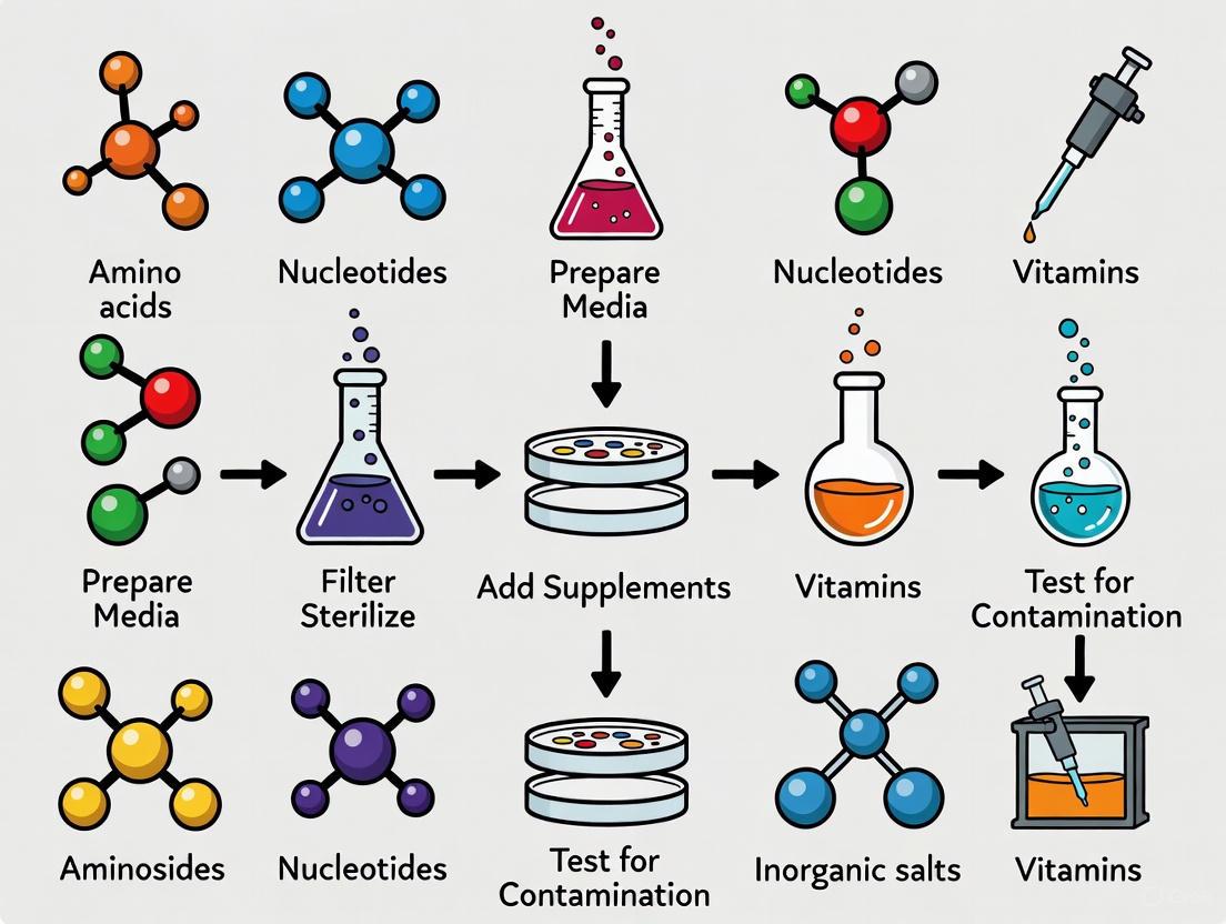
Core Principles of Good Cell Culture Practice (GCCP) for Media Preparation
The reproducibility of in vitro (cell-based) research is fundamentally dependent on the consistent quality of cell culture media [24]. Proper media preparation is not merely a preparatory step but a critical practice that directly influences cellular health, experimental validity, and the reliability of scientific data [25] [4]. Within the context of a broader thesis on contamination prevention, this document outlines how adherence to the core principles of Good Cell Culture Practice (GCCP) 2.0 can mitigate risks and enhance the quality of cell culture media preparation [24] [26]. The advanced, complex culture systems increasingly used in modern research demand more comprehensive quality management than ever before [24].
The GCCP 2.0 Framework and Media Preparation
Good Cell and Tissue Culture Practice 2.0 is an updated guidance document developed for practical use in the laboratory to assure the reproducibility of in vitro work [24] [26]. It is built upon six main principles that provide a framework for all aspects of cell culture, with direct implications for media preparation and contamination control [24]. The relationship between these principles and contamination prevention is illustrated below.
Essential Media Components and Potential Contaminants
Cell culture media is a complex mixture designed to provide a favorable artificial environment for cellular growth [25] [4]. The essential components can also be potential points of introduction for contaminants if not properly managed.
Table 1: Key Media Components and Associated Contamination Risks
| Component Category | Key Examples | Primary Function | Associated Contamination Risks |
|---|---|---|---|
| Inorganic Salts & Buffers | CaCl₂, KCl, MgSO₄, NaHCO₃, NaH₂PO₄ [4] | Maintain osmotic balance, pH, and provide essential ions [4] | Chemical contamination from impurities; microbial growth in stock solutions. |
| Amino Acids | L-Glutamine, L-Arginine, Glycine, L-Isoleucine [4] | Building blocks for protein synthesis [25] | Degradation products (e.g., ammonia from glutamine); microbial introduction. |
| Carbohydrates | Glucose, Galactose | Energy source [25] | Metabolic waste buildup (lactic acid); promotes microbial growth if contaminated. |
| Vitamins | B-group vitamins | Cofactors for enzymatic reactions [25] | Light-sensitive degradation; introduction of impurities. |
| Supplements | Serum (FBS), Growth Factors, Antibiotics | Provides hormones, lipids, and attachment factors [25] | High-risk source of mycoplasma, viruses, and prions; lot-to-lot variability. |
Detailed Protocol for GCCP-Compliant Media Preparation
This protocol is designed to align with GCCP principles, emphasizing documentation, quality control, and aseptic technique to prevent contamination.
Pre-Preparation Planning and Documentation (Principle 3)
- Review Standard Operating Procedure (SOP): Consult the laboratory's specific SOP for the media type (e.g., DMEM, RPMI-1640). Note any modifications from the basal formulation.
- Documentation Initiation: Complete the top section of the Media Preparation Record Sheet (See Section 6.1) before beginning work. This includes assigning a unique batch number.
- Reagent Verification: Confirm that all reagents and water (e.g., ultra-pure, endotoxin-free water) are within their expiration dates and have been stored correctly. Record reagent lot numbers.
Weighing and Dissolution (Principle 2)
- Workspace Preparation: Clean the weighing area and balance with 70% ethanol. Use dedicated, clean spatulas and weighing boats.
- Accurate Weighing: Weigh each powdered component precisely according to the formulation sheet. To prevent cross-contamination, use a fresh weighing boat for each ingredient.
- Dissolution: Transfer the powder to a clean, labeled flask. Add approximately 80% of the final volume of purified water at room temperature while stirring magnetically. Stir until completely dissolved. Avoid heat which can degrade heat-labile components.
Supplementation and pH Adjustment (Principle 1)
- Supplement Addition: Once the basal medium is fully dissolved, add any required supplements such as stable L-glutamine, sodium pyruvate, or HEPES buffer. If using heat-labile supplements (e.g., certain growth factors, antibiotics), wait until after filtration.
- pH Adjustment: Bring the medium to the final volume with purified water. Adjust the pH to the specified level (e.g., 7.2 - 7.4 for most mammalian cells) using sterile, filtered CO₂ or a sterile sodium bicarbonate solution. Note the final pH in the record sheet.
- Osmolality Check: Measure the osmolality of a small sample using an osmometer. Record the value and ensure it falls within the acceptable range for the cell type (typically 280-320 mOsm/kg for mammalian cells).
Sterilization by Filtration (Principle 4)
- Aseptic Setup: Perform all subsequent steps in a Class II biosafety cabinet that has been sanitized with 70% ethanol.
- Membrane Filtration:
- Assemble a sterile, disposable filtration unit with a 0.22 µm polyethersulfone (PES) membrane.
- Pre-wet the membrane with a small volume of sterile water or basal salt solution to reduce protein binding if serum is to be added.
- Pour the medium into the filter reservoir and apply positive pressure (or vacuum) to filter the medium into a sterile, receiving vessel.
- Final Supplementation: If using heat-labile supplements or serum (e.g., Fetal Bovine Serum), aseptically add them to the sterile, filtered medium. Gently mix by swirling.
Quality Control and Storage (Principle 2)
- Sterility Testing: Aseptically withdraw a 5-10 mL sample of the prepared media. Inoculate it into a suitable microbiological broth (e.g., Thioglycollate) or onto agar plates. Incubate at 37°C and 20-25°C for at least 72 hours and observe for microbial growth. Record results.
- Performance Testing: Before use for critical experiments, test the new media batch with a control cell line. Assess cell viability, growth rate, and morphology against a previous, validated batch.
- Labeling and Storage: Label the media container with the media name, unique batch number, date of preparation, expiration date (typically 3-4 weeks for serum-containing media at 2-8°C), and preparer's name. Store in the dark at 2-8°C.
The Scientist's Toolkit: Key Reagents and Materials
The following reagents and equipment are essential for implementing GCCP in media preparation and contamination prevention.
Table 2: Essential Research Reagent Solutions for GCCP-Compliant Media Preparation
| Item Name | Function/Application | GCCP Consideration |
|---|---|---|
| Powdered Basal Medium (e.g., DMEM, RPMI) | Foundation of the culture medium, providing inorganic salts, amino acids, and vitamins [4]. | Source from reputable suppliers; record lot numbers; store in a dry, dark environment. |
| Ultra-Pure Water | Solvent for all media components; must be pyrogen/endotoxin-free. | Use Type I water (e.g., from a Milli-Q system); regularly maintain and test the water purification system. |
| 0.22 µm PES Membrane Filter | Sterilization of the prepared medium by removal of bacteria and fungi [25]. | Pre-wet to reduce adsorption of critical components; do not exceed the recommended volume per filter unit. |
| Fetal Bovine Serum (FBS) | Common supplement providing growth factors, hormones, and attachment factors [25]. | High contamination risk; source from suppliers that perform rigorous viral and mycoplasma screening; heat-inactivate if required. |
| Detachment Agents (e.g., Trypsin, Accutase) | Passaging of adherent cells [25]. | Trypsin can degrade surface proteins; use milder agents (e.g., Accutase) for sensitive applications [25]. Filter sterilize all reagents. |
| Antibiotic-Antimycotic Solution | Suppression of bacterial and fungal growth [25]. | Use is controversial; may mask low-level contaminations. GCCP recommends limited use for primary culture only, not for routine sub-culturing [25]. |
| Mycoplasma Detection Kit | Routine testing for mycoplasma contamination, a common and insidious problem [25]. | Use as part of a regular quality control schedule for both cell stocks and prepared media batches. |
| pH Buffer Systems (e.g., NaHCO₃, HEPES) | Maintenance of physiological pH in the culture medium [4]. | The choice of buffer depends on CO₂ tension of the incubator. HEPES is useful for extra buffering capacity. |
Documentation and Quality Control Workflow
Consistent documentation is not merely administrative; it is a critical scientific and diagnostic tool that enables traceability and problem-solving.
Media Preparation Record Sheet
A comprehensive record sheet should be completed for every media batch prepared.
Table 3: Media Preparation and Quality Control Record Sheet
| Field | Details to Record |
|---|---|
| Media Type & Batch ID | DMEM/F-12, High Glucose; Batch: M-2025-001 |
| Date of Preparation | 21-Nov-2025 |
| Preparer's Name | [Researcher Name] |
| Component Lot Numbers | DMEM Powder: L12345, NaHCO₃: L54321, FBS: L98765 |
| Final Volume | 1000 mL |
| Final pH / Osmolality | 7.38 / 305 mOsm/kg |
| Filtration Details | 0.22 µm PES vacuum filter; Lot: F11223 |
| Supplementation Log | 10% FBS (v/v), 1x GlutaMAX |
| Sterility Test Result | Incubation initiated: 21-Nov-2025; Result (24-Nov): No growth. |
| Performance Test Note | HEK293 control cells: Doubling time ~24h, morphology normal. |
| Expiration Date | 15-Dec-2025 |
The workflow from preparation to quality control release ensures that every batch meets the required standards before being used in experiments.
Adherence to the core principles of GCCP 2.0 during media preparation is a fundamental pillar of reproducible and high-quality in vitro science [24]. By integrating rigorous characterization, robust quality management, and meticulous documentation into every step—from reagent selection to final quality control—researchers can significantly mitigate the risk of contamination [27] [25]. This structured approach not only safeguards precious cell cultures and experimental integrity but also strengthens the overall credibility and acceptance of scientific data generated in fields from basic research to drug development [28]. As cell culture technologies continue to evolve towards more complex 3D and microphysiological systems, the disciplined application of these principles will become even more critical [24] [29].
Practical Aseptic Protocols: Media Preparation and Sterile Handling Techniques
Within cell culture research, the preparation of sterile media is a foundational step upon which experimental validity rests. Contamination during media preparation can compromise years of research, leading to unreliable data and erroneous conclusions. This article details the application of rigorous biosafety cabinet (BSC) management and environmental monitoring protocols, framed within a broader thesis on preventing contamination in cell culture media preparation. The guidance is designed for researchers, scientists, and drug development professionals seeking to uphold the highest standards of aseptic technique and data integrity.
Regulatory Framework and BSC Fundamentals
Biosafety Cabinets are engineered containment devices vital for protecting both the product (e.g., cell culture media) and the personnel preparing it. The primary standard governing their design and performance is NSF/ANSI 49, specifically for Class II (laminar flow) BSCs, which are most common in media preparation workflows [30].
Class II BSCs provide personnel, product, and environmental protection through a combination of HEPA-filtered downward laminar airflow and an inflow air barrier at the front of the cabinet. This design minimizes the inherent hazards of working with agents assigned to biosafety levels 1, 2, or 3 [30]. Adherence to the current version of this standard (NSF/ANSI 49-2024) ensures reliable operation, structural stability, cleanability, and proper performance regarding noise, illumination, and vibration [30].
Recent Updates to NSF/ANSI 49
Staying current with standard revisions is critical for compliance and safety. Key updates in the 2024 edition include [30]:
- Power Failure Disconnection Time: Updated from 1 hour to 5 minutes, enhancing safety protocols.
- New Definitions: Added terms such as "cleanable," "easily cleanable," and "tubing restraint" for clearer interpretation.
- Chemical Resistance: Introduction of new testing language and updated requirements for material durability.
- Noise Level Tests: Addition of new language regarding acoustic performance.
- Filter Replacement: New guidelines for the use of replacement filters.
Biosafety Cabinet Management Protocols
Certification and Field Testing
A BSC must be professionally certified upon installation, annually thereafter, and after any relocation or repair [31]. This certification, performed against NSF/ANSI 49, verifies that the cabinet meets all critical performance criteria. Contracts with specialized certification companies are typically required to maintain this compliance [31].
Table 1: Key NSF/ANSI 49 BSC Field Certification Tests and Criteria
| Test Parameter | Purpose | Acceptance Criteria |
|---|---|---|
| Inflow Velocity | Ensure personnel protection by verifying adequate inward airflow. | Meets minimum velocity per cabinet type and standard [30]. |
| Downflow Velocity | Verify unidirectional laminar airflow for product protection. | Uniform and meets specified velocity requirements [30]. |
| HEPA Filter Integrity | Confirm no leaks in the supply and exhaust HEPA filters. | Prevents passage of particles ≥0.3 µm; no detectable leaks [30]. |
| Visible Aerosol/Mist Test | Visualize airflow patterns to check for turbulence or dead zones. | No penetration of containment barrier; proper airflow pattern over work zone [30]. |
| Noise Level | Ensure operational noise is within acceptable limits. | Conforms to specified decibel levels outlined in the standard [30]. |
Operational Procedures for Sterile Media Preparation
Proper operation is as important as proper certification. The following workflow and protocols are essential for maintaining sterility.
Pre-Use Procedures:
- Personal Protective Equipment (PPE): Always wear a buttoned lab coat and gloves as a minimum [31].
- BSC Decontamination: Before introducing any materials, all internal surfaces of the BSC must be decontaminated. A common and effective method is to use a 1:10 fresh bleach solution, followed by a 70% ethanol rinse to prevent corrosion of metal components [31].
- Supply Preparation: Gather all necessary supplies and surface-decontaminate them in a consistent manner before introducing them into the BSC to minimize the introduction of contaminants [31].
Work Practices Within the BSC:
- Movement: Use slow, deliberate, and linear (not radial) arm movements to minimize airflow disruption [31].
- Workflow: Maintain a clean-to-dirty workflow. Position clean media bottles and sterile utensils away from potentially contaminated items like waste containers [31].
- Sash Height: The BSC is designed to operate at a fixed sash height. Never raise the sash during operation, as this compromises the containment airflow [31].
- Clutter: Avoid overloading the work surface. Do not block the front or rear perforated grilles, as this negatively impacts airflow and containment [31].
Post-Use Procedures:
- Decontaminate Items: Before removing any items from the BSC, decontaminate their exterior surfaces [31].
- Final Decontamination: After all items and waste have been removed, decontaminate the entire interior work surface of the BSC [31].
Practices to Avoid
Certain common but incorrect practices can severely compromise sterility and safety:
- Avoid UV Lights as a Primary Decontamination Method: UV light has limited efficacy, as it only works on direct surfaces, and its germicidal function diminishes long before the blue light burns out. Good chemical disinfection is far more reliable [31].
- Do Not Use Volatile or Flammable Chemicals: BSCs are not designed to handle volatile chemicals, which can be recirculated into the room or create an explosion hazard [31].
- Eliminate Open Flames: Bunsen burners create turbulence that disrupts the protective airflow pattern and present a fire hazard. Use safer alternatives like a Bacticinerator for loop sterilization [31].
Environmental Monitoring and Verification
The Zone Concept for Environmental Monitoring
An Environmental Monitoring Program (EMP) is a systematic approach to validating the effectiveness of your sterile controls. The "Zone Concept" is a widely adopted model for structuring an EMP in a laboratory setting, adapted from food safety [32].
Table 2: Environmental Monitoring Zones for a Media Prep Laboratory
| Zone | Description | Example Locations | Recommended Test & Frequency |
|---|---|---|---|
| Zone 1 | Direct product contact surfaces. | Media dispenser nozzles, inside of sterile flasks/bottles, magnetic stir bars. | Sterility Test: Each media batch.Surface ATP Test: Weekly. |
| Zone 2 | Non-product contact surfaces in close proximity to Zone 1. | BSC work surface (outside immediate work area), BSC interior walls, equipment frames. | Surface Microbial Count: Weekly.Settle Plates: Weekly. |
| Zone 3 | Other surfaces in the open laboratory area. | Lab benchtops, door handles, incubator handles, fridge handles, shared centrifuge keys. | Surface Microbial Count: Monthly. |
| Zone 4 | Areas adjacent to the laboratory. | Hallways, storage rooms, offices. | Surface Microbial Count: Quarterly (for baseline). |
Verification of Sterilization Processes
For reusable equipment, verification and validation of sterilization are distinct but complementary processes [33].
- Verification: Confirms that the sterilizer reached the required physical parameters (e.g., temperature, pressure). This is typically done using chemical indicators (e.g., autoclave tape) that change color, providing instant feedback on a per-cycle basis [33].
- Validation: Confirms that the sterilization process was microbiologically effective. This uses biological indicators (e.g., spore tests like Attest) that are incubated post-cycle to confirm the kill of highly resistant microorganisms. Validation should be performed periodically (e.g., semi-annually) [33].
The Scientist's Toolkit: Essential Reagents and Materials
Table 3: Key Reagents and Materials for BSC Management and Contamination Control
| Item | Function/Application | Key Considerations |
|---|---|---|
| 70% Ethanol | Broad-spectrum disinfectant for surface decontamination and rinsing after bleach use. | Effective concentration for penetration; less corrosive than bleach; flammable [31]. |
| Sodium Hypochlorite (Bleach, 1:10 dilution) | Powerful oxidizing agent for high-level disinfection of surfaces. | Must be freshly diluted; corrosive to metals and requires an ethanol rinse after use [31]. |
| Chemical Indicators (e.g., Autoclave Tape) | Verification of sterilization cycle conditions (e.g., steam penetration, temperature). | Provides immediate, visible feedback for each cycle [33]. |
| Biological Indicators (e.g., Spore Strips/Amps) | Validation of sterilization process efficacy by confirming microbial kill. | Required for periodic (e.g., semiannual) validation of autoclaves [33]. |
| Adenosine Triphosphate (ATP) Monitoring Swabs | Cleaning verification test; measures residual organic matter on surfaces. | Provides rapid, quantitative data on cleaning effectiveness before HLD [34]. |
| Neutralizing Buffer / Letheen Broth | Used in environmental sampling sponges/swabs to inactivate common disinfectants. | Prevents residual sanitizers in samples from killing microbes and yielding false negatives [32]. |
| Sterile Sampling Sponges/Swabs | Aseptic collection of environmental samples from surfaces for microbial analysis. | Allows for standardized sampling of defined surface areas [32]. |
Cell culture media provides the essential nutritional and environmental support required for the survival, growth, and functionality of cells in vitro. Its formulation is a critical bridge between biology and chemistry, directly impacting experimental reproducibility, product yield in biomanufacturing, and the validity of scientific data [35]. A well-formulated medium must not only supply nutrients but also maintain a stable physicochemical environment, primarily through effective buffering systems, and be prepared with high-quality water to avoid introducing chemical contaminants. Within the broader context of contamination prevention research, every component of the media—from the basal nutrients to the water used for reconstitution—represents a potential vector for compromise. This document details the fundamental components of cell culture media, the principles of buffering systems, and the critical role of water quality, providing application notes and protocols to support researchers and drug development professionals in maintaining sterile and consistent culture conditions.
Core Components of Cell Culture Media
The foundation of any cell culture medium is its basal mixture, which is meticulously designed to mimic the natural environment of the cells as closely as possible. Understanding the function of each component is the first step in formulating effective media and troubleshooting culture problems.
Essential Components and Their Functions:
| Component Category | Specific Examples | Primary Function | Key Considerations for Contamination Prevention |
|---|---|---|---|
| Amino Acids | L-glutamine, essential amino acids (e.g., L-arginine, L-lysine) | Building blocks for protein synthesis; some serve as energy sources [35]. | Must be provided in sterile solutions; non-sterile ingredients like L-glutamine must be filter-sterilized before addition to the medium [36]. |
| Vitamins | B-complex vitamins (e.g., B12, Biotin), Vitamin C | Act as cofactors in enzymatic reactions; support cellular metabolism and antioxidative functions [35]. | Quality of raw materials is critical; contaminated supplements are a frequent source of microbial intrusion [6] [17]. |
| Inorganic Salts | NaCl, KCl, CaCl₂, MgSO₄, NaHCO₃ | Maintain osmotic balance and membrane potential; function as enzyme cofactors; participate in signal transduction [35]. | Sodium bicarbonate is a common buffer; its concentration directly affects CO₂ dependence and pH stability [37]. |
| Energy Sources | Glucose, Galactose | Primary fuel for cellular respiration and ATP generation [35]. | High concentrations can lead to metabolic acidosis; byproducts can shift media pH, requiring robust buffering [6]. |
| Serum/Supplements | Fetal Bovine Serum (FBS) | Provides growth factors, hormones, and binding proteins not in basal formulations [35]. | High-risk component for microbial (e.g., mycoplasma, viruses) and chemical contamination [6] [38]. Batch testing is essential. |
| Buffers | HEPES, Sodium Bicarbonate/CO₂ system | Maintain physiological pH by resisting changes from metabolic byproducts [35]. | Discussed in detail in Section 3. |
| Antibiotics/Antimycotics | Penicillin, Streptomycin, Amphotericin B | Prevent bacterial and fungal contamination [3] [35]. | Use is discouraged as it can mask poor aseptic technique; can lead to hidden contaminations and microbial resistance [3]. |
The trend in modern cell culture is moving towards Chemically-Defined (CD) Media, which offer critical advantages for contamination prevention and reproducibility. Unlike serum-containing media, CD media have fully disclosed components of known structure and concentration, eliminating the variability and contamination risks associated with undefined biological fluids like FBS [36]. Studies show that custom CD media, when properly formulated with specific attachment factors like fibronectin, can support robust growth of even sensitive cell types like Human Umbilical Vein Endothelial Cells (HUVECs), thereby enhancing the reliability of bioassays and translational research [36].
Buffering Systems in Cell Culture
The pH of the culture environment is a paramount factor affecting cell morphology, function, and viability. Most mammalian cell lines require a pH of approximately 7.4. As cells metabolize nutrients, they produce acidic byproducts (e.g., lactic acid and CO₂), which can cause the media pH to drop precipitously. Buffering systems are incorporated into media to resist these pH changes.
Types of Buffering Systems
There are two primary buffering mechanisms employed in cell culture:
The Bicarbonate/CO₂ System: This is the most common physiological buffer. It requires a specific equilibrium between the sodium bicarbonate in the medium and a controlled CO₂ atmosphere in the incubator (typically 5-10% CO₂). The chemical equilibrium is:
CO₂ (gas) + H₂O ⇌ H₂CO₃ ⇌ H⁺ + HCO₃⁻When cells produce excess acid (H⁺), the equilibrium shifts to the left, consuming CO₂ and mitigating the pH drop. The concentration of sodium bicarbonate dictates the required CO₂ tension [37]. This system is cost-effective and physiologically normal but is an "open" system dependent on a sealed incubator.Organic Buffers (e.g., HEPES): HEPES (4-(2-hydroxyethyl)-1-piperazineethanesulfonic acid) is a zwitterionic organic buffer effective in the physiological pH range (pKa ~7.3-7.5). It is often used at concentrations of 10-25 mM to provide additional buffering capacity in situations where the culture vessel is frequently opened, disrupting the CO₂ equilibrium [35]. HEPES is considered a "closed" system as it does not require a controlled atmosphere.
Experimental Data on Buffer and Media Component Interactions
Recent research underscores that the efficacy of many common agents, including disinfectants and buffers, can be significantly altered by the components of the cell culture media itself. This has direct implications for contamination control strategies.
Quantitative Impact of Media Components on Inactivating Agents (using Feline Calicivirus as a model) [19]:
| Inactivating Agent | Efficacy in EMEM (Δlog) | Efficacy in Distilled Water (Δlog) | Media Components that Reduce Efficacy | Media Components that Enhance Efficacy |
|---|---|---|---|---|
| SDS (0.5% w/v) | No inactivation | ≥ 4.03 | Inorganic Salts, Basic Amino Acids | - |
| DDAC (0.05% w/v) | ≥ 3.08 | ~2.00 | - | Inorganic Salts, Basic Amino Acids |
| Ethanol (70% v/v) | Consistent effect | 4.00 | Inorganic Salts | - |
| Sodium Hypochlorite (10 ppm) | Low effect | ≥ 4.03 | Basic & Neutral Amino Acids | - |
Table Footnote: Δlog represents the reduction in infectivity titer (log10). A higher Δlog value indicates a stronger inactivation effect. EMEM: Eagle's Minimum Essential Medium; SDS: Sodium Dodecyl Sulfate; DDAC: Didecyl Dimethylammonium Chloride.
This data highlights that media components like inorganic salts and amino acids can directly interfere with decontamination agents, either by protecting the contaminant (e.g., reducing SDS efficacy) or by consuming the active ingredient (e.g., free chlorine in NaClO is consumed by amino acids) [19]. This reinforces the need for rigorous cleaning and sterilization of equipment before it comes into contact with media or cells, as the presence of media residues can shield contaminants during decontamination procedures.
Diagram 1: A workflow for selecting and implementing a buffering system in cell culture media.
The Critical Role of Quality Water
Water is the largest component of any cell culture medium, constituting over 98% of the final solution in powdered media preparations [39]. Therefore, its purity is non-negotiable. Contaminants in water, such as ions, organic molecules, endotoxins, and microorganisms, can directly introduce chemical contamination, alter osmotic balance, inhibit cell growth, or lead to microbial contamination.
In continuous bioprocessing, water is a critical raw material and requires continuous monitoring [38]. The presence of trace chemical contaminants from water sources, including halogenated organic compounds or perfluoroalkyl substances (PFAS), can introduce variability and compromise data reproducibility, particularly in sensitive bioassays [36].
Protocol: Preparation of Cell Culture Media from Powder
This protocol ensures the preparation of sterile, high-quality culture media while minimizing the risk of contamination.
Research Reagent Solutions & Materials:
| Item | Function | Sterilization Requirement |
|---|---|---|
| Powdered Media | Provides basal nutrients, salts, and buffers. | Becomes sterile during filtration. |
| High-Purity Water | Solvent for all medium components; must be Type I ultrapure water (18.2 MΩ·cm). | Pre-sterilized or sterilized during filtration. |
| Sterile Filter Unit (0.22 µm) | Removes microorganisms and particulate matter from the final solution. | Pre-sterilized and single-use. |
| pH Meter & Standard Buffers | To accurately adjust the pH of the medium to the optimal range for the cell type. | Probe must be disinfected with 70% ethanol. |
| Sterile Bottles/Flasks | For storing the finished, sterile media. | Autoclaved or pre-sterilized single-use vessels. |
| Sodium Bicarbonate Solution | If not included in powder, it is added as a sterile stock solution to establish the bicarbonate buffer. | Filter-sterilized (0.22 µm). |
| Additional Supplements | e.g., L-Glutamine, Serum. | Added as sterile solutions after base media filtration. |
Step-by-Step Methodology:
- Dissolution: Add the powdered media to a volumetric flask containing approximately 90% of the final volume of high-purity water at room temperature (15-30°C). Stir gently but thoroughly until all components are completely dissolved. Avoid vigorous stirring to prevent foaming.
- pH Adjustment: Carefully adjust the pH of the solution using sterile 1N HCl or 1N NaOH. The target pH is typically 7.2 - 7.4 at room temperature. Note that the pH will rise approximately 0.2 - 0.3 units upon equilibration with 5% CO₂.
- Final Volume: Bring the solution to the final volume with high-purity water.
- Filtration Sterilization: Aseptically filter the medium through a 0.22 µm pore-size membrane filter into a pre-sterilized receiving vessel. This step is critical for removing all microbial contaminants.
- Supplement Addition: Aseptically add any heat-sensitive or pre-sterilized supplements (e.g., FBS, L-glutamine) to the filtered medium.
- Quality Control & Storage: Perform sterility checks by incubating an aliquot of the media at 37°C for 24-48 hours and observing for turbidity. Label the bottle with the media name, date of preparation, lot number, and initials. Store prepared media protected from light at 2-8°C for no longer than the formulation's specified shelf life, typically no more than 2 re-heatings [37] [36].
Integrated Contamination Prevention Workflow
A proactive, layered strategy is essential for preventing contamination, extending beyond media formulation to encompass all aspects of the cell culture process.
Diagram 2: A multi-layered strategy for comprehensive contamination prevention in cell culture.
As illustrated, contamination prevention is an integrated system. It begins with rigorous quality control (QC) of raw materials, including water and reagents [38]. Preparation must adhere to strict aseptic techniques, utilizing biosafety cabinets and sterile consumables [6] [3]. The process and environment must be controlled through cleanrooms, environmental monitoring, and the use of closed systems where possible [6] [38]. Finally, continuous monitoring through visual checks and specific tests like PCR for mycoplasma is essential for early detection and containment [6] [17] [3]. This holistic approach, where quality assurance is prioritized over mere quality control testing of final products, is the hallmark of a robust contamination prevention strategy in both research and GMP manufacturing [38].
In cell culture laboratories, preventing microbial contamination is a fundamental requirement for ensuring the integrity and reproducibility of research data. Antibiotics and antimycotics are critical tools used to maintain sterile conditions; however, their application must be guided by a precise understanding of their mechanisms, appropriate uses, and associated risks. The escalating global challenge of antimicrobial resistance (AMR), driven in part by misuse of these agents, underscores the need for stringent protocols in all settings, including research laboratories [40]. This document provides detailed application notes and experimental protocols for the proper use of antibiotics and antimycotics within the specific context of cell culture media preparation and contamination prevention, framed within a broader thesis on contamination control.
Fundamental Concepts and Global Context
Definitions and Key Distinctions
- Antibiotics: Medicines that fight bacterial infections by either killing bacteria or making it difficult for them to grow and multiply. They are ineffective against fungi and viruses [40].
- Antimycotics (Antifungals): Medicines that treat fungal infections by killing or stopping the growth of dangerous fungi [40].
- Antimicrobial Resistance (AMR): The ability of microorganisms (including bacteria and fungi) to survive and remain viable despite exposure to antimicrobial agents designed to kill them [41] [40].
The Scale of the Antimicrobial Resistance Threat
The misuse and overuse of antimicrobials in human medicine, veterinary practice, and agriculture are key drivers of AMR [41]. This is not merely a clinical concern; it represents a profound global public health crisis.
Table 1: Global Impact of Antimicrobial Resistance (AMR)
| Metric | Quantitative Data | Source/Context |
|---|---|---|
| Annual global deaths directly attributable to AMR (2019) | At least 1.27 million | [40] |
| Annual global deaths associated with AMR (2019) | Nearly 5 million | [40] |
| Projected annual global deaths by 2050 if unaddressed | 10 million | [41] |
| Annual AMR infections in the United States | More than 2.8 million | [40] |
| Annual U.S. deaths from AMR infections | More than 35,000 | [40] |
| Direct U.S. medical costs of fungal infections (2019) | $7.5 billion | [42] |
| One in six laboratory-confirmed bacterial infections globally were resistant to antibiotics (2023) | 16.7% | [43] |
The World Health Organization (WHO) has classified AMR as one of the top ten global public health threats [44]. The economic burden is substantial, with fungal infections alone costing U.S. healthcare $7.5 billion in direct medical costs in 2019 [42]. For researchers, this global context emphasizes the ethical and practical responsibility to use antimicrobial agents judiciously, even in a laboratory setting, to help mitigate the spread of resistance.
Core Mechanisms of Action and Resistance
Understanding how antibiotics and antimycotics work, and how microorganisms evade them, is crucial for their intelligent application in cell culture.
Mechanisms of Action of Major Antifungal Classes
In cell culture, antimycotics are frequently employed to prevent fungal overgrowth. The three main systemic classes target key fungal cellular structures.
Table 2: Major Classes of Antifungal Drugs and Their Mechanisms
| Antifungal Class | Prototype Agents | Primary Mechanism of Action | Primary Cellular Target |
|---|---|---|---|
| Azoles | Fluconazole, Voriconazole | Inhibition of ergosterol synthesis | Fungal cell membrane |
| Polyenes | Amphotericin B | Binding to ergosterol, forming membrane pores | Fungal cell membrane |
| Echinocandins | Caspofungin, Anidulafungin | Inhibition of β-(1,3)-D-glucan synthesis | Fungal cell wall |
The following diagram illustrates the molecular targets of these major antifungal drug classes within the fungal cell:
Mechanisms of Antimicrobial Resistance
Microbes employ several common strategies to develop resistance to antibiotics and antifungals. These mechanisms are highly relevant in a laboratory setting, where low-level, persistent use of these agents can select for resistant contaminants.
Table 3: Common Microbial Resistance Mechanisms [40]
| Resistance Mechanism | Description | Example in Bacteria | Example in Fungi |
|---|---|---|---|
| Restrict Drug Access | Changing entryways or limiting their number to reduce intracellular drug concentration. | Gram-negative bacteria using their outer membrane. | N/A |
| Drug Efflux | Using pumps in the cell wall to actively remove the drug. | Pseudomonas aeruginosa effluxing fluoroquinolones. | Some Candida species effluxing azoles. |
| Drug Inactivation | Producing enzymes that break down or modify the drug. | Klebsiella pneumoniae producing carbapenemases. | N/A |
| Target Modification | Altering the drug's binding site so it can no longer fit. | E. coli with mcr-1 gene modifying colistin target. | Aspergillus fumigatus modifying the cyp1A gene to resist triazoles. |
| Bypass | Developing new cell processes that avoid using the drug's target. | Some Staphylococcus aureus bypassing trimethoprim. | N/A |
Experimental Protocols for Contamination Control
Protocol: Aseptic Technique for Cell Culture Media Preparation
Principle: To prevent the introduction of microbial contaminants during the preparation and handling of cell culture media and reagents, forming the primary barrier against contamination [15].
Workflow Diagram:
Materials:
- Laminar Flow Hood (Biosafety Cabinet): Provides a sterile working environment [15].
- 70% Ethanol: For disinfecting work surfaces, gloves, and the outside of containers [15].
- Personal Protective Equipment (PPE): Lab coat, gloves, and safety glasses [15].
- Sterile Pipettes and Pipettor: For manipulating liquids without contamination [15].
- Pre-sterilized Reagents and Media: Ensure all components are sterile prior to use within the hood [15].
Procedure:
- Preparation: Clear and declutter the work area. Disinfect the surface of the laminar flow hood and all items to be placed inside it with 70% ethanol [15].
- Personal Hygiene: Wash hands thoroughly. Don appropriate PPE, including a lab coat and gloves. Wipe gloved hands with 70% ethanol before starting work [15].
- Hood Sterilization: Turn on the laminar flow hood and allow it to run for the recommended time. Wipe the interior work surface, walls, and any equipment with 70% ethanol. If available, expose the work surface to ultraviolet (UV) light for at least 15 minutes before use [15].
- Maintaining Sterile Field: Arrange all items neatly within the hood. Wipe the outside of all media bottles, flasks, and reagent containers with 70% ethanol before introducing them. Work deliberately and avoid rapid movements that can disrupt airflow. Never leave sterile containers (e.g., bottles, petri dishes) uncovered for extended periods. If a cap must be placed down, position it with the open face down on the disinfected surface [15].
- Sterile Handling: Use only sterile glass or disposable plastic pipettes. Use each pipette only once to avoid cross-contamination. Never pour media from one sterile container to another; always use a pipette. Be careful not to touch the pipette tip to any non-sterile surface, including the threads of bottles [15].
- Completion: Immediately cap all containers after use. Label prepared media with the contents and date of preparation. Store multi-well plates in sterile, resealable bags. Store media at the recommended temperature (e.g., 4°C for short-term storage) [15].
Protocol: Decision Framework for Antibiotic/Antimycotic Use in Cell Culture
Principle: To provide a logical, step-by-step guide for determining when and how to incorporate antimicrobial agents into cell culture media, prioritizing aseptic technique as the primary defense.
Decision Logic Diagram:
Procedure:
- Assess the Need: The use of antibiotics and antimycotics should be the exception, not the rule. For routine maintenance of established, uncontaminated cell lines, prophylactic use is not recommended as it can mask poor aseptic technique and select for resistant organisms [45] [46].
- Consider Prophylactic Use in Specific Scenarios:
- During the establishment of primary cultures, which have a higher risk of harboring contaminants.
- During complex, lengthy, or high-risk manipulations (e.g., transfection, long-term live imaging) where the risk of contamination is temporarily elevated.
- When a specific, known contaminant is common and difficult to eliminate by other means (guided by experimental history).
- Select the Appropriate Agent:
- Broad-spectrum antibiotics: A combination of penicillin/streptomycin (Pen-Strep) is commonly used to target a wide range of Gram-positive and Gram-negative bacteria.
- Antimycotics: Agents like Amphotericin B are used to prevent fungal contamination.
- Determine Correct Concentration and Duration:
- Always use antimicrobials at the minimum effective concentration as specified by the manufacturer or established literature for your cell type.
- If used prophylactically, consider limiting the duration. Once the culture is established and stable, attempt to wean the cells off antimicrobials by maintaining them in antibiotic-free media to reduce selective pressure.
- Response to Confirmed Contamination:
- Do not automatically add high-dose antibiotics/antimycotics to a contaminated culture in an attempt to "rescue" it. This is rarely successful and poses a significant biohazard.
- The standard and safest protocol is to discard the contaminated culture by autoclaving.
- Decontaminate the work area, incubator, and any equipment that may have been exposed thoroughly with 70% ethanol or other appropriate disinfectants [15] [47].
The Scientist's Toolkit: Essential Reagents and Materials
Table 4: Key Research Reagent Solutions for Contamination Control
| Item | Function/Application in Contamination Control |
|---|---|
| Laminar Flow Hood | Provides a HEPA-filtered, sterile air environment for handling cell cultures and preparing media, serving as the primary physical barrier to contamination [15]. |
| 70% Ethanol Solution | A standard disinfectant used for wiping down work surfaces, equipment, gloves, and the exterior of containers to reduce microbial load [15]. |
| Penicillin-Streptomycin (Pen-Strep) | A common antibiotic solution used prophylactically in cell culture media to prevent bacterial growth. It is broad-spectrum, targeting both Gram-positive and Gram-negative bacteria. |
| Amphotericin B | An antimycotic agent used to prevent fungal and yeast contamination in cell cultures. It is often used in combination with antibiotics. |
| Sterile Serological Pipettes | Disposable, pre-sterilized pipettes for transferring liquid media and reagents without introducing contaminants. Designed for use with a pipettor [15]. |
| Sterile Filters (0.22 µm) | Used for filter-sterilizing heat-labile solutions (e.g., some growth factors, enzymes) that cannot be autoclaved, effectively removing bacteria and fungi from the liquid. |
Table 5: Comprehensive Analysis of Antimicrobial Use in Cell Culture
| Aspect | Antibiotics | Antimycotics |
|---|---|---|
| Benefits | - Can prevent the loss of valuable cultures from bacterial contamination.- Useful as a short-term prophylactic during high-risk procedures. | - Effective at preventing overgrowth by fungi and yeasts.- Can be critical for working with environmental samples prone to fungal load. |
| Limitations | - Ineffective against viral, fungal, and mycoplasma contaminants.- Can be toxic to certain eukaryotic cell types at high concentrations. | - Some formulations (e.g., polyenes) can be toxic to mammalian cells.- Ineffective against bacterial and viral contaminants. |
| Risks | - Masks poor technique: Can lead to a false sense of security and neglect of fundamental aseptic methods [15] [47].- Promotes resistance: Sub-lethal concentrations select for resistant bacteria, which can be a source of persistent, hard-to-treat contamination and contribute to the broader AMR problem [45] [40].- Can hide mycoplasma: Low-level bacterial contamination may be suppressed but not eliminated, masking mycoplasma infections which are unaffected by standard antibiotics [46]. | - Similar risk of promoting selection for resistant fungal strains, such as azole-resistant Aspergillus fumigatus or Candida auris [42] [44].- Cell toxicity can interfere with experimental outcomes unrelated to contamination. |
The proper use of antibiotics and antimycotics in cell culture is a discipline that balances immediate practical benefits against long-term risks, including the contribution to the global AMR crisis. The cornerstone of contamination prevention remains rigorous and unwavering aseptic technique [15] [47]. Antimicrobial agents should be viewed as specialized tools for specific, justified circumstances—not as a universal substitute for sterile practice. By adhering to the protocols and principles outlined in this document, researchers can protect their experiments, their laboratory environment, and public health, ensuring the integrity of scientific research in drug development and beyond.
In cell culture research, the prevention of contamination is not merely a procedural step but the foundational determinant of experimental integrity. Successful cell culture depends heavily on keeping cells free from contamination by microorganisms such as bacteria, fungi, and viruses [15]. Aseptic technique represents a set of rigorous procedures designed to create an impermeable barrier between microorganisms in the environment and the sterile cell culture, thereby preserving sample purity and ensuring reproducible results [15]. Within the specific context of cell culture media preparation—where even a single contaminant can compromise months of research—mastering the trifecta of pipetting, container handling, and personal protective equipment (PPE) usage becomes paramount. These techniques form the essential defense system protecting valuable cultures and reagents from biological contaminants that can alter growth patterns, compromise viability, and ultimately invalidate research findings [15]. This guide provides detailed application notes and protocols specifically framed within contamination prevention research for drug development professionals and scientists.
Fundamental Principles of Aseptic Technique
Defining the Aseptic Barrier Concept
Aseptic technique operates on the principle of establishing and maintaining a protective barrier between sterile materials and non-sterile environments. Unlike sterile technique, which aims to ensure a space is completely free of all microorganisms, aseptic technique focuses on not introducing contamination to a previously sterilized environment [15]. In practical terms, this means that while your cell culture hood might be sterilized using sterile techniques initially, using aseptic techniques maintains its sterility throughout your experimental procedures [15]. This distinction is crucial for researchers to understand, as it emphasizes the ongoing vigilance required throughout every manipulation rather than relying solely on initial sterilization.
The elements of aseptic technique comprise four interconnected pillars: a sterile work area, good personal hygiene, sterile reagents and media, and sterile handling [15]. These components work synergistically to create multiple layers of protection against contamination. When one layer is compromised, the others provide backup protection, creating a robust system for maintaining sterility throughout media preparation and cell culture procedures.
Aseptic Technique Workflow
The following diagram visualizes the integrated relationship between the core components of aseptic technique in a cell culture environment:
Personal Protective Equipment: The First Line of Defense
PPE Selection and Regulatory Standards
Personal protective equipment serves a dual purpose in the cell culture laboratory: protecting the researcher from hazardous materials and protecting the cell cultures from human-associated contaminants [15]. PPE forms an immediate protective barrier between personnel and hazardous agents using items such as gloves, laboratory coats and gowns, shoe covers, respirators, face shields, safety glasses, or goggles [15]. The U.S. Food and Drug Administration regulates PPE intended for medical use, requiring that it meets specific consensus standards for protection, including barrier performance, resistance to tears and snags, and in some cases, sterility [48]. For cell culture work specifically, the minimum PPE requirements typically include gloves, laboratory coats or gowns, and sometimes eye protection or masks, depending on the biosafety level of the materials being handled [15].
Protocol: Proper Donning and Doffing of PPE
Principle: Create an effective personnel barrier that minimizes shed skin, hair, and other potential contaminants while ensuring researcher safety.
Materials:
- Laboratory coat (preferably dedicated for cell culture work)
- Appropriate gloves (nitrile preferred over latex due to allergy concerns)
- Safety glasses or eye protection (if working with splashing hazards)
- Mask (if required by institutional policy or when working with respiratory pathogens)
Procedure:
- Hand Hygiene: Wash hands thoroughly with soap and water for at least 20 seconds, following the World Health Organization recommended technique [49].
- Laboratory Coat: Don a clean laboratory coat, ensuring all buttons/fasteners are secured and sleeves cover arms completely.
- Glove Inspection: Inspect gloves for any visible tears, holes, or defects. Discard any compromised gloves.
- Gloving Technique: Don gloves using proper technique, pulling cuff over the wrist area of the lab coat.
- Pre-Hood Disinfection: Wipe gloved hands with 70% ethanol before entering the laminar flow hood area [15].
- Glove Changes: Change gloves immediately if they become contaminated, torn, or after touching non-sterile surfaces [15].
- PPE Removal: When exiting the culture area, remove gloves first using the "glove-in-glove" technique, followed by the lab coat, and finally perform hand hygiene.
Table: PPE Applications and Specifications for Cell Culture Work
| PPE Item | Primary Function | Quality Standards | Contamination Prevention Role |
|---|---|---|---|
| Gloves | Barrier against hand-borne contaminants | FDA regulations for medical devices [48] | Prevents transfer of microorganisms from hands to culture and vice versa |
| Lab Coat/Gown | Protection from particulate shedding | Fluid-resistant material | Contains skin cells and hair, reduces shed particles |
| Eye Protection | Shield from splashes | ANSI standards for impact resistance | Precludes contamination from reflexive face-touching |
| Mask/Respirator | Respiratory containment | FDA clearance for medical masks [48] | Blocks microorganisms expelled via breathing/talking |
Pipetting Techniques for Contamination Control
Principles of Sterile Pipetting
Proper pipetting technique represents one of the most frequently performed aseptic manipulations in cell culture work, with each transfer representing a potential contamination event. The fundamental principle of sterile pipetting is maintaining the sterility of the pipette tip and any sterile surface it contacts throughout the procedure. According to aseptic technique guidelines, researchers should "use sterile glass or disposable plastic pipettes and a pipettor to work with liquids, and use each pipette only once to avoid cross contamination" [15]. This single-use principle is critical for preventing the transfer of contaminants between different reagent bottles, media stocks, and cell culture vessels.
Protocol: Aseptic Pipetting for Media Preparation
Principle: Transfer sterile liquids without introducing microbial, chemical, or cross-contamination between different reagents and media.
Materials:
- Sterile, disposable plastic pipettes of appropriate volumes
- Pipette aid or controller
- Sterile reagent bottles
- 70% ethanol spray or wipes
- Waste container for used pipettes
Procedure:
- Pipette Inspection: Verify pipette packaging is intact and sterile. Inspect for any damage before opening.
- Packaging Opening: Open sterile pipette packaging within the laminar flow hood, taking care not to touch the tip or shaft to any non-sterile surface [50].
- Pipette Aid Attachment: Carefully attach the pipette to the pipette aid without touching the pipette tip to the pipette aid itself.
- Bottle Access: Loosen, but do not completely remove, caps from reagent bottles. When removing caps, place them with the inner (sterile) surface facing up if they must be placed on the work surface [15] [50].
- Liquid Aspiration:
- Hold the bottle at a slight angle during pipette insertion
- Avoid touching the pipette tip to the neck or threads of the bottle
- Aspirate slightly less than the maximum volume of the pipette to prevent fluid contact with the pipette aid
- Liquid Dispensing:
- Hold the receiving vessel at an angle to facilitate smooth liquid dispensing without splashing
- Dispense along the inner wall of the vessel to minimize aerosol generation
- Avoid touching the pipette tip to any non-sterile surface, including the exterior of the receiving vessel
- Pipette Disposal: Discard used pipettes directly into an appropriate sharps or biohazard container—never reuse disposable pipettes [15].
- Recapping: Immediately recap bottles after use to maintain sterility of remaining contents.
Table: Common Pipetting Errors and Contamination Risks
| Error | Contamination Risk | Prevention Strategy |
|---|---|---|
| Reusing disposable pipettes | Cross-contamination between reagents | Use each sterile pipette only once [15] |
| Touching pipette tip to bottle threads | Introduction of environmental contaminants | careful insertion without contact with non-sterile surfaces |
| Over-aspiration into pipette aid | Microbial growth in pipette aid & aerosol generation | Aspirate less than maximum volume |
| Slow, deliberate movements | Increased exposure to airborne contaminants | Work efficiently but deliberately [15] |
| Uncovered vessels during procedures | Settlement of airborne contaminants | Cap vessels immediately after use [15] |
Sterile Container Handling Protocols
Principles of Container Integrity Maintenance
Maintaining the sterility of culture vessels, reagent bottles, and media containers requires meticulous attention to handling procedures throughout all experimental workflows. The guiding principle is that any sterile surface—whether the inner surface of a flask, the inside of a cap, or the rim of a bottle—must never contact non-sterile surfaces [15]. Even momentary contact can transfer microorganisms capable of proliferating in nutrient-rich culture media. As explicitly stated in aseptic technique guidelines, researchers should "never uncover a sterile flask, bottle, petri dish, etc. until the instant you are ready to use it and never leave it open to the environment" [15]. This practice minimizes the time during which sterile surfaces are exposed to potential contaminants.
Protocol: Sterile Container Management
Principle: Maintain sterility of culture vessels, media bottles, and reagent containers throughout handling procedures to prevent introduction of contaminants.
Materials:
- Culture flasks, plates, or dishes
- Media and reagent bottles
- 70% ethanol spray or wipes
- Permanent laboratory marker
- Sterile wraps or bags for storage
Procedure:
- Surface Decontamination: Wipe the outside of all bottles, flasks, and plates with 70% ethanol before placing them in the cell culture hood [15].
- Labeling: Pre-label all containers with necessary information (date, contents, passage number, etc.) before beginning sterile work to avoid handling markers inside the hood.
- Cap Removal:
- Container Handling:
- Hold vessels at an angle that minimizes horizontal exposure to airborne contaminants
- Avoid breathing or talking directly over open containers [15]
- Work efficiently to minimize exposure time
- Recapping:
- Replace caps as soon as procedures are completed
- Ensure caps are properly sealed to maintain sterility during incubation
- Storage:
- Store multi-well plates in sterile re-sealable bags when not in use [15]
- Ensure all containers are properly sealed before removal from the biosafety cabinet
- Integrity Checking: Before use, inspect all containers for cracks, compromised seals, or other damage that might affect sterility.
Integrated Workflow: Media Preparation with Contamination Control
Comprehensive Aseptic Protocol
Principle: Execute a complete media preparation workflow while maintaining multiple barriers against contamination through coordinated PPE usage, pipetting technique, and container handling.
Materials:
- Personal protective equipment (as detailed in Section 3)
- Sterile pipettes and pipette aid
- Media components and supplements
- Sterile collection vessel
- 70% ethanol and wipes
- Permanent marker
Procedure:
Quality Control and Contamination Monitoring
Principle: Implement systematic quality control measures to detect contamination early and prevent use of compromised media in critical experiments.
Procedure:
- Visual Inspection:
- Sterility Testing:
- Reserve a small aliquot of prepared media for sterility testing
- Incubate test aliquot alongside experimental cultures
- Monitor for signs of contamination before using media for important experiments
- Documentation:
- Record preparation date, components, and lot numbers
- Note quality control observations and any deviations from protocol
- Document storage conditions and expiration dates
Table: Essential Research Reagent Solutions for Aseptic Media Preparation
| Reagent/Equipment | Function in Contamination Prevention | Application Notes |
|---|---|---|
| 70% Ethanol | Surface decontamination through protein denaturation | Wipe work surfaces and container exteriors; effective concentration for microbial control [15] |
| Sterile Disposable Pipettes | Aseptic liquid transfer without cross-contamination | Single-use only; prevent contact with non-sterile surfaces [15] |
| Laminar Flow Hood | HEPA-filtered unidirectional airflow creating sterile work zone | Run continuously; position items without blocking airflow; minimize movement disruptions [15] |
| Autoclave | Sterilization of heat-stable equipment and solutions | Standard sterilization method for laboratory-prepared reagents [51] |
| Sterile Filtration Systems | Sterilization of heat-labile solutions | 0.22μm pore size for complete microbial removal from solutions |
| Personal Protective Equipment | Barrier against human-associated contaminants | Worn consistently; changed when contaminated [15] |
Mastering the integrated techniques of proper pipetting, sterile container handling, and appropriate PPE usage creates a robust defense against contamination in cell culture media preparation. When executed consistently and meticulously, these procedures preserve the integrity of precious cell lines, ensure reproducibility of experimental results, and ultimately uphold the scientific validity of drug development research. As emphasized throughout these application notes, success in aseptic technique lies not only in understanding individual procedures but in recognizing how these elements work synergistically to create multiple layers of protection against contamination. Through diligent application of these protocols and continuous attention to technique refinement, researchers can significantly reduce contamination events and advance the reliability of their cell culture-based research.
Maintaining the integrity of reagents and culture media through optimized storage practices is a critical foundation for successful cell culture research. Proper handling and storage are fundamental to preventing contamination, preserving biochemical stability, and ensuring experimental reproducibility [52] [53]. This application note details evidence-based protocols for implementing effective aliquot systems and maintaining stability, directly supporting contamination prevention strategies in cell culture media preparation.
The Critical Role of Aliquot Systems
Aliquotting—the process of dividing bulk reagents into smaller, single-use volumes—is a primary defense against contamination and reagent degradation. This system minimizes repeated exposure to non-sterile environments, limits the number of freeze-thaw cycles, and preserves the stability of sensitive components [53] [54].
Primary Benefits:
- Contamination Risk Reduction: Using single-use aliquots prevents the entire stock from being compromised if one working container becomes contaminated during use [54].
- Stability Preservation: Sensitive components, such as growth factors, vitamins, and glutamine, are protected from repeated exposure to temperature fluctuations and oxidative stress [53].
- Process Consistency: Aliquots ensure consistent composition and performance across experiments conducted over time, enhancing data reproducibility [55].
Table 1: Reagents Requiring Aliquot Systems and Recommended Storage Conditions
| Reagent Category | Recommended Aliquot Volume | Storage Temperature | Stability Duration | Key Stability Concerns |
|---|---|---|---|---|
| Fetal Bovine Serum (FBS) | 5 - 50 mL [53] | -20°C to -80°C (frozen) [53] | Refer to manufacturer's date; avoid >1 week at 4°C [53] | Loss of potency, microbial growth, formation of precipitates [53] |
| L-Glutamine | Single-experiment volume | -20°C [4] | Degrades rapidly at 4°C or 37°C [4] | Breakdown to ammonia and pyrrolidonecarboxylic acid [4] |
| Antibiotics | Single-experiment volume | -20°C (typically) | Varies by compound | Potential loss of efficacy with repeated freeze-thaw cycles |
| Trypsin/EDTA | Single-experiment volume | -20°C | Varies by formulation | Proteolytic autodegradation |
| Complete Liquid Media | Weekly usage volume | 4°C (short-term) [53] | 2-4 weeks after opening [53] | pH shift (color change), nutrient degradation, microbial contamination [53] |
Comprehensive Storage and Stability Protocols
Sourcing and Quality Control
Begin with high-quality raw materials. For fetal bovine serum (FBS), sourcing from countries with stringent regulations (e.g., the United States, Australia, New Zealand) correlates with lower variability in protein concentrations and improved performance consistency [53]. Conduct upstream testing by reserving specific lots from vendors and performing small-scale tests to ensure the product meets experimental needs before finalizing bulk purchases [53].
Temperature Management and Stability Monitoring
- Short-Term Storage (2–8°C): Liquid media and reagents stored at 4°C must be protected from light and atmospheric CO2 to prevent pH drift. Use sealed containers or CO2-independent buffers to maintain pH stability [56].
- Long-Term Cryogenic Storage (-80°C to -196°C): For long-term viability, cells should be frozen at a controlled rate of approximately -1°C per minute using specialized freezing containers and stored in the vapor phase of liquid nitrogen [57] [56]. Serum loses potency over time when stored at 4°C and should be kept frozen at -20°C to -80°C when not in use [53].
- Stability Monitoring: Visually inspect media for crystal formation, which may indicate high salt content, or turbidity, suggesting microbial contamination [53]. Use phenol red, a common pH indicator in most media, to monitor for acidification (yellow color) indicating metabolite buildup or contamination [57].
Experimental Protocol: Implementing a Media Aliquot System
Principle: To extend the shelf-life of liquid media, preserve nutrient integrity, and prevent microbial contamination by minimizing repeated handling of the primary stock bottle.
Materials:
- Sterile liquid culture media (e.g., DMEM, RPMI-1640)
- Sterile serological pipettes (e.g., Corning)
- Sterile conical centrifuge tubes (e.g., Corning Falcon tubes) or media bottles
- Permanent, ethanol-resistant lab marker
- Personal Protective Equipment (PPE): lab coat, gloves
- Biosafety cabinet
- 70% ethanol spray
- Refrigerator (4°C) and freezer (-20°C)
Procedure:
- Preparation: Disinfect the biosafety cabinet and all materials, including the media bottle, with 70% ethanol. Wear appropriate PPE and work aseptically within the cabinet [52] [54].
- Inspection: Examine the media for any signs of precipitation, unusual color, or turbidity. Do not use if contamination is suspected [57] [53].
- Aliquotting: a. Gently swirl the media bottle to ensure a homogenous solution. b. Using a sterile serological pipette, aseptically transfer the desired volume (e.g., 50 mL) into sterile, labeled tubes or bottles. c. Tightly close the caps on all aliquot containers.
- Sealing and Labeling: a. For added protection against gas exchange, seal the caps of the aliquots with Parafilm to create a better seal [53]. b. Clearly label each aliquot with the reagent name, date of aliquoting, preparation date, and user initials.
- Storage: Place the aliquots at 4°C, protected from light. The master stock bottle should also be resealed, Parafilmed, and returned to 4°C [53].
- Usage: Use individual aliquots on a first-in, first-out basis. Once an aliquot is opened, it should be used promptly. Discard any aliquot if contamination is suspected.
Experimental Protocol: Cryopreservation of Cell Stocks for Long-Term Stability
Principle: Create secure master and working cell banks by freezing cells at a controlled rate in the presence of cryoprotective agents to maintain genetic stability and viability over the long term [57] [55].
Materials:
- Cells in the logarithmic growth phase (>90% viability)
- Appropriate growth medium
- Cryoprotective Agent (CPA): e.g., DMSO (5-10%) or pre-prepared commercial solutions (e.g., Bambanker)
- Programmable freezer or isopropanol freezing container (e.g., Corning CoolCell)
- Cryogenic vials
- Centrifuge
Procedure:
- Cell Preparation: Harvest cells in their log-growth phase using a gentle dissociation method. Count and confirm high viability (>90%) [57] [56].
- Centrifugation and Resuspension: Pellet the cells by gentle centrifugation (e.g., 200 x g for 5 minutes). Resuspend the cell pellet at a high density (e.g., 1-2x10^6 cells/mL for adherent cells) in a freezing medium containing the chosen CPA [57]. DMSO is common, but its toxicity to some cell types should be considered [57].
- Aliquotting: Dispense the cell suspension into cryovials, labeled with cell line, passage number, date, and concentration.
- Controlled-Rate Freezing: Place the cryovials in an isopropanol freezing container and immediately transfer it to a -80°C freezer for 24 hours. This apparatus ensures a cooling rate of approximately -1°C per minute, which is critical for cell survival [57] [56].
- Long-Term Storage: After 24 hours, promptly transfer the vials to a liquid nitrogen tank for long-term storage in the vapor phase (-136°C to -196°C) [57].
The Scientist's Toolkit: Essential Reagent Solutions
Table 2: Key Research Reagent Solutions and Their Functions
| Item | Primary Function | Application Notes |
|---|---|---|
| Corning Cryogenic Vials | Long-term storage of cell stocks at ultra-low temperatures [56] | Certified for leak-free storage in liquid nitrogen; ensures sample integrity [56]. |
| Corning CoolCell | Provides a consistent, controlled freezing rate of -1°C/min [57] [56] | Eliminates the need for manual alcohol handling; ensures reproducible cell freezing. |
| DMSO (Dimethyl Sulfoxide) | Cryoprotective agent; prevents intracellular ice crystal formation [57] | Can be toxic to some cells; use at appropriate concentrations (e.g., 5-10%). |
| Pre-prepared Cryoprotectants (e.g., Bambanker) | Ready-to-use cell freezing media [57] | Offers formulation consistency and convenience; reduces preparation errors. |
| Sterile Serological Pipettes | Aseptic transfer of media and reagents during aliquotting [52] | Single-use to prevent cross-contamination. |
| Sterile Falcon Tubes | Short-term storage of media and reagent aliquots at 4°C [56] | Maintains sterility; minimizes risk of chemical and physical stress on stored liquids. |
| Parafilm | Creates an airtight seal around bottle and tube caps [53] | Helps prevent pH drift and gas exchange in stored media. |
Workflow Visualization
Diagram 1: Reagent and Media Storage Workflow. This diagram outlines the key steps from initial quality control to final usage, highlighting critical contamination control points within the aliquotting and storage process.
Diagram 2: Stability Challenges and Monitoring Framework. This diagram logically relates the primary factors that compromise reagent stability to the essential monitoring actions required for proactive management.
Implementing rigorous aliquot systems and adhering to precise storage protocols are not merely procedural tasks; they are fundamental to ensuring the validity and reproducibility of cell-based research. By systematically managing reagent and media stability, researchers can significantly mitigate the risk of contamination, reduce experimental variables, and protect valuable time and resources. These practices form an essential component of a comprehensive quality management system in any research or drug development laboratory.
Contamination Identification and Resolution: From Detection to Decontamination
Cell culture contamination poses a significant threat to research integrity and biomanufacturing in the pharmaceutical industry, potentially leading to unreliable data, costly setbacks, and health hazards [6] [58]. Visual identification of contamination through microscopic analysis provides the first line of defense, enabling researchers to detect early signs of compromise in cell cultures. This guide details standardized protocols for identifying common contamination indicators—turbidity, pH shifts, and morphological changes—within the broader context of contamination prevention research. The procedures outlined support the critical need for rapid, accessible monitoring techniques that complement advanced diagnostic methods, such as machine learning-assisted UV spectroscopy [21] and automated image analysis [59], ensuring culture purity and data reproducibility.
Visual Indicators of Contamination: A Comparative Analysis
Recognizing the primary visual indicators of contamination is fundamental to maintaining cell culture integrity. The following table summarizes key characteristics and their biological significance.
Table 1: Visual Indicators of Cell Culture Contamination
| Visual Indicator | Description | Common Contaminant Associations | Biological Significance |
|---|---|---|---|
| Increased Turbidity | Cloudiness or haziness in the culture medium; visible as a granular or misty appearance under microscopy [58]. | Bacteria, Yeasts [6] [58] | Indicates rapid microbial proliferation, leading to increased light scattering from suspended cells [58]. |
| pH Shifts | Color change in the culture medium (e.g., from red to yellow) due to metabolic byproducts [58]. | Bacteria, Fungi [6] [58] | Results from acid production (pH drop) from carbohydrate metabolism or alkaline byproducts in some bacterial infections [58]. |
| Morphological Changes | Alterations in host cell size, shape, or structure; includes cellular enlargement or nuclear expansion [59]. | Viral infection, Mycoplasma, Cross-contamination [6] | Reflects underlying stress, metabolic disruption, or cytopathic effects from intracellular pathogens [59]. |
| Floating Clumps/ Filaments | Visible fungal mycelia or yeast clumps suspended in the medium [58]. | Molds (e.g., Aspergillus), Yeasts (e.g., Candida) [6] [58] | Direct evidence of fungal colonization and overgrowth in the culture environment. |
Experimental Protocols for Contamination Identification
Protocol 1: Macroscopic and Microscopic Assessment of Turbidity
This protocol provides a method for detecting microbial contamination through visual and microscopic inspection of culture turbidity.
Principle: Microbial growth increases the particulate load in the culture medium, leading to light scattering that manifests as cloudiness. This can be assessed macroscopically and confirmed under microscopy [58].
Materials:
- Cell culture flask/plate
- Inverted brightfield microscope
- Biosafety cabinet
- Personal protective equipment (gloves, lab coat) [58]
Procedure:
- Macroscopic Inspection: Inside a biosafety cabinet, gently swirl the culture vessel and observe the medium against a white background. Look for any cloudiness, haziness, or suspended particles. Record observations.
- Microscopic Confirmation:
- Place the culture vessel on the stage of an inverted microscope.
- Begin observation at a low magnification (e.g., 40x or 100x) to scan for large clumps or filaments.
- Switch to a higher magnification (200x or 400x) to identify individual microbial cells. Bacteria may appear as small, vibrating spheres or rods, while yeast cells are typically larger, oval-shaped, and may be budding.
- Interpretation: Cloudiness under macroscopic view that correlates with the presence of motile, non-mammalian cells under microscopy indicates microbial contamination. The culture should be discarded following biosafety guidelines [6].
Protocol 2: Monitoring pH Shifts via Colorimetric Change
This protocol standardizes the detection of metabolic contamination through shifts in culture medium pH.
Principle: Phenol red, a common pH indicator in culture media, shifts from red (pH ~7.4) to yellow (acidic, pH <7.0) due to carbohydrate fermentation by contaminants, or to purple (alkaline) under certain conditions [58].
Materials:
- Cell culture with phenol red-containing medium
- Standard color chart for phenol red
Procedure:
- Color Assessment: Compare the color of the culture medium to a standardized phenol red color chart under consistent lighting.
- Documentation: Record the color and the corresponding estimated pH value.
- Correlation: Correlate the pH shift with other visual indicators. A rapid yellow shift (acidification) often accompanies bacterial contamination, while a slower change may indicate other issues [58].
- Action: A significant, unexplained pH shift is a strong preliminary indicator of contamination and should trigger further investigation or culture disposal.
Protocol 3: High-Content Analysis of Host Cell Morphology
This protocol uses high-resolution microscopy to quantify infection-induced morphological changes in host cells, capturing heterogeneity often missed in routine checks [59].
Principle: Intracellular pathogens can induce significant alterations in host cell and nuclear morphology, which can be quantified to assess infection burden and physiological state [59].
Materials:
- Fluorescence microscope (widefield or confocal) capable of high-content imaging [60] [59]
- Stains for nucleus (e.g., Hoechst 33342) and cytoplasm/cell membrane
- Image analysis software (e.g., with convolutional neural network capabilities for automated analysis) [59]
Procedure:
- Cell Staining: Culture and infect cells on chambered coverslips. Stain with Hoechst 33342 (~2 µM) to label nuclei and a suitable cytoplasmic dye (e.g., CellMask) for 30 minutes at 37°C [61] [59].
- Image Acquisition: Acquire high-resolution z-stack images using a 20x or 40x objective. Ensure sufficient cell numbers (e.g., hundreds to thousands) are captured to account for population heterogeneity [59].
- Quantitative Analysis:
- Cellular and Nuclear Area: Use segmentation algorithms to measure the area of the entire cell and its nucleus. Infected cells may show a significant increase in both (e.g., ~37% and ~22%, respectively) [59].
- Nuclear Intensity: Measure the total fluorescence intensity of the nuclear stain. An increase (e.g., ~29%) may indicate altered chromatin condensation or nuclear volume [59].
- Spatial Distribution: Calculate the distance of internal particles (e.g., bacteria) from the cell centroid. This distance often decreases with higher infection burdens [59].
- Interpretation: Systematic increases in cellular/nuclear size and altered spatial organization of internal contents are quantifiable hallmarks of cellular stress or active infection.
Workflow for Contamination Identification
The following diagram illustrates the integrated decision-making process for identifying contamination using the visual indicators and protocols described above.
Advanced and Emerging Detection Methodologies
While visual identification is crucial, it is often supplemented or replaced by more sensitive and rapid technologies, especially in Good Manufacturing Practice (GMP) environments [6] [21].
Table 2: Advanced Methods for Contamination Detection
| Method | Principle | Application | Advantage |
|---|---|---|---|
| Machine Learning-Aided\nUV Spectroscopy [21] | Machine learning analyzes UV absorbance patterns of cell culture fluids to detect microbial contamination. | Early detection of contamination in Cell Therapy Products (CTPs). | Provides a label-free, non-invasive "yes/no" result in under 30 minutes [21]. |
| PCR and ELISA [58] | Molecular detection of specific microbial DNA (PCR) or antigens (ELISA). | Detection of mycoplasma, viruses, and specific bacteria. | High sensitivity and specificity for contaminants not visible by microscopy [6] [58]. |
| STR Profiling [58] | DNA profiling to authenticate cell lines. | Identification of cross-contamination between cell lines. | Ensures cell line purity and genetic identity, critical for data reproducibility [6] [58]. |
The Scientist's Toolkit: Key Research Reagent Solutions
The following table lists essential reagents and their functions for conducting the experiments outlined in this guide.
Table 3: Essential Reagents for Contamination Monitoring Experiments
| Research Reagent | Function/Application | Example Use Case |
|---|---|---|
| Hoechst 33342 [61] [59] | Fluorescent stain for DNA; labels host and bacterial nuclei. | Quantifying nuclear area and intensity changes in infected host cells [59]. |
| Phenol Red [58] | pH indicator in cell culture media. | Macroscopic monitoring of metabolic activity and potential microbial contamination via acid production. |
| Cell Viability/Cytoplasmic Dyes (e.g., Calcein-AM, CellMask) | Stains live cell cytoplasm for morphological analysis. | Demarcating cell boundaries for high-content analysis of changes in cellular area [59]. |
| Click-iT EdU Assay [59] | Labels newly synthesized DNA via Click chemistry. | Monitoring host cell DNA replication shutdown upon infection [59]. |
| Antibiotics/Antimycotics (e.g., Penicillin-Streptomycin) | Suppress microbial growth. | Used strategically to prevent contamination; overuse can mask low-level contamination [58]. |
Within the critical field of cell culture media preparation and contamination prevention, mycoplasma contamination represents a persistent and significant threat to research integrity and biopharmaceutical production. As the smallest free-living organisms, lacking a cell wall and possessing remarkably small genomes, mycoplasmas can alter host cell physiology, metabolism, and signaling without causing overt turbidity in culture media [62] [63]. It is estimated that between 15% to 35% of cell lines worldwide are contaminated with mycoplasma, with extreme incidences reaching 65% to 80% in some settings [62] [64]. This application note provides a detailed comparison of three primary detection methodologies—PCR, ELISA, and DNA staining—and presents standardized protocols for their implementation within a quality assurance framework for cell culture maintenance.
Comparison of Mycoplasma Detection Methods
Selecting an appropriate detection strategy requires a clear understanding of the performance characteristics, advantages, and limitations of each method. The table below provides a quantitative comparison to guide this decision-making process.
Table 1: Quantitative Comparison of Major Mycoplasma Detection Methods
| Method | Principle | Time to Result | Approx. Sensitivity | Key Advantages | Key Limitations |
|---|---|---|---|---|---|
| PCR | Amplification of mycoplasma-specific DNA sequences [65] | 2-5 hours [63] | 6.3 pg DNA or ~8,210 genomic copies [66] | High sensitivity and specificity; rapid; broad species coverage [65] [67] | Risk of false positives from contamination; requires specialized equipment [65] |
| ELISA | Detection of mycoplasma antigens using enzyme-linked antibodies [65] | Several hours to a full day [65] | Varies with antibody specificity | Does not require sophisticated equipment; user-friendly [65] | Lower sensitivity and specificity; may not detect all species [65] [68] |
| DNA Staining (e.g., DAPI, Hoechst) | Fluorescent staining of AT-rich mycoplasma DNA [65] | 4-7 days [67] | ≥10^6 CFU/mL [67] | Direct visualization; provides immediate feedback [65] | Lower sensitivity; subjective interpretation; requires fluorescence microscopy [65] [67] |
| Microbiological Culture | Growth on specialized agar/broth to form "fried egg" colonies [67] [63] | 21-28 days [67] [63] | Varies with strain fastidiousness | Historically the "gold standard"; high specificity [67] [66] | Very slow; cannot detect non-cultivable species [67] [66] |
Detailed Experimental Protocols
Universal PCR-Based Detection Protocol
PCR is currently the most practical and sensitive method for routine mycoplasma screening [66] [63]. This protocol utilizes a four-primer system targeting conserved regions of the mycoplasma 16S rRNA gene and a eukaryotic control to validate the assay.
Table 2: Key Reagents for Universal PCR Detection
| Reagent/Catalog Number | Function |
|---|---|
| Myco-Primer Mix (e.g., targeting 16S-23S rRNA ISR) [67] | Amplifies a 166-191 bp region from mycoplasma DNA. |
| UC48-Primer Mix [66] | Amplifies a 105 bp product from eukaryotic DNA as an internal control. |
| High-Fidelity DNA Polymerase | For accurate and efficient DNA amplification. |
| Template DNA (from test cell culture) | Source for potential mycoplasma and control eukaryotic DNA. |
| Agarose Gel Electrophoresis System | For visualization of PCR amplicons. |
Procedure:
- Sample Preparation: Harvest supernatant from a test cell culture that has been grown for at least 3 days without a medium change [64]. Centrifuge to pellet any cells and debris. Alternatively, use a commercial DNA extraction kit to purify nucleic acids from the cell pellet and supernatant combined.
- PCR Reaction Setup:
- Prepare a master mix containing: 1X PCR buffer, 2.5 mM MgCl₂, 200 µM dNTPs, 0.4 µM of each mycoplasma-specific primer, 0.2 µM of each eukaryotic control primer, 1.25 U DNA polymerase, and 5 µL of template DNA. Adjust the total volume to 50 µL with nuclease-free water.
- Run the reaction with the following cycling parameters:
- Initial Denaturation: 95°C for 5 min
- 35 Cycles: 95°C for 30 sec, 60°C for 30 sec, 72°C for 45 sec
- Final Extension: 72°C for 7 min
- Analysis:
- Resolve 10 µL of the PCR product on a 2-3% agarose gel.
- Visualize under UV light. A positive mycoplasma contamination is indicated by the presence of the mycoplasma-specific band (166-191 bp). The eukaryotic control band (105 bp) must be present to confirm a successful PCR reaction and DNA extraction.
Indirect ELISA Protocol for Specific Mycoplasma Detection
This protocol outlines an indirect ELISA procedure, exemplified by a assay developed for Mycoplasma synoviae (MS) using the recombinant antigen MSLP53, which can be adapted for other specific mycoplasma targets [68].
Procedure:
- Coating: Dilute the purified recombinant antigen (e.g., rMSLP53) to a concentration of 0.63 µg/mL in carbonate-bicarbonate coating buffer (50 mM, pH 9.6). Dispense 100 µL per well into a 96-well microtiter plate. Seal and incubate overnight at 4°C.
- Washing and Blocking: Empty the wells and wash three times with 300 µL PBS containing 0.05% Tween 20 (PBST). Block remaining protein-binding sites by adding 200 µL of blocking buffer (e.g., 5% skim milk in PBST) per well. Incubate for 1-2 hours at 37°C. Wash the plate three times with PBST.
- Sample Incubation: Dilute test serum samples 1:500 in sample dilution buffer [68]. Add 100 µL of diluted sample, positive control, and negative control to designated wells. Incubate for 30 minutes at 37°C. Wash the plate three times with PBST.
- Secondary Antibody Incubation: Dilute HRP-conjugated detection antibody (e.g., goat anti-chicken IgY) 1:20,000 in dilution buffer. Add 100 µL to each well and incubate for 30 minutes at 37°C. Wash the plate three times with PBST.
- Signal Detection and Measurement: Add 100 µL of TMB substrate solution to each well. Incubate in the dark for 15 minutes at room temperature. Stop the reaction by adding 50 µL of 2M H₂SO₄. Immediately measure the absorbance at 450 nm using a microplate reader.
DNA Staining Protocol with DAPI/Hoechst
This method uses fluorescent dyes to visualize mycoplasma DNA attached to the surface of infected cells [65] [64].
Procedure:
- Sample Preparation: Seed indicator cells (e.g., Vero cells) or the test cell line onto a sterile coverslip in a culture dish and incubate until 50-60% confluent.
- Fixation: Remove the culture medium and rinse the cells gently with PBS. Fix the cells with a fresh mixture of methanol and glacial acetic acid (3:1 ratio) for 5-10 minutes at room temperature.
- Staining: Prepare a staining solution of DAPI (1 µg/mL) or Hoechst 33258 (5 µg/mL) in PBS. Apply the stain to the fixed cells and incubate for 15-30 minutes in the dark.
- Washing and Mounting: Rinse the coverslip thoroughly with PBS to remove excess stain. Mount the coverslip onto a glass slide with a drop of antifade mounting medium.
- Visualization: Examine the slides using a fluorescence microscope with a DAPI filter set. Mycoplasma DNA will appear as small, bright extranuclear filaments or granules on the cell surface or in the spaces between cells. The eukaryotic cell nuclei will be larger and brightly stained.
Workflow for Method Selection
The following diagram illustrates a logical decision pathway for selecting the most appropriate mycoplasma detection method based on project requirements and laboratory constraints.
The Scientist's Toolkit: Essential Research Reagents
Table 3: Essential Reagents and Kits for Mycoplasma Detection
| Reagent Solution / Kit | Specific Function in Detection |
|---|---|
| Universal PCR Primers (e.g., targeting 16S-23S rRNA ISR) [67] | Broad-range amplification of mycoplasma DNA for PCR-based screening. |
| Recombinant Mycoplasma Antigens (e.g., MSLP53 lipoprotein) [68] | Highly specific antigens for antibody capture in ELISA-based serology. |
| Fluorescent DNA Stains (DAPI or Hoechst 33258) [65] [67] | Bind AT-rich regions of DNA for direct visualization of mycoplasma. |
| Mycoplasma Removal Agents (MRAs) (e.g., Plasmocin) [69] [64] | Antibiotics for decontaminating infected cultures post-detection. |
| Commercial Detection Kits (e.g., MycoSEQ, LookOut) [63] [70] | Provide validated, standardized reagents for specific detection platforms. |
Vigilant mycoplasma testing is a non-negotiable component of quality control in cell culture-based research and production. While no single method is perfect under all circumstances, PCR-based strategies offer the best combination of speed, sensitivity, and breadth of detection for most routine applications. ELISA provides excellent specificity for targeted species, and DNA staining remains a valuable tool for direct visual confirmation. The integration of these methods into a regular screening schedule, complemented by stringent aseptic technique, is fundamental to ensuring the reliability of scientific data and the safety of biopharmaceutical products derived from cell cultures.
Contamination in cell culture represents a critical failure point in biomedical research and biopharmaceutical manufacturing, with the potential to compromise experimental data, lead to costly production losses, and jeopardize patient safety when producing therapeutics [6]. Unlike predictable experimental variables, contamination events unfold rapidly and require immediate, structured responses. This protocol establishes a comprehensive emergency response framework for managing contamination incidents, encompassing initial isolation, systematic decontamination, and potential culture rescue procedures. The guidance is framed within a broader contamination prevention strategy, emphasizing that robust media preparation and aseptic techniques form the primary defense against such events [71] [72]. The procedures herein are designed for researchers, scientists, and drug development professionals who require definitive, actionable steps to manage contamination crises effectively.
Emergency Response: Phase 1 – Detection and Initial Isolation
The initial response to suspected contamination is critical for limiting its spread and facilitating successful remediation. The first phase focuses on confirmation and containment.
Detection and Confirmation of Contamination
The first step is to recognize signs of contamination and confirm its nature. Different contaminants present distinct symptoms, which are summarized in Table 1 below.
Table 1: Identifying Common Cell Culture Contaminants
| Contaminant Type | Visible Signs | Common Detection Methods |
|---|---|---|
| Bacterial | Cloudy culture medium; rapid pH change (yellow); possible fine granules under microscope [6]. | Microscopy, microbial culture tests, rapid methods like PCR [6] [38]. |
| Fungal/Yeast | Fungal: floating filamentous mycelia. Yeast: turbid medium; slow growth [6]. | Microscopy, microbial culture tests [6]. |
| Mycoplasma | No visible change to medium; subtle cellular effects like altered metabolism and gene expression [6]. | PCR, fluorescence-based staining, ELISA [6]. |
| Viral | Often latent; may cause cytopathic effects (CPE) like cell rounding, syncytia formation, or lysis [18]. | PCR, assays for viral activity, observation of CPE [18]. |
| Cross-Contamination | Overgrowth by an unintended cell line; unexpected morphological or genetic results [6]. | STR (Short Tandem Repeat) profiling for cell line authentication [6]. |
Advanced detection methodologies are continually being developed. For instance, a novel method using UV absorbance spectroscopy combined with machine learning can provide a label-free, non-invasive contamination assessment in under 30 minutes, offering a significant advantage over traditional 7-14 day sterility tests [21].
Immediate Isolation and Quarantine Procedures
Upon confirmation or strong suspicion of contamination, immediate action is required to prevent cross-contamination.
- Alert and Restrict Access: Immediately notify all personnel working in the lab space. Restrict access to the cell culture area to essential staff only.
- Quarantine the Culture: Clearly label the contaminated culture vessel with "CONTAMINATED" and the date. Do not open the dish or flask again.
- Isolate Equipment: Designate a specific biosafety cabinet (BSC) for the contaminated culture if possible. If only one BSC is available, it must be decontaminated after handling the contaminated culture.
- Spatial Separation: Move the contaminated culture to a separate, dedicated incubator if available. If not, place it in a sealed container within the main incubator to prevent aerosol dispersion.
- Document the Incident: Initiate a deviation report or log entry detailing the contaminant type (if known), the affected culture, the date of discovery, and all other cultures handled in the same BSC or incubator [6] [71].
The following workflow outlines the critical first steps from the moment contamination is suspected.
Phase 2 – Systematic Decontamination
Once the immediate isolation is complete, a thorough decontamination process must be executed to eradicate the contaminant from the environment and equipment.
Decontamination of Work Areas and Equipment
All surfaces and equipment that may have been exposed require rigorous decontamination.
- Biosafety Cabinet (BSC) Decontamination:
- Leave the BSC running. Wipe down all interior surfaces—including the work surface, walls, and glass—with a sporicidal-grade disinfectant effective against the identified contaminant [6].
- After wiping, place a open container of the same disinfectant inside the BSC and run it for a recommended contact time (e.g., 20-30 minutes).
- Finally, wipe down all surfaces with sterile water to remove disinfectant residue and irradiate the cabinet with UV light for at least 30 minutes.
- Incubator Decontamination:
- Remove and autoclave all shelves, pans, and water from the incubator.
- Thoroughly scrub the incubator's interior with a disinfectant, then wipe with sterile water.
- Allow the incubator to dry completely before replacing the autoclaved components and resuming use.
- General Lab Area Decontamination:
- Decontaminate all common touchpoints: microscope stages, centrifuge lids, fridge handles, etc. [6].
- Waste Disposal:
- All contaminated cultures, media, and disposable supplies used during the incident must be autoclaved before disposal as biohazardous waste [6].
Efficacy of Decontamination Agents
The efficacy of disinfectants is highly dependent on the context of use, particularly the presence of organic materials from culture media or serum, which can inactivate certain agents [19]. Table 2 summarizes key findings on how suspension media impacts common inactivation agents.
Table 2: Impact of Suspension Media on Inactivation Agent Efficacy (using Feline Calicivirus as a model) [19]
| Inactivation Agent | Efficacy in Culture Medium (EMEM) | Efficacy in Distilled Water (DW) | Key Interfering Components |
|---|---|---|---|
| SDS (0.5% w/v) | No inactivation effect | Strong effect (Δlog ≥ 4.03) | Inorganic salts, Basic Amino Acids (BAA) |
| DDAC (0.05% w/v) | Strong effect (Δlog ≥ 3.08) | Reduced effect (Δlog = ~2) | Efficacy enhanced by inorganic salts & BAA |
| Ethanol (70% v/v) | Reduced effect | Strong effect (Δlog = 4.00) | Inorganic salts |
| Sodium Hypochlorite (NaClO) | Effective only at ≥100 ppm | Strong effect at 10 ppm (Δlog ≥ 4.03) | Basic Amino Acids (BAA), Neutral Amino Acids (NAA), Proteins |
Phase 3 – Culture Rescue and Re-establishment
Not all contaminated cultures can be saved, and the decision to attempt rescue depends on the contaminant type and the value of the culture.
Assessment for Rescue and Decontamination Techniques
- Rescue Viability Assessment:
- Irrecoverable: Cultures with widespread bacterial, fungal, or yeast contamination are typically autoclaved and discarded, as the contamination is often too pervasive to eradicate completely.
- Potentially Recoverable: Mycoplasma contamination or cross-contamination with other cell lines can sometimes be remedied. Viral contamination generally renders a culture unsalvageable [18].
- Rescue Techniques:
- Antibiotic/Antimycotic Treatment: While sometimes used prophylactically, treating a contaminated culture is high-risk. It can lead to persistent, low-level infection and may induce physiological changes in the cells.
- Physical Methods for Mycoplasma:
- Macro-purification: Washing cell monolayers and repeatedly passaging in high-dose antibiotics.
- Co-culture with macrophages: Using macrophages to phagocytose the mycoplasma.
- Passage in media containing specific reagents (e.g., 6-MPDR).
- Cell Line Authentication: If cross-contamination is suspected, the culture must be authenticated via STR profiling. If confirmed, the culture should generally be discarded, and a new stock thawed from an authenticated master cell bank [6].
Re-establishment of Culture
After a contamination event, restarting work with clean cultures requires meticulous planning.
- Source Clean Stock: Always return to a properly validated and authenticated master or working cell bank stored in a liquid nitrogen freezer located in a contamination-free area [6].
- Quarantine New Cultures: Newly thawed cultures should be treated as if they are potentially contaminated until proven otherwise. Maintain them in a separate incubator and BSC for the first few passages while performing thorough mycoplasma and microbial testing [6].
- Review Procedures: The contamination event should trigger a root cause analysis. Review media preparation logs, aseptic techniques, and equipment maintenance records to identify and correct the underlying cause [71] [38].
The complete journey from incident closure to the secure resumption of work is outlined below.
The Scientist's Toolkit
A successful response to contamination relies on having the correct reagents and materials readily available. The following table details key items for an effective contamination control toolkit.
Table 3: Essential Research Reagent Solutions for Contamination Control
| Tool/Reagent | Function/Application | Example & Notes |
|---|---|---|
| Validated Disinfectants | Surface and equipment decontamination. | A sporicidal agent for BSCs; consider efficacy in presence of organics [19] [71]. |
| Mycoplasma Detection Kit | Detecting occult mycoplasma contamination. | PCR-based or fluorescence staining kits for routine screening [6]. |
| STR Profiling Kit | Authenticating cell lines and detecting cross-contamination. | Essential for confirming cell line identity [6]. |
| Authentication Service | Independent verification of cell line identity. | External services provide definitive authentication reports [6]. |
| Master Cell Bank | Preserved, validated stock of cell lines. | The definitive source for restarting cultures post-contamination [6]. |
| Rapid Microbiology Method | Quick sterility testing. | e.g., UV spectroscopy with machine learning for sub-30 min results [21]. |
| Sterile Single-Use Consumables | Preventing contamination introduction. | Pre-sterilized pipettes, flasks, and filters [6]. |
A contamination event is a serious but manageable incident in cell culture laboratories. The key to an effective response lies in a pre-established, systematic protocol that emphasizes rapid detection, immediate isolation to prevent spread, and thorough, knowledge-based decontamination. The choice of disinfectant must be informed by an understanding of how culture media components can alter efficacy. While not all cultures can be saved, the disciplined use of authenticated master cell banks and post-incident quarantine procedures ensures that research and production can resume with minimal delay and maximal confidence in the integrity of the biological systems.
Cell culture is a cornerstone of modern biological research and drug development, yet its reliability is perpetually threatened by microbial contamination. Bacterial and fungal contaminants can compromise experimental integrity, lead to erroneous data, and result in significant losses of valuable time and resources. Within the broader context of cell culture media preparation and contamination prevention research, the strategic selection and application of antibiotics and antifungals form a critical line of defense. This application note provides a detailed guide for researchers and scientists on identifying common cell culture contaminants and implementing effective, evidence-based treatment protocols to safeguard cellular integrity and ensure the validity of experimental outcomes.
Identification of Common Contaminants
Rapid and accurate identification of contaminants is the first critical step in remediation. The following table summarizes the characteristic features of common bacterial and fungal contaminants in cell culture.
Table 1: Identification and Characteristics of Common Cell Culture Contaminants
| Contaminant Type | Examples | Visual/Macroscopic Changes in Media | Microscopic Observations | Impact on Cells |
|---|---|---|---|---|
| Bacteria [73] [74] | White Staphylococcus, E. coli, Pseudomonas | Medium rapidly turns yellow and becomes turbid; may contain floating particles [73] [74]. | Black, fine, sand-like particles moving between cells; can be rod-shaped, spherical, or spiral [73]. | Cell growth slows; cells become rounded and eventually die [73]. |
| Fungi & Molds [73] | Yeast, Aspergillus, Candida | Medium typically remains clear but may have visible white or yellowish floating絮状物 or films [73]. | Visible hyphae (filamentous, tubular, or branched) or chains of spherical yeast cells [73]. | Cell growth rate decreases, and morphology changes [73]. |
| Mycoplasma [73] [74] | M. pneumoniae, M. orale | The medium may quickly turn yellow, but is not cloudy [74]. | No obvious change under ordinary microscopy; possible fine granules or vacuoles in the cytoplasm [73] [74]. | Alters cell metabolism, gene expression; chronic, subtle effects [74]. |
Antimicrobial Agents for Contamination Control
The choice of antimicrobial agent should be guided by the type of contaminant, the value of the cell line, and the need to minimize cytotoxicity. The following section details treatment strategies and provides quantitative data on the application of various antibiotics and antifungals.
Efficacy and Dosage of Common Agents
A systematic approach to treatment involves using agents with demonstrated efficacy against the identified contaminant. The table below outlines recommended compounds and their working concentrations.
Table 2: Antibiotic and Antifungal Agents for Contamination Treatment
| Antimicrobial Agent | Target Contaminants | Common Application & Dosage in Cell Culture | Key Considerations |
|---|---|---|---|
| Penicillin-Streptomycin (P/S) [74] | Broad-spectrum bacteria | Standard use: 1x concentration (e.g., 100 U/mL penicillin, 100 µg/mL streptomycin).Shock therapy: 5-10x the normal dosage for 24-48 hours [74]. | A first-line, general-purpose prophylactic and therapeutic agent. |
| Ciprofloxacin / Levofloxacin [75] [76] | Broad-spectrum bacteria (incl. some resistant strains) | Used for specific contaminations based on sensitivity. Not typically used as a routine supplement. | Fluoroquinolones are effective but should be used judiciously to avoid resistance development [76]. |
| Imipenem-Cilastatin [74] | Broad-spectrum, highly resistant bacteria | Used for severe contamination events (e.g., spilled cultureware, contaminated stock) [74]. | A potent, last-resort option for precious cells. Cilastatin inhibits the degradation of imipenem. |
| Nystatin / Amphotericin B [74] | Fungi, Yeasts, Molds | Used as a therapeutic treatment upon contamination detection [74]. | Can be toxic to cells at effective concentrations; monitor cell health closely [74]. |
| Mycoplasma Removal Reagents [73] | Mycoplasma | Used according to specific commercial kit protocols (e.g., 2-month treatment course) [73]. | Specially formulated kits are the most reliable method for eradicating entrenched mycoplasma contamination. |
Strategic Use of Antibiotics for Resistant Contaminants
In cases of contamination with highly resistant bacteria, strategies from clinical microbiology can be informative. For instance, managing Stenotrophomonas maltophilia, a gram-negative rod with intrinsic resistance to many antibiotics, requires specific approaches [75]. The 2024 IDSA guidelines recommend a combination of two active drugs for serious infections, with preferred options including cefiderocol, minocycline, trimethoprim-sulfamethoxazole (TMP-SMX), or levofloxacin [75]. Another potent strategy for overcoming complex resistance mechanisms (e.g., L1 metallo-β-lactamase and L2 serine β-lactamase) is the combination of ceftazidime-avibactam and aztreonam [75]. While these protocols are derived from clinical practice, the principles of using synergistic combinations can guide the development of last-resort in vitro cell culture rescue attempts for invaluable cell lines.
Experimental Protocols for Contamination Management
Protocol 1: Emergency Treatment of Bacterial Contamination
This protocol is designed for addressing active bacterial contamination in precious cell cultures.
- Identification and Confirmation: Observe culture flasks for yellowing and turbidity. Under an inverted microscope, check for bacterial movement at high magnification (400x) to confirm and identify morphology [73] [74].
- Initial Wash: Aseptically move the culture to a biosafety cabinet. Carefully aspirate and discard the contaminated medium.
- Antibiotic Shock Therapy: Wash the cell monolayer twice with sterile PBS. Add fresh culture medium containing a 5x to 10x concentration of a broad-spectrum antibiotic mixture (e.g., Penicillin-Streptomycin) or a potent agent like Imipenem-Cilastatin [74].
- Incubation and Monitoring: Incubate the culture for 24-48 hours. Monitor closely for signs of cell stress and reduced bacterial load.
- Medium Replacement: After 24-48 hours, carefully aspirate the high-antibiotic medium, wash the cells with PBS, and return to standard culture medium with 1x antibiotics.
- Recovery and Validation: Monitor cell recovery and morphology. Once cells have recovered, validate decontamination by passaging them into antibiotic-free medium and observing for the return of contamination over 3-5 days.
Protocol 2: Eradication of Mycoplasma Contamination
Mycoplasma requires a dedicated and prolonged treatment strategy due to its lack of a cell wall.
- Detection: Confirm mycoplasma contamination using a PCR-based detection kit or a specific fluorescent stain, as it is not visible under standard microscopy [73].
- Reagent Selection: Select a commercial mycoplasma elimination reagent. These are often a combination of antibiotics specifically targeting protein and DNA synthesis in mycoplasma [73].
- Long-term Treatment: Apply the reagent to the contaminated culture according to the manufacturer's instructions. Treatment periods can extend up to 2 weeks or more, with multiple medium changes containing the reagent [73].
- Post-Treatment Culture: After the full treatment course, passage the cells into standard antibiotic-free medium.
- Cure Validation: Maintain the cells for at least 2-3 passages in antibiotic-free medium and then re-test for mycoplasma using a sensitive method (e.g., PCR) to confirm complete eradication [73].
Visual Workflow for Contamination Management
The following decision diagram outlines a systematic workflow for identifying and addressing cell culture contamination.
Decision Workflow for Cell Culture Contamination
The Scientist's Toolkit: Essential Research Reagents
A well-stocked laboratory is essential for effective contamination prevention and management. The following table lists key reagents and their functions.
Table 3: Essential Reagents for Contamination Prevention and Control
| Reagent/Material | Function | Example Application |
|---|---|---|
| Penicillin-Streptomycin (P/S) [74] | Broad-spectrum prophylactic antibiotic. | Routine addition to culture media to prevent bacterial growth. |
| Antimycotic Agents (e.g., Amphotericin B) [74] | Prophylactic and therapeutic antifungal. | Used to prevent or treat yeast and mold contamination. |
| Mycoplasma Detection Kit [73] | Sensitive detection of mycoplasma nucleic acid. | Regular screening (e.g., quarterly) of all cell lines for hidden mycoplasma contamination. |
| Mycoplasma Removal Reagent [73] | Specific combination of antibiotics to eradicate mycoplasma. | Treatment of a precious cell line confirmed to be infected with mycoplasma. |
| Phosphate Buffered Saline (PBS) | Isotonic washing solution. | Washing cell monolayers before applying therapeutic antibiotics. |
| Copper Sulfate [73] | Fungistatic agent for humidification systems. | Added to water trays in CO₂ incubators to inhibit fungal growth. |
Effective management of bacterial and fungal contamination is a non-negotiable aspect of robust cell culture practice. By integrating rapid identification, informed selection of antimicrobial agents based on quantitative efficacy data, and the execution of detailed decontamination protocols, researchers can significantly mitigate the risks posed by microbial contaminants. The strategies and experimental procedures outlined in this application note provide a framework for maintaining the health and authenticity of cell cultures, thereby underpinning the reliability of scientific research and the integrity of the drug development pipeline.
Within the broader research on cell culture media preparation contamination prevention, the implementation of an optimized environmental monitoring (EM) program and a robust process improvement cycle is paramount. Contamination in cell culture remains one of the most persistent challenges in both research and large-scale bioprocessing, leading to experimental failures, compromised production, and regulatory violations [6]. A proactive, holistic Contamination Control Strategy (CCS) is essential for defining all critical control points and assessing the effectiveness of all controls and monitoring measures [77]. This application note provides detailed protocols for establishing these systems, framed within the context of ensuring the sterility and quality of cell culture media and reagents.
Environmental Monitoring System Design
An effective EM program is a foundational element of the CCS, providing the data necessary to verify the state of control of the manufacturing environment.
Core Principles and Sampling Strategy
A successful EM program is not "one-size-fits-all" but must be customized for each facility through a risk-based approach [78]. The core principle is that you cannot control what you do not measure. A cross-functional team familiar with the products and processes should determine sampling locations and frequency [78].
The following table outlines a risk-based sampling approach for media preparation areas:
Table 1: Risk-Based Environmental Monitoring Sampling Strategy
| Risk Category | Description & Examples | Recommended Sampling Frequency |
|---|---|---|
| High Risk | Direct product/Media contact surfaces (e.g., filters, vessels, utensils); Critical zones (e.g., biosafety cabinet interior). | Each operating session or daily. |
| Medium Risk | Adjacent to critical zones (e.g., biosafety cabinet exterior, floor near workbench); Non-product contact equipment. | Weekly or Bi-weekly. |
| Low Risk | Peripheral areas (e.g., room walls, doors, support equipment). | Monthly or Quarterly. |
Key Research Reagent Solutions and Materials
The selection of appropriate tools is critical for meaningful EM results. The collection device must use a neutralizing buffer effective against the sanitizers used in the environment to keep organisms alive for accurate testing [78]. Furthermore, devices with scrub dot technology are recommended to effectively access and collect organisms from biofilms [78].
Table 2: Essential Materials for Environmental Monitoring
| Item | Function/Brief Explanation |
|---|---|
| Contact Plates | Contain solid culture media for sampling flat, regular surfaces to quantify viable particles. |
| Swabs with Neutralizing Buffer | Allow for sampling of irregular, small, or hard-to-reach surfaces. The buffer neutralizes residual disinfectants. |
| Air Samplers | Actively draw a known volume of air to quantify airborne viable and non-viable particulates. |
| Settle Plates | Passively monitor airborne microbial fallout in a given location over a set exposure time. |
| Mycoplasma Detection Kit (e.g., PCR-based) | Routinely screen for this common, often invisible, cell culture contaminant that can alter cellular physiology [6] [23]. |
Process Improvement Cycles and the Contamination Control Strategy
Data from the EM program is futile without a structured process for its analysis and application towards continuous improvement. A comprehensive CCS should be viewed through a lifecycle management lens [79].
The Three Pillars of a Holistic CCS
A robust CCS is built on three inter-related pillars: Prevention, Remediation, and Monitoring with Continuous Improvement [77]. The dynamic relationship between these pillars ensures a state of control is not only achieved but maintained.
Detailed Protocol: Implementing the CCS Lifecycle
This protocol outlines the steps for establishing and maintaining a contamination control strategy, integrating the pillars above.
Protocol Title: Lifecycle Management of a Contamination Control Strategy for Cell Culture Media Preparation
Objective: To provide a structured methodology for implementing a CCS that proactively prevents contamination, effectively remediates events, and drives continuous improvement.
Materials:
- Quality Risk Management (QRM) software or tools (e.g., FMEA templates)
- Environmental Monitoring equipment (see Table 2)
- Documented Standard Operating Procedures (SOPs)
- Laboratory Information Management System (LIMS) or data tracking system
Methodology:
Consider & Create (Phase 1 - Foundational):
- 1.1. Establish a cross-functional team with members from R&D, manufacturing, and quality assurance [78].
- 1.2. Using QRM principles, map the entire media preparation process and identify all potential sources of contamination (e.g., personnel, raw materials, equipment, environment) [6] [77].
- 1.3. Define critical control points and establish control measures for each identified risk. These can be:
- 1.4. Create the formal CCS document, detailing the control measures and the EM program for verifying their effectiveness.
Customize & Implement (Phase 2 - Operational):
- 2.1. Customize the EM program based on the risk assessment (refer to Table 1) [78].
- 2.2. Implement the CCS across the facility. This includes training all personnel, deploying monitoring tools, and establishing data collection workflows [78].
- 2.3. Execute routine monitoring of critical parameters (e.g., viable and non-viable particulates, pressure differentials) [77].
Cultivate & Improve (Phase 3 - Evolutionary):
- 3.1. Cultivate a culture of continuous improvement by regularly reviewing data and trends [78] [79].
- 3.2. Investigate any deviation from control limits through a root cause analysis.
- 3.3. Implement robust Remediation actions, which include decontamination and definitive Corrective and Preventive Actions (CAPA) [6] [77].
- 3.4. Update the CCS document based on learnings from investigations, technological advancements, and process changes. This feedback loop is critical for enhancing the program's effectiveness over time [79].
Quantitative Data Analysis and Application
Rigorous data collection and analysis transform the EM program from a reactive checklist into a proactive management tool. The goal is to seek and address positive findings to strengthen the program long-term [78].
Data Interpretation and Response Levels
Establishing actionable levels for monitoring data is crucial. The following table provides an example framework for interpreting microbial data:
Table 3: Action Levels for Environmental Monitoring Data in a Media Prep Area
| Level | Definition | Example Microbial Count (CFU/plate) | Required Action |
|---|---|---|---|
| Alert Level | A deviation from the normal, baseline state that warrants attention. | 1-5 CFU | Monitor trend; No immediate action unless trend is upward. |
| Action Level | A deviation that requires immediate investigation and corrective action. | 6-10 CFU | Execute investigation and documented corrective action (e.g., enhanced cleaning). |
| Specification Limit | A level that must not be exceeded, indicating a potential loss of state of control. | >10 CFU | Full investigation, root cause analysis, and CAPA implementation. Material processed in that period may be compromised. |
Case Study: Mycoplasma Contamination Control
Background: The NIH National Center for Advancing Translational Sciences (NCATS) implemented a routine, weekly Mycoplasma testing program for all cell lines used in high-throughput screening [23].
Implementation & Workflow:
- A central drop-off location was established for expended culture media samples.
- Samples were tested weekly using the MycoAlert assay (an enzymatic assay detecting Mycoplasma activity).
- Results were communicated to researchers via email.
Outcome and Quantitative Data: Initial testing identified a contamination rate of over 10% [23]. The systematic testing and a clear policy—requiring immediate destruction of contaminated cell lines wherever possible—enabled rapid intervention. Data from over 2,000 cell line samples tested over three years demonstrates the importance of continual vigilance.
Process Improvement Cycle in Action:
- Monitoring: Routine weekly testing.
- Remediation: Immediate destruction of contaminated lines.
- Prevention: Policies requiring cell lines to be certified Mycoplasma-free prior to shipment to NCATS and re-testing upon receipt [23]. This closed-loop system effectively controls the risk.
Optimizing environmental monitoring and embedding its findings into a rigorous process improvement cycle is non-negotiable for modern cell culture research and production. By adopting a holistic Contamination Control Strategy founded on prevention, enabled by precise monitoring, and sustained by a commitment to continuous improvement, organizations can significantly mitigate the risk of contamination in cell culture media preparation. This structured approach ensures data integrity, protects valuable products, and ultimately safeguards patient safety.
Quality Assurance and Control: Authentication, Monitoring, and Compliance Systems
Cell line authentication is a critical quality control process in biomedical research and drug development, serving as a fundamental defense against misidentification and cross-contamination. These issues, pervasive in cell culture laboratories, can lead to unreliable data, irreproducible findings, and a significant waste of scientific resources [80] [81]. It is estimated that the financial cost of invalid research caused by misidentified cell lines exceeds $50 billion, underscoring the economic imperative for rigorous authentication practices [82]. Within a broader research context focused on contamination prevention in cell culture, authentication establishes the foundational integrity of the biological model itself.
The gold-standard method for authenticating human cell lines is Short Tandem Repeat (STR) profiling, which generates a unique genetic "fingerprint" for a cell line [80] [81] [83]. For non-human cell lines or initial screening, species verification techniques are employed. This application note provides detailed protocols and comparative data for these essential methods, framed within a comprehensive strategy for contamination prevention.
Method Comparison: STR Profiling vs. Emerging Techniques
While STR profiling remains the established standard, Next-Generation Sequencing (NGS) is emerging as a powerful, comprehensive alternative. The table below summarizes the core characteristics of these methods.
Table 1: Quantitative Comparison of Cell Line Authentication Methods
| Feature | STR Profiling | NGS-Based SNP Profiling |
|---|---|---|
| Core Principle | PCR amplification and fragment analysis of short, repetitive DNA sequences [81] | High-throughput sequencing to analyze single nucleotide polymorphisms and other genomic features [82] [84] |
| Multiplex Capability | 9-24 loci typically analyzed [82] | 600+ SNPs and chromosomal segments in a single panel [82] |
| Reported Sensitivity | 5-10% (though can miss contaminations up to 20%) [82] | High sensitivity due to deep sequencing coverage (e.g., 3000x) [82] |
| Throughput | Moderate | High, amenable to authenticating large biobanks [82] |
| Key Advantage | Cost-effective, standardized, extensive reference databases [85] [83] | Comprehensive, can simultaneously assess identity, contamination, and genetic stability [84] |
| Primary Limitation | Lower resolution for closely related lines; challenged by microsatellite instability [82] | Higher cost and requires advanced bioinformatics expertise [85] [84] |
Experimental Protocol: STR Profiling for Human Cell Lines
STR profiling analyzes the length variation of 2-7 base pair repetitive DNA sequences across multiple genomic loci. The following protocol is adapted from core facility standards and vendor guidelines [81] [83].
Research Reagent Solutions and Essential Materials
Table 2: Key Reagents and Equipment for STR Profiling
| Item | Function | Example Product/Note |
|---|---|---|
| Commercial STR Kit | Provides validated primers and reagents for multiplex PCR of core STR loci. Essential for reliability and consistency [81]. | AmpFLSTR Identifiler Plus (16 loci) or GlobalFiler (24 loci) kits [81] [83]. |
| DNA Extraction Kit | Isolates high-quality, high-molecular-weight genomic DNA from cell pellets. | QIAamp DNA Blood Mini Kit or equivalent [86]. |
| Capillary Electrophoresis (CE) Instrument | Separates and detects fluorescently labeled PCR amplicons by size to determine allele calls. | Applied Biosystems SeqStudio or 3500 Series Genetic Analyzers [81]. |
| Analysis Software | Compares the sample's STR profile to a reference database and calculates percent match. | GeneMapper Software or microsatellite analysis (MSA) software [81]. |
| STR Reference Database | Public database of authenticated cell line STR profiles for comparison. | Cellosaurus, ATCC, DSMZ, or NCBI BioSample [80] [83]. |
Detailed Step-by-Step Workflow
Sample Preparation and DNA Extraction
- Harvest approximately 5 × 10^6 cells during the log phase of growth [86].
- Extract genomic DNA using a commercial kit. Elute or dilute the final DNA product in a low-EDTA TE buffer (e.g., 0.1 mM EDTA), as high EDTA concentrations can inhibit PCR [83].
- Quantify DNA concentration using a fluorometric method (e.g., Qubit) for accuracy. Assess purity by measuring the 260/280 ratio, ideally between 1.8 and 2.0. The target DNA concentration for the subsequent PCR step is 10 ng/µL, with a minimum required volume of 20 µL [83].
Multiplex PCR Amplification
- Prepare the PCR reaction according to the manufacturer's instructions for the commercial STR kit.
- Use a thermal cycler with validated protocols, such as a VeritiPro or ProFlex PCR System [81].
- The PCR program typically includes an initial denaturation, followed by multiple cycles of denaturation, annealing, and extension, culminating in a final hold.
Capillary Electrophoresis and Data Analysis
Data Interpretation and Authentication
To authenticate a cell line, compare its STR profile (the "query") against a reference profile from a validated source (e.g., ATCC) using a percent match calculation.
Table 3: Example STR Profile Comparison and Match Calculation
| STR Marker | Reference Cell Line U-87 | Test Cell Line | Shared Alleles |
|---|---|---|---|
| D5S818 | 11, 12 | 11, 12 | 2 |
| D13S317 | 8, 11 | 8, 11 | 2 |
| D7S820 | 8, 9 | 8, 9 | 2 |
| D16S539 | 12 | 11 | 0 |
| vWA | 15, 17 | 15, 17 | 2 |
| TH01 | 9.3 | 9.3 | 1 |
| AMEL | X, Y | X | 1 |
| TPOX | 8 | 8 | 1 |
| CSF1PO | 10, 11 | 10, 11 | 2 |
| Total Alleles (Test) | 14 | 13 Shared | |
| Percent Match | (13 / 14) × 100 = 92.8% |
The percent match is calculated based on the eight core STR markers plus amelogenin. The formula recommended is: Percent Match = (Number of Shared Alleles / Total Number of Alleles in Query Profile) × 100% [83]. A match of ≥80% is generally considered to indicate that the query and reference cell lines are related and thus authenticated [83]. The more stringent Tanabe algorithm requires a score of ≥90% to be considered "related" [86].
Experimental Protocol: Species Verification
Species verification is crucial for detecting interspecies contamination, a common problem in cell culture.
PCR-Based Methods with Species-Specific Primers
This is a rapid and specific method to confirm the presence of a single expected species.
- DNA Extraction: Isolve genomic DNA from the cell line as described in Section 3.2.
- PCR Amplification: Design or use validated primers that target a conserved, species-specific genomic region (e.g., cytochrome c oxidase I (COI) gene, 16S rRNA). Perform PCR under optimized conditions.
- Analysis: Analyze the PCR products by agarose gel electrophoresis. The presence of an amplicon of the expected size confirms the species. The absence of a band for the expected species, or the presence of unexpected bands, indicates contamination or misidentification.
DNA Barcoding
DNA barcoding uses PCR to amplify a standardized short genetic sequence from a sample, which is then sequenced and compared to large reference databases to identify the species. This is particularly useful for authenticating non-human cell lines where STR assays are not standardized [83].
Integrating Authentication into a Contamination Prevention Strategy
Cell line authentication is not a one-time event but a component of a continuous quality control program. The following integrated workflow places authentication within the broader context of cell culture media preparation and contamination prevention.
- Upon Receipt: Quarantine all new cell lines. Create a master cell bank from a low-passage culture. Perform initial STR profiling and species verification to confirm identity, and conduct mycoplasma testing [81].
- Media and Culture Practices: Use dedicated media bottles for each cell line to prevent cross-contamination. Work with one cell line at a time in the biosafety cabinet to minimize aerosol-based contamination [81].
- Routine Monitoring and Re-authentication: Monitor cell morphology and growth kinetics as initial indicators of health and potential contamination. Re-authenticate cell lines at key stages, such as before starting a new experiment, after genetic manipulation, and every 3 months for long-term cultures [80] [81]. Limit subculturing to prevent genetic drift, with a general recommendation of not exceeding 20 passages from the original stock [81].
Adhering to these detailed protocols for STR profiling and species verification, and integrating them into a systematic contamination prevention strategy, will significantly enhance the reliability, reproducibility, and integrity of cell-based research and drug development.
Within the context of cell culture media preparation contamination prevention research, implementing a rigorous and routine quality control (QC) testing program is fundamental to ensuring data integrity and product safety. Contamination in cell culture poses a significant threat in both research and Good Manufacturing Practice (GMP) environments, potentially leading to experimental failure, compromised therapeutic products, and substantial financial losses [6]. A Quality Control Plan serves as the documented framework that outlines specific procedures, standards, and responsibilities for maintaining these quality standards [87]. This application note provides detailed protocols and schedules for the routine QC testing essential for preventing contamination in cell culture media, supporting the broader research objective of safeguarding cell cultures from compromise.
Establishing the Quality Control Schedule and Parameters
A robust QC program is built on a defined schedule of testing, encompassing everything from high-frequency checks to long-term validation studies. The parameters chosen must be capable of detecting a wide spectrum of potential contaminants.
Table 1: Routine Quality Control Testing Schedule for Cell Culture Media
| Test Parameter | Contaminant Type | Testing Frequency | Key Documentation |
|---|---|---|---|
| Visual Inspection & pH | Chemical, Microbial | Daily / Pre-use | Media Preparation Log; Incident Report |
| Sterility Testing (Culture) | Bacteria, Fungi | With each media batch [6] | Batch Record; Sterility Test Report |
| Mycoplasma Testing | Mycoplasma | Quarterly; for new cell lines [6] [3] | PCR Assay Report; Cell Line Quarantine Record |
| Endotoxin Testing (LAL) | Endotoxins (Chemical) | With each media batch [6] | Batch Record; LAL Test Report |
| Nutritional Analysis | Chemical | Per batch (Key components) | Certificate of Analysis (CoA) |
| Viral Screening | Viruses | For master cell banks & end-of-production cells [18] | Viral Clearance Validation Report |
The following diagram illustrates the logical workflow integrating these routine QC tests into the media preparation and usage process, highlighting critical control points for contamination prevention.
Detailed Experimental Protocols for Key QC Tests
Protocol for Routine Sterility Testing
This protocol is designed to detect bacterial and fungal contamination in cell culture media samples.
- 1.0 Principle: A sample of the prepared media is inoculated into nutrient-rich broths and monitored for turbidity, which indicates microbial growth.
- 2.0 Scope: Applicable to all cell culture media batches post-filtration.
- 3.0 Responsibilities: Trained QC Technologists.
- 4.0 Materials and Reagents:
- Test sample of cell culture media
- Thioglycollate broth (for aerobes, anaerobes, and microaerophiles)
- Tryptic Soy Broth (TSB) (for general aerobes and fungi)
- Incubator (set at 32°C ± 2°C and 22°C ± 2°C)
- Class II Biosafety Cabinet
- Sterile pipettes and tips
- 5.0 Procedure:
- Aseptic Transfer: Under a biosafety cabinet, aseptically transfer 20 mL of the test media sample into a sterile container of 100 mL of Thioglycollate broth. Repeat for 100 mL of TSB.
- Incubation:
- Incubate the Thioglycollate broth at 32°C ± 2°C for 14 days.
- Incubate the TSB at 22°C ± 2°C for 14 days.
- Observation: Visually examine all test containers for turbidity (cloudiness) on days 3, 7, and 14. Any turbidity is indicative of potential contamination.
- Positive/Negative Control: Include a positive control (broth inoculated with a known non-pathogenic bacteria like E. coli) and a negative control (uninoculated broth) with each test run.
- 6.0 Acceptance Criteria: The test media sample passes sterility testing if no turbidity is observed in any of the test containers after 14 days of incubation. The positive control must show growth and the negative control must remain clear.
- 7.0 Documentation: Record all observations, dates, and results on the Sterility Testing Data Sheet. Any deviation or failure must be documented in an Incident Report.
Protocol for Mycoplasma Detection by PCR
Mycoplasma contamination is common and can significantly alter cell physiology without causing visible media turbidity, making specialized detection crucial [6] [3].
- 1.0 Principle: Polymerase Chain Reaction (PCR) is used to amplify specific DNA sequences unique to mycoplasma, providing a highly sensitive and rapid detection method.
- 2.0 Scope: Used for testing cell cultures, including new cell lines, and can be applied to cell culture media used in suspect cultures.
- 3.0 Responsibilities: Trained Molecular Biologist/QC Technologist.
- 4.0 Materials and Reagents:
- Test sample (200 µL of cell culture supernatant)
- Commercial Mycoplasma PCR Detection Kit
- DNA extraction reagents (if not included in kit)
- PCR tubes, micropipettes, and aerosol-barrier tips
- Thermal Cycler
- Gel electrophoresis equipment (if performing end-point PCR)
- 5.0 Procedure:
- Sample Preparation: Centrifuge the media sample to pellet any cells. Use the supernatant for testing.
- DNA Extraction: Extract DNA from 200 µL of the supernatant following the manufacturer's instructions of the commercial kit.
- PCR Setup: Prepare the PCR master mix on ice. Include a positive control (provided mycoplasma DNA), a negative control (nuclease-free water), and a negative template control (DNA from a known mycoplasma-negative culture).
- Amplification: Load the samples into a thermal cycler and run the program as specified by the kit (typically 30-40 cycles).
- Analysis: Analyze the PCR products using gel electrophoresis or the detection method specified by the kit.
- 6.0 Acceptance Criteria: The test sample is negative for mycoplasma if no amplification band is present at the expected size, and the positive and negative controls perform as expected.
- 7.0 Documentation: Document the PCR run conditions, gel images or fluorescence data, and the final result in the Mycoplasma Testing Log.
Documentation and Data Management Systems
Effective quality control is inseparable from meticulous documentation, which provides traceability and objective evidence of compliance [87] [88].
- Batch Records: Comprehensive documents that track the entire lifecycle of a media batch, including raw material lot numbers, preparation steps, filtration data, and all associated QC test results.
- Quality Control Logs: Bound or electronic logs for each specific test type (e.g., Sterility Test Log, Mycoplasma Test Log) where all testing observations and results are recorded in real-time, signed, and dated.
- Deviations and Corrective Actions: A system for documenting any deviation from established procedures or any QC test failure. This must include an investigation into the root cause and the implementation of a Corrective and Preventive Action (CAPA) [87] [88].
- Electronic Lab Notebooks (ELNs): Digital systems that facilitate secure, searchable, and compliant data management, enhancing data integrity and review efficiency.
The Scientist's Toolkit: Essential Reagents and Materials
Table 2: Key Research Reagent Solutions for Contamination Prevention
| Item | Function / Application | Example / Notes |
|---|---|---|
| HEPA-Filtered Biosafety Cabinet | Provides an aseptic, ISO 5 clean air environment for all open media and cell handling procedures. | Critical physical barrier to airborne contamination [6]. |
| Sterile, Single-Use Consumables | Pre-sterilized filters, pipettes, and culture vessels. | Eliminates risk associated with cleaning validation of reusable items [6]. |
| Mycoplasma Detection Kit | For routine screening of mycoplasma contamination via PCR or ELISA. | Essential as mycoplasma does not cause media turbidity [6] [3]. |
| LAL Endotoxin Assay Kit | Quantifies endotoxin levels from Gram-negative bacteria in media and reagents. | High endotoxin levels can adversely affect cell function [6]. |
| DNA Decontamination Solution | Removes contaminating DNA from surfaces and equipment. | Critical for preparing clean areas for low-biomass work [89]. |
| Validated Sterilizing Filters | For terminal sterilization of heat-labile media components (0.1-0.2 µm pore size). | Must be validated for the specific product being filtered [6]. |
A comprehensive and diligently executed routine quality control testing program is a non-negotiable pillar of successful cell culture media preparation and contamination prevention research. By adhering to a strict schedule, employing sensitive and specific detection protocols, and maintaining impeccable documentation, researchers and manufacturers can significantly mitigate the risks posed by microbial, viral, and chemical contaminants. This proactive approach ensures the integrity of research data, the safety of biopharmaceutical products, and the overall advancement of robust and reliable cell culture technologies.
Within the critical field of cell culture research, the integrity of experimental results is fundamentally dependent on the prevention and detection of contamination. Contaminants, including mycoplasma, viruses, and other biological agents, can profoundly alter cellular behavior and compromise the validity of research data, particularly in drug development and biomedical research [4]. This document provides detailed application notes and protocols for the comparative analysis of detection kits, framed within a broader thesis on contamination prevention in cell culture media preparation. The objective is to equip researchers and scientists with a clear understanding of kit performance metrics—namely, sensitivity and specificity—and to provide robust experimental methodologies for their evaluation and application in a laboratory setting.
Comparative Analysis of Detection Kit Performance
The selection of a detection kit requires a careful balance of sensitivity, specificity, speed, and cost. The following tables summarize the key characteristics and performance data of various contamination detection methods to facilitate this decision-making process.
Table 1: Comparison of Major Contamination Detection Methods
| Detection Method | Principle | Typical Detection Time | Key Advantages | Key Limitations |
|---|---|---|---|---|
| PCR-Based Kits [65] | Amplification of microbial DNA | A few hours | High sensitivity and specificity; broad species detection | Requires specialized equipment; potential for PCR inhibitors |
| ELISA-Based Kits [90] [65] | Antigen-antibody interaction with enzymatic colorimetric detection | Longer than PCR; multiple incubation steps | Ease of use; no sophisticated equipment needed | Variable sensitivity/specificity; may not detect all species |
| Luminescence-Based Kits (e.g., MycoAlert) [91] [92] | Detection of microbial enzyme activity | < 20 minutes | Rapid and simple; minimal training required | May not detect all mollicutes (e.g., Ureaplasma) |
| DAPI Staining [65] | Fluorescent staining of DNA | ~30 minutes (plus microscopy) | Direct visualization of contamination | Lower sensitivity; requires fluorescence microscopy |
| Microscopy (e.g., Kato Katz) [93] | Visual identification of eggs/parasites | Several minutes per sample post-preparation | 100% specificity for target organism | Low sensitivity, especially for light infections; labor-intensive |
Table 2: Quantitative Performance of SARS-CoV-2 Antibody Detection ELISA Kits in Animal Sera [90]
| ELISA Kit | Target Antigen | Reference Test | Sensitivity | Specificity | Key Findings |
|---|---|---|---|---|---|
| ELISA-1 (cPass) | RBD (ancestral B.1) | pVNT | Highest | High | Most reliable for initial high-throughput screening of animal sera. |
| ELISA-2 (NeutraLISA) | RBD (ancestral B.1) | pVNT | Lower than ELISA-1 | High | Demonstrated lower sensitivity for detecting seropositive animals. |
| ELISA-3 (ID Screen) | Nucleoprotein (N) | pVNT | Lower than RBD-based tests | Lower than RBD-based tests | Detection of non-neutralizing antibodies; potential for cross-reactivity. |
Experimental Protocols for Kit Evaluation
Protocol: Evaluation of Mycoplasma Detection Kits
1. Objective: To compare the sensitivity and specificity of PCR, ELISA, and enzymatic luminescence kits for detecting mycoplasma contamination in cell culture supernatants.
2. Materials:
- Cell cultures (both contaminated and clean controls)
- Candidate detection kits (e.g., MycoAlert PLUS, PCR-based kits, ELISA kits)
- Luminometer, PCR machine, or microplate reader as required by the kits
- Sterile tubes and pipettes
3. Methodology:
- Sample Preparation: Collect supernatant from test cell cultures without disturbing the adherent cell layer.
- PCR-Based Detection:
- Extract nucleic acids from the supernatant following the kit's protocol.
- Perform PCR amplification using primers specific for mycoplasma DNA. Include positive and negative controls in each run.
- Analyze amplification curves; a sample is considered positive if the cycle threshold (Ct) is below a pre-defined limit (e.g., Ct < 38) [93].
- Enzymatic Luminescence Detection (e.g., MycoAlert):
- Bring the MycoAlert reagents and sample to room temperature.
- Add a specified volume of cell culture supernatant into a luminometer tube.
- Add MycoAlert Reagent, mix, and measure luminescence (Reading A).
- Add MycoAlert Substrate, incubate, and measure luminescence again (Reading B).
- Calculation: Calculate the ratio of Reading B / Reading A. A ratio ≥ 1.0 indicates mycoplasma contamination [92].
- ELISA-Based Detection:
- Coat a microplate with capture antibody if required.
- Add samples and controls to the wells. Incubate and wash.
- Add enzyme-conjugated detection antibody. Incubate and wash.
- Add substrate solution and measure the colorimetric change.
4. Data Analysis: Compare the results of each method against known positive and negative controls. Calculate the sensitivity and specificity for each kit.
Protocol: Comparing Schistosomiasis Diagnostic Tests
This protocol, adapted from a clinical study, illustrates the framework for a rigorous comparative analysis of diagnostic tests [93].
1. Sample Collection:
- Collect single stool and urine samples from a cohort of participants.
2. Parallel Testing:
- Kato Katz (KK) Technique: Prepare duplicate thick smears from each stool sample. Examine microscopically for eggs after 24 hours [93].
- Point-of-Care Circulating Cathodic Antigen (POC-CCA) Test: Process urine samples using a commercial cassette test. Score results on a negative/positive scale [93].
- Real-time PCR: Preserve stool samples in 70% ethanol. Extract DNA and perform real-time PCR targeting a species-specific tandem repeat sequence (e.g., SM1-7). A Ct value below 38 cycles is considered positive [93].
3. Data Analysis:
- Determine the prevalence of infection by each test.
- In the absence of a perfect "gold standard," use statistical methods like Latent Class Analysis (LCA) to empirically estimate the sensitivity and specificity of each test.
Workflow Visualization for Contaminant Detection
The following diagrams outline the logical workflow for implementing a contamination detection strategy and the specific procedures for two common test types.
Contaminant Detection Workflow
ELISA and PCR Test Procedures
The Scientist's Toolkit: Key Research Reagent Solutions
The following table details essential reagents and kits used in the featured experiments and the broader field of contamination detection.
Table 3: Essential Reagents and Kits for Contamination Detection Research
| Reagent/Kits | Function/Application | Key Features |
|---|---|---|
| cPass SARS-CoV-2 Neutralization Ab Kit (ELISA-1) [90] | Detects neutralizing antibodies against SARS-CoV-2 RBD in sera. | Competitive ELISA format; high diagnostic performance in animal sera. |
| MycoAlert Mycoplasma Detection Kit [91] [92] | Detects mycoplasma contamination in cell culture via enzymatic activity. | Luminescence-based; results in <20 minutes; easy to perform. |
| Mycoplasma PCR Detection Kit [91] [65] | Detects mycoplasma DNA in cell cultures using polymerase chain reaction. | High sensitivity and specificity; detects a wide range of species. |
| ID Screen SARS-CoV-2 Double Antigen Multi-species [90] | Detects total antibodies against SARS-CoV-2 Nucleoprotein (N). | Designed for multiple animal species; detects non-neutralizing antibodies. |
| QuantiFast Pathogen PCR + IC Kit [93] | Used in real-time PCR for pathogen detection (e.g., Schistosoma mansoni). | Includes an internal control (IC) to test for PCR inhibition. |
| POC-CCA Urine Test Cassette [93] | Rapid, lateral flow test for detecting Circulating Cathodic Antigen (CCA) in urine. | Simple, field-deployable test for schistosomiasis. |
| DAPI Stain (4',6-diamidino-2-phenylindole) [65] | Fluorescent stain that binds to A-T rich DNA regions for microscopic visualization. | Allows direct observation of mycoplasma DNA in cell culture. |
Within the framework of cell culture research, particularly in studies focused on contamination prevention, validating media performance is a critical pillar of experimental reproducibility. Media performance is not merely a measure of its ability to support cell growth; it is a comprehensive assessment of its biochemical consistency, sterility, and functional reliability [4]. Contaminated or inconsistent media can lead to false experimental outcomes, compromised cellular health, and significant financial losses, especially in drug development [6] [1]. These application notes provide detailed protocols for the quantitative assessment of growth metrics and biochemical consistency of cell culture media, with all procedures designed to be integrated into a robust contamination prevention strategy.
Core Validation Parameters and Contamination Indicators
A multi-faceted approach is essential for thorough media performance validation. The key parameters, their biological significance, and their role in detecting contamination are summarized in the table below.
Table 1: Key Parameters for Media Performance Validation and Contamination Monitoring
| Parameter Category | Specific Metric | Analytical Method | Significance in Performance Validation | Association with Contamination |
|---|---|---|---|---|
| Cell Growth & Viability | Population Doubling Time | Hemocytometer or automated cell counter [4] | Indicates the ability of media to support rapid and consistent cell proliferation. | Slowed growth can indicate chemical contamination or the presence of mycoplasma [1]. |
| Saturation Density | Hemocytometer or automated cell counter [4] | Reflects the maximum cell yield supported by the nutrient capacity of the media. | Premature saturation or low yield can suggest nutrient degradation or toxicants. | |
| Viability (%) | Trypan Blue exclusion [4] | Measures the percentage of live cells, directly reflecting media toxicity and cell health. | A sudden drop in viability is a hallmark of microbial contamination [1]. | |
| Media Biochemistry | pH Stability | pH meter or indicator dyes (e.g., phenol red) [1] | Critical for enzymatic and metabolic processes; indicates buffering capacity. | Rapid acidification (yellow color) is a classic sign of bacterial metabolism [1]. |
| Nutrient Depletion | Biochemical Profiling (e.g., Hyperspectral Reflectance) [94] | Tracks the consumption of key components like glucose and amino acids. | Unusual depletion patterns may indicate microbial consumption. | |
| Sterility & Purity | Microbial Load | UV Absorbance Spectroscopy with ML, PCR, Mycoplasma kits [21] [3] | Confirms the absence of bacteria, fungi, and yeast. | Directly detects biological contaminants. |
| Mycoplasma Presence | PCR, ELISA-based kits, fluorescence staining [4] [3] [1] | Ensures freedom from this common, stealthy contaminant. | Specifically targets mycoplasma, which does not cause media turbidity [6]. | |
| Cell Line Authenticity | Cross-Contamination | DNA fingerprinting, karyotype analysis, STR profiling [95] [4] [1] | Verifies the identity of the cell line used for testing. | Ensures that perceived "media effects" are not due to an overgrown, contaminating cell line [4]. |
Detailed Experimental Protocols
Protocol 1: Validating Media Performance Through Cell Growth Kinetics
This protocol assesses the fundamental ability of a media batch to support healthy and predictable cell growth, serving as a primary indicator of performance.
Workflow Overview:
Materials:
- Cell Line: A standard, well-characterized line (e.g., HEK293, MSCs) [21].
- Test Media: The media batch to be validated.
- Control Media: A reference batch of known good performance.
- Reagents: Trypsin-EDTA, Trypsin neutralization solution, or a milder dissociation agent like Accutase to preserve surface proteins for subsequent analysis [4]. Phosphate-Buffered Saline (PBS), Trypan Blue solution.
- Equipment: Laminar flow biosafety cabinet, CO₂ incubator, hemocytometer or automated cell counter, inverted microscope.
Procedure:
- Cell Seeding:
- Harvest a culture of the validation cell line in its mid-log phase of growth.
- Count the cells and prepare a suspension at a standardized density (e.g., 1 x 10⁴ cells/mL).
- Seed the cells into multiple T-25 flasks or 6-well plates, ensuring identical volumes and cell numbers per vessel. Use a minimum of triplicates for each media batch (test and control).
- Incubate at 37°C in a 5% CO₂ humidified incubator.
Daily Sampling and Counting:
- Every 24 hours for the duration of the experiment (e.g., 5-7 days), randomly select and process one flask/well from each media group.
- For adherent cells: Aspirate the media, wash with PBS, and add a pre-warmed detachment agent (Trypsin or Accutase). Incubate until cells detach. Neutralize the trypsin with serum-containing media or a specific solution.
- For suspension cells: Directly take an aliquot of the culture.
- Mix a small volume of the cell suspension with Trypan Blue (typically 1:1) and count live (unstained) and dead (blue) cells using a hemocytometer or automated counter.
Data Analysis:
- Growth Curve: Plot the average viable cell density (cells/mL) against time for both test and control media.
- Population Doubling Time (PDT): Calculate during the exponential growth phase (typically between day 1 and day 3) using the formula: ( Td = \frac{t \cdot \ln(2)}{\ln(Nt / N0)} ), where ( t ) is time in hours, ( N0 ) is the initial cell count, and ( N_t ) is the cell count at time ( t ).
- Saturation Density: Determine the maximum cell density achieved at the plateau phase of the growth curve.
Interpretation: Compare the growth curves, PDT, and saturation density of the test media against the control. A significant deviation (>20%) suggests a performance issue. Concurrently, monitor for signs of contamination, such as a sudden pH drop or unexplained cloudiness [1].
Protocol 2: Biochemical Consistency Profiling Using Spectroscopic Methods
This protocol leverages advanced, non-destructive techniques to ensure the biochemical composition of the media remains consistent between batches, which is crucial for process standardization.
Workflow Overview:
Materials:
- Media Samples: Multiple aliquots from different batches of the media.
- Equipment: A spectroscopic system capable of measuring hyperspectral reflectance, such as the Bio-Master phenotyping system or a UV-Vis spectrophotometer [94] [21]. A controlled dark chamber with an independent light source is ideal to minimize external interference.
Procedure:
- Sample Preparation:
- Ensure media samples are clear and free of particulate matter. Gently invert to mix without creating bubbles.
- For systems like Bio-Master, the sample may be segmented into uniform pieces to mitigate structural influences on the reading [94].
- Transfer a representative sample to a cuvette or the instrument's measurement chamber.
Spectral Measurement:
- Place the sample in the controlled dark chamber of the instrument.
- Initiate the measurement to quantify the hyperspectral reflectance across a defined wavelength range. This process is rapid, taking approximately 5 minutes per sample [94].
Data Processing and Analysis:
- The instrument uses an embedded estimation model, such as Gaussian Process Regression, trained on a comprehensive dataset of known biochemical components [94].
- The model processes the reflectance data to provide synchronous estimates for key biochemical components. For cell culture media, relevant outputs could include concentrations of key nutrients, vitamins, or overall organic load, even if the model was originally trained for plant materials.
Interpretation: Compare the quantitative outputs for the test media batch against the established specifications from a reference batch. Consistent values indicate high biochemical lot-to-lot consistency. Discrepancies warrant investigation into the media preparation process or raw material quality.
Advanced Contamination Monitoring Protocol
The following protocol details a novel, rapid method for detecting microbial contamination, which is a critical aspect of media performance validation in a GMP environment or for sensitive applications like cell therapy.
Protocol 3: Rapid, Label-Free Microbial Contamination Detection via UV Spectroscopy and Machine Learning
This method, developed by SMART CAMP, uses the innate "fingerprint" of microbial contamination in cell culture fluids to provide a definitive yes/no assessment within 30 minutes, much faster than traditional 14-day sterility tests [21].
Materials:
- Sample: Cell culture supernatant or media.
- Equipment: UV absorbance spectrometer.
- Software: Pre-trained machine learning model (as described in the publication) [21].
Procedure:
- Sample Collection: Aseptically withdraw a small aliquot of cell culture fluid from the bioreactor or culture vessel. The method is non-invasive and does not require cell extraction [21].
- UV Absorbance Measurement: Transfer the sample to a cuvette and measure its UV light absorbance spectrum. Contaminating microorganisms alter the pattern of light absorption in a characteristic way [21].
- Machine Learning Analysis: Input the absorbance spectrum data into the trained machine learning algorithm. The model is designed to recognize the specific patterns associated with microbial contamination [21].
- Result Output: The system provides a rapid "yes/no" contamination assessment, facilitating early detection and timely corrective actions during the manufacturing process [21].
Interpretation: A positive result indicates likely microbial contamination, and the affected batch should be quarantined. Traditional rapid microbiological methods (RMMs) or PCR can be used for confirmation. This method is ideal for preliminary, continuous safety testing to optimize resource allocation [21].
The Scientist's Toolkit: Essential Research Reagents and Materials
The following table lists key reagents and their critical functions in media validation and contamination prevention.
Table 2: Essential Reagents for Media Validation and Contamination Control
| Reagent / Material | Function in Validation & Research | Contamination Prevention Role |
|---|---|---|
| Dulbecco's Modified Eagle Medium (DMEM) / RPMI-1640 | Standard base media for maintaining a broad spectrum of mammalian cell types; serves as the control or baseline for testing [4]. | N/A |
| Fetal Bovine Serum (FBS) | Provides essential growth factors, hormones, and lipids to support cell proliferation and functions in the media [4]. | Must be sourced from reputable suppliers and tested for viruses and mycoplasma to avoid introducing contaminants [6] [19]. |
| Penicillin-Streptomycin (Antibiotic-Antimycotic) | Supplements media to inhibit bacterial and fungal growth [1]. | Used as a short-term prophylactic, but continuous use is discouraged as it can mask low-level contamination and promote resistant strains [3] [1]. |
| Trypsin-EDTA / Accutase | Enzymatic agents for detaching adherent cells for subculturing and counting during growth assays [4]. | Accutase is milder and helps preserve cell surface integrity, reducing stress during validation assays [4]. |
| Mycoplasma Detection Kit (PCR or ELISA-based) | Specifically and sensitively detects mycoplasma contamination, which is not visible under a standard microscope and alters cell metabolism [4] [3] [1]. | A critical routine test for all cell cultures and media stocks to ensure data integrity. |
| 70% Ethanol / Isopropanol | A potent disinfectant for decontaminating surfaces, gloves, and equipment within the biosafety cabinet and incubator [3]. | A first-line defense against environmental contaminants; essential for maintaining aseptic technique [3]. |
| Sodium Hypochlorite (NaClO) | A common chemical disinfectant and viral inactivation agent [19]. | Efficacy can be reduced by organic matter in media (e.g., serum, amino acids); requires careful concentration validation in the presence of contaminants [19]. |
In the context of cell culture research, the preparation of culture media is a foundational step upon which the validity of all subsequent experimental data rests. Contamination during this critical phase can lead to experimental failure, compromised data integrity, and irreproducible results, ultimately undermining scientific progress and drug development pipelines [4] [6]. Establishing a robust culture of quality is not merely a regulatory formality but a scientific necessity to ensure the reliability and reproducibility of in vitro experimentation [4]. This framework integrates structured training protocols, meticulously developed Standard Operating Procedures (SOPs), and proactive audit preparedness to mitigate contamination risks in cell culture media preparation. Adherence to these principles of Good Cell Culture Practice (GCCP) is essential for both basic research and the manufacturing of cell therapy products, where patient safety is paramount [4] [6].
Training Protocols for Aseptic Technique
A comprehensive training program is the first line of defense against contamination. Effective training transforms theoretical knowledge into consistent, practical competence.
Core Training Modules
All personnel handling cell culture media or reagents must complete and demonstrate proficiency in the following modules:
- Aseptic Technique Theory: Understanding sources of contamination, including microbial (bacteria, fungi, yeast, mycoplasma), viral, and chemical contaminants [6].
- Practical Biosafety Cabinet (BSC) Management: Hands-on training covering proper setup, UV decontamination, organizing materials within the work zone, and minimizing turbulent airflow during manipulations [4].
- Media Preparation Practicals: Supervised practice in reconstituting powdered media, supplementing with sera (e.g., FBS), and adding heat-labile components like L-glutamine using aseptic filtration techniques [6].
- Crisis Management: Protocols for identifying and responding to suspected contamination, including proper disposal and decontamination procedures [6].
Competency Assessment and Certification
Personnel competency is validated through a three-tiered system:
- Written Examination on theoretical principles.
- Practical Demonstration of media preparation under supervision.
- Environmental Monitoring of prepared media plates to validate aseptic technique.
Table 1: Contamination Types and Their Impact on Cell Culture Media
| Contaminant Type | Common Sources in Media Prep | Visible Indicators | Impact on Research |
|---|---|---|---|
| Bacterial | Non-sterile water, contaminated salts, improper handling | Cloudy media, rapid pH shift (color change), cell death [6] | Total experimental loss; altered metabolism [6] |
| Mycoplasma | Contaminated serum, cross-contamination from infected cultures | None visible by light microscopy; requires PCR or fluorescence staining for detection [6] | Altered gene expression, metabolism, and cell function; misleading results [4] [6] |
| Fungal/Yeast | Laboratory environment, airborne spores | Visible filaments (mold) or turbidity (yeast) in media [6] | Slowed cell growth; consumes nutrients [6] |
| Viral | Contaminated raw materials (e.g., serum, trypsin) [6] | Often no immediate visible changes [6] | Altered cellular metabolism; safety concerns for therapeutics [6] |
| Chemical | Endotoxins, residual detergents on glassware, extractables from plastics [6] | Reduced cell viability, altered differentiation potential [6] | Variability in experimental results; toxic effects on cells [6] |
SOP Development for Media Preparation
Standardization is key to preventing variability and contamination. SOPs must be explicit, actionable, and based on risk assessment.
Key Components of a Media Preparation SOP
- Purpose and Scope: Defines the media formulation and applications.
- Responsibilities: Identifies trained personnel authorized to perform the procedure.
- Reagents and Equipment: Lists all materials, including catalog numbers and quality controls.
- Procedure: A step-by-step guide, detailed in the workflow diagram below.
- Quality Control (QC) and Documentation: Specifies QC checks (e.g., pH, osmolarity, sterility testing) and required batch records.
- References and Revision History.
Experimental Protocol: Sterile Preparation of Cell Culture Media
Methodology:
- Preparation: Disinfect all work surfaces and gather materials. Warm reagents (e.g., FBS) to room temperature if required, to prevent precipitation during mixing.
- Reconstitution: Add ~80% of the final volume of high-purity water (WFI or equivalent) to a clean vessel. While stirring, gradually add the powdered media to avoid clumping.
- Supplementation: Add specified supplements (e.g., sodium bicarbonate, FBS). Stir until completely dissolved.
- pH Adjustment: Adjust pH to the specified level using sterile 1N NaOH or 1N HCl.
- Final Volume: Bring the solution to the final volume with purified water.
- Sterile Filtration: Aseptically filter the media through a 0.2 µm pore-size, low-protein-binding membrane filter into a sterile container. Perform this step within a BSC.
- Quality Control: Test an aliquot of the filtered media for sterility (e.g., by incubation or using novel methods like UV absorbance [21]) and confirm pH and osmolarity.
- Aliquoting and Storage: Aseptically aliquot the media into sterile bottles and store at 4°C, protected from light.
Diagram 1: Media preparation and quality control workflow.
Audit Preparedness
A state of continuous audit readiness demonstrates a mature quality culture. It involves meticulous documentation and systematic internal reviews.
Essential Documentation for Audits
- Master and Working Cell Bank Records with authentication data (e.g., STR profiling) [4].
- SOPs with Training Records proving personnel are qualified for assigned tasks.
- Equipment Logs for maintenance, calibration, and cleaning of incubators, BSCs, and refrigerators.
- Reagent and Media Batch Records tracing all materials from receipt to use, including Certificates of Analysis (CoA) from suppliers [6].
- Deviation and Corrective/Preventive Action (CAPA) Reports documenting any process anomalies and the actions taken to resolve and prevent recurrence.
Table 2: Key Research Reagent Solutions for Contamination Prevention
| Reagent/Material | Function in Media Prep | Contamination Control Consideration |
|---|---|---|
| High-Purity Water | Solvent for all media components | Must be endotoxin-free and sterile; use WFI-grade or equivalent to prevent chemical and microbial introduction [6]. |
| Powdered Media | Provides nutrients, salts, buffer | Source from reputable suppliers; store in dry conditions to prevent clumping and microbial growth. |
| Fetal Bovine Serum (FBS) | Provides growth factors and hormones | High-risk for mycoplasma and viruses; use gamma-irradiated or heat-inactivated lots with comprehensive CoA [6]. |
| Sterile Filtration Units | Removal of microbes from liquid media | Use 0.2 µm pore-size membranes; integrity-test filters in GMP settings; ensure compatibility with media components [6]. |
| Antibiotics/Antimycotics | Inhibit growth of contaminants | Should not be used as a substitute for aseptic technique; can mask low-level contamination [4]. |
Advanced Monitoring and Technological Solutions
Leveraging modern technologies enhances the sensitivity and speed of contamination detection, moving quality control from reactive to proactive.
Novel Detection Methods
Recent advancements include machine learning-aided methods, such as UV absorbance spectroscopy, which can provide a label-free, non-invasive, and real-time "yes/no" contamination assessment in under 30 minutes [21]. This is a significant improvement over traditional 14-day sterility tests and can be integrated as a preliminary, continuous safety check during manufacturing [21].
Quality Control Testing Framework
A multi-layered QC strategy is recommended for critical applications:
- Routine Mycoplasma Testing: Employ PCR, fluorescence staining, or ELISA-based assays, as mycoplasma is invisible under standard microscopy and alters cellular functions [4] [6].
- Cell Line Authentication: Use Short Tandem Repeat (STR) profiling to prevent and identify cross-contamination by misidentified cell lines, a widespread problem that contaminates the scientific literature [4].
- Rapid Microbiological Methods (RMMs): Implement these to reduce the traditional sterility testing window, especially crucial in GMP manufacturing for cell therapy products [21].
Diagram 2: Pillars of a contamination prevention quality culture.
Conclusion
Effective contamination prevention in cell culture media preparation requires a multifaceted approach that integrates foundational knowledge, meticulous technique, proactive troubleshooting, and rigorous validation. By implementing the comprehensive strategies outlined across these four intents—from understanding contaminant sources to establishing robust quality control systems—researchers can significantly enhance experimental reproducibility and data reliability. The future of cell-based research and drug development depends on this commitment to quality assurance, which not only safeguards individual experiments but also strengthens the collective scientific knowledge base. Embracing these practices moves the field toward greater standardization and credibility, ultimately accelerating biomedical discoveries and their translation to clinical applications.
References
- 1. Cell Culture Contamination | Thermo Fisher Scientific - US [https://www.thermofisher.com/us/en/home/references/gibco-cell-culture-basics/biological-contamination.html]
- 2. Methods and Analysis of Biological Contaminants in the ... [https://www.mdpi.com/2227-9040/11/5/298]
- 3. Prevention of Contaminations in Cell Culture [https://ibidi.com/content/436-prevention-of-contaminations]
- 4. A Beginner's Guide to Cell Culture: Practical Advice for ... [https://pmc.ncbi.nlm.nih.gov/articles/PMC10000895/]
- 5. Contamination Control Strategies [https://www.ecolab.com/articles/2025/04/contamination-control-strategies]
- 6. Cell culture challenges: Contamination & prevention [https://greenelephantbiotech.com/blog/cell-culture-challenges-contamination-prevention/]
- 7. Mycoplasma Contamination | Thermo Fisher Scientific - KI [https://www.thermofisher.com/ki/en/home/references/gibco-cell-culture-basics/biological-contamination/mycoplasma-contamination.html]
- 8. Microorganism Contamination: Measurement, Control, and ... [https://www.bioprocessintl.com/separation-purification/microorganism-contamination-in-downstream-biopharmaceutical-processes-a-holistic-approach-to-measurement-control-and-prevention]
- 9. Fetal bovine serum, an important factor affecting the ... [https://www.nature.com/articles/s41598-023-29060-7]
- 10. Overview of Endotoxin Testing Methods [https://www.thermofisher.com/us/en/home/life-science/protein-biology/protein-purification-isolation/protein-purification/endotoxin-quantitation-removal/overview-of-endotoxin-testing-methods.html]
- 11. Common lab sources of Endotoxin and strategies for its ... [https://www.wakopyrostar.com/blog/kit-lal/post/common-lab-sources-of-endotoxin-and-strategies-for-its-detection-and-removal/]
- 12. Systematic Comparison of FBS and Medium Variation ... [https://pmc.ncbi.nlm.nih.gov/articles/PMC11898771/]
- 13. Analyzing Single-Use Polymers for Cell Culture Processes [https://www.bioprocessintl.com/process-development/analyzing-single-use-polymers-for-cell-culture-processes-comparison-of-cell-growth-and-viability-test-procedures]
- 14. Plasticizers used in food-contact materials affect adipogenesis ... [https://pmc.ncbi.nlm.nih.gov/articles/PMC5993210/]
- 15. Aseptic Laboratory Techniques and Safety in Cell Culture [https://www.thermofisher.com/us/en/home/references/gibco-cell-culture-basics/aseptic-technique.html]
- 16. Tips and Tricks to Minimize Cell Culture Contamination [https://cellculturecompany.com/tips-and-tricks-to-minimize-cell-culture-contamination/]
- 17. Troubleshooting Common Cell Culture Contamination Issues [https://cellculturecompany.com/troubleshooting-common-cell-culture-contamination-issues/]
- 18. Viral contamination in cell culture: analyzing the impact of ... [https://www.frontiersin.org/journals/microbiology/articles/10.3389/fmicb.2025.1442321/full]
- 19. Influence of cell culture-derived media and environmental ... [https://www.nature.com/articles/s41598-025-05311-7]
- 20. Appendix 3: Cell Culture Hygiene Practices [https://www.isscr.org/basic-research-standards/appendix-3]
- 21. Novel method detects microbial contamination in cell cultures [https://news.mit.edu/2025/novel-method-detects-microbial-contamination-smart-0425]
- 22. Real-time monitoring of cell culture contamination via ... [https://www.sciencedirect.com/science/article/pii/S0039914025014493]
- 23. Keeping it clean: the cell culture quality control experience at ... [https://pmc.ncbi.nlm.nih.gov/articles/PMC8506661/]
- 24. Guidance document on Good Cell and Tissue Culture ... [https://pubmed.ncbi.nlm.nih.gov/34882777/]
- 25. A Beginner's Guide to Cell Culture: Practical Advice for ... [https://www.mdpi.com/2073-4409/12/5/682]
- 26. Guidance Document on Good Cell and Tissue Culture ... [https://pure.johnshopkins.edu/en/publications/guidance-document-on-good-cell-and-tissue-culture-practice-20-gcc]
- 27. Guidance on Good Cell Culture Practice (GCCP) [https://publications.jrc.ec.europa.eu/repository/handle/JRC59782]
- 28. Advancing GCCP [https://caat.jhsph.edu/advancing-gccp/]
- 29. Good cell culture practices & in vitro toxicology [https://www.sciencedirect.com/science/article/abs/pii/S0887233317301078]
- 30. NSF/ANSI 49-2024: Biosafety Cabinets Design and ... [https://blog.ansi.org/ansi/nsf-ansi-49-2024-biosafety-cabinets-bscs/]
- 31. Biological Safety Cabinets | Environment, Health and Safety [https://ehs.cornell.edu/research-safety/biosafety-biosecurity/biological-safety-manuals-and-other-documents/biological/biological-safety-cabinets]
- 32. A Guide to Environmental Monitoring [https://www.eurofinsus.com/food-testing/resources/a-guide-to-environmental-monitoring/]
- 33. Verification and Validation of Sterilization Methods for ... [https://ohiostateresearch.knowledgebase.co/article/verification-and-validation-of-sterilization-methods-for-instruments-equipment-used-in-survival-surgery-47.html]
- 34. Revised Guideline for Manual High-Level Disinfection [https://www.aorn.org/article/revised-guideline-for-manual-high-level-disinfection]
- 35. Cell Culture Media Formulation: Components You Can't ... [https://synapse.patsnap.com/article/cell-culture-media-formulation-components-you-cant-ignore]
- 36. Chemically-defined medium formulation and adaptation ... [https://www.nature.com/articles/s41598-025-19226-w]
- 37. Best Practices for Media Preparation and Exchange in ... [https://cellculturecompany.com/best-practices-for-media-preparation-and-exchange-in-mammalian-cell-culture/]
- 38. Comprehensive Microbial Control Strategy [https://www.bioprocessintl.com/qa-qc/beyond-traditional-microbiology-testing-developing-a-comprehensive-strategy-for-microbial-control-and-detection]
- 39. Cell Culture Protocols | Thermo Fisher Scientific - US [https://www.thermofisher.com/us/en/home/references/gibco-cell-culture-basics/cell-culture-protocols.html]
- 40. About Antimicrobial Resistance [https://www.cdc.gov/antimicrobial-resistance/about/index.html]
- 41. The Global Challenge of Antimicrobial Resistance [https://pmc.ncbi.nlm.nih.gov/articles/PMC12284435/]
- 42. Antifungal Agents in the 21st Century: Advances, Challenges ... [https://pmc.ncbi.nlm.nih.gov/articles/PMC12386373/]
- 43. WHO warns of widespread resistance to common ... [https://www.who.int/news/item/13-10-2025-who-warns-of-widespread-resistance-to-common-antibiotics-worldwide]
- 44. New antifungal strategies and drug development against ... [https://www.frontiersin.org/journals/cellular-and-infection-microbiology/articles/10.3389/fcimb.2025.1662442/full]
- 45. Healthy Habits: Antibiotic Do's and Don'ts [https://www.cdc.gov/antibiotic-use/about/index.html]
- 46. Antibiotics: How they work, uses, side effects and how to use [https://www.medicalnewstoday.com/articles/10278]
- 47. Cell Culture Quality Control: Best Practices [https://cellculturecompany.com/cell-culture-quality-control-best-practices/]
- 48. Personal Protective Equipment for Infection Control [https://www.fda.gov/medical-devices/general-hospital-devices-and-supplies/personal-protective-equipment-infection-control]
- 49. Chapter 4 Aseptic Technique - Nursing Skills - NCBI Bookshelf [https://www.ncbi.nlm.nih.gov/books/NBK593203/]
- 50. Cell Culture Guide - Techniques and Protocols [https://www.assaygenie.com/cell-culture-guide-techniques-and-protocols/?srsltid=AfmBOoqwAWggM_ddtLskJO4i_k_pTkGrWUriF3o0J8KNseA3LVbQiKXi]
- 51. Aseptic Technique: What It Is & What To Know [https://my.clevelandclinic.org/health/treatments/aseptic-technique]
- 52. How to Avoid Contamination in Cell Culture (Best Practices) [https://synapse.patsnap.com/article/how-to-avoid-contamination-in-cell-culture-best-practices]
- 53. Cell Culture Media Quality Control Strategies [https://www.corning.com/worldwide/en/products/life-sciences/resources/stories/at-the-bench/cell-culture-media-quality-control-strategies.html]
- 54. Preventing and Avoiding Cell Culture Contamination [https://www.technologynetworks.com/cell-science/how-to-guides/preventing-and-avoiding-cell-culture-contamination-299231]
- 55. Optimizing Cell Line Stability for Long-Term Research [https://cellculturecompany.com/optimizing-cell-line-stability-for-long-term-research/]
- 56. Cell Storage Protocols: Best Practices for Cell Survival [https://www.corning.com/worldwide/en/products/life-sciences/resources/stories/at-the-bench/cell-storage-protocols-best-practices-for-cell-survival.html]
- 57. Cell culture and maintenance protocol [https://www.abcam.com/en-us/technical-resources/protocols/cell-culture-and-maintenance-protocol]
- 58. Detecting and Preventing Cell Culture Contamination [https://www.technologynetworks.com/cell-science/infographics/detecting-and-preventing-cell-culture-contamination-405162]
- 59. High-content high-resolution microscopy and deep ... [https://elifesciences.org/articles/97495]
- 60. Best practices in plant fluorescence imaging and reporting [https://pmc.ncbi.nlm.nih.gov/articles/PMC12284399/]
- 61. Spectral phasor imaging on a commercial confocal ... [https://www.nature.com/articles/s41598-025-15637-x]
- 62. Prevention and Detection of Mycoplasma Contamination in ... [https://pmc.ncbi.nlm.nih.gov/articles/PMC3584481/]
- 63. Mycoplasma Detection in Cell Cultures [https://www.rapidmicrobiology.com/test-method/rapid-detection-of-mycoplasmas]
- 64. Mycoplasma Contamination: Where Does It Come From ... [https://blog.addgene.org/mycoplasma-contamination-where-does-it-come-from-and-how-to-prevent-it]
- 65. Mycoplasma Detection Kits Compared: PCR vs. ELISA ... [https://synapse.patsnap.com/article/mycoplasma-detection-kits-compared-pcr-vs-elisa-vs-dapi-staining]
- 66. A PCR protocol to establish standards for routine mycoplasma ... [https://pmc.ncbi.nlm.nih.gov/articles/PMC10192841/]
- 67. Development and validation of universal PCR, basic ERA ... [https://www.sciencedirect.com/science/article/abs/pii/S0882401025003948]
- 68. Development of an indirect ELISA based on a new specific ... [https://pmc.ncbi.nlm.nih.gov/articles/PMC12125734/]
- 69. Mycoplasma Contamination: Prevention, Testing & Treatment [https://blog.cellsignal.com/mycoplasma-contamination]
- 70. How Pure is Your Cell Culture Broth? Comparing ... [https://bitesizebio.com/9071/how-pure-is-your-cell-culture-broth-comparing-mycoplasma-detection-kits/]
- 71. USP publishes Draft Chapter on Contamination Control ... [https://www.gmp-compliance.org/gmp-news/usp-publishes-draft-chapter-on-contamination-control-strategy]
- 72. EU GMP Annex 1: Best Practices in Contamination Control [https://www.westpharma.com/blog/2025/june/eu-gmp-annex-1-contamination-control-guidelines?srsltid=AfmBOoryw5pDDUxP97MOYdGrSmJnlde08PiZBKmqbFvwGnCOWQk1JRxj]
- 73. 细胞培养中的污染鉴别与防治策略!-商家动态 [https://www.bioon.com.cn/article/b93d29b0296f]
- 74. 细胞污染类型、预防办法及解决策略 [http://paperbiotec.com/news_detail.php?cateid=25&id=115]
- 75. IDSA指南:2024年美国感染病学会关于治疗抗微生物耐药革兰 ... [https://zyicu.cn/?p=20566]
- 76. 常见抗生素合理使用指南(2025版),分类、禁忌、疗程全解析! [http://www.360doc.com/content/25/1113/21/40398747_1164864008.shtml]
- 77. Contamination Control Strategies: A Path for Quality & Safety [https://www.pda.org/pda-letter-portal/home/full-article/contamination-control-strategies-a-path-for-quality-safety]
- 78. Optimizing Environmental Monitoring Programs [https://www.neogen.com/fr/korea/neocenter/blog/optimizing-environmental-monitoring-programs/]
- 79. Lifecycle Management for Near-Sterile Facility ... [https://www.americanpharmaceuticalreview.com/Featured-Articles/239507-Lifecycle-Management-for-Near-Sterile-Facility-Contamination-Control-Programs/]
- 80. Cell line authentication and validation is a key requirement ... [https://pmc.ncbi.nlm.nih.gov/articles/PMC12181071/]
- 81. Best Practices for Cell Line Authentication [https://cellculturedish.com/best-practices-for-cell-line-authentication/]
- 82. Authenticating Your Cell Lines – Why, When and How! [https://blog.crownbio.com/authenticating-your-cell-lines]
- 83. Human Cell Line Authentication Through STR Profiling [https://www.cgm.northwestern.edu/cores/nuseq/services/cell-line-authentication.html]
- 84. Next-Gen Cell Line Characterization for Safer, Faster Drug ... [https://www.genedata.com/resources/learn/details/blog/next-gen-cell-line-characterization]
- 85. Cell Line Identification and Characterization Testing 2025 ... [https://www.archivemarketresearch.com/reports/cell-line-identification-and-characterization-testing-54406]
- 86. Innovative use of forensic STRs for authentication ... [https://www.oaepublish.com/articles/jtgg.2025.08]
- 87. How to Create an Effective Quality Control Plan ... [https://www.6sigma.us/six-sigma-in-focus/quality-control-plan/]
- 88. What is Quality Planning? Quality Control Plans [https://asq.org/quality-resources/quality-plans?srsltid=AfmBOoqhv7Dgawt5qdMoTr6XsHYnfg2ME1CAKEsfyk37SLuUZzsK3DD-]
- 89. Guidelines for preventing and reporting contamination in ... [https://www.nature.com/articles/s41564-025-02035-2]
- 90. Comparison of Three Commercial ELISA Kits for Detection of ... [https://pmc.ncbi.nlm.nih.gov/articles/PMC12116064/]
- 91. Top Cell Culture Contamination Detection Kits You Should ... [https://synapse.patsnap.com/article/top-cell-culture-contamination-detection-kits-you-should-know]
- 92. Contaminant Detection and Removal Reagents [https://www.fishersci.com/us/en/browse/90217052/contaminant-detection-and-removal-reagents]
- 93. Comparison of sensitivity and specificity of three diagnostic ... [https://journals.plos.org/plosone/article?id=10.1371/journal.pone.0202499]
- 94. Bio-Master: Design and Validation of a High-Throughput ... [https://www.sciencedirect.com/science/article/pii/S2643651524001171]
- 95. Advances in Chemical and Biological Methods to Identify ... [https://pmc.ncbi.nlm.nih.gov/articles/PMC6560418/]