Mastering Cell Cryopreservation: A Step-by-Step CoolCell Protocol for Maximizing Post-Thaw Viability
This comprehensive guide provides researchers, scientists, and drug development professionals with a detailed, evidence-based protocol for cryopreserving mammalian cells using the Corning CoolCell container.
Mastering Cell Cryopreservation: A Step-by-Step CoolCell Protocol for Maximizing Post-Thaw Viability
Abstract
This comprehensive guide provides researchers, scientists, and drug development professionals with a detailed, evidence-based protocol for cryopreserving mammalian cells using the Corning CoolCell container. Covering the entire workflow from foundational principles and preparatory steps to a meticulous step-by-step freezing method, the article ensures standardized, reproducible results. It further delivers crucial troubleshooting advice for common pitfalls and presents comparative data validating the CoolCell's performance against alternative freezing methods. The content is designed to empower laboratories to establish robust cryopreservation practices, thereby safeguarding valuable cell lines, ensuring experimental reproducibility, and supporting critical applications in biomedical research and cell therapy development.
The Science of Cryopreservation and Why Your Freezing Method Matters
Cryopreservation is a vital technique in biological research and clinical applications that uses ultra-low temperatures to preserve the structural and functional integrity of living cells and tissues over indefinite periods. By cooling biological samples to temperatures below -130°C, kinetic and molecular activity within cells is dramatically reduced, effectively suspending cellular metabolism and biological aging [1] [2]. This process enables researchers to maintain valuable cell stocks, prevent genetic drift in continuous cultures, and preserve finite cell lines from senescence and transformation [1].
The fundamental principle underlying cryopreservation is the dramatic reduction of biological and chemical reactions in living cells at low temperatures [2]. When properly executed, this technique bridges the spatiotemporal gap between the sources of biological specimens and their future applications, facilitating widespread distribution and transportation of cellular materials for research and therapeutic purposes [3]. In regulated fields such as cell and gene therapy, cryopreservation serves as a cornerstone technology for ensuring that cellular products are consistently available and maintain their critical quality attributes.
Theoretical Foundations of Cell Freezing
Principles of Low-Temperature Preservation
The effectiveness of cryopreservation hinges on understanding the thermodynamic behavior of cells and their aqueous environment at sub-zero temperatures. When cells are cooled, biological and chemical reactions are dramatically reduced, a phenomenon exploited for long-term storage [2]. At temperatures below -130°C, extracellular ice forms, decreasing kinetic and molecular activity within cells and effectively slowing biological aging [1]. For optimal long-term preservation, cells are typically stored in liquid nitrogen at temperatures ranging from -135°C to -196°C, where all metabolic processes are completely arrested [2] [3].
Two primary thermodynamic paths dominate cryopreservation methodologies. Slow freezing follows the path of gradual cooling, during which ice crystals initiate and propagate in a process known as "freeze concentration," which elevates extracellular osmolality and drives cell dehydration [3]. Vitrification, in contrast, employs rapid cooling to directly transform biospecimens from a liquid state into a glassy state through non-equilibrium cooling, thereby minimizing or eliminating ice formation altogether [3].
Cryoprotectant Mechanisms
Cryoprotective agents (CPAs) are essential components of any cryopreservation protocol, serving to protect cells from the potentially lethal effects of ice formation and osmotic stress. These compounds function by reducing the freezing point of the medium and slowing the cooling rate, which greatly reduces the risk of ice crystal formation that can damage cells and cause cell death [1]. The most commonly used cryoprotectants include dimethyl sulfoxide (DMSO), glycerol, and various sugar-based compounds.
The mechanism of protection varies between penetrating and non-penetrating cryoprotectants. Penetrating agents like DMSO and glycerol readily cross cell membranes and interact directly with intracellular water, reducing ice crystal formation inside the cell. Non-penetrating cryoprotectants such as sucrose remain outside the cell and create an osmotic gradient that promotes gentle cellular dehydration before freezing, thereby reducing the amount of water available for intracellular ice formation [4].
Table 1: Common Cryoprotective Agents and Their Applications
| Cryoprotectant | Concentration Range | Cell Type Applications | Mechanism of Action |
|---|---|---|---|
| DMSO | 5-10% | Mammalian cell lines, stem cells, primary cells | Penetrating agent; reduces ice crystal formation intra- and extracellularly |
| Glycerol | 5-10% | Sperm cells, blood products, some mammalian cells | Penetrating agent; colligatively reduces freezing point |
| Sucrose | 0.1-0.3M | Often used with glycerol for sperm; stabilizes membranes | Non-penetrating agent; creates osmotic gradient for controlled dehydration |
| Egg Yolk | 20% | Sperm cells, specialized applications | Contains lipoproteins that stabilize cell membranes during freezing |
Essential Materials and Reagents
Cryopreservation Equipment
A successful cryopreservation workflow requires specific equipment designed to achieve controlled cooling rates and secure long-term storage. The core equipment includes controlled-rate freezing apparatus, appropriate cryogenic storage vessels, and temperature monitoring systems. For laboratories utilizing the CoolCell system, the patent-pending technology employs a thermo-conductive alloy core and highly insulative outer material to control the rate of heat removal and provide reproducible cell cryopreservation [5] [6].
The CoolCell alcohol-free freezing container represents a significant advancement over traditional isopropanol-based systems by ensuring standardized controlled-rate cooling at approximately -1°C/minute in a standard -80°C freezer without requiring alcohol or any fluids [5] [6]. This system eliminates the maintenance requirements and cost associated with alcohol replacement while delivering highly reproducible freezing profiles across multiple cycles, as demonstrated by performance tests showing identical fusion time and cooling profiles over five consecutive freeze cycles [6].
Reagent Solutions for Cryopreservation
Cryopreservation media formulations vary depending on cell type and application requirements, but typically consist of a base medium, cryoprotectant, and protein source. Traditional laboratory-made formulations often comprise culture media containing fetal bovine serum (FBS) with a cryoprotectant such as DMSO. However, concerns about lot-to-lot variability and potential transmission of infectious agents have driven the development of defined, serum-free alternatives [2].
Table 2: Research Reagent Solutions for Cell Cryopreservation
| Reagent/Material | Function/Purpose | Examples/Specifications |
|---|---|---|
| Cryoprotective Medium | Protects cells from freeze-thaw stress; contains base medium + CPA | Recovery Cell Culture Freezing Medium, Synth-a-Freeze, CryoStor CS10, or lab-made (e.g., 90% FBS + 10% DMSO) [1] [7] |
| DMSO (Dimethyl Sulfoxide) | Penetrating cryoprotectant; reduces ice crystal formation | Use 5-10% concentration; use culture-grade; handle with care due to organic compound facilitation [1] |
| Fetal Bovine Serum (FBS) | Provides extracellular protein protection | 10-20% in lab-made formulations; provides undefined components and risk of variability [7] |
| Serum-Free Alternatives | Defined formulation for regulated applications | Chemically defined, protein-free options (e.g., Synth-a-Freeze) suitable for stem and primary cells [1] |
| Cryogenic Vials | Secure long-term storage at ultra-low temperatures | Internal-threaded recommended to prevent contamination; withstand temperatures down to -196°C [2] |
| CoolCell Container | Achieves controlled-rate freezing without alcohol | Reusable, alcohol-free device for -1°C/minute cooling in -80°C freezer [5] [6] |
Step-by-Step Cell Freezing Protocol with CoolCell
Pre-Freezing Preparation
Proper preparation is essential for successful cell cryopreservation. Begin by ensuring all materials and reagents are prepared and sterile. Log-phase cultured cells with at least 90% viability should be used, as this will lead to the best outcomes when the stock is eventually thawed [1]. Cells should be harvested during their maximum growth phase and should typically have greater than 80% confluency before freezing [2].
Prepare the freezing medium appropriate for your cell type and store at 2° to 8°C until use [1]. For adherent cells, gently detach cells from the tissue culture vessel following standard subculture procedures, using dissociation reagents such as trypsin or TrypLE Express without phenol red [1]. It is critical to perform detachment as gently as possible to minimize damage to the cells that could compromise post-thaw viability.
Cell Harvesting and Suspension
After detaching adherent cells or collecting suspension cells, resuspend the cells in complete growth medium and determine both total cell count and percent viability using a hemocytometer or automated cell counter with Trypan Blue exclusion [1] [7]. Centrifuge the cell suspension at approximately 100-400 × g for 5 to 10 minutes (adjusting speed and duration based on cell type), then aseptically withdraw the supernatant to the smallest volume without disturbing the cell pellet [1].
Resuspend the cell pellet in cold freezing medium at the recommended viable cell density for your specific cell type. Optimal cell concentration varies by cell type but typically falls within a general range of 1×10^3 to 1×10^6 cells/mL [2]. For specific guidelines, approximately 2.0 million cells/mL for adherent cells and 5 million cells/mL for suspension cells are commonly used concentrations [7]. Dispense aliquots of the cell suspension into pre-labeled sterile cryogenic vials, mixing gently but often during aliquoting to maintain a homogeneous cell suspension [1].
Controlled-Rate Freezing with CoolCell
The CoolCell system provides a straightforward method for achieving consistent controlled-rate freezing without the maintenance requirements of alcohol-based systems. Place the cryovials containing the cell suspension into the CoolCell container at room temperature, then transfer the entire unit to a -80°C freezer [5] [6]. The CoolCell container utilizes a thermo-conductive alloy core and highly insulative outer material to control the rate of heat removal, ensuring a consistent cooling rate of approximately -1°C/minute, which is ideal for freezing most cell types [5] [6].
Leave the cells in the -80°C freezer for a minimum of 4 hours, though leaving them overnight is standard practice [2]. The patent-pending technology of the CoolCell system ensures identical freezing profiles across multiple uses, with performance tests demonstrating identical fusion time and cooling profiles over five consecutive freeze cycles [6]. After the freezing period, promptly transfer the cryovials to long-term storage in liquid nitrogen or a -135°C freezer to ensure optimal cell viability over time.
Long-Term Storage and Record Keeping
For optimal long-term stability, store cryogenic vials in the gas phase above liquid nitrogen (below -135°C) rather than in the liquid phase, which reduces the risk of explosion [1]. Short-term storage of cells (<1 month) at -80°C is acceptable but should be minimized to ensure maximum viability and functionality [2]. Cells kept exclusively at -80°C will degrade with time, with the rate of decline dependent on cell type, exposure to thermal cycling, and transient warming events from repeated opening of the freezer door [2].
Proper documentation is crucial for maintaining an effective cell banking system. Label cryovials with all appropriate information including date, researcher's name, cell type, passage number, and any genetic modifications [7]. Use printed cryo labels or a marker resistant to both alcohol and liquid nitrogen. Maintain a comprehensive inventory of banked cells and record whenever a vial is added to or removed from storage to ensure adequate stock management [2].
Troubleshooting and Best Practices
Optimizing Cell Viability
Even with proper technique, researchers may encounter suboptimal post-thaw viability. Several factors can influence cryopreservation success, including cell concentration, freezing rate, and cryoprotectant selection. If experiencing low post-thaw viability, consider testing multiple cell concentrations to determine which provides the desired viability, recovery, and functionality upon thawing [2]. While excessively low concentrations can lead to poor viability, overly high concentrations may cause undesirable cell clumping [2].
The biological state of cells at the time of freezing significantly impacts their recovery. Always use cells in the log phase of growth, as they are healthiest and most resilient to the stresses of cryopreservation [1]. Prior to freezing, ensure that cells are healthy and free of any microbial contamination, including mycoplasma, as contaminated cultures will yield poor results and compromise other samples in storage [2].
Safety Considerations
Cryopreservation involves several important safety considerations. DMSO solutions are known to facilitate the entry of organic molecules into tissues, so always handle reagents containing DMSO using equipment and practices appropriate for the hazards posed by such materials [1]. When storing samples in liquid nitrogen, biohazardous materials should be stored in the gas phase above the liquid nitrogen to reduce explosion risks [1].
For laboratories using alcohol-based freezing containers instead of CoolCell, note that these require regular replacement of isopropanol, typically every five uses, which adds ongoing cost and maintenance [6]. These containers can also be cumbersome to handle and may have inconsistent freezing rates compared to the standardized performance of alcohol-free systems like CoolCell [6].
Applications in Research and Development
The applications of cryopreservation in research and drug development are extensive and continually expanding. In basic research, cryopreservation enables the creation of working cell banks that are essential for ensuring the long-term use of a cell line with reproducible results [2]. This is particularly important for maintaining consistent experimental conditions across research programs that may span months or years.
In the pharmaceutical industry, cryopreservation plays a critical role in drug discovery and development. Preserved primary cells and stem cells provide reproducible model systems for toxicity testing and efficacy studies. For cell-based therapies, cryopreservation allows for quality control testing, transport to clinical sites, and coordination of infusion timing with patient conditioning regimens [3]. The use of defined, GMP-manufactured cryopreservation media is essential in these regulated environments to ensure products are consistently produced and controlled according to quality standards [2].
The global market for assisted reproductive technologies, which heavily relies on cryopreservation of reproductive cells and tissues, is forecasted to reach over $45.4 billion by 2025 [3]. This demonstrates the substantial economic and clinical impact of advanced cryopreservation technologies across multiple sectors of biotechnology and medicine.
Cryopreservation is a fundamental technique for long-term storage of biological specimens, enabling applications in assisted reproduction, stem cell therapy, tissue regeneration, and the preservation of increasingly complex biological systems from cells to organs [8]. Despite its utility, the freeze-thaw process frequently results in irreversible cell injury, primarily through two mechanisms: mechanical damage from intracellular ice crystals and osmotic injury stemming from cell dehydration [8] [9]. The cooling rate is a critical factor determining the dominant injury mechanism.
Controlled-rate freezing, specifically at -1°C per minute, has emerged as the gold standard for preserving a wide variety of cell types. This optimized rate facilitates sufficient water efflux from cells before it freezes, thereby preventing lethal intracellular ice formation (IIF) [6] [10]. This application note details the scientific principles, protocols, and experimental validation of using the CoolCell container to achieve this critical cooling rate reliably, ensuring high post-thaw viability and reproducibility for researchers and drug development professionals.
Scientific Principles: Intracellular Ice Formation vs. Osmotic Shock
During freezing, cells face a fundamental physical challenge. As extracellular water turns to ice, solutes are excluded from the growing ice crystals, creating a hypertonic environment. This imbalance causes intracellular water to osmotically exit the cell. The rate at which the temperature drops determines the cell's fate:
- Slow Cooling: If cooling is too slow, prolonged exposure to hypertonic conditions causes excessive cell dehydration and solute damage, leading to osmotic shock.
- Rapid Cooling: If cooling is too rapid, water does not have sufficient time to leave the cell. Consequently, the supercooled intracellular water reaches a point where it undergoes freezing, forming destructive ice crystals within the cell. These crystals can puncture membranes and organelles, causing irreversible mechanical damage [8] [10].
The cooling rate of -1°C per minute represents a crucial compromise. It is slow enough to allow adequate cellular dehydration, minimizing intracellular ice formation, but fast enough to prevent the extensive dehydration and associated osmotic stress that occurs at slower rates [6]. Advanced models of cell-scale transmembrane transport confirm that this balanced approach is key to predicting and optimizing intracellular ice volume during the freeze-thaw process [8].
The following diagram illustrates the critical trade-off between these two damage pathways and the optimal path achieved with controlled-rate freezing.
Experimental Protocols and Validation
Standardized Freezing Protocol Using CoolCell
The CoolCell container is an alcohol-free, reusable device designed to achieve a consistent cooling rate of -1°C/minute in a standard -80°C freezer. The following protocol is validated for a variety of cell types, including stem cells, primary cells, and cell lines [6].
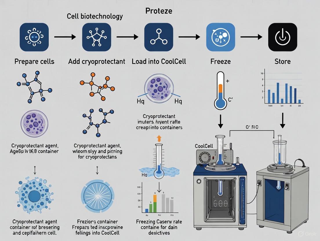
Workflow Overview:
Detailed Methodology:
- Cell Harvest and Preparation: Use healthy, log-phase cells. Gently dissociate cell clusters to ensure cryoprotectant penetration. After harvesting, centrifuge at 200-300 x g for 2 minutes and resuspend in pre-chilled cryopreservation medium at a density of 1-2 x 10^6 cells/mL [10]. High cell density can reduce viability.
- Aliquoting: Dispense the cell suspension into labeled cryogenic vials (e.g., 1 mL per vial). Using internal or external threaded vials is a matter of preference and automation compatibility [10].
- Loading the CoolCell: Place the filled cryovials into a room temperature CoolCell container. Do not pre-cool the device.
- Initiating Freezing: Transfer the entire CoolCell container upright into a -80°C freezer. The patent-pending technology utilizes a thermo-conductive alloy core and highly insulative outer material to control the rate of heat removal, ensuring a consistent -1°C/minute cooling profile [6].
- Post-Freezing Handling: Allow vials to remain in the -80°C freezer for a minimum of 4 hours, or preferably 24 hours, to ensure complete freezing before long-term storage.
- Long-Term Storage: For stable long-term preservation, transfer cryovials to the vapor phase of a liquid nitrogen dewar (typically between -140°C and -180°C). This practice reduces the risk of vial explosion and maintains sample integrity [10].
Validation and Performance Data
The CoolCell system's performance has been rigorously tested. A temperature probe placed into a cryogenic vial containing water and inserted into a room-temperature CoolCell recorded a consistent cooling profile of -1°C per minute when placed in a -80°C freezer. This profile was identical over five consecutive freeze cycles, demonstrating high reproducibility [6].
Empirical data from cell banks further validates the effectiveness of this standardized protocol. The table below summarizes key outcomes from cryopreservation studies.
Table 1: Experimental Outcomes of Optimized Cryopreservation
| Parameter | Experimental Outcome | Cell Type / Model | Significance |
|---|---|---|---|
| Cooling Rate | -1°C/minute [6] | N/A (Validated with water) | Foundational for protocol standardization. |
| Post-Thaw Viability | >80% viability [9] | Human Dermal Fibroblasts (HDF) | Direct indicator of protocol success for primary cells. |
| Phenotype Retention | Positive Ki67 & Collagen-I expression [9] | Human Dermal Fibroblasts (HDF) | Confirms retention of proliferative capacity and specialized function after thawing. |
| Storage Duration | Viability maintained over 3 months [9] | Human Dermal Fibroblasts (HDF) | Validates protocol for medium-term storage. |
The Scientist's Toolkit: Essential Research Reagents and Materials
Successful cryopreservation relies on a defined set of reagents and equipment. The following table details key solutions and materials, their functions, and application notes.
Table 2: Essential Reagents and Materials for Controlled-Rate Freezing
| Item | Function & Role | Application Notes |
|---|---|---|
| CoolCell Container | Provides a standardized, reproducible cooling rate of -1°C/minute in a -80°C freezer without alcohol [6]. | Reusable; offers a cost-effective alternative to programmable freezers. Ensures high post-thaw recovery and viability across cell types [6]. |
| Cryogenic Vials | Temperature-resistant polypropylene vials for containing cell suspensions. | Withstand temperatures down to -196°C. Choose internal or external thread design based on preference and automation needs [6] [10]. |
| Intracellular Cryoprotectant (DMSO) | Penetrates the cell membrane, lowers freezing point, and reduces electrolyte concentration and ice crystal formation [9] [10]. | Commonly used at a final concentration of 10%. Fresh preparation is recommended. Can be cytotoxic at room temperature; handle cells on ice after addition [9] [10]. |
| Fetal Bovine Serum (FBS) | Base constituent of cryomedium; provides proteins and nutrients that stabilize cell membranes and mitigate freezing stress. | Often used in combination with 10% DMSO for freezing primary cells like fibroblasts and keratinocytes [9]. |
| Serum-Free/Commercial Media | Chemically defined, animal-origin-free alternatives for clinical applications or where serum is undesirable. | Products like CryoStor are designed to offer consistency and safety for sensitive cells like stem cells [9]. |
Troubleshooting and Best Practices
Even with optimized protocols, challenges can arise. Below are common issues and their solutions, based on empirical data.
Table 3: Troubleshooting Guide for Controlled-Rate Freezing
| Problem | Potential Cause | Recommended Solution |
|---|---|---|
| Low post-thaw viability | Poor cell health prior to freezing; overexposure to dissociation reagents or CPA at room temperature; incorrect cell density [10]. | Freeze only healthy, log-phase cells. Keep cells on ice after adding cryomedium. Use recommended cell density (1-2 x 10^6 cells/mL). |
| Inconsistent viability between vials | Inhomogeneous cooling from using non-standardized freezing methods like polystyrene boxes [10]. | Use a validated controlled-rate freezer like CoolCell to ensure uniform, reproducible cooling across all vials. |
| Poor attachment/growth after thaw (e.g., iPSCs) | Overgrowth before freezing; large cell clumps preventing CPA penetration; rapid removal of CPA causing osmotic shock [10]. | Feed cells daily pre-freeze. Gently dissociate clumps. Thaw rapidly but remove CPA gently (e.g., drop-wise dilution). |
| Difficulty refreezing cells | Cumulative trauma from multiple freeze-thaw cycles. | Avoid refreezing previously thawed cells. Plan experiments to use all thawed material, as viability plummets after a second freeze [10]. |
Adherence to a standardized, controlled-rate freezing protocol is non-negotiable for maximizing cell viability and functionality post-thaw. The CoolCell container provides a simple, reliable, and reproducible method to achieve the critical -1°C/minute cooling rate, effectively navigating the compromise between lethal intracellular ice formation and damaging osmotic stress. By integrating the detailed protocols, reagent choices, and troubleshooting guidance outlined in this application note, researchers and drug developers can significantly enhance the reliability and reproducibility of their cryopreservation workflows, safeguarding valuable cellular resources for future use.
CoolCell container technology represents a significant advancement in laboratory cryopreservation equipment, offering standardized controlled-rate freezing without the maintenance and contamination concerns associated with alcohol-based systems. This alcohol-free technology utilizes a proprietary thermo-conductive alloy core and highly-insulative outer material to control the rate of heat removal, ensuring reproducible cell cryopreservation at approximately -1°C/minute in standard -80°C freezers. This application note details the design principles, operational protocols, and experimental validation of CoolCell containers, providing researchers with comprehensive guidance for implementing this technology within cell freezing workflows to maximize post-thaw viability and experimental reproducibility.
CoolCell containers employ an innovative physics-based approach to achieve controlled-rate freezing without requiring expensive programmable freezing equipment or the maintenance of alcohol-based systems. The technology centers on a proprietary thermo-conductive alloy core that surrounds the cryovials, coupled with a highly-insulative outer material that precisely regulates heat transfer from the samples to the freezer environment [11]. This specific material composition creates the optimal thermal gradient for biological cryopreservation, achieving a cooling rate of approximately -1°C per minute, which is widely recognized as the ideal rate for freezing most cell types while minimizing intracellular ice crystal formation [2] [12].
The alcohol-free nature of this design eliminates several practical laboratory challenges. Traditional isopropanol freezing containers require careful monitoring and replenishment of alcohol levels to maintain consistent performance, while CoolCell's passive thermal transfer mechanism requires no fluids that can evaporate, leak, or potentially contaminate samples [11] [12]. The radially symmetric construction ensures uniform freezing of all vials placed within the container, eliminating position-based variability that can occur in some freezing systems [11]. The design also features an easy-open lid and exposed vial tops when open, facilitating quick and organized retrieval of frozen samples without struggling to extract vials from a fluid medium [11].
Table: CoolCell Container Technical Specifications
| Parameter | Specification | Significance |
|---|---|---|
| Cooling Mechanism | Thermo-conductive alloy core + insulative shell | Provides consistent -1°C/min cooling without fluids [11] |
| Cooling Rate | Approximately -1°C per minute | Ideal for most cell types; prevents intracellular ice formation [2] [12] |
| Capacity | 6 standard 10 mL cryovials | Accommodates typical experimental batch sizes [11] |
| Freezer Requirement | Standard -80°C freezer | No specialized equipment needed [11] |
| Maintenance | Alcohol-free, no fluid replenishment | Reduced maintenance and contamination risk [11] [12] |
Experimental Protocols and Methodologies
Comprehensive Cell Freezing Protocol Using CoolCell Containers
The following step-by-step protocol ensures optimal cryopreservation results when using CoolCell technology, maintaining cell viability and functionality for long-term storage.
Pre-Freezing Preparation and Cell Harvesting
- Cell Preparation: Begin with healthy, log-phase cells at approximately 80-95% confluency, which handle cryopreservation stress better than stationary-phase cultures [2] [12]. For adherent cells, gently detach using appropriate dissociation reagents like trypsin, TrypLE Express, or Accutase, neutralizing with complete growth medium once cells have detached [7] [1]. For suspension cells, proceed directly with centrifugation.
- Centrifugation and Resuspension: Centrifuge the cell suspension at 100-400 × g for 5-10 minutes to form a pellet [1]. Carefully aspirate the supernatant without disturbing the pellet, then resuspend in an appropriate volume of pre-cooled cryopreservation medium. The optimal cell concentration is typically 1×10^6 to 10×10^6 cells/mL, though this should be optimized for specific cell types [2] [12].
- Cryopreservation Media Selection: Utilize a cryopreservation medium suitable for your cell type. Options include:
- Aliquoting: Dispense 1 mL aliquots of the cell suspension into sterile cryogenic vials [2] [7]. Label all vials comprehensively with cell type, passage number, date, researcher name, and other relevant identifiers [7].
Controlled-Rate Freezing with CoolCell
- Loading: Place the filled cryovials into the CoolCell container, ensuring they are properly seated in the designated slots [11]. Close the lid securely to maintain thermal continuity.
- Freezing Process: Transfer the loaded CoolCell container directly to a -80°C freezer. The proprietary thermal design will automatically implement the optimal -1°C/minute cooling rate without further intervention [11]. Leave the container in the freezer for a minimum of 4 hours, though overnight freezing is typically recommended to ensure complete freezing [2].
- Long-Term Storage: After the freezing period, promptly remove the cryovials from the CoolCell container and transfer them to long-term storage in either the vapor phase of liquid nitrogen (below -135°C) or an ultra-low temperature freezer maintained at -80°C or lower [2] [1]. Note that long-term storage at -80°C is suboptimal as viability declines over time due to temperature fluctuations; liquid nitrogen storage is recommended for indefinite preservation [2] [12].
Comparative Performance Assessment Protocol
To quantitatively evaluate CoolCell performance against alternative freezing methods, researchers can implement the following experimental methodology:
- Experimental Design: Prepare a homogeneous cell suspension from a single culture flask and divide into equal aliquots for freezing using different methods: CoolCell container, isopropanol-based freezing container (e.g., Nalgene Mr. Frosty), and controlled-rate freezer if available [2] [11] [1]. Include multiple biological replicates (minimum n=3) to account for biological variability [13].
- Viability Assessment: After freezing and storage for a standardized period (e.g., 1 week), rapidly thaw one vial from each condition in a 37°C water bath with gentle agitation [2] [12]. Immediately dilute the thawed cell suspension in pre-warmed complete growth medium, centrifuge gently to remove cryoprotectant, and resuspend in fresh medium. Quantify post-thaw viability using Trypan Blue exclusion with automated or manual cell counting [1].
- Functional Assessment: Plate thawed cells at standardized densities and monitor recovery through:
- Attachment efficiency: Count adherent cells 24 hours post-thawing
- Proliferation rates: Perform daily cell counts for 3-5 days
- Cell-specific functionalities: Assess differentiation capacity, marker expression, or other relevant functional metrics for the specific cell type
Table: Essential Research Reagent Solutions for CoolCell-Based Cryopreservation
| Reagent/Material | Function | Examples & Specifications |
|---|---|---|
| Cryopreservation Medium | Protects cells from freeze-thaw stress | CryoStor CS10 (universal), mFreSR (pluripotent stem cells), Synth-a-Freeze (protein-free) [2] [1] |
| Cryogenic Vials | Secure sample containment | Internal-threaded, sterile vials (e.g., Corning Cryogenic Vials) [2] |
| Cell Dissociation Reagents | Detach adherent cells | Trypsin, TrypLE Express, Accutase (cell type-dependent) [7] [1] |
| Viability Assessment | Post-thaw viability quantification | Trypan Blue with automated or manual cell counting [1] |
Results and Data Interpretation
Performance Metrics and Comparative Analysis
CoolCell containers deliver freezing performance comparable to expensive programmable freezers while maintaining the convenience and affordability of passive freezing devices [11]. The technology's consistent -1°C/minute cooling rate ensures minimal intracellular ice formation, a primary cause of cryoinjury that compromises membrane integrity and cellular function [12]. Comparative studies across multiple cell types, including stem cells, primary cells, PBMCs, and established cell lines, demonstrate post-thaw viability metrics equivalent to or exceeding those achieved with isopropanol-based systems [11].
The alcohol-free design eliminates potential contamination routes and maintenance requirements associated with alcohol evaporation or leakage in traditional freezing containers [11] [12]. This feature is particularly valuable in regulated environments like cell therapy manufacturing or Good Manufacturing Practice (GMP) facilities where contamination control is paramount [2]. The radial symmetry of the CoolCell design ensures uniform thermal transfer to all vial positions, eliminating positional variability that can affect freezing outcomes in some container designs [11].
Troubleshooting and Optimization Guidelines
- Suboptimal Post-Thaw Viability: If viability is consistently below expectations, verify that cells are harvested during log-phase growth and at appropriate confluence (80-95%) [2] [12]. Confirm that cryopreservation medium is appropriately formulated for the specific cell type and that DMSO concentration is optimized (typically 5-10%) [1].
- Inconsistent Results Between Vials: Ensure the CoolCell container is placed on a flat surface in the -80°C freezer away from the door or frequently accessed areas to minimize temperature fluctuations [12]. Verify that cryovials are properly seated in the container and that the lid is securely closed before freezing.
- Temperature Monitoring: For critical applications, consider placing a temperature logger inside a mock cryovial within the CoolCell container to validate the achieved cooling rate in your specific freezer model [12].
Application in Research and Development
CoolCell technology integrates seamlessly into comprehensive cell culture workflows, supporting research reproducibility and experimental standardization across biological disciplines. In basic research applications, the consistency of CoolCell freezing reduces experimental variability in cell-based assays by ensuring uniform post-thaw recovery between experiments conducted at different time points [13]. This reproducibility is essential for longitudinal studies and multi-investigator projects where consistent cell performance is critical to data interpretation.
In drug development pipelines, CoolCell containers provide a standardized approach to cell banking that maintains genetic stability and phenotypic consistency of cellular models used for high-throughput screening and toxicity assessment [2] [12]. The elimination of alcohol prevents potential chemical interactions that could compromise sensitive cell types or introduce variables in screening assays.
For regenerative medicine and cell therapy applications, the defined, closed-system nature of CoolCell technology supports compliance with quality assurance standards by eliminating the variability and contamination risks associated with alcohol-based systems [2]. The consistent performance ensures that therapeutic cell products maintain their viability and functional potency throughout the cryopreservation workflow, a critical consideration for clinical applications.
Cryopreservation is a fundamental technique in biomedical research and therapy development, enabling the long-term storage of cells and tissues at ultra-low temperatures, typically below -130°C to -196°C [2] [14]. At these temperatures, all metabolic and biochemical activities are effectively halted, placing cells in a state of suspended animation that preserves their viability and functionality for decades [14]. This technique has become indispensable in the rapidly advancing fields of cell and gene therapy (CGT), serving as a critical component for ensuring manufacturing flexibility, product stability, and global distribution of living cell therapies [15] [14].
For researchers, scientists, and drug development professionals, implementing robust cryopreservation protocols is not merely a convenience but a necessity for maintaining reproducible experimental conditions and ensuring the long-term availability of valuable cell lines [2]. The process involves a delicate balance of cryoprotective agents, controlled cooling rates, and proper storage conditions to minimize the cellular damage that can occur during the freezing and thawing processes [16] [2]. When properly optimized, cryopreservation provides three fundamental benefits: preservation of genetic stability, establishment of standardized cell banks, and prevention of microbial contamination—all essential elements for successful research and therapeutic development.
Key Benefits of Cryopreservation
Ensuring Genetic Stability
Maintaining genetic stability is paramount in cell-based research and therapy development. Cryopreservation effectively halts the biological clock of cells, preventing genetic drift that occurs during continuous passaging and long-term culture [16].
- Suspension of Metabolic Activity: At cryogenic temperatures (below -130°C), molecular motion is minimized to the point where biochemical and enzymatic processes are effectively paused [14]. This metabolic suspension prevents the accumulation of genetic mutations that naturally occur during cell division in continuous culture.
- Minimized Genetic Drift: By establishing cryopreserved cell banks at specific passages, researchers can ensure experimental consistency and reproducibility over time, avoiding the phenotypic and genotypic changes that inevitably occur with prolonged culture [2].
- Reduced Risk of Transformation: For finite cell lines, cryopreservation minimizes the risk of transformation that can occur with extended time in culture, particularly important for primary cells and stem cell populations [17].
The genetic integrity of cryopreserved cells is further supported by minimizing DNA damage during the freezing process. Advanced cryopreservation media containing appropriate cryoprotectants and antioxidants help reduce reactive oxygen species (ROS) that can cause DNA double-strand breaks and histone modifications [14].
Creating Cell Banks
Systematic cell banking represents a cornerstone of reproducible research and therapeutic development, providing a standardized source of cellular materials throughout project lifecycles.
- Working Cell Banks: These banks provide immediate access to quality-controlled cells for daily research activities, typically created from expanded Master Cell Bank vials [2].
- Master Cell Banks: MCBs serve as the foundational stock for all working cells, extensively characterized to ensure identity, purity, and genetic stability [18]. According to market analysis, master cell banks accounted for 38.21% of the cell banking outsourcing market share in 2024 [19].
- Viral Cell Banks: Essential for gene therapy and viral vector production, these banks are experiencing rapid growth (18.25% CAGR) driven by advancing CAR-T, oncolytic virus, and gene-editing modalities [19].
The creation of structured cell banks enables researchers to maintain consistent experimental conditions over extended periods and across multiple locations. For therapeutic development, tiered banking systems are mandatory for regulatory compliance, ensuring traceability from original cell source to final product [19].
Table: Cell Banking Market Analysis (2024)
| Bank Type | Market Share | Projected CAGR | Primary Applications |
|---|---|---|---|
| Master Cell Banks | 38.21% | - | Foundational stock for all downstream operations |
| Viral Cell Banks | - | 18.25% | CAR-T, oncolytic virus, gene-editing therapies |
| Stem Cell Banks | 60.85% (cell type segment) | - | Regenerative medicine, research applications |
Preventing Contamination
Cryopreservation provides a critical barrier against microbial contamination that can compromise research integrity or render therapeutic products unsafe.
- Elimination of Continuous Culture: By cryopreserving cells at specific passages, researchers avoid the cumulative risk of contamination inherent in maintaining continuous cultures, which require regular medium changes and handling [17] [2].
- Controlled Access: Cell banks function as protected repositories, with vials accessed only when needed, minimizing unnecessary exposure to potential laboratory contaminants [2].
- Quality Control Integration: The cell banking process incorporates comprehensive contamination screening, including sterility testing, mycoplasma detection, and viral safety testing before preservation, ensuring only contamination-free stocks are cryopreserved [14].
For cell therapies, cryopreservation enables complete quality control testing before the material is used in manufacturing or administration. Frozen leukopaks or final products can undergo thorough sterility, mycoplasma, and identity testing before release, preventing the use of contaminated materials in downstream applications [14].
Quantitative Data on Cryopreservation Practices
Recent industry surveys provide insight into current cryopreservation practices and their effectiveness in the field. The ISCT Cold Chain Management & Logistics Working Group survey reveals both adoption rates and areas requiring standardization.
Table: Cryopreservation Industry Survey Findings
| Parameter | Survey Result | Industry Implication |
|---|---|---|
| Controlled-Rate Freezing Adoption | 87% of respondents | High penetration in cell-based products, especially late-stage and commercial |
| Default CRF Profile Usage | 60% of users | Majority rely on manufacturer settings without cell-specific optimization |
| Vendor Qualification Reliance | Nearly 30% | Significant portion depend on vendors for system qualification |
| Freeze Curve Utilization in Release | Limited use | Post-thaw analytics preferred over process data for product release |
| Biggest Scaling Hurdle | "Ability to process at large scale" (22% respondents) | Scaling identified as primary challenge for industry growth |
The market context for cryopreservation services demonstrates substantial growth, with the cell banking outsourcing market valued at USD 16.78 billion in 2025 and projected to reach USD 36.64 billion by 2030, reflecting a robust CAGR of 16.91% [19]. This growth is fueled by increasing regulatory requirements for GMP-compliant banking and a surge in cell and gene therapy pipelines exceeding 2,500 active investigational new drug applications in the U.S. alone [19].
Step-by-Step Protocol: Cell Freezing Using CoolCell Container
The following protocol outlines a standardized approach for cryopreserving mammalian cells using the alcohol-free CoolCell cell freezing container, ensuring consistent cooling rates of approximately -1°C/minute without requiring specialized equipment [2].
Pre-freeze Processing
Proper preparation of cells before freezing is critical for maximizing post-thaw viability and functionality.
- Cell Examination and Preparation: Maintain cells in an actively growing state, ideally antibiotic-free for at least one week prior to freezing to identify potential contaminants [17]. Harvest cells during exponential growth phase, just before stationary phase, to maximize viability and uniformity [14]. For adherent cells, renew complete growth medium one day before harvest to improve cell health [14].
- Cell Harvesting: Gently dissociate cells using standard methods (trypsin/EDTA for adherent cells). Handle cells gently throughout harvesting as damaged cells will not survive additional freeze-thaw stress [17]. Determine total cell count and viability using a hemacytometer or automated cell counter with Trypan Blue exclusion [17].
- Centrifugation and Resuspension: Centrifuge cell suspension at 100-200 × g for 5-10 minutes [17]. Aseptically decant supernatant without disturbing cell pellet. Resuspend cells in appropriate cryopreservation medium at optimal concentration (typically 1×10^6 to 1×10^7 cells/mL, though this varies by cell type) [2] [14].
Cryopreservation Medium Preparation
Select and prepare cryopreservation medium appropriate for your cell type and application requirements.
- Cryoprotectant Selection: Dimethyl sulfoxide (DMSO) at 5-10% concentration is most common, often combined with serum or serum-free alternatives [17] [14]. For sensitive cells (e.g., stem cells), use commercial, predefined, serum-free formulations like CryoStor CS10 or cell-type specific media (e.g., mFreSR for pluripotent stem cells) [2].
- Serum Considerations: Traditional homemade freezing medium often uses 90% serum + 10% DMSO [17]. For regulated applications, GMP-manufactured, fully defined cryopreservation media are recommended to avoid lot-to-lot variability and potential infectious agents in serum [2].
- Preparation and Storage: Prepare freezing medium fresh or use commercially prepared, sterile-filtered solutions. Store at 2°C to 8°C until use [17].
Filling and Cooling with CoolCell
The CoolCell system provides a standardized cooling rate without requiring liquid nitrogen or controlled-rate freezers.
- Vial Preparation: Label cryogenic vials with permanent, cryo-resistant labels or markers. Include cell line identifier, passage number, date, and concentration [17] [2]. Place cryovials in a CoolRack CFT30 within the CoolBox CFT30 ice-free cooling station during filling to maintain temperature control [17].
- Aliquoting Cell Suspension: Aliquot 1 mL of cell suspension into each cryovial, frequently and gently mixing the main suspension to maintain homogeneous cell distribution [17].
- CoolCell Setup: Ensure the solid core (black ring) of the CoolCell is at room temperature and seated in the bottom of the central cavity [17]. Place sample vials containing equal volumes (1 mL) into CoolCell wells. Vials should not extend above the CoolCell body. For optimal performance, fill all 12 chambers—use "blank" vials with media only if necessary to fill empty wells, as this ensures radially symmetric cooling [17].
- Freezing Process: Seal the CoolCell lid completely and place the unit in a -80°C freezer with at least 1 inch of clearance on all sides [17]. Leave containers undisturbed for a minimum of 4 hours (up to 24 hours) to ensure complete freezing before transfer to long-term storage [17].
Long-term Storage and Record Keeping
Proper storage conditions and documentation ensure cell viability and traceability.
- Transfer to Long-term Storage: After initial freezing, transfer vials to a continually maintained storage environment below -130°C (typically vapor phase liquid nitrogen or mechanical freezers) [2]. Always use dry ice for transfer—cryovial contents can rise from -75°C to over -50°C in less than one minute if exposed to room temperature air [17].
- Quality Control Testing: Thaw one representative vial after short-term storage to confirm viability and sterility before terminating the stock culture [17].
- Comprehensive Documentation: Maintain detailed records including culture identity, passage number, date frozen, freezing medium, cell concentration, number of vials, storage location, and all quality control test results [17] [2].
Experimental Workflow and Signaling Pathways
Cryopreservation Experimental Workflow
The following diagram illustrates the complete workflow for cryopreservation using the CoolCell system, from cell preparation through to long-term storage and quality control.
Cellular Response to Cryopreservation
The diagram below outlines the key cellular responses and injury mechanisms during cryopreservation, highlighting both the challenges and protective strategies.
The Scientist's Toolkit: Essential Research Reagents and Materials
Successful cryopreservation requires carefully selected reagents and equipment to ensure consistent results and maximum cell viability. The following table details essential materials for standard cryopreservation protocols.
Table: Essential Research Reagents and Materials for Cryopreservation
| Item | Function & Application | Key Considerations |
|---|---|---|
| Cryopreservation Medium | Protects cells from freeze-thaw damage; typically contains cryoprotectants and buffers | Choose serum-free, defined formulations (e.g., CryoStor) for regulated work; DMSO concentration typically 5-10% [2] |
| CoolCell Container | Provides consistent -1°C/min cooling rate in standard -80°C freezer | Alcohol-free; accommodates 12 standard cryovials; ensures reproducible cooling without controlled-rate freezer [17] [2] |
| Cryogenic Vials | Long-term storage of cell suspensions at ultra-low temperatures | Use internal-threaded vials to prevent contamination; label with cryo-resistant markers/tags [2] |
| Controlled-Rate Freezer | Programmable cooling for optimized, cell-specific freezing protocols | Alternative to CoolCell; provides precise control over cooling rate; required for some sensitive cell types [15] |
| Liquid Nitrogen Storage | Long-term storage below -130°C (vapor phase) or -196°C (liquid phase) | Maintains cell viability for decades; vapor phase reduces contamination risk between vials [17] [2] |
| Cell Counting System | Determines cell concentration and viability before freezing | Hemacytometer or automated cell counter with Trypan Blue exclusion; ensures optimal freezing density [17] |
| Cryoprotective Agents | Chemical compounds that protect cells from freezing damage | Permeating (DMSO, glycerol) and non-permeating (sucrose, sugars) agents; toxicity varies by cell type [16] [14] |
Cryopreservation remains an essential tool for modern biological research and therapeutic development, providing the foundation for reproducible science and scalable cell-based therapies. The three core benefits—genetic stability, systematic cell banking, and contamination prevention—create a framework for reliable, long-term cellular preservation. The standardized CoolCell protocol outlined in this application note offers researchers a robust methodology for achieving consistent cryopreservation results without requiring specialized equipment.
As the field advances, with the cell banking outsourcing market projected to grow at 16.91% CAGR [19], implementation of optimized cryopreservation practices becomes increasingly critical. By adhering to these protocols and understanding the underlying principles of cryopreservation, researchers and therapy developers can ensure the integrity of their cellular resources, supporting both basic research and the development of next-generation cell therapies.
A Detailed Step-by-Step Guide to Freezing Cells with Your CoolCell Container
Within a comprehensive cell freezing protocol utilizing a CoolCell container, the steps taken prior to the freezing event are paramount to success. Pre-freezing preparations are a critical phase that directly determines post-thaw cell viability, recovery, and functionality [12]. This document outlines the essential procedures for validating cell health, ensuring optimal growth phase, and confirming a contamination-free status before cryopreservation. Neglecting these steps can compromise even the most technically perfect freezing process, leading to the irrevocable loss of valuable cell stocks and irreproducible experimental results [1] [2]. Adherence to these protocols is fundamental for researchers and drug development professionals aiming to establish reliable, high-quality cell banks.
Assessing Cell Health and Viability
A thorough assessment of cell health is the first mandatory step before initiating cryopreservation. Cells must be in an optimal physiological state to withstand the significant stresses of the freezing process.
Morphological Evaluation
Routinely examine cell cultures under a phase-contrast microscope for key indicators of health. Adherent cells should appear well-attached and spread, exhibiting a classic, uniform morphology for the specific cell type. The culture medium should be clear and free of floating debris or granularity, which can indicate cell death or microbial contamination [2].
Quantitative Viability Assessment
Cell viability must be quantitatively determined using a dye exclusion method, such as Trypan Blue. Only cultures demonstrating high viability, typically greater than 90%, should be considered for cryopreservation [1] [20]. This assessment can be performed manually with a hemocytometer or using an automated cell counter. The table below summarizes the key criteria for cell health assessment.
Table 1: Key Criteria for Pre-Freezing Cell Health Assessment
| Parameter | Optimal Status for Freezing | Assessment Method |
|---|---|---|
| Viability | >90% [1] | Trypan Blue exclusion and cell counting (manual hemocytometer or automated cell counter) [1] |
| Confluency (Adherent Cells) | 80-95% [7] [2] | Visual inspection under a phase-contrast microscope |
| Growth Phase | Mid-log phase (exponential growth) [12] [20] | Cell counting and growth curve analysis |
| Morphology | Healthy, uniform appearance specific to cell type | Visual inspection under a phase-contrast microscope |
| Culture Medium | Clear, no unexpected color change, turbidity, or debris [2] | Visual inspection |
Ensuring Log-Phase Growth
The growth phase of a cell culture at the time of harvesting is a critical biological factor influencing cryopreservation success. Cells harvested during their logarithmic (log) or exponential growth phase are significantly more resilient to the cryopreservation process than those in the stationary or decline phases [12] [20].
Rationale for Log-Phase Freezing
Cells in the log phase are metabolically active, proliferating rapidly, and are in a state of optimal physiological fitness. This vigor enhances their ability to endure the osmotic shifts and metabolic stresses induced by cryoprotective agents (CPAs) and temperature changes [12]. Freezing cells at as low a passage number as possible is also recommended to minimize genetic drift and phenotypic changes [1].
Protocol for Harvesting Log-Phase Cells
- Monitor Growth Dynamics: Establish a growth curve for your specific cell line to understand its typical doubling time and identify the mid-log phase. For many continuous cell lines, this occurs when cultures reach 80-95% confluency for adherent cells, or a predetermined optimal density for suspension cultures [7] [2].
- Plan Your Harvest: Schedule cell passaging and freezing procedures to align with this optimal window. Do not harvest from over-confluent cultures, as nutrient depletion and contact inhibition can induce stress and senescence, rendering cells vulnerable to freezing damage [12].
Contamination Checks
The cryopreservation of contaminated cells leads to the permanent loss of that stock and risks cross-contaminating other samples in the storage tank. Rigorous contamination checks are a non-negotiable pre-freezing requirement.
Mycoplasma Screening
Mycoplasma is a common bacterial contaminant that is not visible under standard microscopy and can profoundly alter cell behavior. It is recommended to include mycoplasma testing in the pre-freezing workflow [2]. This can be done using PCR-based detection kits, enzymatic assays, or fluorescent staining, and should be performed on a representative sample of the culture destined for freezing.
Visual and Culture-Based Checks
- Visual Inspection: As noted in Section 2.1, look for signs of bacterial or fungal contamination, such as sudden, unexplained acidity (yellowing of phenol red-containing media), turbidity, floating fungal hyphae, or rapid cell death [2].
- Aseptic Technique: All procedures leading up to and including the freezing process must be performed using strict aseptic techniques in a Class II biological safety cabinet to prevent the introduction of contaminants [7] [21].
Table 2: Essential Research Reagent Solutions for Pre-Freezing Preparations
| Reagent / Material | Function / Application |
|---|---|
| Trypan Blue Solution | A vital dye used to distinguish live cells (which exclude the dye) from dead cells (which take up the dye) for viability counting [1]. |
| Phosphate Buffered Saline (PBS) | A balanced salt solution used for washing cell monolayers (e.g., of adherent cells prior to dissociation) to remove residual serum and metabolites [1] [7]. |
| Cell Dissociation Reagent (e.g., Trypsin, TrypLE, Accutase) | An enzyme solution used to detach adherent cells from the culture vessel surface for harvesting [1] [7]. |
| Complete Growth Medium | A nutrient-rich medium containing serum and/or supplements, used to culture cells and to neutralize dissociation enzymes after detachment [1]. |
| Mycoplasma Detection Kit | A specialized test (e.g., PCR, ELISA, or staining-based) used to detect the presence of mycoplasma contamination in cell cultures [2]. |
Experimental Workflow for Pre-Freezing Preparation
The following diagram summarizes the logical workflow and decision points for the pre-freezing preparation of cell cultures.
Diagram Title: Pre-Freezing Cell Preparation Workflow This workflow outlines the critical checks and steps required to ensure cells are healthy and contamination-free before cryopreservation. Key decision points include visual inspection for contamination and a quantitative viability threshold check. Failure at any of these points necessitates discarding the culture to protect the integrity of the cell bank.
Cryopreservation is a fundamental technique in biomedical research and drug development, enabling the long-term storage of living cells by suspending cellular metabolism at extremely low temperatures (-80°C to -196°C) [2]. The success of this process hinges on the formulation of the freezing medium, which protects cells from the lethal effects of intracellular ice crystal formation and solute imbalance during the freeze-thaw cycle [2]. This application note examines three principal categories of freezing media: traditional FBS/DMSO-based formulations, serum-free alternatives, and commercial ready-to-use solutions. Framed within broader thesis research on standardized cell freezing protocols utilizing CoolCell containers, we provide a detailed, comparative analysis supported by quantitative data, structured protocols, and decision frameworks to guide researchers in selecting and implementing optimal cryopreservation strategies.
Comparative Analysis of Freezing Medium Formulations
The choice of cryopreservation medium significantly impacts post-thaw cell viability, recovery, and functionality. The core function of any freezing medium is to mitigate ice crystal damage, often achieved through cryoprotective agents (CPAs) like dimethyl sulfoxide (DMSO), and to provide a protective osmotic environment, historically facilitated by serum [22].
Table 1: Key Characteristics of Freezing Medium Formulations
| Formulation Type | Key Components | Typical Cell Viability/Recovery | Major Advantages | Major Limitations |
|---|---|---|---|---|
| Traditional FBS/DMSO [1] [23] | Basal Medium (e.g., DMEM), 10-20% FBS, 10% DMSO | ~80% viability in fish embryonic cells with 10% DMSO/20% FBS [23] | Familiar protocol, cost-effective for labs with readily available FBS [1] | Undefined serum components, lot-to-lot variability, risk of microbial contamination [2] [22] |
| Serum-Free/Protein-Free [24] [22] | Defined basal medium, 7.5-10% DMSO, may include protein substitutes (e.g., BSA) or trehalose | High rates of viability, proliferation, and bioactivity post-thaw [22] | Chemically defined, eliminates serum-associated variability and ethical concerns, suitable for regulatory-sensitive applications [24] [22] | May require optimization for specific cell types; some formulations not suitable for melanocytes [24] |
| Commercial Ready-to-Use [24] [1] [2] | Optimized, predefined mix of cryoprotectants (e.g., DMSO) and protective agents (e.g., in CryoStor, Synth-a-Freeze, mFreSR) | High, reproducible thawing efficiencies; designed for specific cell types like stem cells [2] | Ready-to-use convenience, optimized and reproducible performance, often GMP-manufactured, supports regulatory compliance [2] | Higher cost per unit compared to lab-made media; specific formulation may not be ideal for all cell types |
The experimental data for fish embryonic cell lines (Oryzias dancena) further illustrates the optimization process for a traditional FBS/DMSO formulation. A study systematically testing different component concentrations found that a combination of 10% DMSO, 20% FBS, and 0.1 M trehalose in DMEM yielded optimal post-thaw viability and growth, with the cells showing similar morphology and growth rate to their non-frozen counterparts [23]. This underscores that even within traditional formulations, the exact composition is critical and should be optimized for specific cell types.
Essential Materials and Reagents
A successful cryopreservation workflow requires more than just the freezing medium. The following toolkit lists essential reagents and equipment, with specific product examples relevant to the protocols discussed herein.
Table 2: Research Reagent Solutions for Cell Cryopreservation
| Item | Function/Purpose | Representative Examples |
|---|---|---|
| Freezing Medium | Protects cells from freeze-thaw damage, maintains viability. | Synth-a-Freeze [24], Recovery Cell Culture Freezing Medium [1], CryoStor CS10 [2] |
| Controlled-Rate Freezing Container | Ensures consistent, optimal cooling rate of ~-1°C/minute. | CoolCell (alcohol-free) [5] [2], Mr. Frosty (isopropanol-based) [1] |
| Cryogenic Vials | Safe, sterile containment for long-term storage of cell suspensions. | Corning Cryogenic Vials [2] |
| Cell Dissociation Reagent | Detaches adherent cells from culture vessel for harvesting. | Trypsin, TrypLE Express [1] |
| Viability Assay Reagents | Determines viable cell count pre-freeze and post-thaw. | Trypan Blue Solution [24] [1], Cell Counting Kit-8 [23] |
Experimental Protocols for Freezing Medium Evaluation
Protocol: Cryopreservation of Cells Using a CoolCell Container
This standardized protocol is suitable for use with various freezing media and is central to ensuring reproducible cooling rates in the absence of a programmable freezer [5] [2].
- Harvesting: For adherent cells, gently wash with a balanced salt solution (e.g., DPBS) and detach using an appropriate dissociation reagent like trypsin. Neutralize the enzyme with complete growth medium. For suspension cells, proceed directly to centrifugation [1].
- Centrifugation: Pellet the cells by centrifugation at approximately 100–400 × g for 5–10 minutes. Carefully aspirate the supernatant without disturbing the cell pellet [1].
- Resuspension: Resuspend the cell pellet in pre-chilled (2°–8°C) freezing medium at a concentration of 5 × 10^5 to 3 × 10^6 cells/mL [24] [1]. Gently mix to achieve a homogeneous suspension.
- Aliquoting: Dispense the cell suspension into sterile cryogenic vials (e.g., 1 mL per vial). Label all vials clearly with indelible ink [1] [2].
- Freezing: Immediately transfer the sealed vials into a CoolCell freezing container pre-equilibrated to room temperature. Place the entire container directly into a -80°C freezer for a minimum of 4 hours, though overnight is recommended [5] [2]. The CoolCell container ensures a consistent cooling rate of approximately -1°C/minute [5].
- Long-Term Storage: After the freezing period, promptly transfer the vials to a long-term storage location in the vapor phase of a liquid nitrogen tank (below -135°C) to ensure maximum stability [24] [1] [2].
Protocol: Formulation and Testing of Serum-Free Medium with Trehalose
Based on research into optimized medium composition, this protocol outlines the methodology for creating and validating a serum-free formulation [23] [22].
- Medium Preparation: Prepare the base serum-free freezing medium by supplementing DMEM with 10% DMSO and 0.1 M trehalose [23]. Filter-sterilize the solution and store at 4°C until use.
- Experimental Freezing: Harvest and count the cells as described in section 4.1. Divide the cell pellet into aliquots and resuspend them in: a) the experimental serum-free medium, b) a traditional FBS/DMSO medium (e.g., DMEM + 10% FBS + 10% DMSO), and c) a commercial serum-free medium for comparison.
- Cell Freezing and Storage: Aliquot the cell suspensions into cryovials and freeze them using the standardized CoolCell protocol outlined in section 4.1. Store the vials in liquid nitrogen for a defined period (e.g., 7 days) [23].
- Post-Thaw Analysis:
- Thawing: Rapidly thaw the vials in a 37°C water bath for approximately 2 minutes [23] [2].
- Viability Assay: Immediately after thawing, seed 1 × 10^5 post-thaw cells in a 96-well plate. Assess cell viability using a assay such as the Cell Counting Kit-8 (CCK-8), calculating viability as (Absorbancesample / Absorbancecontrol) × 100, where the control is non-frozen cells [23].
- Growth Activity: Seed 1 × 10^5 post-thaw cells in a 24-well plate and culture for 48 hours. Harvest the cells and count the final cell number with a hemocytometer to assess recovery and proliferation capacity [23].
Diagram 1: Experimental workflow for freezing medium evaluation.
The selection of an appropriate freezing medium is a critical determinant in the establishment of robust and reproducible cell banks. As summarized in Diagram 2, the choice involves a strategic trade-off between protocol familiarity, definition, and regulatory compliance.
For fundamental research where cost is a primary constraint and the use of serum is not prohibitive, a traditional FBS/DMSO medium may be sufficient, provided its limitations are acknowledged [1]. However, for serum-free culture systems, sensitive primary cells, or stem cell applications, commercially available, defined media such as Synth-a-Freeze or cell-type-specific formulations like mFreSR for iPSCs offer significant advantages in performance and consistency [24] [2]. The experimental data confirms that serum-free and commercial media can achieve high post-thaw viability and functionality, often outperforming traditional serum-containing mixes [22].
A key best practice, regardless of the medium chosen, is the use of a controlled-rate freezing device like the CoolCell container. This ensures the critical -1°C/minute cooling rate, standardizing the process and maximizing cell viability upon thawing [5] [2]. In conclusion, researchers and drug development professionals are encouraged to move towards defined, serum-free freezing media where possible, implementing standardized tools like the CoolCell to enhance the reliability and translational potential of their cryopreserved cell stocks.
Diagram 2: Decision pathway for selecting a freezing medium formulation.
Within the broader context of developing a standardized, step-by-step cell freezing protocol utilizing a CoolCell container, the steps of harvesting and resuspending cells at the correct concentration are critical. These steps directly impact post-thaw viability and the reproducibility of future experiments. This application note details a refined methodology for preparing a homogeneous cell suspension at a density of 1x10^6 to 5x10^6 cells/mL, ready for cryopreservation in a CoolCell device, which ensures a consistent cooling rate of -1°C/minute for optimal cell recovery [2] [17].
Key Parameters for Cell Resuspension
Table 1: Summary of key parameters for cell harvesting and resuspension.
| Parameter | Optimal Range or Condition | Rationale & Considerations |
|---|---|---|
| Cell Health & Confluency | >80% confluency; mid-log growth phase [2] | Ensures cells are in a robust, actively dividing state, maximizing post-thaw viability. |
| Pre-harvest Contamination Check | Confirmed absence of microbial contamination (e.g., mycoplasma) [2] [12] | Prevents freezing and storing of contaminated cultures. |
| Final Resuspension Density | General Range: 1x10^3 - 1x10^6 cells/mL [2]Typical Target: 1x10^6 - 5x10^6 cells/mL [25] [17] [20] | Prevents low viability from too few cells and cell clumping or excessive cryoprotectant agent (CPA) exposure from too high a density [2] [12]. |
| Cryoprotectant Agent (CPA) | 10% DMSO in FBS or serum-free medium is common [20] | DMSO mitigates ice crystal formation but is cytotoxic upon prolonged exposure; handle cells quickly after adding CPA [20]. |
| Handling of Cell Pellet | Gentle centrifugation (100-300 x g for 5-10 min) [17] [20] | Hard centrifugation can damage cells, especially fragile ones. Loosen the pellet by gentle agitation [17]. |
Detailed Step-by-Step Protocol
Pre-Harvest Procedures
- Cell Assessment: Confirm cells are healthy, >80% confluent, and in the logarithmic growth phase [2]. Visually inspect cultures for signs of contamination like turbidity or unexpected morphological changes [2] [12].
- Reagent Preparation: Pre-chill the chosen freezing medium (e.g., 90% FBS/10% DMSO or a commercial alternative like CryoStor CS10) to 2-8°C [17]. Prepare all necessary equipment, including labeled cryogenic vials.
Cell Harvesting and Counting
- Harvesting:
- For Adherent Cells: Wash the monolayer with PBS, then dissociate using an appropriate agent like trypsin-EDTA [2] [20]. Neutralize the dissociation reagent with complete growth medium containing serum. Use gentle, non-enzymatic methods like cell scrapers for sensitive cells [12].
- For Suspension Cells: Transfer the cell suspension directly to a centrifuge tube [20].
- Centrifugation: Transfer the cell suspension to a centrifuge tube and spin at 100-300 x g for 5-10 minutes at room temperature [17] [20].
- Supernatant Removal and Counting: Carefully aspirate the supernatant without disturbing the soft cell pellet. Resuspend the pellet in a small volume of fresh medium and perform a cell count and viability assessment using Trypan Blue exclusion on a hemocytometer or automated cell counter [17] [20]. Cell viability should be at least 75% before proceeding with cryopreservation [20].
Resuspension at Optimal Density
- Calculate Volume: Based on the cell count and the target viability (e.g., 2x10^6 cells/mL), calculate the required volume of freezing medium needed.
- Resuspend Pellet: Loosen the cell pellet by gently flicking the tube. Slowly add the calculated, ice-cold freezing medium dropwise while gently swirling the tube or using a wide-bore pipette tip to resuspend the cells [12]. This minimizes shear stress and ensures a homogeneous single-cell suspension without clumping.
- Final Mixing: Mix the cell suspension thoroughly but gently by pipetting up and down slowly to achieve a uniform density. Keep the tube on ice or in a CoolRack to maintain a chilled state and minimize CPA toxicity [17] [20].
Aliquotting and Workflow
The following workflow diagram outlines the entire process from cell culture to storage in the CoolCell container.
Diagram 1: Workflow for harvesting, resuspending, and freezing cells.
The Scientist's Toolkit
Table 2: Essential reagents and equipment for the protocol.
| Item | Function & Application Notes |
|---|---|
| CoolCell Container | A passive cooling device placed in a -80°C freezer to ensure a consistent, controlled freezing rate of -1°C per minute, which is critical for cell viability [2] [17] [20]. |
| Cryoprotective Agent (CPA) | Dimethyl sulfoxide (DMSO) is most common; reduces intracellular ice crystal formation. Glycerol is an alternative for DMSO-sensitive cells [26] [20]. |
| Freezing Medium | Protects cells during freeze-thaw cycle. Can be lab-made (e.g., 90% FBS + 10% DMSO) or commercial, serum-free, GMP-manufactured media (e.g., CryoStor CS10) for higher consistency and safety profiles [2] [25]. |
| Cryogenic Vials | Single-use, sterile vials certified for ultra-low storage. Internal-threaded vials are preferred to minimize contamination risk in liquid nitrogen [2] [12] [26]. |
| Controlled-Rate Freezer | A programmable unit as an alternative to passive coolers for the most precise control over the freezing curve, essential for critical or sensitive applications [2] [26]. |
| Wide-Bore Pipette Tips | Reduce fluid shear stress during resuspension, protecting delicate cells from physical damage [12]. |
Aliquotting into Cryovials and Loading the Room-Temperature CoolCell
Aliquotting cell suspensions into cryovials and correctly loading them into a CoolCell freezing container is a critical step in the cryopreservation workflow. This standardized procedure ensures cells are frozen at the optimal, reproducible cooling rate of approximately -1°C per minute, which is vital for maximizing post-thaw viability and maintaining genetic stability [20] [2]. This application note provides a detailed methodology for this specific phase of the protocol, framed within broader research on standardized cell freezing techniques.
Materials and Reagents
Table 1: Essential Research Reagent Solutions and Materials
| Item Name | Function/Application |
|---|---|
| Cryogenic Vials | For containing and storing the cell suspension; ensure they are sterile and properly labeled [20] [7]. |
| Prepared Freezing Medium | A cryoprotective solution, often containing FBS and DMSO (e.g., 90% FBS + 10% DMSO), which protects cells from ice crystal damage during freezing [20] [7]. |
| CoolCell Freezing Container | An alcohol-free device designed to ensure a consistent, controlled freezing rate of -1°C per minute when placed in a -80°C freezer [20] [27]. |
| Cell Suspension | Harvested and counted cells, resuspended in freezing medium at an optimal density (e.g., 1-5 million cells/mL) [20] [2]. |
Methodology
Aliquotting Cell Suspension into Cryovials
- Labeling: Label cryovials with essential information, including the date, researcher's name, cell line, passage number, and any genetic modifications [20] [7].
- Resuspension and Density: Resuspend the centrifuged cell pellet in the appropriate, pre-warmed freezing medium to achieve the recommended cell density. For most mammalian cells, a density of 1 x 10^6 cells/mL is standard, though certain cell types (e.g., some suspension cells) may require higher densities, such as 5 x 10^6 cells/mL [20] [7].
- Aliquot Volume: Aliquot 1 mL of the cell suspension into each pre-labeled cryovial [20]. Ensure the lids are securely tightened to prevent leakage during storage.
- Time Constraint: Complete the aliquoting process and proceed to the next step promptly. Cells should not remain in the freezing medium at room temperature for more than 10 minutes to minimize cryoprotectant toxicity [20].
Loading the Room-Temperature CoolCell
- Container Preparation: Use the CoolCell at room temperature. The CoolCell LX is an alcohol-free polyethylene foam container [27].
- Loading: Transfer the sealed cryovials directly into the slots of the CoolCell container at room temperature [20].
- Immediate Freezing: Place the entire CoolCell unit containing the cryovials directly into a -80°C freezer [20]. The insulating properties of the container will ensure the temperature decreases at the optimal rate of -1°C per minute [20] [2].
- Long-term Storage: After approximately 24 hours, remove the cryovials from the CoolCell and transfer them to long-term storage in liquid nitrogen [20]. Avoid storing vials at -80°C for extended periods, as this can compromise cell viability [20] [2].
Experimental Workflow and Data
The following diagram illustrates the procedural workflow for aliquotting and freezing cells using the CoolCell system.
Table 2: Key Quantitative Parameters for the Aliquotting and Freezing Process
| Parameter | Specification | Technical Rationale |
|---|---|---|
| Cell Density | 1-5 x 10^6 cells/mL [20] [7] | Prevents low viability from over-dilution and cell clumping from over-concentration. |
| Aliquot Volume | 1.0 mL per cryovial [20] | Standard volume for efficient freezing and storage in cryovials. |
| Time in Freezing Media at RT | ≤ 10 minutes [20] | Limits exposure to cytotoxic cryoprotectants like DMSO. |
| Cooling Rate in CoolCell | -1°C per minute [20] [2] | Optimizes water efflux from cells, minimizing lethal intracellular ice crystal formation. |
| Initial Freezing Duration | ~24 hours [20] | Ensures cells are fully stabilized at -80°C before long-term storage transfer. |
Best Practices and Key Considerations
- Aseptic Technique: Maintain sterility throughout the process to prevent contamination [7] [2].
- Cell Status: Cells should be harvested during their logarithmic growth phase and at high viability (typically >75-80%) prior to cryopreservation [20] [2].
- CoolCell State: Confirm the CoolCell is at room temperature at the start of the protocol to guarantee the correct, controlled freezing rate [20].
- Record Keeping: Meticulous labeling and inventory management are essential for the traceability of cell stocks [2].
Within the framework of a comprehensive thesis on standardized cell freezing, this application note details a critical procedural step: the freezing cycle of the CoolCell container in a -80°C freezer. Successful cryopreservation is not merely about achieving a low temperature; it is about achieving a controlled and reproducible thermal trajectory to maximize post-thaw viability and functionality. The CoolCell system is designed to provide a consistent cooling rate of -1°C per minute, which is widely recognized as optimal for preserving a wide variety of cell types, including sensitive primary cells and cell lines used in therapy development [28] [6] [29]. This document provides a detailed protocol and supporting data for the 4 to 24-hour freezing and stabilization period in a -80°C freezer, a key phase that ensures samples are properly prepared for long-term cryogenic storage.
The Critical Role of the Freezing Cycle
The period during which the CoolCell is placed in a -80°C freezer is functionally divided into two key stages: the active freezing phase and the temperature stabilization phase.
Active Freezing Phase
The proprietary design of the CoolCell, which combines a highly insulative closed-cell polyethylene foam housing with a thermally conductive alloy core, ensures that heat is removed from the cryovials in a radially symmetric and controlled manner [28] [29]. This design passively creates a cooling rate of -1°C per minute as the chamber temperature drops from room temperature to the -80°C setpoint of the freezer [6] [29]. Performance tests demonstrate that this profile is highly reproducible across multiple consecutive cycles, generating identical freezing times and cooling curves [28] [6]. This controlled rate is crucial to minimize intracellular ice crystal formation, a primary cause of cryo-injury and cell death [10].
Stabilization Phase
Once the active freezing period is complete, the samples must remain in the -80°C freezer for a sufficient duration to ensure thermal equilibrium. This stabilization is critical before transferring samples to long-term storage in liquid nitrogen or a -150°C freezer. Premature transfer, before the contents of the vials have reached a stable -80°C, can lead to partial warming, ice recrystallization, and severe loss of viability [17]. The specified hold time of 4 to 24 hours ensures that even the core of the sample vial has reached the target temperature, securing the sample's stability for archival transfer [17].
Materials and Equipment
Table 1: Essential Research Reagent Solutions and Materials
| Item | Function & Specification |
|---|---|
| CoolCell Container | An alcohol-free, passive freezing device that uses a thermo-conductive alloy core and insulative foam to ensure a consistent cooling rate of -1°C/minute in a -80°C freezer [28] [29]. |
| Cryogenic Vials | Temperature-resistant polypropylene vials capable of withstanding temperatures down to -196°C. Internal or external thread designs are available based on user preference and automation compatibility [28] [10]. |
| Cryoprotective Medium | Typically contains a base medium (e.g., 90% serum) and an intracellular cryoprotectant like 10% DMSO, which reduces the freezing point and minimizes ice crystal formation [17] [10]. |
| -80°C Freezer | A mechanical freezer capable of maintaining a stable temperature of -80°C. It must have sufficient free space (at least 1 inch clearance around the CoolCell) for unhindered air circulation [17]. |
| Personal Protective Equipment (PPE) | Insulated cryogenic gloves, a lab coat, and a face shield are mandatory for handling cold surfaces and during sample transfer to protect against cold burns and vial rupture. |
Step-by-Step Protocol
Pre-Freezing Preparation
- Cell Harvesting: Harvest cells in an actively growing state to ensure maximum health. Gently pellet cells via centrifugation and resuspend in an appropriate volume of pre-chilled cryoprotective medium to a final concentration generally between 1 x 10^6 to 4 x 10^6 cells/mL [17] [10].
- Vial Preparation: Aseptically aliquot 1 mL of the cell suspension into each cryogenic vial. Seal the vials tightly and ensure their exteriors are clean and dry [17].
- CoolCell Setup: Ensure the solid alloy core of the CoolCell is at room temperature and seated properly. For radially symmetric cooling, place vials containing an equal volume of media into the wells. If fewer than 12 vials are being frozen, use "blank" vials filled with media or water to occupy the remaining wells [17].
The Freezing Cycle
- Initiation: Place the sealed CoolCell container upright into the -80°C freezer. Ensure there is at least one inch of free space on all sides of the container to allow for proper air circulation [17].
- Duration: Leave the CoolCell container undisturbed in the freezer for a period of at least 4 hours and up to 24 hours [17]. This timeframe guarantees that the active freezing process is complete and the samples have been stabilized at -80°C.
Post-Freezing Transfer to Long-Term Storage
- Preparation: Pre-cool a transfer container or a CoolRack on dry ice.
- Rapid Transfer: Quickly move the cryovials from the CoolCell in the -80°C freezer to the pre-cooled container on dry ice. Minimize the time vials are exposed to ambient temperature, as contents can rise from -75°C to over -50°C in less than a minute, causing viability loss [17].
- Archiving: Transfer the vials on dry ice to their final long-term storage environment, ideally in the vapor phase of liquid nitrogen (typically between -140°C and -180°C) or in a freezer capable of maintaining temperatures below -130°C [17] [10].
The workflow below illustrates the sequence of these critical steps.
Experimental Data and Validation
The efficacy of the CoolCell freezing protocol is supported by rigorous experimental data. The following table summarizes key performance and viability outcomes from validation studies.
Table 2: Performance and Viability Data from CoolCell Validation Studies
| Cell Type / Parameter | Experimental Condition | Result | Citation |
|---|---|---|---|
| CoolCell Performance | Consecutive freeze cycles in -80°C freezer | Identical freezing time and -1°C/min cooling profile reproduced over 5 cycles | [28] [6] |
| Human Embryonic Stem Cells (RC-10) | Post-thaw growth (Day 3) | Cells frozen in CoolCell showed more rapid growth and higher total cell count compared to IPA containers | [28] |
| PBMC & T-Cells | Post-thaw viability | No significant difference in viability between CoolCell and programmable freezer | [29] |
| Ova-Specific Tregs (Ovasave) | Post-thaw viability in GMP trial | High viability (91.7% ± 3.7%) meeting FDA requirements, matching programmable freezer | [29] |
| General Best Practice | Cooling rate for most cells | Controlled rate of -1°C per minute is ideal for viability | [10] [29] |
Interaction of Cooling and Thawing Rates
Recent scientific investigation has provided a more nuanced understanding of how cooling rates interact with thawing rates. A pivotal study demonstrated that for T-cells cooled at a slow rate of -1°C min⁻¹, the warming rate had no significant impact on viable cell number, even with warming rates as slow as 1.6°C min⁻¹ [30]. However, when cells were cooled rapidly at -10°C min⁻¹, a significant reduction in viability was observed following slow warming, which was correlated with damaging ice recrystallization [30]. This evidence underscores that the consistent -1°C min⁻¹ cooling rate provided by the CoolCell not only protects cells during freezing but also offers greater flexibility and robustness during the subsequent thawing process, which is critical in clinical settings where rapid-thaw water baths may not be permissible [30].
Troubleshooting and Best Practices
- Low Post-Thaw Viability: If viability is low, verify that the -80°C freezer is maintaining the correct temperature. Ensure the CoolCell is not overpacked and has adequate air circulation. Confirm that cells were healthy and at an optimal density (e.g., 1-2 x 10^6 cells/mL for iPSCs) prior to freezing [10].
- Avoid Homemade Insulators: Do not substitute the CoolCell with insulated cardboard or polystyrene foam boxes. These lack the thermal control to provide reproducible or uniform cooling, leading to serious viability differences among vials [10].
- GMP Compliance: For cell therapy applications, the CoolCell container can be effectively sanitized with appropriate disinfectant solutions. Validation studies confirm that proper cleaning procedures result in particle-release profiles and microbial counts suitable for Class B cleanrooms [29].
- Storage Temperature: For long-term storage beyond 24 hours, samples must be transferred to a temperature below -130°C. Storage in a -80°C freezer for extended periods (months) will result in a progressive decline in cell viability [17] [10].
Cryopreservation is a vital technique in biological research and drug development, enabling the long-term storage of living cells and tissues by suspending cellular metabolism at ultra-low temperatures [2]. For researchers and scientists, maintaining cell viability and genetic stability from the cryopreservation process through to long-term storage is paramount for ensuring reproducible experimental results and maintaining the integrity of valuable cell lines [31] [1]. This application note details the critical best practices for the long-term storage of cryopreserved samples, with a specific focus on the imperative for rapid sample transfer to either liquid nitrogen freezers (-135°C to -196°C) or mechanical freezers maintained below -130°C. These protocols are framed within the context of a comprehensive cell freezing workflow utilizing a CoolCell container for controlled-rate freezing, ensuring optimal post-thaw viability and functionality for a wide range of cell types, including stem cells and primary cells used in therapeutic development [2] [32].
The Science of Cryogenic Storage
The Critical Temperature Threshold
The fundamental principle of long-term cryogenic storage is the cessation of all biochemical reactions that lead to cellular degradation and death. Biological and chemical activities within cells are dramatically reduced at low temperatures, but only storage below -130°C effectively suspends cellular metabolism indefinitely [2] [31]. At temperatures above this critical threshold, such as in a standard -80°C freezer, intracellular water does not form a stable, glass-like state (vitrification), and slow chemical processes can still occur, leading to a gradual decline in cell viability over time [2] [31]. This degradation is cell-type dependent and can be exacerbated by transient warming events from repeated freezer access [2]. Storage in liquid nitrogen, either in the vapor phase (typically -135°C to -196°C) or the liquid phase (approximately -196°C), provides the temperature stability required for decades of successful preservation [31].
Table 1: Comparison of Common Cell Storage Temperatures
| Storage Temperature | Recommended Duration | Impact on Cell Viability | Key Considerations |
|---|---|---|---|
| -80°C | Short-term (< 1 month) [2] | Gradual decline over time; highly cell-type dependent [2] | Not suitable for long-term storage; sensitive to temperature fluctuations [2] |
| Vapor Phase Liquid Nitrogen (-135°C to -196°C) | Long-term (indefinite) [31] | Optimal for long-term preservation of viability [31] | Prevents risk of vial explosion from liquid nitrogen ingress; requires consistent LN2 supply [31] |
| Liquid Phase Nitrogen (~-196°C) | Long-term (indefinite) [31] | Optimal for long-term preservation of viability [31] | Potential explosion hazard if vials are not properly sealed [31] [1] |
Consequences of Improper Transfer and Storage
Failure to adhere to proper storage temperatures and transfer protocols can compromise entire cell banks. The most significant risks include:
- Ice Recrystallization: During slow warming or storage at inadequate temperatures, small ice crystals can melt and refreeze into larger, more damaging crystals that rupture cell membranes [2] [32].
- Cryoprotectant Toxicity: While cryoprotectants like Dimethyl Sulfoxide (DMSO) are essential for survival during the freezing process, they become increasingly cytotoxic at temperatures above 0°C. Although this is a greater concern during the thawing process, it underscores the need for stable thermal conditions to avoid any partial warming events [32].
- Oxidative Stress and Metabolic Dysfunction: Transient warming during transfer can initiate damaging metabolic pathways that lead to apoptosis upon thawing [33].
Experimental Protocols for Long-Term Storage
Protocol: Rapid Transfer of CoolCell-Frozen Samples to Long-Term Storage
This protocol follows the successful controlled-rate freezing of samples using a CoolCell container in a -80°C freezer.
Materials and Reagents
Table 2: Research Reagent Solutions for Cryopreservation and Storage
| Item | Function/Application | Examples & Notes |
|---|---|---|
| CoolCell Freezing Container | Provides controlled cooling rate of -1°C/minute in a -80°C freezer [34] [35] | CoolCell LX (alcohol-free) [34] |
| Cryogenic Vials | Secure sample containment for ultra-low temperatures | Use sterile, internal-threaded vials with O-rings to prevent contamination [36] [2] |
| Cryopreservation Medium | Protects cells from freezing damage | Standard: 90% FBS + 10% DMSO [36]. Defined/Specialized: CryoStor [2], mFreSR [2] |
| Programmable Freezer (Optional) | Provides precise, controlled-rate freezing | Alternative to freezing containers [36] [31] |
| Liquid Nitrogen Storage Tank | Long-term storage at <-135°C | For vapor phase storage (-135°C to -196°C) [31] |
| Personal Protective Equipment (PPE) | Safety during handling of cryogenic materials | Insulated gloves, lab coat, and face shield [36] |
Step-by-Step Methodology
- Preparation: Pre-cool a labeled storage box or cane system in the vapor phase of the liquid nitrogen tank. Wearing appropriate PPE, retrieve the CoolCell container from the -80°C freezer and place it immediately on a bed of dry ice to maintain a cold environment during the transfer process [31].
- Rapid Vial Transfer: Working quickly, remove the cryovials from the CoolCell container and immediately place them into the pre-cooled storage box or cane. Minimize the time vials spend outside of a sub -80°C environment. Avoid any handling that could warm the vials, such as holding them in bare hands [31].
- Final Storage Placement: Transfer the storage box or canes directly to their designated long-term location in the vapor phase of a liquid nitrogen tank (recommended range: -135°C to -196°C) [31]. If using a mechanical freezer, ensure it maintains a temperature below -130°C at all times [31].
- Inventory Documentation: Update the cell bank inventory log with the vial identifiers, storage location (e.g., tank number, rack, box, and coordinates), and date of storage. This is critical for traceability and efficient retrieval [2].
Supporting Experimental Data: Impact of Storage Conditions on Cell Viability
A 2024 study analyzed cell attachment success after revival based on various cryopreservation conditions, including storage location within the cryo tank. The findings highlight the importance of the storage environment on cell performance [33].
Table 3: Impact of Cryopreservation Conditions on Cell Attachment After 24 Hours (Based on [33])
| Condition | Sub-condition | Observation on Cell Attachment |
|---|---|---|
| Storage Duration | 0 - 6 months | Highest number of vials with optimal cell attachment [33] |
| > 24 months | Decreased performance observed [33] | |
| Storage Location (Phase) | Vapor Phase (Boxes 1-3) | Highest number of vials with optimal cell attachment [33] |
| Liquid Phase (Boxes 4-5) | Lower performance compared to vapor phase [33] | |
| Cell Revival Method | Direct Seeding (no centrifugation) | Highest number of vials with optimal cell attachment [33] |
| Indirect Seeding (with centrifugation) | Lower performance compared to direct method [33] |
Workflow Visualization
The following diagram illustrates the complete workflow from cell preparation to long-term storage, emphasizing the critical control points for ensuring high post-thaw viability.
Discussion and Best Practices
Safety Considerations
- Explosion Hazard: Vials stored submerged in liquid nitrogen (liquid phase) may potentially leak and allow liquid nitrogen to enter. Upon warming, the rapid expansion of this liquid nitrogen can cause the vial to explode with dangerous force. Therefore, storage in the vapor phase of liquid nitrogen is strongly recommended for safety [31] [1].
- Personal Protective Equipment (PPE): Always wear insulated cryogenic gloves, a lab coat, and a full-face shield when handling frozen vials and during liquid nitrogen transfer operations [36].
Record Keeping and Contingency Planning
Robust inventory management is non-negotiable for a successful cell repository. Label vials with alcohol- and liquid nitrogen-resistant markers or printed cryo-labels [2]. Maintain a detailed digital inventory that logs all information, including cell line identity, passage number, date frozen, number of vials, and precise storage coordinates [31]. Furthermore, laboratories should have emergency plans for liquid nitrogen tank failures, including remote monitoring systems with alarms and, for irreplaceable samples, storage backups in a separate physical location [31].
The successful long-term preservation of cellular integrity is contingent upon a seamless workflow that bridges controlled-rate freezing and stable cryogenic storage. Utilizing a CoolCell container for the initial freezing step ensures an optimal cooling rate, but the ultimate post-thaw viability is critically dependent on the immediate transfer of frozen samples to storage environments maintained below -130°C, preferably in the vapor phase of liquid nitrogen. Adherence to the detailed protocols and best practices outlined in this document—encompassing rapid transfer, strict temperature control, meticulous record-keeping, and robust safety measures—will provide researchers and drug development professionals with the reliable, reproducible cell banking system essential for groundbreaking research and the advancement of cell-based therapies.
Troubleshooting Low Viability and Optimizing Your CoolCell Protocol
Cryopreservation is a fundamental technique in biomedical research and therapeutic development, allowing for the long-term storage of cells while maintaining viability and functionality. However, achieving consistently high post-thaw recovery remains challenging. Suboptimal cryopreservation can lead to batch-to-batch variation, lowered cellular functionality, reduced cell yield, and potential selection of subpopulations with genetic or epigenetic characteristics divergent from the original cell line [37]. When cells are frozen using devices like the CoolCell container, which provides a standardized cooling rate, viability issues often stem from other aspects of the protocol. This application note identifies common pitfalls in the cryopreservation workflow and provides detailed methodologies to diagnose and address specific causes of low post-thaw viability and poor recovery, with particular emphasis on proper assessment techniques.
Critical Pitfalls in Cryopreservation and Their Solutions
Inadequate Post-Thaw Assessment Timing
One of the most significant yet overlooked pitfalls is evaluating cell viability too quickly after thawing.
- The Problem: Immediate (0-hour) viability measurements often provide falsely high readings because apoptotic processes take time to manifest [38] [39]. Cells may appear viable initially but die hours later due to cryopreservation-induced stress.
- The Evidence: Research on human bone marrow-derived mesenchymal stem cells (hBM-MSCs) demonstrates that viability decreases significantly within the first 4 hours post-thaw, with recovery only beginning to stabilize after 24 hours [39] [40]. Metabolic activity and adhesion potential remain impaired even after 24 hours, suggesting an extended recovery period is needed for full functional assessment [39].
- The Solution: Implement staggered post-thaw assessments at multiple time points (immediately, 4 hours, and 24 hours) to capture the true recovery profile [38] [39].
Overreliance on Viability Measurements Alone
Another common error is using only viability metrics without considering total cell recovery.
- The Problem: Measuring only viability (the percentage of live cells in the recovered sample) can produce "false positives" if the total number of recovered cells is low [38]. A high viability percentage is meaningless if most cells were lost during the process.
- The Evidence: Studies have shown that some cryoprotectant systems yield high viability percentages but very low total cell recovery, which would be impractical for actual applications [38]. This discrepancy highlights the need for dual metrics.
- The Solution: Always calculate both viability (live cells/total recovered cells × 100) and total cell recovery (total live cells post-thaw/total cells frozen × 100) to obtain an accurate picture of cryopreservation success [38].
Table 1: Quantitative Impact of Cryopreservation on hBM-MSCs Over Time
| Time Post-Thaw | Viability | Apoptosis Level | Metabolic Activity | Adhesion Potential |
|---|---|---|---|---|
| 0 hours | Reduced | Highest | Significantly Impaired | Significantly Impaired |
| 4 hours | Lowest | High | Impaired | Impaired |
| 24 hours | Recovering | Reduced | Below Fresh Levels | Below Fresh Levels |
Data synthesized from quantitative studies on hBM-MSCs [39] [40]
Suboptimal Cryoprotectant Formulation and Handling
The choice and handling of cryoprotectants significantly impact cell recovery.
- The Problem: Standard cryoprotectants like DMSO, while effective, have limitations including toxicity, potential to cause epigenetic changes, and sensitivity in certain cell types [38]. Additionally, improper handling during addition or removal can cause osmotic shock.
- Emerging Solutions: Macromolecular cryoprotectants, particularly polyampholytes, are showing promise in improving post-thaw outcomes. These polymers work through mechanisms like membrane stabilization and reducing intracellular ice formation [38] [41]. Research demonstrates that adding polyampholytes to DMSO-based media can double post-thaw recovery in sensitive cell types like THP-1 monocytes compared to DMSO alone [41].
- Best Practices:
Poor Pre-Freeze Cell Quality and Handling
The starting condition of cells before freezing profoundly affects their post-thaw viability.
- Critical Pre-Freeze Factors:
- Cell Health: Freeze cells during their maximum growth phase (log phase) at >80% confluency [1] [2].
- Passage Number: Use cells at as low a passage number as possible [1].
- Harvesting Technique: Avoid excessive exposure to dissociation reagents like trypsin, which can damage cells [10].
- Cell Concentration: Optimal concentration typically ranges from 1×10³ to 1×10⁶ cells/mL, though this should be optimized for specific cell types [2]. Overly high concentrations can cause clumping, while low concentrations may result in poor viability [2].
Experimental Protocols for Systematic Troubleshooting
Protocol 1: Comprehensive Post-Thaw Recovery Assessment
This protocol provides a standardized method for accurately evaluating post-thaw cell recovery across multiple time points.
Materials:
- Cryopreserved cells frozen in CoolCell or similar device
- Complete growth medium, pre-warmed
- Water bath set to 37°C
- Centrifuge
- Hemocytometer or automated cell counter
- Trypan blue or other viability stain
- Tissue culture incubator (37°C, 5% CO₂)
Procedure:
- Rapid Thawing: Remove vial from liquid nitrogen and immediately place in 37°C water bath with gentle agitation until just thawed (approximately 2-3 minutes) [1] [41].
- Cryoprotectant Dilution: Transfer cell suspension to 10× volume of pre-warmed complete medium added dropwise with gentle mixing [10] [41].
- Centrifugation: Pellet cells at 100-400 × g for 5 minutes [1] [41].
- Resuspension: Discard supernatant and resuspend in fresh complete medium.
- Initial Assessment (0-hour):
- Remove an aliquot for cell counting and viability assessment.
- Calculate: Viability = (live cells/total cells counted) × 100
- Calculate: Total Recovery = (total live cells post-thaw/total cells frozen) × 100
- Plating for Time-Course Assessment:
- Plate remaining cells at appropriate density for the cell type.
- Repeat viability and recovery assessments at 4 hours and 24 hours post-thaw [39].
- Functional Assessment (24+ hours):
Protocol 2: Evaluating Alternative Cryoprotectant Formulations
This protocol enables systematic testing of cryoprotectant additives to optimize recovery for specific cell types.
Materials:
- Base freezing medium (e.g., culture medium with 10% FBS)
- DMSO
- Test macromolecular cryoprotectants (e.g., polyampholytes)
- Sterile filtration equipment (0.22 μm filter)
Procedure:
- Prepare Cryoprotectant Formulations:
- Cell Processing:
- Harvest log-phase cells as previously described.
- Centrifuge and resuspend in test cryoprotectant solutions at optimal density.
- Freezing:
- Aliquot cell suspension into cryovials.
- Place in CoolCell container and transfer to -80°C freezer for 24 hours.
- Transfer to liquid nitrogen for long-term storage.
- Assessment:
- Thaw and assess using Protocol 1.
- Compare viability, total recovery, and functional outcomes between formulations.
Table 2: Research Reagent Solutions for Cryopreservation Optimization
| Reagent/Material | Function | Application Notes |
|---|---|---|
| CoolCell Container | Provides controlled-rate freezing (~-1°C/min) | Standardizes cooling rate without programmable freezer [10] [2] |
| Polyampholytes | Macromolecular cryoprotectants | 20-40 mg/mL in combination with reduced DMSO (5-10%) [38] [41] |
| DMSO | Penetrating cryoprotectant | Typically 5-10% concentration; handle with sterile technique [1] |
| CryoStor CS10 | Commercial, serum-free freezing medium | GMP-manufactured; standardized formulation [2] |
| Synth-a-Freeze | Protein-free, chemically defined cryopreservation medium | Contains 10% DMSO; suitable for stem and primary cells [1] |
| Internal-thread cryogenic vials | Sample storage | Reduce contamination risk during filling and storage [10] [37] |
Visualizing Key Concepts and Workflows
Post-Thaw Cellular Recovery Timeline
Systematic Troubleshooting Workflow
Achieving consistent, high post-thaw viability requires moving beyond simple viability measurements immediately after thawing. Researchers must implement comprehensive assessment strategies that evaluate both viability and total recovery across multiple time points, while paying close attention to pre-freeze cell quality, cryoprotectant formulation, and technical execution. By adopting the systematic troubleshooting approaches and standardized protocols outlined in this application note, researchers can identify specific failure points in their cryopreservation workflow and implement targeted solutions to improve recovery outcomes. Proper assessment is particularly crucial in the context of cell therapy development, where cryopreservation represents a critical bottleneck and product quality is paramount [37].
This application note provides a detailed optimization checklist and supporting protocols for critical stages of cryopreservation, specifically tailored for workflows utilizing CoolCell freezing containers. Focusing on the key interrelated factors of cell confluency, cryoprotectant toxicity, and cold chain management ensures high post-thaw viability and functionality for research and drug development applications. Adherence to these standardized protocols enhances experimental reproducibility and safeguards valuable cellular models and therapeutic products.
Pre-Freezing Optimization Checklist
A systematic approach prior to freezing is fundamental to success. The following checklist outlines critical parameters requiring optimization.
Table 1: Pre-Freezing Optimization Parameters
| Parameter | Optimal Condition | Rationale & Protocol Notes |
|---|---|---|
| Cell Confluency & Health | 70-80% confluency; logarithmic growth phase [12]. | Cells in the log phase are metabolically robust and withstand the stresses of freezing more effectively than those in the stationary phase [12]. |
| Harvesting Method | Use the gentlest method possible; consider non-enzymatic scrapers for sensitive cells [12]. | Overexposure to trypsin/EDTA can damage cellular membranes, compromising post-thaw viability [12]. |
| Cryoprotectant Agent (CPA) Selection | DMSO is common (5-10%), but glycerol, propylene glycol, or sugar-based (trehalose, sucrose) may be preferable for specific cell types [42] [9]. | CPA toxicity is cell-type dependent. DMSO can disrupt membrane integrity and mitochondrial function, while non-penetrating agents like trehalose offer extracellular stabilization with lower toxicity [42]. |
| CPA Exposure Control | Limit exposure time; add CPA to pre-chilled cells and begin freezing process promptly [12] [43]. | CPA toxicity is concentration, time, and temperature-dependent. Toxicity increases at higher temperatures [42] [43]. |
| Cooling Rate Control | Use a controlled-rate freezer or CoolCell container to maintain -1°C/minute [12] [9]. | A slow, controlled cooling rate minimizes lethal intracellular ice formation by allowing water to exit the cell before freezing [12]. |
Experimental Protocols
Protocol 1: Cell Preparation and Harvesting for Cryopreservation
Objective: To ensure cells are harvested at their peak health and viability for optimal cryopreservation outcomes.
Materials:
- Culture vessel with cells at 70-80% confluency
- Pre-warmed dissociation reagent (e.g., trypsin) or a non-enzymatic alternative (e.g., Corning cell scrapers)
- Growth medium containing serum
- Centrifuge tubes
- Centrifuge
Method:
- Monitor Confluency: Regularly observe cultures to determine the optimal harvest time. Cells must be in the log growth phase [12].
- Harvest Cells: Use a standardized dissociation protocol. For sensitive cell types, opt for a gentle, non-enzymatic method to preserve membrane integrity [12].
- Neutralize and Re-suspend: Transfer the cell suspension to a tube containing growth medium with serum to neutralize the dissociation reagent.
- Centrifuge: Pellet cells at a low centrifugal force (<300 × g for <5 minutes) to avoid damaging fragile cells [12].
- Re-suspend in Cryomedium: Carefully decant the supernatant and re-suspend the cell pellet in an appropriate volume of pre-chilled cryopreservation medium to achieve a target density of 1-10 million cells/mL [12].
Protocol 2: Cryoprotectant Toxicity Screening and Formulation
Objective: To identify the least toxic, most effective CPA or CPA mixture for a specific cell type.
Materials:
- Cell suspension
- Selected CPAs (e.g., DMSO, glycerol, ethylene glycol, propylene glycol, trehalose, sucrose)
- Cryovials
- CoolCell freezing container or controlled-rate freezer
- Liquid nitrogen storage tank
- Cell viability assay (e.g., Trypan Blue exclusion, PrestoBlue)
Method:
- Prepare CPA Solutions: Formulate cryomedium with different single CPAs or mixtures. Binary CPA mixtures often reduce toxicity compared to single agents [44]. Example: Combine a penetrating CPA (e.g., DMSO) with a non-penetrating one (e.g., trehalose) [42].
- Equilibrate with CPA: Add CPA solutions to cell pellets. For toxicity screening, perform equilibration at 4°C to minimize toxic effects, allowing 10-15 minutes for CPA penetration [12] [44].
- Freeze and Store: Aliquot cells into cryovials and freeze using a CoolCell container. Transfer frozen vials to liquid nitrogen vapor phase for long-term storage [9].
- Thaw and Assess Viability: Rapidly thaw samples in a 37°C water bath. Perform a viability assay post-thaw and after 24 hours in culture to assess recovery [9].
- Select Optimal Formulation: Choose the CPA formulation that yields the highest post-thaw viability and attachment rate for your cell type.
Table 2: Cryoprotectant Toxicity and Efficacy Profile
| Cryoprotectant | Relative Toxicity | Key Mechanisms & Considerations | Typical Working Concentration |
|---|---|---|---|
| DMSO | Moderate to High | Penetrating; can disrupt membrane integrity, impair mitochondrial function, and induce oxidative stress. Toxicity is strongly temperature-dependent [45] [42]. | 5-10% (v/v) |
| Glycerol | Low to Moderate | Penetrating; lower toxicity than DMSO but slower to enter/exit cells, requiring careful osmotic management. Ideal for RBCs and sperm [42]. | 5-15% (v/v) |
| Ethylene Glycol | Moderate | Penetrating; smaller molecule, faster permeation. Metabolized to toxic compounds at body temperature, but this is less relevant for hypothermic procedures [45]. | 1.5-2.0 M |
| Propylene Glycol | Moderate | Penetrating; often used in vitrification. Can decrease intracellular pH at high concentrations (>2.5 M) [45]. | 1-2.5 M |
| Trehalose | Very Low | Non-penetrating; stabilizes membranes and proteins via water replacement. Often used in combination with penetrating CPAs [42]. | 0.1-0.5 M |
Cold Chain Management Workflow
The following diagram illustrates the critical control points for maintaining temperature stability from pre-freezing preparation to long-term storage.
The Scientist's Toolkit: Research Reagent Solutions
Table 3: Essential Materials for Cryopreservation Workflows
| Item | Function & Application Notes |
|---|---|
| CoolCell Freezing Container | Provides a consistent cooling rate of -1°C/minute in a standard -80°C freezer, eliminating the need for alcohol and ensuring reproducible ice nucleation [12] [9]. |
| Cryogenic Vials | Certified for ultra-low storage; prevents microcracks and leakage. Store upright to minimize contamination risk [12]. |
| DMSO (Cell Culture Grade) | The most common penetrating cryoprotectant. Use high-purity, sterile-filtered grades. Add gradually to pre-chilled cells to mitigate toxicity [42] [43]. |
| Trehalose (USP Grade) | A non-penetrating, low-toxicity disaccharide that stabilizes cell membranes and proteins. Often combined with DMSO to reduce overall CPA toxicity [42]. |
| Serological Pipettes | Ensure precision and reproducibility when preparing cryoprotectant solutions and media, preventing osmolarity errors [12]. |
| Controlled-Rate Freezer | Gold standard for achieving precise, programmable cooling rates for large or sensitive biological samples. Essential for complex tissues. |
Integrating optimized protocols for cell confluency, cryoprotectant toxicity, and cold chain management is essential for successful cell cryopreservation. By systematically applying the checklist and detailed methodologies provided—with a specific focus on using the CoolCell container for controlled freezing—researchers can achieve high post-thaw viability and maintain critical cell functions, thereby enhancing the reliability and reproducibility of downstream research and development processes.
Cryopreservation is an integral activity in most cell culture labs, enabling the long-term storage of cells for future use in research and drug development [10]. However, the process presents unique challenges for different cell types, particularly induced pluripotent stem cells (iPSCs), peripheral blood mononuclear cells (PBMCs), and sensitive primary cells. How cryopreservation is managed—including the materials used and equipment employed—can greatly impact its success [10]. This guide focuses on optimizing cryopreservation protocols using the CoolCell container, which provides standardized controlled-rate freezing at -1°C/minute in a -80°C freezer without requiring isopropanol or other fluids [6].
The fundamental principle of successful cryopreservation involves a controlled cooling rate to minimize the formation of damaging ice crystals [10]. Different cell types require specific optimization to maintain viability, proliferation capacity, and functionality post-thaw. This guide provides targeted troubleshooting approaches for researchers working with challenging cell types within the context of CoolCell container-based freezing protocols.
Cell-Type Specific Parameters for Cryopreservation
Table 1: Optimized cryopreservation parameters for different cell types
| Cell Type | Recommended Cell Density | Cryoprotectant Composition | Cooling Rate | Key Viability Indicators |
|---|---|---|---|---|
| iPSCs | 1-2 × 10⁶ cells/mL [10] | 10% DMSO, often with FBS or Ficoll [10] | -1°C/minute [10] | Colony formation post-thaw, 70-80% confluence at 24-48h [10] |
| PBMCs | 4 × 10⁶ cells/mL [46] | Bambanker hRM or 10% DMSO [46] | -1°C/minute [2] | Cell activity in proliferation assays [46] |
| Hepatocytes | Varies by species | 10% DMSO minimum, with oligosaccharides [10] | -1°C/minute [2] | Trypan blue exclusion [10] |
Table 2: Post-thaw assessment timeline and expected outcomes
| Cell Type | Viability Assessment | Functional Assessment | Expected Recovery Timeframe |
|---|---|---|---|
| iPSCs | Attachment in 30 minutes [10] | Colony formation, pluripotency markers [10] | 70-80% confluence at 24-48 hours [10] |
| PBMCs | Trypan blue exclusion [46] | Proliferation with anti-CD3/CD28 activation [46] | Viability maintained after 30 days in LN₂ [46] |
| Sensitive Primary Cells | Species-specific viability assays [10] | Cell-type specific functional assays | Varies by cell type and application |
Troubleshooting iPSC Cryopreservation
Common Challenges and Solutions
iPSCs present unique cryopreservation challenges due to their sensitivity and complex growth requirements. A frequent problem researchers encounter is iPSCs failing to form colonies after thawing [10]. This issue can stem from multiple factors in the cryopreservation workflow.
Pre-freeze cell condition is paramount—iPSCs should be fed daily before cryopreservation and frozen when they are in their maximum growth phase, typically 2-4 days after passaging [10] [2]. Overgrown cultures demonstrate poor viability after thawing. Additionally, cell clumps must be properly dissolved before freezing, as cryoprotectants cannot adequately penetrate large clusters, resulting in only a small fraction of cells surviving [10]. When collecting iPSCs for freezing, gentle handling is essential; centrifugation at 200-300 × g for 2 minutes and careful pipetting are recommended [10].
For cryoprotectant formulation, DMSO at approximately 10% concentration remains the most common choice [10]. Some researchers supplement with FBS or Ficoll, and various commercial products are also available. To ensure high recovery efficiency, always prepare fresh cryoprotectant mixtures on the day of experimentation [10]. The addition of CEPT (a combination of Chroman 1, Emricasan, Polyamine, and trans-ISRIB) to the culture medium has shown benefits for maintaining iPSC viability during passaging and may improve post-thaw recovery [47].
Protocol for Freezing iPSCs with CoolCell
- Pre-freeze preparation: Ensure iPSCs are healthy and 75-80% confluent with minimal differentiation [47]. Feed cells daily for 2-3 days before freezing.
- Harvesting: Wash cells with DPBS without Ca²⁺ and Mg²⁺ to completely remove medium [47]. Gently dissociate cells using appropriate reagents (Accutase for single-cell suspensions or ReLeSR for clump passaging).
- Centrifugation: Centrifuge at 200-300 × g for 2 minutes [10]. Gently resuspend in freezing medium at 1-2 × 10⁶ cells/mL [10].
- Aliquoting: Dispense cell suspension into cryogenic vials. For automated systems, verify compatibility of vial thread type with instrument grippers [10].
- Freezing: Place vials into a room-temperature CoolCell container and transfer immediately to a -80°C freezer [10]. The CoolCell will maintain the optimal -1°C/minute cooling rate [6].
- Storage: After 24 hours, transfer vials to liquid nitrogen for long-term storage [-135°C to -196°C] [10] [2].
Optimizing PBMC Cryopreservation
Advanced Freezing Methodologies
PBMCs require special consideration due to their heterogeneity and sensitivity to processing variations. Recent research demonstrates that alternative freezing methods can significantly improve operational efficiency while maintaining cell quality. The electromagnetic field (EMF) freezing method, for instance, reduces the minimum freezing time from 3 hours to just 0.25 hours while achieving equivalent results in viable cell count, viability, and cell activity compared to standard slow-freezing methods [46]. This approach allows for earlier transfer of PBMCs to liquid nitrogen, minimizing the risk of viability decline that occurs with extended storage at -80°C [46].
The choice of cryopreservation medium significantly impacts PBMC recovery. While traditional DMSO-containing formulations remain common, specialized media like CryoStor CS10 provide a serum-free, defined alternative that ensures consistency according to quality standards [48] [2]. For researchers using the CoolCell system, the freezing protocol follows the same fundamental principles: resuspend PBMCs at appropriate density (approximately 4 × 10⁶ cells/mL) in cryoprotectant medium, aliquot into cryovials, and place in the CoolCell container for transfer to -80°C freezing [46] [2].
Isolation and Pre-freeze Considerations
The quality of PBMC isolation directly affects cryopreservation success. Traditional density gradient centrifugation methods can introduce variability, while newer immunomagnetic separation technologies like the EasySep Direct Human PBMC Isolation Kit simplify the process and reduce contamination from platelets, red blood cells, and granulocytes [48]. This method enables isolation of highly purified PBMCs in as little as 20 minutes without density gradient centrifugation or RBC lysis [48].
After isolation, proper handling before freezing is crucial. PBMCs should be cryopreserved as soon as possible after isolation to maintain functionality. If immediate freezing isn't possible, hold cells in appropriate media at appropriate temperatures until processing. For biobanking applications, automation using systems like RoboSep instruments can increase throughput and standardization while reducing manual handling time [48].
Table 3: PBMC isolation methods comparison
| Isolation Method | Processing Time | Key Advantages | Contaminant Levels |
|---|---|---|---|
| Density Gradient Centrifugation | 30+ minutes | Traditional method, widely accepted | Higher platelet, RBC, and granulocyte contamination [48] |
| EasySep Direct Isolation | ~20 minutes | No centrifugation or lysis; works with stabilized blood | Significantly lower contamination [48] |
| Automated RoboSep | Varies | High throughput, minimal hands-on time | Consistent, low contamination [48] |
Special Considerations for Sensitive Primary Cells
Hepatocytes and Other Challenging Cell Types
Sensitive primary cells like hepatocytes present unique cryopreservation challenges due to their complex metabolism and sensitivity to cryoprotectant toxicity. Research indicates that 10% DMSO represents the minimum effective concentration for hepatocyte cryopreservation across multiple species [10]. To mitigate DMSO-related toxicity while maintaining viability, researchers can supplement freezing media with oligosaccharides, which have demonstrated significant improvement in cell viability as measured by trypan blue exclusion [10].
Alternative cryopreservation solutions like STEM-CELLBANKER (containing 10% DMSO, glucose, and anhydrous dextrose) have shown higher cell viability compared to standard 12% DMSO-UW medium in hepatocytes [10]. When using the CoolCell system with sensitive primary cells, ensure that all components of the freezing workflow are optimized—from harvesting techniques that minimize cellular stress to appropriate cryoprotectant formulation and controlled cooling rates.
Improving Post-Thaw Viability Across Cell Types
Regardless of cell type, several universal factors significantly impact post-thaw viability. Cell health and density at freezing are critical—healthier cells frozen at optimal densities (typically 1×10³-1×10⁶ cells/mL) yield better recovery [2]. Avoid excessive exposure to dissociation reagents or cryoprotectants at room temperature during harvesting [10]. The cooling rate must be consistently maintained at -1°C/minute, which the CoolCell container reliably provides in a standard -80°C freezer [6]. Finally, prevent temperature fluctuations during transfer to cryogenic storage and ensure proper thawing techniques to avoid osmotic shock [10].
Essential Research Reagent Solutions
Table 4: Key reagents for successful cell cryopreservation
| Reagent/Category | Specific Examples | Function/Application | Cell Type Compatibility |
|---|---|---|---|
| Cryoprotectants | DMSO, Glycerol, Ethylene Glycol [10] | Penetrate cell membrane, prevent ice crystal formation | Broad spectrum |
| Extracellular CPAs | Sucrose, Dextrose, Methylcellulose [10] | Provide extracellular protection, modulate osmotic pressure | Sensitive cells, DMSO-reduced protocols |
| Commercial Freezing Media | CryoStor CS10, mFreSR, Bambanker hRM [46] [2] | Optimized, ready-to-use formulations | Cell-type specific (iPSCs, PBMCs, etc.) |
| Cell Viability Enhancers | CEPT (Chroman 1, Emricasan, Polyamine, trans-ISRIB) [47] | Reduce cellular stress, improve recovery | Particularly beneficial for iPSCs |
| Dissociation Reagents | Accutase, ReLeSR, TrypLE Select [47] [49] | Gentle cell detachment | Varies by cell type and application |
Successful cryopreservation of challenging cell types requires both adherence to fundamental principles and cell-specific optimization. The CoolCell container provides a standardized platform for achieving the critical -1°C/minute cooling rate essential for maintaining cell viability across diverse cell types [6]. By implementing the troubleshooting approaches and optimized protocols outlined in this guide, researchers can significantly improve post-thaw recovery of iPSCs, PBMCs, and sensitive primary cells, enhancing reproducibility in research and drug development workflows.
Best Practices for Aseptic Technique, Vial Labeling, and Comprehensive Record Keeping
This application note provides a detailed protocol and set of best practices for maintaining aseptic conditions, implementing consistent vial labeling, and establishing comprehensive record-keeping systems within the context of cell cryopreservation using CoolCell containers. The guidelines are designed to help researchers, scientists, and drug development professionals standardize their cell freezing workflows to ensure maximum post-thaw viability, genetic stability, and experimental reproducibility. Special emphasis is placed on integrating these practices into a broader cell freezing protocol using CoolCell containers, with all procedures aligned with current quality standards for biological research.
Aseptic Technique in Cell Cryopreservation
Fundamental Principles
Aseptic technique refers to the set of practices performed under controlled conditions to prevent contamination from microorganisms, whereas sterile technique describes processes that destroy all forms of microbial life [50] [51]. In cell culture, you begin with sterile media, vessels, and cells, and aseptic technique maintains this sterility throughout handling procedures [50]. Maintaining strict aseptic conditions is particularly critical during cryopreservation, as contaminants can survive freezing and compromise entire cell banks.
Essential Practices for Cryopreservation Workflows
Personal Protective Equipment (PPE) and Personal Hygiene
Proper PPE forms a protective barrier between personnel and biological materials while simultaneously reducing the probability of contamination from shed skin or clothing [52] [51]. Required PPE includes a clean lab coat, sterile gloves, safety glasses or goggles, and, when working with liquid nitrogen, specific cryogenic apparel including face shields and aprons [52]. Laboratory personnel should wash hands before and after working with cell cultures, tie back long hair, and remove jewelry before beginning procedures [50] [51].
Biosafety Cabinet Management
The biosafety cabinet (BSC) creates a sterile working environment through HEPA-filtered air and serves as the primary containment for aseptic procedures [50]. Proper BSC management includes:
- Allowing the BSC to run for at least 15 minutes before use to stabilize airflow and purge the work surface [50]
- Strategic organization of all necessary materials within the hood before beginning work [50]
- Thorough disinfection of all interior surfaces with 70% ethanol before and after each session, and especially after any spillage [50] [51]
- Maintaining uncluttered work surfaces that are not used for storage [51]
- Positioning all items at least six inches from the front grille to avoid disrupting protective airflow patterns [50]
Sterile Handling During Cell Freezing
Specific handling techniques are required when preparing cells for cryopreservation:
- Wipe the outside of all containers, flasks, and freezing media vials with 70% ethanol before introducing them to the BSC [2] [51]
- Work deliberately and minimize rapid movements that could disrupt laminar airflow [50]
- Minimize the time that culture vessels remain open to the environment [50] [51]
- Use sterile pipettes only once to avoid cross-contamination [51]
- When removing caps, place them with the opening facing down on the sterile work surface [50] [51]
- Flame the necks of bottles and flasks before opening and again before closing to create upward convection currents that prevent airborne contamination [50]
Contamination Monitoring
Regular monitoring for microbial contamination is essential for maintaining cell bank integrity. Researchers should regularly check for:
- Bacterial contamination: Appears as small, discrete floating particles or cloudy turbidity in the culture medium, often within 24-48 hours [50]
- Fungal contamination: Visible as fuzzy, off-white, or black growth on the medium surface [50]
- Mycoplasma contamination: Requires specialized testing as it doesn't cause visible turbidity but subtly affects cell growth and experimental results [2] [50] Routine mycoplasma testing should be included in the pre-freezing workflow, and any contaminated cultures should be immediately quarantined [2] [50].
Vial Labeling and Identification Systems
Critical Label Information
Proper vial identification ensures traceability throughout the cell banking lifecycle. Each cryogenic vial should be labeled with the following essential information [2]:
- Cell line designation or unique identifier
- Passage number
- Date of cryopreservation
- Freezing medium composition (including cryoprotectant concentration)
- Cell concentration
- Researcher initials or identifier
Labeling Technologies
Advanced labeling systems provide permanent identification solutions for cryogenic storage:
- Cryo-resistant markers: Use markers specifically designed to withstand both alcohol and liquid nitrogen [2]
- Printed cryo labels: Specialized labels engineered to maintain adhesion and legibility at ultra-low temperatures [2]
- Barcoded systems: Corning offers 1D/2D barcoded cryogenic vials with laser-etched 2D barcodes on the vial bottom and linear barcodes on the side for efficient sample tracking and handling [28] [6] These systems enable reliable sample identification and maintain a secure chain of custody, particularly important in regulated research environments [28].
Comprehensive Record Keeping
Essential Documentation
Comprehensive record keeping ensures the long-term usability and regulatory compliance of cell banks. Maintain detailed records including:
Table: Essential Records for Cell Banking
| Record Type | Specific Elements | Importance |
|---|---|---|
| Cell Line Documentation | Source, passage history, population doublings, authentication data | Tracks genetic stability and prevents cross-contamination [12] |
| Freezing Protocol | Cryoprotectant formulation, freezing rate, equipment used | Ensures protocol reproducibility [2] |
| Quality Control Data | Pre-freeze viability, mycoplasma testing results, post-thaw recovery rates | Validates cell bank quality [2] [50] |
| Inventory Logs | Vial location, date stored, removal dates, researcher identification | Enables inventory management and prevents loss of samples [2] |
Inventory Management Systems
Maintain an up-to-date inventory of banked cells that records whenever a vial is added to or removed from storage [2]. This practice helps ensure adequate stock of frozen cells is maintained and prevents the accidental loss of valuable cell lines. Digital inventory systems coupled with barcoded vials can significantly enhance tracking efficiency and reduce human error [28] [6].
Integrated Cell Freezing Protocol with CoolCell Container
Pre-Freezing Preparation
- Cell Assessment: Ensure cells are healthy, in logarithmic growth phase, and have >80% confluency before harvesting [2] [12]. Confirm absence of microbial contamination through microscopic examination and routine mycoplasma testing [2].
- Cell Harvesting: Use the gentlest dissociation method appropriate for your cell type to minimize membrane damage [12]. Overexposure to trypsin or EDTA can harm cellular membranes, particularly in sensitive cell types [12].
- Cell Counting and Concentration: Determine cell concentration and viability using a hemocytometer or automated cell counter. For most cell types, aim for a final concentration of 1×10^6 to 10×10^6 cells/mL in freezing medium [2]. Note that very low concentrations may lead to poor viability after thawing, while very high concentrations can cause undesirable clumping [2].
Cryoprotectant Preparation and Selection
Choose an appropriate cryoprotectant based on cell type and research requirements:
Table: Cryoprotectant Options for Cell Freezing
| Cryoprotectant Type | Concentration | Advantages | Disadvantages |
|---|---|---|---|
| DMSO with Serum | 5-10% in culture medium with serum | Cost-effective; widely used [53] | DMSO toxicity to some cells; undefined serum components [2] [53] |
| Glycerol with Serum | 2-20% in serum | Less toxic than DMSO; non-toxic [53] | Less effective at preventing ice crystals [53] |
| Serum-Free Commercial Media (e.g., CryoStor) | Ready-to-use | Defined formulation; optimized performance; GMP options available [2] | Higher cost [2] [53] |
Controlled-Rate Freezing with CoolCell
The CoolCell container provides a consistent -1°C/minute freezing rate when placed in a -80°C freezer, utilizing a proprietary combination of insulative polyethylene foam and a thermally conductive alloy core [28] [6] [29]. This rate is considered optimal for post-thaw cell viability and is identical to that obtained with programmable freezers [29].
Procedure:
- Aliquot cell suspension into appropriately labeled cryogenic vials [2]
- Place vials in CoolCell container, ensuring proper arrangement for uniform freezing [28]
- Transfer the entire CoolCell unit to a -80°C freezer for overnight freezing [2]
- For long-term storage, transfer vials to liquid nitrogen storage (-135°C to -196°C) the following day [2]
Post-Freezing Procedures
- Storage Conditions: For optimal long-term stability, store cryogenic vials in the vapor phase of liquid nitrogen (-135°C to -196°C) [2] [12]. Storage at -80°C is acceptable for short periods (<1 month) but not recommended for long-term preservation as cells will gradually degrade [2].
- Inventory Update: Record the storage location and date of transfer in your cell bank inventory system [2].
Experimental Workflow and Materials
Research Reagent Solutions
Table: Essential Materials for Cell Cryopreservation
| Item | Function | Examples |
|---|---|---|
| Cryoprotectant | Prevents ice crystal formation and osmotic shock during freezing | DMSO, Glycerol, CryoStor CS10 [2] [53] |
| Freezing Container | Provides controlled cooling rate | Corning CoolCell [28] [54] |
| Cryogenic Vials | Safe containment at ultra-low temperatures | Corning Cryogenic Vials [2] [28] |
| Cell Culture Media | Maintains cell viability during processing | Appropriate basal medium with necessary supplements [2] |
| Detachment Reagents | Releases adherent cells from culture surface | Trypsin, EDTA, non-enzymatic alternatives [12] |
Workflow Visualization
Implementing robust practices for aseptic technique, vial labeling, and comprehensive record keeping establishes a foundation for successful cell cryopreservation using CoolCell containers. These standardized protocols ensure maximum post-thaw viability, genetic stability, and experimental reproducibility—critical factors in both basic research and clinical applications. The CoolCell system provides a cost-effective, reproducible method for achieving the optimal -1°C/minute freezing rate without the need for expensive programmable freezers or maintenance-intensive alcohol-based systems [28] [6] [29]. By integrating these best practices into routine cryopreservation workflows, research and development teams can maintain high-quality cell banks that support reliable, reproducible scientific outcomes.
Validating Performance: How CoolCell Compares to Other Freezing Methods
Within the framework of research on step-by-step cell freezing protocols, selecting the appropriate cryopreservation container is a critical determinant of experimental success. This application note provides a detailed comparative analysis of two prevalent technologies: the alcohol-free Corning CoolCell and traditional isopropanol (IPA) containers (commonly known as "Mr. Frosty"). For researchers, scientists, and drug development professionals, the choice between these systems impacts cell viability, workflow efficiency, operational costs, and the reproducibility of results—factors paramount to high-quality research and biobanking. We evaluate these containers based on empirical data, focusing on cost, reproducibility, and ease of use to guide informed decision-making.
Comparative Analysis: CoolCell vs. Isopropanol Containers
The following table summarizes the key quantitative and qualitative differences between the two cryopreservation systems, drawing from performance tests and manufacturer specifications.
| Feature | CoolCell Alcohol-Free Container | Traditional Isopropanol (IPA) Container |
|---|---|---|
| Freezing Mechanism | Proprietary thermo-conductive alloy core and insulated foam [28] [6] | Isopropanol solution [28] [55] |
| Freezing Rate | Consistent -1°C/minute [28] [55] [6] | Variable -1°C/minute (rate can be inconsistent) [55] |
| Cell Viability & Growth | High post-thaw viability and proliferation; comparable to programmable freezers [28] [55] | Viable, but subject to variability due to mechanism [55] |
| Reproducibility | High (identical cooling profile over 5 consecutive cycles) [28] [6] | Low to Moderate (performance depends on vial position and IPA replenishment) [55] |
| Ease of Use | No fluid required; quick reuse without pre-cooling [56] [28] | Requires careful handling and periodic replacement of IPA [28] [55] |
| Maintenance | Maintenance-free; reusable indefinitely without performance loss [56] [55] | Requires costly isopropanol replacement every ~5 uses [28] [55] [6] |
| Safety | Eliminates hazardous chemicals [56] [28] | Involves handling flammable alcohol [28] |
| Throughput | Multiple runs per day possible (rapid return to room temperature) [55] | Typically limited to one run per day (slow IPA equilibration) [55] |
| Cost of Ownership | Reasonable one-time cost; lower long-term cost [28] [6] | Repeated purchases of isopropanol increase long-term cost [28] |
Economic Considerations
A quantitative cost-of-use analysis reveals a significant financial advantage for the CoolCell system. While the initial purchase price of an isopropanol container may be lower, the recurring cost of reagent-grade isopropanol and the requirement to replace it approximately every five uses lead to a higher total cost of ownership over time [28] [55] [6]. The CoolCell container, with its one-time purchase and indefinite reusability, presents a more economical solution for laboratories with frequent cryopreservation needs [28] [6].
Experimental Protocols for Cryopreservation
Protocol: Cell Freezing Using CoolCell Container
The following step-by-step protocol ensures standardized, controlled-rate freezing for a variety of cell types.
Research Reagent Solutions & Essential Materials
| Item | Function/Benefit |
|---|---|
| Corning CoolCell LX | Alcohol-free freezing container ensuring a -1°C/minute rate [28] [27]. |
| Cryogenic Vials | Temperature-resistant polypropylene vials; barcoded for sample tracking [28] [6]. |
| Cell Suspension | Single-cell suspension in growth medium with cryoprotectant (e.g., 10% DMSO) [55]. |
| Cooling Agent | Standard -80°C freezer [28] [55]. |
Methodology
- Preparation: Create a single-cell suspension and combine with chilled freezing medium (e.g., containing 10% DMSO) to the desired final cell concentration [55].
- Aliquoting: Dispense the cell suspension into cryogenic vials. Ensure vials are tightly closed.
- Loading: Place the filled cryogenic vials into the vial holders of the room-temperature CoolCell container. The radially symmetric design ensures identical heat-removal profiles for each vial [55].
- Freezing: Immediately transfer the loaded CoolCell container directly to a -80°C freezer. Leave it undisturbed for a minimum of 3 hours to ensure complete freezing [28] [6].
- Storage: After 3–4 hours, quickly remove the cryogenic vials from the CoolCell container and transfer them to a long-term storage system (e.g., liquid nitrogen vapor phase) [55].
- Reuse: The CoolCell container can be used immediately for another freezing run once it has returned to room temperature [56] [55].
Workflow Comparison: CoolCell vs. IPA Container
The logical workflow for each method highlights key differences in steps, time, and potential bottlenecks. The CoolCell protocol is more streamlined, eliminating the isopropanol handling steps.
Performance Validation and Key Considerations
Validation of Controlled Rate: Performance tests were conducted with a temperature probe placed in a cryogenic vial containing water. The CoolCell container was placed in a -80°C freezer, and the temperature profile was recorded. The test demonstrated that the CoolCell generated an identical fusion time and a consistent -1°C/minute cooling profile over five consecutive freeze cycles, confirming high reproducibility [28] [6].
Cell Type Applicability: The CoolCell system has been validated for a wide variety of cell types, including stem cells, primary cells, PBMCs, and common cell lines (e.g., HeLa, CHO-K), delivering high post-thaw viability and growth rates comparable to those achieved with programmable freezers [28] [55].
The diagram below synthesizes the decision-making pathway for selecting a cryopreservation container, based on key performance metrics.
For research and drug development applications where data integrity, sample quality, and workflow efficiency are non-negotiable, the Corning CoolCell system presents a superior alternative to traditional isopropanol containers. Its alcohol-free, standardized protocol eliminates a significant source of variability, enhances laboratory safety, and provides a more reproducible and cost-effective solution for critical cryopreservation workflows. By adopting the CoolCell container, researchers can better ensure the viability and genetic stability of precious cellular samples, thereby improving the reliability and translational potential of their scientific findings.
Within the critical field of cell cryopreservation, the transition from room temperature to -80°C represents a delicate phase where improper cooling can severely compromise cell viability. The established gold standard for this process has long been the programmable controlled-rate freezer, a device engineered to precisely manage temperature descent. However, these sophisticated units present significant barriers, including high acquisition costs, substantial operational complexity, and considerable physical footprints, rendering them inaccessible for many research and development laboratories [55].
In response to these challenges, passive cooling devices such as the Corning CoolCell container have emerged as viable alternatives. This application note provides a detailed, evidence-based comparison between these two technologies, demonstrating that CoolCell containers can deliver comparable freezing performance at a dramatically reduced cost. Framed within a broader thesis on step-by-step cell freezing protocols, this document provides researchers, scientists, and drug development professionals with the experimental data and standardized methodologies needed to implement a reliable, cost-effective cryopreservation strategy.
Technology Comparison: Mechanisms and Cost Analysis
How They Work
- Programmable Freezers: These are sophisticated instruments that use liquid nitrogen or specialized compressor systems to execute user-defined freezing profiles. They actively monitor sample temperature via probes and adjust coolant delivery to maintain a precise, linear cooling rate, typically at -1°C/minute for many cell types. This process is documented and reproducible but requires significant capital investment and maintenance [55] [57] [58].
- CoolCell Containers: These are passive, alcohol-free devices engineered from a closed-cell polyethylene foam housing and a proprietary solid-state, thermo-conductive alloy core. When placed in a standard -80°C freezer, the container's specific material composition and radially symmetric vial design orchestrate a consistent, reproducible heat removal profile, achieving the critical -1°C/minute cooling rate without active mechanical intervention [28] [54] [55].
Performance and Cost Comparison
The following table summarizes the key operational and economic differences between the two technologies.
Table 1: Quantitative Comparison of CoolCell Containers vs. Programmable Freezers
| Feature | CoolCell Container | Programmable Freezer |
|---|---|---|
| Freezing Mechanism | Passive, conduction-based cooling [55] | Active, programmable liquid nitrogen or compressor-based cooling [57] [58] |
| Typical Cooling Rate | -1°C/minute [28] [2] | User-programmable (e.g., -1°C/minute) [57] |
| Upfront Cost | ~$150 - $520 per unit [54] | >$10,000 [59] |
| Operational Costs | None (relies on existing -80°C freezer) | High (liquid nitrogen, electricity, maintenance) |
| Footprint | Compact, benchtop | Large, requires dedicated space |
| Ease of Use | Simple; place vials in container and put in -80°C freezer [2] | Complex; requires training, protocol setup, and thermocouple installation [55] |
| Throughput | One freeze run per container; multiple units can be used simultaneously | Typically one run at a time per machine |
| Reproducibility | High; identical heat-removal profile for all vials [55] | High, when operated correctly [55] |
| Documentation | No integrated data logging | Integrated temperature data logging and report generation [57] |
Independent validation studies confirm the performance parity of CoolCell technology. As noted in Nature Protocols, CoolCell containers provide a standardized freezing method that "greatly increased the reproducibility of the freeze process, with increased cell viability and cell growth post thaw" [55]. Furthermore, in direct comparisons for cell therapy production, CoolCell containers have been shown to yield increased post-thaw cell viability over programmable freezers while simultaneously lowering costs and improving scalability [55].
Essential Reagents and Materials for Cryopreservation
Successful cryopreservation relies on a suite of specialized reagents and materials designed to protect cells during the freezing and thawing processes. The following table details the essential components of the researcher's toolkit.
Table 2: Research Reagent Solutions for Controlled-Rate Cryopreservation
| Item | Function | Examples & Specifications |
|---|---|---|
| Cryopreservation Medium | Provides a protective environment; contains cryoprotectants to prevent ice crystal formation. | CryoStor CS10: A ready-to-use, serum-free formulation [2]. mFreSR: Specialized for human ES and iPS cells [2]. |
| Cryogenic Vials | Secure, sterile containment for samples during ultra-low temperature storage. | Corning Cryogenic Vials: Made of temperature-resistant polypropylene (withstands -196°C); available with 2D barcodes for sample tracking [28] [2]. |
| Controlled-Rate Freezing Device | Governs the critical cooling phase to achieve the optimal -1°C/minute rate. | Corning CoolCell LX: Passive, alcohol-free container [54]. Strex CytoSensei: Active, liquid nitrogen-free programmable freezer [57]. |
| -80°C Freezer | Provides the cold sink for passive freezing devices and enables intermediate or long-term sample storage. | Upright or chest-style mechanical freezers; essential for use with CoolCell containers [59]. |
| Liquid Nitrogen Storage | Enables long-term preservation of cell viability at temperatures of -135°C to -196°C. | Long-term storage after initial freezing in CoolCell or programmable freezer is required for maximum viability [2] [12]. |
Recommended Experimental Protocol: Cell Cryopreservation Using CoolCell
This standardized protocol is designed for freezing mammalian cells, including immortalized cell lines and primary cells, using the CoolCell container to ensure high post-thaw viability and reproducibility.
Pre-Freezing: Cell Harvest and Preparation
- Cell Health Check: Begin with healthy, contamination-free cultures during their maximum growth phase (log phase), ideally at >80% confluency [2] [12].
- Gentle Harvesting: Harvest cells using the gentlest dissociation method appropriate for the cell type (e.g., low-concentration trypsin or non-enzymatic scrapers) to minimize membrane damage. Limit pipetting and centrifugation force (<300 × g for 5 minutes) to reduce shear stress [12].
- Resuspension in Freezing Medium:
- Centrifuge the cell suspension and carefully remove the supernatant.
- Resuspend the cell pellet in an appropriate, pre-cooled cryopreservation medium (e.g., CryoStor CS10) to achieve a final concentration generally between 1x10^6 to 1x10^7 cells/mL [2] [12].
- Keep the cell suspension on ice or at 4°C after adding the cryoprotectant-containing medium.
- Aliquoting: Gently aliquot the cell suspension into sterile, labeled cryogenic vials. Using wide-bore pipette tips is recommended to minimize shear stress. Ensure vials are tightly closed [12].
Controlled-Rate Freezing with CoolCell
- Loading: Immediately place the sealed cryogenic vials into the CoolCell LX container at room temperature, ensuring they are fully seated in the designated slots [54] [2].
- Initiating Freeze: Transfer the entire loaded CoolCell container directly into a -80°C freezer. Do not pre-chill the container [2].
- Freezing Duration: Leave the CoolCell container in the -80°C freezer for a minimum of 4 hours, or preferably overnight. This ensures samples complete the freezing process and reach the -80°C temperature [2].
Post-Freezing and Long-Term Storage
- Rapid Transfer: After the freezing period in the CoolCell, quickly remove the cryogenic vials from the container and transfer them to a long-term storage system.
- Long-Term Storage: For optimal long-term viability, store vials in the vapor or liquid phase of a liquid nitrogen tank (-135°C to -196°C). Storage in a -80°C freezer is acceptable only for short durations (less than one month), as cell viability will decline over time at this temperature [2] [12].
The workflow for this protocol is illustrated below.
Discussion and Best Practices
Maximizing Post-Thaw Viability
The success of cryopreservation is measured by post-thaw cell viability and functionality. Adhering to the following best practices is critical:
- Rapid Thawing: Thaw cryopreserved cells quickly by immersing the vial in a 37°C water bath until only a small ice crystal remains. This minimizes exposure to damaging solute concentrations and prevents ice recrystallization [2] [12].
- Immediate Dilution: Immediately upon thawing, transfer the cell suspension to a pre-warmed culture vessel containing fresh medium. Gently mix to dilute the cryoprotectant (e.g., DMSO). Some protocols recommend gentle centrifugation to remove the cryoprotectant entirely [2] [12].
- Aseptic Technique and Record Keeping: Perform all steps under sterile conditions within a biosafety cabinet. Maintain meticulous records, including cell passage number, freezing date, and vial location, to ensure a reliable chain of custody and experimental reproducibility [2] [12].
The Corning CoolCell container presents a compelling, evidence-supported alternative to programmable freezers for achieving controlled-rate freezing. It delivers comparable performance in post-thaw viability and process reproducibility while offering profound advantages in cost-effectiveness, operational simplicity, and scalability. For most research and bioprocessing applications, particularly those requiring standardization across multiple sites or operating under budget constraints, the CoolCell container is an indispensable tool that democratizes high-quality cryopreservation, ensuring that more researchers can reliably preserve their most valuable cellular resources.
Within the broader research on step-by-step cell freezing protocols using CoolCell containers, this application note provides a critical, data-driven validation of cooling rate consistency and its direct impact on post-thaw viability across diverse cell types. Cryopreservation is a cornerstone technology in biomedical research and drug development, enabling the long-term storage and banking of precious cellular samples [12] [2]. The central challenge lies in mitigating cryoinjury, primarily caused by intracellular ice formation and osmotic stress, which is largely managed by controlling the cooling rate during the freezing process [60] [61].
While the principle of slow freezing is universally acknowledged, the optimal rate can be cell type-specific. This document synthesizes experimental data from multiple studies to quantitatively review these differences. Furthermore, we validate the use of passive cooling devices, such as the CoolCell container, which is engineered to provide a consistent cooling rate of approximately -1°C/min in a standard -80°C freezer [12] [2]. This consistency is vital for reproducible cryopreservation outcomes, minimizing the variability that can arise from manual or less controlled freezing methods. The following sections present summarized quantitative data, detailed protocols for key experiments, and analytical workflows to guide researchers in validating and optimizing their freezing protocols.
The following tables consolidate experimental data on optimal cooling rates and post-thaw outcomes for various cell types, emphasizing the critical window between 0°C and -10°C where the risk of intracellular ice formation is highest [61].
Table 1: Comparison of Optimized Cooling Rates and Post-Thaw Viability by Cell Type
| Cell Type | Optimal Cooling Rate | Post-Thaw Viability / Recovery | Key Metric and Method | Source |
|---|---|---|---|---|
| General Mammalian Cells | ~ -1°C/min | Maximized viability & recovery | Standardized protocol for most cell types [12] [1] [2] | |
| Sheep Spermatogonial Stem Cells (SSCs) | -1°C/min (0°C to -10°C) | Significantly greater viability, proliferation, and stemness | Controlled-rate freezing vs. faster protocols [61] | |
| Human iPSCs | Between -1°C/min and -3°C/min | Better post-thaw recovery | Comparison to a rate of -10°C/min [60] | |
| Human Oocytes | -0.3°C/min to -30°C, then -50°C/min to -150°C | Good recovery upon thawing | Multi-step cooling profile for a highly sensitive cell type [60] | |
| Adipose-Derived Stem Cells (ADSCs) | -1°C/min | High cell viability, attachment, and differentiation capacity | Programmed cryopreservation [62] |
Table 2: Impact of Cooling Profile on Sheep Spermatogonial Stem Cells (SSCs) [61]
| Parameter | Pre-Freeze Control | Cooling Profile 1 (Isopropanol, -1°C/min) | Cooling Profile 2 (Programmable) | Cooling Profile 3 (Uncontrolled Rapid) |
|---|---|---|---|---|
| Description | N/A | Isopropanol chamber in -80°C freezer | Programmable freezer | Direct placement in vapor phase of LN₂ |
| Proportion of Viable Cells | 95.4% ± 0.3% | 76.6% ± 0.5% | 69.8% ± 0.4% | 59.3% ± 0.5% |
| Proliferation Rate (Absorbance) | 0.82 ± 0.01 | 0.72 ± 0.01 | 0.66 ± 0.01 | 0.59 ± 0.01 |
| Stemness Activity (Absorbance) | 0.81 ± 0.01 | 0.71 ± 0.01 | 0.65 ± 0.01 | 0.58 ± 0.01 |
Detailed Experimental Protocols for Data Generation
Protocol: Validating Cooling Rate with a CoolCell Container
This protocol outlines the steps for using a CoolCell container to achieve a consistent cooling rate of approximately -1°C/min for freezing mammalian cells [12] [2].
Materials:
- Log-phase cells at >90% confluency and >80% viability [2].
- Cryoprotective medium (e.g., complete growth medium with 10% DMSO or a commercial formulation like CryoStor CS10) [1] [2].
- Sterile cryogenic vials.
- CoolCell freezing container (e.g., Corning CoolCell).
- -80°C freezer.
- Liquid nitrogen storage tank.
Methodology:
- Harvest and Prepare Cell Suspension: Detach adherent cells gently using a non-enzymatic method or trypsin, and neutralize with complete media. For suspension cells, directly transfer to a centrifuge tube [12] [7].
- Count and Centrifuge: Determine total and viable cell count using a hemocytometer or automated cell counter. Centrifuge the cell suspension at approximately 100–400 × g for 5–10 minutes [1] [7].
- Resuspend in Cryoprotective Medium: Carefully aspirate the supernatant and resuspend the cell pellet in pre-chilled cryoprotective medium. A typical target concentration is 1x10^6 to 5x10^6 cells/mL, which should be optimized for specific cell types [12] [2] [7].
- Aliquot and Load: Dispense 1 mL of cell suspension into each pre-labeled cryogenic vial. Securely close the vials and place them into the CoolCell container [2].
- Controlled-Rate Freezing: Immediately transfer the loaded CoolCell container to a -80°C freezer for a minimum of 4 hours, or preferably overnight. The CoolCell is designed to ensure a cooling rate of ~-1°C/min [12] [2].
- Long-Term Storage: After the freezing period, quickly transfer the cryovials from the CoolCell to a long-term storage system, such as the vapor phase of a liquid nitrogen tank (below -135°C) [12] [1].
Protocol: Assessing Post-Thaw Viability and Recovery
Accurate assessment of cryopreservation success requires evaluating both immediate viability and the ability of cells to recover and proliferate in culture [38].
Materials:
- Water bath or automated thawing device (e.g., ThawSTAR) set to 37°C.
- Pre-warmed complete growth medium.
- Centrifuge.
- Trypan blue or other viability stain.
- Cell counter.
- Tissue culture plates.
Methodology:
- Rapid Thawing: Remove a cryovial from long-term storage and immediately thaw it by gentle agitation in a 37°C water bath until only a small ice crystal remains [12] [1]. It is critical to minimize the time the cells are exposed to the high concentration of cryoprotectants.
- Dilution and Washing: Transfer the thawed cell suspension to a sterile tube containing a pre-calculated volume (e.g., 9 mL) of pre-warmed growth medium. This step dilutes the potentially toxic cryoprotectant. Gently mix the cells [12] [62].
- Centrifugation and Resuspension: Centrifuge the cell suspension at a gentle speed (e.g., 100–400 × g for 5 minutes) to pellet the cells. Carefully aspirate the supernatant and resuspend the cell pellet in fresh, pre-warmed complete medium [1] [7].
- Immediate Viability Count: Mix a sample of the cell suspension with trypan blue and count the live (unstained) and dead (blue) cells using a hemocytometer or automated cell counter. Calculate the percentage of viable cells. Note: This measurement immediately post-thaw can overestimate true recovery and should be followed by a culture period [38].
- Seeding and Culture Monitoring: Seed the resuspended cells at an appropriate density into a culture vessel. Monitor the cells over the next 24-72 hours for attachment, morphology, and confluence. A successful freeze-thaw cycle is confirmed by healthy, adherent, and proliferating cells after several days in culture [60] [38].
The experimental workflow for the entire validation process, from cell preparation to data analysis, is outlined below.
Diagram 1: Experimental workflow for validating cell freezing protocols with a CoolCell container, covering from cell preparation to post-thaw analysis.
The Scientist's Toolkit: Essential Research Reagents and Materials
Table 3: Key Research Reagent Solutions for Cell Cryopreservation
| Item | Function & Rationale |
|---|---|
| CoolCell Container | Passive cooling device that ensures a consistent cooling rate of ~-1°C/min in a -80°C freezer, eliminating the need for expensive programmable freezers and standardizing protocols [12] [2]. |
| Cryoprotective Agents (CPAs) | Protect cells from ice crystal damage. DMSO is the gold standard for many cells [1] [2]. Glycerol is a less toxic alternative used for specific applications like adipose tissue [62]. Polyampholytes are emerging macromolecular CPAs that can reduce DMSO requirements [38]. |
| Defined Cryopreservation Media | Ready-to-use, serum-free media (e.g., CryoStor, mFreSR) provide a controlled, xeno-free environment for freezing, enhancing consistency and compliance for clinical applications [2]. |
| Liquid Nitrogen Storage | Long-term storage at or below -135°C (in vapor phase) is essential to halt all metabolic activity and prevent ice recrystallization, which can occur at warmer temperatures [12] [60]. |
Analysis and Data-Driven Decision Framework
The data clearly demonstrates that a cooling rate of -1°C/min is highly effective for a wide range of cell types, from general mammalian cells to specialized stem cells like SSCs and ADSCs [61] [62]. The consistency offered by devices like the CoolCell is critical, as fluctuations in the cooling rate, particularly through the critical -1°C to -10°C zone, can drastically alter outcomes by tipping the balance between cellular dehydration and intracellular ice formation [12] [61].
A key insight from recent research is the risk of "false positives" in post-thaw assessment. Relying solely on viability measurements taken immediately after thawing can be misleading, as cells may undergo apoptosis hours later [38]. Therefore, a robust validation protocol must include both an immediate viability count and an assessment of cellular attachment and proliferation after 24-72 hours in culture. This combined approach provides a true measure of recovery and functionality.
The following diagram illustrates the critical trade-offs and decision process involved in optimizing the cooling rate to minimize cryoinjury.
Diagram 2: The balance between cooling rate and cryoinjury. An optimal rate of approximately -1°C/min balances the risks of dehydration and ice formation to maximize cell survival.
This data-driven review validates that consistent cooling at a rate of approximately -1°C/min, achievable with standardized tools like the CoolCell container, is a fundamental requirement for achieving high post-thaw viability and functionality across a spectrum of cell types. The quantitative data provided offers a benchmark for researchers to compare their own cryopreservation outcomes. By adhering to the detailed protocols and incorporating a dual-point assessment of cell health—immediately post-thaw and after several days in culture—scientists and drug developers can significantly enhance the reproducibility and reliability of their work, ensuring that valuable cellular models are preserved with maximum integrity for future experiments.
The burgeoning field of cell therapy presents unique regulatory challenges for manufacturing processes, particularly in cryopreservation where traditional methods introduce significant variability. Programmable freezers, while effective, are costly, difficult to maintain, and susceptible to malfunction, creating bottlenecks in multi-site clinical trials [29]. Isopropanol-filled devices consume substantial volumes of alcohol, introduce positional variability for cryovials, and lack the reproducibility demanded by current Good Manufacturing Practices (cGMP) [5] [29]. The CoolCell cell-freezing container addresses these limitations as a passive freezing device that delivers a consistent -1°C/minute freeze rate to all cryogenic vials when placed in a standard -80°C freezer [5] [29]. This standardized approach ensures high thermal control and reproducibility through a unique design incorporating a highly insulative closed-cell polyethylene foam material and a solid alloy thermal core that ensures identical heat removal profiles for each vial [29]. This technology supports regulatory compliance by providing a robust, reproducible, and practical solution for cryopreservation across all stages of cell therapy development and manufacturing.
The Critical Role of Controlled-Rate Freezing in Cell Viability
Successful cryopreservation depends on a tightly controlled rate of temperature decrease during freezing, specifically -1°C/min, which is considered optimal for post-thaw cell viability [29]. The biological rationale for this controlled rate is to minimize intracellular ice crystal formation, which can cause irreversible membrane damage and cell death, while simultaneously preventing solute imbalances caused by excessive dehydration [2] [20].
The CoolCell container's patented technology utilizes a thermo-conductive alloy core and highly insulative outer material to control the rate of heat removal, providing reproducible cryopreservation comparable to expensive programmable freezers [5]. This consistent cooling profile ensures that all vials experience identical freezing conditions, eliminating the inter-vial variability commonly associated with isopropanol-filled containers that can depend on vial position within the device [29]. The insulative outer housing and thermo-conductive solid-state core work in concert to ensure consistent heat removal from all vials throughout the freezing period, making the process inherently more reproducible and suitable for regulated environments [5].
Quantitative Performance Validation
The performance of CoolCell containers has been rigorously validated against controlled-rate freezers in clinical-grade applications. The table below summarizes key comparative data from these studies:
Table 1: Post-Thaw Viability Comparison Between CoolCell and Programmable Freezers
| Cell Type | CoolCell Viability | Programmable Freezer Viability | Study Context |
|---|---|---|---|
| PBMCs | No significant difference | No significant difference | Phase IIb Clinical Trial (TxCell) [29] |
| Ova-Treg Cells | 91.7% ± 3.7% | 91.7% ± 4.0% | Crohn's Disease Therapy [29] |
| Various Immune Cells | 94.3% - 97.9% (CryoStor CS10) | N/A | Multiple Donors [63] |
In a Phase IIb clinical trial conducted by TxCell investigating Ovasave, an immunotherapy for Crohn's disease, researchers demonstrated that CoolCell containers produced equivalent post-thaw viability to controlled-rate freezers for both peripheral blood mononuclear cells (PBMCs) and the target Ova-Treg cells [29]. This study is particularly significant as it led to the decision to switch from programmable freezers to CoolCell containers during the trial—a testament to the device's performance and reliability [29].
cGMP-Compliant Cryopreservation Protocol for Cell Therapy Products
The following standardized protocol ensures reproducible, high-viability cryopreservation of cell therapy products using CoolCell containers, designed to meet regulatory requirements for manufacturing processes.
Materials and Equipment
Table 2: Essential Materials for cGMP Cryopreservation
| Category | Specific Products | Function and Regulatory Considerations |
|---|---|---|
| Freezing Container | CoolCell Alcohol-Free Cell Freezing Container | Provides consistent -1°C/minute freeze rate in -80°C freezer; alcohol-free design eliminates maintenance and variability [5] [29] |
| Cryopreservation Media | CryoStor CS10, NutriFreez D10 | cGMP-manufactured, serum-free media with 10% DMSO; mitigates temperature-induced molecular stress [64] [63] |
| Cryogenic Vials | Internal-threaded cryogenic vials | Gamma-irradiated with sterility assurance; internal thread minimizes contamination risk during filling [37] [2] |
| Storage Equipment | -80°C freezer, Liquid nitrogen tank | Short-term holding at -80°C; long-term storage in vapor phase liquid nitrogen (-135°C to -196°C) [2] [10] |
Step-by-Step Procedure
Cell Harvest and Preparation
- Harvest cells during maximum growth phase (log phase) at >80% confluency for optimal post-thaw recovery [2].
- Perform mycoplasma testing and ensure cells are free from microbial contamination before cryopreservation [2].
- For adherent cells, wash with PBS, dissociate using appropriate enzyme (e.g., trypsin), and neutralize with culture medium [20].
- For suspension cells, transfer directly to centrifuge tubes [20].
Cell Counting and Centrifugation
Cryoprotectant Addition and Aliquotting
- Resuspend cell pellet in pre-chilled cGMP-compliant cryopreservation medium at recommended density (typically 1-10 × 10^6 cells/mL, cell-type dependent) [2] [63].
- Gently mix cell suspension to ensure uniform distribution without creating bubbles.
- Aliquot 1-2 mL of cell suspension into pre-labeled, sterile cryogenic vials [20].
- Keep cells in cryoprotectant solution at room temperature for no longer than 10 minutes to minimize DMSO toxicity [20].
Controlled-Rate Freezing with CoolCell
Long-Term Storage
- After 24 hours, quickly transfer cryovials from CoolCell container to long-term storage in vapor-phase liquid nitrogen (-135°C to -196°C) [2] [20].
- Critical Step: Minimize time cryovials spend outside controlled temperatures during transfer to prevent temperature fluctuations.
- Maintain detailed inventory records with unique identifiers for each vial, including donor/cell line information, passage number, freezing date, and location [2].
Figure 1: cGMP-Compliant Cell Cryopreservation Workflow Using CoolCell
Regulatory and Practical Advantages for Cell Therapy Manufacturing
Meeting Regulatory Requirements
The CoolCell system directly addresses several key regulatory requirements for advanced therapy medicinal products:
- Reproducibility and Control: Regulatory frameworks require robust, reproducible processes [37]. CoolCell containers provide a consistent -1°C/minute freeze rate for all vials, eliminating inter-vial and inter-batch variability [29].
- Cleanroom Compatibility: For cell therapy manufacturing requiring EU GMP cleanroom standards, CoolCell containers can be effectively sanitized with surface cleaning and disinfectant solutions [29]. Validation studies demonstrate that properly decontaminated CoolCell containers maintain particle-release profiles well below acceptable levels and microbial contamination suitable for class B cleanrooms [29].
- Closed System Protection: When used with appropriate cryogenic vials, the system helps maintain a closed environment that prevents cross-contamination during storage, a critical concern in multi-product facilities [37].
- Documentation and Validation: The predictable performance of CoolCell containers facilitates process validation, with extensive documentation available from clinical implementations supporting their use in regulatory submissions [29].
Operational and Economic Benefits
Beyond regulatory compliance, CoolCell containers offer significant practical advantages for cell therapy development and manufacturing:
- Cost Efficiency: CoolCell containers eliminate the need for expensive programmable freezers, leveraging standard -80°C freezers already available in most facilities [29]. The passive design requires no maintenance or consumable purchases beyond the initial unit, unlike isopropanol-based systems that require continuous alcohol replenishment [5].
- Flexibility and Scalability: The small footprint and portability of CoolCell containers enable use across multiple sites without dedicated equipment, facilitating technology transfer in multi-center trials [29]. The containers allow rapid processing of multiple consecutive runs with mere 5-10 minute wait periods between cycles, significantly improving workflow efficiency compared to programmable freezers [29].
- Risk Mitigation: The simple, maintenance-free design eliminates risks associated with equipment malfunction in programmable freezers [29]. Alcohol-free operation removes potential contamination issues and safety concerns associated with isopropanol handling [5].
Standardized cryopreservation using CoolCell containers represents a critical advancement in cell therapy manufacturing, directly addressing the regulatory requirements for reproducibility, control, and documentation while offering practical operational benefits. The validation data from clinical trials, combined with the systematic protocol outlined in this document, provides a framework for implementing this technology in cGMP environments. As cell therapies continue to evolve and scale toward commercial distribution, standardized, reliable cryopreservation methods will play an increasingly vital role in ensuring product quality, patient safety, and manufacturing efficiency.
Conclusion
Mastering the CoolCell freezing protocol is more than a technical skill—it is a critical component of reliable and reproducible cell culture management. By integrating the foundational principles of cryobiology with a standardized, accessible method for controlled-rate freezing, the CoolCell system empowers researchers to create high-quality cell banks with maximized post-thaw viability and functionality. This robust approach directly addresses key challenges in modern biomedical research, from reducing batch-to-batch variation in basic science to ensuring the consistency and safety of cellular starting materials in advanced therapeutic products. As the field of cell and gene therapy continues to expand, the adoption of such validated, reproducible cryopreservation techniques will be paramount for successful clinical translation and commercial scalability, ultimately ensuring that the integrity of cellular products is maintained from the research bench to the patient bedside.
References
- 1. Cell Freezing Protocols | Thermo Fisher Scientific - US [https://www.thermofisher.com/us/en/home/references/gibco-cell-culture-basics/cell-culture-protocols/freezing-cells.html]
- 2. Protocols and Best Practices for Freezing Cells [https://www.stemcell.com/cryopreservation-basics-protocols-and-best-practices-for-freezing-cells]
- 3. Advanced technologies for the preservation of mammalian ... [https://pmc.ncbi.nlm.nih.gov/articles/PMC8765766/]
- 4. Impact of cryopreservation agents on sperm quality, DNA ... [https://www.nature.com/articles/s41598-025-15456-0]
- 5. CoolCell® Cell Freezing Containers [https://www.azenta.com/consumables-instruments/sample-cooling-heating-labware/coolcell-cell-freezing-containers]
- 6. Ensuring Standardized Controlled-Rate Freezing For All ... [https://www.cellandgene.com/doc/cryogenic-storage-ensuring-standardized-controlled-rate-freezing-for-all-cell-types-0001]
- 7. Freezing Protocol [https://www.cytion.com/us/Knowledge-Hub/Cell-Culture-Techniques/Freezing-Protocol/]
- 8. Modeling and typical cases analyze at the cell-scale of ... [https://www.sciencedirect.com/science/article/abs/pii/S0011224025000161]
- 9. Optimisation of cryopreservation conditions, including storage ... [https://bmcmolcellbiol.biomedcentral.com/articles/10.1186/s12860-024-00516-6]
- 10. Cryopreservation of Cells: Dos and Don'ts [https://www.corning.com/worldwide/en/products/life-sciences/resources/stories/at-the-bench/the-dos-and-donts-of-cryopreservation.html]
- 11. Corning® CoolCell® SV10 Freezing Container, for 6 x 10 ... [https://ecatalog.corning.com/life-sciences/b2c/US/en/General-Labware/Cryogenic-Storage-and-Accessories/CoolCell%C2%AE/Corning%C2%AE-CoolCell%C2%AE-Containers/p/432011]
- 12. Cell Storage Protocols: Best Practices for Cell Survival [https://www.corning.com/worldwide/en/products/life-sciences/resources/stories/at-the-bench/cell-storage-protocols-best-practices-for-cell-survival.html]
- 13. Practical considerations for data exploration in quantitative ... [https://pmc.ncbi.nlm.nih.gov/articles/PMC12045597/]
- 14. Cryopreservation: Key to Scalable & Global Cell Therapy ... [https://www.atlantisbioscience.com/blog/cryopreservation-in-cell-therapy-enabling-scalability-quality-and-global-distribution/?srsltid=AfmBOooC3iqvmFbBa7iisEsms4FOpyzrKcNpZ3oD5ZGfLvWIOcAmg4wU]
- 15. Cryopreservation - Key Survey Findings from the ISCT ... [https://www.isctglobal.org/telegrafthub/blogs/ken-ip1/2025/05/28/cryopreservation-key-survey-findings-from-the-isct]
- 16. Cryopreservation Techniques for Plant Germplasm ... [https://plantcelltechnology.com/blogs/blog/cryopreservation-techniques-for-plant-germplasm-conservation?srsltid=AfmBOopyjpN2hGLTotUgFijIXWqm1nE0LtrjhYkn83Uf5oW5NWg_GN9h]
- 17. Protocol for Rate-Controlled Cryopreservation and ... [https://www.creative-biogene.com/support/protocol-for-rate-controlled-cryopreservation-and-thawing-of-mammalian-cells.html]
- 18. Reassessing Long-Term Cryopreservation Strategies for ... [https://www.sciencedirect.com/science/article/pii/S0022202X24018980]
- 19. Cell Banking Outsourcing Market Size, Growth Report ... [https://www.mordorintelligence.com/industry-reports/cell-banking-outsourcing-market]
- 20. Cryopreservation of mammalian cell lines video protocol [https://www.abcam.com/en-us/technical-resources/protocols/cryopreservation-of-mammalian-cell-lines]
- 21. Protocol for generation and utilization of patient-derived ... [https://pmc.ncbi.nlm.nih.gov/articles/PMC12395520/]
- 22. Serum-Free Cell Freezing Medium [https://www.creative-diagnostics.com/Serum-Free-Cell-Freezing-Medium.htm]
- 23. Medium composition for effective slow freezing of ... [https://pmc.ncbi.nlm.nih.gov/articles/PMC4698261/]
- 24. Synth-a-Freeze™ Cryopreservation Medium - FAQs [https://www.thermofisher.com/order/catalog/product/A1254201/faqs]
- 25. Optimizing cryopreservation conditions for use ... [https://pmc.ncbi.nlm.nih.gov/articles/PMC11007032/]
- 26. Cryopreservation of Cells Guide [https://www.corning.com/worldwide/en/products/life-sciences/resources/stories/at-the-bench/your-guide-to-the-cryopreservation-of-cells.html]
- 27. Corning® CoolCell® LX Cell Freezing Container [https://www.stemcell.com/products/corning-coolcell-lx-cell-freezing-container.html]
- 28. Cryogenic Storage and Preservation | CoolCell [https://www.corning.com/worldwide/en/products/life-sciences/products/general-labware/cryogenic-storage.html]
- 29. Innovative Temperature Standardization Technology ... [https://drug-dev.com/standardization-technology-innovative-temperature-standardization-technology-supports-cell-therapy-clinical-trials/]
- 30. The Impact of Varying Cooling and Thawing Rates on ... [https://pmc.ncbi.nlm.nih.gov/articles/PMC6399228/]
- 31. Cryogenic Storage of Animal Cells [https://www.atcc.org/resources/technical-documents/cryogenic-storage-of-animal-cells]
- 32. Optimizing cryopreservation strategies for scalable cell therapies [https://pmc.ncbi.nlm.nih.gov/articles/PMC11659802/]
- 33. Optimisation of cryopreservation conditions, including ... [https://pmc.ncbi.nlm.nih.gov/articles/PMC11441136/]
- 34. Corning CoolCell LX, orange Cell Freezing Containe [https://www.netascientific.com/cryogenic-storage-container-accessories/432003]
- 35. CoolCell® 5ml LX Cell Freezing Container [https://www.azenta.com/products/coolcell-5ml-lx-cell-freezing-container-for-12-x-3-5ml-to-5ml-cryo-tubes]
- 36. Cell culture protocols [https://www.culturecollections.org.uk/training-and-support/technical-support/cell-culture-protocols/]
- 37. Technical Considerations in the Freezing, Low ... [https://pmc.ncbi.nlm.nih.gov/articles/PMC6558338/]
- 38. Post-Thaw Culture and Measurement of Total Cell Recovery Is Crucial in the Evaluation of New Macromolecular Cryoprotectants - PMC [https://pmc.ncbi.nlm.nih.gov/articles/PMC7362331/]
- 39. Quantitative assessment of the impact of cryopreservation ... [https://pmc.ncbi.nlm.nih.gov/articles/PMC7734731/]
- 40. Quantitative assessment of the impact of cryopreservation on human bone marrow-derived mesenchymal stem cells: up to 24 h post-thaw and beyond | Stem Cell Research & Therapy | Full Text [https://stemcellres.biomedcentral.com/articles/10.1186/s13287-020-02054-2]
- 41. Cryopreservation and post-thaw differentiation of monocytes enabled by macromolecular cryoprotectants which restrict intracellular ice formation - RSC Applied Polymers (RSC Publishing) DOI:10.1039/D5LP00131E [https://pubs.rsc.org/en/content/articlehtml/2025/lp/d5lp00131e?page=search]
- 42. Cryoprotectant Toxicity: Balancing Safety and Efficacy [https://allanchem.com/cryoprotectant-toxicity-safety-efficacy/]
- 43. Cryoprotectant Use: Common Limitations [https://allanchem.com/cryoprotectant-use-limitations/]
- 44. Screening for cryoprotective agent toxicity and ... [https://www.sciencedirect.com/science/article/abs/pii/S001122402500121X]
- 45. Cryoprotectant Toxicity: Facts, Issues, and Questions - PMC [https://pmc.ncbi.nlm.nih.gov/articles/PMC4620521/]
- 46. Comparing the Activity of Peripheral Blood Mononuclear ... [https://pmc.ncbi.nlm.nih.gov/articles/PMC12631015/]
- 47. Protocol for Maintaining iPSC Cultures [https://www.protocols.io/view/protocol-for-maintaining-ipsc-cultures-g788bzrzx.html]
- 48. Simplify Your PBMC Isolations with the EasySep™ Direct ... [https://www.stemcell.com/simplify-your-pbmc-isolations.html]
- 49. Reprogramming Peripheral Blood Mononuclear Cells ... [https://www.thermofisher.com/us/en/home/references/protocols/cell-culture/stem-cell-protocols/ipsc-protocols/reprogramming-peripheral-blood-mononuclear-cells.html]
- 50. Mastering Aseptic Technique: The Foundation of ... [https://www.labmanager.com/mastering-aseptic-technique-the-foundation-of-successful-cell-culture-34208]
- 51. Aseptic Laboratory Techniques and Safety in Cell Culture [https://www.thermofisher.com/us/en/home/references/gibco-cell-culture-basics/aseptic-technique.html]
- 52. What are Safety Protocols in Handling Frozen Cells? [https://www.coleparmer.com/blog/safety-protocols-for-handling-frozen-cells/]
- 53. Cell culture and maintenance protocol [https://www.abcam.com/en-us/technical-resources/protocols/cell-culture-and-maintenance-protocol]
- 54. Corning® CoolCell® Containers [https://ecatalog.corning.com/life-sciences/b2c/US/en/General-Labware/Cryogenic-Storage-and-Accessories/CoolCell%C2%AE/Corning%C2%AE-CoolCell%C2%AE-Containers/p/corningCoolCellContainers]
- 55. Freezing Containers for Cell Preservation [https://www.genengnews.com/insights/freezing-containers-for-cell-preservation/]
- 56. Keep Your Cells Safe – Special Promotion on Corning® ... [https://www.chemie-brunschwig.ch/news/keep-your-cells-safe-special-promotion-on-corning-coolcell-products]
- 57. What is a Controlled-Rate Freezer [https://strexcell.com/blog/overview-of-the-controlled-rate-freezer/]
- 58. FARRAR™ Programmable Control Rate Freezer, Water ... [https://www.bsilab.com/product/farrar-programmable-control-rate-freezer-water-cooled-40c-to-80c/]
- 59. How Much Does a Cryogenic Freezer Cost? [https://www.excedr.com/blog/how-much-does-a-cryogenic-freezer-cost]
- 60. Improving Cell Recovery: Freezing and Thawing ... [https://pmc.ncbi.nlm.nih.gov/articles/PMC8909336/]
- 61. Comparison of different freezing rates on post-thaw viability ... [https://www.sciencedirect.com/science/article/abs/pii/S0011224025000094]
- 62. The effect of glycerol as a cryoprotective agent in the ... [https://stemcellres.biomedcentral.com/articles/10.1186/s13287-022-02817-z]
- 63. Cell Freezing and Preservation Media | Cell Storage [https://www.stemcell.com/cell-storage-media-overview.html]
- 64. Evaluation of the viability and functionality of human ... [https://pmc.ncbi.nlm.nih.gov/articles/PMC12355051/]