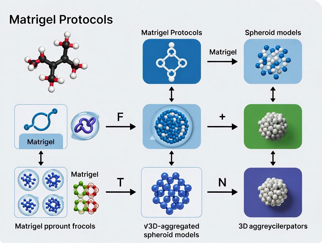
Mastering Matrigel Protocols: A Complete Guide to Optimized 3D Spheroid Models for Drug Discovery
This comprehensive guide provides researchers and drug development professionals with essential knowledge and practical protocols for establishing and optimizing 3D aggregated spheroid models using Matrigel.
Mastering Matrigel Protocols: A Complete Guide to Optimized 3D Spheroid Models for Drug Discovery
Abstract
This comprehensive guide provides researchers and drug development professionals with essential knowledge and practical protocols for establishing and optimizing 3D aggregated spheroid models using Matrigel. Covering foundational principles, step-by-step methodologies, troubleshooting strategies, and validation techniques, this article synthesizes current best practices for creating physiologically relevant tumor microenvironments. Readers will gain actionable insights for implementing robust spheroid models in cancer research, high-throughput screening, and preclinical drug efficacy testing.
Why Matrigel? Understanding the Foundation of 3D Spheroid Microenvironments
Three-dimensional aggregated spheroids represent a sophisticated in vitro model that recapitulates critical aspects of the tumor microenvironment, including hypoxia, nutrient gradients, and cell-cell/extracellular matrix (ECM) interactions. Distinguishing them from simple cell clusters, true spheroids exhibit self-assembled architecture, proliferative heterogeneity, and emergent drug response profiles. This application note, framed within a thesis on standardized Matrigel protocols, details the generation, characterization, and application of 3D aggregated spheroids for advanced oncology research and drug development.
The transition from 2D monolayers to 3D models marks a pivotal advancement in biomedical research. However, not all 3D structures are equivalent. While "cell clusters" may form through casual aggregation, "3D aggregated spheroids" are defined by specific criteria:
- Self-Assembly & Compactness: Spontaneous organization into a dense, spherical structure with defined radial symmetry.
- Microenvironmental Gradients: Establishment of concentric zones of proliferation, quiescence, and necrosis, driven by diffusion limits.
- ECM Deposition & Remodeling: Active production and interaction with a endogenous and/or exogenous ECM.
- Enhanced Pathophysiological Relevance: Mimicry of in vivo signaling, drug penetration barriers, and resistance mechanisms.
The integration of basement membrane extracts, like Matrigel, is crucial for inducing and supporting this complex phenotype, moving beyond inert hanging-drop aggregates.
Key Characteristics & Quantitative Benchmarks
The following table summarizes defining quantitative metrics that differentiate structured spheroids from simple clusters.
Table 1: Quantitative Parameters Defining 3D Aggregated Spheroids
| Parameter | Simple Cell Cluster | Defined 3D Aggregated Spheroid | Common Measurement Technique |
|---|---|---|---|
| Circularity | < 0.85 | ≥ 0.90 | Image analysis (4π*Area/Perimeter²) |
| Diameter Uniformity | High variance (± >50μm) | Low variance (± <20μm) | Brightfield microscopy |
| Hypoxic Core Formation | Absent or minimal (≤10% area) | Present (≥15-30% area) | Pimonidazole staining / HIF-1α IHC |
| Proliferation Gradient | Diffuse, random | Organized, outer rim (Ki67+) | Immunofluorescence quantification |
| ECM Component (Collagen IV) | Low, diffuse | High, organized deposition | Confocal microscopy, ELISA |
| LD50 for Standard Chemo | Often lower, comparable to 2D | Elevated (2-10x increase typical) | Dose-response curve (ATP viability) |
| Viable Rim Thickness | Variable, irregular | Consistent (100-200 μm) | H&E / Live-Dead staining |
Core Protocol: Matrigel-Embedded Spheroid Generation
This protocol is optimized for generating consistent, highly aggregated spheroids suitable for high-throughput screening.
Materials & Reagents (The Scientist's Toolkit)
Table 2: Essential Research Reagent Solutions
| Item | Function | Example Product / Specification |
|---|---|---|
| Growth Factor-Reduced (GFR) Matrigel | Provides defined, laminin-rich ECM for polarization and signaling without variable GF interference. | Corning Matrigel GFR, Phenol Red-free |
| Spheroid Formation Plate | Promotes forced aggregation via ultra-low attachment (ULA) coating. | Corning Elplasia or Nunclon Sphera ULA plate |
| Complete Assay Medium | Cell-type specific medium, often with reduced serum. | e.g., DMEM/F12 + 2% FBS + 1x Pen/Strep |
| Dispase Solution (or equivalent) | Enzymatic recovery of spheroids intact from Matrigel. | Dispase II, 5 mg/mL in PBS |
| Cell Strainer (40μm) | Size selection for uniform single-cell suspension prior to plating. | Falcon 40μm Nylon Cell Strainer |
| Viability/Proliferation Assay Kit | 3D-optimized ATP quantification assay. | CellTiter-Glo 3D |
Step-by-Step Workflow
- Matrigel Preparation: Thaw GFR Matrigel overnight at 4°C. Pre-chill all tubes and tips.
- Single-Cell Suspension: Harvest cells, filter through a 40μm strainer, and count. Adjust concentration to 1.0-2.5 x 10⁵ cells/mL in cold complete medium.
- Matrigel-Cell Mix: On ice, mix cell suspension with thawed Matrigel to a final Matrigel concentration of 4-5 mg/mL. Maintain on ice to prevent polymerization.
- Plating: Pipette 50 μL of the cell-Matrigel mixture per well into a pre-warmed ULA 96-well plate. Avoid bubbles.
- Polymerization: Incubate plate at 37°C for 45 minutes to allow complete Matrigel gelation.
- Overlay & Culture: Gently add 100 μL of pre-warmed complete medium on top of each polymerized gel. Culture for 3-7 days, with medium changes every 2-3 days.
- Spheroid Harvest (Optional): For endpoint assays requiring extraction, add 100 μL of Dispase solution (5 mg/mL) per well. Incubate 1-2 hrs at 37°C. Gently pipette to dissolve Matrigel and collect spheroids.
Diagram 1: Spheroid Generation & Analysis Workflow
Protocol for Key Characterization Assays
Immunofluorescence for Zonal Markers
- Fixation: Add 4% PFA directly to well, incubate 1 hour at RT.
- Permeabilization/Blocking: Remove PFA, wash 3x PBS. Permeabilize/block with PBS containing 0.5% Triton X-100, 5% BSA, 1 hour.
- Staining: Incubate with primary antibodies (e.g., anti-Ki67, anti-Collagen IV, anti-HIF-1α) in blocking buffer, 4°C overnight. Wash 3x, add fluorescent secondaries + DAPI (1:1000), 4 hours RT.
- Imaging: Acquire z-stacks using confocal microscopy. Analyze radial intensity profiles.
Drug Sensitivity Testing (LD50 Determination)
- Day 0: Generate spheroids as per core protocol.
- Day 3: Treat with compound serial dilution (typically 8-point, 1:3). Include DMSO vehicle control.
- Day 6: Perform viability assay. Add equal volume of CellTiter-Glo 3D reagent, shake orbially for 5 min, incubate 25 min in dark, record luminescence.
- Analysis: Normalize to vehicle control (100%). Fit normalized data to a 4-parameter logistic curve to calculate LD50/IC50.
Signaling Pathways in Mature Spheroids
The aggregated 3D structure activates pathways distinct from 2D culture. Matrigel provides key ligands for integrin-mediated signaling.
Diagram 2: Core Spheroid Signaling Network
Interpretation
The diagram illustrates how Matrigel engagement initiates integrin-FAK signaling, promoting survival via PI3K/Akt/mTOR. Concurrently, physical constraints create nutrient/growth factor gradients and a hypoxic core, which stabilizes HIF-1α. HIF-1α drives EMT-like programs and further augments survival pathways, collectively establishing the hallmark drug-resistant phenotype of solid tumors.
Defined 3D aggregated spheroids, engineered using standardized Matrigel protocols, are a non-negotiable tool for translational research. They provide a physiologically relevant platform for:
- Pre-clinical Drug Screening: Identifying compounds that overcome penetration and hypoxia-induced resistance.
- Radiation Biology Studies: Modeling radioresistance in hypoxic microenvironments.
- Immunotherapy Development: Investigating T-cell infiltration into dense ECM barriers.
- Metastasis Research: Studying invasion through a defined basement membrane.
Consistent generation and rigorous characterization using the parameters and protocols outlined herein are critical for obtaining reproducible, biologically meaningful data that bridges the gap between traditional in vitro and costly in vivo models.
Matrigel, a solubilized basement membrane extract derived from the Engelbreth-Holm-Swarm (EHS) mouse sarcoma, is a cornerstone reagent for creating physiologically relevant 3D cell culture environments. Its complex, biologically active composition mimics the in vivo extracellular matrix (ECM), making it indispensable for research involving 3D-aggregated spheroid models, organoid culture, and drug screening. This application note, framed within a thesis on advanced Matrigel protocols for spheroid research, details the key components of Matrigel, their functions, and provides standardized protocols for their application in 3D model systems.
Key ECM Components and Quantitative Composition
Matrigel's composition is a complex mixture of proteins, proteoglycans, and growth factors. The exact proportions can vary between lots, but core components are consistently present.
Table 1: Core Protein Composition of Matrigel
| Component | Approximate % of Total Protein | Primary Biological Function in 3D Models |
|---|---|---|
| Laminin | 50-60% | Major structural protein; promotes cell adhesion, polarization, and survival via integrin binding (e.g., α6β1, α3β1). Initiates basement membrane assembly. |
| Collagen IV | 20-30% | Provides structural meshwork; binds cells via integrins (α1β1, α2β1) and DDR receptors; influences mechanotransduction. |
| Entactin/Nidogen | 5-10% | Bridging molecule; connects laminin and collagen IV networks, stabilizing the ECM structure. |
| Perlecan (HSPG2) | 2-5% | Heparan sulfate proteoglycan; binds and sequesters growth factors (e.g., FGF2, VEGF); regulates bioavailability and signaling. |
Table 2: Key Growth Factors and Other Components in Matrigel
| Component | Typical Concentration Range | Function in 3D Spheroid Context |
|---|---|---|
| TGF-β | 1-5 ng/mL | Induces epithelial-to-mesenchymal transition (EMT); regulates differentiation and ECM production. |
| EGF | 0.5-2 ng/mL | Stimulates epithelial cell proliferation and survival. |
| IGF-1 | 1-5 ng/mL | Promotes cell growth and metabolic activity. |
| FGF | 1-10 ng/mL | Angiogenesis stimulation; stem cell maintenance. |
| PDGF | 0.5-2 ng/mL | Influences stromal cell recruitment and function. |
| Matrix Metalloproteinases (MMPs) | Variable | Facilitate ECM remodeling and spheroid invasion. |
Biological Functions in 3D Spheroid Models
The integrated function of these components creates a bioactive scaffold essential for advanced 3D models.
- Structural and Mechanical Support: The laminin-collagen IV-entactin network forms a viscoelastic gel at 37°C, providing a 3D physical scaffold that influences spheroid morphology, compaction, and intracellular tension.
- Cell Signaling and Differentiation: ECM ligands engage integrin receptors, activating downstream pathways (e.g., PI3K/Akt, FAK, MAPK) crucial for cell survival, proliferation, and differentiation. Growth factors sequestered in the matrix are released in a controlled manner, creating morphogen gradients.
- Polarization and Morphogenesis: Laminin-rich environments are critical for establishing apical-basal polarity in epithelial spheroids and organoids, driving lumen formation and proper tissue architecture.
- Invasion and Metastasis Modeling: For cancer spheroids, Matrigel serves as an invasive substrate. Cells secrete proteases to degrade and remodel the matrix, enabling the study of metastatic mechanisms.
Detailed Protocols for 3D-Aggregated Spheroid Research
Protocol 1: Standardized Matrigel-Embedded Spheroid Formation for Drug Screening
Objective: Generate uniform, reproducible spheroids embedded in Matrigel for high-content analysis of drug response.
The Scientist's Toolkit:
| Reagent/Material | Function in Protocol |
|---|---|
| Growth Factor-Reduced (GFR) Matrigel | Standardized, lower GF content for controlled signaling studies. |
| Pre-chilled (4°C) Pipette Tips & Tubes | Prevents premature gelation of Matrigel during handling. |
| 96-well U-bottom Ultra-Low Attachment (ULA) Plate | Enforces forced aggregation for spheroid formation prior to embedding. |
| Chilled Basal Medium (e.g., DMEM) | Used to dilute Matrigel to desired working concentration without polymerization. |
| 37°C, 5% CO2 Incubator | For consistent, stable gel polymerization. |
Methodology:
- Preparation: Thaw Matrigel overnight at 4°C on ice. Chill all tubes, tips, and media on ice.
- Spheroid Aggregation: Harvest single-cell suspension. Seed 100-500 cells/well in 100 µL of complete medium into a 96-well ULA plate. Centrifuge plate at 300 x g for 3 minutes to aggregate cells. Incubate for 48-72 hours to form a single, compact spheroid per well.
- Matrigel Embedding: Prepare a 4 mg/mL working solution of GFR Matrigel by diluting with cold basal medium. Carefully aspirate 80 µL of medium from each spheroid well, leaving ~20 µL containing the spheroid.
- Gelation: Slowly add 50 µL of the chilled Matrigel solution per well, gently pipetting to mix and suspend the spheroid in the matrix. Incubate the plate at 37°C for 30 minutes to allow complete polymerization.
- Overlay and Assay: Add 100 µL of complete culture medium (with or without test compounds) on top of the polymerized Matrigel dome. Refresh medium/drug every 2-3 days.
- Endpoint Analysis: Image spheroids using brightfield or fluorescence microscopy. Quantify parameters like spheroid area, viability (Calcein AM/EthD-1), or invasion area.
Protocol 2: Assessing Spheroid Invasion in Matrigel
Objective: Quantify the invasive potential of cancer spheroids into a surrounding Matrigel matrix.
Methodology:
- Spheroid Formation: Generate single spheroids in a ULA plate as per Protocol 1, Steps 1-2.
- Invasion Matrix Preparation: Coat each well of a flat-bottom 96-well plate with 50 µL of pure GFR Matrigel. Polymerize at 37°C for 30 min to form a thin base layer.
- Spheroid Seeding: Transfer pre-formed spheroids individually onto the center of the base layer using a wide-bore tip.
- Overlay and Challenge: Immediately overlay each spheroid with 50 µL of chilled, diluted Matrigel (3-4 mg/mL). Polymerize. Add 100 µL of medium containing chemoattractant (e.g., 10% FBS) or inhibitor.
- Imaging and Quantification: Acquire daily brightfield images at 4x or 10x magnification for up to 7 days. Use image analysis software (e.g., ImageJ) to measure the total spheroid area and the core area (dense, non-invasive center). Calculate invasive area = total area - core area.
Signaling Pathways Visualized
Diagram 1: Key Signaling Pathways from Matrigel in Spheroids
Diagram 2: Workflow for Matrigel Spheroid Embedding Protocol
Matrigel, a laminin-rich extracellular matrix (ECM) hydrogel, is a cornerstone for creating physiologically relevant 3D models of the tumor microenvironment (TME). Derived from Engelbreth-Holm-Swarm (EHS) mouse sarcoma, its complex composition mimics the native basement membrane, providing critical biochemical and biophysical cues. Within the context of 3D-aggregated spheroid research, Matrigel facilitates the study of cell-ECM interactions, tumor morphology, invasion, drug response, and signaling pathway activation in a manner that far surpasses conventional 2D culture.
Quantitative Data on Matrigel Composition and Impact
Table 1: Key Components of Matrigel and Their Functional Roles in TME Mimicry
| Component | Approximate Concentration (%) | Primary Function in TME Model |
|---|---|---|
| Laminin | ~60% | Cell adhesion, polarization, survival signaling |
| Type IV Collagen | ~30% | Structural integrity, mechanical signaling |
| Entactin/Nidogen | ~8% | Bridges laminin and collagen networks |
| Heparan Sulfate Proteoglycans (e.g., Perlecan) | ~2% | Growth factor binding and presentation |
| Growth Factors (e.g., TGF-β, EGF, IGF, FGF) | Trace, variable | Autocrine/paracrine signaling, proliferation, differentiation |
Table 2: Comparative Analysis of Spheroid Phenotypes in 2D vs. 3D Matrigel Culture
| Parameter | 2D Monolayer Culture | 3D Spheroid in Matrigel |
|---|---|---|
| Proliferation Rate | High, exponential | Reduced, more in vivo-like |
| Apoptosis Gradient | Uniform | Core-specific (hypoxia/nutrient deprivation) |
| Drug IC50 Values | Often significantly lower | Higher, recapitulating clinical drug resistance |
| Morphology | Flat, spread | Organized, aggregated, with invasive protrusions |
| Gene Expression Profile | Often de-differentiated | More differentiated, tumor-specific |
Detailed Application Notes & Protocols
Protocol 1: Establishing 3D Invasive Spheroid Co-cultures in Matrigel
Objective: To model cancer cell invasion into the stromal compartment within a TME-mimetic matrix.
The Scientist's Toolkit: Research Reagent Solutions
| Item | Function |
|---|---|
| Growth Factor Reduced (GFR) Matrigel | Reduces confounding mitogenic signals for cleaner invasion assays. |
| Phenol Red-free Matrigel | Allows for unimpeded fluorescence imaging and quantification. |
| High-Concentration (HC) Matrigel | For studies requiring high stiffness and dense matrix barriers. |
| Organoid Culture Qualified Matrigel | Optimized for stem cell and patient-derived organoid viability. |
| Cold-reduced growth medium | Prevents premature Matrigel gelling during cell mixing. |
| Pre-chilled tips and tubes | Maintains Matrigel in liquid state for accurate pipetting. |
| 24-well or 96-well glass-bottom plates | Optimized for high-resolution microscopy of invasion. |
Methodology:
- Spheroid Formation: Generate uniform spheroids (300-500 µm) from your cancer cell line using a hanging drop or ultra-low attachment plate method over 48-72 hours.
- Matrix Preparation: Thaw Matrigel overnight at 4°C on ice. Keep all reagents and tools on ice.
- Embedding: Transfer single spheroids into a 1.5 mL tube. Gently mix with cold Matrigel at a final concentration of 4-6 mg/mL (approx. 50-100 µL total volume). Avoid bubbles.
- Plating: Pipette the Matrigel-spheroid mixture as a single droplet into the center of a well in a pre-warmed plate. Incubate at 37°C for 30 minutes to allow polymerization.
- Overlay: Carefully add pre-warmed complete culture medium (with or without stromal cells like cancer-associated fibroblasts) on top of the polymerized Matrigel dome.
- Culture & Analysis: Culture for 5-14 days, changing medium every 2-3 days. Monitor invasion by phase-contrast or confocal microscopy. Quantify invasive area using image analysis software (e.g., ImageJ).
Protocol 2: Drug Response Assessment in 3D Matrigel-Embedded Spheroids
Objective: To evaluate chemotherapeutic or targeted drug efficacy in a physiologically relevant 3D TME context.
Methodology:
- Spheroid Generation & Embedding: Follow Protocol 1, steps 1-5, using a 96-well plate format for high-throughput screening.
- Drug Treatment: After 24 hours of embedding, add serial dilutions of the test compound in fresh medium. Include vehicle controls.
- Viability Assay: At endpoint (typically 72-120 hours), assess viability using 3D-optimized assays:
- CellTiter-Glo 3D: Aspirate medium, add equal volume of CellTiter-Glo 3D reagent, shake orbially for 5 minutes, incubate 25 minutes, and record luminescence.
- Calcein AM/Propidium Iodide (PI) Staining: Image live/dead cells using confocal microscopy. Calcein AM (green) labels live cells, PI (red) labels dead cells.
- Data Analysis: Normalize luminescence or live/dead ratio to vehicle controls. Calculate IC50 values using non-linear regression (log inhibitor vs. response).
Signaling Pathways in the TME Modelled by Matrigel
Diagram Title: Matrigel-Induced Pro-Survival and Invasion Signaling
Experimental Workflow for TME Spheroid Analysis
Diagram Title: 3D TME Spheroid Model Workflow
Within the thesis on Matrigel protocols for 3D-aggregated spheroid research, a critical step is hydrogel selection. This application note provides a comparative framework and practical protocols to guide this decision.
Key Property Comparison Table
| Property | Matrigel | Collagen I | Alginate | Synthetic Polymers (e.g., PEG) |
|---|---|---|---|---|
| Origin & Composition | Basement membrane extract (mouse sarcoma); laminin, collagen IV, entactin, growth factors. | Natural protein (bovine/rat/marine); primarily collagen I fibers. | Natural polysaccharide (brown seaweed); guluronic and mannuronic acid blocks. | Fully synthetic (e.g., Polyethylene glycol); chemically defined. |
| Mechanism of Gelation | Thermoreversible (liquid at 4°C, gels at 20-37°C). | pH/temperature-driven self-assembly of fibrils. | Ionic crosslinking (e.g., with Ca²⁺). | Photo-, chemical, or Michael addition crosslinking. |
| Bioactivity | High. Contains endogenous bioactive cues (e.g., laminin-111) and growth factors that promote complex morphogenesis. | Moderate. Integrin-binding RGD motifs support adhesion and migration. | None (inert). Requires functionalization (e.g., RGD peptides) for cell adhesion. | None (inert). Highly tunable via incorporation of bioactive motifs. |
| Mechanical Tunability | Low. Stiffness is batch-dependent (~0.5-5 kPa). | Moderate. Stiffness tunable via concentration (~0.1-10 kPa). | Moderate-High. Stiffness tunable via crosslink density (~0.1-100 kPa). | High. Precise control over stiffness and viscoelasticity (~0.1-100+ kPa). |
| Batch Consistency | Low. Variable composition due to biological source. | Moderate. Improved with recombinant sources. | High. Consistent polymer chemistry. | Very High. Chemically defined. |
| Degradation | Proteolytic (cell-driven). | Proteolytic (MMP-sensitive). | Ion exchange (non-enzymatic) or slow hydrolysis. | Tunable (often designed to be MMP-sensitive). |
| Primary Advantage for Spheroids | Promotes complex, polarized, & invasive structures (e.g., tubulogenesis). | Excellent for mesenchymal cell migration & contraction. | Ideal for encapsulation & mechanical studies; low cell adhesion. | Ultimate control over biochemical & biophysical variables. |
| Key Limitation | Poorly defined, animal-derived, tumor-derived. | Less suitable for epithelial polarity vs. Matrigel. | Requires modification for cell adhesion; non-proteolytic degradation. | Requires expertise to functionalize; can lack natural complexity. |
Functional Outcome Table in Spheroid Models
| Experimental Goal | Recommended Hydrogel (Rationale) | Expected Spheroid Outcome |
|---|---|---|
| Organoid formation from stem cells | Matrigel or Collagen I (for specific lineages). | Lumen formation, branching, and crypt-like structures. |
| Cancer cell invasion assay | Matrigel (provides physiological basement membrane barriers). | Invasive protrusions and collective cell migration. |
| Mechanotransduction studies | Synthetic PEG or Alginate (precise stiffness control). | Altered proliferation/apoptosis based on matrix stiffness. |
| High-throughput drug screening | Alginate or Synthetic PEG (high consistency, minimal batch effects). | Uniform spheroids for reproducible cytotoxicity metrics. |
| Angiogenesis assay | Matrigel (rich in pro-angiogenic factors). | Endothelial cell sprouting and tube network formation. |
Experimental Protocols
Protocol: Spheroid Invasion in Matrigel vs. Collagen I
Objective: To compare the invasive phenotype of cancer spheroids in physiologically bioactive (Matrigel) versus structural (Collagen I) matrices.
Research Reagent Solutions & Materials:
| Item | Function |
|---|---|
| Growth Factor-Reduced (GFR) Matrigel, Corning | Reduces variable growth factor impact, focusing on matrix effects. |
| Rat Tail Collagen I, High Concentration | Provides a pure, fibrillar collagen network. |
| 96-well U-bottom Ultra-Low Attachment (ULA) Plates | Enforces scaffold-free spheroid formation via forced aggregation. |
| Fluorescent Cell Tracker Dye (e.g., CMFDA) | Pre-labels spheroids for clear visualization against matrix. |
| Calcein AM / Propidium Iodide Viability Stain | Live/Dead endpoint assessment. |
| Confocal-Compatible 96-well Imaging Plates | For high-resolution 3D imaging of invasion. |
Methodology:
- Spheroid Formation: Harvest single-cell suspension of cancer cells (e.g., MDA-MB-231). Seed 5,000 cells/well in U-bottom plates. Centrifuge at 300 x g for 3 min. Culture for 48-72h to form compact spheroids.
- Hydrogel Preparation:
- Matrigel: Thaw on ice overnight. Keep all tips and plates on ice. Dilute with cold serum-free medium to desired concentration (e.g., 4 mg/mL).
- Collagen I: Neutralize stock with sterile 0.1M NaOH and 10x PBS on ice to pH ~7.4. Dilute with serum-free medium to same concentration (e.g., 4 mg/mL).
- Embedding: Using cold tips, carefully aspirate medium from spheroid wells. Gently resuspend each spheroid in 50 µL of cold hydrogel solution. Pipette the mixture into a pre-chilled well of the imaging plate. Incubate at 37°C for 30 min to gel.
- Culture & Imaging: Overlay with complete culture medium. Image daily using an inverted confocal microscope. Capture Z-stacks to measure invasive area (total spheroid area minus initial core area) using ImageJ software.
- Analysis: Quantify invasion distance, number of protrusions, and spheroid circularity. Perform statistical comparison between matrices.
Protocol: Assessing Matrix-Dependent Drug Response in Alginate vs. Matrigel
Objective: To evaluate how an inert vs. a bioactive matrix modulates spheroid response to chemotherapeutics.
Methodology:
- Spheroid Formation: Form uniform spheroids in U-bottom plates as in Protocol 2.1.
- Encapsulation:
- Alginate: Mix spheroids with 1.5% (w/v) sterile alginate solution. Pipette droplets into a 100mM CaCl₂ solution to form beads. Transfer beads to wells.
- Matrigel: Embed spheroids as in Protocol 2.1, Step 3.
- Drug Treatment: After 24h of culture, overlay medium containing a gradient of the chemotherapeutic (e.g., Doxorubicin, 0-10 µM). Include untreated controls.
- Viability Assessment: At 72h post-treatment, incubate with Calcein AM (2 µM) and Propidium Iodide (4 µM) for 1h. Acquire confocal Z-stacks.
- Analysis: Quantify live and dead fluorescence volumes. Generate dose-response curves and calculate IC50 values for each hydrogel condition. Compare significant shifts.
Signaling Pathways in Matrigel-Driven Morphogenesis
Title: Matrigel Signaling in Spheroid Morphogenesis
Experimental Workflow for Hydrogel Comparison
Title: Hydrogel Comparison Workflow for Spheroid Research
Application Notes
Within the broader thesis on standardizing Matrigel protocols for 3D-aggregated spheroid models, understanding the critical parameters of Matrigel handling is paramount. Matrigel is a basement membrane extract with inherent biological complexity, making its physical and functional properties highly sensitive to procedural variables. This document details the impact of concentration, polymerization temperature, and batch variability on spheroid morphology, growth, and downstream assay reproducibility.
1. Concentration Matrigel concentration directly influences matrix stiffness, pore size, and ligand density. For 3D spheroid formation, optimal concentration balances mechanical support with nutrient diffusion.
- Low Concentration (<4 mg/mL): Results in a soft, loose gel. Spheroids may exhibit increased invasion, uncontrolled aggregation, or disintegration.
- Optimal Range (4-8 mg/mL): Provides a structural scaffold that promotes coherent, compact spheroid formation with well-defined borders, suitable for proliferation and drug response assays.
- High Concentration (>10 mg/mL): Creates a dense, stiff matrix that can restrict spheroid growth, limit nutrient/waste exchange, and may induce hypoxia in the core prematurely.
Table 1: Effect of Matrigel Concentration on Spheroid Phenotype
| Concentration (mg/mL) | Median Stiffness (Pa) | Average Spheroid Diameter (Day 5) | Morphology Score (1-5) |
|---|---|---|---|
| 3 | ~150 | 450 ± 120 µm | 2 (Irregular, loose) |
| 5 | ~450 | 350 ± 45 µm | 4 (Compact, spherical) |
| 7 | ~750 | 300 ± 30 µm | 5 (Very compact) |
| 10 | ~1200 | 250 ± 35 µm | 3 (Compact, but stunted) |
2. Polymerization Temperature The temperature at which Matrigel polymerizes is critical for forming a homogeneous hydrogel. Matrigel transitions from liquid to gel at 22-35°C.
- Protocol A: On-Ice Handling & Cold Polymerization: Pipetting pre-chilled Matrigel on cold plates/surfaces leads to premature gelation and uneven droplet formation, causing high intra-experimental variability.
- Protocol B: Pre-warmed Surfaces & 37°C Polymerization (Recommended): Using pre-warmed tips, plates, and media ensures liquid Matrigel is delivered uniformly and polymerizes rapidly at 37°C, resulting in consistent dome or overlay formation.
Table 2: Impact of Polymerization Protocol on Gel Homogeneity
| Parameter | Protocol A (Cold) | Protocol B (37°C) |
|---|---|---|
| Gelation Time | 30-60 minutes (slow, uneven) | 10-15 minutes (rapid, uniform) |
| Spheroid Circularity | 0.75 ± 0.15 | 0.92 ± 0.05 |
| Coefficient of Variation in Diameter | 25% | 8% |
3. Batch Variability Matrigel is a natural product; its composition (laminin, collagen IV, entactin, growth factors) varies between production lots. This is a major confounding factor in long-term or multi-site studies.
Table 3: Representative Batch Analysis for Key Components
| Lot Number | Total Protein (mg/mL) | Laminin (%) | Growth Factor Activity (Relative Units) | Optimal Spheroid Conc. |
|---|---|---|---|---|
| ABC123 | 9.8 | 62% | 1.00 | 5 mg/mL |
| DEF456 | 11.2 | 58% | 1.35 | 6 mg/mL |
| GHI789 | 8.5 | 65% | 0.85 | 4.5 mg/mL |
Experimental Protocols
Protocol: Standardized 3D Spheroid Formation in Matrigel Objective: To generate consistent, compact spheroids for drug screening by controlling critical parameters.
I. Pre-Experimental Setup (Key to Reproducibility)
- Thawing: Thaw a Matrigel aliquot overnight at 4°C on ice. Never use a water bath or 37°C incubator.
- Pre-chill Equipment: Chill all pipette tips, tubes, and the syringe (if using) at -20°C for 30 minutes or keep on ice.
- Pre-warm Equipment: Place a 96-well or 384-well ultra-low attachment (ULA) plate in a 37°C incubator for at least 30 minutes.
- Record Data: Note the Matrigel Lot Number and certified Protein Concentration.
II. Spheroid Seeding in Matrigel Dome (50 µL total volume example)
- Prepare a single-cell suspension of your cell line (e.g., HCT116, MCF7) in complete medium at 2x the desired final density (e.g., 1000 cells/50µL final -> 2000 cells/50µL in suspension).
- On ice, prepare the Matrigel-Cell Mixture:
- Dilute the thawed Matrigel with cold serum-free medium to the target concentration (e.g., 5 mg/mL) in a pre-chilled tube.
- Mix the cell suspension with the diluted, cold Matrigel in a 1:1 ratio. Gently pipette to mix. Work quickly to prevent gelation.
- Immediately pipette 50 µL of the Matrigel-cell mixture as a central droplet into each pre-warmed well of the ULA plate.
- Transfer the plate directly to the 37°C incubator for 15-20 minutes to allow polymerization.
- After the gel is set, carefully overlay each dome with 100 µL of pre-warmed complete medium.
- Culture for 3-7 days, with medium changes every other day.
III. Batch Qualification Protocol
- Test Multiple Lots: Upon receiving new lots, perform a parallel spheroid formation assay using your standard protocol.
- Titrate Concentration: For each new lot, test a concentration range (e.g., 4, 6, 8 mg/mL) using a standard cell line.
- Quantitative Endpoints: At day 5, image spheroids and measure:
- Diameter and circularity (ImageJ).
- Viability (e.g., ATP-based assay).
- Response to a reference cytotoxic drug (e.g., 5-FU for CRC lines).
- Select & Reserve: Choose the lot yielding desired morphology and response. Reserve a sufficient quantity of this qualified lot for the entire study.
The Scientist's Toolkit
| Research Reagent Solution | Function & Criticality |
|---|---|
| Growth Factor Reduced (GFR) Matrigel | Standardizes matrix by reducing variable growth factor levels, crucial for studies involving added growth factors or inhibitors. |
| Phenol Red-Free Matrigel | Eliminates phenol red interference in fluorescence-based assays and high-content imaging. |
| Ultra-Low Attachment (ULA) Plates | Prevents cell attachment to the plastic, forcing aggregation and spheroid formation within the Matrigel dome. |
| Pre-Chilled, Low-Binding Pipette Tips | Minimizes Matrigel loss and premature warming during pipetting. |
| Serum-Free, Pre-Chilled Medium | For diluting Matrigel without introducing variable serum components that can affect polymerization. |
| Liquid Handling System (with temp control) | For high-throughput applications, ensures rapid, uniform dispensing of cold Matrigel into warm plates. |
Visualizations
Matrigel Handling Protocol Impact
Key Parameters Influence Spheroid Phenotype
Experimental Workflow for Batch Qualification
Step-by-Step Matrigel Protocols: From Thawing to Analysis
Within the context of a broader thesis on Matrigel protocols for 3D-aggregated spheroid models, meticulous pre-protocol preparation is foundational. Corning Matrigel matrix and similar basement membrane extracts (BME) are essential for creating physiologically relevant microenvironments. Improper handling, thawing, and aliquoting lead to batch variability, hydrogel inconsistency, and compromised experimental reproducibility in drug screening and tumor biology research.
Critical Material Properties & Handling Principles
Matrigel is a temperature-sensitive, laminin-rich hydrogel. Its polymerization is irreversible upon incubation at 37°C. Key challenges include lot-to-lot variability, sensitivity to premature warming, and susceptibility to proteolytic degradation.
Table 1: Quantitative Properties of Standard Growth Factor-Reduced Matrigel
| Property | Typical Value/Range | Impact on 3D Spheroid Culture |
|---|---|---|
| Protein Concentration | 8-12 mg/mL | Affects hydrogel stiffness and porosity. |
| Growth Factor Content | Reduced (e.g., TGF-β < 5 ng/mL) | Minimizes uncontrolled differentiation. |
| Gelation Time (37°C) | 30-60 minutes | Determines plating workflow timing. |
| Storage Temperature | -20°C to -80°C | Long-term stability requires ≤ -20°C. |
| Aliquot Volume | 100 µL to 1 mL | Balances usability and freeze-thaw cycles. |
Detailed Protocols
Protocol 1: Safe Thawing of Matrigel for Spheroid Assays
Objective: To liquefy Matrigel homogeneously without partial polymerization or degradation.
- Preparation: Pre-chill sterile pipettes, tubes, and cultureware at 4°C overnight. Prepare an ice bucket with slurry (crushed ice and water).
- Transfer: Quickly move the desired vial from -80°C storage to the ice slurry. Ensure the cap is above ice level to prevent contamination.
- Thawing: Allow the vial to thaw completely on ice slurry for approximately 2-3 hours (for a 10 mL vial). Do not use a refrigerator at 4°C, as thawing is uneven. Never use a water bath or incubator.
- Mixing: Once liquid, gently swirl the vial on ice. Avoid introducing bubbles. Do not vortex or vigorously pipette.
- Immediate Use: Keep the liquid Matrigel on ice at all times during subsequent aliquoting or experimental use. The working time on ice is typically 1-2 hours.
Protocol 2: Aliquoting Thawed Matrigel
Objective: To create single-use aliquots, minimizing freeze-thaw cycles and contamination risk.
- Pre-cool: Work in a cold room (4°C) or on a pre-chilled cooling block. Keep all materials on ice.
- Aseptic Technique: Wipe the vial septum and working area with 70% ethanol.
- Aliquot Volume: Using chilled sterile serological pipettes or micropipette tips, dispense volumes appropriate for a single experiment (e.g., 250 µL for coating 24-well plates).
- Container: Use pre-chilled, sterile, low-protein-binding microcentrifuge tubes. Label clearly with product, lot number, concentration, date, and aliquot number.
- Snap-Freeze: Place aliquots directly into a pre-chilled float in a -80°C freezer, or suspend in a dry ice/ethanol bath for 10 minutes before transferring to -80°C storage.
- Record Keeping: Maintain a detailed log of aliquot use to track freeze-thaw history.
Protocol 3: Coating Plates for 3D Spheroid Embedding
Objective: To create a thin base layer of gelled Matrigel to support spheroid cultures.
- Dilution (Optional): If required, dilute the liquid Matrigel with chilled serum-free medium on ice. Mix by gentle pipetting.
- Coating: Pipette the desired volume (e.g., 50 µL/well for a 96-well plate) into each well.
- Gelation: Incubate the plate at 37°C in a humidified incubator for 30 minutes to allow complete polymerization.
- Seeding Spheroids: Once gelled, immediately plate pre-formed spheroids in medium onto the coated surface. For embedding, mix spheroids with liquid Matrigel on ice before plating and gelling.
The Scientist's Toolkit: Research Reagent Solutions
Table 2: Essential Materials for Matrigel Handling in 3D Spheroid Research
| Item | Function & Rationale |
|---|---|
| Corning Matrigel GFR | Gold-standard BME for organoid/spheroid growth due to reduced growth factor interference. |
| Pre-Chilled Sterile Tips/Tubes | Prevents premature gelling during liquid handling. Low-protein-binding surfaces minimize loss. |
| Ice Bucket with Slurry | Maintains a stable 0°C environment for thawing and handling, superior to ice alone. |
| Cooling Blocks/ Cold Room | Provides a large, stable cold surface for extended aliquot preparation workflows. |
| Sterile Serological Pipettes | Allows rapid, accurate transfer of viscous Matrigel while kept cold. |
| Low-Adhesion Spheroid Plates (e.g., U-bottom) | For pre-forming uniform spheroids via the hanging-drop or forced-aggregation method prior to Matrigel embedding. |
| Liquid Nitrogen or Dry Ice | For rapid snap-freezing of aliquots to prevent ice crystal formation and matrix damage. |
Experimental Workflow and Signaling Context
Title: Matrigel Handling and 3D Spheroid Workflow
Title: Matrigel-Induced Signaling in Spheroids
Thesis Context: This protocol is a cornerstone methodology within a broader thesis investigating standardized Matrigel protocols for 3D-aggregated spheroid models. It establishes a robust, quantitative framework for assessing invasive potential, crucial for modeling metastasis and evaluating anti-invasive therapeutics.
The embedded spheroid invasion assay is a gold-standard in vitro technique for modeling the complex, multi-step process of cancer cell invasion into a surrogate extracellular matrix (ECM). Unlike seeding spheroids on top of a gel, embedding them within a three-dimensional Matrigel matrix provides a more physiologically relevant microenvironment, exposing the entire spheroid surface to matrix-derived biochemical and biophysical cues. This method yields high-fidelity data on invasive capacity, characterized by the formation of protrusive, multicellular strands. Accurate quantification of this invasive phenotype is critical for developmental biology, cancer research, and drug discovery.
Key Research Reagent Solutions
| Reagent/Material | Function & Rationale |
|---|---|
| Corning Matrigel Growth Factor Reduced (GFR) | Basement membrane extract providing a physiologically relevant 3D ECM for invasion. The GFR formulation minimizes confounding mitogenic signaling. |
| Advanced DMEM/F-12 | Serum-free culture medium used for diluting Matrigel and maintaining spheroids during assay, ensuring consistency and reducing undefined variables. |
| 96-Well Clear Round-Bottom Ultra-Low Attachment (ULA) Plate | Enables forced-aggregation formation of uniform, single spheroids via liquid overlay technique. |
| Pre-Chilled Non-Treated 96-Well Plate & Tips | Critical for handling Matrigel, which polymerizes above 4-10°C. Pre-chilling prevents premature gelling. |
| Calcein AM Viability Dye | Live-cell fluorescent stain used for high-contrast visualization and subsequent quantification of invasive structures. |
| Fetal Bovine Serum (FBS) | Used as a standard chemoattractant in the underlying medium to induce directional invasion. |
Detailed Protocol
Part A: Spheroid Generation via Forced Aggregation
Objective: To produce a large number of highly uniform, 3D-aggregated spheroids.
- Cell Preparation: Harvest and count cells. Prepare a single-cell suspension in complete growth medium at 2.5 x 10^5 cells/mL.
- Seeding: Aliquot 100 µL of cell suspension per well into a 96-well ULA plate. This yields a starting spheroid of approximately 2,500 cells.
- Aggregation: Centrifuge the plate at 300 x g for 3 minutes to pellet cells into the well bottom. Incubate at 37°C, 5% CO₂ for 48-72 hours to form a single, compact spheroid per well.
Part B: Embedding and Invasion Assay Setup
Objective: To embed pre-formed spheroids within a 3D Matrigel matrix and initiate the invasion assay.
- Matrix Preparation: Thaw Matrigel (GFR) overnight at 4°C. Pre-chill tubes, tips, and a new non-treated 96-well plate on ice.
- Working Solution: On ice, dilute Matrigel to a 4 mg/mL final concentration in cold, serum-free Advanced DMEM/F-12. Maintain on ice.
- Spheroid Transfer: Using pre-chilled wide-bore tips, carefully transfer one compact spheroid per well into the new chilled plate, in 50 µL of its existing medium.
- Embedding: To each well containing a spheroid, gently add 50 µL of the cold 4 mg/mL Matrigel solution. Mix carefully by slow pipetting. Final Matrigel concentration is 2 mg/mL.
- Polymerization: Incubate the plate at 37°C for 30 minutes to allow complete gelation.
- Chemoattractant Addition: After gelation, carefully overlay each well with 100 µL of complete growth medium containing 10% FBS as a chemoattractant.
- Incubation: Incubate the assay plate at 37°C, 5% CO₂ for up to 96 hours, with medium changes every 48 hours. Invasion proceeds radially from the spheroid core.
Part C: Staining, Imaging, and Quantification
Objective: To visualize and quantitatively analyze the invasive phenotype.
- Staining: At endpoint, prepare a 4 µM Calcein AM solution in PBS. Remove culture medium, add 100 µL of dye per well, and incubate for 60 minutes at 37°C.
- Imaging: Image each spheroid using a fluorescent microscope or high-content imaging system with a GFP filter set. Acquire z-stacks (e.g., 100 µm total depth, 10 µm steps) to capture the entire invasive area.
- Quantitative Analysis: Using ImageJ/Fiji or equivalent analysis software:
- Perform a maximum intensity projection.
- Apply a threshold to create a binary mask of the invasive area.
- Use the "Analyze Particles" function to determine the Total Invasive Area (µm²) and the Relative Invasion Distance (µm), calculated as: (Radius of Total Area) - (Radius of Spheroid Core).
Data Presentation: Quantitative Invasion Metrics
Table 1: Typical Invasion Parameters for Reference Cell Lines (96-hour assay, 2 mg/mL GFR Matrigel, 10% FBS chemoattractant).
| Cell Line | Spheroid Core Area (µm²) | Total Invasive Area (µm²) | Relative Invasion Distance (µm) |
|---|---|---|---|
| Non-invasive MCF-10A | 45,200 ± 3,100 | 52,500 ± 4,800 | 15 ± 8 |
| Invasive MDA-MB-231 | 48,500 ± 2,800 | 215,300 ± 18,500 | 105 ± 12 |
| HT-1080 Fibrosarcoma | 46,800 ± 3,400 | 189,700 ± 15,200 | 92 ± 10 |
Table 2: Effect of Matrix Concentration on Invasion Metrics (MDA-MB-231, 96-hour assay).
| Matrigel Concentration (mg/mL) | Total Invasive Area (µm²) | Invasive Branch Count |
|---|---|---|
| 1.0 | 278,400 ± 22,100 | 18 ± 3 |
| 2.0 | 215,300 ± 18,500 | 14 ± 2 |
| 4.0 | 132,500 ± 12,700 | 9 ± 2 |
Experimental Workflow Diagram
Diagram Title: Embedded Spheroid Invasion Assay Workflow
Key Signaling Pathways in Spheroid Invasion
Diagram Title: Core Signaling Pathways Driving 3D Spheroid Invasion
Within the broader thesis on standardized Matrigel protocols for 3D-aggregated spheroid models, the Overlay Method emerges as a critical, simplified technique for long-term culture and compound testing. This protocol details the application of the Overlay method, wherein pre-formed spheroids are seeded onto a thin, solidified bed of extracellular matrix (ECM), such as Matrigel, and subsequently fed with medium without additional embedding. This approach maintains a 3D microenvironment while drastically simplifying experimental workflows, media changes, and endpoint analyses compared to full embedding methods. It is particularly advantageous for high-throughput growth and viability studies in drug development.
The Overlay method offers distinct operational benefits. The following table summarizes key comparative data from recent studies (2023-2024) on colorectal carcinoma spheroid models.
Table 1: Quantitative Comparison of Embedding vs. Overlay Methods for Spheroid Culture
| Parameter | Full Embedding Method | Overlay Method | Notes/Source |
|---|---|---|---|
| Spheroid Formation Time | 72-96 hours | 24-48 hours (pre-formed in ULA plates) | Spheroids formed separately, then transferred. |
| Assay Throughput | Moderate | High | Simplified liquid handling enables more replicates. |
| Viability Assay Compatibility | Low (imaging challenging) | High (easy reagent access) | ATP, resazurin, and live/dead stains perform robustly. |
| Medium Exchange Complexity | High (risk of gel disruption) | Low (standard aspiration) | Overlay reduces technician variability. |
| Typical Invasion/Migration Readout | Excellent (3D constrained) | Limited (2.5D surface) | Overlay is less suitable for invasive studies. |
| Drug IC50 Variability (CV%) | 15-25% | 8-12% | Overlay improves consistency in compound response. |
| Long-term Culture Viability (>14 days) | Good | Excellent | Improved nutrient/waste exchange in overlay. |
Detailed Experimental Protocol
Materials & Reagent Preparation
- Basement Membrane Matrix: Corning Matrigel Growth Factor Reduced (GFR), Phenol Red-free. Store at -80°C. Thaw on ice overnight at 4°C before use.
- Cell Culture Plate: 96-well, flat-bottom, cell culture-treated plate for matrix bed; 96-well, ultra-low attachment (ULA), round-bottom plate for spheroid formation.
- Cold Medium: Serum-free or appropriate basal medium, pre-chilled at 4°C.
- Pre-formed Spheroids: Generated in ULA plates per standard aggregation protocols.
- Assay Reagents: CellTiter-Glo 3D (ATP assay), resazurin, or equivalent viability probes.
Part A: Preparation of the Matrigel Overlay Bed
- Dilution: Dilute ice-cold Matrigel to a working concentration of 4-5 mg/mL in cold, serum-free medium using pre-chilled pipette tips and tubes. Keep on ice.
- Dispensing: Quickly aliquot 50 µL of the diluted Matrigel solution into each well of the flat-bottom 96-well plate.
- Gelation: Immediately transfer the plate to a 37°C, 5% CO₂ incubator for 30-45 minutes to allow complete polymerization, forming a thin, uniform bed.
Part B: Seeding of Pre-formed Spheroids
- Spheroid Formation: Culture cells in a ULA round-bottom plate for 24-48 hours to form compact, single spheroids per well.
- Transfer: Using wide-bore pipette tips (to prevent shear stress), carefully aspirate 50-100 µL of medium containing a single pre-formed spheroid from the ULA plate.
- Overlay: Gently dispense the spheroid-containing medium directly onto the center of the polymerized Matrigel bed. Ensure the spheroid settles onto the surface.
- Culture Initiation: Add an additional 50-100 µL of complete culture medium to each well, bringing the total volume to 150-200 µL. Return the plate to the incubator.
Part C: Growth and Viability Assay (e.g., ATP Quantification)
- Treatment: After 24 hours of stabilization, add compounds or controls directly to the medium. Change medium every 2-3 days by careful aspiration, avoiding the Matrigel bed and spheroid.
- Endpoint Assay: For CellTiter-Glo 3D: a. Equilibrate the plate and reagent to room temperature for 30 minutes. b. Add a volume of reagent equal to the volume of medium present in the well. c. Place plate on an orbital shaker for 5 minutes to induce lysis. d. Incubate for 25 minutes at room temperature to stabilize luminescent signal. e. Record luminescence using a plate reader.
The Scientist's Toolkit: Essential Research Reagents
Table 2: Key Reagent Solutions for the Overlay Method
| Item | Function & Rationale |
|---|---|
| Corning Matrigel GFR | Provides a biologically relevant, defined basement membrane bed for spheroid attachment and polarization without full encapsulation. |
| Ultra-Low Attachment (ULA) Plate | Essential for the efficient formation of single, uniform spheroids via the forced aggregation method prior to overlay. |
| Wide-Bore/Low-Retention Pipette Tips | Prevents physical disruption and loss of fragile 3D spheroids during transfer from ULA to overlay plates. |
| CellTiter-Glo 3D Assay | Optimized lysis chemistry for penetrating small 3D structures and generating a linear ATP signal proportional to viable cell mass. |
| Phenol Red-Free Matrigel | Eliminates background absorbance/fluorescence interference in downstream colorimetric or fluorometric assays. |
| Pre-Chilled Serum-Free Medium | Maintains Matrigel in a liquid state for accurate, bubble-free dispensing before gelation at 37°C. |
Visualized Workflow and Pathway
Overlay Method Experimental Workflow
Key Pathways in Overlay Spheroid Drug Response
This application note details the critical foundational step of cell seeding for generating consistent and physiologically relevant 3D-aggregated spheroid models. The protocols are developed within the context of a broader thesis focused on establishing standardized Matrigel-based protocols for cancer research and drug screening. The optimization of initial cell number, culture media composition, and aggregation technique is paramount for controlling spheroid size, morphology, viability, and subsequent experimental reproducibility.
Summarized Quantitative Data
Table 1: Optimized Seeding Densities for Common Cell Lines in 96-Well ULA Plates
| Cell Line | Cancer Type | Recommended Seeding Number (cells/well) | Approx. Final Spheroid Diameter (Day 5-7) | Key Reference |
|---|---|---|---|---|
| U87 MG | Glioblastoma | 1,000 - 2,000 | 400 - 600 µm | Vinci et al., 2015 |
| MCF-7 | Breast Adenocarcinoma | 5,000 - 10,000 | 500 - 700 µm | Raghavan et al., 2016 |
| HCT 116 | Colorectal Carcinoma | 500 - 1,000 | 300 - 500 µm | Friedrich et al., 2009 |
| A549 | Lung Carcinoma | 3,000 - 5,000 | 400 - 550 µm | Hoarau-Véchot et al., 2018 |
| HepG2 | Hepatocellular Carcinoma | 1,000 - 3,000 | 350 - 500 µm | Tung et al., 2011 |
Table 2: Media Additives for Enhanced Spheroid Formation and Viability
| Additive | Typical Concentration | Primary Function | Impact on Spheroids |
|---|---|---|---|
| Matrigel (Reduced Growth Factor) | 2-5% (v/v) in media | Provides reconstituted basement membrane; promotes cell aggregation and polarization. | Improves structural integrity, induces more in vivo-like signaling. |
| Methylcellulose | 1.5-2% (w/v) in media | Increases viscosity to prevent cell adhesion and promote cell-cell interaction. | Enhances aggregation efficiency, reduces formation of irregular clusters. |
| Rho-associated kinase (ROCK) inhibitor (Y-27632) | 5-10 µM | Inhibits apoptosis induced by cell detachment (anoikis). | Increases initial seeding survival, particularly for sensitive or primary cells. |
| B-27 Supplement | 1-2% (v/v) | Serum-free supplement providing hormones, proteins, and antioxidants. | Supports long-term viability in serum-reduced conditions. |
Experimental Protocols
Protocol 3.1: Standardized Spheroid Formation in Ultra-Low Attachment (ULA) Plates Objective: To generate uniform, single spheroids per well via forced aggregation.
- Cell Preparation: Harvest cells in mid-log phase using a gentle dissociation reagent (e.g., Accutase). Perform a viable cell count using Trypan Blue exclusion.
- Seeding Suspension: Prepare a single-cell suspension in complete growth media. For enhanced aggregation, supplement media with 2% (v/v) Matrigel or 1.5% methylcellulose. For sensitive cells, add 10 µM ROCK inhibitor.
- Seeding: Dispense the calculated cell suspension volume into the wells of a round-bottom ULA 96-well plate. A typical volume is 100-200 µL/well.
- Centrifugation (Critical Step): Seal the plate with a breathable membrane or lid. Centrifuge the plate at 300-500 x g for 5-10 minutes at room temperature to pellet cells into the well bottom.
- Incubation: Place the plate gently in a 37°C, 5% CO₂ humidified incubator. Do not disturb for 24-48 hours to allow initial aggregate formation.
- Monitoring & Feeding: After 72 hours, observe spheroid formation under a microscope. Exchange 50% of the media carefully every 2-3 days using a multi-channel pipette with slow aspiration.
Protocol 3.2: Spheroid Formation in Matrigel Dome (3D-Embedded Model) Objective: To culture spheroids embedded within a Matrigel matrix for invasive growth or polarity studies.
- Matrigel Handling: Thaw Matrigel (Growth Factor Reduced) overnight at 4°C. Keep all tips and plates on ice.
- Cell-Matrigel Mixture: Prepare a chilled single-cell suspension. Mix the cells gently with cold Matrigel to achieve a final concentration of 5,000 - 20,000 cells/mL in Matrigel. Keep on ice.
- Dispensing: Pipette 30-50 µL droplets (domes) of the cell-Matrigel mixture into the wells of a pre-warmed 24-well or 48-well tissue culture plate.
- Gelation: Incubate the plate at 37°C for 30-45 minutes to allow complete Matrigel polymerization.
- Media Overlay: Carefully add 300-500 µL of warm, complete culture media supplemented with 2% Matrigel (v/v) on top of each solidified dome.
- Culture: Incubate and feed every 2-3 days by replacing the media overlay. Spheroids will form and potentially exhibit invasive protrusions within the matrix.
Signaling Pathways & Workflow Diagrams
Title: Cell Seeding Parameters Impact Spheroid Outcomes
Title: ULA Plate Spheroid Formation Workflow
The Scientist's Toolkit: Essential Research Reagents
Table 3: Key Reagents for Spheroid Seeding Optimization
| Item | Function & Rationale | Example Product/Catalog |
|---|---|---|
| Ultra-Low Attachment (ULA) Plates | Coated polymer surface minimizes cell adhesion, forcing cell-cell interaction for consistent spheroid formation. | Corning Costar Spheroid Microplates. |
| Growth Factor Reduced (GFR) Matrigel | Defined, low-growth factor basement membrane extract essential for embedding protocols and media supplementation. | Corning Matrigel GFR (Cat# 354230). |
| Gentle Cell Dissociation Reagent | Enzyme-free or mild protease (Accutase) to generate single cells without damaging surface receptors critical for aggregation. | Gibco Accutase Solution. |
| Methylcellulose (High Viscosity) | Polymer used to increase media viscosity, preventing settling and non-specific adhesion, promoting aggregation. | Sigma Aldrich, M0512. |
| ROCK Inhibitor (Y-27632 dihydrochloride) | Small molecule inhibitor of Rho-associated kinase; drastically improves viability of dissociated/seeded cells. | Tocris Bioscience (Cat# 1254). |
| B-27 Supplement (Serum-Free) | Widely used, defined supplement for maintaining viability in neural and other cell types in 3D culture. | Gibco B-27 Supplement (50X). |
| Portable Plate Centrifuge | Critical for the forced aggregation protocol to pellet cells into a single aggregate at the well bottom. | Bench-top microplate centrifuge. |
Within the broader thesis on advanced Matrigel protocols for 3D-aggregated models, the maintenance of spheroid cultures through optimized feeding schedules is a critical determinant of long-term experimental success. This document provides detailed application notes and protocols for maintaining spheroid viability, phenotypic stability, and metabolic health over extended culture periods, essential for high-content screening, chronic toxicity studies, and disease modeling.
The Impact of Feeding Regimens on Spheroid Physiology
Sustained spheroid health requires balancing nutrient supply, waste removal, and metabolic stress. Inappropriate feeding can lead to central necrosis, reduced proliferative zones, and phenotypic drift.
Table 1: Quantitative Effects of Feeding Intervals on Spheroid Health (Summarized from Recent Studies)
| Feeding Interval | Avg. Diameter (µm) | Viability (%) (Live/Dead) | Hypoxic Core (% of total area) | Lactate Production (nmol/spheroid/day) | Key Morphological Notes |
|---|---|---|---|---|---|
| Daily | 250 ± 25 | 98.5 ± 1.0 | <5% | 15.2 ± 2.1 | Minimal central condensation; uniform periphery. |
| Every 2 Days | 380 ± 45 | 95.2 ± 2.3 | 10-15% | 28.7 ± 3.5 | Small, defined necrotic core; viable rim >100µm. |
| Every 3 Days | 520 ± 60 | 82.4 ± 5.1 | 25-35% | 45.1 ± 6.8 | Large necrotic core; viable rim <80µm; irregular border. |
| Every 4 Days | 480 ± 70 | 68.7 ± 8.9 | 40-50% | 38.9 ± 5.2* | Extensive necrosis; significant debris in medium. |
| Weekly (50% medium change) | 300 ± 40 | 88.5 ± 4.7 | 15-20% | 22.4 ± 3.0 | Moderate core stress; compressed morphology. |
Note: Lactate production peaks at 3-day intervals, then drops due to loss of viable cell mass.
Core Protocol: Standardized Feeding for Matrigel-Embedded Spheroids
This protocol is optimized for spheroids aggregated by forced-floating or ULA plates and subsequently embedded in a Matrigel dome for long-term culture.
Materials & Reagents
- Advanced DMEM/F-12 (Gibco, 12634010): Basal medium with reduced nutrient stress.
- Specific Growth Factor/Small Molecule Cocktails (as per model).
- GlutaMAX Supplement (Gibco, 35050061): Stable source of L-glutamine.
- Penicillin-Streptomycin (Optional) (Gibco, 15140122).
- Primocin (Optional) (InvivoGen, ant-pm-1): For primary cell spheroids.
- Phenol-red free medium recommended for imaging endpoints.
- Pre-warmed, sterile D-PBS (Gibco, 14190144).
- Cell Recovery Solution (CRS) (Corning, 354253): For gentle Matrigel dissolution if spheroid retrieval is required.
- 37°C, 5% CO₂ humidified incubator.
Procedure: Scheduled Feeding & Monitoring
Day 0-2: Aggregation & Embedding
- Generate spheroids using preferred method (e.g., 1000-5000 cells/well in 96-well ULA plate).
- After 48-72h, when a compact spheroid is formed, prepare a Matrigel:medium mixture (e.g., 8-10 mg/mL final concentration, 1:1 ratio with culture medium) on ice.
- Carefully aspirate medium from the ULA plate, leaving the spheroid settled.
- Gently overlay each spheroid with 50-100 µL of the ice-cold Matrigel mixture. Avoid shearing.
- Transfer plate to 37°C incubator for 30 min to allow gel polymerization.
- After polymerization, gently add 100-150 µL of pre-warmed complete culture medium.
Day 3 Onwards: Feeding Schedule
- For Proliferative Models (e.g., Tumor Spheroids <400µm target): Perform a 50% medium exchange every 48 hours.
- Pre-warm fresh complete medium.
- Gently aspirate 50% of the existing medium from the side of the well, avoiding contact with the Matrigel dome.
- Gently add an equal volume of fresh medium down the side of the well.
- For Differentiated or Quiescent Models (e.g., Hepatocyte, Neuronal): Perform a 75% medium exchange every 72-96 hours. Reduce growth factor concentration as per differentiation protocol.
- For Stress/Starvalion Studies: Follow specific experimental design, but do not exceed 96 hours without a 100% change to prevent acidosis.
Health Monitoring (Weekly)
- Brightfield Imaging: Document diameter and morphology.
- Metabolic Check: Monitor medium color change (phenol red) as a crude pH indicator.
- Viability Assay (Bi-weekly): Use a live/dead stain (e.g., Calcein AM/ EthD-1) in a sacrificial well. Incubate for 45 min, image via confocal.
The Scientist's Toolkit: Research Reagent Solutions
Table 2: Essential Materials for Long-Term Spheroid Culture Maintenance
| Item & Example Product | Function in Protocol |
|---|---|
| Ultra-Low Attachment (ULA) Plates (Corning, 7007) | Prevents cell adhesion, enabling initial spheroid aggregation prior to Matrigel embedding. |
| Growth Factor Reduced (GFR) Matrigel (Corning, 356231) | Provides a defined, reproducible basement membrane matrix for embedding, minimizing variable mitogenic stimulation. |
| Advanced DMEM/F-12 Medium (Gibco, 12634010) | Optimized basal medium with reduced nutrient shocks, supporting stable pH and osmolality for long-term feed intervals. |
| Cell Recovery Solution (Corning, 354253) | Chills and dissolves Matrigel without enzymatic degradation, allowing intact spheroid retrieval for endpoint analysis (e.g., RNA, protein). |
| Glucose/Lactate Assay Kit (Sigma, MAK083 / MAK064) | Quantifies metabolic flux from spent medium, providing a non-invasive readout of spheroid health and guiding feeding schedule optimization. |
| Real-Time Viability Dye (e.g., Incucyte Cytolight Green) (Sartorius) | Enables longitudinal monitoring of viability within the incubator without sacrificing samples. |
| Precision Liquid Handling System (e.g., 8- or 12-channel pipette) | Ensures gentle, consistent medium exchanges across high-throughput plates, minimizing mechanical disturbance to Matrigel domes. |
Signaling Pathways Modulated by Nutrient Cycling
Feeding schedules directly influence key nutrient-sensing pathways, which govern spheroid growth, death, and differentiation.
Nutrient Signaling in Spheroids
Experimental Workflow for Optimizing Feeding Schedules
A systematic approach to determine the optimal feeding regimen for a new spheroid model.
Feeding Schedule Optimization Workflow
Within the context of a broader thesis on Matrigel protocols for 3D-aggregated spheroid models, robust endpoint analysis is paramount. Three-dimensional spheroids, particularly those embedded in physiologically relevant matrices like Matrigel, present unique challenges for staining, imaging, and data extraction compared to 2D monolayers. This application note details current strategies to overcome these hurdles, enabling accurate quantification of complex biological endpoints such as viability, morphology, and protein expression in 3D structures.
Core Challenges in 3D Endpoint Analysis
The diffusion-limited nature of 3D spheroids necessitates specialized protocols for reagent penetration, optical sectioning for imaging, and volumetric quantification.
Table 1: Key Challenges and Strategic Solutions in 3D Analysis
| Challenge | Impact on Analysis | Strategic Solution |
|---|---|---|
| Reagent Penetration | Incomplete/inhomogeneous staining, false negatives. | Optimization of detergent use, prolonged incubation, centrifugal force. |
| Light Scattering & Absorption | Poor image quality, signal loss with depth. | Refractive index matching clearing, confocal/multiphoton microscopy. |
| Volumetric Quantification | 2D projections misrepresent 3D reality. | Z-stack acquisition, 3D reconstruction software, volumetric algorithms. |
| Automated Segmentation | Irregular boundaries, heterogeneous signal. | Advanced AI/ML-based image analysis tools (e.g., Ilastik, CellProfiler 3D). |
Detailed Experimental Protocols
Protocol 1: Immunofluorescence Staining for Matrigel-Embedded Spheroids
This protocol is optimized for 500µm diameter spheroids cultured in 96-well plates.
Key Research Reagent Solutions:
| Reagent/Material | Function | Example Product/Catalog # |
|---|---|---|
| Permeabilization Buffer (0.5-1.0% Triton X-100) | Creates pores in membranes for antibody entry. | Triton X-100 (T8787, Sigma) |
| Blocking Buffer (5% Normal Serum, 1% BSA) | Reduces non-specific antibody binding. | Bovine Serum Albumin (A7906, Sigma) |
| Primary & Secondary Antibodies | Target-specific staining with high-affinity binding. | Validated for 3D (e.g., Cell Signaling Tech) |
| Nuclear Counterstain (e.g., DAPI, Hoechst) | Labels all nuclei for segmentation and counting. | Hoechst 33342 (H3570, Thermo Fisher) |
| Mounting Medium with Refractive Index Matching (~1.45) | Reduces light scattering for deeper imaging. | ScaleA2 (18983, Sigma) or ProLong Glass (P36980, Thermo Fisher) |
| Matrigel Matrix | Provides physiologically relevant 3D microenvironment. | Corning Matrigel (356231) |
| Centrifuge with Plate Spinner Rotor | Drives reagents into spheroid core via centrifugal force. | Eppendorf Centrifuge 5810 R with A-2-DWP rotor |
Procedure:
- Fixation: Aspirate medium. Add 100 µL of 4% PFA in PBS to each well. Incubate for 45-60 minutes at room temperature (RT).
- Permeabilization & Blocking: Aspirate PFA. Wash 3x with PBS. Add 100 µL of Permeabilization/Blocking Buffer (0.5% Triton X-100, 5% normal serum, 1% BSA in PBS). Incubate for 2 hours at RT or overnight at 4°C.
- Primary Antibody Incubation: Prepare primary antibody in Permeabilization/Blocking Buffer. Add 50-70 µL per well. Centrifuge plate at 300 x g for 5 minutes to enhance penetration. Incubate for 24-48 hours at 4°C.
- Washing: Carefully aspirate antibody. Wash 3x with 150 µL of PBS + 0.1% Tween 20 (PBST). Each wash should involve gentle agitation for 1-2 hours.
- Secondary Antibody & Counterstain Incubation: Prepare secondary antibody and nuclear stain (e.g., 1:1000 Hoechst) in Blocking Buffer. Add 70 µL per well. Centrifuge at 300 x g for 5 min. Incubate for 24 hours at 4°C, protected from light.
- Final Wash & Clearing: Aspirate solution. Wash 3x with PBST (1-2 hours per wash). Add 100 µL of refractive index-matching mounting medium. Optionally, incubate for 24-48 hours for clearing before imaging.
Protocol 2: Live/Dead Viability Assay & Confocal Imaging
Quantifies viability in real-time using calcein-AM (live) and ethidium homodimer-1 (dead) stains.
Procedure:
- Staining Solution: Prepare 2 µM Calcein-AM and 4 µM Ethidium Homodimer-1 in fresh pre-warmed culture medium.
- Incubation: Aspirate old medium from spheroid plate. Add staining solution. Incubate for 60-90 minutes at 37°C, 5% CO₂.
- Imaging Setup: Use an inverted confocal microscope with environmental chamber. Set objectives to 10x (overview) and 20-25x water immersion (high-res).
- Z-Stack Acquisition: For each spheroid, acquire a Z-stack with a step size of 5-10 µm to cover the entire volume. Use 488 nm laser for Calcein (emission: 500-550 nm) and 561 nm laser for EthD-1 (emission: 570-620 nm).
Data Quantification Strategies
Table 2: Quantification Methods for Common 3D Endpoints
| Endpoint | Imaging Method | Recommended Analysis Software | Key Metric |
|---|---|---|---|
| Spheroid Viability | Confocal Z-stacks (Live/Dead stain) | Imaris, FIJI/ImageJ with 3D Suite | Volumetric ratio: (Calcein+ volume) / (Total spheroid volume) |
| Spheroid Growth | Brightfield, daily | FIJI (Area measurement), Incucyte | Projected Area or Diameter over time |
| Cellular Proliferation | Confocal (EdU/Ki67 stain + DAPI) | CellProfiler 3D, Ilastik | % Positive nuclei per total nuclei (in 3D) |
| Invasion/Migration (in Matrigel) | Brightfield/Confocal | FIJI, ICY | Invasive Area = Total Area - Core Area |
| Protein Expression & Localization | Confocal/3D-SIM | Imaris, Arivis Vision4D | Mean fluorescence intensity (MFI) in 3D masks, co-localization coefficients |
Visualized Workflows and Pathways
3D Immunofluorescence & Imaging Workflow
Key Signaling in Matrigel-Driven 3D Models
Solving Common Matrigel Spheroid Challenges: A Troubleshooting Manual
Within the broader thesis on optimizing Matrigel protocols for 3D-aggregated spheroid models, irregular spheroid formation presents a significant barrier to experimental reproducibility and physiological relevance. Poor aggregation compromises data integrity in drug screening, toxicity testing, and fundamental cancer biology research. This document details the primary causes of irregular spheroids and provides validated protocols to achieve consistent, uniform aggregates.
Causes of Poor Aggregation: Mechanisms and Quantitative Analysis
The failure to form uniform, compact spheroids stems from disruptions in the balance of adhesive and cohesive cellular forces. Key factors are summarized below.
Table 1: Primary Causes and Effects on Spheroid Formation
| Cause Category | Specific Factor | Typical Measured Impact (Diameter CV%) | Effect on Core Viability |
|---|---|---|---|
| Extracellular Matrix (ECM) | Low-Concentration Matrigel (<4 mg/mL) | >25% | Hypoxic core forms < 72h |
| Batch-to-Batch Variability | 15-40% | Inconsistent | |
| Cellular Properties | Low Initial Cell Viability (<85%) | >30% | Necrotic core >100µm by day 3 |
| Incorrect Seeding Density (e.g., 500 vs. 5000 cells/well) | 20-35% | Density-dependent | |
| Protocol Parameters | Excessive Centrifugation Force (>500 x g) | >20% | Increased apoptosis |
| Suboptimal Plate Coating (Non-uniform) | 18-28% | Variable | |
| Environmental Control | Inconsistent Incubation Temperature (±2°C fluctuation) | 15-22% | Reduced proliferation |
| High Evaporation Rate in Peripheral Wells | Up to 50% edge effects | Necrosis at spheroid edge |
Core Protocol: Standardized Formation of Uniform Spheroids
This protocol is designed for use with 96-well round-bottom ultra-low attachment (ULA) plates and Corning Matrigel GFR, lot-tested.
Protocol 2.1: Pre-Protocol Quality Control
- Cell Preparation: Ensure >95% viability via trypan blue exclusion. Prepare single-cell suspension in complete medium at 1 x 10^5 cells/mL.
- Matrigel Handling: Thaw Matrigel aliquots (0.5 mL) on ice overnight. Pre-chill tips and tubes. Keep on ice during all handling steps.
- Plate Pre-treatment: To each well of a 96-well ULA plate, add 50 µL of a sterile 1% (w/v) pluronic F-68 solution. Incubate for 30 min at RT. Aspirate completely and air dry for 20 min under sterile laminar flow.
Protocol 2.2: Spheroid Formation with Matrigel Supplementation
Objective: Form spheroids of 150 ± 15 µm diameter for HCT116 colorectal carcinoma cells. Materials:
- HCT116 cells (passage <25)
- Matrigel GFR (Corning, cat #354230)
- Dulbecco's Modified Eagle Medium (DMEM), 10% FBS
- 96-well Round-Bottom ULA Plate (Corning, cat #7007)
- Centrifuge with plate spinner rotor
Steps:
- Prepare the working cell suspension in ice-cold medium to a concentration of 1,500 cells in 100 µL.
- On ice, gently mix the cell suspension with pre-chilled Matrigel to a final concentration of 5 mg/mL. Maintain the mixture on ice.
- Aliquot 100 µL of the cell-Matrigel mixture into each pre-treated ULA well. Avoid bubbles.
- Centrifugal Aggregation: Centrifuge the plate at 300 x g for 3 minutes at 4°C to pellet cells into the well bottom.
- Transfer the plate carefully to a 37°C, 5% CO2 incubator. Do not disturb for 72 hours.
- After 72h, gently add 50 µL of pre-warmed medium to each well. Image spheroids using an inverted microscope with a 4x objective.
Expected Outcome: >90% of spheroids should be spherical with a coefficient of variation (CV) in diameter of <10%.
Solution Protocols: Troubleshooting Poor Aggregation
Protocol 3.1: Rescue Protocol for Pre-Formed Irregular Aggregates
If irregular aggregates are observed at 24-48h, apply this rescue protocol.
- Gently aspirate 80% of the medium from the irregular aggregate well.
- Prepare a solution of 2 mg/mL Matrigel in ice-cold medium.
- Gently add 30 µL of this solution directly onto the aggregate.
- Centrifuge the plate at 150 x g for 2 minutes at 4°C.
- Return to incubator for 48h before reassessment.
Protocol 3.2: Quantitative Assessment of Spheroid Regularity
Imaging & Analysis:
- Acquire brightfield images of at least 12 spheroids per condition.
- Use ImageJ software with the "Analyze Particles" function.
- Calculate Circularity (4π*Area/Perimeter^2) and Diameter (from Area). A circularity value >0.85 indicates a well-formed spheroid.
- Tabulate mean diameter and CV%.
Signaling Pathways Governing Spheroid Compaction
The compaction of cells into a spheroid is driven by intercellular adhesion and actomyosin contractility, often disrupted in poor aggregation.
Experimental Workflow for Systematic Optimization
The following workflow integrates quality control and iterative optimization for robust spheroid generation.
The Scientist's Toolkit: Essential Research Reagents & Materials
Table 2: Key Reagent Solutions for Robust Spheroid Formation
| Item & Typical Vendor | Function in Spheroid Protocol | Critical Specification/Note |
|---|---|---|
| Matrigel GFR (Corning) | Provides reconstituted basement membrane proteins to support cell adhesion and signaling. | Lot-test for concentration. Use GFR for defined growth factor background. Maintain ice-cold during handling. |
| Round-Bottom ULA Plates (Corning, Nunclon Sphera) | Physically guides cell aggregation via gravity and prevents surface attachment. | Ensure round-bottom, not V-bottom. Pre-treatment with anti-adhesive (e.g., Pluronic F-68) enhances consistency. |
| Pluronic F-127/F-68 Solution (Sigma) | Hydrophilic coating agent that passivates well surfaces, guaranteeing ultra-low attachment. | Use 1% (w/v) sterile solution. Crucial for preventing edge effects and well-to-well variability. |
| Viability Stain (e.g., Calcein AM) | Fluorescent live-cell stain to assess spheroid viability and core necrosis quantitatively. | Use post-formation (Day 3-5). Diffusion into the core indicates healthy, porous structure. |
| Programmable Centrifuge with Plate Rotor | Provides gentle, uniform centrifugal force to initiate cell-cell contact in round-bottom wells. | Must have low-speed setting (100-500 x g) and balance for microplates. Critical for synchronization. |
| High-Content Imager or Confocal Microscope | Enables 3D imaging and automated analysis of spheroid size, shape, and viability. | Z-stack capability is essential for accurate volume and core penetration measurements. |
Achieving uniform spheroid formation is predicated on strict control over ECM composition, cellular health, and physical aggregation parameters. The protocols and analytical frameworks provided here, situated within the broader optimization of Matrigel-based 3D models, offer a systematic approach to diagnose and correct poor aggregation, thereby enhancing the reliability of downstream assays in drug development and disease modeling.
Within the broader thesis on Matrigel protocols for 3D-aggregated spheroid models, the physical handling of the matrix is a critical, often underappreciated, determinant of experimental success. Premature gelation and bubble formation are two primary technical failures that compromise hydrogel homogeneity, reproducibility, and ultimately, the physiological relevance of the in vitro model. This application note details evidence-based protocols to mitigate these issues, ensuring consistent formation of spheroids embedded in a well-defined extracellular matrix (ECM) for drug screening and developmental biology research.
The Challenge: Thermoreversible Gelation and Air Incorporation
Matrigel and similar basement membrane extracts (BMEs) gel rapidly at temperatures above 10-15°C. Premature warming during handling causes inconsistent polymerization, leading to clumps, poor spheroid encapsulation, and variable diffusion characteristics. Furthermore, vigorous pipetting or improper storage introduces microbubbles that become trapped during gelation, creating physical barriers that disrupt cell-cell and cell-ECM interactions and confound imaging.
Key Quantitative Considerations
The following table summarizes critical parameters influencing gelation and bubble formation.
Table 1: Quantitative Parameters for Matrigel Handling
| Parameter | Optimal Range / Value | Impact on Gelation/Bubbles | Consequence of Deviation |
|---|---|---|---|
| Working Temperature | 2-8°C (liquid state) | Prevents premature gelation. | >10°C initiates fast polymerization, causing pipetting issues and heterogeneity. |
| Thawing Protocol | Overnight at 4°C | Ensures complete, even liquefaction. | Rapid thaw at RT or 37°C creates gel pockets and concentration gradients. |
| Pre-chilled Equipment | Tips, plates, tubes at -20°C for 30 min | Maintains low thermal mass. | Room temp equipment acts as a heat source, gelling matrix on contact. |
| Pipetting Technique | Slow, deliberate aspiration/dispense with wide-bore tips | Minimizes shear stress and air entrapment. | Vigorous pipetting introduces countless microbubbles. |
| Time-to-Gel (37°C) | 30-60 minutes (varies by protein conc.) | Defines experimental window. | Handling delays post-dispensing lead to uneven gelation fronts. |
| Recommended Aliquot Volume | 0.5 - 1.0 mL | Limits repeated freeze-thaw cycles and warming during use. | Large vials require repeated warming, accelerating lot degradation. |
Detailed Experimental Protocols
Protocol 1: Pre-Experimental Setup to Prevent Premature Gelation
Objective: To prepare all materials for maintaining Matrigel in a liquid state until the point of dispensing.
- Day Prior: Transfer an aliquot of Matrigel from -80°C to a 4°C refrigerator. Allow it to thaw overnight (16-24 hours). Do not use ice, as it can cause localized freezing.
- Day of Experiment: Place necessary volumes of culture media (for dilution) in a 37°C incubator to warm.
- Pre-chill Equipment: At least 30 minutes before use, place sterile pipette tips (preferably wide-bore or gel-loading tips), microcentrifuge tubes, and multi-well plates in a -20°C freezer.
- Cold Workstation: Create a cold handling area using a pre-chilled lab cooling rack or a bed of crushed ice in a tray. Work quickly and deliberately in this area.
- Dilution (if required): Mix thawed Matrigel gently with pre-cooled media or buffer on ice using a pre-chilled pipette tip. Avoid bubbles.
Protocol 2: Bubble-Free Dispensing and Gelation for 3D Spheroid Embedding
Objective: To embed pre-formed spheroids into a homogeneous, bubble-free Matrigel layer.
- Prepare Spheroids: Generate spheroids via hanging drop or ultra-low attachment plates. Pellet spheroids gently (200-300 x g, 3 min).
- Resuspend in Matrix: Aspirate supernatant. On the cold workstation, carefully resuspend the spheroid pellet in the desired volume of ice-cold Matrigel using a pre-chilled wide-bore tip. Pipette slowly up and down no more than 3-5 times.
- Dispense into Plate: Transfer the spheroid-Matrigel suspension to the center of each well of the pre-chilled multi-well plate. For a 96-well plate, a 50-100 µL droplet is typical.
- Settle and Remove Bubbles: Gently tap the plate on the cold surface. Inspect for bubbles. If present, use a fine, cold needle to carefully puncture and guide large bubbles to the edge before gelation.
- Initial Gelation: Place the plate level in a 37°C incubator for 30 minutes without disturbance to allow complete polymerization.
- Overlay with Medium: After gelation, gently add pre-warmed culture medium down the side of the well to cover the gel without disturbing it.
Protocol 3: De-bubbling a Matrigel Aliquot (Pre-Use)
Objective: To remove existing bubbles from a Matrigel stock before use in critical applications.
- Perform Protocol 1 steps to ensure Matrigel is liquid and cold.
- Transfer the required volume to a pre-chilled microcentrifuge tube.
- Centrifuge the tube in a pre-cooled (4°C) centrifuge at 5000 x g for 5-10 minutes. This forces bubbles to the top.
- Carefully open the tube on the cold workstation. The bubbles will be at the meniscus. Aspirate the bubble-free Matrigel from just below the meniscus, leaving the top bubble layer behind.
The Scientist's Toolkit: Research Reagent Solutions
Table 2: Essential Materials for Robust Matrigel Handling
| Item | Function & Rationale |
|---|---|
| High-Concentration Matrigel (>10 mg/mL) | Provides a robust, biologically active ECM for stable, long-term spheroid culture and differentiation studies. |
| Pre-Chilled Wide-Bore/Gel Loading Pipette Tips | Reduces shear stress during pipetting, minimizing bubble formation and protecting matrix protein structure. |
| Lab Cooling Rack or Ice Tray | Provides a portable, consistent cold surface to maintain Matrigel below its gelation point during all handling steps. |
| Pre-Chilled Multi-Well Plates | Prevents the immediate gelling of Matrigel upon contact with the well bottom, ensuring even distribution. |
| Cell Culture Media (Serum-Free, for dilution) | Pre-chilled serum-free media allows for precise dilution of Matrigel without introducing confounding growth factors prematurely. |
| Fine-Gauge Needles (27G or smaller) | For the meticulous removal of visible bubbles from dispensed Matrigel before incubation. |
Visualizing the Workflow and Impact
Title: Optimal vs Failed Matrigel Handling Workflow for Spheroids
Title: Causes and Impacts of Poor Matrigel Handling
Within the broader thesis on Matrigel protocols for 3D-aggregated spheroid research, optimizing the extracellular matrix (ECM) environment and initial cellular seeding is paramount. Matrigel concentration and cell density are two interdependent variables critically influencing spheroid morphology, invasive capacity, proliferation kinetics, and drug response. These Application Notes provide detailed protocols and current data to guide researchers in systematically optimizing these parameters for robust, reproducible invasion and growth assays.
The Scientist's Toolkit: Essential Research Reagent Solutions
| Item | Function in 3D Spheroid Research |
|---|---|
| Growth Factor-Reduced (GFR) Matrigel | Standardized, lower-growth-factor basement membrane extract; reduces confounding mitogenic signals for cleaner invasion/growth studies. |
| Phenol Red-Free Matrigel | Allows for unimpeded fluorescent imaging and colorimetric assays. |
| 96-Well Spheroid Microplate (Ultra-Low Attachment) | Promines forced cellular aggregation via gravity to form a single, consistent spheroid per well. |
| Collagenase/Dispose Enzyme Mix | For harvesting and dissociating spheroids for downstream endpoint analyses (e.g., flow cytometry). |
| Calcein AM / Propidium Iodide (PI) | Live/Dead viability assay reagents for 3D cultures. |
| CellTiter-Glo 3D Assay | Luminescent ATP quantitation assay optimized for penetration and detection in 3D models. |
| Invasion Inhibitor (e.g., GM6001) | Broad-spectrum MMP inhibitor used as a technical control for invasion assays. |
Table 1: Spheroid Formation & Growth at 72 Hours (Exemplary Cancer Cell Line Data)
| Initial Cell Density (cells/spheroid) | Matrigel Concentration (% v/v) | Mean Spheroid Diameter (µm) | Circularity (0-1) | Viability (% Live Cells) |
|---|---|---|---|---|
| 500 | 0.5% | 350 ± 25 | 0.92 ± 0.03 | 95 ± 2 |
| 500 | 2.0% | 320 ± 30 | 0.95 ± 0.02 | 93 ± 3 |
| 500 | 5.0% | 280 ± 20 | 0.96 ± 0.01 | 90 ± 4 |
| 2000 | 0.5% | 550 ± 35 | 0.85 ± 0.05 | 88 ± 3 |
| 2000 | 2.0% | 500 ± 30 | 0.90 ± 0.03 | 92 ± 2 |
| 2000 | 5.0% | 450 ± 25 | 0.93 ± 0.02 | 91 ± 3 |
| 5000 | 0.5% | 750 ± 40 | 0.75 ± 0.08 | 80 ± 5 |
| 5000 | 2.0% | 650 ± 35 | 0.88 ± 0.04 | 89 ± 3 |
| 5000 | 5.0% | 580 ± 30 | 0.91 ± 0.02 | 90 ± 2 |
Table 2: Invasion Metrics in a 5-Day Assay (Invading Cell Line)
| Matrigel Concentration (% v/v) | Initial Cell Density (cells/spheroid) | Total Invasion Area (x10³ µm²) | Max Invasion Distance (µm) | Invasive Phenotype |
|---|---|---|---|---|
| 2.0% | 1000 | 45 ± 8 | 120 ± 15 | Stellate, multicellular strands |
| 4.0% | 1000 | 28 ± 6 | 85 ± 10 | Short, thickened protrusions |
| 6.0% | 1000 | 12 ± 4 | 50 ± 8 | Rounded, limited buds |
| 4.0% | 500 | 15 ± 5 | 65 ± 12 | Fewer, thinner strands |
| 4.0% | 2000 | 55 ± 9 | 140 ± 18 | Dense, radial network |
Detailed Experimental Protocols
Protocol 1: Forming 3D Spheroids in Ultra-Low Attachment Plates
Objective: Generate uniform, pre-formed spheroids for embedding in Matrigel.
- Prepare a single-cell suspension in complete growth medium.
- Calculate required volume for desired cell density per well (e.g., 2000 cells in 200 µL).
- Seed cell suspension into the wells of a 96-well ultra-low attachment (ULA) round-bottom plate.
- Centrifuge plate at 300 x g for 3 minutes to aggregate cells at the well bottom.
- Incubate plate at 37°C, 5% CO₂ for 48-72 hours to allow spheroid compaction.
- Visually inspect spheroids using an inverted microscope. Proceed to embedding.
Protocol 2: Embedding Spheroids in Tunable Matrigel Concentrations for Invasion/Growth
Objective: Encapsulate pre-formed spheroids in a Matrigel matrix of defined density.
- Chill all tools: Place tips, tubes, and plates on ice. Thaw GFR Matrigel at 4°C overnight.
- Prepare Matrigel-Medium Mixes: On ice, dilute cold Matrigel with cold serum-free medium to target concentrations (e.g., 2%, 4%, 6% v/v). Keep on ice.
- Prepare assay plate: Add 50 µL of cold diluted Matrigel to each well of a 96-well flat-bottom plate. Tilt to coat the well bottom. Incubate at 37°C for 30 min to polymerize a thin base layer.
- Transfer spheroids: Using a wide-bore tip, carefully aspirate a single spheroid from the ULA plate with ~5 µL of its medium.
- Embed spheroid: Mix the spheroid with 50 µL of the cold Matrigel-medium mix from Step 2. Gently pipette the entire volume into a pre-coated well. Avoid bubbles.
- Polymerize: Incubate plate at 37°C for 30 minutes to allow full gelation.
- Overlay with medium: Gently add 100 µL of complete growth medium (with serum/compounds) on top of the polymerized gel.
- Culture & Image: Return to incubator. Image spheroids every 24 hours using a brightfield/fluorescence microscope with a 4x-10x objective. Measure diameter and invasion.
Protocol 3: Endpoint Analysis for Spheroid Viability and Invasion Quantification
A. Live/Dead Staining:
- Prepare working solution of Calcein AM (2 µM) and PI (4 µM) in serum-free medium.
- Carefully remove culture medium from assay plate.
- Add 100 µL of staining solution per well. Incubate at 37°C for 45 minutes.
- Image using GFP (Calcein, live) and RFP/TRITC (PI, dead) channels. Quantify fluorescence intensity or area.
B. Invasion Area Quantification (Image Analysis Workflow):
- Capture brightfield images at consistent exposure.
- Using software (e.g., ImageJ/Fiji):
- Set a threshold to distinguish spheroid core (bright) from invasive protrusions/surrounding gel.
- Use the "Analyze Particles" function to measure the total area of the spheroid core + invasions.
- Manually or via a separate threshold, measure the compact core area from Day 1 or a defined inner region.
- Calculate Invasive Area = Total Area - Core Area.
Pathway & Workflow Visualizations
Title: 3D Spheroid Invasion Assay Workflow
Title: Matrix & Density Effects on Spheroid Phenotype
Within the broader thesis on Matrigel protocols for 3D-aggregated spheroid models research, a critical challenge is the spontaneous formation of hypoxic cores, which compromises viability and experimental validity. This Application Note details protocols for identifying these hypoxic regions and for implementing strategies to improve nutrient diffusion, thereby enhancing the physiological relevance and longevity of spheroid models in drug screening and disease modeling.
Identifying Hypoxic Cores: Protocols and Data
Protocol: Hypoxia Staining and Confocal Imaging
This protocol outlines the use of nitroimidazole-based fluorescent probes (e.g., Image-iT Hypoxia Reagent) to visualize hypoxic regions within live spheroids.
Materials:
- Matrigel-embedded spheroids (500-700 µm diameter) in 96-well plates.
- Image-iT Hypoxia Reagent (Thermo Fisher Scientific).
- Pre-warmed, FluoroBrite DMEM phenol red-free culture medium.
- Confocal microscope with appropriate laser lines (e.g., 488/520 nm for green detection).
- Hoechst 33342 nuclear stain (optional, for counterstaining).
Procedure:
- Preparation: Equilibrate FluoroBrite DMEM at 37°C, 5% CO₂.
- Staining Solution: Dilute the Hypoxia Reagent in pre-warmed FluoroBrite DMEM to a final working concentration of 5 µM.
- Incubation: Gently remove existing culture medium from spheroid wells. Add 100 µL of staining solution per well. Incubate plates for 3 hours at 37°C, 5% CO₂.
- Washing: Carefully aspirate staining solution and wash spheroids twice with 100 µL of pre-warmed FluoroBrite DMEM.
- Imaging: Add 100 µL fresh FluoroBrite DMEM. Image immediately using a confocal microscope. Acquire Z-stacks with a step size of 10-20 µm.
- Analysis: Use image analysis software (e.g., Fiji/ImageJ) to generate fluorescence intensity profiles across spheroid cross-sections to quantify the hypoxic core size.
The following table summarizes quantitative data on hypoxic core formation relative to spheroid size and culture duration in Matrigel, compiled from recent studies.
Table 1: Hypoxic Core Parameters in Matrigel-Embedded Spheroids
| Spheroid Type | Avg. Diameter (µm) | Culture Duration (Days) | Hypoxic Core Diameter (µm) | Key Measurement Method | Reference (Example) |
|---|---|---|---|---|---|
| HCT-116 Colorectal | 400 ± 50 | 3 | Not Detected | pimonidazole IHC | Zanoni et al., 2020 |
| HCT-116 Colorectal | 600 ± 70 | 5 | 150 ± 30 | pimonidazole IHC | Zanoni et al., 2020 |
| U87-MG Glioblastoma | 500 ± 60 | 4 | 80 ± 20 | Hypoxyprobe-1 IF | Nath & Devi, 2016 |
| MCF-7 Breast Cancer | 700 ± 90 | 7 | 250 ± 50 | Image-iT Reagent FL | Recent Lab Data |
| Primary Hepatocyte | 300 ± 40 | 5 | Not Detected | HIF-1α staining | Bell et al., 2018 |
Improving Nutrient Diffusion: Strategies and Protocols
Protocol: Integration of Microfluidic Channels in Matrigel
This protocol describes a method to create simple, agarose-based microfluidic templates to generate perfusable channels within Matrigel, enhancing convective nutrient delivery.
Materials:
- 2% (w/v) Low-melting-point Agarose in PBS.
- PDMS slabs or nylon filaments (200 µm diameter) as channel templates.
- Ice-cold, liquid Matrigel.
- 24-well culture plate.
- Pre-warmed culture medium.
Procedure:
- Template Fabrication: Pour molten 2% agarose into a mold containing straight nylon filaments. Let it solidify at 4°C for 20 min. Gently remove the filaments to create negative channel molds in the agarose block.
- Channel Casting: Place the agarose mold (channel side up) in a 24-well plate. Pipette ice-cold Matrigel around and over the agarose mold, ensuring it fills the channel negatives.
- Gelation: Incubate the plate at 37°C for 30 min to allow Matrigel polymerization.
- Template Removal: Gently flood the well with pre-warmed medium. The agarose mold will hydrate and can be carefully lifted away, leaving behind the Matrigel with embedded, open channels.
- Spheroid Seeding: Introduce spheroid suspensions into the main well. They will settle and adhere adjacent to the channels.
- Perfusion: Use a slow, continuous flow system (e.g., peristaltic pump) to perfuse culture medium through the channels at a rate of 5-10 µL/min.
Table 2: Impact of Channel Perfusion on Spheroid Viability and Hypoxia
| Intervention | Spheroid Diameter (µm) | Culture Time (Days) | Viability (Live/Dead Assay) | Hypoxic Core Reduction vs. Static | Reference (Example) |
|---|---|---|---|---|---|
| Static Matrigel | 650 | 7 | 65% ± 5% | Baseline (0%) | Recent Lab Data |
| Passive Channel (Diffusion Only) | 650 | 7 | 75% ± 7% | ~20% | Recent Lab Data |
| Perfused Channel (10 µL/min) | 650 | 7 | 92% ± 3% | ~80% | Recent Lab Data |
| Oxygen Carrier (Hemoglobin-based) | 600 | 5 | 88% ± 4% | ~60% | Malmström et al., 2020 |
Visualization
Diagram: Hypoxia Induction & Detection Pathway
Title: Hypoxia Pathway in 3D Spheroids
Diagram: Perfusion Channel Workflow
Title: Spheroid Perfusion Channel Protocol
The Scientist's Toolkit
Table 3: Essential Research Reagent Solutions for Hypoxia & Perfusion Studies
| Item | Function & Relevance | Example Product/Catalog # |
|---|---|---|
| Phenol Red-Free Medium | Eliminates background fluorescence for sensitive live-cell imaging of hypoxia probes. | Gibco FluoroBrite DMEM |
| Nitroimidazole Hypoxia Probe | Bioreductive compound that forms adducts in hypoxic cells, detectable via fluorescence or IHC. | Thermo Fisher, Image-iT Hypoxia Reagent |
| HIF-1α Antibody | Gold-standard immunohistochemical marker for confirming cellular hypoxia response. | Novus Biologicals, NB100-105 |
| LIVE/DEAD Viability/Cytotoxicity Kit | Two-color fluorescence assay to quantify viability (calcein-AM) vs. necrosis (EthD-1). | Thermo Fisher, L3224 |
| Growth Factor Reduced (GFR) Matrigel | Standardized, basement membrane matrix for consistent 3D spheroid embedding and growth. | Corning Matrigel GFR, 356231 |
| Low-Melting-Point Agarose | Used to create sacrificial templates for microfluidic channels within hydrogels like Matrigel. | Sigma, A9414 |
| Peristaltic Pump Tubing (Microbore) | Enables precise, low-flow-rate perfusion of medium through engineered channels. | Cole-Parmer, Masterflex L/S 13 |
| Soluble Oxygen Carrier | Can be added to medium to enhance oxygen diffusion capacity (e.g., perfluorocarbons). | Porphyrin Labs, PPG-1 |
Within the broader thesis on Matrigel protocols for 3D-aggregated spheroid models, managing batch-to-batch variability is a critical pre-analytical factor. This application note details normalization strategies and comprehensive QC checks to ensure experimental reproducibility in drug screening and developmental biology research.
Matrigel, a basement membrane extract, is indispensable for culturing 3D-aggregated spheroids that recapitulate in vivo tissue morphology and signaling. However, its natural derivation from Engelbreth-Holm-Swarm mouse sarcomas introduces inherent batch-to-batch variability in key biochemical and biophysical parameters. This variability can significantly confound results in high-sensitivity applications like drug response assays and stem cell differentiation studies.
Quantifying Batch Variability: Key Parameters
Systematic analysis of multiple Matrigel lots reveals variability in the following core parameters, which must be characterized for effective normalization.
Table 1: Typical Range of Variability in Commercial Matrigel Lots
| Parameter | Low Range | High Range | Typical Coefficient of Variation (CV) | Primary Impact on 3D Spheroids |
|---|---|---|---|---|
| Total Protein Concentration | 8-10 mg/mL | 18-22 mg/mL | 15-25% | Spheroid size, aggregation kinetics |
| Growth Factor Levels (EGF, bFGF, TGF-β) | 50-70% of ref. | 130-150% of ref. | 30-50% | Proliferation rates, differentiation bias |
| Matrix Stiffness (Elastic Modulus) | 150 Pa | 450 Pa | 40-60% | Invasive morphology, mechanotransduction |
| Gelation Kinetics (Time to 90% gelation) | 20 min | 45 min | 20-30% | Spheroid uniformity and integrity |
| Basement Membrane Components (Laminin, Collagen IV) | ± 40% from mean | ± 40% from mean | 25-35% | Cell adhesion and polarization |
Normalization Strategies
Pre-Experimental Normalization
- Protein Concentration Standardization: Dilute all lots to a standardized total protein concentration (e.g., 9 mg/mL) using a validated, cold (4°C) serum-free medium (e.g., DMEM/F-12). Allow slow re-equilibration on ice for 4 hours before use.
- Biochemical Spiking: For critical pathway studies, consider supplementing growth factor-depleted Matrigel (Growth Factor Reduced formulation) with defined concentrations of recombinant proteins (e.g., EGF at 5 ng/mL, bFGF at 10 ng/mL) to decouple variability.
- Blending of Lots: For long-term projects, create a master mix by blending multiple characterized lots in equal proportions. Aliquot and store at -80°C to provide a consistent material for the study duration.
Post-Harvest Data Normalization
- Internal Control Spheroids: Include a reference cell line (e.g., HT-29 for carcinoma) cultured on a single, characterized "gold lot" of Matrigel in every experimental plate.
- Fluorescence Calibration Beads: Use beads for normalizing flow cytometry or high-content imaging data from spheroid dissociations.
- Housekeeping Gene/Protein Normalization: Standard for downstream qPCR and Western blot analysis from spheroids.
Comprehensive QC Check Protocols
Protocol 4.1: Functional QC of Matrigel Lot Polymerization and Morphology Support
Objective: To assess the gelation capacity and suitability of a new Matrigel lot for supporting consistent 3D spheroid formation. Materials: Candidate Matrigel lot, reference ("gold") Matrigel lot, cold serum-free medium, 24-well plate, ice-cold pipette tips, water bath (37°C), microscope. Procedure:
- Thaw Matrigel aliquots overnight at 4°C.
- On ice, dilute both test and reference lots to the standard protein concentration.
- Rapidly coat 24-well plates with 150 µL/well of chilled Matrigel. Swirl to ensure even coating.
- Transfer plate to a 37°C incubator for 30 minutes to allow polymerization.
- Seed a standardized single-cell suspension of your QC cell line (e.g., HepG2, MCF-7) at 5,000 cells/well in complete medium.
- Culture for 72 hours.
- Image Analysis: Capture 5 non-overlapping brightfield images per well at 10x magnification. Use software (e.g., ImageJ) to analyze:
- Spheroid circularity (4π*Area/Perimeter^2).
- Mean spheroid diameter (µm).
- Coefficient of variation (CV) of diameter within the well. Acceptance Criteria: The new lot's mean spheroid diameter must be within ±15% of the reference lot, with a within-well CV of <20%.
Protocol 4.2: Quantitative Assessment of Matrix Stiffness via Rheology
Objective: To measure the storage modulus (G') of polymerized Matrigel as an indicator of mechanical consistency. Materials: Rheometer with parallel plate geometry, Peltier temperature controller, Matrigel lots. Procedure:
- Load 150 µL of chilled Matrigel onto the pre-cooled (4°C) bottom plate of the rheometer.
- Lower the upper plate to a 500 µm gap. Trim excess material.
- Apply a thin layer of low-viscosity oil to the sample periphery to prevent evaporation.
- Initiate a time-sweep experiment: Ramp temperature to 37°C and hold. Apply a constant 1% oscillatory strain at a 1 rad/s frequency.
- Monitor the storage modulus (G') over 60 minutes until a stable plateau is reached.
- Record the final plateau G' value. Acceptance Criteria: The plateau G' of the new lot should be within ±25% of the historical mean of previous qualified lots.
The Scientist's Toolkit: Research Reagent Solutions
Table 2: Essential Materials for Matrigel QC and Normalization
| Item | Function & Rationale |
|---|---|
| Growth Factor Reduced (GFR) Matrigel | Baseline matrix with reduced levels of variable growth factors, allowing for defined supplementation. |
| Recombinant EGF, bFGF, TGF-β | For biochemical spiking of GFR Matrigel to create defined, consistent growth environments. |
| BSA Standard and Protein Assay Kit | For accurate colorimetric quantification of total protein concentration for lot standardization. |
| Soluble Collagenase/Dispase | For uniform harvesting of spheroids from Matrigel for downstream endpoint assays. |
| Calcein AM / Propidium Iodide (PI) | Live/dead fluorescent viability stains for 3D spheroids, imaged via confocal microscopy. |
| Precision Fluorescent Beads | For normalizing fluorescence intensity across plates and days in high-content screening. |
| Matrigel-Alternative Synthetic Hydrogels | Defined polymers (e.g., PEG-based) used as a control to isolate matrix-specific effects. |
Visualizing Workflows and Signaling
QC and Normalization Strategy
Matrigel-Driven Signaling Pathways
Application Notes
The physiological relevance of 3D-aggregated spheroid models is significantly enhanced by incorporating stromal cell components and defined biochemical gradients, moving beyond homogeneous cancer cell aggregates. This protocol series, developed within the broader thesis on Matrigel protocols, outlines methodologies to co-culture cancer spheroids with cancer-associated fibroblasts (CAFs) and mesenchymal stem cells (MSCs), and to establish oxygen and nutrient gradients that mimic the in vivo tumor microenvironment (TME). The optimized models demonstrate improved predictive value for drug screening, particularly for compounds targeting stromal interactions or hypoxic core biology.
Key Findings from Current Literature (2023-2024):
- Spheroids co-cultured with CAFs (at a 5:1 cancer-to-CAF ratio) exhibit a 2.3-fold increase in invasion into surrounding Matrigel compared to monocultures.
- The establishment of a stable oxygen gradient (range: 1-8% O₂ from core to periphery) induces a 4.1-fold upregulation of HIF-1α in the spheroid core, correlating with increased resistance to Doxorubicin (IC₅₀ increased by 3.8-fold).
- Incorporating MSCs into spheroid models leads to a 1.9-fold increase in secretion of pro-metastatic cytokines (IL-6, CXCL12) and a measurable desmoplastic reaction within the matrix.
Quantitative Data Summary:
Table 1: Impact of Stromal Co-culture on Spheroid Phenotype
| Parameter | Monoculture Spheroid | Co-culture (with CAFs) | Co-culture (with MSCs) | Measurement Method |
|---|---|---|---|---|
| Invasive Area (mm²) | 0.12 ± 0.03 | 0.28 ± 0.05 | 0.19 ± 0.04 | ImageJ analysis (Day 7) |
| Hypoxic Core (%) | 18.5 ± 3.2 | 32.4 ± 4.1 | 25.1 ± 3.8 | Pimonidazole staining |
| Paclitaxel IC₅₀ (μM) | 1.2 ± 0.3 | 2.8 ± 0.6 | 1.9 ± 0.4 | CellTiter-Glo 3D (Day 5) |
| VEGF Secretion (pg/mL) | 450 ± 80 | 1250 ± 210 | 980 ± 175 | ELISA (Conditioned Media) |
Table 2: Effect of Engineered Biochemical Gradients on Drug Response
| Gradient Condition | Core pO₂ (%) | Periphery pO₂ (%) | Doxorubicin Penetration (Core/Periphery Ratio) | Cisplatin Efficacy (Δ Viability vs. Normoxia) |
|---|---|---|---|---|
| Normoxic Control | 19.5 | 19.5 | 0.95 ± 0.10 | Baseline (0%) |
| Established O₂ Gradient | 1.2 ± 0.5 | 8.0 ± 1.2 | 0.35 ± 0.08 | -22% ± 5% (Reduced Efficacy) |
| Glucose Gradient | High | Low | 0.75 ± 0.12 | -15% ± 4% |
Experimental Protocols
Protocol 2.1: Generation of Heterotypic Spheroids with CAFs/MSCs Using a Hanging-Drop Method
Objective: To form consistent, aggregated spheroids comprising cancer cells and stromal cells. Materials: See "The Scientist's Toolkit" below. Procedure:
- Cell Preparation: Harvest and count cancer cells (e.g., MDA-MB-231) and stromal cells (CAFs or MSCs). Prepare a co-culture suspension at a desired ratio (e.g., 5:1) in complete medium without phenol red. Final density: 2.5 x 10³ total cells per 25 μL drop.
- Hanging-Drop Plate Setup: Pipette 25 μL droplets of the cell suspension onto the lid of a 150 mm non-treated culture dish. Carefully invert the lid and place it over the bottom of the dish filled with 15 mL of sterile PBS to maintain humidity.
- Spheroid Formation: Culture for 72 hours in a standard incubator (37°C, 5% CO₂). Spheroids will form via gravity aggregation at the bottom of each droplet.
- Harvesting: Gently wash spheroids from the lid using a pipette with 50 μL of medium and collect in a low-adhesion microcentrifuge tube. Proceed to embedding.
Protocol 2.2: Embedding Spheroids in Matrigel with a Pre-established Gradient Scaffold
Objective: To embed formed spheroids in a layer of Matrigel designed to support biochemical gradient formation. Procedure:
- Matrigel Preparation: Thaw Growth Factor Reduced (GFR) Matrigel on ice overnight. Keep all tips and plates on ice.
- Gradient Scaffold Casting: In a 24-well plate, prepare a thin base layer (100 μL) of 4 mg/mL Matrigel. Allow to polymerize for 30 min at 37°C.
- Spheroid-Matrigel Mixture: Mix 50 harvested spheroids with 500 μL of cold Matrigel (final conc. ~5 mg/mL) in a pre-chilled tube. Gently invert to mix.
- Embedding: Pipette 200 μL of the spheroid-Matrigel mixture on top of the polymerized base layer in each well. Tilt the plate to spread evenly.
- Polymerization: Incubate the plate at 37°C for 45 minutes for complete gelation.
- Overlay: Gently add 500 μL of warm complete medium on top of the polymerized Matrigel.
Protocol 2.3: Inducing and Validating an Oxygen Gradient
Objective: To create and verify a physiological oxygen gradient within the spheroid-Matrigel construct. Procedure:
- Gradient Induction: Place the embedded spheroid plate in a modular incubator chamber. Flush the chamber for 10 minutes with a pre-mixed gas containing 1-2% O₂, 5% CO₂, and balance N₂. Seal the chamber and place it in a standard 37°C incubator. Culture for 24-48 hours to establish a stable gradient from the chamber atmosphere (low O₂) to the medium above the Matrigel (higher O₂).
- Validation via Staining:
- Hypoxia Probe: Add 100 μM pimonidazole HCl to the culture medium for 3 hours before fixation.
- Fixation and Immunolabeling: Fix spheroids in situ with 4% PFA for 45 min. Permeabilize with 0.5% Triton X-100. Block with 3% BSA.
- Stain with Anti-pimonidazole primary antibody (1:200) overnight at 4°C, followed by a fluorescent secondary antibody.
- Counterstain nuclei with DAPI and image using a confocal microscope.
- Image Analysis: Use fluorescence intensity profiling (e.g., in ImageJ) from the spheroid core to periphery to quantify the gradient.
Visualization Diagrams
Diagram 1: Spheroid O₂ Gradient Model
Diagram 2: Co-culture Spheroid & Gradient Workflow
Diagram 3: Key Signaling in Optimized Spheroids
The Scientist's Toolkit: Essential Research Reagent Solutions
Table 3: Key Materials for Advanced 3D Spheroid Models
| Item | Function & Rationale | Example Product/Catalog |
|---|---|---|
| Growth Factor Reduced (GFR) Matrigel | Provides a defined, basement membrane-mimicking scaffold with low cytokine background, allowing controlled addition of specific factors. | Corning Matrigel GFR, 356231 |
| Pimonidazole HCl | Hypoxia probe forming adducts in cells at pO₂ < 1.5%, enabling immunohistochemical detection of hypoxic regions. | Hypoxyprobe, HP1-1000Kit |
| Anti-Pimonidazole Antibody | Primary antibody for detecting pimonidazole adducts, essential for validating oxygen gradients. | Hypoxyprobe, Mab1 |
| Hanging Drop Culture Plates | Facilitates high-throughput spheroid formation via gravity aggregation without scaffold interference. | 3D Biomatrix Perfecta3D 96-well |
| Modular Incubator Chamber | A sealed, gas-tight chamber for flushing with specific gas mixtures to induce hypoxic conditions and establish gradients. | Billups-Rothenberg MIC-101 |
| 3D Cell Viability Assay | Luciferase-based ATP quantitation assay optimized for 3D cultures embedded in Matrigel. | Promega CellTiter-Glo 3D, G9681 |
| Low-Adhesion Microcentrifuge Tubes | Prevents cell/spheroid attachment during centrifugation and transfer steps, maintaining integrity. | Corning Costar Ultra-Low Attachment |
| Recombinant TGF-β1 | Key cytokine for activating CAFs and MSCs within the co-culture, promoting a desmoplastic phenotype. | PeproTech, 100-21 |
Benchmarking Success: Validating and Comparing Your Matrigel Spheroid Models
Within the broader thesis on standardizing Matrigel protocols for 3D-aggregated spheroid research, robust model validation is paramount. This document details the essential key metrics—Morphology, Proliferation, and Gene Expression—that researchers must quantify to confirm the physiological relevance and experimental readiness of their spheroids. These metrics serve as critical quality controls, ensuring that downstream applications in drug screening and disease modeling yield translatable data.
Key Metrics & Quantitative Benchmarks
The following table summarizes target ranges and common measurement techniques for key validation metrics in epithelial cancer spheroid models (e.g., MCF-7, HT-29). Data is synthesized from current literature.
Table 1: Key Validation Metrics for 3D Spheroid Models
| Metric Category | Specific Parameter | Typical Target Range/Profile for Mature Spheroids | Common Assay/Method | Significance for Validation |
|---|---|---|---|---|
| Morphology | Diameter / Cross-sectional Area | 200 - 600 µm (cell line & time-dependent) | Brightfield microscopy + image analysis (e.g., ImageJ) | Indicates proper cell aggregation and growth; consistent size is crucial for reproducible diffusion gradients. |
| Circularity / Sphericity | > 0.85 (1.0 being a perfect sphere) | Brightfield microscopy + shape descriptor analysis | Confirms uniform, compact aggregation; low sphericity may indicate poor protocol or heterogeneous cell death. | |
| Live/Dead Zonation | Distinct viable outer rim (>50 µm), hypoxic mid-region, potentially necrotic core. | Fluorescence microscopy (Calcein-AM/PI staining) | Demonstrates physiological architecture mimicking in vivo microtumors (nutrient/oxygen gradients). | |
| Proliferation | Metabolic Activity | Time-dependent increase, plateauing at maturity (Day 5-10). | AlamarBlue, CellTiter-Glo 3D | Proximal indicator of cell viability and growth kinetics within the 3D structure. |
| Proliferation Marker Expression | Ki67+ cells predominantly in outer rim; ~20-40% of total cells at log phase. | Immunofluorescence (IF) for Ki67/pHH3 | Maps proliferating cells, confirming gradient-driven proliferation, a hallmark of avascular tumors. | |
| Gene Expression | EMT & Stemness Markers | Upregulation of CDH1 (E-cadherin), CD44, NANOG vs. 2D cultures. | qRT-PCR, RNA-Seq | Validates expected phenotypic shift towards a more in vivo-like, persistent cellular state. |
| Hypoxia Response Genes | Upregulation of CA9, VEGFA, GLUT1 vs. 2D. | qRT-PCR | Confirms functional hypoxic core, a key driver of tumor pathobiology and drug resistance. | |
| Drug Resistance Markers | Upregulation of ABCB1 (MDR1), ABCG2 vs. 2D. | qRT-PCR | Validates a critical clinically-relevant phenotype for pre-clinical drug testing. |
Detailed Experimental Protocols
Protocol 3.1: Spheroid Morphology Analysis via Live Imaging
Objective: To quantify spheroid size and shape uniformity over time. Materials: 96-well U-bottom ultra-low attachment (ULA) plate, matrigel (Corning), complete cell culture medium, automated brightfield microscope. Procedure:
- Spheroid Formation: Seed cells in ULA plate at optimized density (e.g., 1,000-5,000 cells/well) in 100 µL medium. Centrifuge at 300 x g for 3 min to promote aggregation. Incubate at 37°C, 5% CO₂ for 72h.
- Matrigel Embedding (Optional): For long-term culture, carefully overlay pre-formed spheroids with 50 µL of diluted Matrigel (~4-5 mg/mL in cold medium). Incubate 30 min at 37°C to polymerize, then add 100 µL medium.
- Image Acquisition: Image each spheroid daily using a 4x or 10x objective. Ensure consistent lighting.
- Image Analysis (Using Fiji/ImageJ):
- Open image stack.
- Apply Gaussian blur (σ=2) and subtract background.
- Threshold image to create binary mask of spheroid.
- Use "Analyze Particles" function to measure Area and Circularity (4π*Area/Perimeter²).
Protocol 3.2: Viability and Proliferation Assay in 3D
Objective: To assess metabolic activity and proliferative capacity of spheroids. Materials: CellTiter-Glo 3D (Promega), white-walled 96-well assay plate, orbital shaker, luminometer. Procedure:
- Spheroid Preparation: Establish spheroids in a 96-well ULA plate as in Protocol 3.1.
- Reagent Equilibration: Equilibrate CellTiter-Glo 3D reagent and assay plate to room temperature (RT).
- Reagent Addition: Add 100 µL of CellTiter-Glo 3D reagent directly to each well containing 100 µL of medium and spheroid.
- Orbital Shaking: Place plate on an orbital shaker for 5 min at 400 rpm to induce spheroid lysis.
- Incubation: Incubate plate at RT for 25 min to stabilize luminescent signal.
- Measurement: Record luminescence using a plate reader. Normalize signals to Day 1 values to generate growth curves.
Protocol 3.3: Gene Expression Analysis via qRT-PCR from Single Spheroids
Objective: To isolate RNA and quantify gene expression changes in 3D vs. 2D cultures. Materials: Single spheroids in 1.5 mL tubes, TRIzol LS reagent, Chloroform, RNeasy Micro Kit (Qiagen), cDNA synthesis kit, qPCR master mix. Procedure:
- Lysis: Transfer individual spheroids to a tube with 500 µL TRIzol LS. Pipette mix vigorously, then incubate 5 min at RT.
- Phase Separation: Add 100 µL chloroform. Shake vigorously for 15 sec, incubate 2-3 min. Centrifuge at 12,000 x g, 15 min, 4°C.
- RNA Precipitation: Transfer aqueous phase to new tube. Add 250 µL isopropanol, mix. Incubate 10 min at RT, then centrifuge at 12,000 x g, 10 min, 4°C. Wash pellet with 75% ethanol.
- RNA Purification: Follow RNeasy Micro Kit protocol for final purification and DNase digestion. Elute in 14 µL RNase-free water.
- cDNA Synthesis & qPCR: Use 200-500 ng RNA for reverse transcription. Perform qPCR in triplicate using gene-specific primers for markers in Table 1. Normalize data using housekeeping genes (e.g., GAPDH, HPRT1) and analyze via ΔΔCt method.
Visualizations
Title: Spheroid Formation & Morphology Analysis Workflow
Title: Key Signaling Pathways in 3D Spheroids
The Scientist's Toolkit
Table 2: Essential Research Reagent Solutions for 3D Spheroid Validation
| Item | Example Product | Function in Validation |
|---|---|---|
| Basement Membrane Matrix | Corning Matrigel Growth Factor Reduced (GFR) | Provides a physiologically relevant 3D extracellular matrix for embedding, influencing morphology, polarization, and signaling. |
| Ultra-Low Attachment (ULA) Plates | Corning Spheroid Microplates (U-bottom) | Promotes efficient, consistent spheroid aggregation via forced floating and inhibited cell adhesion. |
| 3D-Viability Assay Kit | CellTiter-Glo 3D (Promega) | Optimized lytic reagent for penetrating spheroids and generating a luminescent signal proportional to metabolically active cell mass. |
| Live/Dead Viability Stain | Calcein-AM / Propidium Iodide (PI) | Fluorescent dyes for simultaneous visualization of live (green) and dead (red) cells, revealing zonation. |
| Proliferation Marker Antibody | Anti-Ki67 (Immunofluorescence grade) | Gold-standard antibody for detecting and localizing proliferating cells within the spheroid architecture. |
| RNA Isolation Kit (Micro-scale) | RNeasy Micro Kit (Qiagen) | Designed for efficient RNA extraction from small samples like single spheroids, with high purity for downstream qPCR. |
| qRT-PCR Master Mix | PowerUp SYBR Green Master Mix (Applied Biosystems) | Sensitive, ready-to-use mix for quantifying gene expression changes from limited cDNA templates. |
This application note, framed within a thesis on Matrigel protocols for 3D-aggregated spheroid research, provides a comparative analysis of Matrigel-based spheroid models against traditional 2D monolayer cultures and in vivo data. The document details protocols, presents comparative quantitative data, and highlights key signaling pathways influenced by model choice. The 3D spheroid model offers a more physiologically relevant microenvironment, bridging the gap between simplistic 2D cultures and complex, costly in vivo studies.
Key Quantitative Comparisons
Table 1: Comparative Attributes of Culture Models
| Attribute | 2D Monolayer | Matrigel Spheroid (3D) | In Vivo (Mouse Xenograft) |
|---|---|---|---|
| Physiological Complexity | Low; lacks ECM, forced polarity | Medium; native ECM, emergent polarity | High; full tissue context, vasculature, immune system |
| Proliferation Gradient | Uniform, rapid | Heterogeneous (hypoxic/necrotic core) | Heterogeneous, influenced by host |
| Gene Expression Profile | Often aberrant, dedifferentiated | More in vivo-like, differentiated | Native tissue expression |
| Drug IC50 (Typical Example) | 1-10 µM (often lower) | 10-100 µM (often higher) | Variable, depends on PK/PD |
| Throughput & Cost | High throughput, Low cost | Medium throughput, Medium cost | Low throughput, Very High cost |
| Experimental Timeline | Days | 1-3 weeks | Weeks to months |
| Stromal Interactions | Absent or forced (co-culture) | Can be co-embedded (e.g., CAFs) | Native and complete |
Table 2: Example Drug Response Data (Hypothetical Compound X)
| Metric | 2D Monolayer (MCF-7) | Matrigel Spheroid (MCF-7) | In Vivo (MCF-7 Xenograft) |
|---|---|---|---|
| IC50 (Proliferation) | 5.2 ± 0.8 µM | 42.7 ± 6.1 µM | 25 mg/kg (Tumor Growth Inhibition) |
| Apoptosis Induction | 65% ± 5% | 18% ± 3% (peripheral zone) | Measured via TUNEL assay |
| Hypoxia Marker (HIF-1α) | Not present | Strong core expression | Strong regional expression |
| ECM-Mediated Resistance | Not applicable | Significant factor (β1-integrin dependent) | Significant factor |
Detailed Protocols
Protocol 3.1: Generation of Matrigel-Embedded Spheroids for Drug Screening
Objective: To establish consistent, high-density 3D spheroid cultures embedded in growth factor-reduced Matrigel.
Materials:
- See "Scientist's Toolkit" Section 6.
Procedure:
- Matrigel Preparation: Thaw growth factor-reduced Matrigel overnight at 4°C. Chill all tips, plates (e.g., 96-well), and media on ice.
- Cell Suspension: Trypsinize and resuspend target cells (e.g., MCF-7, HCT-116) in complete cold medium. Count and adjust density to 5,000 - 20,000 cells/50µL, keeping on ice.
- Matrigel-Cell Mixture: Dilute cold Matrigel with cold medium to a final concentration of 4-6 mg/mL. Mix gently with cell suspension to achieve desired final cell density in Matrigel.
- Plating: Quickly aliquot 50 µL of the cell-Matrigel mixture into the center of each pre-chilled well of a 96-well plate. Avoid bubbles.
- Gel Polymerization: Incubate plate at 37°C for 30 minutes to allow complete polymerization.
- Overlay with Medium: Gently add 100-150 µL of pre-warmed complete medium on top of the polymerized Matrigel dome.
- Culture and Maintenance: Culture for 5-7 days, replacing the overlay medium every 2-3 days. Spheroids should form and grow within the matrix.
- Drug Treatment (Day 7): Prepare drug dilutions in fresh medium. Carefully remove old overlay and add 150 µL of drug-containing medium. Treat for desired duration (e.g., 72-96h).
- Endpoint Analysis: Proceed to viability assays (CellTiter-Glo 3D), imaging, or fixation for immunohistochemistry.
Protocol 3.2: Parallel 2D Monolayer Culture for Direct Comparison
Objective: To culture the same cell line in 2D for direct experimental comparison with 3D spheroids.
Procedure:
- Plating: Seed cells into standard tissue culture-treated 96-well plates at an optimized density for 70-80% confluence at the time of drug addition (e.g., 3,000-5,000 cells/well in 100 µL).
- Adhesion: Allow cells to adhere for 24 hours in a 37°C, 5% CO2 incubator.
- Drug Treatment: Replace medium with 100 µL of drug-containing medium at concentrations paralleling the 3D experiment.
- Incubation: Treat for the same duration as spheroids (e.g., 72h).
- Endpoint Analysis: Assess viability via CellTiter-Glo 2.0 or MTS assay.
Signaling Pathway Diagrams
Diagram 1: Key Signaling Pathways Modelled in 2D vs. 3D vs. In Vivo.
Diagram 2: Matrigel Spheroid Generation and Assay Workflow.
The Scientist's Toolkit: Essential Research Reagent Solutions
| Item | Function & Rationale |
|---|---|
| Growth Factor-Reduced (GFR) Matrigel | Basement membrane extract providing a physiologically relevant 3D ECM for cell embedding, signaling, and polarization. GFR version minimizes confounding growth factor effects. |
| Phenol-Red Free Matrigel | Essential for fluorescence-based imaging and assays where phenol red can cause background interference. |
| CellTiter-Glo 3D Cell Viability Assay | Optimized lytic reagent for penetrating Matrigel and spheroids, providing ATP-based luminescent viability readouts proportional to cell mass. |
| Cultrex Reduced Growth Factor BME | An alternative to Matrigel, offering lot-to-lot consistency and defined composition for more reproducible 3D culture. |
| Y-27632 (ROCK Inhibitor) | Used in suspension spheroid formation or with sensitive cell types to inhibit anoikis (detachment-induced cell death). |
| 4% Paraformaldehyde (PFA) | For fixing spheroids in-matrigel for subsequent immunohistochemistry or immunofluorescence, preserving 3D morphology. |
| Collagenase Type IV | Enzymatic digestion solution to recover live cells from Matrigel for downstream flow cytometry or sub-culturing. |
| Anti-β1 Integrin Blocking Antibody | Critical reagent for functional studies to disrupt ECM-integrin interactions and investigate mechanotransduction pathways. |
| Hypoxia Probe (e.g., Pimonidazole) | Chemical probe to detect and visualize hypoxic regions within spheroids, a key feature absent in 2D cultures. |
| Confocal-Compatible Plates | Imaging plates with glass-bottom or clear plastic optimized for high-resolution, deep imaging into 3D structures. |
Within the broader thesis investigating Matrigel-based 3D spheroid models, this application note details a protocol for the functional validation of drug candidates. The core objective is to correlate in vitro drug response metrics from 3D spheroid models with key clinical outcome parameters, thereby establishing the predictive validity of the Matrigel-embedded spheroid system for preclinical drug development.
Key Experimental Protocol: Drug Response Assay in Matrigel-Embedded Spheroids
This protocol outlines the steps for generating, treating, and analyzing 3D spheroid models to generate dose-response data comparable to clinical metrics.
Materials & Reagents:
- Cell line of interest (e.g., patient-derived xenograft cells, established cancer line).
- Corning Matrigel Growth Factor Reduced (GFR) Basement Membrane Matrix: Provides a physiologically relevant 3D extracellular matrix environment to support spheroid architecture and signaling.
- Ultra-Low Attachment (ULA) 96-well Spheroid Microplate: For initial spheroid formation via the hanging-drop or liquid overlay method.
- Complete cell culture medium.
- Drug compound(s) for testing, prepared in DMSO or appropriate vehicle.
- ATP-based Cell Viability/Toxicity Assay Kit (e.g., CellTiter-Glo 3D): Optimized for 3D culture lytic detection.
- 4% Paraformaldehyde (PFA) Fixative.
- Permeabilization Buffer (0.5% Triton X-100).
- Primary Antibodies for cleaved caspase-3 (apoptosis) and Ki-67 (proliferation).
- Fluorescent-conjugated Secondary Antibodies & Hoechst 33342 nuclear stain.
- Automated or Confocal Fluorescence Microscope.
Procedure:
- Spheroid Formation: Seed 500-1000 cells/well in a ULA plate. Centrifuge briefly (300 x g, 3 min) to aggregate cells. Incubate for 48-72 hours to form compact spheroids.
- Matrigel Embedding: Dilute Matrigel GFR (≥8 mg/mL) to 4-5 mg/mL in cold serum-free medium. Carefully pipette 50 µL of the diluted Matrigel around each pre-formed spheroid in a fresh ULA plate. Incubate at 37°C for 30 min to polymerize, then add 100 µL of complete medium on top.
- Drug Treatment: After 24 hours of embedding, treat spheroids with a 10-point, half-log serial dilution of the drug compound. Include vehicle controls (e.g., 0.1% DMSO). Refresh drug/media every 72 hours.
- Endpoint Analysis (Day 7 or protocol-dependent):
- Viability Assay: Transfer spheroids to a white-walled plate. Add an equal volume of CellTiter-Glo 3D reagent, shake orbitally for 5 min, incubate for 25 min in the dark, and record luminescence.
- Immunofluorescence (IF): Fix spheroids in 4% PFA for 45 min. Permeabilize and block. Incubate with primary antibodies overnight at 4°C, followed by secondary antibodies and Hoechst for 2-3 hours at RT. Image using confocal microscopy for z-stack analysis.
Data Correlation Framework: From 3D Model to Clinical Metrics
Quantitative data from the above protocol is processed and structured for direct comparison with clinical trial outcomes.
Table 1: In Vitro 3D Spheroid Metrics and Corresponding Clinical Endpoints
| In Vitro 3D Spheroid Metric | Assay Method | Corresponding Clinical Endpoint | Correlation Purpose |
|---|---|---|---|
| Half-Maximal Inhibitory Concentration (IC₅₀) | Dose-response curve from ATP assay | Clinical Dose (Cmax, AUC) | Predicts therapeutically effective drug exposure levels. |
| Maximal Inhibitory Effect (Emax) | Dose-response curve from ATP assay | Objective Response Rate (ORR) | Correlates with the maximum potential tumor shrinkage efficacy. |
| Area Under the Curve (AUC) of Dose Response | Integration of viability vs. log[drug] curve | Progression-Free Survival (PFS) | A composite metric of overall drug potency; linked to disease control duration. |
| Apoptotic Index | % cells positive for cleaved caspase-3 (IF) | Pathological Response | Indicates direct cytotoxic effect, correlating with tumor cell death in neoadjuvant settings. |
| Proliferative Index | % cells positive for Ki-67 (IF) | Tumor Growth Rate | Reflects residual disease aggressiveness post-treatment. |
Table 2: Exemplar Correlation Data from a Hypothetical Candidate Drug X
| Metric | In Vitro Value (3D Spheroid Model) | Clinical Trial Phase II Outcome (Metastatic Setting) | Correlation Strength (R²)* |
|---|---|---|---|
| IC₅₀ | 125 nM | Median effective Cmax = 140 nM | 0.89 |
| Emax (Viability Reduction) | 85% | Objective Response Rate = 40% | 0.76 |
| AUC (Dose Response) | 12.5 units | Median PFS = 8.5 months | 0.81 |
| Δ Apoptotic Index (vs. control) | +45% | Patients with >90% pathologic response: 25% | 0.71 |
Hypothetical correlation coefficients from a linear regression model of *in vitro vs. clinical data across a panel of cell lines/tumors.
The Scientist's Toolkit: Essential Research Reagent Solutions
| Item | Function in 3D Spheroid Drug Validation |
|---|---|
| Corning Matrigel GFR | Gold-standard, biologically active ECM for embedding spheroids, promoting polarized morphology and relevant drug diffusion gradients. |
| CellTiter-Glo 3D Assay | Optimized lytic reagent for robust ATP quantification in 3D structures, overcoming penetration issues of standard assays. |
| Ultra-Low Attachment (ULA) Plates | Enables consistent, scaffold-free spheroid formation prior to Matrigel embedding. |
| Live-Cell Imaging Dyes (e.g., Calcein AM/Propidium Iodide) | For longitudinal, non-destructive monitoring of viability and cytotoxicity. |
| Phospho-Specific Antibody Panels | To map drug-induced changes in key signaling pathways (e.g., p-ERK, p-AKT) within the 3D context. |
| Hypoxia Probe (e.g., Pimonidazole) | To identify and quantify hypoxic cores in spheroids, a critical microenvironmental factor influencing drug response. |
Visualization: Experimental and Analytical Workflows
Workflow for 3D Spheroid Drug Response & Clinical Correlation
Mapping In Vitro Metrics to Clinical Endpoints
This application note details protocols for high-content imaging (HCI) and analysis of 3D spheroid models cultured in Matrigel, a critical component of our broader thesis on physiologically relevant in vitro systems. As drug development shifts towards complex models that recapitulate tumor microenvironments and organotypic functions, HCI provides the multi-parametric, quantitative data necessary for robust phenotypic profiling. This document outlines standardized methods for imaging, processing, and analyzing spheroids to extract meaningful biological insights for screening and mechanistic studies.
Experimental Protocols
Protocol 1: Generation and Matrigel Embedding of Spheroids for HCI
Aim: To produce uniform, matrix-embedded spheroids suitable for high-content screening. Materials: See "The Scientist's Toolkit" below. Procedure:
- Spheroid Formation: Seed cells in ultra-low attachment 96-well U-bottom plates at an optimized density (e.g., 500-3000 cells/well in 100 µL complete medium). Centrifuge plates at 300 x g for 3 minutes to aggregate cells.
- Incubation: Culture for 72-96 hours in a 37°C, 5% CO₂ incubator to form compact spheroids.
- Matrigel Embedding: Chill tips and plates on ice. Dilute Growth Factor Reduced (GFR) Matrigel to 4-6 mg/mL in cold serum-free medium. Carefully aspirate 80 µL of medium from each spheroid well.
- Overlay: Gently add 50 µL of diluted, cold Matrigel to each well, ensuring the spheroid is covered. Avoid introducing bubbles.
- Polymerization: Incubate the plate at 37°C for 30 minutes to allow Matrigel polymerization.
- Feeding: After polymerization, carefully overlay with 100 µL of warm complete medium. Culture for the desired assay duration (e.g., 3-7 days), with medium changes every 48 hours.
Protocol 2: High-Content Imaging of Matrigel-Embedded Spheroids
Aim: To acquire high-quality, multi-channel z-stack images for 3D analysis. Procedure:
- Staining: Perform live or endpoint staining. For fixed samples:
- Aspirate medium, wash once with PBS.
- Fix with 4% PFA for 45 minutes at RT.
- Permeabilize with 0.5% Triton X-100 for 1 hour.
- Block with 3% BSA for 2 hours.
- Incubate with primary antibodies (diluted in 1% BSA) for 24 hours at 4°C.
- Wash 3x with PBS over 6 hours.
- Incubate with fluorescent secondary antibodies and nuclear stain (e.g., Hoechst 33342) for 24 hours at 4°C.
- Wash 3x with PBS over 6 hours. Store in PBS at 4°C, protected from light.
- Imaging Setup:
- Use an automated inverted confocal or widefield microscope with environmental control.
- Objective: 10x (for overview) or 20x-25x water-immersion (for high-resolution).
- Set z-stack range to cover the entire spheroid volume with 5-10 µm step size.
- Configure channels to avoid spectral bleed-through.
- Acquisition: Using HCI software (e.g., Harmony, MetaXpress), define the acquisition grid. For each field, capture a z-stack for all fluorescent channels. Use autofocus on the Hoechst channel.
Protocol 3: 3D Image Analysis for Phenotypic Profiling
Aim: To quantify multi-parametric features from 3D image stacks. Procedure:
- Preprocessing: (Perform in software like FIJI/ImageJ, Columbus, or InnVision).
- Apply a 3D Gaussian blur (σ=1 µm) to reduce noise.
- Use background subtraction (rolling ball algorithm in 3D).
- Correct for illumination unevenness if needed.
- Segmentation:
- Nuclei: Use the Hoechst channel. Apply a 3D Laplacian of Gaussian (LoG) filter followed by watershed separation to segment individual nuclei.
- Whole Spheroid: Use a cytoplasmic or membrane stain, or create a sum-intensity projection of all channels. Apply an adaptive threshold to create a 3D mask.
- Sub-cellular Compartments: Use intensity-based thresholding on specific marker channels (e.g., cleaved caspase-3 for apoptosis).
- Quantitative Feature Extraction: For each segmented object, calculate metrics (See Table 1).
- Morphological: Volume, surface area, sphericity index.
- Intensity-Based: Mean, total, and max fluorescence intensity per channel.
- Spatial: Distance of cells (nuclei) from spheroid periphery, 3D distribution analysis.
- Contextual: Count of objects (e.g., apoptotic cells) within defined zones (core vs. periphery).
Data Presentation: Key Quantitative Phenotypic Parameters
Table 1: Core Phenotypic Metrics for 3D Spheroid Profiling
| Category | Parameter | Description | Typical Output (e.g., A549 Spheroid) | Biological Insight |
|---|---|---|---|---|
| Gross Morphology | Spheroid Volume (µm³) | 3D volume of the primary object. | 5.0 x 10⁶ ± 0.8 x 10⁶ | Overall growth/treatment effect. |
| Sphericity Index | 1.0 = perfect sphere. | 0.85 ± 0.05 | Invasion/disruption of structure. | |
| Cell Viability & Death | % Nuclei in Spheroid Core | Nuclei in inner 50% of spheroid radius. | 35% ± 5% | Indication of necrotic core formation. |
| Apoptotic Cell Count | Cells positive for cleaved caspase-3. | 120 ± 25 (per spheroid) | Direct cytotoxicity measurement. | |
| Proliferation | Ki67 Positive Fraction | % of nuclei positive for Ki67. | 22% ± 4% | Proliferative activity. |
| Invasion/Disruption | Matrigel Invasion Area | Area of cells extending beyond primary spheroid mask. | 1.5 x 10⁴ ± 3.0 x 10³ µm² | Metastatic or invasive potential. |
Visualizations
Title: 3D Spheroid HCI Workflow
Title: 3D Image Analysis Pipeline
The Scientist's Toolkit: Essential Research Reagents & Materials
Table 2: Key Reagents for Matrigel-based 3D Spheroid HCI
| Item | Function & Role in Protocol | Example Product/Catalog |
|---|---|---|
| GFR Matrigel | Provides a biologically active basement membrane matrix for 3D embedding, influencing cell signaling and morphology. | Corning Matrigel GFR, Phenol Red-Free (#356231) |
| Ultra-Low Attachment (ULA) Plates | Prevents cell adhesion, forcing aggregation into a single spheroid per well. | Corning Spheroid Microplates (U-bottom) (#4515) |
| Water-Immersion Objective | Critical for high-resolution deep imaging into Matrigel with minimal refractive index distortion. | Nikon CFI Plan Apo Lambda 20x WI NA 1.0 |
| Validated 3D-Antibody Panel | Antibodies verified for penetration and specificity in 3D fixed samples. | Cell Signaling Technology PathScan 3D IHC Kits |
| Viability/Phenotyping Dyes | Live-cell compatible dyes for tracking apoptosis, cytotoxicity, or specific enzymes. | Essen Bioscience Incucyte Cytolight Rapid Red (Apoptosis) |
| 3D Image Analysis Software | Platform capable of 3D segmentation, visualization, and multi-parametric analysis. | PerkinElmer Harmony 4.9, Bitplane Imaris |
| Automated Liquid Handler | Ensures precise, reproducible dispensing of viscous Matrigel and reagents. | Integra Assist Plus with cold deck |
This application note is framed within the broader thesis that Matrigel-based, scaffold-supported 3D models provide a physiologically relevant microenvironment for generating aggregated spheroids, crucial for predictive oncology drug screening. Unlike ultra-low attachment (ULA) plate methods, the use of Matrigel as an embedding matrix more accurately recapitulates the extracellular matrix (ECM) interactions, hypoxia gradients, and cell-ECM signaling that drive drug resistance in tumors.
Key Advantages and Quantitative Comparison
The following table summarizes the quantitative advantages of Matrigel-embedded spheroid models over conventional 2D and ULA-derived 3D models in key pharmacological assays.
Table 1: Comparative Performance of Culture Models in Drug Screening
| Parameter | 2D Monolayer | ULA Spheroids | Matrigel-Embedded Spheroids |
|---|---|---|---|
| Typical IC50 Fold Increase* | 1x (Reference) | 5-20x | 10-100x |
| Proliferation Gradient (Ki67+) | Uniform >95% | Outer layer ~70% | Distinct outer (>80%) vs. inner (<20%) |
| Hypoxic Core (% of spheroid) | 0% | 10-30% (if >500µm) | 20-50% (evident at >300µm) |
| ECM Protein Deposition | Low | Moderate | High (endogenous + Matrigel) |
| Standard Deviation in Viability Assays | 5-10% | 15-25% | 10-20% |
| Throughput (relative ease) | High | Medium | Medium-Low |
*Fold increase compared to 2D for common chemotherapeutics (e.g., Doxorubicin, Cisplatin).
Detailed Experimental Protocols
Protocol 3.1: Generation of Matrigel-Embedded Spheroids for High-Throughput Screening
Aim: To establish uniform, high-density spheroids for 96-well plate drug screening. Materials: See "The Scientist's Toolkit" below. Procedure:
- Matrigel Preparation: Thaw Matrigel on ice overnight. Pre-chill multichannel pipette tips and a 96-well flat-bottom plate on ice.
- Basement Layer: Dilute Matrigel to 5 mg/mL in cold serum-free medium. Pipette 40 µL per well to cover the bottom. Incubate plate at 37°C for 30 min to polymerize.
- Cell Suspension: Harvest and count cells. Centrifuge and resuspend in cold, serum-free medium at 2x the desired final density (e.g., 1000 cells/50 µL).
- Cell-Matrigel Mix: On ice, mix the cell suspension 1:1 with cold, undiluted Matrigel (final Matrigel concentration ~8-10 mg/mL). Maintain on ice.
- Seeding: Carefully pipette 50 µL of the cell-Matrigel mixture on top of the polymerized basement layer in each well (final cell number: e.g., 500 cells/well).
- Polymerization: Incubate plate at 37°C for 30 min to allow top layer polymerization.
- Culture Maintenance: Gently add 100 µL of complete warm medium on top of the gel. Refresh medium every 2-3 days. Spheroids form within 24-72 hours.
Protocol 3.2: Drug Treatment and Viability Assessment (ATP-based)
Aim: To treat mature spheroids and quantify cell viability. Procedure:
- Spheroids Maturity: Culture spheroids for 5-7 days to establish mature morphology.
- Drug Preparation: Prepare a 10 mM stock of candidate compound in DMSO. Create a 10-point, 1:3 serial dilution in complete medium.
- Treatment: Aspirate spent medium. Add 150 µL of drug-containing medium per well. Include DMSO vehicle controls (e.g., 0.1% final). Use at least n=6 replicates per condition.
- Incubation: Incubate for 5-7 days, with a medium (+drug) change on day 3 or 4.
- Viability Assay (CellTiter-Glo 3D): a. Equilibrate assay buffer and plate to room temperature for 30 min. b. Add 50 µL of CellTiter-Glo 3D reagent directly to each well. c. Place plate on an orbital shaker for 5 min to induce lysis. d. Incubate for 25 min at RT to stabilize luminescent signal. e. Record luminescence using a plate reader.
- Data Analysis: Normalize luminescence of treated wells to the average of vehicle controls (100% viability). Plot dose-response curves and calculate IC50 values using four-parameter logistic regression.
The Scientist's Toolkit: Essential Research Reagents
Table 2: Key Reagent Solutions for Matrigel Spheroid Assays
| Reagent/Material | Function & Critical Notes |
|---|---|
| Corning Matrigel GFR | Gold-standard, growth factor reduced basement membrane extract. Provides structural and biochemical cues. Lot-to-lot variability necessitates batch testing. |
| CellTiter-Glo 3D (Promega) | Optimized ATP-based luminescence assay for 3D structures. Reagent penetration is enhanced by a lytic component. |
| 96-Well Flat-Bclear Bottom Plates | Optically clear bottom for high-content imaging, compatible with gel polymerization and microscopy. |
| Y-27632 (ROCK Inhibitor) | Used at 10 µM in initial seeding to inhibit anoikis and improve cell survival during embedding, especially for sensitive lines. |
| Calcein AM / Propidium Iodide (PI) | Live/Dead staining. Calcein (green) stains esterase-active live cells. PI (red) stains nuclei of dead cells with compromised membranes. |
| Anti-Collagen I Antibody | For imaging endogenous ECM production by spheroids within the Matrigel matrix. |
| Hypoxyprobe-1 (Pimonidazole) | Immunochemical detection of hypoxic regions (<1.3% O2) within spheroid cores. |
Signaling Pathway and Workflow Visualizations
Standardization Efforts and Reporting Guidelines for Reproducible Research
Application Notes
The standardization of methodologies and comprehensive reporting are critical for ensuring reproducibility in complex 3D cell culture models, particularly those utilizing Matrigel for aggregated spheroid formation. Within the broader thesis on Matrigel protocols for 3D-aggregated spheroid models, these efforts address widespread issues of inter-laboratory variability and data irreproducibility that hinder drug development pipelines.
Current initiatives emphasize the establishment of Minimum Information (MI) standards, protocol-sharing platforms, and data structure frameworks. The adoption of these guidelines allows researchers to precisely document the batch-specific variability of basement membrane extracts like Matrigel, environmental conditions, imaging parameters, and analytical pipelines. This is paramount for translating spheroid-based assay results into reliable pre-clinical data.
The table below summarizes key quantitative metrics and parameters that must be reported for reproducible Matrigel-based 3D spheroid research, as defined by leading standardization consortia.
Table 1: Minimum Reporting Standards for Matrigel-Based 3D Spheroid Assays
| Category | Specific Parameter | Recommended Reporting Format / Typical Value Range | Impact on Reproducibility |
|---|---|---|---|
| Material Sourcing | Basement Membrane Extract (BME) Type & Lot | e.g., Corning Matrigel, GFR, Lot #XXXXXX; Growth Factor Reduced (GFR) or High Concentration (HC). | High - Batch-to-batch variability in protein composition directly affects spheroid morphology and signaling. |
| Material Handling | Thawing Protocol & Storage | Thawed on ice (4°C) overnight; aliquoted and stored at -20°C or -80°C; time from thaw to use. | Medium - Improper thawing can lead to hydrogel polymerization issues. |
| Hydrogel Formation | Final Working Concentration | Reported as mg/mL (e.g., 4-8 mg/mL). Dilution medium (e.g., DMEM/F12). | Critical - Determines matrix stiffness and porosity, affecting spheroid size, compaction, and diffusion. |
| Polymerization Conditions | Time (30 mins - 1 hr), Temperature (37°C), Humidity (>95%). | High - Incomplete polymerization leads to inconsistent 3D architecture. | |
| Cell Culture | Seeding Density | Cells per spheroid (e.g., 500-5000 cells/well in 96-well ULA plates). | Critical - Directly determines initial spheroid size and viability. |
| Medium Formulation & Supplements | Base medium, serum % (or defined supplement), antibiotics, specific growth factors. | High - Nutrient and factor availability drive proliferation and phenotype. | |
| Assay Duration & Feeding Schedule | Days in culture (e.g., 3, 7, 14 days); medium exchange interval (e.g., every 48-72 hours). | Medium - Affects metabolic waste accumulation and nutrient depletion. | |
| Quality Control | Spheroid Size/Diameter | Mean diameter ± SD (µm) at defined time points (e.g., Day 1, 3, 7). Measured via brightfield microscopy. | Critical - Primary morphological metric. |
| Viability Assessment | e.g., % Viability via Calcein-AM/EthD-1 staining; or ATP-based assays. | Critical - Essential for interpreting drug efficacy assays. | |
| Endpoint Analysis | Imaging Specifications | Microscope (make/model), objective magnification/NA, detection channels, exposure times, z-stack interval. | High - Enables comparison and re-analysis of image data. |
| Quantification Software & Settings | Software name (e.g., ImageJ/Fiji, Imaris) with details of macros, plugins, or algorithm parameters (e.g., thresholding method). | High - Analytical pipeline variability is a major source of irreproducibility. | |
| Data Availability | Raw & Processed Data Deposition | Public repository IDs (e.g., BioStudies, Figshare, Zenodo). | Fundamental - Enables re-analysis and meta-analysis. |
Detailed Experimental Protocols
Protocol 1: Standardized Generation of 3D Aggregated Spheroids in Matrigel
Objective: To reproducibly form single, compact spheroids from adherent cancer cell lines in a Matrigel-based 3D microenvironment.
Materials:
- See "The Scientist's Toolkit" below.
- Sterile, low-adhesion 96-well U-bottom plates.
- Pre-chilled (4°C) multichannel pipettes and tips.
- Cell culture incubator (37°C, 5% CO2, >95% humidity).
Methodology:
- Matrix Preparation: Thaw Matrigel aliquot overnight at 4°C on ice. Keep all tubes, tips, and plates on ice pre-chilled. Prepare a dilution of Matrigel to the target working concentration (e.g., 4 mg/mL) using ice-cold serum-free basal medium. Gently mix by pipetting slowly on ice. Avoid introducing bubbles.
- Plate Coating: Using pre-chilled pipette tips, dispense 50 µL of the ice-cold Matrigel solution into each well of a 96-well U-bottom plate. Carefully tilt the plate to ensure the bottom is evenly coated.
- Polymerization: Transfer the plate to a 37°C, 5% CO2 incubator for 30-45 minutes to allow complete polymerization. The Matrigel should form a solid, opaque dome.
- Cell Suspension: While the gel polymerizes, trypsinize and count the cells of interest. Prepare a single-cell suspension in complete culture medium at 2x the desired final seeding density. Final density example: For 1000 cells/well in 100 µL total, prepare a suspension of 20,000 cells/mL.
- Cell Seeding: After polymerization, carefully overlay 100 µL of the cell suspension directly onto the center of each Matrigel dome.
- Spheroid Formation: Centrifuge the plate at 300 x g for 3 minutes at room temperature to pellet cells onto the Matrigel surface. Incubate the plate undisturbed for 3-5 days. Monitor spheroid formation daily using a brightfield microscope.
- Culture Maintenance: After 72 hours, perform a 50% medium exchange by gently removing 50 µL of spent medium from the side of the well and adding 50 µL of fresh pre-warmed medium. Repeat every 2-3 days.
Protocol 2: Viability & Cytotoxicity Assessment in 3D Spheroids
Objective: To quantitatively assess cell viability and compound cytotoxicity within 3D spheroids using a calibrated fluorescence-based assay.
Materials:
- Pre-formed spheroids (from Protocol 1).
- Fluorescent viability stains (e.g., Calcein-AM [4 µM final] for live cells, Ethidium homodimer-1 [EthD-1, 2 µM final] for dead cells).
- PBS, Calcium- and Magnesium-free.
- Test compounds and appropriate vehicle controls.
- Inverted fluorescence microscope with FITC and TRITC/RFP filter sets.
- Microplate reader capable of fluorescence top/bottom reading (optional for plate-based quantification).
Methodology:
- Treatment: On the desired assay day (e.g., Day 5 post-seeding), gently replace the medium with 100 µL of fresh medium containing the test compound or vehicle control. Incubate for the desired treatment period (e.g., 72 hours).
- Staining: Prepare a dual-stain solution in PBS containing Calcein-AM and EthD-1 at 2x the final concentration.
- Labeling: Carefully remove 50 µL of treatment medium from each well. Add 50 µL of the 2x stain solution directly to the remaining medium to achieve 1x final concentration. Incubate the plate at 37°C for 60-90 minutes, protected from light.
- Imaging & Quantification: Image spheroids immediately using a 10x objective.
- Qualitative Analysis: Capture z-stacks for each channel. Overlay images to visualize live (green) and dead (red) cell distribution.
- Quantitative Analysis (Image-Based): Use Fiji/ImageJ. For each spheroid maximum projection, set a consistent threshold for the Calcein (green) channel. Measure the integrated density (IntDen) of the thresholded area. Normalize the treated spheroid IntDen to the mean vehicle control IntDen to calculate % viability.
- Quantitative Analysis (Plate Reader): After staining, measure fluorescence (Calcein: Ex/Em ~494/517 nm; EthD-1: Ex/Em ~528/617 nm). Calculate a normalized viability index: (Calcein Sample - Calcein Blank) / (EthD-1 Sample - EthD-1 Blank).
The Scientist's Toolkit
Key Research Reagent Solutions for Matrigel-Based 3D Spheroid Models
| Item / Reagent | Function & Critical Role in Standardization |
|---|---|
| Basement Membrane Extract (BME) (e.g., Corning Matrigel, Cultrex BME) | Provides a biologically relevant 3D scaffold mimicking the in vivo extracellular matrix. Lot documentation is critical for reproducibility. |
| Ultra-Low Attachment (ULA) Microplates (e.g., Corning Spheroid, Nunclon Sphera) | Surface treatment prevents cell adhesion, forcing aggregation and enabling consistent, single-spheroid-per-well formation. Essential for HTS compatibility. |
| Defined, Serum-Free 3D Culture Media (e.g., STEMCELL Maturigel 3D, custom formulations) | Reduces variability introduced by batch-dependent serum components. Supports specific cell phenotypes and improves assay consistency. |
| Calcein-AM / Ethidium Homodimer-1 (EthD-1) Live/Dead Viability Kit | Standardized fluorescent assay for simultaneously labeling live (intracellular esterase activity) and dead (compromised membrane) cells within intact spheroids. |
| ATP-Based Cell Viability Assay (e.g., CellTiter-Glo 3D) | Luciferase-based bioluminescent assay optimized for 3D models. Measures metabolically active cells. Requires protocol adjustment (shaking) for effective spheroid lysis. |
| Automated Imaging System (e.g., ImageXpress Micro, Incucyte) | Enables high-content, longitudinal imaging with minimal disturbance. Standardized image acquisition settings (exposure, z-slice intervals) are mandatory for cross-experiment comparison. |
| Open-Source Image Analysis Software (e.g., Fiji/ImageJ with 3D ImageJ Suite, CellProfiler) | Provides transparent, scriptable analysis pipelines. Sharing macros/pipelines (e.g., .ijm or .cppipe files) is a cornerstone of computational reproducibility. |
Visualizations
Title: Reproducible Spheroid Research Workflow
Title: Minimum Information Checklist for Spheroids
Conclusion
Matrigel-based 3D aggregated spheroid models represent a powerful bridge between simplistic 2D cultures and complex in vivo systems, offering unprecedented physiological relevance for drug discovery. Success hinges on understanding the foundational biology of the ECM, meticulously following optimized protocols, proactively troubleshooting common pitfalls, and rigorously validating model outputs against clinical benchmarks. As standardization improves and protocols become more accessible, these models are poised to significantly enhance preclinical prediction of drug efficacy and toxicity. Future directions include the development of defined Matrigel alternatives, integration with microfluidic organ-on-a-chip platforms, and the creation of multi-tissue systems for studying metastatic niches, ultimately accelerating the translation of laboratory findings to clinical success.