Mastering Matrigel-Based 3D Organoid Culture: A Complete Protocol from Foundations to Clinical Translation
This article provides a comprehensive guide to Matrigel-based 3D organoid culture, a transformative technology that bridges the gap between traditional 2D cell cultures and in vivo physiology.
Mastering Matrigel-Based 3D Organoid Culture: A Complete Protocol from Foundations to Clinical Translation
Abstract
This article provides a comprehensive guide to Matrigel-based 3D organoid culture, a transformative technology that bridges the gap between traditional 2D cell cultures and in vivo physiology. Tailored for researchers, scientists, and drug development professionals, it covers the foundational principles of organoid biology, detailed step-by-step protocols for establishing and maintaining cultures from cryopreserved material, and advanced troubleshooting strategies to overcome common challenges like batch variability and heterogeneity. Furthermore, it validates the technology through comparative analyses with 2D models and clinical data, highlighting its superior predictive power in drug screening and personalized medicine applications. By synthesizing the latest research and practical insights, this resource aims to empower scientists to robustly implement organoid models in their preclinical workflows.
Understanding Organoids and Why Matrigel is the Gold Standard
Organoids are defined as three-dimensional (3D) multi-cellular, microtissues derived from stem cells that are designed to closely mimic the complex structure and functionality of human organs [1]. They are considered a critical bridge between conventional two-dimensional (2D) cell lines and in vivo models, encapsulating the genetic profiles, cellular characteristics, cell–cell interactions, and physiological functions of organ-specific cells [2]. Three distinct criteria differentiate a true organoid: it must be a 3D biological microtissue containing several cell types, represent the complexity and organization of native tissue, and resemble at least some aspect of the tissue's actual functionality [1].
These self-organizing structures are generated from various stem cell sources, including pluripotent stem cells (such as embryonic stem cells and induced pluripotent stem cells) and adult stem cells (also known as tissue-resident stem cells) [2] [3]. Depending on the tissue of origin, organoids can lack stromal, vascular, neural, and immune cells, but otherwise typically contain cells from all the respective tissue-specific cell lineages found in vivo [3]. Their ability to preserve the heterogeneity of original tissues makes them particularly valuable for studying human development, disease modeling, and drug discovery [4] [5].
Table 1: Key Characteristics of Organoid Model Systems
| Characteristic | Description | Research Significance |
|---|---|---|
| 3D Architecture | Multi-cellular microtissues with spatial organization | Provides physiologically relevant context for cell signaling and drug responses [6] |
| Self-Renewal Capacity | Derived from stem cells with continuous proliferation potential | Enables long-term culture and expansion for extended studies [3] |
| Functional Mimicry | Recapitulates at least some aspects of native organ function | Allows for realistic disease modeling and therapeutic testing [1] |
| Tissue Heterogeneity | Contains multiple cell types found in the original tissue | Preserves cellular diversity and interactions seen in vivo [6] [5] |
| Genetic Stability | Maintains genetic and molecular profiles of source tissue | Crucial for personalized medicine and accurate disease modeling [6] |
Key Applications in Biomedical Research
Disease Modeling and Drug Development
Organoids have revolutionized disease modeling by providing human-relevant systems that accurately recapitulate pathological features. They have proven instrumental in elucidating genetic cell fate in hereditary diseases, infectious diseases, metabolic disorders, and malignancies [2]. For example, in cancer research, patient-derived tumor organoids preserve the native cellular elements and structural organization of tissues, maintaining genetic and histological heterogeneity that significantly influences tumor behavior [7]. Brain organoids have been used to study Zika virus infection, which causes reduced organoid size and loss of surface folds, and SARS-CoV-2 infection, which leads to neuron-neuron and neuron-glial cell fusion, resulting in cell death and synaptic loss [2].
In drug development, organoids serve as valuable tools for toxicity and efficacy assessments, providing a more accurate representation of human tissue responses than traditional models [2] [8]. The integration of organoid technology with artificial intelligence and microfluidics has significantly advanced large-scale, rapid, and cost-effective drug evaluation [2]. Furthermore, the U.S. FDA has invested in exploring organoids as non-animal methods that can potentially replace, reduce, or refine animal testing in drug development and evaluation [8].
Personalized Medicine and Regenerative Applications
In personalized medicine, patient-derived organoids enable functional drug testing and precision medical diagnostics [2]. For instance, in pancreatic cancer research, 3D organoid models have demonstrated the ability to more accurately mirror patient clinical responses to standard chemotherapy regimens like gemcitabine plus nab-paclitaxel and FOLFIRINOX compared to 2D cultures [6]. This approach holds promise for identifying predictive biomarkers and advancing precision medicine in cancer treatment [6].
In regenerative medicine, organoids are gaining prominence with advances in high-performance materials, 3D printing technology, and gene editing [2]. Human brain organoids have been successfully transplanted into the striatum of immunodeficient mice, human bile duct organoids have been implanted into human liver tissue, and human intestinal organoids have been used in clinical trials for ulcerative colitis [2]. These advancements highlight the potential of organoid technology for tissue repair and replacement therapies.
Establishing Matrigel-Based Organoid Cultures: Core Methodologies
Extracellular Matrix Foundations
The extracellular matrix (ECM) plays a critical role in organoid culture by providing not only physical support but also regulating cell behavior to maintain cell fate [5]. Matrigel, extracted from Engelbreth-Holm-Swarm (EHS) mouse sarcomas, is a widely used ECM material that forms a 3D gel at 37°C and provides a suitable environment for various cell types [5] [7]. This natural matrix contains adequate naturally occurring cell-adhesive regions that facilitate cell attachment and can undergo degradation and remodeling through enzymes expressed during organoid development [7].
Matrigel's complex composition includes ECM proteins such as laminin, collagen IV, and entactin, along with more than 1800 identified proteins including numerous intracellular proteins involved in metabolic pathways and other important biological processes [4]. However, due to its animal origin, Matrigel demonstrates significant batch-to-batch variability in its mechanical and biochemical properties, which can affect experimental reproducibility [5] [7]. This inherent variability makes it unsuitable for certain clinical applications and difficult to tailor to the specific requirements of various organoid environments [7].
Step-by-Step Protocol for Organoid Culture
The following workflow outlines the standard methodology for establishing and maintaining Matrigel-based organoid cultures, applicable to both normal and diseased tissues [3]:
Thawing of Cryopreserved Organoids
- Preparation: Pre-warm culture vessels (e.g., 6-well plate) in a 37°C incubator for at least 60 minutes. Thaw ECM components at 4°C, keeping them on ice once thawed. Prepare organoid-specific complete medium [3].
- Thawing Process: Remove cryovial from liquid nitrogen storage and rapidly thaw. Transfer contents to a conical tube with warm basal medium and centrifuge to generate a cell pellet [3].
- Reseeding: Resuspend the cell pellet in liquid ECM (e.g., Corning Matrigel Matrix) at recommended concentrations (typically 10-18 mg/ml). Dispense as small droplets onto pre-warmed tissue culture plastic and incubate at 37°C for 20 minutes to form solid gel domes. Carefully overlay with pre-warmed complete medium [3].
Maintenance and Expansion
- Medium Refreshment: Change culture medium every 2-3 days, depending on cell type and growth rate [6] [3].
- Passaging: Harvest organoids once >50% exceed 300μm in size (typically 2-4 weeks after seeding) [6]. Remove ECM using appropriate dissolving methods (see Section 3.3), then enzymatically and/or mechanically dissociate organoids. Return dissociated cells to 3D culture conditions for continued expansion [3].
- Cryopreservation: For long-term storage, dissociate organoids into single cells or small fragments, resuspend in cryopreservation medium, and slowly freeze before transfer to liquid nitrogen storage [3].
Matrigel Dissolving Methods for Downstream Analysis
Separating organoids from Matrigel is essential for various downstream applications. A comparative study of three common dissolving methods revealed significant differences in efficiency and suitability for proteomic analysis [4]:
Table 2: Comparison of Matrigel Dissolving Methods for Organoid Recovery
| Method | Mechanism | Protocol | Efficiency & Suitability |
|---|---|---|---|
| Dispase | Enzymatic digestion | Incubate with 1 U/ml dispase at 37°C for 30-60 minutes [4] | Optimal efficiency with highest peptide yield (97.1% SILAC incorporation); minimal Matrigel contaminants [4] |
| Cell Recovery Solution | Non-enzymatic dissociation | Incubate with commercial solution at 4°C for 30 minutes [4] | Moderate efficiency; potential for Matrigel contaminants in proteomic analysis [4] |
| PBS-EDTA Buffer | Chemical chelation | Incubate with PBS-EDTA at 4°C for 30-60 minutes [4] | Lower efficiency; higher potential for Matrigel contaminants affecting proteomic quantification [4] |
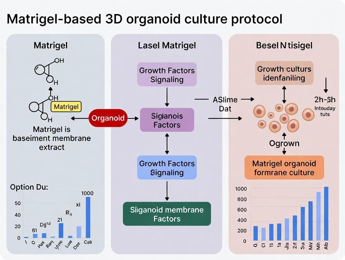
Diagram 1: Complete workflow for Matrigel-based organoid culture
Essential Signaling Pathways and Growth Factors
Organoid culture media require tailored combinations of growth factors and signaling molecules that address the specific needs of different tumor types and tissues [7]. These components activate critical signaling pathways that maintain stemness and promote differentiation:
Diagram 2: Key signaling pathways in organoid development and maintenance
Research Reagent Solutions for Organoid Culture
Table 3: Essential Materials for Matrigel-Based Organoid Culture
| Reagent Category | Specific Examples | Function & Application |
|---|---|---|
| Extracellular Matrix | Corning Matrigel Matrix [6] [9], Collagen [7] | Provides 3D scaffold mimicking native tissue environment; supports cell attachment and organization [5] [7] |
| Base Medium | Advanced DMEM/F12 [6] [3] | Nutrient foundation supporting cell growth and metabolism |
| Essential Supplements | HEPES, L-Glutamine, N-Acetylcysteine, B-27, Nicotinamide [6] [3] | Maintains physiological pH, reduces oxidative stress, provides essential nutrients |
| Growth Factors | EGF, Noggin, R-spondin, FGF-10, FGF-7, Wnt3A [6] [3] | Activates critical signaling pathways for stemness and differentiation (see Diagram 2) |
| Small Molecule Inhibitors | A83-01, SB202190, Y-27632 [6] [3] | Modulates TGF-β, p38 MAPK, and ROCK signaling to enhance growth and survival |
| Dissociation Reagents | Dispase [4], Cell Recovery Solution [4], PBS-EDTA [4] | Dissolves Matrigel for organoid recovery and passaging |
| Tissue-Specific Additives | Gastrin (gastric/pancreatic) [3], Heregulin-beta (mammary) [3] | Addresses specific requirements of different organoid types |
Quantitative Analysis of Organoid Drug Responses
The physiological relevance of organoid models is particularly evident in drug sensitivity testing. A 2025 study on pancreatic cancer organoids demonstrated that 3D organoid models more accurately mirrored patient clinical responses to standard chemotherapy regimens compared to traditional 2D cultures [6]. Notably, the IC₅₀ values for the 3D organoids were generally higher, reflecting the structural complexity and drug penetration barriers observed in vivo [6].
Table 4: Drug Response Profiling in Pancreatic Cancer Organoid Models
| Chemotherapy Regimen | 2D vs. 3D Model Response | IC₅₀ Values | Clinical Correlation |
|---|---|---|---|
| Gemcitabine + Nab-paclitaxel | 3D organoids showed higher resistance than 2D cultures [6] | Generally higher in 3D models [6] | 3D responses more accurately mirrored patient outcomes [6] |
| FOLFIRINOX | 3D organoids demonstrated different sensitivity profiles than 2D [6] | Generally higher in 3D models [6] | Better prediction of clinical response [6] |
| KRAS Inhibition | Patient-derived organoids revealed chemotherapy resistance mechanisms [10] | Variable based on genetic profile [10] | Identified novel therapeutic vulnerabilities [10] |
These quantitative assessments highlight the value of organoid models in preclinical drug evaluation. The integration of organoid technology with artificial intelligence and microfluidics further enables large-scale, rapid, and cost-effective drug testing, advancing the field of personalized medicine [2].
The Crucial Role of the Extracellular Matrix (ECM) in 3D Culture
Application Note & Protocol
The Extracellular Matrix (ECM) is far more than a static scaffold; it is a dynamic, bioactive environment that regulates essential cellular processes such as proliferation, differentiation, migration, and survival through bi-directional communication [11] [12]. In traditional two-dimensional (2D) culture, cells are forced into an unnatural state, often losing their native phenotype and function. Three-dimensional (3D) cultures within an ECM context bridge this gap, providing a physiologically relevant model that recapitulates the in vivo tumor microenvironment (TME) and tissue architecture [11] [12]. For organoid culture and cancer research, the ECM provides crucial mechanical and biochemical cues that direct cell fate, making the choice of 3D matrix a fundamental determinant of experimental success.
This document outlines the pivotal role of the ECM in 3D cultures, with a specific focus on Matrigel-based protocols for organoid generation. We provide detailed methodologies and data demonstrating how the ECM influences cellular behavior, underpinning its critical role in advanced in vitro models.
Key Applications: How the ECM Directs Cell Fate
The ECM's composition and physical properties directly dictate cellular outcomes in 3D culture. The following table summarizes key experimental findings that highlight the ECM's instructive role.
Table 1: Experimental Evidence of ECM Influence in 3D Cultures
| Application / Cell Type | ECM System Used | Key Findings on ECM Role | Reference / Experimental Context |
|---|---|---|---|
| Breast Cancer Cell Behavior | Patient-Derived Scaffolds (PDS) from normal vs. tumor tissue | - Tumor PDS had significantly higher stiffness (Young's modulus) and overexpression of Collagen IV and Vimentin [13].- Cells on tumor PDS showed higher viability, proliferation, and secreted 4x more IL-6 (122.91 vs. 30.23 pg/10⁶ cells) [13].- Tumor PDS upregulated invasiveness genes (CAV1, CXCR4, CNN3, MYB, TGFB1) [13]. | [13] |
| Extracellular Vesicle (EV) Biogenesis | 3D Tunable CNF/GelMA Hydrogel (Soft vs. Stiff) | - EVs from stiff 3D matrices (StEVs) had distinct cargo and physicochemical traits [14].- StEVs more potently promoted tumor cell proliferation, migration, and in vivo tumor growth via MAPK/ERK1/2 pathway activation [14]. | [14] |
| Stem Cell Tissue Regeneration | Matrigel-based 3D Culture of hGMSCs | - 3D culture significantly enhanced cell viability and adipogenic differentiation capacity [15].- hGMSCs/Matrigel construct injected in a rat model accelerated soft tissue repair by promoting autologous stem cell proliferation and collagen fiber generation [15]. | [15] |
| Intestinal Organoid Culture | Matrigel Domes with Specialized Medium | - Provides the structural and biochemical foundation for crypt cells to form complex, multi-lobed organoid structures [16].- The matrix supports the self-organization and differentiation of intestinal stem cells into all the requisite epithelial lineages [16]. | [16] |
Detailed Protocol: Establishing Mouse Intestinal Organoids in Matrigel
This protocol is adapted from established methods for creating 3D intestinal organoid cultures from isolated mouse crypts using Corning Matrigel [16].
Materials and Reagents
- IntestiCult Organoid Growth Medium (Mouse) (Catalog #06005)
- Corning Matrigel Matrix, GFR (Phenol Red-Free, Catalog #356231)
- DMEM/F-12 medium
- Phosphate-Buffered Saline (PBS), cold
- 24-well cell culture plate
- Pre-chilled pipette tips and 15 mL conical tubes
- Centrifuge
Method
- Crypt Isolation and Counting: Isolate intestinal crypts from mouse tissue and resuspend the selected crypt fraction in cold DMEM/F-12. Count the crypts using a hemocytometer. Desirable crypts are rectangular or circular with smooth edges, while villi, single cells, and debris should be excluded [16].
- Crypt-Matrigel Mixture Preparation:
- Centrifuge the volume containing 500-3000 crypts at 200 x g for 5 minutes at 2-8°C. Aspirate the supernatant.
- Add 150 µL of room temperature complete IntestiCult medium to the pellet.
- Add 150 µL of undiluted, thawed Matrigel to the tube. Pipette up and down carefully ten times to resuspend the pellet without introducing bubbles [16].
- Plating:
- Quickly pipette 50 µL of the crypt-Matrigel suspension as a dome into the center of each well of a pre-warmed 24-well plate.
- Place the plate at 37°C for 10 minutes to allow the Matrigel to solidify into a dome.
- Culture Maintenance:
- After gelation, gently add 750 µL of room temperature complete IntestiCult medium to each well, pipetting down the sidewall to avoid disturbing the dome.
- Incubate the culture at 37°C and 5% CO₂.
- Monitor daily for organoid formation. Spherical structures typically appear within days, developing into budded, complex organoids over 5-7 days for small intestine, or slower for colon [16].
- Fully exchange the culture medium three times per week. Passage organoids every 7-10 days to prevent overgrowth [16].
Workflow Diagram: Organoid Culture Establishment
Mechanistic Insights: How the ECM Signals to Cells
The ECM influences cell behavior through two primary, interconnected mechanisms: mechanotransduction and biochemical signaling.
Diagram: ECM-Mediated Signaling in 3D Culture
- Mechanotransduction in 3D: In a 3D context, cells are mechanically confined by the surrounding matrix. They generate force through actomyosin-based contractility and protrusion extension. Integrin-mediated focal adhesions sense mechanical properties like stiffness and viscoelasticity, converting these cues into biochemical signals that ultimately regulate transcription and cell phenotype [11].
- Biochemical Signaling: ECM components like laminin and collagen IV (the primary components of Matrigel) bind to cell surface receptors such as integrins and CD44. This binding facilitates intracellular signaling. Furthermore, the ECM acts as a reservoir for growth factors and hormones, presenting them to cells or withholding them, thereby directly influencing signaling pathways like MAPK/ERK1/2 [14] [12].
The Scientist's Toolkit: Essential Reagent Solutions
Table 2: Key Research Reagents for 3D ECM-Based Culture
| Reagent / Material | Function and Role in 3D Culture | Example Use Case |
|---|---|---|
| Corning Matrigel Matrix | A solubilized basement membrane extract from the EHS mouse tumor, containing key ECM proteins like Laminin (~60%), Collagen IV (~30%), and Entactin. It forms a biologically active 3D gel at 37°C, providing a reconstituted basement membrane for cell growth [17]. | The foundational scaffold for organoid culture (e.g., intestinal, mammary) and for assessing complex cell behaviors in a physiologically relevant 3D context [15] [16]. |
| TOCNF/GelMA Hybrid Hydrogel | A tunable, biomimetic synthetic hydrogel. Cellulose Nanofibrils (TOCNF) provide structural fidelity and control over mechanical properties (stiffness), while Gelatin Methacryloyl (GelMA) provides bioactive RGD motifs for cell adhesion [14]. | Ideal for mechanobiology studies where precise, independent control over matrix stiffness is required to investigate its effect on cell behavior and EV biogenesis [14]. |
| Patient-Derived Scaffolds (PDS) | A decellularized native human or animal tissue that retains the original ECM's unique composition, architecture, and mechanical properties. This provides the most authentic ex vivo model of a specific tissue's TME [13]. | Used to compare the specific effects of normal vs. diseased ECM (e.g., from tumor tissue) on cell phenotype, invasiveness, and drug response [13]. |
| Specialized Growth Media (e.g., IntestiCult) | Medium formulations supplemented with specific growth factors and inhibitors (e.g., Wnt agonists, R-spondin) that are essential for the survival and proliferation of stem cells and the formation of specific organoid types. | Essential for organoid culture to provide the necessary biochemical signals that, in concert with the ECM, guide self-organization and differentiation [16]. |
| Laminin-Rich ECM | A key attachment factor and major component of the basement membrane. It is critical for cell polarization, survival, and the maintenance of stemness [12]. | Used to enhance the aggressiveness of engineered tumor models and to differentiate between benign and malignant phenotypes based on morphology and proliferation [12]. |
Matrigel is a solubilized basement membrane preparation extracted from the Engelbreth-Holm-Swarm (EHS) mouse sarcoma, a tumor rich in extracellular matrix (ECM) proteins [18] [19] [20]. Since its development nearly 30 years ago, it has become one of the most extensively referenced and trusted tools in cell culture, providing a natural hydrogel that closely mimics the in vivo basement membrane environment [19] [20]. Its unique property of being a liquid at low temperatures (4°C) and polymerizing into a solid gel at physiological temperatures (37°C) makes it exceptionally useful for creating 3D cell culture environments, supporting cell attachment, differentiation, and morphogenesis in vitro [18] [21]. For researchers developing 3D organoid cultures, Matrigel provides a complex biological matrix that is often indispensable for recapitulating native tissue architecture and function.
Composition and Sourcing
Core Biochemical Composition
Matrigel's composition is complex, reflecting the natural heterogeneity of a basement membrane. The table below summarizes its major constituents.
Table 1: Major Constituents of Corning Matrigel Matrix
| Component | Approximate Percentage | Primary Function |
|---|---|---|
| Laminin | ~60% | Major structural component; promotes cell adhesion, signaling, and polarization [20] [21] |
| Collagen IV | ~30% | Provides structural integrity and forms a network [20] [21] |
| Nidogen (Entactin) | ~8% | Bridges laminin and collagen IV networks, stabilizing the matrix [20] [21] |
| Heparan Sulfate Proteoglycans (e.g., Perlecan) | 1-2% | Binds and sequesters growth factors, modulating their bioavailability [20] [21] |
In addition to these structural proteins, Matrigel contains a myriad of embedded growth factors present at varying concentrations due to its biological source. These include Transforming Growth Factor-β (TGF-β), Epidermal Growth Factor (EGF), Fibroblast Growth Factor (FGF), and Vascular Endothelial Growth Factor (VEGF) [22] [21]. Proteomic analyses have identified over 1,800 unique proteins in Matrigel, underscoring its compositional complexity [22] [21].
Sourcing and Production
Matrigel is sourced from the EHS mouse sarcoma, a tumor model that was extensively characterized at the National Institutes of Health (NIH) in the 1970s and 1980s [18] [21]. The production process involves several key steps [18] [21]:
- Tumor Harvesting: EHS tumors are grown in C57BL/6 mice, often under conditions that inhibit collagen cross-linking to enhance matrix yield.
- Homogenization and Extraction: The harvested tumors are homogenized, and soluble proteins are washed away. The insoluble basement membrane matrix is then extracted using chaotropic agents like urea or guanidine hydrochloride.
- Dialysis and Sterilization: The extract is dialyzed against a buffer to remove the chaotropic agents, resulting in a sterile, viscous solution.
- Quality Control: The final product undergoes rigorous testing for sterility, endotoxin levels, protein concentration, and biological activity.
The term "Matrigel" was coined in the early 1980s by John R. Hassell, and the product was subsequently commercialized, with Corning Life Sciences now being the primary manufacturer [18] [21].
Product Variants and Specifications
To suit different research applications, Corning offers several formulations of Matrigel. The growth factor-reduced (GFR) formulation is particularly useful for studies where the effects of endogenous growth factors need to be minimized [23] [20].
Table 2: Common Matrigel Product Variants and Specifications
| Product Type | Typical Protein Concentration | Key Features | Primary Applications |
|---|---|---|---|
| Standard Matrigel | 8-12 mg/mL [20] | Contains native levels of growth factors | General cell culture, differentiation studies [20] |
| Growth Factor Reduced (GFR) | 8-12 mg/mL [23] [20] | Levels of TGF-β, EGF, and other GFs are significantly reduced | Applications requiring a more defined basement membrane [23] [20] |
| High Concentration (HC) | 18-22 mg/mL [20] | Provides greater matrix stiffness and scaffold integrity | In vivo cell delivery, tumor augmentation [20] |
| hESC-qualified | Varies by lot | Pre-screened for feeder-free culture of human embryonic and induced pluripotent stem cells | Maintenance and expansion of hESCs and hiPSCs [20] |
| For Organoid Culture | Varies by lot | Optimized for organoid culture and differentiation | Generation and maintenance of 3D organoids [20] |
Mechanism of Action
Physical Properties and Gelation
Matrigel's mechanism of action is rooted in its physical transformation and biochemical composition. At 4°C, it remains in a liquid state, allowing for easy handling and mixing with cells. Upon warming to 37°C, its protein components self-assemble into a hydrogel with pore sizes of approximately 1-5 micrometers [21]. The mechanical properties of the gelled matrix are soft and tissue-like, with an elastic modulus (G') generally ranging from 50 to 250 Pa for standard concentrations, which closely mimics the compliance of natural basement membranes [21]. This 3D scaffold provides a physical support structure that enables cells to adopt polarized morphologies and organize into complex structures, a fundamental requirement for organoid development.
Biochemical Signaling and Cellular Interactions
The biological activity of Matrigel is mediated through its interactions with cell surface receptors and its ability to present growth factors. The diagram below illustrates the key signaling and mechanical interactions that underpin Matrigel's function in supporting epithelial and stem cell morphogenesis.
Diagram 1: Matrigel's dual mechanism provides a physical scaffold and biochemical signals that drive cell differentiation and organization.
The matrix provides a reservoir of growth factors that are presented to cells in a controlled, physiological manner. For instance, FGF signaling is intimately connected to the ECM, with heparan sulfate proteoglycans in Matrigel forming a ternary complex with FGF and its receptor (FGFR) to activate downstream pathways like PI3K/AKT that are crucial for survival, proliferation, and differentiation [24] [25]. Research has shown that the basement membrane components in Matrigel can directly activate inherent developmental programs in stem cells, promoting the differentiation of columnar ectoderm and cavitation in embryoid bodies [25].
Application Notes and Protocols for 3D Organoid Culture
Within the context of a broader thesis on Matrigel-based 3D organoid culture, the following detailed protocols are provided as foundational methodologies.
The Scientist's Toolkit: Essential Reagents
Table 3: Key Research Reagent Solutions for Matrigel-based 3D Organoid Culture
| Reagent / Material | Function / Application | Example / Notes |
|---|---|---|
| Corning Matrigel for Organoid Culture | Optimized matrix for organoid generation and differentiation. Provides the essential 3D scaffold. | Phenol red-free formulation is recommended for assays requiring color detection (e.g., fluorescence) [20]. |
| hESC-qualified Matrigel | For feeder-free culture and maintenance of human pluripotent stem cells (hPSCs), the starting material for many organoid lines. | Pre-screened for compatibility with defined media like mTeSR1 [20]. |
| Growth Factor Reduced (GFR) Matrigel | Provides a more defined basement membrane preparation where minimizing the influence of endogenous GFs is critical for experimental consistency. | Useful for isolating the effects of exogenously added growth factors [23] [20]. |
| ROCK Inhibitor (Y-27632) | Improves cell survival after thawing, passaging, and during initial seeding in 3D matrices. | Shown to increase efficiency of primary cell isolation and proliferation [22]. |
| Suspension Culture Plates | Low-attachment plates are essential for allowing embedded organoids to form and grow freely in three dimensions. | Corning spheroid microplates can be used for high-throughput organoid formation [19]. |
Standard Protocol: Establishing 3D Organoid Cultures from Single Cells
This protocol outlines the foundational steps for generating organoids by embedding single cells within Matrigel droplets, a widely used method for intestinal, mammary, and other epithelial organoids.
Workflow Overview:
Diagram 2: The standard workflow for establishing 3D organoid cultures in Matrigel.
Detailed Methodology:
- Thawing Matrigel: Slowly thaw a vial of Corning Matrigel for Organoid Culture overnight on ice at 4°C or in a refrigerator. Keep the vial on ice and pre-chill all pipette tips and tubes throughout the handling process. Critical: Avoid premature gelling. [19]
- Cell Preparation: Harvest and dissociate your starting cell population (e.g., dissociated tissue, pluripotent stem cell-derived progenitors) into a single-cell suspension. It is highly recommended to supplement the cell suspension media with a ROCK inhibitor (Y-27632, 5-10 µM) to enhance cell viability during the embedding process [22]. Centrifuge and resuspend the cell pellet in cold culture medium at the desired density (e.g., 1x10⁵ to 5x10⁵ cells/mL, depending on organoid type).
- Mixing Cells with Matrigel: Combine the cold single-cell suspension with an equal volume of liquid Matrigel on ice. Gently pipette up and down to mix thoroughly, avoiding bubble formation. The final Matrigel concentration should be at least 5-10 mg/mL. Note: Working quickly on ice is essential.
- Plating: Using pre-chilled tips, pipette 20-50 µL droplets of the cell-Matrigel mixture onto the center of each well of a pre-warmed multi-well cell culture plate.
- Polymerization: Carefully transfer the plate to a 37°C, 5% CO₂ incubator for 30-60 minutes to allow the Matrigel droplets to polymerize into solid gels.
- Feeding: After polymerization, gently overlay each droplet with pre-warmed organoid-specific culture medium. Change the medium every 2-4 days, depending on the metabolic rate of the organoids.
Protocol for In Vitro Angiogenesis (Tube Formation) Assay
While the tube formation assay is distinct from organoid culture, it is a critical application of Matrigel for modeling vascularization within the tumor microenvironment, a key aspect of cancer organoid research [26].
Workflow Overview:
Diagram 3: Standard workflow for the endothelial tube formation assay.
Detailed Methodology:
- Coating: Thaw Matrigel on ice as described in section 4.2. Add 50 µL of liquid Matrigel per well of a pre-chilled 96-well plate. Gently tilt the plate to ensure the entire well bottom is covered.
- Polymerization: Place the coated plate in a 37°C incubator for at least 30 minutes to form a solid gel layer. Avoid letting the gel dry out.
- Cell Seeding: Trypsinize and count human endothelial cells, such as Human Umbilical Vein Endothelial Cells (HUVECs). Resuspend cells in endothelial growth medium (EGM-2) at a density of 50,000 - 100,000 cells/mL. Gently add 100-150 µL of this cell suspension on top of the polymerized Matrigel in each well.
- Incubation and Imaging: Return the plate to the 37°C incubator. Capillary-like tube networks will typically begin to form within 2-6 hours. Incubate for up to 18 hours. Observe and image the networks using an inverted microscope.
- Quantification: Analyze images using software to quantify key parameters such as total tube length, number of branches, and number of meshes per field of view.
Critical Considerations for Research
- Batch-to-Batch Variability: As a naturally derived product, Matrigel exhibits inherent lot-to-lot variation in its precise protein and growth factor composition [22] [21]. For a multi-year thesis project, it is critical to test and qualify a new lot of Matrigel for your specific organoid system before committing to a large purchase. Whenever possible, purchase a sufficient quantity of a single lot to complete a major set of experiments.
- Experimental Controls: The presence of endogenous growth factors can confound experiments. For studies where defined conditions are paramount, the Growth Factor Reduced (GFR) formulation should be used [23]. Furthermore, appropriate positive and negative controls (e.g., using neutralizing antibodies against key matrix components or growth factors) should be included to validate that observed biological effects are indeed due to the Matrigel microenvironment.
- Moving Beyond Matrigel: While Matrigel is an invaluable tool, researchers should be aware of its limitations, including its tumor-derived origin and complex, undefined nature [22] [26]. The field is increasingly moving towards defined synthetic hydrogels that allow for precise control over mechanical properties (stiffness, degradability) and biochemical cues (adhesive ligands, growth factors) [26]. For the long-term progression of the field, validating key findings in a more defined system can significantly strengthen research conclusions.
Matrigel, a solubilized basement membrane extract derived from the Engelbreth-Holm-Swarm (EHS) mouse sarcoma, has become the gold standard substrate for three-dimensional (3D) organoid culture, playing a pivotal role in advancing personalized medicine and drug development research [18] [19]. This natural hydrogel provides a complex, biologically active microenvironment that closely mimics the in vivo extracellular matrix (ECM), supporting cell differentiation, polarization, and morphogenesis [22] [21]. However, its widespread adoption coexists with significant challenges rooted in its murine tumor origin and substantial batch-to-batch variability [27] [28]. These inherent limitations pose considerable obstacles for reproducible research and clinical translation, creating a paradox where Matrigel is simultaneously indispensable and problematic. This application note examines this duality, providing researchers with a detailed analysis of Matrigel's properties, documented limitations, and practical protocols for its use within the context of 3D organoid culture, specifically framing these discussions within ongoing thesis research aimed at optimizing organoid culture protocols.
Composition, Properties, and Functional Advantages
Biochemical and Physical Characteristics
Matrigel's functional superiority stems from its complex composition, which recreates a native basement membrane environment. The major components include laminin (approximately 60%), collagen type IV (approximately 30%), entactin/nidogen (approximately 8%), and heparan sulfate proteoglycans (such as perlecan, 1-2%) [22] [21]. Critically, it also contains a myriad of embedded growth factors—including transforming growth factor-β (TGF-β), epidermal growth factor (EGF), fibroblast growth factor (FGF), and vascular endothelial growth factor (VEGF)—which are essential for cell proliferation and differentiation [22] [21]. Proteomic analyses have identified over 1,800 unique proteins within Matrigel, contributing to its biological complexity but also to its compositional variability [22].
Physically, Matrigel undergoes temperature-dependent gelation, transitioning from a viscous liquid at 4°C to a stable hydrogel at 37°C within 30-60 minutes [21]. The resulting matrix has pore sizes of 1-5 micrometers, facilitating cell embedding, migration, and nutrient diffusion [21]. Its mechanical properties are characterized by low elastic modulus, typically ranging from 10 to 400 Pascals, depending on protein concentration, which mimics the softness of natural basement membranes [21].
Table 1: Key Characteristics of Standard Corning Matrigel Matrix Formulations
| Matrigel Type | Key Features | Primary Applications | Notable Growth Factor Levels |
|---|---|---|---|
| Standard | Complete basement membrane profile; contains phenol red | General 3D cell culture, angiogenesis assays | Endogenous growth factors present |
| Growth Factor Reduced (GFR) | Selectively reduced TGF-β and EGF | Studies requiring defined soluble factors | TGF-β reduced to <0.3 ng/mL |
| Phenol Red-Free | Absence of phenol red dye | Assays sensitive to color interference (e.g., fluorescence) | Similar to standard Matrigel |
| High Concentration | Elevated protein concentration | In vivo implantation, tumor studies | More concentrated growth factors |
| hESC-Qualified | Tested for human stem cell culture | Feeder-free culture of pluripotent stem cells | Optimized for stem cell maintenance |
| For Organoid Culture | Specifically optimized for organoids | Organoid culture and differentiation | Tailored for epithelial organoid growth |
Mechanism of Action: Creating a Physiologic Microenvironment
Matrigel facilitates organoid development through multiple synergistic mechanisms. Its structural proteins, particularly laminin-111, provide essential cell-adhesive ligands that engage integrin receptors on progenitor cells, activating intracellular signaling pathways that promote survival, proliferation, and polarization [22] [29]. The embedded growth factors function as soluble signaling cues that guide morphogenesis and differentiation, while heparan sulfate proteoglycans act as reservoirs for factor sequestration, creating concentration gradients that direct cellular self-organization [29] [21].
The 3D architecture of the gel imposes physical constraints and mechanical cues that influence cell polarity and cytoskeletal organization, driving the formation of complex structures with central lumens—a hallmark of organoid development [6] [29]. Furthermore, the matrix is susceptible to proteolytic remodeling by matrix metalloproteinases (MMPs) secreted by embedded cells, enabling organoid expansion and morphological changes over time [29] [21]. This dynamic reciprocity between cells and their matrix is crucial for establishing the feedback loops that guide self-organization in organoid cultures.
Diagram 1: Matrigel-induced signaling and organoid morphogenesis. Matrigel components activate synergistic pathways driving 3D organization.
Documented Limitations and Research Implications
Batch-to-Batch Variability
The most frequently cited limitation of Matrigel is its inherent batch-to-batch variability, which arises from the biological nature of its production from EHS mouse tumors [27] [28] [29]. Proteomic studies reveal that only approximately 53% of identified proteins are consistent across different lots, with significant fluctuations in the concentrations of major components like laminin, collagen IV, and entactin [21]. This variability extends to growth factor content; for instance, TGF-β concentrations can range from 1.7 to 4.7 ng/mL between batches [21]. These compositional differences directly impact mechanical properties, with the elastic modulus of standard Matrigel preparations varying between 50 and 250 Pa [21].
For organoid culture, this variability translates into substantial experimental challenges. Studies demonstrate differential organoid formation efficiency, growth rates, and morphological phenotypes when identical progenitor cells are cultured in different Matrigel batches [28] [29]. In drug screening applications, such variability can compromise the reproducibility of IC50 values for chemotherapeutic agents, potentially leading to inconsistent conclusions about drug efficacy [6] [29]. This lack of reproducibility poses particular problems for long-term thesis research and multi-center preclinical studies, where standardized conditions are essential for valid comparisons.
Murine Origin and Clinical Translation Challenges
The murine sarcoma origin of Matrigel presents both scientific and clinical limitations. The presence of xenogeneic components, particularly mouse-specific laminin isoforms and growth factors, introduces interspecies differences that may not accurately recapitulate human tissue microenvironments [27] [28]. These differences can skew cellular responses and signaling pathway activation in human organoid models [28].
For clinical applications, the undefined nature and animal origin raise significant safety concerns regarding potential immunogenic reactions if organoids are used for transplantation therapies [28]. Regulatory agencies like the FDA typically require fully defined, xeno-free culture systems for cellular therapies, making Matrigel unsuitable for these applications [27] [28]. Furthermore, the tumor-derived nature of Matrigel introduces theoretical risks of transferring potentially oncogenic factors, though commercial processing minimizes this concern for research use [22] [21].
Additional Limitations in Advanced Applications
Beyond variability and origin concerns, researchers should consider several other limitations:
- Limited Tunability: Unlike synthetic hydrogels, Matrigel's mechanical properties cannot be independently adjusted without altering its biochemical composition, making it difficult to decouple biochemical from biomechanical cues [29].
- Rapid Polymerization: The irreversible thermal gelation requires precise, rapid handling on ice to prevent premature gelling, complicating experimental procedures [19].
- Structural Limitations: For some organoid types, such as intestinal organoids, Matrigel lacks specific ECM components (e.g., laminin-511) present in native human tissue, potentially limiting architectural fidelity [28].
Table 2: Quantitative Impact of Matrigel Limitations on Research Applications
| Limitation Category | Quantitative Measure | Impact on Research | Potential Consequence |
|---|---|---|---|
| Compositional Variability | ~47% protein difference between batches [21] | Reduced reproducibility across experiments | Inconsistent organoid formation efficiency |
| Growth Factor Variability | TGF-β range: 1.7-4.7 ng/mL [21] | Altered differentiation outcomes | Variable lineage specification in stem cell organoids |
| Mechanical Variability | Elastic modulus range: 50-250 Pa [21] | Changed morphogenetic responses | Different organoid size and morphology |
| Murine Components | Laminin-111 (mouse) vs. human isoforms [28] | Species-specific signaling discrepancies | Reduced predictive value for human physiology |
| Undefined Composition | >1,800 proteins [22] | Difficulty identifying critical factors | Challenges in mechanistic studies |
Practical Applications and Protocols
Establishing Pancreatic Cancer Organoid Cultures
The following protocol, adapted from recent literature, details the establishment of patient-derived pancreatic cancer organoids using Matrigel, demonstrating a key application in cancer research [6]:
Materials Required:
- Corning Matrigel Matrix, Phenol Red-free (Catalog #356231) [19]
- Patient-derived pancreatic cancer cells (conditionally reprogrammed cells)
- F medium: Ham's F-12 nutrient mix (70%) + Dulbecco's Modified Eagle's Medium (25%)
- Supplement cocktail: hydrocortisone (0.4 μg/mL), insulin (5 μg/mL), cholera toxin (8.4 ng/mL), EGF (10 ng/mL), FBS (5%), adenine (24 μg/mL)
- Rho-associated kinase inhibitor Y-27632 (5 μM)
- 6-well cell culture plates
- Pre-chilled pipettes and tips
Procedure:
- Thawing and Handling: Thaw Matrigel overnight at 4°C. Keep all tubes and tips on ice throughout the procedure.
- Cell Preparation: Harvest patient-derived pancreatic cancer cells and resuspend in cold F medium. Count cells and adjust concentration.
- Mixing with Matrix: Centrifuge required number of cells and carefully resuspend in cold Matrigel at a density of 5,000-10,000 cells per 20 μL, avoiding bubble formation.
- Plating: Aliquot 20 μL drops of the cell-Matrigel mixture into 6-well plates (7 domes per plate). Avoid disturbing the drops.
- Gelation: Incubate plates at 37°C for 20-30 minutes to allow complete polymerization.
- Medium Addition: Gently overlay each dome with 4 mL of pre-warmed F medium supplemented with Y-27632.
- Culture Maintenance: Refresh medium every 3-4 days. Monitor organoid formation daily.
- Passaging: Harvest organoids when >50% exceed 300 μm in size (approximately 2-4 weeks). Dissociate using mechanical disruption or enzymatic digestion and replate in fresh Matrigel.
Technical Notes: For rapidly growing cells, use 5,000 cells/20 μL dome; for slower-growing cells, use 10,000 cells/20 μL dome. This protocol specifically avoids using organoid culture media components like Wnt3a, R-spondin, and Noggin to preserve intrinsic molecular subtypes of the cancer cells [6].
Diagram 2: Workflow for establishing pancreatic cancer organoids in Matrigel. Critical temperature-sensitive steps ensure proper matrix polymerization.
The Scientist's Toolkit: Essential Reagents and Materials
Table 3: Essential Research Reagents for Matrigel-Based Organoid Culture
| Reagent/Material | Function/Application | Example Product | Protocol Notes |
|---|---|---|---|
| Corning Matrigel Matrix | Basement membrane extract for 3D support | Corning #356231 (Organoid Culture) | Maintain at 4°C during handling; avoid repeated freeze-thaw |
| Rho-associated Kinase (ROCK) Inhibitor | Enhances cell survival after passage | Y-27632 (5 μM) | Critical for initial 2-3 days after plating |
| Growth Factor-Reduced Matrigel | For studies requiring defined factors | Corning #356231 | Reduces TGF-β to <0.3 ng/mL, EGF levels |
| Tumor Dissociation Kit | Tissue processing to single cells | Human Tumor Dissociation Kit | Enzymatic and mechanical digestion |
| Basal Medium | Nutrient foundation for culture | Ham's F-12/DMEM mix | Must be supplemented with specific factors |
| Growth Factor Cocktail | Directs cell fate and proliferation | EGF, FGF, Noggin, R-spondin | Organ-type specific combinations required |
| Matrix Metalloproteinase Inhibitors | Controls ECM remodeling | GM6001, Marimastat | Regulates organoid invasion in Matrigel |
Mitigation Strategies and Alternative Approaches
Minimizing the Impact of Batch Variability
To address batch variability in research, implement these practical strategies:
- Batch Testing and Validation: Before starting critical experiments, test multiple Matrigel lots using standardized organoid formation assays. Select a single lot for all experiments in a study and purchase sufficient quantity for the entire project [19].
- Quality Control Assessment: Characterize each batch for protein concentration (standard: 8-12 mg/mL) and gelation properties. Corning provides lot-specific certificates of analysis with this information [19] [21].
- Internal Standards: Include control cell lines with known organoid formation efficiency in each experiment to normalize for batch-specific effects [29].
- Biochemical Supplementation: Add defined ECM components (e.g., collagen V) to correct for missing elements in specific Matrigel batches, as demonstrated in pancreatic differentiation studies [28].
Emerging Alternatives to Matrigel
Research into defined matrices addresses both variability and murine origin concerns:
- Synthetic Hydrogels: PEG-based and other polymer hydrogels offer precisely tunable mechanical properties and defined chemical compositions, though they may lack native bioactivity [28] [29].
- Recombinant ECM Proteins: Engineered proteins (e.g., recombinant laminins) provide defined human components but at higher cost and complexity [27] [28].
- Decellularized ECM: Human or porcine tissue-derived matrices better replicate organ-specific composition but still face batch variability challenges [28].
- Hybrid Approaches: Combining synthetic polymers with defined bioactive peptides offers intermediate solutions with some customizability [29].
Each alternative presents trade-offs in cost, complexity, and biological performance, necessitating careful selection based on research goals [27] [28].
Matrigel remains an indispensable tool in 3D organoid culture, providing an unmatched biologically active microenvironment that supports the complex process of self-organization and tissue maturation [6] [19]. Its advantages in supporting physiologically relevant models are evidenced by successful applications in pancreatic cancer research, where Matrigel-based organoids have demonstrated superior drug response prediction compared to 2D models [6]. However, researchers must acknowledge and actively manage its inherent limitations, particularly batch variability and murine origin, through careful experimental design and appropriate controls.
Future developments in organoid technology will likely focus on defined, xeno-free matrices that recapitulate the supportive qualities of Matrigel while ensuring reproducibility and clinical compatibility [27] [28] [29]. Until such alternatives mature, understanding Matrigel's properties and limitations remains essential for generating robust, reproducible organoid data. For thesis research specifically, documenting Matrigel lot numbers and implementing consistent handling protocols will strengthen the validity and reproducibility of findings, contributing to the broader effort to standardize organoid culture methodologies.
Three-dimensional (3D) organoid cultures have emerged as a transformative technology in biomedical research, bridging the gap between conventional two-dimensional (2D) cell cultures and in vivo models. These self-organizing 3D structures are derived from pluripotent stem cells or adult stem cells (ASCs) and recapitulate key aspects of the architecture and functionality of native organs [2]. The foundation of successful organoid culture often relies on a supportive extracellular matrix (ECM), with Corning Matrigel matrix being one of the most widely used and published hydrogels for this purpose [30]. Matrigel provides the necessary biochemical and structural cues that mediate cell migration, behavior, and polarization, enabling researchers to generate mini-organs of the kidney, thyroid, liver, brain, lung, intestine, prostate, breast, esophagus, gastric, ovarian, and pancreas [30] [2]. This application note details the use of Matrigel-based 3D organoid cultures within the key areas of disease modeling, drug screening, and personalized medicine, providing standardized protocols for researchers and drug development professionals.
Disease Modeling with 3D Organoids
Protocol: Establishing Patient-Derived Cancer Organoids
Background: Patient-derived organoids (PDOs) have proven instrumental in elucidating genetic cell fate in hereditary diseases, infectious diseases, metabolic disorders, and malignancies [2]. For instance, pancreatic cancer organoids have been shown to retain the molecular characteristics, transcriptomic, and mutational profiles of the parental tumors, displaying distinct morphologies corresponding to cancer stages and differentiation [6].
Materials:
- Corning Matrigel matrix for organoid culture (e.g., Catalog #356231) [6] [4]
- Patient-derived tissue sample (e.g., from endoscopic ultrasound-guided fine-needle biopsy or surgical resection)
- Human Tumor Dissociation Kit (e.g., Miltenyi Biotec)
- F medium: 70% Ham’s F-12, 25% complete DMEM, 0.4 mg/mL hydrocortisone, 5 mg/mL insulin, 8.4 ng/mL cholera toxin, 10 ng/mL epidermal growth factor, 5% FBS, 24 mg/mL adenine, 10 mg/mL gentamicin, 250 ng/mL Amphotericin B [6]
- Rho-associated kinase inhibitor Y-27632
- 6-well cell culture plates
Methodology:
- Tissue Processing: Mechanically and enzymatically dissociate the fresh tumor tissue using a Human Tumor Dissociation Kit according to the manufacturer's instructions. Filter the cell suspension through a 40 µM-pore cell strainer [6].
- Initial 2D Culture (Conditional Reprogramming): Seed the cell suspension on a feeder layer of lethally irradiated J2 murine fibroblasts in F medium supplemented with 5 µM Y-27632. Incubate at 37°C in a humidified atmosphere with 5% CO₂ [6].
- 3D Organoid Culture: a. Harvest the conditionally reprogrammed cells (CRCs). b. Mix cells with 90% growth factor-reduced Matrigel. For rapidly growing cells, use a density of 5,000 cells per 20 µL of Matrigel; for slower-growing cells, use 10,000 cells per 20 µL [6]. c. Aliquot 20 µL of the cell-Matrigel mixture into a 6-well plate, forming dome structures. Solidify the domes at 37°C for 20 minutes. d. Carefully add 4 mL of F medium to each well. Refresh the medium every 3–4 days.
- Passaging: Harvest organoids for subculturing or downstream assays once more than 50% exceed 300 μm in size. This typically occurs 2–4 weeks after seeding [6].
The workflow below summarizes the key steps in establishing and utilizing patient-derived organoids for disease modeling and drug screening.
The Scientist's Toolkit: Essential Reagents for Organoid Research
Table 1: Key Research Reagent Solutions for Matrigel-based 3D Organoid Culture.
| Item | Function | Example |
|---|---|---|
| Corning Matrigel Matrix for Organoids | Provides a biologically active basement membrane extract to support 3D organoid growth, differentiation, and structural integrity. | Corning Catalog #356231 [30] |
| Rho-associated Kinase (ROCK) Inhibitor | Enhances cell survival and prevents anoikis during the initial phases of cell seeding and passaging. | Y-27632, 5 µM [6] |
| Tissue Dissociation Kit | Enzymatically and mechanically dissociates patient tissue samples to a single-cell suspension for culture initiation. | Human Tumor Dissociation Kit (Miltenyi Biotec) [6] |
| Dispase Solution | An enzymatic Matrigel dissolving method optimal for downstream proteomic analysis, providing high peptide yield and minimal Matrigel contaminants. | 1 U/ml dispase solution [4] |
| Defined Media Supplements | Provides niche factors (e.g., growth factors, cytokines) necessary for the expansion and differentiation of specific organoid types. | EGF, Noggin, R-spondin-1, Wnt3a [6] [2] |
Drug Screening and Toxicity Assessment
Protocol: Drug Sensitivity Profiling in 3D Organoids
Background: A pivotal advantage of 3D organoids is their ability to mirror patient clinical responses to drugs more accurately than 2D cultures. In pancreatic cancer, drug response profiling of regimens like gemcitabine plus nab-paclitaxel (Abraxane) and FOLFIRINOX demonstrated that 3D organoids better predicted patient outcomes, with IC₅₀ values that were generally higher, reflecting the structural complexity and drug penetration barriers observed in vivo [6].
Materials:
- Mature organoids (200–300 μm in diameter)
- Dispase solution (for harvesting)
- 96-well or 384-well cell culture plates (including spheroid microplates for an all-in-one workflow) [30] [10] * Drug compounds of interest (e.g., chemotherapeutics) * Cell viability assay kit (e.g., ATP-based luminescence assay)
Methodology:
- Organoid Harvesting: a. Discard the supernatant medium and collect organoids embedded in Matrigel using PBS. b. Wash twice with PBS. c. Add 1 U/ml pre-warmed dispase solution (1 ml/well of a 6-well plate) and incubate at 37°C for 30 minutes [4]. d. Centrifuge to pellet the organoids, discard the supernatant, and add fresh dispase for a second 30-minute incubation. e. Pellet and wash the organoid cells twice with PBS before resuspending in an appropriate medium for counting.
- Assay Setup: a. Seed a single-cell suspension or small organoid fragments in a Matrigel dome or pre-coated Matrigel matrix-3D plate [30]. b. Allow organoids to reform for 24-48 hours. c. Treat organoids with a concentration gradient of the drug(s) of interest. Include negative (vehicle) and positive (cytotoxic) controls. d. Incubate for a predetermined period (e.g., 3-7 days), refreshing drug/media as needed.
- Viability Assessment: a. At the endpoint, perform a cell viability assay according to the manufacturer's instructions. b. Measure the signal (e.g., luminescence) and calculate the percentage of viability relative to the vehicle control.
- Data Analysis: a. Generate dose-response curves. b. Calculate the half-maximal inhibitory concentration (IC₅₀) using appropriate non-linear regression models.
Table 2: Quantitative Drug Response Data from Pancreatic Cancer Organoids [6].
| Chemotherapy Regimen | 2D Culture IC₅₀ | 3D Organoid IC₅₀ | Clinical Response Correlation |
|---|---|---|---|
| Gemcitabine + Nab-paclitaxel | Lower | Generally Higher | 3D organoid responses more accurately mirrored patient clinical outcomes. |
| FOLFIRINOX | Lower | Generally Higher | 3D organoid responses more accurately mirrored patient clinical outcomes. |
The following diagram illustrates the logical relationship and signaling crosstalk between key pathways often dysregulated in cancer and targeted in drug screening.
Personalized Medicine Applications
Protocol: Utilizing Organoids for Patient-Specific Therapeutic Insights
Background: The integration of organoid technology with high-throughput screening holds promise for advancing precision medicine. Creating biobanks of patient-derived organoids (PDOs) enables high-throughput pharmacotyping, where the sensitivity of a patient's organoids to a panel of drugs can be tested to guide therapeutic selection [10] [2]. This "clinical trial in a dish" approach is being applied to cancers, including pancreatic cancer, and complex neurological diseases [10].
Materials:
Methodology:
- Organoid Biobanking: Establish and expand multiple PDO lines from a cohort of patients, ensuring stable passage and cryopreservation [6].
- High-Throughput Screening (HTS): a. Scale down the drug screening protocol to 384-well formats to enable testing of many compounds or combinations. b. Use automation for seeding, drug dispensing, and assay readout to ensure consistency and efficiency. c. For complex phenotypes (e.g., morphology changes, biomarker expression), employ high-content imaging rather than a single viability endpoint.
- Data Integration and AI Analysis: a. Collect multi-parametric data (viability, morphology, -omics data). b. Leverage artificial intelligence tools to analyze the complex datasets, identify response patterns, and predict effective patient-specific drug combinations [2].
- Clinical Correlation: Correlate the ex vivo drug sensitivity data (e.g., IC₅₀) with the patient's actual clinical response to treatment to validate the predictive power of the platform [6].
Matrigel-based 3D organoid cultures represent a robust and physiologically relevant platform that is revolutionizing biomedical research. As detailed in these application notes, their ability to accurately model diseases, recapitulate patient-specific drug responses, and serve as a tool for personalized therapeutic discovery is unparalleled. While challenges regarding standardization and scalability persist, the integration of these models with advanced bioengineering, AI, and high-throughput screening technologies promises to significantly accelerate drug discovery and the implementation of precision medicine.
A Step-by-Step Protocol for Robust Organoid Culture
Essential Materials and Pre-culture Preparation
In Matrigel-based three-dimensional (3D) organoid culture, proper preparation of materials and pre-culture procedures are fundamental to success. This protocol details the essential reagents, equipment, and preparatory steps required to establish a robust environment for organoid development and maintenance. The foundational role of the extracellular matrix (ECM) cannot be overstated—it provides the critical biochemical and structural support that mediates cell signaling, behavior, and polarization necessary for organoid formation [30]. By standardizing these preparatory phases, researchers can enhance experimental reproducibility and ensure the generation of high-quality organoids that accurately mimic in vivo physiology.
The Scientist's Toolkit: Essential Materials
Successful organoid culture requires specific, high-quality reagents and specialized cultureware. The following table catalogs the core components of the organoid culture toolkit.
Table 1: Key Research Reagent Solutions for Matrigel-Based Organoid Culture
| Item | Function & Importance | Examples & Specifications |
|---|---|---|
| Extracellular Matrix (ECM) | Provides the 3D structural scaffold and biochemical cues; critical for self-organization. | Corning Matrigel Matrix (for organoids, GFR, or standard); kept at 4°C during handling [30] [31] [3]. |
| Organoid Culture Medium | Supplies nutrients and specific signaling factors to support stem cell maintenance and differentiation. | Tissue-specific formulations (e.g., IntestiCult); often includes supplements (B-27, N-Acetylcysteine) and growth factors (EGF, Noggin, R-spondin) [3]. |
| Dissociation Reagent | Gently breaks down the ECM and dissociates organoids for passaging without damaging cells. | Gentle Cell Dissociation Reagent (GCDR) or enzyme mixes (e.g., Trypsin/EDTA for some protocols) [31] [32]. |
| ROCK Inhibitor | Improves cell survival after thawing and passaging by inhibiting apoptosis. | Y-27632, typically used at a final concentration of 5-10 µM in the medium for the first 24-48 hours after seeding [3]. |
| Basal Wash Medium | Used for washing cell pellets and diluting reagents; free of growth factors. | DMEM/F-12 with HEPES buffer, kept ice-cold for handling Matrigel suspensions [31] [3]. |
Specialized Equipment
- Culture Vessels: Standard multi-well plates (e.g., 24-well or 6-well) are used for embedded dome cultures [3]. For suspension culture methods, Ultra-Low Adherence Plates are required to prevent cell attachment [31].
- Temperature Control Equipment: A 37°C water bath for warming media, a refrigerator at 4°C, and a cooling rack or ice bucket are essential for the proper handling of temperature-sensitive Matrigel [3].
Preparative Workflow and Experimental Protocol
The process of establishing organoid cultures from cryopreserved stocks involves a critical pre-culture phase to ensure high cell viability and successful embedding. The workflow is designed to maintain the integrity of both the cells and the ECM.
Detailed Pre-culture Protocol
Protocol: Initiating Organoid Culture from Cryopreserved Vials
Materials: Cryopreserved organoids, EHS-based ECM (e.g., Corning Matrigel), complete organoid culture medium, basal medium (e.g., DMEM/F-12), ROCK inhibitor (Y-27632), 15 mL conical tubes, multi-well tissue culture plates.
Step-by-Step Method:
- Thaw ECM: The day before culture, transfer the required volume of ECM from a -20°C or -80°C freezer to a refrigerator at 4°C to thaw overnight. Once liquid, keep the vial on ice during all subsequent handling to prevent premature gelling [3]. For some protocols, growth factor-reduced (GFR) Matrigel is specified [31].
- Prepare Cultureware: Warm the tissue culture plate (e.g., a 6-well plate) in a 37°C incubator for at least 60 minutes. This prevents the rapid cooling of Matrigel when it is dispensed [3].
- Thaw Cryopreserved Organoids:
- Prepare a 15 mL conical tube with 10 mL of room temperature basal medium.
- Remove the vial of organoids from liquid nitrogen storage and thaw rapidly in a 37°C water bath for approximately 1-2 minutes.
- Gently transfer the thawed cell suspension to the prepared conical tube containing basal medium [3].
- Wash and Pellet Cells:
- Resuspend Pellet in ECM:
- Keep the cell pellet on ice. Gently resuspend the pellet in a predetermined volume of the thawed, ice-cold ECM. The required cell density is model-specific but often ranges from 5,000 to 50,000 cells per 20-50 µL of ECM [6] [3].
- To increase initial cell viability, add ROCK inhibitor (Y-27632) to the cell-ECM mixture at the recommended concentration (e.g., 5-10 µM) [3]. Mix thoroughly by pipetting gently, avoiding air bubbles.
- Plate ECM-Cell Mixture:
- Using pre-chilled pipette tips, quickly dispense the desired volume (e.g., 20-50 µL) of the cell-ECM suspension as individual droplets (domes) onto the pre-warmed culture plate [3].
- Solidify and Add Medium:
- Transfer the plate to the 37°C, 5% CO₂ incubator for 20-30 minutes to allow the domes to polymerize into a solid gel.
- Once solidified, gently add pre-warmed complete culture medium, supplemented with ROCK inhibitor, to each well, taking care not to disrupt the domes. For a 24-well plate, 500 µL of medium is typical [31] [3].
- Return the plate to the incubator. Perform a half-medium change every 2-3 days, and monitor organoid growth under a microscope.
Quantitative Data and Formulations
Standardization is key to reproducibility. Documenting the physical properties of ECM lots and using consistent medium formulations are critical steps in the pre-culture phase.
Table 2: Representative Medium Formulations for Human Cancer Organoid Culture (Final Concentrations)
| Component | Colon | Pancreatic | Mammary |
|---|---|---|---|
| Advanced DMEM/F12 | Base | Base | Base |
| HEPES | 10 mM | 10 mM | 10 mM |
| B-27 Supplement | 1x | 1x | 1x |
| N-Acetylcysteine | 1 mM | 1.25 mM | 1.25 mM |
| EGF | 50 ng/mL | 50 ng/mL | 5 ng/mL |
| Noggin | 100 ng/mL | 100 ng/mL | 100 ng/mL |
| A83-01 | 500 nM | 500 nM | 500 nM |
| R-spondin1 CM | 20% | 10% | 10% |
| Wnt-3A CM | Not included | 50% | Not included |
| Gastrin | Not included | 10 nM | Not included |
| FGF-10 | Not included | 100 ng/mL | 20 ng/mL |
| FGF-7 | Not included | Not included | 5 ng/mL |
Adapted from ATCC Organoid Culture Guide [3]. CM = Conditioned Medium.
Meticulous attention to the "Essential Materials and Pre-culture Preparation" phase lays the groundwork for successful and reproducible Matrigel-based 3D organoid cultures. The integrity of the ECM, the precision of reagent preparation, and the careful handling of cells during thawing and embedding are non-negotiable aspects of the protocol. By adhering to these detailed procedures, researchers can create a biomimetic environment that robustly supports the complex process of organoid development, thereby providing a reliable platform for advanced biomedical research.
Within the framework of Matrigel-based three-dimensional (3D) organoid culture research, the successful initiation of viable cultures from cryopreserved material is a critical first step. This protocol standardizes the process of thawing and establishing organoid cultures, a step that is fundamental to ensuring experimental reproducibility and reliability in downstream applications such as disease modeling, drug screening, and personalized medicine [33] [34]. Using a defined extracellular matrix (ECM) like Corning Matrigel matrix provides the necessary biochemical and structural cues to support the survival, proliferation, and self-organization of thawed stem cells into functional organoids [30] [7].
Materials
Reagents and Solutions
Table 1: Essential Reagents for Thawing and Initiating Organoid Cultures
| Reagent/Solution | Function/Purpose | Examples/Notes |
|---|---|---|
| Cryopreserved Organoids/Stem Cells | Starting biological material. | Dental Pulp Stem Cells (DPSCs) [33], Patient-Derived Organoids (PDOs) [34]. |
| Pre-warmed Complete Culture Medium | Provides nutrients and essential signaling factors for growth and maintenance. | Advanced DMEM/F12, supplemented with specific growth factors (e.g., EGF, Noggin, R-Spondin-1) [6] [34]. |
| Basement Membrane Extract (BME) | Acts as a 3D scaffold mimicking the in vivo extracellular matrix. | Corning Matrigel matrix for organoids [30]. Must be kept on ice to prevent premature polymerization. |
| ROCK Inhibitor (Y-27632) | Enhances cell survival post-thaw by inhibiting apoptosis. | Used at a final concentration of 10 µM in the recovery medium [34]. |
| Phosphate Buffered Saline (PBS) | For washing cells to remove residual cryoprotectant. | Calcium- and magnesium-free is recommended. |
| Cell Recovery Solution | Facilitates the dissociation of organoids from the Matrigel dome for passaging or analysis. | Corning Cell Recovery Solution or similar [34]. |
| Trypsin/EDTA or Accutase | Enzymatic dissociation reagents for passaging organoids into single cells or small clumps. | Choice depends on organoid type and sensitivity [33]. |
Laboratory Equipment
- Water bath or bead bath (set to 37°C)
- Centrifuge
- Biological safety cabinet
- Refrigerated centrifuge (capable of 4°C)
- Incubator (37°C, 5% CO₂)
- Pipettes and sterile tips
- Sterile centrifuge tubes (15 mL and 50 mL)
- Cell culture plates (e.g., 24-well or 48-well)
Methodology
Thawing Cryopreserved Cells
- Preparation: Pre-warm a sufficient volume of complete organoid culture medium, supplemented with 10 µM ROCK inhibitor (Y-27632). Place the medium in a 37°C water bath briefly, then hold at room temperature. Chill the required number of centrifuge tubes on ice.
- Rapid Thaw: Remove the cryovial of cells from liquid nitrogen storage and immediately place it in a 37°C water bath. Gently agitate the vial until only a small ice crystal remains (approximately 1-2 minutes).
- Transfer and Dilute: Wipe the cryovial with 70% ethanol. Gently transfer the thawed cell suspension to the pre-chilled 15 mL centrifuge tube containing 10 mL of pre-warmed, ROCK inhibitor-supplemented medium. This step dilutes the cytotoxic cryoprotectant (e.g., DMSO).
- Centrifuge: Centrifuge the cell suspension at approximately 300 × g for 5 minutes at 4°C.
- Aspirate and Resuspend: Carefully aspirate the supernatant without disturbing the cell pellet. Gently resuspend the pellet in a small volume (e.g., 1-2 mL) of fresh, ROCK inhibitor-supplemented medium.
Embedding in Matrigel and Initial Plating
- Matrigel Handling: Keep an aliquot of Corning Matrigel matrix for organoids on ice at all times to prevent premature gelling. Use pre-cooled pipette tips.
- Mixing: Combine the resuspended cell pellet with an appropriate volume of chilled Matrigel. Gently mix by pipetting slowly to avoid introducing air bubbles, ensuring a homogeneous cell suspension within the matrix. The final cell density should be optimized for the specific organoid type [33].
- Plating Domes: For a 24-well plate, pipette a 20-50 µL droplet of the cell-Matrigel mixture onto the center of each well.
- Polymerization: Place the culture plate in a 37°C, 5% CO₂ incubator for 20-30 minutes to allow the Matrigel to form a solid dome.
- Overlay with Medium: After polymerization, carefully add pre-warmed, ROCK inhibitor-supplemented culture medium to each well, ensuring the medium overlays the dome without dislodging it.
- Culture Initiation: Return the plate to the incubator. The medium should be replaced every 2-3 days, and the ROCK inhibitor is typically only required for the first 2-3 days post-thaw to support initial survival [34].
The workflow below summarizes the key steps from thawing to the establishment of the 3D culture.
Anticipated Results and Quality Control
Within 3-7 days post-thaw, significant cellular aggregation should be observable, marking the initial stage of organoid development [33]. Organoids derived from tissues such as dental pulp (DPSCs) will begin to form complex 3D structures. Quality control is essential at this stage. Characterization can include:
- Viability Assays: Using assays like Calcein-AM (for live cells) and Propidium Iodide (for dead cells) to assess health [34].
- Immunofluorescence (IF) Analysis: Staining for key markers to confirm identity and differentiation state, such as Runx2 for osteogenic differentiation or CD105 for an undifferentiated state in DPSC-organoids [33].
- Functional Assays: For example, Alizarin Red staining can be used to detect calcium deposits, indicating mineralization in bone-like organoids [33].
Table 2: Key Signaling Pathways and Their Roles in Organoid Initiation
| Signaling Pathway | Key Components | Role in Organoid Culture | Common Modulators |
|---|---|---|---|
| Wnt/β-catenin | Wnt3a, R-spondin | Critical for stem cell self-renewal and proliferation. Often required for initiating and maintaining organoid growth [6] [34]. | CHIR99021 (activator) |
| BMP (Bone Morphogenetic Protein) | BMP, Noggin (inhibitor) | Regulates differentiation and patterning. Noggin is frequently added to inhibit BMP signaling and promote epithelial growth [6] [5]. | Recombinant Noggin |
| EGF (Epidermal Growth Factor) | EGF | Promoves cell proliferation and survival in many organoid types [33] [6]. | Recombinant EGF |
| TGF-β (Transforming Growth Factor Beta) | TGF-β, A-83-01 (inhibitor) | A complex pathway that can inhibit cell proliferation; its inhibition is often beneficial for certain organoid cultures [34]. | A-83-01 (inhibitor) |
| Rho-associated kinase (ROCK) | Y-27632 (inhibitor) | Promotes cell survival and inhibits anoikis (detachment-induced cell death), crucial for recovery after thawing and passaging [6] [34]. | Y-27632 |
The interactions of these pathways in the context of the Matrigel microenvironment are crucial for successful organoid formation, as illustrated below.
Troubleshooting
Table 3: Common Issues and Proposed Solutions
| Problem | Potential Cause | Solution |
|---|---|---|
| Low Cell Viability Post-Thaw | Slow or improper thawing process; insufficient ROCK inhibitor. | Ensure rapid thawing; always use ROCK inhibitor (Y-27632) in the recovery medium for the first 2-3 days [34]. |
| No Organoid Formation | Incorrect cell density; suboptimal growth factor composition; inactive Matrigel. | Optimize seeding density; verify growth factor activity and concentration in the medium; use a qualified lot of Matrigel matrix [33] [30]. |
| Organoid Cultures Display High Variability | Inconsistent handling of Matrigel; variable passaging techniques. | Standardize all procedures, including consistent Matrigel mixing and plating. For some organoids, single-cell passaging can improve uniformity [33] [34]. |
| Excessive Cell Death Following Passaging | Harsh enzymatic dissociation; lack of survival factors. | Optimize dissociation time and reagent; include ROCK inhibitor in the medium for 24-48 hours after passaging [34]. |
The embedded 3D 'dome' culture technique is a foundational method for establishing and expanding organoids, providing a physiologically relevant environment that closely mimics the in vivo extracellular matrix (ECM) [3] [35]. This protocol details the procedure for seeding cryopreserved organoids within a dome of basement membrane extract, such as Corning Matrigel matrix, which is critical for supporting self-organization, proliferation, and the maintenance of tissue-specific functions [36] [37]. Standardizing this seeding process is essential for generating reproducible and reliable organoid models for downstream applications in cancer research, drug screening, and personalized medicine [38] [39].
Materials and Reagent Solutions
The following materials and reagents are required for the successful execution of this protocol.
Table 1: Essential Materials and Reagents
| Item | Specification/Function | Examples & Notes |
|---|---|---|
| Extracellular Matrix (ECM) | Basement membrane extract providing 3D structural and biochemical support. | Corning Matrigel matrix [10] [36] or Geltrex [40]; kept on ice during handling. |
| Organoid Culture Medium | Serum-free medium supplemented with specific growth factors. | Advanced DMEM/F12 base with additives (e.g., B-27, N-2, N-Acetylcysteine, EGF, Noggin) [3]. |
| ROCK Inhibitor | Y-27632; enhances cell survival post-thawing and during passaging by inhibiting apoptosis. | Use at 5-10 µM in culture medium during seeding and initial recovery [40] [36]. |
| Culture Vessels | Standard tissue culture-treated multiwell plates. | Pre-warmed 6-well, 12-well, or 24-well plates [3]. |
| Basal Wash Medium | For diluting and washing cells. | Advanced DMEM/F12 or PBS without Ca2+/Mg2+ [3]. |
Experimental Workflow
The overall process of establishing organoid cultures from cryopreserved material is summarized below.
Detailed Methodology
Preparation of Reagents and Culture Ware
- Thaw ECM: Overnight at 4°C. For smaller aliquots (<1 ml), thaw on ice for several hours. Keep all ECM components on ice during use to prevent premature polymerization [3].
- Prepare Complete Medium: Warm basal medium to room temperature. Add all growth factors and supplements according to the specific organoid type (see Table 2). Supplement with 5-10 µM Y-27632 ROCK inhibitor [40] [36].
- Pre-warm Culture Plates: Place empty tissue culture multiwell plates in a 37°C incubator for at least 60 minutes before seeding [3].
Thawing and Preparation of Organoids
- Rapid Thaw: Remove cryovial from liquid nitrogen and immediately place in a 37°C water bath until only a small ice crystal remains [3].
- Sanitize and Transfer: Wipe the vial with 70% ethanol and gently transfer the contents to a 15 ml conical tube containing at least 10 ml of cold basal wash medium [3].
- Pellet Cells: Centrifuge the cell suspension at 1100 rpm for 5 minutes. Carefully aspirate the supernatant, which contains the cryopreservation medium [3].
- Resuspend Pellet: Gently resuspend the cell pellet in a small volume of cold basal medium. Avoid creating bubbles [3].
Seeding in Embedded 3D 'Dome' Culture
- Combine Cells and ECM: Mix the resuspended cell pellet with a predetermined volume of thawed, liquid ECM. Keep the mixture on ice. The final ECM concentration should typically be 8-18 mg/ml [3] [36].
- Critical Parameter: Cell density must be optimized. For rapidly growing cells, a density of 5,000 cells per 20 µL of ECM is recommended, while slower-growing cells may require 10,000 cells per 20 µL [39].
- Plate as Domes: Pipette 20-30 µL drops of the cell-ECM mixture onto the pre-warmed culture plate. Work quickly to prevent the ECM from solidifying [3] [36].
- Polymerize ECM: Place the culture plate in a 37°C, 5% CO₂ incubator for 20-30 minutes to allow the ECM domes to solidify [3] [39].
- Overlay with Medium: After polymerization, carefully add pre-warmed complete organoid culture medium (supplemented with ROCK inhibitor) to each well, gently covering the dome. For a 6-well plate, typically add 2-4 ml per well [3] [39].
Culture Maintenance and Monitoring
- Feeding: Replace the culture medium every 2-3 days. For embedded cultures, carefully aspirate the spent medium and gently overlay with fresh, pre-warmed complete medium [40].
- Monitoring: Check organoids daily using brightfield microscopy. Organoids are typically ready for passaging when the majority reach 100-300 µm in diameter, which usually occurs within 5-14 days [40].
- Preventing Necrosis: Avoid overgrowth. Organoids that become too large may develop dark, necrotic cores due to nutrient and oxygen diffusion limitations [40].
Quantitative Data and Parameters
Key parameters for successful organoid culture are summarized below.
Table 2: Key Quantitative Parameters for Seeding and Culture
| Parameter | Typical Range | Application Context & Notes |
|---|---|---|
| ECM Concentration | 8 - 18 mg/ml | Standard for dome formation [3] [36]. |
| Seeding Density | 5,000 - 10,000 cells/20 µL dome | Lower end for fast-growing lines; higher for slower-growing lines [39]. |
| ROCK Inhibitor (Y-27632) | 5 - 10 µM | Critical for initial 2-3 days post-seeding to improve viability [40] [36]. |
| Feeding Frequency | Every 2 - 3 days | Prevents metabolic waste buildup [40]. |
| Optimal Passaging Size | 100 - 300 µm | Prevents necrosis and maintains culture health [40]. |
| Medium Volume (6-well plate) | 2 - 4 ml/well | Sufficient to cover dome and nourish organoids [3]. |
Troubleshooting Guide
Common challenges and solutions in the initial stages of organoid culture include:
Table 3: Troubleshooting Common Issues
| Observation | Potential Cause | Recommended Solution |
|---|---|---|
| Poor cell viability post-thaw | Apoptosis due to thawing stress. | Ensure ROCK inhibitor is included in the seeding medium [40] [36]. |
| ECM dome does not solidify | Plates were not pre-warmed; ECM was warmed during handling. | Pre-warm plates sufficiently and keep ECM-cell mixture on ice during pipetting [3]. |
| Organoids fail to form | Incorrect cell density; suboptimal medium. | Optimize seeding density and verify all medium components are fresh and correctly formulated [3] [39]. |
| Necrotic centers in organoids | Overgrowth; infrequent feeding. | Passage organoids before they exceed 300 µm and adhere to a strict feeding schedule [40]. |
Within the broader context of Matrigel-based three-dimensional (3D) organoid culture protocol research, the processes of maintaining, expanding, and passaging organoids are critical for the long-term study of stem cell and tissue biology ex vivo [41]. These procedures enable the continuous propagation of tissue stem cells in vitro, supporting complex multicellular phenomena like patterning and morphogenesis [41]. Organoids grown in 3D cultures better represent in vivo physiology and genetic diversity than traditional two-dimensional cell lines, making them invaluable tools for disease modeling and drug discovery [3] [42]. This protocol outlines standardized methodologies for the routine handling of organoids that can be applied to both normal and diseased tissue from various tissue types, with a focus on maintaining phenotypic stability throughout serial passages.
Materials and Equipment
Research Reagent Solutions
| Item | Function | Examples & Notes |
|---|---|---|
| Basal Medium | Nutrient foundation for culture media | Advanced DMEM/F12 [3] |
| Extracellular Matrix (ECM) | Provides 3D structural support for organoid growth | Corning Matrigel Matrix [30]; EHS-derived (e.g., ATCC ACS-3035) [3] |
| Dissociation Reagent | Breaks down ECM and dissociates organoids into fragments/cells | Enzymatic (e.g., dispase, collagenase) and/or mechanical means [3] |
| ROCK Inhibitor (Y-27632) | Improves cell viability after passaging/thawing; inhibits apoptosis | Optional; used at 5-10 µM concentration [3] [6] |
| Growth Factors & Supplements | Direct stem cell maintenance and lineage differentiation | EGF, Noggin, R-spondin, Wnt3a, B-27, N-Acetylcysteine [3] |
| Cryopreservation Medium | Long-term storage of organoids | Typically contains DMSO and culture medium [3] |
Workflow for Organoid Maintenance and Expansion
The following diagram illustrates the cyclical workflow for maintaining and expanding organoid cultures, from established 3D structures to new passages.
Detailed Methodology
Maintenance of Organoid Cultures
4.1.1 Medium Exchange
- Frequency: Fully exchange culture medium three times per week to avoid accumulation of waste and ensure consistent nutrient delivery [16].
- Technique: Carefully aspirate the existing liquid medium, keeping the pipette tip at the edge of the well bottom to avoid disturbing the Matrigel dome. Replace with fresh, room temperature complete organoid growth medium pipetted gently down the sidewall of the well [16].
- Volume: Use 750 µL per well of a 24-well plate format; adjust accordingly for other plate formats [16].
4.1.2 Growth Monitoring and Morphological Assessment
- Regularly monitor cultures for organoid growth and typical morphology using an inverted microscope.
- Small Intestine: Crypts typically form spherical structures after ~3 hours. Budding begins after 2-4 days, forming complex, multi-lobed structures by day 5-7 [16].
- Colon: Growth is slower. Small cystic organoids appear by day 2, grow in size between days 3-7, and may develop less defined budding structures by days 7-10 [16].
- Organoids are typically ready for passaging when they reach a density of 150-300 µm in size or show extensive budding [6] [16].
Passaging of Organoid Cultures
4.2.1 Harvesting and Dissociation
- Dissociation Methods: Organoids can be propagated by removal of the ECM followed by enzymatic and/or mechanical dissociation [3].
- Mechanical Dissociation: For gentle passaging, use a pipette tip to mechanically break up organoid structures into smaller fragments. This method preserves some cell-cell contacts and can improve viability.
- Enzymatic Dissociation: For single-cell suspensions or complete dissociation, use reagents such as TrypLE, accutase, or collagenase/dispase to digest the ECM and break cell junctions. Incubation times vary (typically 5-15 minutes at 37°C) and should be optimized for each organoid line.
- Rinse: Transfer the organoid suspension to a conical tube and add chilled PBS. Centrifuge at 200 × g for 5 minutes at 2-8°C. Carefully remove supernatant and repeat washing step if needed to efficiently isolate organoids from Matrigel [6] [16].
4.2.2 Re-plating and Expansion
- Resuspension: Resuspend the final pellet in undiluted, ice-cold Matrigel [16]. For rapidly growing cells, adjust density to 5,000 cells per 20 µL of Matrigel; for slower-growing cells, use 10,000 cells per 20 µL [6]. Pipette carefully to mix without introducing bubbles.
- Seeding: Working quickly before the Matrigel solidifies, pipette 50 µL of the suspension into the center of each well of a pre-warmed multiwell plate to form a dome [16].
- Solidification: Place the plate at 37°C for 10-20 minutes to allow the Matrigel to set into a gel [6] [16].
- Medium Addition: Add pre-warmed complete organoid growth medium gently down the sidewall of each well. Use 750 µL for a 24-well plate format [16].
- Split Ratios: Recommended split ratios vary by organoid type and purpose:
Advanced Culture Configurations
4.3.1 Triple-Decker Sandwich Cultures For improved imaging and uniform growth, consider the triple-decker sandwich method [41]:
- Base Layer: Pre-coat glass-bottom dishes with a non-adherent layer of PolyHEMA to prevent organoids from contacting the dish and reverting to monolayers.
- Cell Layer: Mix organoids with Matrigel and plate on top of the PolyHEMA base.
- Top Layer: Carefully overlay with a thin layer of diluted Matrigel. This configuration aligns organoids in a common z-plane with uniform access to media, facilitating long-term imaging [41].
Troubleshooting and Optimization
Table 2: Common Issues and Solutions in Organoid Maintenance and Passaging
| Problem | Potential Cause | Solution |
|---|---|---|
| Poor Growth After Passaging | Low viability due to harsh dissociation | Include ROCK inhibitor Y-27632 (5-10 µM) in medium for 2-3 days after passaging [6]. Optimize dissociation time/temperature. |
| Excessive Cell Death | Nutrient depletion or waste accumulation | Increase medium change frequency. Ensure proper seeding density to avoid overcrowding. |
| Loss of Budding Morphology | Incorrect growth factor composition; Over-digestion during passaging | Verify growth factor concentrations and activity (e.g., Wnt, R-spondin, Noggin) [3]. Use gentler mechanical dissociation. |
| Organoid Size Variability | Inconsistent seeding density; Non-uniform cell suspension | Ensure a uniform single cell/fragment suspension before seeding [43]. Standardize counting and seeding protocols. |
| Cystic, Differentiated Organoids | Overgrown cultures; Infrequent passaging | Passage more frequently (every 5-7 days) before organoids become over-confluent. |
| Contamination | Non-sterile technique | Use antibiotics (e.g., penicillin/streptomycin) in culture medium, though note they can mask low-level contamination [3] [16]. Perform regular mycoplasma testing. |
Timing and Scheduling
The typical timeline for organoid culture maintenance follows a weekly cycle:
- Days 1, 3, 5: Medium changes [16].
- Day 7-10: Organoids typically reach appropriate density for passaging [16].
- Passaging Day: Allow 30-60 minutes for harvesting, dissociation, and reseeding.
Patient-derived organoid lines may behave differently, and each line should be tested for optimal density and fragmentation size during passaging, as these factors greatly impact viability [43].
Within Matrigel-based 3D organoid culture research, the ability to reliably cryopreserve and bank organoids is a critical cornerstone. It enables the creation of biobanks for drug discovery and preserves precious patient-derived models that recapitulate original tissue architecture and function [38]. Effective cryopreservation maintains cellular viability and genetic stability, ensuring that organoids recovered after thawing retain the key phenotypic and functional characteristics of the original tissue. This protocol provides a detailed, standardized procedure for the long-term cryopreservation of organoids, framed within the context of a broader Matrigel-based 3D organoid culture workflow.
Materials
Research Reagent Solutions
The following table details the essential materials required for the successful cryopreservation of organoids.
| Item | Function/Application | Example Specifications |
|---|---|---|
| Cryopreservation Solution | Protects cells from ice crystal damage during freeze-thaw cycle; typically contains a cryoprotectant like DMSO. | Ready-made solution (e.g., Gibco, catalog #12648010) or lab-made (e.g., 90% FBS + 10% DMSO) [44]. |
| Dimethyl Sulfoxide (DMSO) | Penetrating cryoprotectant agent; reduces ice crystal formation. | Cell culture grade, sterile-filtered (e.g., Sigma, catalog #D2650) [44]. |
| Fetal Bovine Serum (FBS) | Base component for lab-made freezing media; provides nutrients and proteins. | Certified, heat-inactivated (e.g., Gibco, catalog #A5669710) [44]. |
| Cell Recovery Solution | Dissolves Matrigel to harvest organoids prior to cryopreservation. | Commercial solution (e.g., Corning, catalog #354270) [44] [4]. |
| Dispase Solution | Enzymatic alternative to cell recovery solution for Matrigel dissolution. | 1-5 U/ml concentration in basal medium [4]. |
| PBS-EDTA Buffer | Chemical method for Matrigel dissolution and cell harvesting. | Freshly prepared before use [4]. |
| Advanced DMEM/F-12 | Basal medium for washing and resuspending organoids. | With HEPES and GlutaMAX [44] [45]. |
| Cryogenic Vials | Secure, leak-proof containers for storage in liquid nitrogen. | Internally threaded, self-standing, 1.0-2.0 ml capacity. |
| Controlled-Rate Freezer | Provides a reproducible, optimal cooling rate (typically -1°C/min). | Optional but recommended for maximizing viability. |
Equipment
- Biosafety Cabinet
- Centrifuge (capable of 300g)
- Water Bath or Bead Bath (set to 37°C)
- Pipettes and pre-cooled tips
- Programmable Freezing Container ("Mr. Frosty" or equivalent)
- Liquid Nitrogen Tank for long-term storage
Methodology
Organoid Harvesting and Preparation
This initial phase focuses on isolating organoids from their Matrigel matrix with high viability.
- Matrigel Dissolution: Using a pre-cooled pipette tip, gently add an appropriate volume of chilled Cell Recovery Solution to the Matrigel dome (approximately 2 mL per 100 μL of Matrigel). Incubate at 4°C for 30-60 minutes, gently pipetting up and down every 15 minutes to aid dissolution [44] [4].
- Collection and Washing: Transfer the organoid suspension to a 15 mL conical tube. Wash the culture vessel with cold Advanced DMEM/F-12 to collect any remaining organoids and pool the washes. Centrifuge the suspension at 300g for 5 minutes at 4°C to pellet the organoids [44].
- Supernatant Removal: Carefully aspirate the supernatant without disturbing the organoid pellet.
- Viability Assessment (Optional): Resuspend a small aliquot of the pellet in culture medium and assess organoid integrity and count under a microscope.
Cryopreservation Medium Preparation
Prepare the cryopreservation medium under sterile conditions. Two common formulations are widely used, with their key characteristics compared below.
Table: Cryopreservation Medium Formulation Comparison
| Component | Formulation A: Commercial Solution | Formulation B: Laboratory-Made |
|---|---|---|
| Base Solution | Ready-to-use Cryopreservation Solution (e.g., Gibco) [44] | 90% Fetal Bovine Serum (FBS) [44] |
| Cryoprotectant | Pre-formulated, concentration unspecified | 10% Dimethyl Sulfoxide (DMSO) |
| Preparation | Ready-to-use; no preparation needed | Requires sterile formulation in-lab |
| Advantage | High consistency and convenience; optimized for performance | Cost-effective; allows for customization |
Freezing and Storage
This controlled-rate freezing process is critical for maintaining high post-thaw viability. The following workflow diagram illustrates the entire cryopreservation process.
- Resuspension: Gently resuspend the organoid pellet in the chosen, pre-chilled cryopreservation medium. A typical density is 1-5 x 10^5 organoids per mL of freezing medium.
- Aliquoting: Quickly aliquot 1.0-1.5 mL of the organoid suspension into each labeled cryogenic vial. Tighten the caps securely.
- Controlled-Rate Freezing:
- Using a Programmable Freezer: Place vials in the chamber and initiate a freeze program with a standard rate of -1°C per minute until reaching -80°C.
- Using an Isopropanol Chamber: Place vials into the chamber and immediately transfer it to a -80°C freezer. The isopropanol provides an approximately -1°C/minute cooling rate. Leave the chamber for 24 hours.
- Long-Term Storage: After 24 hours at -80°C, promptly transfer the cryogenic vials to a liquid nitrogen tank for long-term storage, either in the vapor phase (below -150°C) or the liquid phase (below -196°C). Maintain detailed inventory records.
Quality Control and Validation
Rigorous post-thaw analysis is essential to validate the success of the cryopreservation protocol. The table below outlines standard quality control metrics.
Table: Post-Thaw Quality Control Assessment Metrics
| Assay Type | Method | Success Criteria |
|---|---|---|
| Viability Assay | Trypan Blue exclusion or fluorescent live/dead staining (e.g., Calcein AM/EthD-1). | >70-80% post-thaw viability [45]. |
| Recovery Rate | Measure the time for organoids to resume normal growth and morphology post-thaw. | Re-establishment of typical growth within 3-5 days. |
| Phenotypic Validation | Immunofluorescence staining for tissue-specific and stem cell markers (e.g., CK7, CK20). | Retention of original marker expression patterns [45]. |
| Functional Assay | Drug sensitivity testing (e.g., IC50 determination) compared to pre-freeze profiles. | Similar drug response profiles between pre-freeze and post-thaw organoids [38] [45]. |
Thawing and Recovery Protocol
For completeness, a brief thawing procedure is included:
- Rapidly thaw a vial by gentle agitation in a 37°C water bath until only a small ice crystal remains.
- Decontaminate the vial with 70% ethanol and transfer the contents to a 15 mL tube.
- Slowly add 10 mL of pre-warmed organoid culture medium drop-wise to dilute the DMSO.
- Centrifuge at 300g for 5 minutes, aspirate the supernatant, and resuspend the organoid pellet in fresh, cold Matrigel for dome culture as per the standard protocol [44].
Within the established framework of Matrigel-based 3D organoid culture protocols, the precise formulation of culture media is not merely a supportive element but a deterministic factor for success. While the extracellular matrix (ECM) provides the essential physical scaffold and mechanochemical cues for three-dimensional growth, the culture medium constitutes the biochemical niche, directing cell fate, lineage specification, and long-term functional maintenance [46]. Traditional two-dimensional (2D) culture systems often fail to maintain the specialized functions of primary cells; for instance, gastrointestinal cells quickly lose function in 2D, necessitating the development of more advanced models [46]. Organoid technology bridges the gap between simple cell cultures and complex animal models by enabling the growth of structures that mimic the functional, structural, and biological complexities of organs through the self-organization of stem cells in a 3D matrix supplemented with specific factors [46].
The development of organoid cultures has been pivotal for long-term studies of development, physiology, and pathology across numerous tissues, including those of the gastrointestinal system, mammary gland, and pancreas [46] [47]. A key challenge in this field is that nutrients, growth factors, and other soluble cues in the media profoundly influence baseline cellular signaling pathways and phenotypes. These media-dictated states, in turn, critically affect how organoids respond to subsequent genetic or environmental perturbations [46]. Therefore, optimizing tissue-specific media formulations is paramount for ensuring that organoid phenotypes accurately mirror in vivo biology, enabling their reliable application in disease modeling, drug screening, and personalized medicine [38] [46] [48]. This application note provides a detailed guide to the medium formulations and associated protocols essential for cultivating physiologically relevant organoids from diverse tissues.
Media Formulations and Quantitative Compositions
The following tables summarize the core components and key growth factors required for the establishment and maintenance of organoids from different tissues. These defined formulations are designed to recapitulate the specific signaling environments of the native stem cell niches.
Table 1: Key Growth Factor Compositions for Tissue-Specific Organoid Media
| Tissue Type | Essential Growth Factors & Signaling Modulators | Key Functions & Targeted Pathways |
|---|---|---|
| Intestinal | Wnt-3A, R-spondin 1, Noggin [46] [48] | Activates Wnt/β-catenin signaling; maintains stemness |
| Pancreatic | R-spondin 1, WNT3, FGF10, EGF [49] [48] | Supports ductal progenitor growth; mimics stromal signaling |
| Mammary | EGF, FGF2, Neuregulin 1, R-spondin 1 [47] | Promoves luminal and basal cell expansion; maintains ERα expression |
| Generic PSC-Derived | BMP4, FGF2, Activin A, Nodal | Directs lineage specification from pluripotent state |
Table 2: Basal Media and Supplement Formulations for Organoid Culture
| Tissue Type | Basal Medium | Serum Replacement | Additional Critical Supplements |
|---|---|---|---|
| Intestinal (Human) | Advanced DMEM/F12 | B-27 Supplement, N-2 Supplement | N-Acetylcysteine, Gastrin I [50] |
| Pancreatic (Mouse) | DMEM/F12 | ITS Supplement (Insulin, Transferrin, Selenium) | Nicotinamide, A83-01 (TGF-β inhibitor) [49] |
| Mammary | DMEM/F12 | B-27 Supplement | Heparin, Y-27632 (Rho kinase inhibitor) [47] |
Tissue-Specific Protocols for Organoid Culture
Gastrointestinal (Intestinal) Organoid Culture
Protocol: Establishment of Human Intestinal Organoids from Crypts
- Objective: To generate and maintain 3D human intestinal organoids ("mini-guts") from intestinal crypts or single cells that retain crypt-villus architecture and all major epithelial lineages [50].
- Materials:
- Methodology:
- Embedding in Matrix: Resuspend the isolated crypts in ice-cold Matrigel and plate as domes in a pre-warmed culture plate. Allow the domes to polymerize for 20-30 minutes in a 37°C incubator.
- Initiation of Culture: Carefully overlay the polymerized Matrigel domes with pre-warmed IntestiCult medium.
- Maintenance and Passaging: Change the medium every 2-3 days. For passaging (typically every 7-10 days), mechanically disrupt or enzymatically digest organoids, then re-embed the fragments in fresh Matrigel at an appropriate split ratio.
- Key Applications: This protocol is instrumental for studying intestinal stem cell biology, host-pathogen interactions (e.g., Salmonella norovirus), inflammatory bowel disease, colorectal cancer, and for high-throughput drug screening [50]. The diagram below outlines the core workflow and the signaling pathways recapitulated by the key media components.
Mammary Organoid Culture
Protocol: Suspension Culture for Long-term Estrogen Receptor Maintenance
- Objective: To establish and passage normal breast organoids in a suspension culture system that enhances growth uniformity and, crucially, maintains estrogen receptor (ERα) expression and responsiveness over the long term, which is often lost in conventional dome cultures [47].
- Materials:
- Culture Medium: DMEM/F12 base supplemented with B-27, growth factors (e.g., FGF2, EGF), and hormones (e.g., prolactin, hydrocortisone) [47].
- Matrix Component: 5% (v/v) Matrigel in suspension, as opposed to solid domes.
- Culture Vessel: Ultra-low attachment plates.
- Methodology:
- Tissue Processing: Dissociate human breast tissue to small cell clusters (20–60 cells).
- Suspension Culture Setup: Seed the cell clusters into ultra-low attachment plates with culture medium containing a low concentration (5%) of Matrigel.
- Maintenance: Culture the organoids in suspension, passaging using TryPLE or similar enzymes when organoids reach confluence. The suspension method produces larger, more uniform organoids with a higher proportion of proliferating cells across all lineages (hormone-sensing, luminal adaptive secretory precursor, and basal/myoepithelial) compared to the dome method [47].
- Key Applications: This advanced protocol is a valuable platform for investigating the initiation and evolution of ER-positive breast cancer, studying the effects of estrogenic compounds, and performing high-throughput drug screens on physiologically relevant models [47].
Pancreatic Organoid Culture
Protocol: Generation of Patient-Derived Pancreatic Cancer Organoids
- Objective: To establish 3D patient-derived organoid (PDO) models from pancreatic ductal adenocarcinoma (PDAC) that retain the molecular and phenotypic characteristics of the original tumor for preclinical drug evaluation [38] [49] [48].
- Materials:
- Culture Medium: Serum-free, defined medium such as PancreaCult Organoid Medium (Human), typically containing R-spondin 1, WNT3, FGF10, and EGF [49] [48].
- Matrix: Matrigel-based dome embedding.
- Starting Material: Patient-derived conditionally reprogrammed cells (CRCs), pancreatic duct fragments, or fine-needle aspiration (FNA) biopsies [38] [48].
- Methodology:
- Sample Preparation: Embed pancreatic tissue fragments or CRC-derived cells in Matrigel domes.
- Culture Initiation and Expansion: Culture the embedded samples in the defined pancreatic organoid medium. Organoids can be passaged every 3-6 days for long-term maintenance [49].
- Drug Sensitivity Testing: Use early-passage PDOs for drug screening assays (e.g., gemcitabine plus nab-paclitaxel, FOLFIRINOX). Studies show that 3D organoids more accurately mirror patient clinical responses than 2D cultures, often exhibiting higher IC50 values that reflect the structural complexity and drug penetration barriers seen in vivo [38].
- Key Applications: Pancreatic PDOs are transformative tools for biomarker discovery, studying therapy resistance mechanisms, personalized medicine approaches, and bridging preclinical and clinical insights [38] [48]. The workflow from patient sample to functional assay is depicted below.
The Scientist's Toolkit: Essential Research Reagent Solutions
Successful organoid culture relies on a suite of specialized reagents and tools designed to mimic the in vivo niche. The following table catalogues essential solutions for setting up a robust organoid culture laboratory.
Table 3: Essential Research Reagent Solutions for Organoid Culture
| Item | Function & Utility | Example Products & Specifications |
|---|---|---|
| Basement Membrane Extract (BME) | Provides a complex, biologically active 3D scaffold for organoid growth, rich in ECM proteins like laminin, collagen IV, and entactin. | Corning Matrigel Matrix, Geltrex [46] [10] |
| Defined Organoid Media Kits | Complete, serum-free media formulations optimized for specific tissues, ensuring consistency and reproducibility. | IntestiCult (Intestinal), PancreaCult (Pancreatic) [50] [49] |
| Stem Cell Factor Cocktails | Critical recombinant proteins and small molecules that activate or inhibit key developmental pathways. | Recombinant R-spondin 1, Wnt-3A, Noggin, FGF10 [46] [48] |
| Dissociation Reagents | Enzymatic solutions for gentle passaging and dissociation of organoids into single cells or small fragments. | TrypLE, Accutase, Dispase [47] |
| Specialized Cultureware | Plates designed to prevent cell attachment, facilitating suspension-based organoid culture methods. | Ultra-low attachment (ULA) plates [47] |
The refinement of tissue-specific media formulations is a cornerstone of modern 3D organoid technology, directly impacting the physiological relevance and experimental utility of these models. As demonstrated, optimized media for intestinal, mammary, and pancreatic systems enable the maintenance of lineage fidelity, transcriptional profiles, and key functional characteristics like hormone responsiveness and drug resistance [38] [50] [47]. The future of organoid culture lies in further enhancing the complexity of these models through defined, synthetic matrices to replace animal-derived products like Matrigel, and the integration of immune and stromal components to create more holistic in vitro microenvironments [46] [51]. By adhering to the detailed protocols and formulations outlined in this application note, researchers can leverage organoids to their full potential, accelerating discoveries in basic biology and translational medicine.
Solving Common Challenges and Enhancing Reproducibility
Troubleshooting Poor Growth, Cell Death, and Lack of Structure
Patient-derived organoids (PDOs) have emerged as powerful three-dimensional (3D) tools in personalized medicine and cancer research, replicating tumor heterogeneity and enabling personalized drug screening. A critical component in the generation of these organoids is the Corning Matrigel basement membrane matrix, a soluble basement membrane extract of the Engelbreth-Holm-Swarm (EHS) mouse tumor that gels at room temperature to form a reconstituted basement membrane. The major components of Matrigel matrix are laminin (~60%), collagen IV (~30%), entactin (~8%), and heparan sulfate proteoglycan [52]. However, researchers often encounter significant challenges with poor growth, cell death, and lack of 3D structure when establishing Matrigel-based 3D organoid cultures. This application note addresses these critical issues within the broader context of Matrigel-based 3D organoid protocol research, providing evidence-based troubleshooting strategies, standardized protocols, and quantitative benchmarks to improve experimental reproducibility and success rates across diverse sample types.
Common Challenges and Troubleshooting Strategies
Organoid culture success depends on multiple interdependent factors, with Matrigel handling being particularly crucial. The following sections detail specific failure modes and their solutions.
Poor Growth and Proliferation
Inadequate organoid growth often stems from suboptimal culture conditions or handling errors. The composition and preparation of the extracellular matrix are fundamental.
- Root Cause: Suboptimal Matrigel polymerization and quality. Matrigel is temperature-sensitive and must be handled at 4°C until polymerization. Premature warming leads to inconsistent gel formation, compromising the 3D scaffold for organoid development [52].
- Solution: Implement strict cold-chain handling. Thaw Matrigel overnight by submerging the vial in a 4°C refrigerator, swirl gently to disperse the material, and use pre-chilled tips and cultureware for all steps. For the embedded 3D culture protocol, dilute Matrigel to 5 mg/mL with ice-cold cell culture medium and incubate at 37°C for 30-45 minutes to form a proper gel [52].
- Root Cause: Inappropriate cell seeding density. Seeding too few cells can limit growth potential and paracrine signaling, while excessive density can lead to nutrient exhaustion and central necrosis [53].
- Solution: Standardize cell counting and optimize seeding density. For MDCK cells in embedded 3D culture, a final density of 5 × 10⁵ cells/mL in the Matrigel solution is recommended, ensuring the cell volume does not exceed 10% of the total Matrigel solution volume to allow proper polymerization [52].
Cell Death and Viability Loss
Rapid cell death following plating indicates critical failures in initial processing or medium composition.
- Root Cause: Delayed tissue processing and poor initial viability. Cell viability decreases significantly with processing delays, directly impacting organoid formation efficiency [53].
- Solution: Prioritize prompt tissue processing and use appropriate preservation methods. For colorectal tissues, transfer samples in cold Advanced DMEM/F12 medium supplemented with antibiotics. If processing is delayed beyond 14 hours, cryopreservation in a specialized medium (e.g., 10% FBS, 10% DMSO in 50% L-WRN conditioned medium) is recommended over refrigerated storage, as cryopreservation results in 20-30% higher live-cell viability compared to short-term cold storage [53].
- Root Cause: Inadequate niche factor supplementation. Organoid medium requires specific growth factors to maintain stemness and promote proliferation.
- Solution: Use complete, freshly prepared organoid medium. For example, a standardized organoid medium should include B27 supplement, N-acetylcysteine, and essential growth factors like FGF10 (100 ng/mL), EGF (50 ng/mL), Noggin (25 ng/mL), and Gastrin (10 nM) [54]. Aliquots of growth factors should be stored at -20°C, and complete medium should be pre-warmed to 37°C before use with a recommended shelf life of one week at 4°C [54].
Lack of 3D Structure Formation
Failure to form proper 3D structures indicates fundamental issues with the microenvironment or differentiation signals.
- Root Cause: Incorrect Matrigel concentration and embedding technique. The concentration of Matrigel directly influences the mechanical properties of the 3D environment.
- Solution: Optimize Matrigel concentration for specific applications. For the "on-top" MDCK 3D culture protocol, use Matrigel at 8-11 mg/mL for the base layer and then culture cells in a medium containing 10% Matrigel (final concentration 0.8-1.1 mg/mL) [52]. For the embedded method, dilute Matrigel to 5 mg/mL with ice-cold medium [52].
- Root Cause: Batch-to-batch variability in Matrigel. As a biologically derived product from murine sarcoma, Matrigel has inherent compositional variability between lots that can dramatically affect organoid formation capacity [55].
- Solution: Test new Matrigel lots before full implementation and consider animal-free alternatives for enhanced reproducibility. Recent studies demonstrate that fibrin-based hydrogels can effectively support vascular organoid differentiation, promoting vascular network formation and endothelial cell sprouting comparable to Matrigel-based cultures [55]. For 2D culture of induced pluripotent stem cells (iPSCs) prior to 3D differentiation, Vitronectin serves as a suitable xeno-free replacement for Matrigel, maintaining pluripotency and facilitating subsequent differentiation [55].
Quantitative Data and Benchmarks
The following tables summarize critical quantitative parameters for successful organoid culture, derived from established protocols and experimental observations.
Table 1: Tissue Processing Methods and Impact on Cell Viability
| Preservation Method | Processing Delay | Cell Viability | Recommended Application |
|---|---|---|---|
| Refrigerated Storage | ≤6-10 hours | 70-80% | Short-term storage with antibiotic supplementation [53] |
| Cryopreservation | >14 hours | 90-100% | Long-term storage; preferred for delays exceeding 14 hours [53] |
Table 2: Matrigel Protocol Specifications for 3D Culture
| Culture Method | Matrigel Concentration | Cell Seeding Density | Polymerization Conditions |
|---|---|---|---|
| On-Top Method | 8-11 mg/mL (base layer)0.8-1.1 mg/mL (medium) | 3 × 10⁵ cells/mL | 37°C for 30 minutes [52] |
| Embedded Method | 5 mg/mL | 5 × 10⁵ cells/mL | 37°C for 30-45 minutes [52] |
Table 3: Key Growth Factors for Organoid Medium
| Component | Stock Concentration | Final Concentration | Function |
|---|---|---|---|
| FGF10 | 100 μg/mL | 100 ng/mL | Promotes proliferation and morphogenesis [54] |
| EGF | 100 μg/mL | 50 ng/mL | Stimulates epithelial cell growth and survival [54] |
| Noggin | 100 μg/mL | 25 ng/mL | BMP antagonist; maintains stem cell niche [54] |
| Gastrin | 100 μM | 10 nM | Regulates epithelial cell growth and differentiation [54] |
| B27 Supplement | 50× | 1× | Provides essential nutrients and hormones [54] |
| N-acetylcysteine | 1 M | 1 mM | Antioxidant; reduces cellular oxidative stress [54] |
Experimental Protocols
Standardized Protocol for Embedded 3D Organoid Culture
This protocol adapts established methods for robust organoid generation [52], incorporating critical steps to prevent common failure points.
- Preparation (Day 0): Thaw Matrigel overnight at 4°C. Pre-chill all cultureware, reagents, and pipette tips. Prepare complete organoid medium and pre-warm it in a 37°C water bath.
- Matrigel Dilution: On ice, dilute the thawed Matrigel to 5 mg/mL using ice-cold basal organoid medium.
- Cell Preparation: Trypsinize healthy, sub-confluent cells to create a single-cell suspension. Pellet cells via centrifugation (125 × g for 5 minutes at room temperature). Resuspend the cell pellet in a small volume of cold medium and perform a viable cell count.
- Mixing and Seeding: Combine the cell suspension with the diluted Matrigel solution on ice. The final cell density should be 5 × 10⁵ cells/mL, and the volume of the cell suspension should not exceed 10% of the Matrigel solution. Using pre-chilled tips, plate the cell-Matrigel mixture into pre-chilled cultureware.
- Polymerization: Incubate the plate at 37°C for 30-45 minutes to allow the Matrigel to form a gel.
- Medium Addition: Gently add pre-warmed complete organoid medium overlay, pipetting down the side of the well to avoid disturbing the gel.
- Culture Maintenance: Culture for 8-10 days, changing the medium every 2 days. Monitor organoid formation and morphology regularly using microscopy.
Protocol for Troubleshooting Structural Formation
This protocol specifically addresses the lack of 3D structure by systematically evaluating matrix and signaling conditions.
- Matrix Quality Control: Test a new aliquot of Matrigel alongside the current batch using a standard cell line (e.g., MDCK) and the embedded protocol above. Compare gel formation consistency and organoid morphology after 5 days.
- Signaling Pathway Modulation: If using a new Matrigel batch does not resolve the issue, empirically adjust key signaling pathways. Prepare media with varying concentrations of Wnt agonists (e.g., CHIR99021) or BMP antagonists (e.g., Noggin), as these pathways are critical for stem cell maintenance and differentiation in many organoid systems [53].
- Alternative Matrix Validation: For translational applications or to eliminate batch variability, test an animal-free hydrogel system. For vascular organoids, a fibrin-based hydrogel (formed by combining fibrinogen and thrombin) has been shown to support vascular network formation and endothelial sprouting comparable to Matrigel [55].
- Assessment: Quantify organoid formation efficiency, diameter, and structural complexity (e.g., presence of lumens, budding) after 7 days in culture using brightfield imaging and subsequent immunohistochemistry for lineage-specific markers.
Signaling Pathways and Workflows
Organoid Culture Troubleshooting Workflow
The following diagram outlines a systematic decision-making process for diagnosing and resolving common organoid culture failures.
Critical Signaling Pathways in Organoid Development
This diagram conceptualizes the key signaling pathways that require precise modulation for successful organoid growth and structure formation, integrating inputs from the extracellular matrix.
The Scientist's Toolkit: Research Reagent Solutions
Table 4: Essential Reagents for Matrigel-Based 3D Organoid Culture
| Reagent/Category | Specific Examples | Function & Importance |
|---|---|---|
| Basement Membrane Matrix | Corning Matrigel Growth Factor Reduced (GFR) [52] | Provides the 3D scaffold; essential structural and biochemical support. |
| Animal-Free Matrix Alternatives | Vitronectin (for 2D culture), Fibrin-based hydrogels (for 3D culture) [55] | Enhances reproducibility and translational potential; reduces batch variability. |
| Critical Medium Supplements | B27 Supplement, N-acetylcysteine, GlutaMAX [54] | Provides essential nutrients, antioxidants, and stable glutamine for cell health. |
| Key Growth Factors | Recombinant human FGF10, EGF, Noggin, R-Spondin 1 conditioned medium [54] | Activates signaling pathways critical for stemness, proliferation, and patterning. |
| Dissociation Enzymes | Collagenase XI, Dispase, TrypLE Express [54] | Gentle enzymatic digestion for tissue processing and organoid passaging. |
| Viability Assay Kits | Cyto X, Trypan blue stain [54] | Quantifies cell viability and drug response in 3D cultures. |
Matrigel, a solubilized basement membrane preparation extracted from Engelbreth-Holm-Swarm (EHS) mouse sarcoma, is a cornerstone of three-dimensional (3D) organoid culture systems [4] [56]. Its complex composition, which includes laminin (56%), collagen IV (31%), heparan sulfate proteoglycans, entactin, nidogen, and various growth factors, provides the biochemical and structural cues necessary for organoid development, proliferation, and differentiation [56]. However, this very complexity is the root cause of a significant challenge in organoid research: substantial batch-to-batch variation. This variation arises from the biological nature of the source material and can manifest in differences in protein concentration, growth factor abundance, biomechanical properties (such as stiffness and polymerization kinetics), and overall biochemical composition.
For researchers relying on Matrigel-based 3D organoid cultures, this inconsistency poses a direct threat to experimental reproducibility and data reliability. Variations can lead to inconsistent organoid formation rates, morphology, differentiation efficiency, and ultimately, experimental outcomes [57]. Therefore, implementing a rigorous strategy for testing and normalizing Matrigel batches is not merely a best practice but an essential component of robust scientific methodology in organoid research. This Application Note provides detailed protocols and strategies to identify, quantify, and mitigate the effects of Matrigel batch variation, ensuring consistency within the broader context of a Matrigel-based 3D organoid culture protocol.
Quantitative Assessment of Matrigel Batches
A systematic approach to characterizing new Matrigel batches is the first critical step toward normalization. The following suite of assays provides a quantitative profile of each batch's key attributes, enabling informed decisions about their suitability and use.
Table 1: Key Quality Attributes for Matrigel Batch Testing
| Quality Attribute | Description & Impact | Recommended Assay |
|---|---|---|
| Total Protein Concentration | Fundamental measure; affects gel stiffness & porosity. | Bradford Assay [56] |
| Growth Factor Profile | Influences stem cell maintenance & differentiation. | ELISA / Growth Factor Array |
| Mechanical Stiffness (Elastic Modulus) | Critical for mechanotransduction & organoid morphology. | Rheometry |
| Biochemical Composition | Verifies relative levels of core ECM components. | SDS-PAGE & Western Blot [56] |
| Functional Performance | Ultimate test of batch efficacy in supporting organoids. | Organoid Formation Assay |
Experimental Protocol: Total Protein Concentration via Bradford Assay
The Bradford assay is a colorimetric method for determining total protein concentration, a primary differentiator between batches.
Materials:
- Bradford reagent
- Matrigel samples (new and reference batches)
- Bovine Serum Albumin (BSA) standards
- Spectrophotometer and cuvettes
- Borate buffer (10 mM Na₂B₄O₇, 180 mM H₃BO₃, 18 mM NaCl, pH 7.4) [56]
Method:
- Prepare Standards: Create a series of BSA standards in borate buffer, typically ranging from 0 to 1000 µg/mL.
- Dilute Matrigel: Thaw Matrigel samples on ice and dilute them serially with borate buffer to fall within the standard curve's range (e.g., 114 - 570 µg/mL) [56].
- Perform Assay: Mix 0.1 mL of each standard and diluted sample with 3 mL of Bradford reagent in a disposable cuvette.
- Incubate and Measure: Let the mixtures stand for 10 minutes at room temperature. Measure the absorbance of each at 595 nm.
- Calculate Concentration: Generate a standard curve from the BSA standards and use it to determine the protein concentration of the unknown Matrigel samples.
Experimental Protocol: Biochemical Composition via Western Blot
Monitoring the levels of major components like laminin and collagen IV is crucial, as they dissolve at different rates and may vary between batches [56].
Materials:
- Primary antibodies for laminin-1 and collagen IV [56]
- Matrigel samples and molecular weight standard
- Laemmli buffer (62.5 mM TRIS-HCL, pH 6.8, 2% SDS, 25% glycerol, 0.01% Bromophenol Blue) [56]
- Dithiothreitol (DTT) solution
- Apparatus for SDS-PAGE and Western Blot
Method:
- Prepare Samples: Mix 20 µL of Matrigel sample with 10 µL of Laemmli buffer and 10 µL of 0.1 M DTT solution. Boil the samples for 5 minutes.
- Electrophoresis: Load samples onto an SDS-PAGE gel and run to separate proteins by molecular weight.
- Transfer and Block: Transfer proteins from the gel to a membrane and block non-specific binding sites.
- Probe with Antibodies: Incubate the membrane with primary antibodies against laminin and collagen IV, followed by appropriate secondary antibodies.
- Visualize and Analyze: Detect the bands and analyze their intensity using a system like FluoChem 5500 to compare relative abundance between batches [56].
Normalization and Mitigation Strategies
Once a new batch has been characterized, several strategies can be employed to normalize its performance against an established reference batch.
Protocol: Functional Normalization Using an Organoid Formation Assay
This bioassay is the most relevant method for normalization, as it directly measures the biological performance of Matrigel.
Materials:
- Gastric cancer organoids or other relevant cell line (e.g., mouse ESCs) [4] [58]
- Organoid culture medium (e.g., Advanced DMEM/F12 supplemented with B27, N2, GlutaMAX, and growth factors) [4]
- Test and reference batches of Matrigel
- Cell Recovery Solution (Corning) or dispase (Stemcell) [4]
Method:
- Embed Organoids: Mix dissociated organoid cells with the test and reference Matrigel batches at a standardized dilution and plate them in pre-warmed plates to form domes. Allow the Matrigel to polymerize at 37°C for 20-30 minutes.
- Culture: Overlay with organoid culture medium and culture under standard conditions (e.g., 37°C, 5% CO₂), refreshing the medium every 2-3 days.
- Quantify Output: After 5-7 days, quantify the organoid formation efficiency, average diameter, and budding morphology. Key quantitative metrics are summarized in Table 2.
- Normalize Concentration: If the test batch shows significantly higher or lower performance, adjust its protein concentration for subsequent experiments to match the functional output of the reference batch.
Table 2: Key Metrics for Functional Normalization Assays
| Metric | Measurement Method | Acceptance Criterion vs. Reference Batch |
|---|---|---|
| Formation Efficiency (%) | (Number of organoids / Number of seeded cells) x 100 | ±15% |
| Average Diameter (µm) | Brightfield imaging and analysis with software (e.g., ImageJ) [58] | ±10% |
| Budding Morphology Index | Qualitative scoring (e.g., 0=spherical, 1=1-2 buds, 2=>2 buds) | No significant difference (p > 0.05) |
Strategic Mitigation Approaches
- Biochemical Supplementation: If a batch is deficient in specific components, it can be supplemented with purified proteins like laminin to restore functionality. Research shows that laminin requires a negatively charged synthetic hydrogel (e.g., PeptiGel Alpha7) to function correctly, suggesting the Matrigel environment must be compatible [57].
- Blending Batches: For long-term studies, blending multiple lots into a single, large master mix can ensure remarkable consistency throughout a project.
- Adoption of Synthetic Matrices: For ultimate control and reproducibility, consider transitioning to synthetic hydrogels. Self-assembling peptide hydrogels (SAPHs) are reproducible, mechanically tuneable, and biocompatible. They can be engineered with specific ECM-derived adhesion motifs (e.g., laminin-functionalized) to support tissue-specific organoids, thereby eliminating batch variability concerns [57].
The Scientist's Toolkit: Essential Research Reagents
Table 3: Key Research Reagent Solutions for Matrigel Management
| Reagent / Material | Function & Application | Example Product |
|---|---|---|
| Corning Matrigel Matrix for Organoids | Optimized, lot-tested matrix for organoid growth and differentiation. | Corning [30] |
| Dispase | Enzymatic dissolution of Matrigel for optimal organoid recovery and minimal proteomic interference. | Stemcell [59] [4] |
| Cell Recovery Solution | Non-enzymatic, cold-sensitive solution for dissolving Matrigel to recover intact organoids. | Corning [4] |
| Laminin-1 | Key ECM component for functional supplementation of suboptimal Matrigel batches. | BD Biosciences [56] |
| Self-Assembling Peptide Hydrogels (SAPHs) | Biochemically defined, synthetic alternative to Matrigel to eliminate batch variation. | PeptiGel [57] |
Experimental and Data Workflows
The following diagrams outline the logical workflows for batch assessment and decision-making.
Matrigel Batch Assessment Workflow
Batch Normalization Decision Strategy
Optimizing Matrix Stiffness and Concentration for Specific Tissues
In Matrigel-based three-dimensional (3D) organoid culture, the optimization of matrix stiffness and concentration is not merely a technical consideration but a fundamental determinant of biological fidelity. The extracellular matrix (ECM) provides both structural support and biomechanical signaling that collectively guide cellular processes including stemness, differentiation, morphogenesis, and drug response [46]. Traditional basement membrane extracts (BMEs) like Matrigel, while widely used, present significant challenges including batch-to-batch variability and an undefined composition that complicates the dissection of specific mechanical cues [7]. This application note synthesizes recent advances in understanding and controlling matrix properties to enhance the physiological relevance and reproducibility of organoid models across tissue types, with particular emphasis on quantitative relationships between matrix parameters and biological outcomes.
The mechanical properties of the ECM, particularly stiffness and viscoelasticity, serve as critical regulators of cell fate through mechanotransduction pathways. Cells sense and respond to their physical environment through integrin-mediated signaling, activating downstream effectors such as YAP/TAZ that translocate to the nucleus and influence transcriptional programs [46]. In glandular epithelia, including mammary and prostate tissues, matrix stiffness has been shown to directly regulate stem cell multipotency through specific signaling cascades [60]. Similarly, in immunotherapy applications, matrix properties significantly influence T cell activation, proliferation, and functional polarization, potentially skewing preclinical evaluations of immunotherapies if not properly controlled [61]. This document provides evidence-based guidance for optimizing these essential parameters across different tissue contexts and experimental applications.
Quantitative Matrix Properties and Biological Responses
Comparative Mechanical Properties of Hydrogel Platforms
Table 1: Mechanical properties and functional characteristics of matrices used in 3D organoid culture
| Matrix Type | Storage Modulus (G') | Stiffness Range | Key Advantages | Documented Limitations |
|---|---|---|---|---|
| NFC Hydrogel | ~40 Pa [61] | High stiffness | Chemically defined, preserves T-cell function, room temperature handling | Significantly stiffer than natural soft tissues |
| Matrigel/BME | ~3-20 Pa [61] | Low to medium stiffness | Versatile, commercially available, supports diverse organoid types | Batch variability, undefined composition, temperature-sensitive gelation |
| Collagen I | Adjustable via concentration (2-8 mg/ml) [60] | Tunable stiffness | Defined composition, concentration-dependent multipotency induction | Requires optimization for each tissue type |
| PEG-based | Programmable stiffness [60] | Highly tunable | Inert backbone, precise mechanical control, incorporation of adhesive ligands | Requires functionalization for cell adhesion |
Tissue-Specific Optimization Parameters
Table 2: Documented optimal matrix stiffness and concentration ranges for specific tissues and applications
| Tissue/Application | Optimal Matrix/Concentration | Biological Outcome | Key Signaling Pathways |
|---|---|---|---|
| Mammary Gland Organoids | Collagen I (4-8 mg/ml) [60] | Promotes basal stem cell multipotency | β1 integrin/FAK/AP-1 axis |
| Prostate Organoids | Collagen I (4-8 mg/ml) [60] | Enhances basal cell multipotency | β1 integrin/FAK signaling |
| CAR-T Cell Immunotherapy | Nanofibrillar Cellulose [61] | Preserves T-cell effector function | Not specified |
| General Soft Tissue Morphogenesis | Matrigel (70-100%) [60] | Supports organoid formation and growth | YAP/Notch signaling |
Experimental Protocols for Matrix Optimization
Protocol 1: Assessing Matrix-Dependent Stem Cell Multipotency in Glandular Organoids
This protocol outlines methods for evaluating the influence of collagen concentration and matrix stiffness on basal stem cell (BaSC) multipotency in mammary gland and prostate organoids, based on established methodologies [60].
Materials Required:
- K5CreER/Rosa-tdTomato or K5CreER/Rosa-YFP mice
- Collagen I solution (2 mg/ml, 4 mg/ml, and 8 mg/ml concentrations)
- Polyethylene glycol (PEG) gels with varying elastic modulus
- Tamoxifen (TAM) for lineage tracing
- Culture media appropriate for organoid type
- Flow cytometry equipment for analysis
Procedure:
- Organoid Isolation and Culture: Isplicate mammary gland or prostate organoids from transgenic mice expressing lineage tracing reporters.
- Matrix Embedding: Embed organoids in either:
- Collagen I gel at concentrations of 2 mg/ml, 4 mg/ml, and 8 mg/ml
- PEG gels with programmed stiffness levels
- Lineage Tracing: Add tamoxifen (TAM) to culture media 48 hours after embedding to activate lineage tracing in basal cells.
- Culture Maintenance: Maintain organoids for 5-7 days post-TAM administration with regular media changes.
- Analysis: Quantify the proportion of Tomato-positive (TOM+) luminal cells (K8+) using flow cytometry or immunofluorescence to assess BaSC multipotency.
Expected Results: Higher collagen concentrations (8 mg/ml) and increased matrix stiffness should yield significantly greater proportions of TOM+ luminal cells, indicating enhanced BaSC multipotency.
Protocol 2: Evaluating T-cell Function in Alternative Hydrogel Matrices
This protocol describes the comparison of nanofibrillar cellulose (NFC) hydrogel with traditional Matrigel/BME for T-cell and CAR-T cell functional assays [61].
Materials Required:
- Primary human CD4+ T cells or CAR-T cells
- Murine CD4+ T cells from C57BL/6 Foxp3eGFP mice
- NFC hydrogel
- Matrigel and BME
- Anti-CD3/CD28 monoclonal antibodies for activation
- Recombinant IL-2
- Flow cytometry equipment
- Cytokine detection assays (e.g., ELISA)
Procedure:
- Hydrogel Preparation: Prepare NFC, Matrigel, and BME according to manufacturer specifications, noting NFC's reversible gelation properties.
- Cell Encapsulation: Embed T cells or CAR-T cells in each matrix type at equivalent cell densities.
- T cell Activation: Stimulate encapsulated cells with anti-CD3/CD28 antibodies plus soluble IL-2.
- Culture Monitoring: Maintain cultures for 5-7 days, observing morphological changes and cluster formation.
- Functional Assessment:
- Analyze cell viability and proliferation by flow cytometry
- Quantify cytokine secretion using ELISA or multiplex assays
- For murine T cells, assess regulatory T cell (Treg) differentiation via Foxp3eGFP expression
- For CAR-T cells, evaluate cytotoxic function against target cells
Expected Results: NFC hydrogels should support significantly higher T-cell activation and proliferation (>10-fold) compared to Matrigel/BME, with reduced induction of regulatory phenotypes in murine T cells.
Signaling Pathways in Matrix Mechanotransduction
Diagram 1: Matrix stiffness regulation of stem cell multipotency. Research demonstrates that increased collagen concentration and matrix stiffness activate integrin-mediated signaling through the β1 integrin/FAK/AP-1 axis, while parallel stiffness sensing through YAP/TAZ and Notch signaling converges to promote stem cell multipotency in glandular epithelia [62] [60].
The Scientist's Toolkit: Essential Research Reagents
Table 3: Key research reagents for matrix optimization studies
| Reagent/Category | Specific Examples | Function in Matrix Optimization |
|---|---|---|
| Natural Matrices | Matrigel, BME, Collagen I | Provide biologically active substrates with native ECM components |
| Synthetic Hydrogels | Nanofibrillar Cellulose (NFC), PEG gels | Offer defined composition and tunable mechanical properties |
| Mechanical Probes | Rheometers, Atomic Force Microscopy | Quantify storage modulus (G') and stiffness parameters |
| Lineage Tracing Systems | K5CreER/Rosa-tdTomato, Foxp3eGFP | Enable fate mapping of specific cell populations |
| Signaling Inhibitors | FAK inhibitors, YAP/TAZ pathway modulators | Dissect mechanistic pathways in mechanotransduction |
| Analysis Tools | Flow cytometry, Immunofluorescence, scRNA-seq | Characterize cellular responses to matrix variations |
The systematic optimization of matrix stiffness and concentration represents a critical advancement in organoid technology, moving beyond traditional "one-size-fits-all" approaches with Matrigel toward precision microenvironments tailored to specific tissues and research questions. The quantitative relationships outlined in this application note provide a framework for researchers to design more physiologically relevant and reproducible organoid cultures. Particularly compelling is the emerging evidence that different tissue types require distinct mechanical niches – for instance, the promotion of basal stem cell multipotency in glandular tissues at higher collagen concentrations versus the preservation of T-cell effector function in stiffer NFC hydrogels [61] [60].
Future directions in matrix optimization will likely incorporate even greater sophistication, including dynamic hydrogels with tunable properties that can evolve alongside developing organoids, and patient-specific matrices tailored to individual disease states. The integration of these advanced matrix systems with other technological innovations such as organ-on-a-chip platforms, 3D bioprinting, and automated high-throughput screening will further enhance their utility in drug development and personalized medicine applications [5]. As the field progresses, standardized reporting of matrix parameters including stiffness, concentration, and composition will be essential for comparing results across studies and building a comprehensive understanding of how mechanical cues shape biological outcomes in 3D organoid models.
Addressing Heterogeneity and Improving Organoid Maturity
Organoids, which are primary patient-derived micro-tissues grown within a three-dimensional extracellular matrix, represent a transformative technology that better represents in vivo physiology and genetic diversity than traditional two-dimensional cell lines [3]. Despite their significant potential in modeling organ development, disease mechanisms, and drug responses, the widespread adoption of organoid technology in clinical trial validation and pharmaceutical development faces two major limitations: high variability in organoid morphology, function, and formation efficiency, alongside challenges in achieving consistent functional maturation [63]. This heterogeneity stems from the inherently non-linear and deterministic nature of organogenesis, where even slight deviations in initial conditions can lead to significant differences in final morphogenesis [63]. Simultaneously, conventional culture methods often fail to support the complete developmental trajectory required for full functional maturity, particularly in complex organ systems [64]. This Application Note presents integrated strategies and standardized protocols to address these interconnected challenges, enabling the production of organoids with enhanced uniformity and maturation for more reliable research and drug screening applications.
Technological Platforms for Enhanced Uniformity and Maturation
Engineered Microenvironment Platforms
Recent advances in culture platform design have focused on providing both geometrical constraints for uniformity and optimized microenvironments for maturation. The UniMat (Uniform and Mature organoid culture platform) incorporates a 3D geometrically-engineered, permeable membrane that serves as a microwell array to physically partition individual organoids [63]. This design provides geometrical constraints that ensure consistent organoid growth while facilitating unrestricted exchange of soluble factors—including nutrients, growth factors, and oxygen—essential for maturation [63]. The platform is fabricated from a polycaprolactone (PCL) and Pluronic F108 nanofiber membrane, which combines excellent biocompatibility with enhanced hydrophilicity, and can be tuned to various sizes (UniMat400, UniMat600, UniMat800) to accommodate different organoid types [63].
An alternative automated microfluidic approach generates uniform organoid precursors by forming monodisperse Matrigel droplets containing precisely controlled cell numbers [65]. This system utilizes a droplet-based microfluidics module coupled with a 3D droplet printing module that sequentially places individual organoid precursors into culture wells with a success rate exceeding 95% [65]. Each Matrigel droplet (approximately 0.08 μL) encapsulates a defined number of cells (e.g., 1,500 cells), creating standardized starting conditions that significantly reduce inter-organoid variability [65].
Matrigel-Free Methods for Improved Standardization
While Matrigel remains widely used, its batch-to-batch variability can contribute to experimental inconsistency. Microwell-based Matrigel-free systems have been developed for cerebral organoid generation, utilizing 3D-printed devices with specific geometry and surface coatings (e.g., mPEG) to promote self-organization without exogenous extracellular matrices [66]. These systems generate cerebral organoids with robust formation of high-level features, including wrinkling/folding, lumens, and neuronal layers, with improved consistency compared to conventional Matrigel-embedding methods [66]. The elimination of Matrigel also enhances long-term culture stability and reduces experimental variables, particularly important for clinical applications.
Quantitative Assessment of Organoid Quality
Morphological Quality Metrics
Systematic analysis has identified reliable morphological parameters for quality assessment, particularly for brain organoids. The Feret diameter (the longest distance between any two points of the organoid) has emerged as a robust, single parameter that characterizes organoid quality, with a threshold of 3050 μm demonstrating high predictive value for classifying brain organoid quality (Youden index of 0.68) [67]. Additional morphological parameters correlating with expert quality assessment include Area, Perimeter, Cysts Amount, and Cysts Area [67].
Table 1: Morphological Parameters for Brain Organoid Quality Assessment
| Parameter | Definition | Correlation with Quality | Optimal Threshold |
|---|---|---|---|
| Feret Diameter | Maximal caliper diameter | Negative correlation | 3050 μm |
| Area | Two-dimensional projected area | Negative correlation | ~6.5 mm² |
| Perimeter | Outer boundary length | Negative correlation | ~11.5 mm |
| Cysts Amount | Number of fluid-filled cavities | Negative correlation | - |
| Cysts Area | Total area occupied by cysts | Negative correlation | - |
Cellular and Molecular Quality Determinants
Beyond morphology, cellular composition provides critical insights into organoid quality. Mesenchymal cell (MC) content has been identified as a major confounder in brain organoid differentiation, showing a significant positive correlation with Feret diameter and negative correlation with overall quality [67]. High-quality brain organoids consistently display lower MC proportions (typically <20%), while organoids with MC content exceeding 40-50% generally represent lower-quality specimens [67]. Transcriptomic analysis reveals that high-quality organoids exhibit enhanced expression of tissue-specific maturation markers, such as nephron transcripts in kidney organoids [63] and region-specific cholangiocyte markers in liver organoids [64].
Signaling Pathways in Organoid Maturation
The maturation of organoids depends on precise regulation of key developmental signaling pathways. The diagram below illustrates the core signaling networks involved in directing differentiation and maturation across multiple organoid types:
Comprehensive Protocol for Uniform and Mature Organoid Generation
Automated High-Throughput Organoid Production
This protocol enables scalable production of uniform organoids using microfluidic templating, adapted from Jiang et al. [65]:
Materials Required:
- Microfluidic droplet generation system with PTFE tubing
- Growth-factor reduced Matrigel (kept at 4°C)
- HFE7000 oil (3M)
- Cell suspension of interest (e.g., dissociated tumor tissue or stem cells)
- 96-well or 384-well cell culture plates
- Organoid culture medium (tissue-specific)
Procedure:
- System Preparation: Assemble the microfluidic system in a temperature-controlled environment maintaining Matrigel at 4°C to prevent premature gelation.
- Droplet Generation: Co-inject cell-laden Matrigel and HFE7000 oil through a flow-focusing junction to generate monodisperse droplets (~0.08 μL volume) at a frequency of 10-100 Hz.
- Droplet Incubation: Pass droplets through a 10m incubation tubing immersed in a 37°C water bath to facilitate Matrigel gelation during transit.
- Precision Printing: Synchronize the printing head XY motion with droplet formation frequency to pattern individual gelled Matrigel spheres into discrete culture wells.
- Culture Initiation: After oil evaporation, add appropriate tissue-specific culture medium to each well.
- Medium Refreshment: Replace 50% of medium every 2-3 days, monitoring organoid development.
Validation Metrics:
- Organoid precursor diameter: 400-500 μm with <5% coefficient of variation
- Cell number per droplet: 1,500 ± 50 cells
- Printing success rate: >95% (≤4 missing placements in a 96-well plate)
Kidney Organoid Differentiation in UniMat Platform
This protocol generates uniform, mature kidney organoids using the UniMat platform, adapted from the method described in Nature Communications [63]:
Materials Required:
- UniMat400 inserts (400 μm width, 343 μm depth)
- Agarose hydrogel for coating
- Nephron progenitor cells (NPCs) derived from human iPSCs
- Kidney organoid differentiation medium
- 24-well cell culture plates
Procedure:
- Platform Preparation: Coat UniMat400 inserts with a thin layer of agarose hydrogel to enhance low-attachment conditions.
- Cell Seeding: Seed NPCs derived from hiPSCs (day 9 of differentiation) onto the UniMat400 platform at a density of 5-10 × 10³ cells/cm².
- Guided Aggregation: Culture for 24-48 hours to allow cell aggregation facilitated by the V-shaped microwell design.
- Differentiation Induction: Induce kidney organoid differentiation using established protocols (e.g., Morizane protocol adaptation).
- Long-term Culture: Maintain cultures for 24-26 days with medium changes every 2-3 days.
- Quality Assessment: Monitor formation of nephron-like structures (podocytes, proximal tubules, distal tubules).
Expected Outcomes:
- Aggregation efficiency: >90% formation of pretubular aggregates
- Differentiation success: 87 ± 5% development into nephron-like kidney organoids
- Yield: Approximately 5 organoids per mm²
- Structural features: Presence of PODXL+ (podocytes), LTL+ (proximal tubules), and CDH1+ (distal tubules) structures
Cholangiocyte Organoid Maturation in 3D Culture
This protocol enhances functional maturation of cholangiocyte organoids from human pluripotent stem cells, adapted from Frontiers in Cell and Developmental Biology [64]:
Materials Required:
- Human PSCs (iPSCs or ESCs)
- Matrigel or similar ECM matrix
- Cholangiocyte differentiation medium
- 6-well low attachment plates
- Growth factors (EGF, FGF, BMP, etc.)
Procedure:
- Definitive Endoderm Induction: Differentiate PSCs into definitive endoderm using Activin A (100 ng/mL) for 3 days.
- Anterior Foregut Patterning: Pattern definitive endoderm into anterior foregut fate using BMP (10 ng/mL) and FGF (50 ng/mL) inhibition for 5 days.
- Hepatic Specification: Induce hepatic specification using FGF (50 ng/mL) and BMP (10 ng/mL) for 5 days.
- 3D Organoid Formation: Dissociate cells and embed in Matrigel domes (50-100 × 10³ cells per dome) in 6-well plates.
- Cholangiocyte Maturation: Culture in cholangiocyte maturation medium containing EGF (50 ng/mL), FGF (25 ng/mL), and TGF-β inhibitor (10 μM) for 14-21 days.
- Functional Assessment: Evaluate CFTR channel activity and multidrug resistance protein 1 function.
Maturation Markers:
- Core biliary markers: CK7, CK19, CFTR
- Intrahepatic cholangiocytes: YAP1, JAG1
- Extrahepatic cholangiocytes: AQP1, MUC1
- Functional assays: CFTR-dependent fluid transport, rhodamine 123 efflux
Research Reagent Solutions for Organoid Culture
Table 2: Essential Research Reagents for Advanced Organoid Culture
| Reagent Category | Specific Examples | Function & Application | Considerations |
|---|---|---|---|
| Extracellular Matrices | Matrigel, Cultrex BME, Collagen I, Synthetic PEG-based hydrogels | Provides 3D scaffolding and biochemical cues for cell organization | Matrigel shows batch variability; synthetic alternatives offer better standardization |
| Signaling Modulators | Y-27632 (ROCK inhibitor), CHIR99021 (Wnt activator), A83-01 (TGF-β inhibitor) | Controls stem cell survival, differentiation patterning, and maturation | Concentration and timing critically influence lineage specification |
| Growth Factors | EGF, FGF-10, FGF-7, Noggin, R-spondin, Wnt-3A | Directs tissue-specific differentiation and supports progenitor expansion | Recombinant proteins vs. conditioned media; cost considerations for screening |
| Platform Materials | PCL/Pluronic F108 nanofiber membranes, PDMS microfluidics, 3D-printed microwells | Provides geometrical constraints for uniformity and enhances soluble factor exchange | Biocompatibility, permeability, and manufacturing scalability vary |
| Quality Control Tools | Anti-MAP2, SOX2, PAX6 antibodies (neural); PODXL, LTL (kidney); CK7, CK19 (liver) | Enables assessment of structural and cellular composition | Validation for 3D imaging and tissue clearing compatibility essential |
Applications in Disease Modeling and Drug Screening
The implementation of these standardized protocols significantly enhances the reliability of organoid-based disease modeling and drug screening. Patient-derived cancer organoids generated using automated platforms recapitulate 97% of gene mutations present in parental tumors and accurately reflect patient-specific drug responses, achieving >80% accuracy in predicting patient responses across 21 individuals investigated [65]. Similarly, pancreatic cancer organoids established using Matrigel-based 3D culture demonstrate superior correlation with clinical patient responses to standard chemotherapy regimens (gemcitabine plus nab-paclitaxel and FOLFIRINOX) compared to 2D cultures, with generally higher IC50 values that better reflect the structural complexity and drug penetration barriers observed in vivo [6]. The enhanced uniformity provided by these platforms reduces inter-organoid variability, enabling more reproducible high-throughput screening and reliable statistical analysis in pharmaceutical applications.
Workflow Diagram: Integrated Approach to Quality Organoid Generation
The following diagram illustrates the comprehensive workflow from organoid initiation through quality validation, incorporating the critical control points for enhancing uniformity and maturation:
The integration of immune and stromal cells into Matrigel-based 3D organoid cultures represents a transformative approach for creating more physiologically relevant human tissue models. While conventional organoids recapitulate epithelial architecture and function, they often lack the critical cellular interactions within the native tissue microenvironment. Co-culture systems address this limitation by incorporating essential non-epithelial components, enabling researchers to model complex biological processes such as immune response, inflammation, and stromal-epithelial crosstalk. These advanced platforms have become indispensable tools for studying host-pathogen interactions, inflammatory diseases, cancer immunology, and for developing more predictive drug screening platforms [44] [68].
The foundation of these techniques builds upon established 3D organoid culture systems, where primary epithelial cells or stem cells are embedded in Matrigel to form self-organizing structures that mimic organ architecture and function. Traditional organoid models have proven valuable for studying tissue development, homeostasis, and disease pathogenesis, but their utility in immunology and microenvironmental studies has been limited. The incorporation of immune cells, particularly peripheral blood mononuclear cells (PBMCs), and stromal components creates integrated systems that more accurately replicate the cellular heterogeneity and functional complexity of human tissues [68]. These co-culture platforms have enabled unprecedented insights into human biology and disease mechanisms, bridging critical gaps between conventional 2D cultures and in vivo models.
Key Applications and Advantages of Co-culture Systems
Primary Research Applications
Infection and Inflammatory Disease Modeling: Co-culture systems enable detailed investigation of host-pathogen interactions and inflammatory responses. For instance, polarity-reversed endometrial organoids with exposed epithelial surfaces allow natural bacterial infection routes, effectively modeling infectious conditions like endometritis. These systems recapitulate key pathological features including epithelial barrier disruption, inflammatory cytokine release, and cellular damage, providing robust platforms for studying disease mechanisms and therapeutic interventions [44].
Immunotherapy Screening and Evaluation: Patient-derived organoids (PDOs) co-cultured with HLA-matched PBMCs create powerful ex vivo platforms for assessing immunotherapeutic efficacy. These systems preserve patient-specific genetic and phenotypic heterogeneity while incorporating autologous immune components, making them ideal for personalized medicine approaches and preclinical drug evaluation [68].
Real-time Functional Analysis: The combination of co-culture systems with advanced live-cell imaging technologies enables continuous, non-invasive monitoring of dynamic cellular interactions. This approach provides unprecedented resolution for studying immune cell recruitment, tumor cell killing, and other time-dependent processes within a physiologically relevant 3D context [68].
Advantages Over Conventional Models
Enhanced Physiological Relevance: Co-culture systems replicate critical cellular interactions absent in monoculture organoids, including immune-epithelial and stromal-epithelial crosstalk. These interactions significantly influence cellular behavior, differentiation, and drug responses, yielding more clinically predictive data [44] [68].
Preservation of Patient-Specific Characteristics: Patient-derived organoids maintain the genetic and phenotypic heterogeneity of the original tissue, while autologous immune cells retain patient-specific functional attributes. This preservation is crucial for personalized medicine applications and understanding inter-individual variations in treatment responses [38] [68].
Integration with Advanced Imaging Modalities: Optimized co-culture conditions facilitate stable long-term imaging, enabling detailed analysis of dynamic processes. When combined with AI-based analytical pipelines like 3DCellScope, these systems provide deep insights into morphological and topological changes at multiple scales, from subcellular to whole-organoid levels [69].
Experimental Protocols
Organoid-PBMC Co-culture for Live Imaging
This protocol details the establishment of robust co-culture systems suitable for stable live imaging, enabling real-time assessment of immune-organoid interactions [68].
Materials and Reagents
- Patient-derived organoids (PDOs) embedded in Matrigel
- HLA-matched peripheral blood mononuclear cells (PBMCs)
- Complete organoid culture medium
- Live-cell imaging-compatible culture vessels
- Environmental control system for maintained temperature, humidity, and CO₂
- Time-lapse microscopy system with appropriate fluorescence capabilities
Procedure
Phase 1: Organoid Generation and Maturation
- Establish PDO Cultures: Generate patient-derived organoids from tumor tissue or primary epithelium using standard Matrigel-embedding protocols. Maintain cultures in appropriate differentiation media for 10-14 days to ensure full maturation [38].
- Quality Assessment: Verify organoid morphology, viability, and characteristic markers before initiating co-cultures. Organoids should exhibit typical architecture and size uniformity.
Phase 2: Immune Cell Preparation
- PBMC Isolation: Isolate PBMCs from HLA-matched donor blood using density gradient centrifugation.
- Immune Cell Activation: If required for specific experimental questions, activate PBMCs with appropriate cytokines (e.g., IL-2 for T cell activation) for 24-48 hours before co-culture.
Phase 3: Co-culture Establishment for Live Imaging
- Experimental Setup: Transfer Matrigel-embedded organoids to live-imaging compatible vessels.
- PBMC Addition: Gently add prepared PBMCs in fresh culture medium at optimized effector-to-target ratios (typically 5:1 to 20:1, determined empirically for each model system).
- Stabilization Period: Allow co-cultures to stabilize for 4-6 hours before initiating imaging to minimize drift during time-lapse acquisition.
- Image Acquisition: Implement fixed Z-plane acquisition protocols to maintain consistent focal planes throughout extended imaging sessions. Acquire images at appropriate intervals (15-60 minutes) depending on the biological process being investigated.
Phase 4: Data Analysis
- Image Processing: Utilize AI-based tools like 3DCellScope for automated segmentation and analysis of 3D imaging data [69].
- Quantitative Assessment: Extract metrics including immune cell infiltration dynamics, organoid morphological changes, and viability parameters.
Table 1: Critical Parameters for Organoid-PBMC Co-culture
| Parameter | Optimization Guidelines | Technical Notes |
|---|---|---|
| Organoid Size | 100-300 μm diameter | Uniform size ensures reproducible interactions |
| PBMC:Organoid Ratio | 5:1 to 20:1 | Must be determined empirically for each model |
| Matrix Density | Standard Matrigel concentration | Avoid excessive density that impedes immune cell migration |
| Imaging Duration | 24-72 hours | Balance between data collection and viability maintenance |
| Environmental Control | 37°C, 5% CO₂, humidity >90% | Critical for long-term viability during live imaging |
Polarity-Reversed Epithelial Organoids for Host-Pathogen Studies
This protocol describes generating apical-out organoids to model natural infection routes, particularly relevant for studying bacterial-induced inflammatory conditions [44].
Materials and Reagents
- Primary human endometrial epithelial cells or tissue fragments
- Collagenase II and IV solutions
- Matrigel (Corning, 356255)
- Complete endometrial organoid medium: DMEM/F12 with B27, N2, EGF, Noggin, R-spondin-1, A83-01, Y-27632
- Bacterial strains (e.g., Escherichia coli ATCC-25922)
- Low-adhesion plates for polarity reversal
- Cell recovery solution (Corning, 354270)
Procedure
Phase 1: Primary Organoid Establishment
- Tissue Processing: Digest 0.5 × 1.0 cm human endometrial tissue with collagenase II/IV solution (2 mg/mL) at 37°C for 60-90 minutes with gentle agitation.
- Epithelial Isolation: Separate epithelial glands from stromal components through differential sedimentation.
- Matrigel Embedding: Resuspend isolated epithelial structures in Matrigel and plate as domes in pre-warmed culture dishes. Allow Matrigel to polymerize for 20-30 minutes at 37°C.
- Culture Initiation: Overlay with complete endometrial organoid medium and culture for 7-10 days, refreshing medium every 2-3 days.
Phase 2: Polarity Reversal
- Organoid Release: Dissolve Matrigel using cell recovery solution (30 minutes at 4°C) and collect organoids by gentle centrifugation.
- Polarity Reversal: Transfer organoids to low-adhesion plates in organoid medium without Matrigel. Culture for 24-48 hours to establish apical-out polarity.
- Polarity Validation: Confirm polarity reversal through immunohistochemical staining for apical markers (e.g., gp135) and basal markers (e.g., integrins).
Phase 3: Bacterial Infection
- Bacterial Preparation: Grow Escherichia coli to mid-logarithmic phase in LB broth, wash, and resuspend in organoid culture medium without antibiotics.
- Infection Protocol: Infect polarity-reversed organoids with bacteria at defined multiplicity of infection (MOI), typically 10-100 bacteria per organoid cell.
- Incubation: Centrifuge bacteria-organoid co-cultures briefly (500 × g, 5 minutes) to facilitate contact, then incubate at 37°C for 1-4 hours.
- Assessment: Monitor epithelial barrier integrity, cytokine release, and cellular damage through appropriate assays.
Table 2: Key Components for Polarity-Reversed Organoid Co-culture
| Component | Function | Concentration/Details |
|---|---|---|
| Matrigel | Extracellular matrix for initial 3D growth | High concentration, growth factor reduced |
| B27 Supplement | Neuronal and epithelial survival | 1× final concentration |
| N2 Supplement | Epithelial growth and differentiation | 1× final concentration |
| Y-27632 | ROCK inhibitor, prevents anoikis | 10 μM |
| A83-01 | TGF-β receptor inhibitor, supports epithelial proliferation | 0.5 μM |
| EGF | Epithelial growth and maintenance | 50 ng/mL |
| Noggin | BMP inhibitor, promotes epithelial fate | 100 ng/mL |
| R-spondin-1 | WNT agonist, supports stemness | 100 ng/mL |
Technical Considerations and Optimization Strategies
Experimental Design and Controls
Appropriate Control Conditions: Include essential controls such as organoids alone (without immune cells), immune cells alone (without organoids), and appropriate baseline measurements before experimental interventions. For infection studies, include non-pathogenic bacterial strains as negative controls.
Timeline Optimization: Coordinate the development of organoids and preparation of immune components to ensure both are at optimal states when co-cultures are initiated. Typically, organoids require 1-3 weeks of maturation before co-culture establishment.
Replication and Sample Size: Account for biological variability by incorporating sufficient replicates. Patient-derived systems particularly require multiple biological replicates (typically 3-5) to capture inter-individual heterogeneity.
Technical Challenges and Solutions
Viability Maintenance: Extended co-culture periods, particularly under imaging conditions, can challenge cellular viability. Mitigate this through optimized environmental control, specialized imaging media, and limitation of light exposure during live imaging.
Model Validation: Rigorously characterize both organoid and immune cell components before and after co-culture. Assess maintenance of cell-type-specific markers, functional responses, and overall viability throughout the experimental timeframe.
Imaging Optimization: Balance temporal resolution with phototoxicity concerns. Use the lowest light intensity that provides sufficient signal-to-noise ratio and maximize acquisition intervals while still capturing biological processes of interest.
The Scientist's Toolkit: Essential Research Reagents
Table 3: Key Research Reagent Solutions for Organoid Co-culture Systems
| Reagent/Category | Specific Examples | Function & Application Notes |
|---|---|---|
| Basal Media | DMEM/F12, Advanced DMEM/F12 | Foundation for organoid culture media; Advanced DMEM/F12 is optimized for sensitive primary cultures |
| Media Supplements | B27, N2 | Provide hormones, growth factors, and micronutrients essential for epithelial and neuronal survival |
| Growth Factors | EGF, Noggin, R-spondin-1 | Regulate stemness, proliferation, and differentiation pathways in epithelial organoids |
| Small Molecule Inhibitors | Y-27632 (ROCKi), A83-01 (TGF-βi) | Enhance survival of dissociated cells and maintain progenitor cell populations |
| Extracellular Matrix | Matrigel, Collagen I/IV | Provide 3D structural support and biochemical cues for organoid formation and polarity |
| Dissociation Reagents | TrypLE Express, Cell Recovery Solution | Gentle enzymatic and non-enzymatic methods for organoid processing and passaging |
| Immune Cell Media Additives | IL-2, IL-15, Immune cell activation cocktails | Maintain immune cell viability and functionality in co-culture systems |
| Cryopreservation Solutions | Commercial cryomedium with DMSO | Enable long-term storage and biobanking of established organoid lines |
Visualizing Experimental Workflows and Signaling Pathways
Organoid-PBMC Co-culture Workflow
Signaling Pathways in Organoid Co-culture Systems
The integration of co-culture systems with Matrigel-based 3D organoids represents a significant advancement in experimental biology, enabling unprecedented modeling of human physiology and disease. These sophisticated platforms bridge critical gaps between conventional 2D cultures and in vivo models, providing more physiologically relevant contexts for studying cellular interactions, disease mechanisms, and therapeutic interventions. The protocols and methodologies detailed in this application note provide researchers with robust frameworks for implementing these advanced techniques, with particular emphasis on standardization, reproducibility, and integration with cutting-edge analytical approaches. As the field continues to evolve, further refinements in matrix composition, cellular complexity, and analytical capabilities will undoubtedly expand the utility of these powerful experimental platforms across basic research, drug discovery, and personalized medicine applications.
Benchmarking Performance: Organoids vs. 2D Models and Clinical Data
Faithfully Retaining Parental Tumor Mutational and Transcriptomic Profiles
The transition from traditional two-dimensional (2D) cell cultures to three-dimensional (3D) Matrigel-based organoid models represents a paradigm shift in cancer research. These advanced models are pivotal for faithfully recapitulating the complex architecture and cellular heterogeneity of original tumors, thereby providing a more physiologically relevant platform for preclinical drug evaluation and personalized medicine [6] [5]. A critical benchmark for these models is their ability to retain the genetic and transcriptomic identity of the parental tumor, a feature where traditional 2D cultures often fall short due to selective pressure and the lack of a native microenvironment [6] [70]. This protocol details the establishment and validation of patient-derived conditionally reprogrammed cell (CRC) organoids using a Matrigel-based platform, specifically designed to preserve the intrinsic molecular subtypes—including mutational profiles and transcriptomic signatures—of the original pancreatic ductal adenocarcinoma (PDAC) tissue [6]. The reliability of this approach is underscored by DNA methylation profiling studies, which demonstrate that 3D cultures, particularly those maintained in serum-free conditions, show significantly greater fidelity to their parental tumors compared to their 2D counterparts [70].
Materials
Research Reagent Solutions
The following reagents are essential for the successful establishment and maintenance of patient-derived organoid cultures.
Table 1: Essential Research Reagents and Materials
| Item | Function/Description | Example/Reference |
|---|---|---|
| Corning Matrigel Matrix for Organoids | An extracellular matrix (ECM) hydrogel providing structural support and biochemical cues for 3D organoid growth and differentiation. | [30] |
| Growth Factor-Reduced Matrigel | Used for specific protocols requiring a defined matrix with reduced growth factor interference. | [6] |
| F Medium | Base medium for conditional reprogramming, supporting the growth of patient-derived cells. | [6] |
| Y-27632 (ROCK Inhibitor) | Enhances cell survival and prevents anoikis during initial plating and passaging. | [6] |
| Human Tumor Dissociation Kit | For the enzymatic and mechanical digestion of solid tumor samples to a single-cell suspension. | [6] |
| J2 Murine Fibroblasts | Irradiated feeder cells used in the initial 2D conditional reprogramming of patient cells. | [6] |
| CancerSCAN/GliomSCAN Panels | Targeted sequencing panels for validating mutational retention in patient-derived cells (PDCs). | [71] |
Specialized Equipment
- Cell culture incubator: Maintained at 37°C with 5% CO₂.
- Class II biological safety cabinet.
- Centrifuge.
- Inverted microscope with imaging capabilities for morphological assessment.
- Water bath set to 37°C for thawing Matrigel.
- ImageXpress Confocal HT.ai High-Content Imaging System or similar automated confocal microscope for 3D organoid imaging and analysis [1].
Methods
Establishment of Patient-Derived Conditionally Reprogrammed Cells (CRCs)
- Tissue Acquisition and Dissociation: Obtain pancreatic cancer tumor tissues via endoscopic ultrasound-guided fine-needle biopsy or surgical resection, with appropriate ethical approval and patient consent [6]. Mechanically dissect fresh tumor tissues into 2–4 mm pieces using dissection scissors. Subject the pieces to enzymatic digestion using a Human Tumor Dissociation Kit according to the manufacturer's instructions to achieve a single-cell suspension [6]. Filter the cell suspension through a 40 µm-pore cell strainer.
- Initial 2D CRC Culture: Seed the single-cell suspension onto a feeder layer of lethally irradiated (30 Gy) J2 murine fibroblasts in F medium [6]. The F medium should be supplemented with 5 µM Y-27632 (a ROCK inhibitor) to enhance cell survival. Incubate the culture at 37°C in a humidified atmosphere with 5% CO₂, refreshing the medium every 2-3 days.
Transitioning from 2D CRC to 3D Matrigel-Based Organoid Culture
- Harvesting and Preparing CRCs: Once the 2D CRC cultures are established and proliferating, harvest the cells using a standard method like trypsinization. Centrifuge the cells and resuspend the pellet in an appropriate buffer.
- Mixing with Matrigel: Thaw Corning Matrigel matrix for organoids on ice overnight. Carefully mix the harvested CRC cells with cold, liquid Matrigel to a concentration of 5,000–10,000 cells per 20 µL of 90% Matrigel, depending on cell growth rates [6]. Keep the mixture on ice to prevent premature gelling.
- Plating the Organoids: Aliquot 20 µL of the cell-Matrigel mixture into each well of a 6-well cell culture plate, forming dome-shaped structures. Transfer the plate to a 37°C incubator for 20 minutes to allow the Matrigel to polymerize and form a solid dome [6] [1].
- Adding Culture Medium: After the Matrigel has solidified, gently overlay each dome with 4 mL of pre-warmed F medium. Refresh the medium every 3–4 days [6].
- Monitoring and Passaging: Monitor organoid growth regularly. Organoids are typically ready for harvesting or subculturing when more than 50% exceed 300 µm in size, which usually occurs 2–4 weeks after seeding [6]. For passaging, dissociate the organoids using mechanical disruption or enzymatic digestion, and repeat the process from step 3.2.2.
The following workflow diagram summarizes the key steps in the establishment and analysis of tumor organoids.
Molecular Validation of Parental Tumor Profile Retention
To confirm that the established 3D CRC organoids faithfully retain the key characteristics of the parental tumor, perform the following analyses:
DNA Methylation (DNAm) and Copy Number Variation (CNV) Profiling:
- Perform genome-wide DNAm profiling (e.g., using the DNAm-based CNS tumor classifier) on both the parental tumor and derived organoids [70].
- Analyze CNV profiles from the DNAm array data to assess the retention of gross structural alterations [70].
- Interpretation: High-fidelity organoids will maintain a consistent methylation class and show coincident or similar CNV profiles with the original tumor. Serum-free 3D cultures have been shown to significantly improve the maintenance of these epigenetic and genetic profiles compared to 2D or serum-containing cultures [70].
Mutational and Transcriptomic Analysis:
- Conduct Whole Exome Sequencing (WES) or targeted sequencing (e.g., using CancerSCAN) on paired tumor and organoid samples to identify somatic mutations in key driver genes (e.g., KRAS, TP53, CDKN2A, SMAD4 for PDAC) [6] [71].
- Perform RNA sequencing to compare transcriptomic profiles.
- Interpretation: A strong positive correlation in mutational spectra and gene expression profiles confirms the recapitulation of the parental tumor's molecular landscape. Studies show that patient-derived cells (PDCs) are significantly closer to tumor tissues from The Cancer Genome Atlas (TCGA) than traditional cell lines are [71].
Immunofluorescence (IF) Staining:
Results and Data Analysis
Quantitative Assessment of Molecular Fidelity
Systematic comparison of 3D organoids with their parental tumors and 2D cultures reveals superior retention of molecular profiles.
Table 2: Molecular Fidelity of 3D CRC Organoids vs. 2D Cultures and Parental Tumors
| Analysis Type | Key Finding | Implication/Interpretation | Reference |
|---|---|---|---|
| Mutational Profile | Somatic variations in major driver genes (e.g., TP53, KRAS, EGFR, APC) were well preserved from parental tumors to PDCs. | 3D PDCs serve as reliable genomic proxies for primary tumors, maintaining the genetic drivers of cancer. | [71] |
| Transcriptomic Similarity | Strong positive correlation (R value) of gene expression between parent tumors and PDCs. | PDCs more accurately reflect the gene expression landscape of in vivo tumors compared to conventional cell lines. | [71] |
| DNA Methylation Class Fidelity | 3D cultures and serum-free conditions significantly contributed to maintaining the original tumor's DNAm class. | Culture conditions critically impact epigenetic fidelity; optimized 3D protocols minimize divergence. | [70] |
| CNV Profile Maintenance | Coincident CNV profiles were significantly increased in serum-free vs. serum cell cultures. | Serum-free conditions help preserve the gross structural genomic alterations of the original tumor. | [70] |
Functional Validation through Drug Sensitivity Profiling
The true test of a model's physiological relevance is its ability to mimic clinical drug responses.
Table 3: Drug Response Profiling of 3D CRC Organoids
| Metric | Finding in 3D Organoids | Comparison to 2D Cultures | Clinical Correlation |
|---|---|---|---|
| IC₅₀ Values | Generally higher IC₅₀ values for chemotherapeutics (e.g., Gemcitabine + nab-paclitaxel, FOLFIRINOX). | 2D cultures showed lower IC₅₀ values, potentially overestimating drug efficacy. | The higher IC₅₀ in 3D models reflects the structural complexity and drug penetration barriers observed in vivo [6]. |
| Response Accuracy | Drug response profiles more accurately mirrored patient clinical responses. | 2D cultures failed to predict clinical non-responses in some cases. | Enables more predictive pre-clinical drug evaluation and personalized therapy selection [6]. |
| Lineage-Specific Sensitivity | Pharmacological landscape revealed distinct, lineage-specific drug sensitivity clusters (e.g., glioma vs. GI cancers). | N/A | Recapitulates the known variation in drug efficacy across different cancer types, supporting the model's biological relevance [71]. |
The following diagram illustrates the key signaling pathways often dysregulated in cancers like PDAC and how they contribute to the phenotypes observed in faithful organoid models.
Discussion
The protocol outlined herein demonstrates that Matrigel-based 3D organoid cultures, derived from conditionally reprogrammed cells, provide a robust and faithful model for cancer research. The critical success factors are the use of a defined Matrigel matrix as a physiological scaffold and the avoidance of culture components that could artificially alter molecular subtypes [6] [30]. The data confirms that these 3D models consistently outperform 2D cultures in retaining the parental tumor's mutational spectrum, transcriptomic profile, and epigenetic landscape [6] [70] [71].
A key implication of this fidelity is the model's enhanced predictive power in drug sensitivity screening. The higher IC₅₀ values observed in 3D organoids are not a flaw but a feature, mirroring the drug penetration barriers present in in vivo tumors [6]. This makes them indispensable for personalized cancer therapy, as they can more accurately forecast patient-specific responses to regimens like FOLFIRINOX and gemcitabine plus nab-paclitaxel, thereby guiding clinical decision-making [6].
Troubleshooting and Technical Considerations
- Organoid Formation Failure: Optimize the initial cell seeding density and ensure Matrigel is handled on ice to prevent premature gelling. Verify the quality and activity of growth factors in the culture medium [6] [1].
- Loss of Molecular Fidelity Over Time: Culture cells in serum-free conditions whenever possible and use early-passage organoids (≤ passage 5) for critical experiments to minimize clonal selection and epigenetic drift [70].
- Heterogeneity in Drug Response: This may reflect intrinsic tumor heterogeneity. Consider screening multiple organoid lines from the same tumor or using high-throughput systems to capture this diversity [5] [71].
This application note establishes a standardized and detailed protocol for generating Matrigel-based 3D tumor organoids that consistently preserve the genetic and functional essence of the parental tumor. By bridging the gap between traditional in vitro models and clinical reality, this approach offers a powerful and predictive platform for accelerating oncology drug discovery and advancing the field of precision medicine.
Three-dimensional (3D) organoid cultures have emerged as a transformative technology in preclinical drug development, demonstrating superior predictive power for patient-specific drug responses compared to traditional two-dimensional (2D) models. This application note details the implementation of Matrigel-based 3D organoid culture protocols through case studies in pancreatic cancer, malignant mesothelioma, and breast cancer. Data presented herein quantitatively show that 3D organoid models consistently mirror clinical drug responses, capture tumor heterogeneity, and recapitulate native tissue architecture and drug penetration barriers. The provided methodologies and analytical frameworks support researchers in implementing these physiologically relevant models for enhanced drug sensitivity and resistance testing.
The high failure rates of oncology drug candidates in clinical trials often stem from the poor predictive value of conventional 2D cell culture models, which lack the structural complexity and cellular heterogeneity of human tumors. Patient-derived tumor organoids (PDTOs) grown in Matrigel-based 3D cultures address these limitations by preserving the genetic, transcriptomic, and phenotypic diversity of the original tumor microenvironment [6] [72]. These models maintain patient-specific characteristics, including distinct morphologies corresponding to cancer stages and differentiation states, enabling more accurate prediction of therapeutic outcomes [6]. This application note provides validated protocols and analytical methods for establishing robust organoid culture systems for drug sensitivity testing, supported by quantitative case study data demonstrating their superior predictive power.
Case Studies in Predictive Drug Response
Pancreatic Cancer Organoids for Chemotherapy Prediction
Background: Pancreatic ductal adenocarcinoma (PDAC) remains one of the most lethal malignancies, with standard chemotherapy regimens (FOLFIRINOX and gemcitabine plus nab-paclitaxel) offering limited survival benefits and no validated predictive biomarkers [6].
- Protocol Overview: Researchers established 3D organoid cultures from patient-derived conditionally reprogrammed cell (CRC) lines using a Matrigel-based platform without organoid-specific medium components (Wnt3a, R-Spondin-1, Noggin) to preserve intrinsic molecular subtypes [6].
- Key Findings: The established CRC organoids retained molecular characteristics, transcriptomic profiles, and mutational signatures of parental tumors. Drug sensitivity profiling revealed that 3D organoids accurately mirrored patient clinical responses, unlike their 2D counterparts [6].
Table 1: Drug Response in Pancreatic Cancer 2D vs. 3D Models
| Culture Model | Therapeutic Regimen | Predictive Accuracy for Clinical Response | IC50 Values | Key Observations |
|---|---|---|---|---|
| 2D Culture | FOLFIRINOX | Low | Generally lower | Failed to recapitulate clinical resistance patterns |
| 3D Organoid | FOLFIRINOX | High | Generally higher | Reflected clinical response; captured drug penetration barriers |
| 2D Culture | Gemcitabine + Nab-paclitaxel | Low | Generally lower | Overestimated drug efficacy |
| 3D Organoid | Gemcitabine + Nab-paclitaxel | High | Generally higher | Accurately predicted patient responder/non-responder status |
The quantitative data demonstrates the critical advantage of 3D models: their generally higher IC50 values more accurately reflect the structural resistance and drug penetration barriers encountered in vivo, enabling more clinically relevant therapeutic predictions [6].
Malignant Mesothelioma Organoids for Cisplatin Sensitivity
Background: Malignant mesothelioma (MM) is an aggressive malignancy with limited treatment options, where cisplatin resistance represents a major clinical challenge [73].
- Protocol Overview: A protocol was developed to generate murine MM organoids from p53+/- or wild-type C57BL/6 mice using growth factor-reduced Matrigel [73].
- Key Findings: RNA sequencing revealed significant expressional differences between 2D and 3D cultures, particularly in receptor tyrosine kinases (e.g., IGF1R, EGFR), glycosylation, and cholesterol/steroid metabolism. The 3D MM-organoids demonstrated enhanced cisplatin sensitivity compared to 2D cultures, attributable to stable plasma membrane localization of the major cisplatin transporter, copper transporter 1 (Ctr1) [73].
Table 2: Cisplatin Response in Malignant Mesothelioma Models
| Feature | 2D Culture | 3D Organoid | Biological Significance |
|---|---|---|---|
| Ctr1 Localization | Diffuse/cytoplasmic | Stable plasma membrane | Facilitates improved drug uptake in 3D models |
| Cisplatin IC50 | Higher | Lower | Increased sensitivity in organoids |
| Cellular Architecture | Monolayer, loss of polarity | Apical-basal polarity, in vivo-like structure | Recapitulates native tissue transport dynamics |
| Pathway Activation | Altered RTK signaling | In vivo-like RTK, glycosylation, and metabolism | Mimics the transcriptional profile of original tumors |
This case highlights how the 3D architecture of Matrigel-grown organoids restores physiological drug transporter localization and cellular polarity, leading to more accurate modeling of chemotherapeutic agent uptake and efficacy [73].
Breast Cancer Organoids for Modeling Tumor Heterogeneity and Therapy Resistance
Background: Intra-tumor heterogeneity is a major driver of therapy resistance in breast cancer. A key challenge for organoid models is to faithfully capture and maintain this heterogeneity in vitro [72].
- Protocol Overview: Organoids were generated from normal breast and breast cancer (ER+ and TNBC) tissues using a Matrigel culture system supplemented with essential factors like amphiregulin (AREG) and FGF7 [72].
- Analytical Method: The Jensen-Shannon Divergence (JSD) index was employed to quantitatively compare the distribution of cellular phenotypes (based on Cytokeratin 8/14 staining) between starting tissue (ST) and derived organoids. A low JSD score indicates high similarity, confirming the organoid's ability to recapitulate original tumor heterogeneity [72].
- Key Findings: The JSD method provided a quantifiable measure to validate that organoid cultures preserved the phenotypic diversity of the original tumor. This methodology enabled the tracking of therapy-resistant cellular populations in response to drug treatments, demonstrating that specific microenvironmental factors (e.g., HER1, FGFR signaling) could drive the emergence of divergent phenotypes with different drug sensitivities [72].
Detailed Experimental Protocols
Protocol 1: Establishing Patient-Derived Organoids in Matrigel
This protocol adapts methodologies from pancreatic cancer and mesothelioma studies for robust organoid generation [6] [73].
Materials:
Methodology:
- Matrix Preparation: Thaw Matrigel on ice overnight at 4°C. Keep all tubes and tips at -20°C prior to use to prevent premature polymerization.
- Cell Preparation: Harvest and count cells. For rapidly growing cells, adjust density to 5,000 cells per 20 μL of 90% Matrigel; for slower-growing cells, use 10,000 cells per 20 μL [6].
- Mixing and Seeding: Thoroughly mix the cell suspension with chilled 90% Matrigel. Pipette 20 μL aliquots of the cell-Matrigel mixture into the center of each well of a multi-well plate.
- Polymerization: Incubate the plate at 37°C for 20-30 minutes to allow the Matrigel to form solid domes.
- Media Addition: Gently overlay each dome with pre-warmed complete organoid culture medium (e.g., F medium). Refresh the medium every 2-3 days.
- Passaging: Organoids are typically ready for passaging in 2-4 weeks when >50% exceed 300 μm in size. For passaging, mechanically and enzymatically dissociate organoids, then re-seed in fresh Matrigel as described [6].
Protocol 2: Drug Sensitivity Screening (DSS) in 3D Organoids
This protocol outlines a standardized workflow for assessing drug efficacy, adaptable from leukemia and solid tumor studies [74] [75].
Materials:
- Matrigel-embedded organoids
- Drug library (e.g., 30+ FDA-approved compounds)
- 96-well or 384-well cell culture plates
- Cell viability assay reagent (e.g., PrestoBlue)
Methodology:
- Organoid Preparation: Harvest and dissociate organoids to single cells or small clusters. Seed them uniformly into 96-well plates pre-coated with a thin layer of Matrigel, or as suspended domes.
- Drug Printing: Prepare a master drug plate with compounds at desired concentrations. Using liquid handling robots, transfer nanoliter to microliter volumes of drugs into assay plates to create a drug-printed plate [74].
- Treatment: Add the prepared organoid suspension to the drug-printed plate. Incubate at 37°C, 5% CO2 for a defined period (e.g., 5-7 days).
- Viability Assessment: Add cell viability reagent (e.g., PrestoBlue) to each well and incubate for several hours. Measure fluorescence or absorbance according to manufacturer instructions [74].
- Data Analysis: Calculate percentage viability normalized to untreated controls. Generate dose-response curves and determine IC50 values. Use RelRMSE (Relative Root Mean Squared Error) for robust performance comparison across different compounds [76].
Advanced Analytical Technique: Quantifying Phenotypic Heterogeneity
To ensure organoids faithfully represent the original tumor, employ the following quantitative method: 1. Staining: Fix organoids and process for paraffin embedding. Section and perform immunofluorescence (IF) staining for key phenotypic markers (e.g., Cytokeratin 8 and Cytokeratin 14 for breast cancer) [72]. 2. Imaging and Quantification: Acquire high-resolution images (minimum of 23 sections per sample recommended for statistical power). Quantify the area positive for each marker. 3. JSD Calculation: Calculate the ratio of marker expression (e.g., K8/K14) for each image and bin the data. The JSD index is then computed to measure the similarity between the probability distributions of the starting tissue and the organoids. A lower JSD value indicates superior recapitulation of original tumor heterogeneity [72].
The Scientist's Toolkit: Essential Research Reagents
Table 3: Key Reagents for Matrigel-based 3D Organoid Culture
| Item | Function | Application Note |
|---|---|---|
| Corning Matrigel Matrix for Organoids | Basement membrane matrix providing structural and biochemical support for 3D growth. | Optimized for organoid culture; lots are tested for stable dome formation and elastic modulus [30]. |
| Rho-associated kinase (ROCK) inhibitor (Y-27632) | Inhibits anoikis (cell death upon detachment); enhances survival of dissociated cells. | Critical during initial seeding and passaging phases [6]. |
| Tissue Dissociation Kit | Enzymatically dissociates tumor tissue to single cells or small clusters for culture initiation. | Essential for primary culture establishment; follow manufacturer's instructions to maintain viability. |
| Organoid Culture Medium | Typically a base medium supplemented with specific growth factors, nutrients, and antibiotics. | Composition is tissue-specific. Some protocols avoid Wnt3a/R-Spondin to preserve molecular subtypes [6]. |
| PrestoBlue / Cell Titer-Glo 3D | Cell viability assays optimized for 3D culture models. | Provide a quantitative readout for drug sensitivity screens [74]. |
Signaling Pathways and Experimental Workflows
Key Signaling Pathways in 3D Organoid Drug Response
Diagram 1: Mechanism of enhanced cisplatin sensitivity and resistance modeling in 3D organoids. The 3D architecture facilitated by Matrigel promotes proper cellular polarity and stable membrane localization of the cisplatin transporter Ctr1, leading to enhanced drug uptake and sensitivity, as observed in mesothelioma organoids [73]. Concurrently, the structural complexity inherent to 3D models recreates drug penetration barriers, resulting in higher IC50 values that better mirror clinical resistance patterns seen in vivo [6].
Integrated Workflow for Predictive Drug Testing
Diagram 2: End-to-end workflow for patient-specific drug response prediction. The process begins with a patient tumor sample used to establish Matrigel-based organoids. These organoids undergo molecular and phenotypic validation (e.g., via JSD analysis) to confirm they recapitulate the original tumor. Subsequently, high-throughput drug sensitivity screening is performed, and the resulting data is analyzed for correlation with clinical outcomes, ultimately providing guidance for personalized therapy [6] [72] [74].
Matrigel-based 3D organoid cultures represent a paradigm shift in preclinical drug testing, consistently demonstrating superior predictive power over traditional 2D models across multiple cancer types. The detailed case studies and protocols provided herein empower researchers to implement these advanced models for more accurate assessment of drug sensitivity and resistance. By faithfully preserving tumor heterogeneity, native tissue architecture, and in vivo-like drug response mechanisms, these organoid platforms significantly de-risk drug development and pave the way for truly personalized cancer medicine.
The high failure rate of novel therapeutics in clinical trials, often attributed to the poor predictive power of traditional two-dimensional (2D) cell cultures, has driven the adoption of more physiologically relevant models [77]. Three-dimensional (3D) organoid cultures have emerged as a transformative technology, bridging the gap between conventional 2D monolayers and in vivo physiology [73] [1]. Framed within research on Matrigel-based protocols, this application note provides a comparative analysis of these systems. We detail key methodological approaches and present quantitative data demonstrating that 3D organoids offer superior mimicry of tissue architecture, gene expression, and drug response, thereby enabling more predictive preclinical screening [78] [79].
Key Comparative Differences Between 2D and 3D Systems
The transition from 2D to 3D culture represents a fundamental shift in cell biology. While 2D cultures are characterized by cells growing as a single, flat layer on a plastic surface, 3D organoids are complex, self-organizing microtissues that recapitulate the structure and function of native organs [80] [1]. The following workflow illustrates the foundational differences in their culture processes, with 3D systems requiring a supportive extracellular matrix (ECM) like Matrigel.
Figure 1: A comparison of the fundamental workflows for establishing 2D cell cultures and 3D organoid cultures. The 3D process relies on an ECM for support and enables self-organization that mimics in vivo conditions [80] [1].
This fundamental difference in culture environment leads to significant disparities in physiological relevance, as summarized in the table below.
Table 1: A systematic comparison of 2D cell culture and 3D organoid characteristics.
| Feature | 2D Cell Culture | 3D Organoid Culture | Ref. |
|---|---|---|---|
| Spatial Architecture | Flat, monolayer | Three-dimensional, microtissue with lumen and complex structures | [80] [1] |
| Cell–ECM Interactions | Limited or aberrant | Physiologically relevant, Matrigel-based | [80] [77] |
| Cellular Polarity | Lost | Apical-basal polarity maintained | [80] [73] |
| Proliferation & Gradients | Uniform, unlimited nutrient access | Heterogeneous, with hypoxic cores and nutrient gradients | [80] [42] |
| Gene Expression Profile | Does not mimic in vivo tissue | Closer to in vivo gene expression and splicing patterns | [80] [78] |
| Drug Response | Often overestimates efficacy; lacks resistance mechanisms | Better predicts in vivo efficacy; models chemoresistance | [81] [42] [78] |
| Cost & Throughput | Low cost, high throughput, standardized | More expensive, lower throughput, protocol optimization needed | [81] [80] |
| Typical Applications | High-throughput initial compound screening, genetic manipulation | Disease modeling, personalized therapy testing, advanced toxicology | [81] [42] |
Quantitative Data from Comparative Studies
Empirical evidence underscores the enhanced predictive power of 3D organoid models. A 2023 study on colorectal cancer (CRC) provided direct, quantitative comparisons between 2D and 3D cultures, revealing significant differences in key experimental outcomes [78].
Table 2: Quantitative outcomes from a comparative study on colorectal cancer models. Data derived from a 2023 study comparing five CRC cell lines in 2D and 3D culture systems [78].
| Assay / Parameter | 2D Culture Findings | 3D Organoid Findings | Statistical Significance |
|---|---|---|---|
| Proliferation (MTS Assay) | Rapid, continuous proliferation over time | Significantly different, moderated proliferation pattern | p < 0.01 |
| Apoptosis Profile (Flow Cytometry) | Altered distribution of live/early/late apoptotic cells | Profile more representative of in vivo behavior | p < 0.01 |
| Drug Response (IC50) | Higher sensitivity to 5-FU, Cisplatin, Doxorubicin | Increased resistance, mimicking in vivo tumor response | Significant |
| Methylation & miRNA | Elevated methylation rate; altered miRNA expression | Pattern closely matched patient-derived FFPE samples | N/A |
| Transcriptomics (RNA-seq) | Significant dissimilarity involving thousands of genes | Pathway expression more reflective of in vivo physiology | p-adj < 0.05 |
A pivotal finding from another study on malignant mesothelioma (MM) showed that organoids cultured in Matrigel exhibited enhanced sensitivity to cisplatin compared to their 2D counterparts. This was mechanistically linked to the stable plasma membrane localization of the cisplatin transporter CTR1, a phenomenon that was replicated in the original MM tumors and xenografts but was absent in 2D cultures [73]. This highlights how the 3D Matrigel environment restores crucial biology relevant to drug uptake and action.
Matrigel-Based Organoid Protocol: An Application Workflow
The following section outlines a standardized protocol for establishing and analyzing 3D organoid cultures, with Matrigel as a foundational component. The workflow from stem cell to analyzed organoid is complex and requires careful quality control at multiple stages, as visualized in the diagram below.
Figure 2: The end-to-end workflow for generating and analyzing 3D organoids, from initial stem cell isolation to final quantitative readouts [1] [82].
Detailed Experimental Methodology
A. 3D Organoid Culture from Colorectal Cancer Cell Lines [78]
Materials:
Protocol:
- Harvest Cells: Grow cells to 80-90% confluency in 2D culture and detach using trypsin-EDTA.
- Prepare Suspension: Centrifuge cells and resuspend in complete medium to a density of 5 x 10³ cells per 200 µL aliquot.
- Seed Spheroids: Pipette a 200 µL aliquot of cell suspension into individual wells of the U-bottom 96-well plate.
- Culture Maintenance: Maintain spheroids in a humidified incubator (37°C, 5% CO₂). Perform a 75% medium change every 24 hours for three consecutive days to support growth without disrupting the forming spheroids.
B. Organoid Culture for Drug Screening and QC [1] [82]
Materials:
- Stem Cells: Primary cells or induced pluripotent stem cells (iPSCs).
- Matrix: Corning Matrigel (for dome formation).
- Growth Factors: Tissue-specific factors (e.g., R-Spondin-1, Noggin, EGF, Wnt3A) [73].
- Supplements: B-27, N-2, N-Acetyl-L-cysteine [73].
- Small Molecule Inhibitors: Y-27632 (ROCK inhibitor) to improve cell viability [73].
Protocol:
- 2D Pre-culture: Expand and maintain the stem cell population.
- 3D Embedding:
- Thaw Matrigel on ice and gently mix with cells to form a single-cell suspension.
- Plate the cell-Matrigel mixture as droplets into a 24-well plate (e.g., 30-50 µL domes per well).
- Incubate the plate for 20-30 minutes at 37°C to allow the Matrigel to polymerize into a solid dome.
- Overlay with Medium: Carefully add pre-warmed, tissue-specific complete medium over the solidified Matrigel domes.
- Long-term Culture: Culture organoids for 7 days or more, with medium changes every 2-3 days. The entire culture process can be kinetically monitored using live-cell analysis systems (e.g., Incucyte) to track organoid formation, growth, and morphology without disturbing the culture [82].
The Scientist's Toolkit: Essential Reagent Solutions
Table 3: Key reagents and their functions in Matrigel-based 3D organoid culture protocols.
| Reagent / Kit | Function / Application in Protocol | Reference |
|---|---|---|
| Corning Matrigel GFR | Provides a biologically active ECM scaffold for 3D growth and self-organization. | [73] [1] |
| Nunclon Sphera Plates | Super-low attachment surface to promote 3D spheroid formation in suspension. | [78] |
| Tissue-specific Growth Factors (e.g., R-Spondin, EGF) | Directs stem cell differentiation and maintains organoid viability and phenotype. | [73] |
| B-27 & N-2 Supplements | Serum-free supplements providing essential hormones and proteins for cell growth. | [73] |
| Y-27632 (ROCK inhibitor) | Improves cell survival after passaging and freezing by inhibiting apoptosis. | [73] |
| Incucyte Organoid Analysis Software Module | Automated, label-free kinetic analysis of organoid count, size, and morphology. | [82] |
| CellTiter 96 MTS Assay Kit | Colorimetric measurement of cell proliferation in 2D and 3D cultures. | [78] |
| FITC Annexin V Apoptosis Kit | Flow cytometry-based detection of apoptotic cells using Annexin V and PI staining. | [78] |
Technical Considerations and Implementation
Successfully integrating 3D organoid models requires addressing specific technical challenges. A primary hurdle is the batch-to-batch variability of Matrigel, a complex mixture of ECM proteins and growth factors, which can affect experimental reproducibility [80]. Furthermore, 3D cultures demand more sophisticated and often more expensive reagents and equipment than 2D cultures [81] [80].
A critical step is the implementation of robust quality control (QC). Automated live-cell imaging systems are invaluable for this, allowing for the non-invasive, kinetic monitoring of organoid development. Key QC metrics include organoid size, count, and morphology (e.g., "eccentricity" for budding, "darkness" for lumen debris), which help define the optimal timing for passaging or experimental use [82]. Finally, the analysis of 3D models necessitates advanced confocal imaging and 3D image analysis software to accurately quantify structures throughout the entire microtissue volume, moving beyond simple 2D projections [1].
The comparative data and protocols presented herein firmly establish that 3D organoid cultures, particularly those utilizing Matrigel-based matrices, provide a profoundly more physiologically relevant model system than traditional 2D cultures. They excel in recapitulating critical in vivo characteristics such as tissue architecture, cellular heterogeneity, gene expression, and, most importantly, patient-specific drug responses [73] [78]. While 2D cultures remain useful for high-throughput initial screens, 3D organoids are an indispensable tool for predictive preclinical validation, disease modeling, and the advancement of personalized medicine. Their continued integration into the drug development pipeline holds the promise of significantly improving clinical trial success rates.
Patient-derived tumor organoids (PDTOs) represent a transformative three-dimensional (3D) in vitro model that faithfully preserves the genetic, phenotypic, and morphological heterogeneity of original patient tumors. When cultured within a physiologically relevant extracellular matrix (ECM), such as Matrigel, these organoids develop architectures and microenvironment interactions that closely mimic the in vivo setting. This biomimetic quality is crucial for generating clinically predictive data in drug development. This Application Note provides a consolidated framework of the quantitative evidence validating PDTOs as predictors of clinical outcome, alongside detailed protocols for their establishment, drug sensitivity testing, and advanced analysis within a Matrigel-based 3D culture system, contextualized within a broader thesis on standardized organoid culture methodologies.
Clinical Validation: Correlating Organoid and Patient Drug Responses
Substantial clinical evidence from diverse cancer types demonstrates a strong correlation between drug sensitivity in patient-derived organoids and actual patient treatment outcomes. The data below summarize key quantitative findings from recent studies.
Table 1: Clinical Validation of Patient-Derived Organoids in Predicting Treatment Response
| Cancer Type | Therapeutic Regimen | Correlation Metric | Clinical Correlation | Reference |
|---|---|---|---|---|
| Metastatic Colorectal Cancer | 5-FU & Oxaliplatin | PPV: 0.78, NPV: 0.80, AUROC: 0.78-0.88 | Significant correlation with lesion size change (R=0.54-0.60) & associated with PFS/OS | [83] |
| Pancreatic Ductal Adenocarcinoma | Gemcitabine + nab-paclitaxel; FOLFIRINOX | IC50 values | 3D organoids mirrored patient clinical responses more accurately than 2D cultures | [6] |
| High-Grade Serous Ovarian Cancer | Carboplatin, PARP inhibitors, and 19 other FDA-approved drugs | Drug sensitivity (AUC) | In vitro drug screening outcomes correlated with clinical data; recapitulated known resistance (e.g., BRCA1 mutation) | [84] |
The predictive power of organoids extends beyond simple chemotherapy. Their application in cancer immunotherapy is rapidly advancing through co-culture models that incorporate immune cells to evaluate therapies like immune checkpoint inhibitors (ICIs) and CAR-T cells, providing a more comprehensive platform for assessing personalized treatment strategies [5].
Matrigel-Based 3D Organoid Culture Establishment Protocol
This protocol details the establishment of organoids from patient-derived conditionally reprogrammed cell (CRC) lines, adapted from a validated pancreatic cancer model [6]. The use of a Matrigel-based platform without specific organoid medium components helps preserve intrinsic molecular subtypes.
Materials and Reagents
Table 2: Key Research Reagent Solutions for Organoid Culture
| Item | Function/Description |
|---|---|
| Growth Factor-Reduced Matrigel | Provides a 3D biomimetic scaffold for organoid growth and polarization. |
| F Medium | Base nutrient medium for conditional reprogramming. |
| Rho-associated kinase (ROCK) inhibitor Y-27632 | Enhances cell survival by inhibiting apoptosis during initial culture and passaging. |
| J2 Murine Fibroblasts (Lethally Irradiated) | Feeder layer cells that support the growth and reprogramming of primary epithelial cells. |
| Human Tumor Dissociation Kit | Enzymatic and mechanical digestion of tumor tissue to a single-cell suspension. |
Step-by-Step Workflow
Step 1: Cell Preparation
- Use pre-established patient-derived pancreatic cancer CRC lines previously grown in 2D co-culture with irradiated J2 feeders in F medium supplemented with 5 µM Y-27632 [6].
- Harvest 2D CRC cells at an appropriate confluence.
Step 2: Matrigel Embedding
- Resuspend the harvested CRC cells in 90% growth factor-reduced Matrigel on ice. For rapidly growing cells, use a density of 5,000 cells per 20 µL of Matrigel; for slower-growing cells, use 10,000 cells per 20 µL [6].
- Pipette 20 µL aliquots of the cell-Matrigel suspension onto a 6-well culture plate, forming dome structures. Avoid bubbles.
- Transfer the plate to a 37°C incubator for 20 minutes to allow the Matrigel to polymerize.
Step 3: Culture Initiation and Maintenance
- Gently overlay each Matrigel dome with 4 mL of pre-warmed F medium.
- Refresh the culture medium every 3-4 days.
- Monitor organoid growth. Organoids are typically ready for harvesting or passaging when more than 50% exceed 300 µm in size, which usually occurs within 2-4 weeks [6].
Step 4: Passaging
- To passage, mechanically break up Matrigel domes and extract organoids. Dissociate organoids into smaller clusters or single cells using a suitable dissociation reagent.
- Re-embed the dissociated cells into fresh Matrigel at the densities described in Step 2 to initiate new cultures.
Drug Sensitivity Screening and Response Analysis Protocol
This protocol outlines the process for testing chemotherapeutic agents on established organoids, quantifying response, and correlating results with clinical data.
Drug Preparation and Treatment
- Drug Stock Solutions: Prepare high-concentration stock solutions of chemotherapeutics (e.g., Gemcitabine, nab-paclitaxel, Oxaliplatin, 5-FU) in appropriate solvents. Aliquot and store at -80°C.
- Dose-Response Matrix: Serially dilute drugs in the organoid culture medium (F medium) to create a concentration gradient covering a clinically relevant range (e.g., from nM to µM). Include a vehicle control.
- Organoid Treatment:
- Harvest mature organoids and dissociate them into single cells or small, uniform clusters.
- Re-embed the organoid material in Matrigel as described in Section 3.2, but scale down to a 96-well plate format suitable for high-throughput screening.
- After 24-48 hours of recovery, carefully remove the culture medium and add the drug-containing medium to the organoids.
Viability Assay and Data Analysis
- Incubation and Viability Readout: Incubate organoids with drugs for a predetermined period (e.g., 5-7 days). Measure cell viability using a fluorescent DNA-binding dye (e.g., CyQUANT) according to manufacturer instructions. This dye quantifies cell density, which is proportional to viability [83].
- Dose-Response Curve Fitting: For each drug concentration, calculate the percentage of viable cells relative to the vehicle control. Fit a dose-response curve to the data using non-linear regression analysis in software such as GraphPad Prism.
- Response Metric Calculation: From the fitted curve, calculate the Half-Maximal Inhibitory Concentration (IC50) and the Area Under the dose-response Curve (AUC). AUC provides a robust and integrated measure of overall drug sensitivity [85].
- Clinical Correlation: Compare the in vitro IC50 or AUC values with the patient's clinical response (e.g., radiological tumor size change, progression-free survival). Statistical analyses like Pearson correlation can be used to quantify the relationship [83].
Advanced Applications and Integrative Technologies
To fully leverage the predictive potential of organoid models, several advanced technologies can be integrated into the workflow.
AI-Driven Analysis for Enhanced Prediction
The inherent complexity and high-dimensional data from organoid drug screens benefit from advanced computational approaches.
- PharmaFormer Model: This AI model uses a Transformer architecture and transfer learning. It is first pre-trained on extensive pan-cancer cell line gene expression and drug response data, then fine-tuned with limited tumor-specific organoid data. This process enhances the accuracy of clinical drug response predictions for specific tumor types from patient RNA-seq data [85].
- Digitalized Organoids with 3DCellScope: This pipeline employs AI-based multilevel segmentation for high-speed 3D analysis of organoid structures. It quantifies morphological and topological changes at nuclear, cytoplasmic, and whole-organoid scales in response to drugs, providing a deep and unbiased phenotypic profile [69].
Recapitulating the Tumor Microenvironment (TME)
Basic organoids are primarily epithelial. To study immunotherapy, the TME must be modeled through co-culture techniques.
- Immune Reconstitution Models: Autologous immune cells (e.g., peripheral blood lymphocytes) are isolated and co-cultured with organoids to study the efficacy of immunotherapies like Immune Checkpoint Inhibitors (ICIs) and CAR-T cells [5].
- Air-Liquid Interface (ALI) Method: This holistic culture method maintains native tumor fragments, including endogenous immune, stromal, and vascular components, preserving the original TME for ex vivo drug testing without the need for reconstitution [7].
Table 3: Advanced Models for Complex TME and Therapy Screening
| Model/Technology | Key Application | Core Advantage |
|---|---|---|
| Organoid-Immune Co-culture | Evaluation of ICIs, CAR-T cell therapy, oncolytic viruses. | Reconstructs patient-specific tumor-immune cell interactions. |
| AI-Powered Drug Screening (PharmaFormer) | Clinical drug response prediction from transcriptomic data. | Integrates large-scale cell line data with biomimetic organoid data for accuracy. |
| 3D Bioprinting & Microfluidics | Incorporation of stromal cells and controlled ECM architecture. | Enables precise spatial control over the TME and vascular structures. |
Matrigel-based 3D patient-derived organoids represent a robust and physiologically relevant platform that effectively bridges the gap between in vitro models and clinical patient outcomes. The protocols and data outlined in this Application Note provide a validated roadmap for researchers to establish this technology, perform predictive drug sensitivity assays, and integrate advanced analytical methods. The consistent correlation between organoid drug responses and clinical results across multiple cancer types underscores the transformative potential of this model in advancing personalized oncology and streamlining the drug development pipeline.
The field of three-dimensional (3D) cell culture, particularly organoid technology, has revolutionized biological research by providing models that more accurately recapitulate the structural and functional complexity of in vivo tissues compared to traditional two-dimensional (2D) systems. For years, Matrigel, a basement membrane matrix derived from Engelbreth-Holm-Swarm (EHS) mouse sarcoma, has been the cornerstone of 3D culture methodologies. Its complex composition of laminin (~60%), collagen IV (~30%), entactin (~8%), and heparin sulfate proteoglycan perlecan (~2-3%) creates a biologically active environment that supports cell growth, differentiation, and morphogenesis [86]. However, the very properties that made Matrigel indispensable now limit its utility in advanced research and clinical applications. Its ill-defined composition, significant batch-to-batch variability, and xenogenic origin introduce experimental uncertainty and raise safety concerns for therapeutic development [86].
The scientific community is increasingly transitioning toward xeno-free, chemically defined culture systems that eliminate animal-derived components while providing precise control over the cellular microenvironment. This shift is driven by the need for enhanced reproducibility, translational relevance, and compliance with regulatory standards for clinical applications. Research demonstrates that culture conditions significantly influence cellular phenotypes, as shown in studies where immune cells exhibited different surface marker expression profiles when cultured in xeno-free human AB serum versus xenogeneic fetal bovine serum (FBS) [87]. Similarly, adipose tissue-derived stem cells expanded in xeno-free conditions demonstrated enhanced proliferative capacity and superior adipogenic differentiation potential compared to their FBS-cultured counterparts [88]. This application note examines the current landscape of engineered matrices and provides detailed protocols for implementing xeno-free cultures within the context of Matrigel-based 3D organoid research.
Limitations of Natural Matrices and the Case for Transition
Fundamental Drawbacks of Matrigel
While Matrigel has been instrumental in advancing 3D cell culture, its limitations present significant challenges for rigorous scientific investigation:
- Batch-to-Batch Variability: The tumor-derived nature of Matrigel results in substantial composition fluctuations between production lots, introducing unwanted experimental variables that compromise reproducibility and data interpretation [86].
- Ill-Defined Composition: Beyond its major components, Matrigel contains an array of growth factors, cytokines, and enzymes including transforming growth factor-β (TGF-β), fibroblast growth factors (FGFs), and matrix metalloproteinases (MMPs) that influence cellular behavior in unpredictable ways [86].
- Xenogenic Origin: As an animal-derived product, Matrigel poses risks of immunogenic responses and pathogen transmission, rendering it unsuitable for cell therapies destined for clinical applications [89] [86].
- Limited Tunability: The fixed biochemical and biophysical properties of Matrigel prevent researchers from systematically manipulating specific parameters to discern their individual effects on cellular behavior [86].
Functional Consequences in Biological Systems
These limitations translate directly to experimental outcomes. Studies comparing xeno-free and xenogeneic culture conditions have revealed significant phenotypic differences in primary cells. For instance, monocyte-derived immune cells cultured in FBS exhibited significantly upregulated expression of CD16 and CD163, along with altered costimulatory molecule profiles compared to those maintained in human AB serum [87]. Such serum-induced variations complicate data interpretation and may obscure biologically relevant findings.
Advanced Solutions: Engineered Matrices and Xeno-Free Systems
Synthetic Hydrogel Platforms
Synthetic matrices represent the forefront of defined culture environments, offering precise control over biochemical and mechanical properties. These systems typically employ polyethylene glycol (PEG)-based hydrogels that can be functionalized with specific adhesion peptides and tailored to exhibit desired mechanical characteristics and degradation kinetics [86].
Table 1: Synthetic Scaffold Materials and Their Applications in Cell Culture
| Synthetic Scaffold Material | Cells and Application | Key Features |
|---|---|---|
| PMEDSAH [86] | Long-term 2D hESC and hiPSC culture and maintenance | Synthetic polymer surface |
| Peptide-acrylate surfaces with vitronectin-derived peptide [86] | Long-term 2D hESC culture and maintenance | Chemically defined adhesion motifs |
| RGD-functionalized PEG hydrogel crosslinked using factor XIIIa [86] | 3D human fibroblast reprogramming to hiPSCs and 3D hiPSC culture | Protease-sensitive, customizable mechanical properties |
| Protease-degradable, RGD-functionalized PEG-MAL hydrogel [86] | Human intestinal organoids and lung organoids | Matrix remodeling capability |
| MMP-sensitive, heparin-functionalized biohybrid PEG hydrogel [86] | Renal tubulogenesis, mammary epithelial morphogenesis | Growth factor presentation |
Xeno-Free Culture Media and Supplements
The transition to completely defined systems requires both solid substrates and soluble components free of animal derivatives. Recent advances have demonstrated the feasibility of deriving and maintaining various cell types under xeno-free conditions:
- Stem Cell Culture: Researchers have successfully established human extended pluripotent stem cells (hEPS) from discarded blastocysts using chemically defined, xeno-free media, achieving an isolation efficiency of 46% while maintaining normal karyotype and differentiation potential through multiple passages [89].
- Primary Cell Applications: Adipose tissue-derived stem cells (ASCs) expanded in xeno-free, serum-free medium (XV) proliferated significantly faster than those in FBS or platelet lysate (PLT) conditions and demonstrated enhanced adipogenic differentiation and angiogenic activity [88].
- Reprogramming Systems: Xeno-free, feeder-free reprogramming of cord blood progenitors to induced pluripotent stem cells (iPSCs) has been achieved using defined conditions with synthetic substrates like Corning Synthemax, facilitating clinical-grade cell line development [90].
Experimental Protocols: Implementing Defined Culture Systems
Protocol 1: Establishing Pancreatic Cancer Organoids in Defined Conditions
Background: Patient-derived organoid models of pancreatic ductal adenocarcinoma (PDAC) provide powerful tools for drug screening and personalized medicine. This protocol adapts traditional Matrigel-based methods to defined conditions [6].
Materials:
- Synthetic hydrogel matrix: Protease-degradable, RGD-functionalized PEG-MAL hydrogel [86]
- Base medium: Advanced DMEM/F12
- Defined supplements: N-acetylcysteine, B27, Nicotinamide
- Growth factors: Recombinant human EGF, FGF2, Noggin
- Enzymes: Accutase for dissociation
Procedure:
- Matrix Preparation:
- Prepare PEG-MAL hydrogel precursor solution according to manufacturer's instructions.
- Functionalize with RGD peptide (0.5-1 mM) and matrix metalloproteinase (MMP)-sensitive crosslinker.
- Pipette 20 μL droplets into each well of a 6-well plate and expose to UV light (365 nm) for 60 seconds to crosslink.
Cell Seeding:
- Harvest patient-derived pancreatic cancer cells using Accutase enzyme digestion.
- Resuspend cells at a density of 5,000-10,000 cells per 20 μL of synthetic hydrogel.
- Carefully plate cell-hydrogel mixture as domes onto pre-warmed culture plates.
- Solidify at 37°C for 20 minutes.
Culture Maintenance:
- Add 4 mL of defined organoid medium per well.
- Refresh medium every 2-3 days.
- Monitor organoid growth and passage when structures exceed 300 μm in diameter (typically 2-4 weeks).
Passaging:
- Remove hydrogel domes and dissociate using 2 mg/mL collagenase type I for 30 minutes at 37°C.
- Centrifuge at 1500 RPM for 3 minutes and resuspend in fresh hydrogel.
- Plate at appropriate dilution (typically 1:3 to 1:5 ratio).
Technical Notes: The stiffness of PEG hydrogels can be tuned between 150-5700 Pa to mimic normal or tumorigenic tissue microenvironments [10]. Higher stiffness (1100-5700 Pa) may promote invasive behavior in cancer organoids.
Protocol 2: Xeno-Free Differentiation of Adipose Tissue-Derived Stem Cells
Background: Adipose tissue-derived stem cells (ASCs) hold promise for regenerative applications but require defined culture conditions for clinical translation. This protocol promotes adipogenic differentiation under xeno-free conditions [88].
Materials:
- Culture vessel: Tissue culture plastic coated with recombinant human fibronectin (5 μg/cm²)
- Expansion medium: PRIME-XV MSC expansion XSFM
- Adipogenic differentiation medium: Xeno-free adipogenic induction supplement
- Staining solutions: Oil Red O working solution
Procedure:
- Cell Expansion:
- Coat culture vessels with recombinant human fibronectin for 1 hour at 37°C.
- Seed stromal vascular fraction cells at 10,000 cells/cm² in PRIME-XV MSC expansion XSFM.
- Incubate at 37°C with 5% CO₂.
- Passage cells at 80-90% confluence using recombinant trypsin-like enzyme.
Adipogenic Differentiation:
- Seed expanded ASCs at 50,000 cells/cm² in fibronectin-coated plates.
- Culture until 100% confluence (day 0).
- Replace expansion medium with xeno-free adipogenic induction medium.
- Refresh medium every 3-4 days for 14-21 days.
Analysis:
- Fix differentiated cells with 4% paraformaldehyde for 15 minutes.
- Stain lipid droplets with Oil Red O working solution for 30 minutes.
- Quantify adipogenic differentiation by measuring extracted Oil Red O dye at 510 nm.
Technical Notes: ASCs expanded in xeno-free conditions demonstrate significantly enhanced adipogenic differentiation capacity compared to those cultured with FBS, with higher expression of adipogenic markers and more extensive lipid accumulation [88].
Research Reagent Solutions: Essential Materials for Defined Culture
Table 2: Key Reagents for Xeno-Free 3D Cell Culture Systems
| Reagent Category | Specific Products | Function and Application |
|---|---|---|
| Synthetic Substrates | Corning Synthemax [90], PEG-based hydrogels [86], Peptide-functionalized acrylate surfaces [86] | Defined surfaces for cell attachment and growth in 2D and 3D culture |
| Xeno-Free Media | PRIME-XV MSC expansion XSFM [88], STEMPRO hESC SFM [89], Defined xeno-free reprogramming media [90] | Chemically defined, animal component-free nutrient solutions |
| Attachment Factors | Recombinant human fibronectin, Recombinant laminin-521 [89], Vitronectin-derived peptides [86] | Promote cell adhesion in defined culture systems |
| Enzymatic Dissociation Reagents | Recombinant trypsin, Accutase [6], Collagenase type I [6] | Defined enzymes for cell passaging and recovery |
| Soluble Supplements | Recombinant growth factors (EGF, FGF, TGF-β inhibitors), Chemically defined lipid concentrates, Albumin human | Replace animal-derived supplements in media formulations |
Signaling Pathways in Xeno-Free Culture Systems
The following diagram illustrates key signaling pathways modulated by xeno-free culture conditions and their functional impacts on cell behavior:
Pathway Regulation in Defined Systems: Xeno-free culture conditions modulate critical signaling pathways that direct cell fate and function. Studies indicate that cells cultured in defined environments show altered activation of PI3K/AKT signaling, which enhances proliferative capacity as observed in adipose-derived stem cells [88]. The mTOR pathway regulates differentiation processes, potentially explaining improved adipogenic potential in xeno-free systems. Under 3D culture conditions, HIF-1α stabilization occurs even under normoxic conditions, promoting angiogenic factor secretion [91]. Furthermore, engagement with defined ECM components through integrin-mediated signaling enhances cell survival and function.
Comparative Analysis: Performance Metrics of Culture Systems
Table 3: Functional Comparison of Matrigel versus Engineered Matrix Performance
| Parameter | Matrigel | Synthetic PEG Hydrogels | Xeno-Free Culture Systems |
|---|---|---|---|
| Composition Definition | Poorly defined, variable [86] | Fully defined, reproducible [86] | Chemically defined, lot-to-lot consistent |
| Drug Screening Accuracy | Moderate correlation with clinical response [6] | High predictive value (requires validation) | Improved clinical correlation [88] |
| Stem Cell Expansion | Supported with xenogeneic factors | RGD-functionalized PEG supports 3D hiPSC culture [86] | 46% efficiency in hEPS derivation [89] |
| Differentiation Capacity | Influenced by variable growth factors | Tunable to direct specific lineages | Enhanced adipogenic differentiation in ASCs [88] |
| Cost Considerations | Moderate expense, high variability costs | Higher initial investment, lower experimental failure | Reduced batch-testing requirements |
| Regulatory Compliance | Limited for clinical applications | Suitable for clinical-grade manufacturing [90] | Compatible with cGMP standards [90] |
The transition from ill-defined, xenogenic matrices like Matrigel to engineered, xeno-free culture systems represents a paradigm shift in 3D cell culture and organoid technology. The evidence demonstrates that chemically defined environments enhance experimental reproducibility, improve differentiation outcomes, and facilitate clinical translation. Synthetic hydrogel platforms offer unprecedented control over biochemical and biophysical cues, enabling researchers to deconstruct the complexities of the extracellular microenvironment and elucidate specific mechanisms governing cell behavior.
Future developments in this field will likely focus on increasingly sophisticated biomaterial systems that can dynamically respond to cellular cues and provide spatiotemporal control over signaling presentation. The integration of organ-on-a-chip technologies with defined matrices will further enhance the physiological relevance of these models. Additionally, the continued refinement of xeno-free differentiation protocols will accelerate the development of clinically applicable cell therapies.
For researchers embarking on this transition, a phased approach is recommended—beginning with the adoption of xeno-free media for established models before progressing to fully defined synthetic matrices. This strategy allows for systematic optimization and validation while maintaining cellular stability. As the toolkit of engineered matrices and defined culture components expands, the scientific community moves closer to the ultimate goal: highly predictive human cell models that faithfully recapitulate in vivo biology while satisfying the rigors of regulatory standards for therapeutic development.
Conclusion
Matrigel-based 3D organoid culture has firmly established itself as an indispensable platform that more accurately recapitulates the structural and functional complexity of human tissues compared to traditional 2D models. While challenges such as batch variability, limited maturation, and the need for vascularization persist, the protocols and optimization strategies outlined provide a robust framework for achieving reproducible and physiologically relevant results. The compelling validation data, demonstrating strong correlation between organoid drug responses and clinical outcomes, underscores its transformative potential in drug discovery and personalized medicine. Future advancements will likely focus on engineering defined, xeno-free matrices, integrating organoids with microfluidic organ-on-a-chip systems to model multi-organ interactions, and leveraging artificial intelligence for high-throughput analysis. By continuing to refine these models, the scientific community can accelerate the development of more effective, personalized therapies.
References
- 1. Organoids, 3D Organoid Research [https://www.moleculardevices.com/applications/3d-cell-models/organoids]
- 2. Organoids: development and applications in disease models ... [https://pmc.ncbi.nlm.nih.gov/articles/PMC11416091/]
- 3. ATCC Organoid Culture Guide [https://www.atcc.org/resources/culture-guides/organoid-culture-guide]
- 4. In-Depth Comparison of Matrigel Dissolving Methods on ... [https://pmc.ncbi.nlm.nih.gov/articles/PMC8733271/]
- 5. Breakthroughs and challenges of organoid models for ... [https://www.nature.com/articles/s41420-025-02505-w]
- 6. Establishing 3D organoid models from patient-derived ... [https://pmc.ncbi.nlm.nih.gov/articles/PMC12131615/]
- 7. Reproducible extracellular matrices for tumor organoid culture [https://translational-medicine.biomedcentral.com/articles/10.1186/s12967-025-06349-x]
- 8. Organoids and organs-on-chips: Recent advances ... [https://www.sciencedirect.com/science/article/abs/pii/S2666634025000947]
- 9. Corning® Matrigel® Matrix and Organoids [https://www.corning.com/worldwide/en/products/life-sciences/applications/cell-culture/3D-cell-culture/organoid-models/form-corning-matrigel-matrix-and-organoids-guide.html]
- 10. 3D Cell Culture Summit Roadshow 2025 [https://www.corning.com/worldwide/en/products/life-sciences/news-events/events/2025/09/3d-summit-2025.html]
- 11. Cell–extracellular matrix mechanotransduction in 3D - PMC [https://pmc.ncbi.nlm.nih.gov/articles/PMC10656994/]
- 12. Attachment Factors for Three-dimensional Cell Culture [https://www.sigmaaldrich.com/US/en/technical-documents/technical-article/cell-culture-and-cell-culture-analysis/3d-cell-culture/3-dimensional-cell?srsltid=AfmBOorYgCVBe3Pak3Ob4NBrGb5nHjVVDaztfS2zFgfQEaXxY56R7rpj]
- 13. Tumor extracellular matrix enhances invasive gene ... [https://www.nature.com/articles/s41598-025-12196-z]
- 14. 3D Nanofibrillar Matrix Stiffness Modulates Extracellular ... [https://pmc.ncbi.nlm.nih.gov/articles/PMC12508261/]
- 15. three-dimensional culture of stem cells in Matrigel for tissue ... [https://pmc.ncbi.nlm.nih.gov/articles/PMC11128131/]
- 16. Establishing Organoid Culture from Mouse Intestinal Crypts [https://www.stemcell.com/how-to-establish-mouse-intestinal-organoid-culture.html]
- 17. Corning® Matrigel® Basement Membrane Matrix for 3D ... [https://www.sigmaaldrich.com/US/en/technical-documents/protocol/cell-culture-and-cell-culture-analysis/3d-cell-culture/corning-matrigel-basement-membrane-matrix-for-3d-culture-in-vitro?srsltid=AfmBOorOaxP0pxcjArqVvZeIFUJSfgHZKPgI-DuVRHqILIx5ERgYGzpQ]
- 18. Matrigel: history/background, uses, and future applications [https://pmc.ncbi.nlm.nih.gov/articles/PMC9733768/]
- 19. Matrigel Matrix | Trusted Extracellular Matrices [https://www.corning.com/worldwide/en/products/life-sciences/products/surfaces/matrigel-matrix.html]
- 20. Matrigel Matrix | Trusted Extracellular Matrices [https://www.corning.com/emea/en/products/life-sciences/products/surfaces/matrigel-matrix.html]
- 21. Matrigel [https://grokipedia.com/page/Matrigel]
- 22. Matrigel - an overview [https://www.sciencedirect.com/topics/medicine-and-dentistry/matrigel]
- 23. Corning® Matrigel® Growth Factor Reduced (GFR) ... [https://ecatalog.corning.com/life-sciences/b2c/US/en/Surfaces/Extracellular-Matrices-ECMs/Corning%C2%AE-Matrigel%C2%AE-Matrix/p/354230]
- 24. Cardiac Progenitor Cells from Stem Cells [https://www.mdpi.com/2073-4409/8/12/1536]
- 25. Fibroblast Growth Factor Signaling and Basement Membrane ... [https://pmc.ncbi.nlm.nih.gov/articles/PMC2192393/]
- 26. In vitro 3D Systems to Model Tumor Angiogenesis and ... [https://www.frontiersin.org/journals/cell-and-developmental-biology/articles/10.3389/fcell.2020.594903/full]
- 27. Rethinking Matrigel: The Complex Journey to Matrix ... [https://pubmed.ncbi.nlm.nih.gov/41204762/]
- 28. Towards organoid culture without Matrigel [https://www.nature.com/articles/s42003-021-02910-8]
- 29. Reproducible extracellular matrices for tumor organoid ... [https://pmc.ncbi.nlm.nih.gov/articles/PMC12044958/]
- 30. Corning® Matrigel® Matrix for Organoids | Disease ... [https://www.corning.com/worldwide/en/products/life-sciences/products/surfaces/matrigel-matrix-for-organoids.html]
- 31. Passaging Intestinal Organoids into Suspension Culture ... [https://www.stemcell.com/intestinal-organoid-suspension-culture-protocol.html]
- 32. Corning® Matrigel® Basement Membrane Matrix for 3D ... [https://www.sigmaaldrich.com/US/en/technical-documents/protocol/cell-culture-and-cell-culture-analysis/3d-cell-culture/corning-matrigel-basement-membrane-matrix-for-3d-culture-in-vitro?srsltid=AfmBOorCqejSILpuJp-YMMjc1N61e_QWN4lXp-OPNt8-XdCKOXYJG1fY]
- 33. Protocol for the generation and analysis of organoids ... [https://www.sciencedirect.com/science/article/abs/pii/S0040816625002903]
- 34. Organoid Culture Frequently Asked Questions [https://www.sigmaaldrich.com/US/en/technical-documents/technical-article/cell-culture-and-cell-culture-analysis/3d-cell-culture/organoid-culture-faqs?srsltid=AfmBOooSI7cJqQDCxws2tSg6KRxRGAYMA1ZsV-ZYS3mpfAsgepw5tBwu]
- 35. International Journal of Molecular Medicine [https://www.spandidos-publications.com/10.3892/ijmm.2025.5626]
- 36. Organoid Culture Frequently Asked Questions [https://www.sigmaaldrich.com/US/en/technical-documents/technical-article/cell-culture-and-cell-culture-analysis/3d-cell-culture/organoid-culture-faqs?srsltid=AfmBOorokNgluft1iU2ziWrfN8rhclkEd2TkzhSPbsbx-Mfn_utAlPlL]
- 37. Scaffold-based 3D cell culture models in cancer research [https://pmc.ncbi.nlm.nih.gov/articles/PMC10789053/]
- 38. Establishing 3D organoid models from patient-derived ... [https://pubmed.ncbi.nlm.nih.gov/40462147/]
- 39. Establishing 3D organoid models from patient-derived ... [https://molecular-cancer.biomedcentral.com/articles/10.1186/s12943-025-02374-y]
- 40. Effective Techniques for Cancer Organoid Culture [https://www.thermofisher.com/blog/behindthebench/effective-techniques-for-cancer-organoid-culture/]
- 41. Triple-decker sandwich cultures of intestinal organoids for ... [https://pmc.ncbi.nlm.nih.gov/articles/PMC9006308/]
- 42. Three-Dimensional Cell Cultures in Drug Discovery and ... [https://pmc.ncbi.nlm.nih.gov/articles/PMC5448717/]
- 43. Cell Culture Protocol: Best Practice in Spheroid and ... [https://www.corning.com/worldwide/en/products/life-sciences/resources/stories/at-the-bench/cell-culture-protocol-best-practices-in-spheroid-and-organoid-cultures.html]
- 44. The Establishment of 3 Polarity-Reversed D From Human... Organoids [https://bio-protocol.org/en/bpdetail?id=5349&type=0]
- 45. Establishment of 2.5D organoid culture model using 3D ... [https://www.nature.com/articles/s41598-020-66229-w]
- 46. Developments in gastrointestinal organoid cultures to ... [https://www.frontiersin.org/journals/bioengineering-and-biotechnology/articles/10.3389/fbioe.2025.1521044/full]
- 47. Breast organoid suspension cultures maintain long-term ... [https://www.nature.com/articles/s41523-024-00714-7]
- 48. Advancing pancreatic cancer research and therapeutics [https://www.nature.com/articles/s12276-024-01378-w]
- 49. PancreaCult™ Media for Pancreatic Organoid Culture [https://www.stemcell.com/products/brands/pancreacult-pancreatic-culture.html]
- 50. IntestiCult™ Intestinal Organoid Culture Media [https://www.stemcell.com/products/brands/intesticult-intestinal-organoid-media.html]
- 51. Examining the Market for Organoid Culture Systems in 2025 [https://eureka.patsnap.com/report-examining-the-market-for-organoid-culture-systems-in-2025]
- 52. Corning® Matrigel® Basement Membrane Matrix for 3D ... [https://www.sigmaaldrich.com/US/en/technical-documents/protocol/cell-culture-and-cell-culture-analysis/3d-cell-culture/corning-matrigel-basement-membrane-matrix-for-3d-culture-in-vitro?srsltid=AfmBOooaowpQlMfs9OqB1bDh_wSuB2B9zGg5VijUOj0ZTNd7ojoj7AoL]
- 53. A Practical Guide to Developing and Troubleshooting Patient ... [https://pmc.ncbi.nlm.nih.gov/articles/PMC12566426/]
- 54. Protocol to calculate the synergy of drug combinations in ... [https://pmc.ncbi.nlm.nih.gov/articles/PMC11472603/]
- 55. Animal-free alternatives for Matrigel in human iPSC- ... [https://www.nature.com/articles/s41598-025-20091-w]
- 56. Quantitative characterization of binding of small molecules ... [https://pmc.ncbi.nlm.nih.gov/articles/PMC2896049/]
- 57. Optimising a self-assembling peptide hydrogel as ... [https://www.sciencedirect.com/science/article/pii/S2772950824000906]
- 58. Matrigel is required for efficient differentiation of isolated ... [https://pmc.ncbi.nlm.nih.gov/articles/PMC8360351/]
- 59. In-Depth Comparison of Matrigel Dissolving Methods on ... [https://www.sciencedirect.com/science/article/pii/S1535947621001535]
- 60. Collagen signaling and matrix stiffness regulate ... [https://www.nature.com/articles/s41467-024-54843-5]
- 61. Impact of 3D cell culture hydrogels derived from basement ... [https://pmc.ncbi.nlm.nih.gov/articles/PMC12362413/]
- 62. Dynamic hydrogel mechanics in organoid engineering [https://www.sciencedirect.com/science/article/pii/S2452199X2500427X]
- 63. Scalable production of uniform and mature organoids in a ... [https://www.nature.com/articles/s41467-024-53073-z]
- 64. 3D organoid cultivation improves the maturation and ... [https://www.frontiersin.org/journals/cell-and-developmental-biology/articles/10.3389/fcell.2024.1361084/full]
- 65. An Automated Organoid Platform with Inter ... [https://pmc.ncbi.nlm.nih.gov/articles/PMC7762778/]
- 66. A matrigel-free method to generate matured human ... [https://www.sciencedirect.com/science/article/pii/S2452199X20302541]
- 67. Deciphering brain organoid heterogeneity by identifying ... [https://www.nature.com/articles/s42003-025-08855-6]
- 68. Organoid-Immune Cell Co-culture for Stable Live Imaging [https://pubmed.ncbi.nlm.nih.gov/40146522/]
- 69. Digitalized organoids: integrated pipeline for high-speed ... [https://www.nature.com/articles/s41592-025-02685-4]
- 70. Evaluating cell culture reliability in pediatric brain tumor ... [https://www.nature.com/articles/s41698-024-00578-x]
- 71. Pharmacogenomic landscape of patient-derived tumor cells ... [https://pmc.ncbi.nlm.nih.gov/articles/PMC8514738/]
- 72. Identifying the effectiveness of 3D culture systems to ... [https://pmc.ncbi.nlm.nih.gov/articles/PMC11090829/]
- 73. Matrigel-based organoid culture of malignant ... [https://pmc.ncbi.nlm.nih.gov/articles/PMC10230733/]
- 74. The first-in-Africa ex vivo drug sensitivity testing platform ... [https://www.nature.com/articles/s41598-025-93634-w]
- 75. Comparison of Cell and Organoid-Level Analysis of Patient ... [https://www.sciencedirect.com/science/article/pii/S2472555222066059]
- 76. Feature selection strategies for drug | Scientific... sensitivity prediction [https://www.nature.com/articles/s41598-020-65927-9?error=cookies_not_supported&code=b36ade77-7e05-4d05-af48-de4293ffd63e]
- 77. A comprehensive review on 3D tissue models [https://www.sciencedirect.com/science/article/abs/pii/S0142961223004167]
- 78. Comparative analysis between 2D and 3D colorectal ... [https://www.nature.com/articles/s41598-023-45144-w]
- 79. Advanced Cellular Models for Preclinical Drug Testing [https://pmc.ncbi.nlm.nih.gov/articles/PMC9367322/]
- 80. 2D and 3D cell cultures – a comparison of different types of ... [https://pmc.ncbi.nlm.nih.gov/articles/PMC6040128/]
- 81. 3D vs 2D Cell Culture: Which Model Suits Your Lab in 2025? [https://www.pharmanow.live/knowledge-hub/research/3d-vs-2d-cell-culture-comparison]
- 82. Organoid Culture QC [https://www.sartorius.com/en/applications/life-science-research/live-cell-assays/organoid-culture-qc]
- 83. Patient-derived organoids predict treatment response in ... [https://pubmed.ncbi.nlm.nih.gov/40982295/]
- 84. Longitudinal prediction of drug response in high-grade ... [https://www.sciencedirect.com/science/article/pii/S2090123225005983]
- 85. PharmaFormer predicts clinical drug responses through ... [https://www.nature.com/articles/s41698-025-01082-6]
- 86. Synthetic alternatives to Matrigel - PMC - PubMed Central [https://pmc.ncbi.nlm.nih.gov/articles/PMC7500703/]
- 87. Optimizing monocyte-derived immune cell cultures [https://www.frontiersin.org/journals/immunology/articles/10.3389/fimmu.2025.1589553/full]
- 88. The influence of xeno-free culture conditions on ... [https://www.sciencedirect.com/science/article/pii/S2352320424001743]
- 89. Chemically defined and xeno-free media enables the ... [https://pmc.ncbi.nlm.nih.gov/articles/PMC12123846/]
- 90. 脐带血祖细胞向诱导多能和卵裂球样干细胞的定义无异种重 ... [https://app.jove.com/cn/t/68613/defined-xenofree-reprogramming-cord-blood-progenitors-to-induced]
- 91. 3-D 成球培养MSCs 的研究进展及临床应用前景 [https://pmc.ncbi.nlm.nih.gov/articles/PMC8498185/]