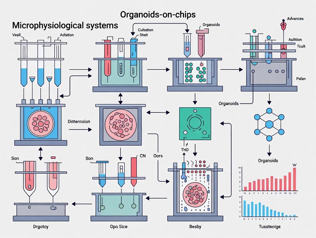
Organoids-on-Chips: Revolutionizing Biomedical Research with Next-Generation Microphysiological Systems
This article explores the transformative potential of organoids-on-chips technology, an innovative platform that integrates self-assembling 3D organoids with microfluidic systems.
Organoids-on-Chips: Revolutionizing Biomedical Research with Next-Generation Microphysiological Systems
Abstract
This article explores the transformative potential of organoids-on-chips technology, an innovative platform that integrates self-assembling 3D organoids with microfluidic systems. Tailored for researchers, scientists, and drug development professionals, it provides a comprehensive analysis spanning foundational principles, methodological applications, optimization strategies, and validation frameworks. The content covers how these systems enhance physiological relevance in disease modeling, drug testing, and personalized medicine by overcoming the limitations of traditional 2D cultures and animal models. By synthesizing the latest research and technological advances, this guide serves as an essential resource for leveraging organoids-on-chips to improve predictive accuracy in preclinical research and accelerate therapeutic discovery.
The Foundation of Organoids-on-Chips: Bridging Biology and Engineering
Abstract Organoids-on-chips represents a transformative microphysiological system (MPS) born from the synergistic integration of two pioneering technologies: self-assembling, stem-cell-derived 3D organoids and microfluidic organ-on-a-chip (OOC) devices [1] [2] [3]. This convergence creates in vitro human models that recapitulate complex organ-level physiology with high fidelity, addressing critical limitations of conventional 2D cell cultures and animal models in drug development [1] [4]. Organoids contribute multicellular architecture and patient-specific pathophysiology, while microfluidic chips provide dynamic microenvironments with perfusion, mechanical cues, and physiological gradients [1] [2]. This application note details the quantitative advantages, provides established protocols for model setup, and outlines essential reagent solutions to guide researchers in deploying this technology for predictive preclinical research.
Quantitative Performance of Organoids-on-Chips
Meta-analyses of perfused cultures compared to static controls reveal that cellular responses to flow are biomarker-specific and cell-type-dependent. The following table summarizes key quantitative findings from systematic comparisons.
Table 1: Quantitative Impact of Perfusion in Microphysiological Systems
| Cell Type / Model | Key Biomarker / Function | Fold-Change (Perfused vs. Static) | Physiological Relevance |
|---|---|---|---|
| CaCo-2 (Intestine) | CYP3A4 Activity | >2-fold increase [5] | Enhanced metabolic competence for drug absorption studies. |
| Hepatocytes | PXR mRNA Levels | >2-fold increase [5] | Improved regulation of xenobiotic metabolism and transport. |
| Blood Vessel Walls | Various Functional Biomarkers | Strong response to flow [5] | Better mimicry of vascular shear stress and barrier function. |
| General 3D Cultures | Overall Functionality | Slight improvement over 2D [5] | Perfusion benefits high-density cell cultures by improving nutrient/waste exchange. |
The data underscores that perfusion, a hallmark of organ-on-a-chip systems, drives specific functional enhancements critical for drug metabolism and toxicity studies [5]. The integration of organoids within these perfused systems leverages these benefits while adding human-specific cellular complexity.
Experimental Protocols
Protocol: Establishing a Basic Microfluidic Organoid Culture
This protocol outlines the process for loading and maintaining patient-derived or stem-cell-derived organoids in a microfluidic chip, such as the Emulate Chip-S1 or a PDMS-based custom device [3] [6].
Workflow Diagram: Organoid-on-a-Chip Setup
Materials:
- Organoids: Mature, defined organoids (e.g., intestinal, hepatic, cerebral).
- Microfluidic Chip: Commercially available (e.g., Emulate Chip-S1) or fabricated PDMS chip.
- Extracellular Matrix (ECM): Cultrex Basement Membrane Extract (BME) Type 3 (R&D Systems) or similar.
- Cell Culture Medium: Organoid-specific serum-free medium.
- Perfusion System: Syringe or pressure-driven pump capable of low, continuous flow rates (e.g., 50-500 µL/h).
- Tubing and Connectors: Sterile, gas-permeable or impermeable tubing as required.
Method:
- Organoid Preparation: Harvest mature organoids from their initial 3D culture. Gently break down large organoid structures into smaller, uniform aggregates (100-200 µm) using mechanical dissociation or gentle enzymatic treatment to prevent channel clogging [1] [3].
- Chip Priming and Coating: Sterilize the microfluidic chip (e.g., via UV light or 70% ethanol). Introduce a liquid ECM solution (e.g., BME diluted in medium) into the main cell culture chamber. Incubate (37°C, 30-60 min) to form a thin, stable gel layer that mimics the in vivo basement membrane.
- Organoid Loading: Resuspend the organoid aggregates in a low-viscosity, ECM-supplemented medium to facilitate loading. Using a pipette, carefully introduce the organoid suspension into the primed cell culture chamber via the designated inlet port. Allow the organoids to settle onto the coated surface for 15-30 minutes.
- Initiation of Perfusion: Connect the chip to the perfusion system. Initiate a very low flow rate (e.g., 50 µL/h) to minimize initial shear stress. Gradually ramp up the flow rate over 24-48 hours to the final desired rate, which applies a physiologically relevant shear stress (typically 0.02–0.5 dyne/cm² for epithelial barriers) [1] [5].
- Maintenance and Monitoring: Culture the organoids-on-chip under continuous perfusion, replacing the medium reservoir every 2-3 days. Monitor cell viability and morphology daily via integrated or microscope imaging. Assess barrier integrity in real-time if using chips with embedded electrodes (TEER) [4].
- Endpoint Analysis: At the experiment conclusion, the chip can be disassembled for endpoint readouts. These include:
- Immunofluorescence: Fix and stain the organoids within the chip for confocal microscopy.
- Effluent Collection: Analyze secreted biomarkers, metabolites, or drug compounds from the outflow medium.
- Omics Analysis: Recover organoids for transcriptomic, proteomic, or metabolomic profiling.
Protocol: Drug Absorption and Toxicity Screening using a Gut-Liver Axis Model
This protocol describes a multi-organ setup to study first-pass metabolism and organ crosstalk, a key application for pharmacokinetic analysis [7] [4].
Workflow Diagram: Gut-Liver Axis Assay
Materials:
- Multi-Organ Chip: A microfluidic platform with at least two interconnected culture chambers (e.g., Emulate's linked organ system or similar) [6].
- Organoids: Intestinal organoids (from primary cells or iPSCs) and liver organoids (from iPSCs or primary hepatocytes).
- Common Circulation Medium: A serum-free medium suitable for both intestinal and liver cell types, such as Williams E Medium supplemented with necessary growth factors.
- Analytical Equipment: LC-MS/MS for drug and metabolite quantification, ELISA kits for toxicity biomarkers (e.g., Albumin, ALT).
Method:
- Chip Seeding: Establish mature intestinal and liver organoids in their respective, fluidically isolated chambers of the multi-organ chip, following the basic protocol in Section 2.1.
- System Connection and Baseline: Connect the outlet of the "Gut Chip" compartment to the inlet of the "Liver Chip" compartment via microfluidic tubing. Initiate a common recirculating medium flow to establish a baseline and allow the systems to equilibrate for 24-48 hours.
- Drug Administration: Introduce the drug candidate at a physiologically relevant concentration directly into the inlet stream of the "Gut Chip" compartment.
- Sampling: Collect effluent samples from the outlet of the "Liver Chip" compartment at predetermined time intervals (e.g., 0, 1, 2, 4, 8, 24 hours).
- Analysis:
- Use LC-MS/MS to quantify the concentration of the parent drug and its major metabolites in the sampled medium. This provides kinetic data on absorption by the gut and metabolism by the liver.
- Use ELISA to measure the release of liver-specific enzymes like Alanine Aminotransferase (ALT) as a biomarker of drug-induced liver injury [7] [6].
- Data Interpretation: Plot concentration-time profiles for the parent drug and metabolites. Calculate pharmacokinetic parameters like half-life and clearance. Correlate metabolite appearance with markers of toxicity to assess the safety profile of the drug candidate.
The Scientist's Toolkit: Essential Research Reagent Solutions
Successful implementation of organoids-on-chips technology relies on a suite of specialized reagents and materials. The table below catalogs key solutions and their critical functions.
Table 2: Essential Reagents and Materials for Organoids-on-Chips Research
| Research Reagent / Material | Function and Application |
|---|---|
| Cultrex BME / Matrigel | A basement membrane extract providing a 3D scaffold that supports organoid growth, differentiation, and polarization [1] [8]. |
| Chip-R1 Rigid Chip (Emulate) | A consumable made from minimally drug-absorbing plastics, critical for obtaining accurate pharmacokinetic (ADME) and toxicology data by reducing compound loss [6]. |
| Induced Pluripotent Stem Cells (iPSCs) | The primary cell source for generating patient-specific organoids, enabling disease modeling and personalized medicine applications [1] [9] [8]. |
| Polydimethylsiloxane (PDMS) | The most common elastomer for fabricating microfluidic chips; prized for its gas permeability, optical clarity, and ease of prototyping [3] [4]. |
| Advanced 3D Culture Media | Chemically defined, serum-free media formulations supplemented with niche-specific growth factors (e.g., Wnt, R-spondin, Noggin) to maintain stemness and drive organ-specific differentiation in organoids [1] [8]. |
| Syringe / Pressure-Driven Pumps | Provide precise, active control over fluid flow in the microfluidic system, enabling the application of physiologically relevant shear stresses [3]. |
The Evolution from 2D Cultures to Dynamic 3D Microphysiological Systems
The failure of animal models and traditional two-dimensional (2D) cell cultures to accurately predict human therapeutic responses is a major challenge in drug development, contributing to high clinical attrition rates [10] [11]. Microphysiological Systems (MPS), often termed organ-on-a-chip (OOC) technologies, represent a transformative evolution from static 2D cultures to dynamic three-dimensional (3D) models that recapitulate critical aspects of human physiology [7]. These systems bridge the gap between basic biology and human health by incorporating 3D cellular architecture, fluid flow, and multi-cellular interactions, thereby offering more precise diagnostic and therapeutic strategies for patients [12]. The integration of organoid technology—self-assembling 3D cellular aggregates derived from stem cells—with sophisticated microfluidic chips has further accelerated this paradigm shift, enabling unprecedented modeling of human development, disease, and drug responses outside the body (organoids-on-chips) [13] [2]. This article details the core applications and provides actionable protocols for implementing these advanced models in biomedical research.
Core Comparative Analysis: 2D, 3D, and MPS Models
The limitations of traditional models are well-documented. While 2D cell cultures are useful for basic assays, they cannot replicate the complex 3D environment of human tissues, often leading to misleading or inaccurate data [10]. Animal models, though valuable, are expensive, time-consuming, and limited by species differences that often result in poor prediction of human outcomes [10] [14]. MPS address these shortcomings by mimicking the dynamic microenvironment of human organs, including fluid flow, mechanical stresses, and cell-cell interactions, leading to more physiologically relevant responses [12] [2].
Table 1: Comparative Analysis of Preclinical Model Systems
| Feature | In vitro 2D Cell Culture | In vitro 3D Spheroid | In vivo Animal Models | Microphysiological System (MPS) |
|---|---|---|---|---|
| Human Relevance | Low | Medium | Variable (Species-Dependent) | High |
| Complex 3D Architecture | No | Yes | Yes | Yes |
| (Blood)/Flow Perfusion | No | No | Yes | Yes |
| Innate & Adaptive Immune System | No | No | Yes | Emerging |
| Multi-organ Capability | No | No | Yes | Yes |
| Longevity | < 7 days | < 7 days | > 4 weeks | ~ 4 weeks |
| Acute and Chronic Dosing | Limited | Limited | Yes | Yes |
| New Drug Modality Compatibility | LOW | MEDIUM | LOW | MEDIUM / HIGH |
| Throughput | High | High | Low | Medium |
| Time to Result | FAST | FAST | SLOW | FAST |
| High-content Data | Limited | Limited | Yes | Yes [14] |
Table 2: Quantitative Advantages of MPS in Drug Metabolizing Enzyme (CYP) Expression
| Study Model | CYP Enzyme | Expression/Activity in MPS vs. Static Culture | Significance |
|---|---|---|---|
| Liver acinus dynamic (LADY) chip [10] | CYP2E1 | Remarkably increased | Improved drug metabolism capability |
| General Liver-on-chip [10] | Multiple CYPs | Higher than conventional plate cultures | More accurate prediction of drug availability and toxicity |
| Kidney epithelial cells in microfluidic device [10] | P-glycoprotein (P-gp) | Higher expression and activity | Better recapitulation of drug transport and clearance |
Application Notes: Key Use Cases in Biomedical Research
Disease Modeling and Drug Screening
MPS excel in modeling complex human diseases. For example, JAX scientists grow tumor organoids from human colon cancers, which not only recreate cancer cell behavior but also provide a platform for high-throughput drug screening [12]. Similarly, patient-derived organoids (PDOs) from rare malignancies, such as malignant peritoneal mesothelioma, faithfully recapitulate tumor histopathology and genomic heterogeneity, enabling personalized drug testing [13]. The "gut-on-a-chip" platform developed by Jalili et al. features intestinal epithelial cells that form finger-like villi and secrete mucus. When populated with bacteria and immune cells, this model allows real-time observation of host-microbe-immune interactions, crucial for studying Inflammatory Bowel Disease (IBD) and colorectal cancer [12].
ADME and Toxicology Profiling
A major application of MPS is the evaluation of a drug's Absorption, Distribution, Metabolism, and Excretion (ADME) properties and its toxicity. MPS provide a more sensitive system to uncover potential adverse effects early in development [14]. These systems are highly metabolically competent, expressing a full range of cytochrome P450 enzymes and transporters. Multi-organ MPS can recreate the process of drug absorption and first-pass metabolism to derive human bioavailability, offering enhanced accuracy over animal models [10] [14]. This capability is vital for de-risking the development of new drug modalities, including antibody-drug conjugates and CAR-T cell therapies, for which animal models are often less suitable [10].
Rare Disease Research
For over 7,000 rare diseases—most of which are hereditary—traditional models have struggled to recapitulate human-specific pathology. Organoids-on-chips offer a powerful platform to parse rare-disease pathogenesis [13]. For instance, spinal muscular atrophy (SMA) has been modeled using patient-derived organoids, which successfully replicated early disease features like motor neuron defects [13]. These models provide a much-needed resource for understanding disease mechanisms and accelerating therapeutic discovery for conditions that affect small patient populations.
Experimental Protocols
Protocol 1: Establishing a Gut-on-a-Chip Model for Host-Microbiome Interaction Studies
This protocol outlines the creation of a human gut-on-a-chip model to study real-time interactions between the intestinal barrier, microbiome, and immune system [12].
I. Materials
- Microfluidic Device: A multi-channel PDMS-free chip with a porous membrane separating apical and basolateral channels.
- Cells: Primary human intestinal epithelial cells or patient-derived intestinal organoid cells.
- Media: Intestinal cell culture medium, bacterial culture medium, and endothelial cell medium.
- Extracellular Matrix (ECM): Reduced-growth-factor MATRIGEL or similar ECM hydrogel.
- Other Reagents: Fluorescent dyes for viability and barrier integrity (e.g., FIT-dextran), fixatives, and antibodies for immunostaining.
II. Methodology
Step 1: Device Preparation
- Sterilize the microfluidic chip using UV light or 70% ethanol.
- Coat the porous membrane of the apical channel with ECM (e.g., MATRIGEL) and incubate at 37°C for at least 2 hours to form a gel.
Step 2: Cell Seeding and Monoculture Formation
- Introduce a suspension of intestinal epithelial cells into the apical channel.
- Apply a gentle vacuum to the basolateral channel to facilitate cell attachment to the ECM-coated membrane.
- Perfuse the basolateral channel with cell culture medium at a low flow rate (0.1-0.5 µL/min) for 24-48 hours to establish a monolayer.
Step 3: 3D Co-culture and Differentiation
- Seed human endothelial cells into the basolateral channel to form a vascular layer.
- Increase the flow rate to 10-30 µL/min to apply physiological shear stress.
- Culture the system for 5-10 days to promote the formation of 3D intestinal villi-like structures and a mature endothelial layer.
Step 4: Introduction of Microbiome and Immune Cells
- Introduce a defined bacterial community suspended in medium into the apical channel to colonize the mucus layer.
- Circulate immune cells (e.g., peripheral blood mononuclear cells) through the vascular (basolateral) channel.
Step 5: Real-time Monitoring and Endpoint Analysis
- Monitor barrier integrity in real-time by measuring the translocation of fluorescent molecules from the apical to the basolateral channel.
- At endpoint, fix the tissues within the chip and perform immunostaining for tight junction proteins (e.g., ZO-1) or specific cell markers.
- Collect effluent from the basolateral channel for cytokine analysis to quantify immune responses.
Protocol 2: Multi-organ MPS for ADME and Toxicity Assessment
This protocol describes the operation of a multi-organ MPS, such as the commercially available PhysioMimix system, to study inter-organ crosstalk and systemic drug effects [14].
I. Materials
- MPS Controller Hardware: A system capable of housing and perfusing multi-chip plates (e.g., PhysioMimix Controller, Docking Stations, and MPS Drivers).
- Multi-chip Plates: Organ-specific plates (e.g., liver, gut, kidney).
- Cells: 3D-validated primary cells or stem-cell derived organoids for each organ model.
- Assay Kits: Validated protocol kits, including custom media, supplements, and controls.
II. Methodology
Step 1: System Setup and Priming
- Mount the organ-specific multi-chip plates onto the MPS drivers and dock them into the controller within a standard cell culture incubator (37°C, 5% CO2).
- Prime the entire fluidic network of the system with appropriate culture medium to remove air bubbles and condition the channels.
Step 2: Tissue Model Loading
- Load pre-formed 3D organoids or tissue spheroids into their respective compartments on the multi-chip plate.
- Alternatively, seed single-cell suspensions into organ-specific scaffolds to form tissues in situ under perfusion.
Step 3: System Interconnection and Maintenance
- Connect the individual organ compartments via microfluidic channels to establish a shared circulatory flow.
- Set the recirculating flow rate to match physiological velocities for the specific organ combination (e.g., 0.5-5 µL/min).
- Maintain the system without daily maintenance for up to 4 weeks, with periodic sampling from the common medium reservoir.
Step 4: Dosing and Metabolite Tracking
- Introduce the drug candidate into the common medium reservoir at clinically relevant concentrations.
- Collect time-point samples from the reservoir to track the parent drug depletion and the formation of metabolites using LC-MS/MS.
- Monitor organ-specific toxicity in real-time via inline sensors or by analyzing effluent for released biomarkers (e.g., ALT for liver injury, KIM-1 for kidney injury).
Step 5: Multi-omic Endpoint Analysis
- At the end of the experiment, disassemble the system and extract microtissues from each organ compartment for analysis.
- Perform transcriptomic (RNA-seq), proteomic, or histopathological analysis on the tissues to uncover deep mechanistic insights into drug effects and organ-organ crosstalk.
The Scientist's Toolkit: Essential Research Reagents and Materials
Successful implementation of MPS relies on a suite of specialized materials and reagents designed to mimic the in vivo microenvironment.
Table 3: Key Research Reagent Solutions for Organoids-on-Chips
| Item Category | Specific Examples | Function & Importance |
|---|---|---|
| Microfluidic Hardware | PhysioMimix Controller & Docking Stations [14]; PDMS-free Multi-chip Plates [14] | Provides the engineered infrastructure for housing tissues, applying fluid shear stress, and connecting multiple organ models. PDMS-free materials prevent small molecule absorption. |
| 3D Scaffolds & ECM | Reduced-growth-factor MATRIGEL; Synthetic PEG-based hydrogels; Organ-specific scaffolds [2] | Provides the critical 3D biochemical and biophysical microenvironment for cell attachment, migration, and tissue organization. |
| Cell Sources | Primary human cells (e.g., hepatocytes, intestinal epithelial cells) [10]; Induced Pluripotent Stem Cell (iPSC)-derived organoids [13]; 3D-validated cell lines [14] | Forms the biological basis of the model. Patient-derived cells enable personalized medicine approaches, while validated cells ensure reliability. |
| Specialized Media | Organ-specific culture media (e.g., for liver, gut, kidney); Co-culture media; Media for host-microbiome studies [12] [14] | Supplies tailored nutrients, growth factors, and hormones to support the viability and function of complex, multi-cellular tissues. |
| Sensing & Assay Kits | TEER measurement electrodes; Metabolic activity assays (e.g., Albumin, Urea for liver); Cytokine detection kits; Live-dead staining kits [2] [14] | Enables real-time, non-destructive monitoring of tissue health, barrier function, and functional output. Critical for longitudinal studies. |
| Integrated Sensors | Oxygen sensors; pH sensors [13] | Monitors the physicochemical microenvironment in real-time within the microfluidic channels, providing data on metabolic activity and culture conditions. |
The evolution from simple 2D cultures to dynamic 3D Microphysiological Systems marks a fundamental shift in how researchers model human biology and disease. By integrating organoid biology with microfluidic engineering, MPS provide a physiologically relevant platform that bridges the translational gap between preclinical models and human patients [12] [2]. As these technologies continue to mature, supported by advances in 3D bioprinting, multi-omics integration, and automation, their adoption in drug development pipelines and regulatory decision-making is poised to accelerate [7] [13]. This promises not only to reduce the pharmaceutical industry's reliance on animal models but also to usher in a new era of personalized medicine, where a patient's own cells can be used to identify the most effective therapeutic strategies [11].
The field of microphysiological systems (MPS) has been revolutionized by the synergistic integration of stem cell biology and microfluidic engineering. This convergence has given rise to sophisticated organoids-on-chips platforms that overcome critical limitations of conventional organoid culture. While stem cell biology provides the foundational building blocks through self-organizing human organoids (HOs), microfluidic engineering delivers the precise environmental control required for enhanced physiological relevance. Together, they enable the creation of 3D organotypic living models that recapitulate critical tissue-specific properties and functions, representing a significant advancement over traditional two-dimensional cell cultures and animal models for biomedical research and drug development [2].
The core innovation lies in how microfluidic technology addresses the inherent challenges of traditional organoid culture. Standard organoid methods suffer from limited long-term functional culture, lack of maturation, and high batch-to-batch variability, primarily due to their dependence on passive diffusion for nutrient exchange and waste removal [15] [16]. Microfluidic organ-on-a-chip (OoC) systems tackle these limitations by providing dynamic perfusion, biomechanical stimulation, and precise control over the cellular microenvironment. This integration creates a more in vivo-like ecological niche that supports enhanced organoid maturation, viability, and functionality [15] [2].
Core Biological Components: Stem Cells and Their Microenvironment
The biological foundation of organoids-on-chips technology rests on the utilization of various stem cell sources, each offering distinct advantages for specific research applications. The appropriate selection of stem cell type is crucial for successfully modeling target tissues or disease states.
Pluripotent Stem Cells (PSCs): This category includes both embryonic stem cells (ESCs) and induced pluripotent stem cells (iPSCs). iPSCs, in particular, have transformed the field by enabling the generation of patient-specific organoids. These cells can differentiate into any cell type derived from the three germ layers—endoderm, mesoderm, and ectoderm—making them ideal for modeling a wide range of tissues, including brain, kidney, liver, and intestine [17] [2]. Their indefinite self-renewal capacity provides a scalable source for high-throughput applications.
Adult Stem Cells (ASCs): Also known as tissue-specific stem cells, ASCs are multipotent cells found in specific adult tissues. They are responsible for natural tissue maintenance and repair. Organoids derived from ASCs, such as intestinal organoids from Lgr5+ crypt base columnar cells, typically model the epithelial layer of their organ of origin and are widely used for disease modeling and drug screening [17] [18].
Differentiated Primary Cells: Recent advancements have demonstrated that certain differentiated cell types, such as cholangiocytes and hepatocytes, can also be reprogrammed to form organoids, expanding the potential cell sources for specific applications [17].
The Extracellular Matrix (ECM) and Niche Factors
The stem cell microenvironment, or niche, is a critical component that guides organoid self-assembly, differentiation, and maturation. It provides both physical scaffolding and essential biochemical signals.
ECM Scaffolds: The extracellular matrix provides the physical scaffold for 3D organoid growth, influencing cell polarity, migration, and differentiation. The most commonly used ECM materials include:
- Matrigel: A basement membrane extract rich in laminin, collagen IV, and growth factors. It is the gold standard for many organoid cultures but has limitations due to its batch-to-batch variability and undefined composition [17] [19].
- Collagen-Based Matrices: Often used as a more defined alternative to Matrigel, particularly for in vivo transplantation studies due to reduced angiogenic potential [19].
- Synthetic Hydrogels: Engineered polymers, such as PEG-based hydrogels, are gaining traction as they offer precise control over mechanical and biochemical properties, enhancing reproducibility for clinical applications [17] [19].
Biochemical Niche Factors: A precise combination of growth factors and small molecules is required to mimic the endogenous signaling landscape and guide stem cell fate. These factors modulate key evolutionary conserved signaling pathways such as Wnt, BMP, TGF-β, and EGF. The required niche factors vary significantly depending on the organoid type, as detailed in Table 1 [19].
Table 1: Essential Niche Factors for Various Organoid Types
| Organoid Type | Essential Proteins & Growth Factors | Key Small Molecules | Common ECM |
|---|---|---|---|
| Intestinal/Colon | EGF, Noggin, R-spondin, Wnt-3A | A83-01, Y-27632, SB202190, Gastrin | Matrigel, GFR-BME [19] |
| Cerebral | EGF, Noggin, R-spondin | A83-01, SB202190 | Matrigel [15] [19] |
| Hepatic | EGF, R-spondin, FGF10, HGF | Nicotinamide, Gastrin, Forskolin | BME, PEG Hydrogel [19] |
| Pancreatic | EGF, Noggin, R-spondin, FGF10 | Wnt-3A, Retinoic Acid, A83-01 | Matrigel, Collagen [19] |
| Lung | EGF, Noggin, R-spondin, FGF7, FGF10 | A83-01 | Matrigel [19] |
Core Engineering Principles: Microfluidic Design and Fabrication
Key Microfluidic Features and Their Physiological Relevance
Microfluidic engineering contributes functionalities that are indispensable for creating physiologically relevant organoid models. The design of these systems is guided by the reductionist analysis of the target organ's functional unit [2].
Dynamic Perfusion and Mimicking Vasculature: Microfluidic channels enable continuous, controlled fluid flow. This perfusion mimics blood flow, ensuring efficient delivery of nutrients and oxygen while removing metabolic waste. This solves the diffusion limitation inherent in static cultures, preventing necrotic core formation in larger organoids and enabling long-term culture [15] [16]. The resulting fluid shear stress also serves as a key biomechanical cue for endothelial and epithelial cells.
Biomechanical Cues: Organ-on-chip platforms can incorporate physiological mechanical forces such as cyclic strain (to mimic breathing motions in lung alveoli or peristalsis in intestine) and compressive forces. These cues are critical for proper tissue maturation and function [15] [2] [20].
Spatial Control and Partitioned Co-culture: Micrometer-sized channels and chambers allow for the precise spatial patterning of cells and tissues. This enables the creation of complex, multi-cellular interfaces, such as the blood-brain barrier or gut-epithelium-microbe interfaces, which are fundamental to studying organ-level interactions and drug permeability [2].
Automation and High-Throughput Screening: Microfluidic platforms are inherently scalable and amenable to automation. They can be designed as multi-well array systems, allowing for the parallel culture and analysis of hundreds of organoids under controlled conditions. This significantly enhances experimental reproducibility and throughput for drug screening and toxicology studies [15] [18].
Fabrication Technologies and Material Selection
The physical realization of organoids-on-chips relies on advanced microfabrication techniques.
Photolithography and Soft Lithography: These are the most established methods. Photolithography is used to create a master mold with defined microstructures on a silicon wafer. Soft lithography, typically using the polymer Polydimethylsiloxane (PDMS), is then employed to replicate these structures into a transparent, gas-permeable, and biocompatible chip [2] [20]. PDMS is popular for its optical clarity and ease of use but can absorb small hydrophobic molecules, which is a consideration for drug screening.
3D Printing: An emerging and highly versatile technology, 3D bioprinting allows for the direct fabrication of microfluidic devices, integrated sensors, and even the printing of cells and matrices (bioprinting) within the platform. It offers rapid prototyping and the creation of more complex, multi-layer architectures [2].
Etching Techniques: Both wet (chemical) and dry (e.g., reactive ion) etching are used to fabricate microfluidic channels in materials like glass and silicon, offering high precision for smaller channel sizes [2].
Integrated Experimental Protocols
Protocol 4.1: Establishing a Brain Organoid-on-Chip Culture
This protocol adapts the pioneering work of Lancaster et al. and subsequent studies for embedding brain organoids into a microfluidic platform to enhance neural development and reduce necrotic core formation [15].
Workflow Overview:
Materials:
- Microfluidic Chip: e.g., OrganoidChip+ design or commercial equivalent [18].
- Cell Source: Human induced Pluripotent Stem Cells (iPSCs).
- ECM: Growth Factor Reduced Matrigel.
- Media: Neural induction medium, followed by neuronal differentiation medium.
- Key Reagents: Growth factors (EGF, Noggin, R-spondin), small molecules (e.g., SMAD inhibitors), and staining antibodies (e.g., against Nestin, SOX2, TUJ1) [15] [19].
Step-by-Step Procedure:
- EB Formation (Days 1-6): Generate embryoid bodies (EBs) from iPSCs using AggreWell plates or the forced aggregation method according to established protocols [15].
- Neuroectoderm Induction (Days 7-11): Culture EBs in static conditions in neural induction medium supplemented with dual SMAD inhibitors to direct differentiation toward the neuroectodermal lineage.
- On-Chip Seeding (Day 11): a. Pre-coat the culture chamber of the microfluidic chip with a thin layer of Matrigel. b. Resuspend the neuroectoderm-induced EBs in a cold, diluted Matrigel solution. c. Carefully pipette the EB-Matrigel suspension into the main culture chamber of the chip. d. Allow the Matrigel to polymerize at 37°C for 30 minutes.
- On-Chip Perfusion Culture (Days 12-30+): a. Connect the chip to a programmable perfusion system. b. Initiate a continuous flow of neuronal differentiation medium. The initial flow rate should be low (e.g., 0.1-0.5 µL/min) to avoid dislodging organoids, and can be gradually increased. c. Culture the organoids under perfusion for the desired period, with medium changes every 2-3 days.
- Analysis: Monitor organoid growth via brightfield microscopy. For endpoint analysis, fix organoids on-chip and perform immunostaining for neural markers (e.g., Nestin for progenitors, TUJ1 for neurons). Image using confocal or two-photon microscopy directly on the chip [15] [18].
Protocol 4.2: High-Content Imaging and Viability Assay of Intestinal Organoids-on-Chip
This protocol utilizes the "OrganoidChip+" platform to enable transferless culturing, staining, and high-resolution imaging of adult stem cell-derived intestinal organoids (ASOs) [18].
Workflow Overview:
Materials:
- Microfluidic Platform: OrganoidChip+ or similar device with immobilization traps and a thin glass substrate [18].
- Cell Source: Canine or human colon adult stem cells (ASCs).
- ECM: Matrigel.
- Staining Reagents: Live/Dead viability/cytotoxicity kit (e.g., Calcein AM / Ethidium homodimer-1).
- Imaging Equipment: Widefield fluorescence microscope and/or two-photon microscope.
Step-by-Step Procedure:
- On-Chip Seeding and Culture: a. Mix intestinal ASCs with cold Matrigel and seed ~5 µL of the suspension into the culture chamber of the chip. b. Polymerize the Matrigel at 37°C. c. Connect the chip to perfusion and culture for 7 days with appropriate intestinal organoid medium, monitoring growth via intermittent brightfield imaging.
- Endpoint Staining and Immobilization (for Viability Assay): a. On day 7, stop perfusion and inject a Matrigel digestion solution into the culture chamber. Incubate until the matrix is dissolved. b. Introduce the Live/Dead staining solution and incubate according to the manufacturer's protocol. c. Apply a controlled flow to transfer the now-freed and stained organoids from the culture chamber into the dedicated trapping areas (TAs) for immobilization.
- Imaging: a. For viability: Perform widefield fluorescence imaging on the immobilized organoids. Capture multiple z-stacks for 3D analysis. b. For label-free metabolic assessment: On a separate set of organoids still embedded in Matrigel (Day 7), perform two-color, two-photon microscopy to measure the autofluorescence of NAD(P)H and FAD to calculate the redox ratio, an indicator of metabolic activity [18].
- Data Analysis: Quantify the percentage of live vs. dead cells using image analysis software (e.g., ImageJ, CellProfiler). Calculate the redox ratio as FAD/(NAD(P)H+FAD) for metabolic comparison.
Quantitative System Parameters and Performance Metrics
The successful integration of biology and engineering is reflected in quantifiable parameters that define system performance and physiological relevance. Table 2 summarizes key quantitative data from established organoids-on-chips platforms.
Table 2: Quantitative Parameters for Organoids-on-Chips Culture and Analysis
| Parameter | Typical Range / Value | Significance / Impact | Reference Example |
|---|---|---|---|
| Culture Chamber Height | 550 µm - 610 µm | Limits z-axis span of organoids, facilitating high-resolution imaging with high-NA objectives. | [15] [18] |
| Perfusion Flow Rate | 0.1 - 5.0 µL/min (organ-dependent) | Mimics physiological shear stress; prevents necrotic cores; improves nutrient/waste exchange. | [15] [20] |
| Organoid Size Range | 400 - 600 µm (for imaging) | Compatible with trapping and immobilization chambers in microfluidic devices. | [18] |
| Culture Duration | Weeks to >8 months | Enables study of chronic toxicity, disease progression, and long-term maturation. | [15] [19] |
| Growth Rate (on-chip vs off-chip) | Superior or comparable | Indicates a healthy culture environment within the microfluidic device. | [18] |
| Redox Ratio (Metabolic Activity) | Comparable or slightly better than off-chip | Suggests enhanced or maintained metabolic health under perfusion culture. | [18] |
The Scientist's Toolkit: Essential Research Reagent Solutions
A successful organoids-on-chips experiment relies on a suite of well-defined reagents and materials. The following table details key components and their functions.
Table 3: Essential Reagents and Materials for Organoids-on-Chips Research
| Item Category | Specific Examples | Primary Function | Application Notes |
|---|---|---|---|
| Stem Cell Sources | iPSCs, Adult Stem Cells (ASCs) | Self-renewing foundation that differentiates into complex 3D tissue structures. | iPSCs for patient-specific & multi-tissue models; ASCs for epithelial organoids. [17] [2] |
| ECM Scaffolds | Matrigel, Collagen I, Synthetic PEG Hydrogels | Provides a 3D biomechanical scaffold mimicking the native extracellular matrix. | Matrigel is common but undefined; synthetic hydrogels offer control and reproducibility. [17] [19] |
| Key Growth Factors | EGF, Noggin, R-spondin, FGF families, Wnt-3A | Activates signaling pathways critical for stem cell maintenance and directed differentiation. | Combinations are tissue-specific (see Table 1). Required for long-term culture. [15] [19] |
| Small Molecule Inhibitors/Activators | Y-27632 (ROCKi), A83-01 (TGF-βi), CHIR99021 (Wnt activator) | Precisely controls signaling pathways to enhance viability and guide cell fate. | Y-27632 reduces anoikis; A83-01 promotes epithelial growth. [19] |
| Microfluidic Device Materials | PDMS, PMMA, PS, Glass | Forms the physical structure of the chip, with properties like gas permeability and optical clarity. | PDMS is most common; absorption of small molecules can be a limitation for drug studies. [2] [20] |
The biopharmaceutical industry is facing a critical productivity challenge. Despite record levels of research and development activity, with over 23,000 drug candidates in development and $300 billion spent annually on R&D, success rates have been declining precipitously [21]. The most striking evidence of this crisis is the plummeting success rate for Phase 1 drugs, which fell to just 6.7% in 2024 compared to 10% a decade ago [21]. This attrition problem has driven the internal rate of return for R&D investment down to 4.1% – well below the cost of capital [21].
A fundamental cause of this high failure rate is the poor predictivity of traditional preclinical models, particularly animal testing. Statistics show that over 90% of drugs that appear safe and effective in animals ultimately fail in human clinical trials, with 60% failing due to lack of efficacy and 30% due to toxicity issues in humans [22] [23]. This failure highlights the profound scientific limitations of interspecies extrapolation and reinforces the need for human-relevant models that can better predict human responses [22].
Table 1: Contemporary Analysis of Clinical Trial Success Rates (2001-2023)
| Development Phase | Historical Success Rate (%) | Key Failure Drivers |
|---|---|---|
| Phase I to Phase II | 6.7% (2024) [21] | Lack of efficacy (60%), toxicity (30%) [22] |
| Phase II to Phase III | Varies by therapeutic area | Inaccurate disease modeling, off-target effects |
| Phase III to Approval | Recently shows improvement | Commercial viability, confirmatory trial requirements |
| Overall Likelihood of Approval | 7-20% (varies by study) [24] | Composite of all above factors |
The Regulatory Mandate for Human-Relevant Models
A seismic regulatory shift is underway, moving the industry from animal-first to human-relevant testing paradigms. The landmark FDA Modernization Act 2.0, passed in late 2022, provided the critical legal authorization for utilizing non-animal methods in Investigational New Drug (IND) applications [22] [23]. This act transformed animal testing from a mandatory requirement into a permissible option, effectively establishing New Approach Methodologies (NAMs) as legally viable alternatives for demonstrating safety and efficacy [23].
In 2025, this transition accelerated significantly. The FDA announced a groundbreaking plan to phase out animal testing requirements for monoclonal antibody therapies and other drugs, replacing them with more effective, human-relevant methods [25]. The agency's "Roadmap to Reducing Reliance on Animal Testing in Preclinical Safety Studies" identifies monoclonal antibodies (mAbs) as an immediate focus area, noting that current requirements for mAbs mandate extensive, costly repeat-dose toxicity studies in animals, often requiring up to 144 non-human primates over periods of one to six months at a cost of up to $750 million per therapeutic [23].
Further momentum comes from the National Institutes of Health (NIH), which launched an $87 million Standardized Organoid Modeling (SOM) Center to address the primary hurdle to NAM adoption: the lack of standardized, reproducible protocols across different laboratories [23]. This investment structurally validates the use of robust, high-throughput 3D microtissues as essential technology for achieving newly prioritized goals of scientific reproducibility and regulatory acceptance.
Human-Relevant Model Technologies: Organoids and Organ-on-a-Chip
Microphysiological Systems (MPS), including organoids and organ-on-a-chip (OoC) technologies, represent promising alternatives to animal testing that offer in vitro models with high physiological relevance [7]. Organoids are 3D cell aggregates that self-organize into structures resembling native organs, while organ-on-a-chip systems are microfluidic devices lined with living human cells that mimic the physiological environment and mechanical forces experienced by cells in vivo [7].
These technologies are transitioning from exploratory tools to established, versatile platforms for real-world biomedical problems. The 2025 MPS World Summit showcased this maturation with the introduction of next-generation platforms like the AVA Emulation System, a 3-in-1 Organ-Chip platform designed specifically for high-throughput experiments, enabling researchers to run 96 independent Organ-Chip samples in a single run [6].
Table 2: Comparative Analysis of Human-Relevant Preclinical Models
| Model Type | Key Advantages | Current Limitations | Lead Applications |
|---|---|---|---|
| Organoids | Human genetic background, 3D architecture, patient-specific [22] | Batch-to-batch variability, limited maturity [7] | Disease modeling, personalized medicine [26] |
| Organ-on-a-Chip | Controlled biomechanical cues, fluid flow, multi-tissue integration [7] [26] | Technical complexity, cost [22] | Toxicity testing, ADME studies [6] |
| Integrated Organoid-on-a-Chip | Combines physiological relevance of organoids with controlled environment of OoC [26] | Nascent technology, standardization challenges | Complex disease modeling, pharmacokinetic studies [26] |
Experimental Protocol: Establishing a Human-Relevant Testing Platform
Protocol 1: Liver-Chip for Predictive Toxicology Studies
Background: Drug-induced liver injury (DILI) remains a leading cause of drug attrition and post-market withdrawals. Conventional models (animal testing, 2D hepatocyte cultures) show poor predictivity for human DILI. The Emulate Liver-Chip has demonstrated superior performance in predicting drug-induced liver injury compared to animal and hepatic spheroid models [22] [6].
Materials & Reagents:
- Emulate Chip-S1 Stretchable Chips or Chip-R1 Rigid Chips (for ADME/toxicology applications) [6]
- Primary human hepatocytes (donor-matched if possible)
- Primary human liver sinusoidal endothelial cells (LSECs)
- Primary human Kupffer cells (for immune-competent models)
- Liver-Chip specific extracellular matrix (e.g., collagen IV/fibronectin)
- Hepatocyte maintenance medium + endothelial cell-specific medium
- Test compounds + reference controls (e.g., acetaminophen, troglitazone)
- Effluent collection plates for biomarker analysis
Methodology:
- Chip Preparation:
- Coat the top (parenchymal) channel with liver-specific extracellular matrix (0.1 mg/mL collagen IV, 0.02 mg/mL fibronectin) for 2 hours at 37°C.
- Coat the bottom (vascular) channel with 0.1 mg/mL collagen I for 1 hour at 37°C.
Cell Seeding:
- Day 0: Seed primary human hepatocytes (1.0×10^6 cells/mL) in the top channel. Allow attachment for 4-6 hours.
- Day 1: Seed human LSECs (0.5×10^6 cells/mL) in the bottom channel.
- For immune-competent models: Add Kupffer cells (0.2×10^6 cells/mL) to the top channel following hepatocyte attachment.
Culture & Maintenance:
- Maintain chips under continuous, unidirectional flow (30 μL/hour vascular channel, 10 μL/hour parenchymal channel) using the Zoë-CM2 Culture Module or AVA Emulation System.
- Culture for 7-10 days to establish mature phenotype before compound testing.
- Confirm functionality through albumin/urea production (hepatocytes) and factor VIII expression (LSECs).
Compound Testing:
- Prepare test compounds in endothelial cell-specific medium at 100X final concentration.
- Dilute 1:100 when perfusing through vascular channel to achieve desired concentration.
- Include vehicle controls and benchmark compounds (both safe and hepatotoxic).
- Expose chips for 7 days with daily medium renewal.
Endpoint Analysis:
- Daily: Collect effluent from both channels for biomarker analysis (ALT, AST, albumin).
- Post-experiment: Fix and immunostain for zonula occludens-1 (ZO-1), CYP3A4, and CD31.
- Quantify viability via intracellular ATP content.
- Optional: Transcriptomic/proteomic analysis of retrieved cells.
Validation: Benchmark against known hepatotoxicants (e.g., acetaminophen, troglitazone) and clinically safe compounds. Compare predictivity to historical animal model performance using metrics like sensitivity, specificity, and overall concordance with human clinical outcomes.
Integrated Testing Strategies and Advanced Applications
Multi-Organ Systems for Complex Biology
While single-organ models provide valuable insights, many drug effects involve complex inter-organ interactions. Advanced MPS platforms now enable the linking of multiple organ chips to create human-relevant systems for studying pharmacokinetics and pharmacodynamics [7] [26]. For instance, a Liver-Chip can be integrated with Gut-Chip and Kidney-Chip models to simulate first-pass metabolism and systemic clearance, providing a more comprehensive prediction of human drug responses [7].
The workflow below illustrates the experimental process for establishing and applying these human-relevant models in drug development.
The Scientist's Toolkit: Essential Research Reagent Solutions
Table 3: Key Research Reagents for Organoid and Organ-on-a-Chip Applications
| Reagent/Material | Function | Example Application |
|---|---|---|
| Chip-R1 Rigid Chips (Emulate) | Low-drug-absorbing plastic chips for ADME/toxicology [6] | Pharmacokinetic studies, chronic toxicity testing |
| Liver-Chip Extracellular Matrix | Provides physiological scaffold for cell attachment and polarization [6] | Maintaining hepatocyte polarity and function |
| Primary Human Hepatocytes | Gold standard for liver functionality assessment [6] | Drug metabolism, transporter studies, DILI prediction |
| Immune Cell Supplements (e.g., Kupffer cells) | Introduces immune competence to organ models [23] | Immunotoxicity assessment, cytokine release syndrome |
| Multi-organ linking medium | Universal medium supporting multiple cell types in linked systems [7] | Multi-organ pharmacokinetic studies |
| Tissue-specific differentiation factors | Directs stem cell differentiation toward target lineages [22] | Generation of patient-specific organoids |
Protocol 2: Multi-organ Platform for ADME and Toxicity Assessment
Background: Predicting systemic exposure and organ-specific toxicity requires understanding of a drug's journey through the body. Integrated multi-organ systems can provide a more comprehensive assessment before clinical trials.
Materials & Reagents:
- Organ-specific chips (Liver, Gut, Kidney at minimum)
- Multi-organ circulation module (Emulate Zoë-CM2 or equivalent)
- Universal circulation medium (compatible with all cell types)
- Precision peristaltic pumps with adjustable flow rates
- Automated sampling system for temporal profiling
- LC-MS/MS system for compound quantification
Methodology:
- Individual Chip Preparation:
- Establish individual organ chips (Liver, Gut, Kidney) following protocol 1.
- Confirm tissue-specific functionality before linking.
System Integration:
- Connect chips in physiologically relevant order: Gut → Liver → Kidney.
- Establish circulation using universal medium at flow rates scaling to human organ blood flows.
- Include a mixing reservoir to represent systemic circulation.
Dosing and Sampling:
- Introduce compound through Gut chip (oral) or mixing reservoir (IV).
- Collect temporal samples from each organ effluent and systemic reservoir.
- Analyze compound and metabolite concentrations using LC-MS/MS.
Endpoint Analysis:
- Assess functional markers for each organ (e.g., albumin for liver, TEER for gut, KIM-1 for kidney).
- Evaluate tissue integrity and specific toxicity markers.
- Calculate pharmacokinetic parameters (Cmax, Tmax, AUC, clearance).
Validation: Compare multi-organ system predictions of human pharmacokinetics and toxicity for known drugs with established clinical profiles to validate predictivity.
Quantitative Success and Future Outlook
The adoption of human-relevant models is demonstrating tangible impacts on drug development efficiency. Companies implementing these approaches report significant reductions in preclinical timelines and improved decision-making quality. The workflow below outlines the strategic integration of these models into the drug development pipeline to de-risk programs before clinical stages.
The future of human-relevant testing will be increasingly powered by computational integration and artificial intelligence. As noted by Dr. Greg Tietjen, CEO of Revalia Bio, "The future is human-centered, and we stand on the shoulders of all the work that came before. But the biggest conceptual takeaway is that we must get to a place where failing a human experiment is no longer a catastrophic event, as it is in a failed clinical trial, but rather a catalytic engine for learning" [27].
The FDA is supporting this integration through its Modeling and Simulation Working Group, which focuses on computational tools, including AI, Machine Learning, and Physiologically Based Pharmacokinetic (PBPK) modeling [23]. These in silico technologies can inform first-in-human dosing and justify waiving certain animal studies, particularly when combined with high-quality data from human-relevant models [23].
The transition to human-relevant models represents a fundamental transformation in drug development philosophy. By anchoring science in human biology from the outset, rather than attempting to translate from other species, the industry can address the root causes of drug attrition. The convergence of advanced model systems (organoids, organ-on-a-chip), regulatory evolution (FDA Modernization Act 2.0, 2025 FDA roadmap), and technological innovation (AI, digital twins) creates an unprecedented opportunity to make drug development faster, cheaper, and more predictive.
While challenges remain in standardizing and scaling these technologies, the coordinated push from regulators, industry, and academia suggests that human-relevant models will soon become the default rather than the alternative in preclinical testing. For researchers, early adoption and mastery of these platforms will be crucial for maintaining competitiveness in the evolving drug development landscape.
The U.S. Food and Drug Administration (FDA) has initiated a groundbreaking strategic plan to reduce and ultimately replace animal testing requirements in drug development, particularly for monoclonal antibodies and other biological products [25]. This landmark decision marks a fundamental transformation in regulatory science, transitioning from traditional animal models to human-relevant, advanced technological solutions. The FDA's new approach embraces New Approach Methodologies (NAMs)—including AI-based computational models, organoids, and organ-on-a-chip (OoC) technologies—designed to improve drug safety prediction while accelerating therapeutic development [25] [7]. This shift responds to both the ethical imperative to reduce animal use and the scientific limitation of animal models, which often fail to adequately recapitulate human physiology and disease pathology [9] [28]. For researchers and drug development professionals, this regulatory evolution necessitates familiarity with emerging human-relevant testing platforms and their integration into preclinical workflows.
FDA Regulatory Roadmap: Strategic Framework and Timelines
Core Components of the FDA Initiative
The FDA's comprehensive framework outlines a multi-faceted approach to modernizing drug safety evaluation:
- Reduction, refinement, and potential replacement of animal testing requirements using advanced technologies [25]
- Immediate implementation for investigational new drug (IND) applications, with encouragement for including NAMs data [25]
- Utilization of pre-existing, real-world safety data from other countries with comparable regulatory standards where drugs have already been studied in humans [25]
- Pilot programs for select monoclonal antibody developers to use primarily non-animal-based testing strategies under close FDA consultation [25]
- Regulatory incentives for companies that submit strong safety data from non-animal tests, potentially including streamlined review processes [25]
Strategic Implementation Timeline
Table 1: FDA Implementation Timeline for Alternative Testing Methods
| Timeframe | Regulatory Goals and Milestones | Expected Impact |
|---|---|---|
| Immediate (Initiated) | Acceptance of NAMs data in IND applications; Launch of pilot programs for monoclonal antibodies | Early adoption encouraged; foundational data collection |
| Short-term (1-3 years) | Phase-out of specific animal tests for biologics; Development of updated guidance documents | Reduced animal use for highly human-specific products |
| Mid-term (3-5 years) | Make animal studies "the exception rather than the norm" for preclinical safety/toxicity testing [28] | Transformative shift in regulatory standards; increased reliance on human-relevant data |
FDA Commissioner Dr. Martin A. Makary emphasized the far-reaching significance of this initiative: "For too long, drug manufacturers have performed additional animal testing of drugs that have data in broad human use internationally. This initiative marks a paradigm shift in drug evaluation and holds promise to accelerate cures and meaningful treatments for Americans while reducing animal use" [25].
Advanced Technological Platforms: Organoids and Organ-on-Chip Systems
Scientific Foundations and Convergence
The FDA's regulatory shift is enabled by significant advancements in microphysiological systems (MPS), particularly organoids and organ-on-chip technologies:
Organoids are three-dimensional, multicellular, self-assembling structures derived from various types of stem cells (pluripotent stem cells, embryonic stem cells, or tissue-specific stem cells) that retain characteristic features of corresponding organs [16]. These models effectively recapitulate human physiology more accurately than conventional 2D cultures or animal models.
Organ-on-a-Chip platforms are engineered microfluidic cell culture devices that simulate the functional units of human organs. These systems recreate tissue-tissue interfaces and incorporate biomechanical cues and vascular flow to mimic the in vivo microenvironment [16].
Organoids-on-Chips represents an integrative approach that combines the physiological relevance of organoids with the controlled microenvironment and perfusion capabilities of microfluidic chips [9] [16]. This synergy addresses key limitations of conventional organoid culture, including lack of maturation, limited reproducibility, and absence of physiological cues.
Technical Advantages of Integrated Systems
Table 2: Comparative Analysis of Traditional vs. Advanced Testing Platforms
| Parameter | Traditional Animal Models | Conventional Organoids | Organoids-on-Chips |
|---|---|---|---|
| Physiological Relevance | Limited by species differences | High cellular complexity but static environment | High, with dynamic microenvironment |
| Predictive Value for Human Response | Variable, often poor | Improved but limited by maturation | Enhanced through mechanical cues and perfusion |
| Throughput and Scalability | Low, time-consuming | Moderate | High with automated systems [6] |
| Reproducibility | Moderate to high | Variable, batch-to-batch variability | Improved through environmental control |
| Cost and Timeline | High cost, lengthy studies | Moderate cost and time | Higher initial investment but reduced long-term costs |
The integration of organoids with chip technology addresses several critical limitations of conventional organoid culture:
- Perfusable microfluidic networks mimic vascular function, overcoming diffusion limitations that restrict organoid size and maturation [16]
- Biomechanical stimulation through application of fluid flow and pressure recapitulates in vivo mechanical cues important for tissue development and function [16]
- Multi-organoid systems enable the study of organ-organ interactions, crucial for understanding systemic drug effects and disease mechanisms [16]
- Automation and environmental control enhance reproducibility and enable higher-throughput screening [16]
Application Notes: Implementation in Drug Development Workflows
Protocol: Establishing Intestinal Organoids-on-Chips for Barrier Integrity Assessment
This protocol adapts established methodologies for generating human intestinal MPS compatible with FDA's emphasis on human-relevant testing platforms [29]:
Organoid Line Establishment and Maintenance
- Source human intestinal stem cells from patient biopsies or commercially available stem cell lines
- Culture in 3D Matrigel domes with intestinal stem cell medium containing Noggin, R-spondin, and EGF for 7-10 days
- Passage organoids every 7-14 days using mechanical dissociation and enzymatic digestion with TrypLE Express
- Validate organoid phenotype through immunohistochemistry for intestinal markers (Villin, Mucin-2, Lysozyme) and functional assays
Microfluidic Chip Preparation and Seeding
- Select appropriate chip platform (e.g., Emulate Chip-S1 or Chip-R1 for low drug absorption [6])
- Coat microfluidic channels with collagen IV (10 µg/mL) for 2 hours at 37°C
- Prepare single-cell suspension from mature organoids using TrypLE Express digestion
- Seed intestinal epithelial cells at density of 2×10^6 cells/mL in the top channel of the chip
- Add human vascular endothelial cells (HUVECs or intestinal microvascular endothelial cells) at 1×10^6 cells/mL in the bottom channel
- Culture under static conditions for 24 hours to allow cell attachment, then initiate perfusion at 30 µL/hour
Barrier Integrity Assessment and Functional Testing
- Monitor transepithelial electrical resistance (TEER) daily using integrated or external electrodes
- Perform permeability assays with fluorescent dextran (4 kDa) applied to the apical channel
- Sample effluent from basal channel at timed intervals for quantification of analyte transport
- Fix and stain for tight junction proteins (ZO-1, occludin) to confirm barrier formation
- Challenge with inflammatory cytokines (TNF-α, IL-1β) or test compounds to model disease and assess therapeutic responses
Protocol: Multi-Organoid Platform for Systemic Toxicity Assessment
Advanced MPS platforms now enable connected multi-organ systems for evaluating complex drug effects:
System Configuration and Operational Parameters
- Select organ types based on compound absorption, metabolism, and target tissues (typically intestine, liver, and kidney)
- Establish individual organoid models for each system using standardized protocols
- Connect organoid chips in physiologically relevant sequence using low-volume tubing
- Maintain appropriate flow rates and medium composition to support all organ types
- Implement real-time monitoring of metabolic markers, oxygen consumption, and functional outputs
The Scientist's Toolkit: Essential Research Reagents and Platforms
Table 3: Essential Research Reagents and Platforms for Organoids-on-Chips Research
| Category/Item | Function and Application | Examples/Specifications |
|---|---|---|
| Stem Cell Sources | Foundation for generating patient-specific organoids | Human induced pluripotent stem cells (iPSCs), adult stem cells |
| Extracellular Matrix | Provides 3D scaffolding for organoid development | Matrigel, collagen-based hydrogels, synthetic PEG hydrogels |
| Microfluidic Platforms | Housing for organoids with controlled perfusion | Emulate Chip-S1, Chip-R1 [6], custom PDMS chips |
| Advanced Culture Systems | Automated, high-throughput MPS culture | AVA Emulation System (96 Organ-Chips) [6] |
| Characterization Tools | Assessment of barrier integrity and function | TEER measurement systems, fluorescent dextrans, ELISA assays |
| Imaging and Analysis | Structural and functional assessment | Confocal microscopy, live-cell imaging, automated image analysis |
Visualization: Experimental Workflow and Regulatory Impact
The following diagram illustrates the integrated workflow for implementing organoids-on-chips technology within the new regulatory framework:
Industry Adoption and Validation Case Studies
The transition toward human-relevant testing platforms is already underway across pharmaceutical development, with several compelling case studies demonstrating practical implementation:
Safety Assessment Applications
- Boehringer Ingelheim and Daiichi Sankyo have advanced the use of Liver-Chip systems for cross-species DILI prediction and comparative liver toxicity studies [6]
- UCB has validated a Kidney-Chip model for antisense oligonucleotide de-risking, addressing safety concerns for this emerging therapeutic modality [6]
- Pfizer has developed a Lymph Node-Chip capable of predicting antigen-specific immune responses, representing a significant advancement for preclinical immunotoxicity testing [6]
Disease Modeling Applications
- Inflammatory Bowel Disease (IBD) Modeling: AbbVie, Institut Pasteur, and London South Bank University have employed Intestine-Chip platforms to study therapeutic interventions on goblet cells and barrier integrity in IBD [6]
- Infectious Disease Modeling: Institut Pasteur has developed comprehensive lung infection models using lung-derived airway and alveolar organoids cultured on chips, demonstrating infection and barrier disruption from Streptococcus pneumoniae and SARS-CoV-2 variants [6]
- Rare Disease Research: Organoids-on-chips approaches enable modeling of rare diseases through patient-derived cells, overcoming the limitations of traditional models for conditions like Duchenne muscular dystrophy and spinal muscular atrophy [9]
The FDA's strategic initiative to phase out animal testing requirements represents a transformative moment in drug development and regulatory science. The integration of organoids-on-chips platforms with AI-based computational modeling creates unprecedented opportunities to enhance the predictive accuracy of preclinical safety assessment while accelerating therapeutic development [25] [7]. For researchers and drug development professionals, successful navigation of this new landscape requires developing expertise in these advanced MPS platforms, understanding their validation requirements, and actively contributing to the refinement of regulatory standards based on human biology rather than animal models.
While significant challenges remain—including standardization, validation, and implementation of complex multi-organ systems—the coordinated efforts of regulatory agencies, academic researchers, and industry partners are rapidly addressing these hurdles. The continued development of these technologies, aided by in silico, automation, and AI approaches, promises to further advance their capabilities and regulatory acceptance [7]. As this field evolves, researchers should prioritize generating high-quality, reproducible data from these human-relevant systems to both advance their own drug development programs and contribute to the broader transformation of regulatory science.
Methodologies and Real-World Applications in Disease Modeling and Drug Development
The emergence of organoids-on-chips (OoCs) represents a paradigm shift in the development of microphysiological systems for biomedical research. These systems synergistically combine the organotypic fidelity of stem-cell-derived organoids with the precise microenvironmental control afforded by microfluidic organ-on-a-chip technology [2] [16]. The fabrication techniques underpinning these advanced in vitro models have evolved substantially, transitioning from established methods like soft lithography to innovative approaches utilizing 3D printing [30]. This evolution addresses the growing demand for more accessible, scalable, and customizable platforms that can better recapitulate human physiology for applications in disease modeling, drug screening, and personalized medicine [31] [9]. This Application Note provides a detailed overview of these fabrication methodologies, complete with structured protocols and technical specifications to guide researchers in selecting and implementing the most appropriate technique for their organoids-on-chips research.
Comparative Analysis of Fabrication Techniques
Table 1: Comparison of Key Fabrication Techniques for Organoids-on-Chips
| Feature | Soft Lithography (PDMS-based) | 3D Printing (SLA/DLP) | Injection Molding (Thermoplastics) |
|---|---|---|---|
| Primary Material | Polydimethylsiloxane (PDMS) [30] | Photopolymer resins (e.g., Dental SG, Biocompatible resins) [30] [32] | Polymethyl methacrylate (PMMA), Polycarbonate (PC), Polystyrene (PS) [33] |
| Typical Resolution | Sub-micrometer to hundreds of micrometers [30] | ~25-200 µm [30] [32] | Tens to hundreds of micrometers [33] |
| Relative Cost | Low for prototyping [30] | Moderate (printer cost, but falling) [30] | High initial tooling, low per-unit cost [33] |
| Throughput | Low to medium (prototyping) [30] | Low to medium (prototyping and small batches) [30] | High (mass production) [33] |
| Key Advantage | High transparency, gas permeability, biocompatibility, well-established [33] [30] | High design freedom, rapid prototyping, no cleanroom needed [30] | High throughput, suitable for mass production, material diversity [33] |
| Key Limitation | Hydrophobicity, absorbs small molecules, time-consuming molding [33] [30] | Limited material properties vs. PDMS, potential cytotoxicity requiring washing [30] [32] | High upfront cost and lead time for mold creation, less suited for prototyping [33] |
| Best Suited For | Fundamental research, complex cell culture microenvironments [20] | Rapid design iteration, complex 3D architectures, vascularized models [30] [32] | Commercial applications, production of standardized devices [33] |
Detailed Fabrication Protocols
Protocol 1: Soft Lithography for Microfluidic Chip Fabrication
This protocol details the creation of a PDMS-based microfluidic device using soft lithography, the longstanding cornerstone technique for research-grade organ-on-a-chip systems [30] [20].
Step 1: Master Mold Fabrication
- Procedure: A master mold with positive relief of the desired microchannel network is created on a silicon wafer using SU-8 photoresist and standard photolithography techniques. The wafer is spin-coated with SU-8, exposed to UV light through a photomask defining the channel design, and then developed to reveal the patterned structures [30].
- Technical Notes: The height of the microchannels is determined by the thickness of the SU-8 layer. Feature sizes can range from sub-micrometer to hundreds of micrometers. This step requires access to a cleanroom facility.
Step 2: PDMS Replica Molding
- Procedure: A degassed mixture of PDMS base and curing agent (typically at a 10:1 ratio) is poured onto the master mold and cured in an oven at ~60-80°C for several hours. Once cured, the solid PDMS slab containing the imprinted microchannels is carefully peeled from the master mold [30].
- Technical Notes: PDMS gas permeability is critical for long-term cell culture. Its optical transparency is ideal for microscopic observation.
Step 3: Device Assembly and Bonding
- Procedure: Inlet and outlet ports for fluidic connections are punched into the PDMS slab. The patterned PDMS surface and a glass slide (or another PDMS layer) are treated with oxygen plasma for ~30-60 seconds. The activated surfaces are immediately brought into contact, forming an irreversible seal that encloses the microchannels [30] [20].
- Technical Notes: Plasma treatment also renders the naturally hydrophobic PDMS surface temporarily hydrophilic, facilitating initial channel wetting. For organoid culture, the device may be sterilized via autoclaving or UV light exposure at this stage.
Step 4: Surface Functionalization (Optional)
- Procedure: To enhance cell adhesion or create specific bioactive surfaces, the sealed device can be filled with solutions of extracellular matrix (ECM) proteins like collagen or fibronectin, or coated with a thin layer of Matrigel, and incubated [16].
- Technical Notes: This step is crucial for anchoring organoids or guiding cell growth within the microchannels.
The following workflow diagram illustrates the soft lithography fabrication process:
Protocol 2: 3D Printing for Vascularized Organoid-on-Chip
This protocol describes the use of consumer-grade stereolithography (SLA) 3D printing to create a customized microfluidic chip designed for co-culturing and vascularizing organoids, enabling the study of neurovascular interactions [32].
Step 1: Chip Design and 3D Modeling
- Procedure: Design the microfluidic chip using computer-aided design (CAD) software. The design features an "open-well" central chamber for organoid placement, flanked by dedicated microchannels for endothelial cell seeding, separated by micropillars or a narrow gap (~50 µm) to allow for cellular sprouting and interaction [32].
- Technical Notes: This open-well design facilitates easy access for precise organoid loading and retrieval for downstream analysis.
Step 2: 3D Printing and Post-Processing
- Procedure: Print the chip using a high-resolution SLA 3D printer (e.g., Formlabs Form 2/3) and a biocompatible resin (e.g., Dental SG resin was validated for this application [32]). After printing, wash the chip in isopropanol to remove uncured resin. Then, post-cure the device under UV light according to the manufacturer's specifications to ensure complete polymerization and enhance material stability.
- Technical Notes: Print orientation on the build platform is critical to minimize the need for support structures on internal channel surfaces, which could be difficult to remove.
Step 3: Biocompatibility Rendering
- Procedure: To eliminate residual cytotoxicity from unreacted polymers, subject the cured chip to an extensive washing procedure. This involves soaking and agitating the device in successive baths of ethanol and cell culture-grade water for several days, with regular solvent changes [32].
- Technical Notes: Biocompatibility must be empirically validated for each new resin and printer combination using cell viability assays.
Step 4: Chip Sealing and Sterilization
- Procedure: Bond a sterile glass coverslip to the bottom of the 3D printed chip using a biocompatible, transparent adhesive (e.g., silicone sealant) to create enclosed microchannels. Sterilize the assembled device under UV light for at least 30 minutes per side [32].
- Technical Notes: Ensure the adhesive creates a water-tight seal without leaking and does not release cytotoxic compounds into the culture medium.
Step 5: On-Chip Cell Seeding and Culture
- Procedure: Embed the cerebral organoid in a hydrogel (e.g., Matrigel) and place it into the open central chamber. Seed hPSC-derived endothelial cells and pericytes into the side microchannels via the fluidic inlets. Connect the chip to a microfluidic perfusion system to provide continuous medium flow, encouraging vascular sprouting and infiltration into the organoid [32].
- Technical Notes: The perfusion flow rate should be optimized to provide adequate nutrient supply while avoiding excessive shear stress on the developing vascular networks.
The following workflow diagram illustrates the 3D printing and organoid integration process:
The Scientist's Toolkit: Essential Research Reagents and Materials
Table 2: Key Research Reagent Solutions for Organoids-on-Chips Fabrication and Culture
| Item Name | Function/Application | Technical Notes |
|---|---|---|
| Polydimethylsiloxane (PDMS) | Elastomeric polymer for soft lithography; forms the body of microfluidic chips. [33] [30] | High gas permeability crucial for cell viability. Prone to absorption of small hydrophobic molecules; surface treatment often required. [33] |
| SU-8 Photoresist | Negative photoresist for creating high-aspect-ratio master molds on silicon wafers. [30] | Enables definition of microchannel patterns with sub-micron to ~1 mm feature heights. Process requires cleanroom facilities. |
| Biocompatible SLA Resins (e.g., Dental SG) | Photopolymer materials for 3D printing microfluidic chips. [32] | Requires rigorous post-printing washing and biocompatibility validation. Offers high design freedom and rapid prototyping. |
| Matrigel / Hydrogels (e.g., GelMA) | Basement membrane extract or engineered hydrogels used as 3D extracellular matrix (ECM) for embedding organoids and supporting 3D cell culture. [16] [33] | Provides biochemical and structural cues for cell growth and organization. Mechanical properties can be tuned. [33] |
| Oxygen Plasma Treater | Instrument for surface activation of PDMS and glass to enable irreversible bonding and create hydrophilic surfaces. [30] | Critical for device assembly. Effect is time-sensitive; bonding must be performed shortly after treatment. |
| hPSC-Derived Endothelial Cells | Differentiated endothelial cells for creating human-relevant vascular networks within chips. [32] | Developmentally matched to hPSC-derived organoids, enabling better interaction than primary cells like HUVECs. [32] |
The fabrication landscape for organoids-on-chips is dynamically evolving. While soft lithography remains the gold standard for creating high-fidelity, PDMS-based devices for complex cell culture microenvironments, 3D printing is rapidly advancing as an accessible and versatile technology that democratizes fabrication and enables novel designs, such as integrated neurovascular models [30] [32]. The choice of technique involves a careful trade-off between resolution, material properties, throughput, cost, and accessibility. Future developments in printable, PDMS-like biocompatible materials and increases in printing resolution and speed will further accelerate the adoption of 3D printing, pushing the boundaries of what is possible in modeling human physiology and disease on a chip [30]. These advanced fabrication techniques collectively empower researchers to build more physiologically relevant microsystems, thereby enhancing the predictive power of organoids-on-chips in drug development and disease research.
The convergence of organoid biology and microfluidic organ-on-a-chip (OoC) technology has given rise to advanced organoids-on-chips (OrgOCs) systems, representing a transformative approach in microphysiological systems research [16] [2]. These systems integrate the physiological relevance of organoids with the precise environmental control enabled by microfluidic devices, enabling researchers to overcome critical limitations of conventional organoid culture [16] [34]. This protocol details standardized methodologies for the three fundamental aspects of OrgOC systems: seeding techniques, perfusion parameters, and co-culture strategies, providing researchers with a comprehensive framework for establishing robust, physiologically relevant models for drug development and disease modeling.
Seeding and Immobilization Protocols
The method of organoid integration into microfluidic devices significantly impacts subsequent development, functionality, and experimental reproducibility. Several established techniques offer flexibility depending on research requirements and organoid characteristics.
Pre-formed Organoid Seeding in Matrix Scaffolds
The most common approach involves embedding pre-formed organoids within a hydrogel matrix before loading into chip culture chambers [16].
- Protocol:
- Organoid Formation: Generate organoids using standard protocols from adult stem cells (ASCs) or pluripotent stem cells (PSCs) in Matrigel or other extracellular matrix (ECM) substitutes [35].
- Matrix Preparation: Gently mix pre-formed organoids with a liquid ECM solution (e.g., Matrigel, synthetic PEG hydrogels, collagen) at 4°C to create a uniform suspension. The ECM should be growth-factor reduced for drug testing applications [35] [36].
- Chip Loading: Pipette the organoid-ECM mixture into the designated culture chamber(s) of the microfluidic device.
- Polymerization: Incubate the chip at 37°C for 15-30 minutes to induce complete gelation, immobilizing the organoids within the 3D scaffold [16].
- Perfusion Initiation: Connect the chip to a perfusion system and begin medium flow at a low rate (e.g., 0.1-1 µL/min) to avoid disrupting the newly formed gel.
On-Chip Self-Assembly from Single Cells
For studies requiring high uniformity or specific patterning, organoids can be assembled directly within the microfluidic device from dissociated single cells [16] [2].
- Protocol:
- Cell Preparation: Dissociate organoids or start with organoid-derived single cells. Create a cell-ECM mixture at a defined density (e.g., 1-10 x 10^6 cells/mL) [16].
- Chip Loading and Gelling: Load the cell-laden hydrogel into the microfluidic culture chamber and polymerize as described above.
- Dynamic Culture: Maintain the culture under continuous perfusion. The combination of biochemical cues from the culture medium and biomechanical forces from fluid flow guides the self-organization of single cells into mature organoid structures over several days [16] [2].
Adhesive Coating Methods
For specific applications where direct contact with a coated surface is beneficial, organoids can be seeded onto pre-coated surfaces without bulk ECM embedding [16].
- Protocol:
- Surface Coating: Pre-coat the microfluidic culture chamber with a thin layer of adhesive substrate like Matrigel, collagen, or poly-L-lysine and allow it to set.
- Organoid Seeding: Transfer pre-formed organoids directly onto the coated surface, allowing them to adhere.
- Perfusion Initiation: Begin perfusion with caution, using a low flow rate that permits organoid attachment while delivering nutrients.
Table 1: Comparison of Organoid Seeding and Immobilization Methods
| Method | Procedure Summary | Advantages | Limitations | Best Applications |
|---|---|---|---|---|
| Pre-formed in Matrix [16] | Organoids mixed with liquid hydrogel, loaded, and gelled in chip. | Preserves initial organoid structure; high success rate. | Potential for heterogeneity in organoid size/distribution. | General culture, long-term maintenance. |
| On-Chip Self-Assembly [16] [2] | Single cells in hydrogel loaded and gelled; organoids form under flow. | Improved uniformity; direct control over initial microenvironment. | Longer culture time required; may not suit all organoid types. | High-throughput screening, patterning studies. |
| Adhesive Coating [16] | Pre-formed organoids seeded onto a pre-coated chip surface. | Direct access to organoid surface; simpler retrieval. | Limited 3D support; may not mimic native ECM interaction fully. | Surface-based assays, imaging-intensive studies. |
The following workflow diagram illustrates the primary methods for integrating organoids into microfluidic chips, from initial cell preparation to final analysis.
Perfusion System Configuration
Dynamic perfusion is a cornerstone of OrgOC technology, overcoming diffusion limitations and introducing physiological biomechanical cues.
Establishing Physiological Flow Parameters
Precise control of fluid flow is critical for nutrient delivery, waste removal, and application of physiologically relevant shear stresses [16] [2].
- Protocol: Perfusion Setup and Calibration
- System Assembly: Connect the microfluidic chip to a precision pump (e.g., syringe, peristaltic) via sterile tubing. Ensure all connections are airtight to prevent bubble formation.
- Flow Rate Calculation: Calculate initial flow rates based on chip chamber geometry and desired shear stress. Typical shear stresses for epithelial tissues range from 0.02 to 0.1 dyn/cm² [2].
- Priming and Initiation: Prime the entire system with culture medium to remove air bubbles. Initiate perfusion at the calculated low flow rate (e.g., 0.1 - 1 µL/min) to allow organoid adaptation.
- Gradual Ramping: Over 24-48 hours, gradually increase the flow rate to the final setpoint for long-term culture, typically between 1 - 20 µL/min, depending on the organ model and device design [16].
- Medium Renewal: Use perfusion for continuous medium renewal or set the pump for periodic intervals (e.g., 15 minutes flow every 3 hours) to simulate peristalsis or pulsatile flow where relevant.
Mimicking Vasculature for Enhanced Viability
Perfusable microfluidic channels adjacent to the organoid culture chamber can mimic vascular function, promoting oxygenation and nutrient penetration into larger organoid structures [16] [11].
- Protocol:
- Chip Design: Utilize a chip with at least two parallel channels separated by a porous membrane or an ECM-filled gap.
- Channel Seeding: Seed organoids in one channel (the "parenchymal" side). Optionally, seed endothelial cells in the adjacent channel to form a vascular lumen under flow.
- Perfusion Management: Apply independent or interconnected flow to both channels to study nutrient transport, barrier function, and immune cell recruitment.
Table 2: Standard Perfusion Parameters for Different Organoid Models
| Organ Model | Typical Flow Rate Range | Target Shear Stress | Key Physiological Cues |
|---|---|---|---|
| Intestinal Organoids [2] | 5-20 µL/min | 0.02 - 0.1 dyn/cm² | Peristalsis-like flow, fluid shear stress. |
| Liver Organoids [11] | 1-10 µL/min | 0.001 - 0.05 dyn/cm² | Low, continuous flow mimicking sinusoids. |
| Renal Organoids [11] | 0.5-5 µL/min | 0.5 - 5 dyn/cm² | Higher shear stress for filtrating function. |
| Neural Organoids [9] | 0.1-2 µL/min | < 0.01 dyn/cm² | Very low flow to minimize mechanical disturbance. |
| Tumor Organoids [36] | 2-15 µL/min | 0.01 - 0.1 dyn/cm² | Mimics interstitial flow and drug delivery. |
Co-culture Strategies
Integrating multiple cell types within OrgOCs is essential for modeling complex tissue-tissue interfaces, host-microbiome interactions, and immune responses.
Multi-Organoid Systems for Organ-Organ Interaction
Linking different organoid models on a single chip platform allows for the study of systemic processes like absorption, distribution, metabolism, and excretion (ADME) [35] [11].
- Protocol: Sequential Fluidic Linking
- Chip Selection: Use a multi-chamber chip where separate chambers are connected by microfluidic channels in a physiologically relevant sequence (e.g., Gut-Liver-Kidney) [11].
- Individual Seeding: Seed different organoid types (e.g., gut and liver) in their respective chambers using the preferred embedding method.
- Interconnection: Connect the chambers via microfluidic channels to allow shared medium perfusion. The medium from the first organoid chamber (e.g., gut) flows directly to the second (e.g., liver), mimicking systemic circulation.
- Flow Control: Implement a single pump with a common flow rate or use multiple pumps to fine-tune the microenvironment for each organoid type.
Tumor Organoid-Immune Cell Co-culture
This specialized co-culture is pivotal for advancing cancer immunotherapy research by modeling the tumor immune microenvironment [36].
- Protocol:
- Tumor Organoid Culture: Establish patient-derived tumor organoids (PDOs) within the chip's main culture chamber under perfusion.
- Immune Cell Introduction: After tumor organoids have stabilized, introduce immune cells (e.g., peripheral blood mononuclear cells - PBMCs, T cells, macrophages) directly into the culture chamber via the perfusion inlet or a dedicated injection port.
- Dynamic Co-culture: Maintain the system under continuous, low-flow perfusion to enable constant interaction between immune cells and tumor organoids while supplying nutrients and removing waste.
- Analysis: Monitor immune cell infiltration, tumor organoid killing, and cytokine production using on-chip sensors or endpoint imaging and molecular analysis [36].
The following diagram outlines the strategic setup for co-culturing different organoids and cells to model complex physiological interactions.
The Scientist's Toolkit: Essential Research Reagents and Materials
Successful implementation of OrgOC protocols relies on a carefully selected set of reagents and materials. The table below details key components and their functions in establishing robust systems.
Table 3: Essential Research Reagent Solutions for Organoids-on-Chips
| Category | Specific Examples | Function & Application Note |
|---|---|---|
| ECM Hydrogels | Matrigel, Collagen I, Synthetic PEG-based hydrogels [35] | Provides 3D structural support and biochemical cues. Matrigel is common but biologically variable; synthetic PEG hydrogels offer defined composition and tunable stiffness. |
| Cell Sources | Induced Pluripotent Stem Cells (iPSCs), Adult Stem Cells (ASCs), Patient-Derived Cells [35] [36] | iPSCs offer unlimited expansion and multi-lineage potential. ASC-derived organoids retain adult tissue functionality. Patient-derived cells enable personalized disease modeling. |
| Essential Growth Factors | R-spondin1, Noggin, Wnt3a, EGF [35] [36] | Critical niche factors for maintaining stemness and guiding differentiation in many epithelial organoid types (e.g., gut, liver). |
| Microfluidic Device Materials | PDMS, PMMA, PS [35] [2] | PDMS is widely used for its gas permeability and ease of prototyping but can absorb small molecules. Thermoplastics (PMMA, PS) reduce compound absorption. |
| Perfusion Equipment | Syringe Pumps, Peristaltic Pumps, Microfluidic Flow Sensors | Provide precise, continuous medium flow. Syringe pumps offer high precision for low flow rates; peristaltic pumps are suitable for higher flow rates and longer durations. |
| Specialized Medium Additives | TGF-β inhibitors, BMP inhibitors, FGF10 [36] | Used for directed differentiation and patterning of PSC-derived organoids towards specific lineages (e.g., pulmonary, gastric). |
The detailed protocols for seeding, perfusion, and co-culture outlined herein provide a foundational framework for the development and application of organoids-on-chips systems. By standardizing these critical integration strategies, researchers can enhance the physiological relevance, functional maturity, and reproducibility of their models [16] [37]. The adoption of these microphysiological systems, supported by the essential toolkit of reagents, is poised to significantly accelerate drug development, improve the predictive power of preclinical testing, and advance the frontier of personalized medicine. As the field progresses, continued refinement of these protocols—particularly through automation and advanced biosensing—will further solidify the role of OrgOCs as an indispensable technology in biomedical research.
Organoids-on-chips (OrgOCs) represent a transformative microphysiological system (MPS) born from the integration of organ-on-a-chip (OoC) technology and human organoids (HOs) [2]. This synergy creates a robust, biomimetic platform that surpasses the limitations of conventional two-dimensional (2D) cell cultures and animal models by more accurately recapitulating the complex three-dimensional (3D) architecture, cellular heterogeneity, and dynamic microenvironment of human tissues [13] [37]. These systems are particularly vital for researching rare diseases and cancer, where patient scarcity, complex pathophysiology, and high degrees of heterogeneity present significant challenges for traditional research paradigms and drug development [13] [38]. This Application Note provides detailed protocols and case studies demonstrating the application of organoids-on-chips for modeling these complex conditions.
The Organoids-on-Chips Platform
The power of OrgOCs stems from combining the strengths of its constituent technologies:
- Human Organoids (HOs): These are 3D, multicellular, self-assembling structures derived from pluripotent stem cells (iPSCs), embryonic stem cells (ESCs), or tissue-specific stem cells [13] [2]. They retain the characteristic cellular diversity and organization of the corresponding organ, making them ideal for modeling human development, genetic diseases, and patient-specific responses [13] [37].
- Organs-on-Chips (OoCs): These are microfluidic devices designed to simulate the in vivo physiological conditions of living organs [13] [20]. They provide dynamic control over critical microenvironmental factors such as fluid shear stress, mechanical stretching (e.g., breathing motions, peristalsis), oxygen gradients, and partitioned co-culture spaces, thereby mimicking the ecological niches of human native organs [2].
The integrated OrgOC platform enables the multifaceted exploration of disease pathologies through controlled integration of multiple microenvironmental factors, real-time monitoring with integrated biosensors, and the simulation of multi-organ interactions [13] [2]. The recent U.S. FDA Modernization Act 2.0, which no longer mandates animal testing prior to clinical trials, further underscores the translational potential of these advanced human-relevant models [13] [38].
Application Note: Modeling a Rare Neuromuscular Disease
Case Study: Spinal Muscular Atrophy (SMA)
Background: Spinal muscular atrophy is a rare, hereditary neuromuscular disease caused by loss of spinal motor neurons and muscle atrophy due to decreased levels of survival motor neuron (SMN) protein [13]. Traditional models struggle to recapitulate the human-specific traits and complex pathological features of this disease.
Objective: To model the early neurodevelopmental defects of SMA using patient-derived organoids within a microfluidic chip system, enabling the study of disease progression and screening of therapeutic candidates.
Key Quantitative Findings from the Case Study
Table 1: Key experimental outcomes from SMA patient-derived organoid model.
| Parameter Investigated | Finding in SMA Organoids | Implication for Disease Modeling |
|---|---|---|
| Motor Neuron Differentiation | Significant defects observed | Recapitulates core pathology of motor neuron loss |
| Neural Stem Cell Differentiation | Aberrant patterns identified | Suggests developmental origin for the disease |
| Self-organization capacity | Altered 3D structure | Reflects impact of genetic mutation on tissue development |
| Drug response profiling | Enabled for SMN-enhancing compounds | Provides a platform for personalized therapeutic discovery |
Protocol: Establishing an SMA Neuromuscular Organoid-on-a-Chip
Principle: This protocol details the generation of spinal cord organoids from SMA patient-derived iPSCs and their culture in a dedicated microfluidic chip to model disease-specific phenotypes.
Workflow:
Materials:
Table 2: Research reagent solutions for SMA organoid-on-a-chip.
| Item | Function/Description | Example |
|---|---|---|
| SMA Patient iPSCs | Genetically defined starting material containing SMN1 mutation. | Line from commercial or academic biobank. |
| Neural Induction Medium | Directs pluripotent stem cells toward neural lineage. | Contains SMAD inhibitors (e.g., LDN-193189, SB431542). |
| Motor Neuron Patterning Factors | Specifies spinal motor neuron fate. | Retinoic Acid (RA), Smoothened Agonist (SAG). |
| Extracellular Matrix (ECM) Hydrogel | Provides a 3D scaffold for organoid self-assembly. | Cultrex Basement Membrane Extract, Matrigel. |
| Microfluidic Chip | Provides perfusion, mechanical cues, and tissue organization. | Commercial or custom-made polydimethylsiloxane (PDMS) device. |
| Perfusion Bioreactor System | Maintains medium flow and gas exchange. | Peristaltic or syringe pump system. |
Step-by-Step Procedure:
- iPSC Maintenance: Culture SMA patient-derived iPSCs in feeder-free conditions using mTeSR Plus medium. Passage cells regularly using EDTA or enzyme-free dissociation reagents to maintain pluripotency.
- Neural Induction: Dissociate iPSCs into single cells and aggregate into embryoid bodies in low-attachment plates using neural induction medium. Culture for 7-10 days, with medium changes every other day.
- Motor Neuron Patterning: At day 10, supplement the medium with patterning factors: 1µM Retinoic Acid (RA) and 1µM Smoothened Agonist (SAG) for 14 days to drive differentiation toward spinal motor neurons.
- 3D Organoid Maturation: Transfer the neural aggregates to an ECM hydrogel droplet (e.g., Matrigel) to support 3D organization. Culture in motor neuron maturation medium containing BDNF, GDNF, and CNTF for an additional 21-28 days.
- Chip Seeding and Perfusion: a. Fabricate or procure a multi-channel microfluidic chip with a central gel chamber. b. Mix the matured organoids with a liquid ECM hydrogel and pipette into the central chamber. Allow the gel to polymerize at 37°C for 30 minutes. c. Connect the chip to the perfusion system and initiate a continuous flow of maturation medium at a low shear stress (0.1 - 0.5 dyne/cm²) to mimic interstitial fluid flow.
- Phenotypic Analysis: a. Immunofluorescence: Fix organoids on-chip and stain for markers like ISLET1 (motor neurons), TUJ1 (neurons), and SMN protein. Quantify motor neuron density and neurite outgrowth. b. Functional Assay: Perform calcium imaging or patch-clamp electrophysiology on-chip to assess neuronal activity and network formation.
Application Note: Modeling Cancer for Therapeutic Screening
Case Study: Personalized Cancer Therapy using Patient-Derived Organoids (PDOs)
Background: Cancer is a highly heterogeneous disease, and the low success rate of therapies in clinical trials is partly due to the inability of existing models (e.g., 2D cell lines, xenografts) to predict patient-specific responses [38]. Cancer-on-a-Chip (CoC) models incorporating PDOs can closely mimic the complex tumor microenvironment (TME) and retain original patient tumor characteristics [38].
Objective: To create a personalized CoC model from a patient's tumor biopsy for high-throughput drug screening to identify the most effective therapeutic regimen.
Key Quantitative Findings from the Case Study
Table 3: Key performance metrics of a Cancer-on-a-Chip platform for drug screening.
| Platform Capability | Cancer-on-a-Chip Performance | Advantage over Traditional Models |
|---|---|---|
| TME Recapitulation | Incorporates vasculature, immune cells, and stromal components | Moves beyond simplistic 3D structures to model complex cell-cell interactions [38]. |
| Throughput | Enables multiplexed and parallel drug testing on a single chip | Accelerates screening process compared to low-throughput animal models [38]. |
| Predictive Accuracy | Higher correlation with patient clinical response | Reduces attrition in drug development pipelines [38]. |
| Analysis Readouts | Real-time, multiparametric (cell death, proliferation, morphology) | Provides rich, high-resolution data for mechanistic insights [38]. |
Protocol: Developing a Cancer-on-a-Chip for Drug Screening
Principle: This protocol describes the process of establishing a biomimetic tumor model on a microfluidic chip using patient-derived cancer organoids and stromal cells to screen chemotherapeutics and targeted agents.
Workflow:
Materials:
Table 4: Research reagent solutions for Cancer-on-a-Chip.
| Item | Function/Description | Example |
|---|---|---|
| Patient-Derived Tumor Organoids (PDOs) | Maintains genomic and phenotypic heterogeneity of the original tumor. | Biopsy-derived organoids from colorectal, breast, or other cancers. |
| Stromal Cells | Recapitulates the tumor microenvironment. | Cancer-associated fibroblasts (CAFs), endothelial cells, immune cells. |
| Tumor-Specific Culture Medium | Supports the growth of patient-derived organoids. | Advanced DMEM/F12 with specific growth factors (e.g., EGF, Noggin, R-spondin). |
| Microfluidic CoC Device | Platform for 3D co-culture, perfusion, and drug exposure. | Chip with multiple tissue chambers connected by microchannels. |
| Fluorescent Viability/Cytotoxicity Kits | For real-time, non-invasive monitoring of drug response. Probes for live/dead cells, caspase activity. | CellTracker, Calcein AM / Propidium Iodide, Caspase-3/7 reagents. |
| In-situ Sensor Pods (Optional) | Integrated sensors for continuous monitoring of metabolic parameters. | pH, oxygen, or glucose/lactate sensors. |
Step-by-Step Procedure:
- PDO Generation: Process the patient tumor biopsy by mechanical and enzymatic dissociation. Embed the resulting cell clusters in an ECM hydrogel and culture in tumor-specific organoid medium. Expand PDOs for 2-3 passages to establish a stable line.
- CoC Device Preparation: Sterilize the microfluidic chip (e.g., via UV light or ethanol). Pre-warm the chip and culture medium to 37°C.
- Chip Seeding with TME Components: a. Harvest and dissociate PDOs into small fragments (50-100 µm in diameter). b. Resuspend PDO fragments with CAFs and other stromal cells in a liquid ECM hydrogel. c. Inject the cell-ECM mixture into the central tissue chamber of the chip. Allow it to polymerize. d. Introduce endothelial cells into the adjacent vascular channels to form a perfusable vessel-like network.
- Perfusion Culture and Maturation: Connect the chip to the perfusion system. Circulate complete tumor medium through the vascular channels at a flow rate simulating capillary blood flow (0.1-1 µL/min). Culture for 3-7 days to allow for TME maturation and self-organization.
- On-chip Drug Screening: a. After maturation, switch the perfusion medium to contain the chemotherapeutic or targeted agent at clinically relevant doses. Multiple chips or parallel channels can be used for different drugs or concentrations. b. Maintain drug exposure for a predetermined period (e.g., 72-96 hours) under continuous perfusion.
- Analysis of Drug Response: a. Viability Imaging: At endpoint, introduce fluorescent live/dead stains directly into the perfusion system and image using confocal microscopy. Quantify the percentage of dead cells within the organoids. b. Invasion Assay: If the chip design allows, track the invasion of cancer cells into the surrounding matrix or vascular channel in response to treatment. c. Molecular Analysis: Collect effluent from the chip outlet for analysis of secreted biomarkers (e.g., cytokines, cell-free DNA) as indicators of treatment efficacy and resistance.
The high failure rate of drug candidates in clinical trials, predominantly due to inadequate efficacy or unanticipated toxicity, remains a critical challenge in pharmaceutical development [39]. Over 90% of therapeutics that enter clinical trials ultimately fail, a problem largely attributed to the poor predictive power of conventional preclinical models [39] [1]. Two-dimensional (2D) cell cultures oversimplify biological systems by lacking three-dimensional tissue structure, essential cell-cell and cell-matrix interactions, and the complexity of native microenvironments [40] [39]. Meanwhile, animal models often fail to accurately predict human responses due to fundamental interspecies differences in metabolism, genetics, and immune function [40] [11].
Organoids-on-chips technology represents a transformative approach that merges patient-derived organoids with microfluidic engineering to create highly predictive microphysiological systems (MPS) [40] [39]. This integrative strategy combines the biological relevance of organoids—three-dimensional, self-organizing structures derived from pluripotent or adult stem cells that replicate structural and functional characteristics of human organs—with the precise microenvironmental control offered by organ-on-a-chip platforms [39] [1]. These advanced systems recapitulate organ-level physiology and pathophysiology with high fidelity, enabling more accurate prediction of human pharmacokinetic and pharmacodynamic responses during preclinical drug development [11].
Advantages of Organoids-on-Chips for PK/PD Studies
Technical Advantages Over Conventional Models
Organoids-on-chips technology offers several distinct advantages for drug screening and toxicity testing. The integration of microfluidic systems addresses key limitations of traditional organoid cultures by providing continuous perfusion that enhances nutrient delivery and waste removal, thereby supporting long-term viability and functional maturation [39]. These platforms enable precise control over microenvironmental elements including spatial organization, mechanical cues, biochemical signals, vascularized structure, and organ-organ interplay [40]. The technology also allows for the incorporation of physiological fluid flow, which exerts shear stress and other mechanical forces that influence cell differentiation and function [39] [1].
Compared to conventional models, organoids-on-chips demonstrate superior predictive accuracy. Studies have shown an overall consistency of 83.33% between drug sensitivity observed in these systems and actual clinical responses [40]. In specific applications such as colorectal cancer, patient-derived organoid (PDO) models on chips have demonstrated drug-response accuracy exceeding 87% compared to patient clinical outcomes [39]. This enhanced predictive power stems from the capacity of these systems to more faithfully replicate human physiology, including tissue-specific polarization, cell-matrix interactions, and paracrine signaling networks [40].
Quantitative Performance Comparison
Table 1: Comparison of Drug Screening and Toxicity Testing Platforms
| Model System | Physiological Relevance | Predictive Accuracy for Human Response | Throughput Potential | Key Limitations |
|---|---|---|---|---|
| 2D Cell Cultures | Low - Lacks 3D architecture, tissue-specific polarization, and cell-matrix interactions [40] | Limited - Cannot reflect physiological complexity of organ interactions and PK processes [40] | High - Scalable and easy to use [39] | Oversimplifies biological systems; lacks tumor microenvironment complexity [39] |
| Animal Models | Moderate - Has systematic features but shows essentially different symptoms from humans [40] | Poor - 71% prediction of human toxicity based on animal tests alone [1]; >80% failure rate of human trial drugs [40] | Low - Lengthy time for results, high financial costs [1] | Interspecies divergence in metabolism, genetics, immune function [40] [39] |
| Organoids (Static) | Moderate - 3D min-organs with self-renewal and self-organization; preserve genetic heterogeneity [40] [39] | Good - Retain histopathological and phenotypic features of parent tissue [39] | Moderate - Limited by small-scale batches, reproducibility issues [1] | Necrotic cores due to inadequate diffusion; limited maturation [39] |
| Organoids-on-Chips | High - Recapitulates 3D microenvironments, dynamic processes, tissue-specific responses [40] [9] | High - 83.33% overall consistency with clinical responses [40]; >87% accuracy in colorectal cancer PDOs [39] | Moderate-High - Improving with automation and high-throughput systems [6] | Standardization challenges; complexity of data analysis [41] [42] |
Application Notes
Single-Organ Systems for Targeted PK/PD Studies
Single-organ chips allow detailed investigation of organ-specific drug responses and have been successfully implemented for key metabolic organs including liver, intestine, and kidney. These systems demonstrate particular utility for studying tissue-specific drug absorption, metabolism, and toxicity profiles.
Liver-on-a-Chip: Liver chips support primary hepatocytes in a physiologically relevant microenvironment, maintaining metabolic function for over four weeks, enabling both acute and chronic toxicity studies [43]. These systems have been validated for detecting phase I and II metabolites, identifying liver toxicity markers, and modeling liver diseases with high accuracy [43]. The incorporation of immune cells further enhances their utility for detecting adverse drug effects across various therapeutic modalities, including monoclonal antibodies, oligonucleotides, and traditional small molecules [43]. A notable application includes the prediction of drug-induced liver injury (DILI), where liver chips have demonstrated superior performance compared to conventional models in detecting human-specific toxicities [6].
Gut-on-a-Chip: Gut chips recapitulate the intestinal epithelium using primary human cells, including epithelial and goblet cells, forming complex 3D-like morphology under perfusion conditions [43]. These models exhibit absorptive functions, biological barrier function with permeability aligned to the human gut, and express tight junctions while secreting mucus [43]. The continuous flow enables the formation of a complex 3D-like morphology that closely models human gut physiology, making it suitable for studying drug absorption, barrier integrity, and inflammatory bowel disease [6] [43]. Research institutions including AbbVie and Institut Pasteur have utilized human Intestine-Chip models to study therapeutic interventions in inflammatory bowel disease, examining impacts on goblet cells and barrier integrity [6].
Kidney-on-a-Chip: Kidney chips replicate critical aspects of renal function, including reabsorption and nephrotoxicity responses. These systems have been specifically applied for de-risking novel therapeutic modalities such as antisense oligonucleotides, with demonstrated validation by pharmaceutical companies including UCB [6]. The capacity to maintain primary renal tubular cells under flow conditions enables more accurate prediction of drug-induced kidney injury compared to static culture systems.
Multi-Organ Systems for Integrated ADME/Tox Profiling
Multi-organ chips interconnected through microfluidic channels enable the study of complex organ-organ crosstalk, providing unprecedented insights into systemic drug responses. These systems recapitulate interdependent pharmacokinetic and pharmacodynamic relationships, allowing for the simulation of whole-body responses to drug compounds [40] [43].
Gut/Liver-on-a-Chip: This dual-organ system enables the investigation of first-pass metabolism by combining intestinal absorption with hepatic transformation [43]. The interconnected configuration allows sampling of circulating drugs, metabolites, and biomarkers to generate concentration-time profiles, facilitating comparison between oral and intravenous dosing regimens [43]. This system has been successfully employed to study the combined effects of intestinal and liver metabolism, predicting oral bioavailability and metabolite-mediated toxicity with human-relevant accuracy [43].
Lung/Liver-on-a-Chip: This model captures the crosstalk between the lung (a primary entry point for inhaled medications and environmental exposures) and the liver (the main metabolic organ) [43]. The system enables researchers to determine the pharmacokinetics of inhaled or intravenously dosed drugs, particularly uptake through either organ and subsequent metabolism in both healthy and diseased models [43]. Additionally, it facilitates the study of inflammatory responses and the interactions of circulating immune cells with both organs [43].
Quantitative validation studies have demonstrated the predictive power of multi-organ systems. Research using a multi-organ chip platform connecting gut, liver, and kidney modules successfully predicted human pharmacokinetic parameters for orally administered nicotine that closely matched clinical data [39] [11]. Similarly, a system linking bone marrow, liver, and kidney chips accurately simulated the pharmacokinetics and toxicity of cisplatin administered intravenously [39]. These studies confirm that quantitative in vitro-to-in vivo translation (IVIVT) can successfully predict human PK parameters that align with real-world clinical observations [39].
Table 2: Multi-Organ Chip Applications in PK/PD Studies
| Organ Combination | Key Applications | Measurable Endpoints | Validation Outcomes |
|---|---|---|---|
| Gut/Liver | First-pass metabolism, oral bioavailability, metabolite-mediated toxicity [43] | Drug and metabolite concentrations, barrier integrity biomarkers, cytotoxicity markers [43] | Accurate prediction of oral drug absorption and hepatic clearance; identification of human-specific metabolites [39] |
| Lung/Liver | Inhaled drug pharmacokinetics, systemic toxicity of pulmonary-administered drugs, inflammatory responses [43] | Compound uptake rates, metabolic conversion, cytokine profiles, tissue damage markers [43] | Successful modeling of inter-organ inflammatory crosstalk; prediction of systemic exposure from pulmonary delivery [6] |
| Gut/Liver/Kidney | Integrated ADME profiling, organ-specific toxicity, metabolite excretion [39] [11] | Parent compound and metabolite kinetics, organ-specific toxicity markers, clearance rates [39] | Quantitative prediction of human pharmacokinetic responses to drugs; IVIVT correlation with clinical data [39] [11] |
Regulatory Adoption and Future Outlook
The regulatory landscape is increasingly recognizing the value of organoids-on-chips technologies. The 2022 FDA Modernization Act 2.0 removed the mandatory animal testing requirement for Investigational New Drug applications, explicitly authorizing the use of non-animal alternatives including organ-on-chip platforms [39]. This legislation specifically recognizes cell-based assays, microphysiological systems, and computer models as acceptable alternatives for safety and efficacy testing [39] [1]. In April 2025, the FDA further reinforced this direction by proposing a plan to phase out traditional animal experiments in favor of laboratory-cultured organoids and organ chip systems for drug safety testing [9].
Future developments in organoids-on-chips technology will likely focus on enhancing physiological complexity through the integration of immune components, vascular networks, and nervous system elements [41]. The field is also moving toward greater standardization, validation, and interoperability through initiatives such as the International MPS Society and World Summits [42]. Additionally, the integration of artificial intelligence and machine learning with organoids-on-chips is emerging as a powerful approach for analyzing complex datasets, identifying novel biomarkers of drug response and resistance, and improving predictive accuracy [39] [41].
Experimental Protocols
Protocol 1: Establishing a Liver-on-a-Chip for Metabolism and Toxicity Studies
Purpose: This protocol describes the procedure for creating a functional liver-on-a-chip model using primary human hepatocytes for drug metabolism and toxicity screening.
Materials:
- PhysioMimix Liver-on-a-chip kit or equivalent microfluidic device [43]
- Primary human hepatocytes (cryopreserved)
- Liver sinusoidal endothelial cells (LSECs) and hepatic stellate cells (optional)
- Hepatocyte culture medium (Williams E Medium supplemented with hepatocyte maintenance supplements)
- Collagen I-coated culture plates
- Inlet and outlet reservoirs
- Peristaltic pump or pneumatic flow control system
- Lactate dehydrogenase (LDH) assay kit
- Albumin ELISA kit
- CYP450 activity assay substrates (e.g., Luciferin-IPA for CYP3A4)
Procedure:
- Device Preparation:
- Sterilize the microfluidic chip by UV exposure for 30 minutes per side.
- Coat the chip channels with collagen I (50 µg/mL in PBS) and incubate overnight at 4°C.
- Before cell seeding, wash channels three times with sterile PBS.
Cell Preparation:
- Thaw cryopreserved primary human hepatocytes quickly at 37°C.
- Centrifuge at 100 × g for 5 minutes and resuspend in hepatocyte culture medium at a concentration of 8 × 10^6 cells/mL.
- If creating a more complex model, prepare non-parenchymal cells (LSECs and stellate cells) separately at appropriate concentrations.
Cell Seeding:
- Introduce the hepatocyte suspension into the main chamber of the chip (25 µL containing 2 × 10^5 cells).
- Allow cells to attach for 4-6 hours in a stationary incubator (37°C, 5% CO2).
- Carefully connect the chip to the perfusion system and begin medium flow at 5 µL/hour, gradually increasing to 30 µL/hour over 48 hours.
Culture Maintenance:
- Maintain the liver chip under continuous perfusion for the duration of the experiment.
- Replace 50% of the medium in the reservoir daily.
- Monitor cell viability and functionality daily through microscopy, LDH release, and albumin production.
Functional Validation (Day 7-10):
- Confirm hepatic functionality by measuring albumin secretion (expected range: 5-20 µg/10^6 cells/24h) [43].
- Assess CYP450 activities using isoform-specific substrates.
- Evaluate ammonia metabolism or lidocaine conversion as additional functional markers.
Drug Exposure:
- After functional validation (typically 7-10 days), introduce test compounds dissolved in culture medium.
- Collect effluent samples at predetermined time points (e.g., 0, 2, 6, 12, 24 hours) for pharmacokinetic analysis.
- Assess toxicity endpoints through LDH release, ATP content, and glutathione depletion.
Quality Control:
- Acceptable models should maintain >85% viability through the culture period.
- Albumin production should remain stable throughout the experiment.
- CYP3A4 activity should be maintained at levels comparable to fresh human hepatocytes.
Protocol 2: Multi-Organ Gut/Liver Chip for First-Pass Metabolism Studies
Purpose: This protocol describes the interconnection of gut and liver chips to model first-pass metabolism and predict oral bioavailability.
Materials:
- PhysioMimix Gut/Liver-on-a-chip system or equivalent interconnected platform [43]
- Human primary intestinal epithelial cells or intestinal organoids
- Primary human hepatocytes
- Intestinal cell culture medium (DMEM/F12 with specific growth factors)
- Hepatocyte culture medium (as in Protocol 1)
- Common circulation medium (DMEM with 1% FBS and supplements)
- Test compounds for absorption and metabolism studies
- UPLC-MS/MS system for compound quantification
Procedure:
- Individual Organ Preparation:
- Establish the gut chip according to manufacturer's protocols, using primary human intestinal epithelial cells or intestinal organoids.
- Establish the liver chip separately following Protocol 1.
- Culture each chip individually for 5-7 days to ensure proper tissue maturation before interconnection.
System Interconnection:
- Connect the vascular channel of the gut chip to the vascular inlet of the liver chip using sterile tubing.
- Establish a common circulation medium reservoir that perfuses both organs sequentially.
- Set the flow rate to 30 µL/hour for the combined system.
- Allow the system to stabilize for 24-48 hours after interconnection.
System Validation:
- Verify tissue viability post-connection through microscopic examination.
- Confirm barrier function in the gut chip by measuring TEER (transepithelial electrical resistance) or FITC-dextran permeability.
- Assess liver function through albumin and urea production.
Drug Dosing and Sampling:
- Introduce the test compound to the intestinal lumen compartment to simulate oral administration.
- Collect serial samples from the common vascular circulation at designated time points (e.g., 0, 0.5, 1, 2, 4, 8, 24 hours).
- Process samples for LC-MS/MS analysis to quantify parent compound and metabolites.
Data Analysis:
- Calculate pharmacokinetic parameters including Cmax, Tmax, AUC, and half-life.
- Determine metabolite profiles and identify major metabolic pathways.
- Compare results to known human data for validation compounds.
Troubleshooting:
- If tissue viability decreases after interconnection, check for bubble formation in the microfluidic circuit.
- If metabolic capacity is lower than expected, verify that the liver-to-gut cell ratio is appropriate (typically 2:1 hepatocytes to intestinal epithelial cells).
- If barrier function comprimises, ensure that shear stress levels are within physiological range (0.02-0.2 dyne/cm² for intestinal epithelium).
The Scientist's Toolkit: Research Reagent Solutions
Table 3: Essential Research Reagents and Materials for Organoids-on-Chips
| Reagent/Material | Function | Examples/Specifications | Application Notes |
|---|---|---|---|
| Microfluidic Chips | Provides 3D scaffold and fluidic control for tissue culture | PDMS-based chips (e.g., Emulate Chip-S1); Polymer-based chips (e.g., Emulate Chip-R1 with minimal drug absorption) [6] | Chip-R1 preferred for ADME studies due to low drug-binding properties [6] |
| Primary Human Cells | Forms physiologically relevant tissues | Primary hepatocytes, intestinal epithelial cells, renal proximal tubule cells [43] | Donor-matching crucial for multi-organ systems; use early passage cells [43] |
| Stem Cell-Derived Organoids | Patient-specific disease modeling | iPSC-derived organoids, patient-derived tumor organoids (PDOs) [39] [9] | Preserve genetic and phenotypic features of original tissue; ideal for personalized medicine [39] |
| Extracellular Matrix | Provides biochemical and structural support for 3D growth | Matrigel, collagen I, fibrin, synthetic hydrogels [1] [6] | Tissue-specific matrix compositions support organization and function [39] |
| Perfusion Media | Nutrient delivery and waste removal | Organ-specific media (e.g., hepatocyte maintenance medium); common circulation medium for multi-organ systems [43] | Continuous flow enhances tissue maturation and function [39] |
| Biosensors | Real-time monitoring of microenvironment | Oxygen, pH, glucose/lactate sensors; TEER measurement electrodes [40] | Integrated sensors facilitate continuous monitoring without disrupting system [40] |
Workflow and System Diagrams
The emergence of microphysiological systems (MPS), particularly organoids-on-chips, represents a transformative approach in preclinical research and therapeutic development [7]. These technologies synergize the physiological relevance of organ-specific microenvironments with the genetic fidelity of patient-derived tissues, creating powerful platforms for predicting individual treatment responses [2]. By integrating patient-derived cells into precisely controlled microfluidic devices, researchers can now replicate critical aspects of human physiology and disease states, enabling more accurate therapeutic selection and potentially reducing reliance on traditional animal models [35]. This paradigm shift is supported by recent regulatory changes, including the U.S. Food and Drug Administration's 2025 guidance that prioritizes non-animal testing methods for drug evaluation [26].
The convergence of patient-derived organoids and organ-on-chip technologies addresses a fundamental challenge in precision medicine: the inability of conventional models to fully capture inter-individual variation in drug response [44]. These integrated systems maintain patient-specific biological characteristics while introducing physiologically relevant mechanical and chemical cues, such as fluid shear stress, mechanical strain, and multicellular interactions [45]. This combination enables researchers to bridge the translational gap between biological research and clinical applications, potentially accelerating the development of personalized treatment strategies for cancer, rare diseases, and other complex disorders [9].
Comparative Analysis of Patient-Derived Models
Model Characteristics and Applications
Table 1: Comparison of Patient-Derived Model Platforms for Therapeutic Selection
| Model Type | Key Features | Applications in Therapeutic Selection | Limitations | Validation Data |
|---|---|---|---|---|
| Patient-Derived Organoids (PDOs) | 3D self-organizing structures from patient tissue; maintain genetic and histological features of original tumor [44] | High-throughput drug screening; response prediction for chemotherapy and targeted therapies [46] | Lack vascularization, neural input, and immune components; limited microenvironmental cues [46] | 60-80% success rate in establishing from CRC samples; 84-100% predictive accuracy for treatment response in proof-of-concept studies [44] |
| Organ-on-Chip (OoC) with Cell Lines | Microfluidic devices with controlled fluid flow, mechanical forces, and multi-cellular architecture [35] | Preclinical toxicity assessment; ADME (Absorption, Distribution, Metabolism, Excretion) studies; mechanistic studies | Limited patient-specificity when using established cell lines; may not capture individual variations | Recapitulates key physiological functions; enables real-time monitoring of barrier function and cellular responses |
| Organoids-on-Chips (Integrated Platform) | Combines patient-derived organoids with microfluidic control of microenvironment [2] | Personalized therapy prediction; disease modeling with physiological relevance; study of complex tissue interactions | Technical complexity; standardization challenges; higher resource requirements | Quantitative correlation with PDX drug response (R² > 0.9 for some chemotherapy agents); improved morphological and functional maturation |
| Patient-Derived Xenograft (PDX) | Human tumor tissue implanted in immunodeficient mice; preserves tumor microenvironment | Gold standard for validating drug efficacy; biomarker discovery; co-clinical trials | Time-consuming (4-8 months); expensive; low throughput; ethical concerns | High correlation with patient clinical response; maintains tumor heterogeneity and stromal components |
Quantitative Performance Metrics
Table 2: Validation Metrics for Patient-Derived Models in Predicting Therapeutic Response
| Validation Parameter | Patient-Derived Organoids | Organoids-on-Chips | Traditional 2D Cultures | PDX Models |
|---|---|---|---|---|
| Establishment Success Rate | 60-90% for most carcinomas [44] | 50-75% (protocol-dependent) | >95% | 30-70% (variable by cancer type) |
| Time to Result | 2-4 weeks | 3-5 weeks | 1-2 weeks | 4-8 months |
| Predictive Accuracy for Clinical Response | 84-100% in multiple CRC studies [46] | Improved correlation with PDX response demonstrated [47] | 50-70% | 85-95% |
| Cost per Test (Drug Screen) | $500-1000 | $1000-2000 | $100-500 | $5000-10000 |
| Throughput (Compounds) | High (96-384 well formats) | Medium (increasing with new platforms) | Very High | Very Low |
| Microenvironment Complexity | Moderate (cell-autonomous, some ECM) | High (dynamic flow, mechanical cues) [45] | Low | High (in vivo context) |
Experimental Protocols for Therapeutic Selection
Protocol 1: Establishing Patient-Derived Organoid Biobanks for Drug Screening
Principle: Generate and expand patient-derived organoids that retain original tumor characteristics for high-throughput drug testing [44].
Materials:
- Tumor tissue from biopsy or surgical resection
- Digestion enzyme mix (Collagenase/Dispase)
- Basal medium (Advanced DMEM/F12)
- Growth factor cocktail (EGF, Noggin, R-spondin) [44]
- Extracellular matrix (Matrigel or synthetic alternatives)
- 24-well or 96-well cell culture plates
Procedure:
- Tissue Processing: Mechanically dissociate tumor tissue into small fragments (0.5-1 mm³) using sterile scalpel.
- Enzymatic Digestion: Incubate tissue fragments with digestion enzyme mix at 37°C for 30-60 minutes with gentle agitation.
- Cell Isolation: Centrifuge digested tissue at 300 × g for 5 minutes. Resuspend pellet in basal medium.
- Matrix Embedding: Mix cell suspension with extracellular matrix at 1:1 ratio. Plate 30-50 μL drops in pre-warmed culture plates. Polymerize at 37°C for 20-30 minutes.
- Culture Initiation: Overlay polymerized matrix drops with complete culture medium containing appropriate growth factors.
- Expansion and Passage: Culture at 37°C with 5% CO₂, refreshing medium every 2-3 days. Passage organoids when reaching 200-500 μm diameter by mechanical or enzymatic dissociation.
- Cryopreservation: Suspend organoids in freezing medium (90% FBS, 10% DMSO). Cool at controlled rate (-1°C/min) before storage in liquid nitrogen.
Quality Control:
- Validate genomic stability by STR profiling or SNP analysis every 3 passages
- Confirm retention of original tumor histology through H&E staining and immunohistochemistry
- Test for mycoplasma contamination monthly
Diagram Title: Patient-Derived Organoid Biobanking Workflow
Protocol 2: Microfluidic Tumor-on-Chip Platform for Drug Response Assessment
Principle: Recapitulate patient-specific tumor microenvironment and drug exposure using microfluidic perfusion culture [47].
Materials:
- Integrated Microfluidic Tumour Culture Array (IMITA) device or equivalent
- Polydimethylsiloxane (PDMS) or polymer microchips
- Syringe pumps or hydrostatic pressure-driven flow system
- Patient-derived tumor spheroids or dissociated organoids
- Culture medium appropriate for tumor type
- Test compounds at clinical relevant concentrations
Procedure:
- Device Preparation: Sterilize microfluidic device by UV irradiation or ethanol flushing. Coat with appropriate extracellular matrix if required.
- Cell Loading: Introduce patient-derived tumor spheroids (100-300 μm diameter) at concentration of 1-5 × 10⁶ cells/mL into device loading ports.
- Spheroid Trapping: Apply flow rate of 10-20 μL/min to distribute spheroids into culture chambers. Micro-pillar arrays will trap spheroids while allowing medium perfusion.
- Acclimation Period: Maintain perfusion at low flow rate (1-5 μL/min) for 24-48 hours to allow spheroid recovery and adaptation.
- Compound Exposure: Generate concentration gradient using integrated gradient generator or direct injection. For the IMITA device, this enables testing of 5 drugs at 8 concentrations with 4 replicates simultaneously [47].
- Response Monitoring: Continuously perfuse compounds for 3-7 days, collecting effluent for analysis. Monitor viability in real-time using integrated sensors or endpoint analysis.
- Endpoint Analysis: At conclusion of experiment, extract spheroids for:
- Viability assays (CellTiter-Glo, calcein-AM/ethidium homodimer)
- Immunofluorescence staining (cleaved caspase-3, Ki67)
- RNA/DNA extraction for molecular profiling
- Histological processing
Validation:
- Compare chip-based drug response with available clinical outcomes or PDX data
- Establish correlation metrics (e.g., IC50 values) between chip results and clinical response
Signaling Pathways in Patient-Derived Models
The successful culture of patient-derived organoids and their integration into organ-on-chip platforms requires precise regulation of key developmental signaling pathways. Understanding these pathways is essential for optimizing culture conditions and interpreting drug response data.
Diagram Title: Key Signaling Pathways in Patient-Derived Organoids
The Wnt/β-catenin pathway plays a particularly crucial role in maintaining stemness in gastrointestinal organoids, with pathway activation through R-spondin and Wnt agonists being essential for normal intestinal organoid growth [44]. Notably, approximately 90% of colorectal cancers harbor mutations in the Wnt pathway, primarily through APC mutations, making these organoids less dependent on exogenous Wnt activation [44]. Similarly, the EGFR pathway promotes cancer cell proliferation and requires epidermal growth factor (EGF) supplementation in the culture medium, though tumors with constitutive activation mutations may show reduced dependence [44].
The Scientist's Toolkit: Essential Research Reagents and Materials
Table 3: Essential Research Reagents for Organoids-on-Chips Research
| Reagent/Material | Function | Examples/Alternatives | Application Notes |
|---|---|---|---|
| Extracellular Matrix | Provides 3D scaffold for cell growth and organization | Matrigel, BME, synthetic PEG hydrogels, collagen-based hydrogels | Matrigel shows batch variability; defined synthetic hydrogels improve reproducibility [35] |
| Stem Cell Media Supplements | Maintain stemness and support proliferation | EGF, R-spondin, Noggin, Wnt3a, FGF | Composition varies by tissue type; CRC organoids may not require Wnt/R-spondin due to pathway mutations [44] |
| Microfluidic Chips | Provide microenvironment control and perfusion | PDMS chips, plastic chips (Chip-R1), multi-well MPS platforms | PDMS can absorb small molecules; new minimally-absorbing plastics address this limitation [6] |
| Cell Sources | Patient-derived biological material | Primary tumor cells, iPSCs, tissue-specific stem cells, immune cells | Patient-derived cells maintain genetic features of original tissue; iPSCs enable disease modeling [2] |
| Viability Assays | Assess drug response and toxicity | CellTiter-Glo, calcein-AM/ethidium homodimer, caspase assays | 3D viability assays require optimization; ATP-based assays show good reliability in organoids [44] |
| Characterization Tools | Validate model fidelity and response | Single-cell RNA sequencing, immunohistochemistry, live imaging | Multi-omics approaches recommended for comprehensive characterization; daily imaging possible in new platforms [6] |
Advanced Applications and Case Studies
Case Study: Colorectal Cancer Therapeutic Selection
A landmark study demonstrated the direct correlation between tumor-on-chip predictions and patient-derived xenograft responses for colorectal cancer [47]. Researchers developed an Integrated Microfluidic Tumour Culture Array (IMITA) device featuring a 32-plex culture chamber system coupled to a concentration gradient generator. Patient-derived colorectal cancer spheroids were subjected to five standard-of-care chemotherapeutic drugs (5-fluorouracil, oxaliplatin, irinotecan, and combination regimens) at eight concentrations with four replicates. The rank-ordered drug efficacies predicted by the microfluidic perfusion culture strongly correlated with responses observed in matched PDX models, establishing a quantitative framework for validating chip-based predictions against established preclinical models [47].
Case Study: Personalized Bone Marrow Toxicity Assessment
The Bone Marrow-on-a-Chip platform exemplifies the application of patient-specific models for predicting treatment-related toxicity [45]. This system recreated bone marrow architecture by housing CD34⁺ hematopoietic progenitor and stromal cells within a 3D extracellular matrix adjacent to a perfused vascular channel lined with endothelial cells. When exposed to clinically relevant chemotherapy doses, the chip accurately recapitulated lineage-specific depletion patterns observed in patients. Furthermore, chips seeded with cells from patients with Shwachman-Diamond syndrome reproduced disease-specific features including impaired neutrophil maturation, demonstrating the platform's capacity for modeling patient-specific pathophysiology and predicting individualized toxicity risks [45].
Emerging Platform: High-Throughput Systems
Recent technological advances have addressed throughput limitations in organoids-on-chips research. The 2025 introduction of the AVA Emulation System represents a significant scaling achievement, enabling simultaneous culture of 96 independent Organ-Chip samples in a single run [6]. This platform reduces consumable costs four-fold and decreases cell and media requirements by up to 50% per sample while generating AI-ready datasets through automated imaging and monitoring. Such systems enable the side-by-side comparison of dozens of compounds or doses, making personalized therapeutic screening more practical for clinical applications [6].
Organoids-on-chips technology has revolutionized the landscape of personalized medicine by providing physiologically relevant, patient-specific platforms for therapeutic selection. The integration of patient-derived cells with precisely controlled microenvironments enables more accurate prediction of drug efficacy and toxicity than conventional models. As these technologies continue to evolve with improvements in standardization, throughput, and analytical capabilities, they are poised to become indispensable tools in clinical decision-making. The ongoing validation of these systems against clinical outcomes will further solidify their role in advancing precision medicine, ultimately enabling the selection of optimal therapies for individual patients based on their unique biological characteristics.
Overcoming Technical Challenges: Strategies for Enhanced Reproducibility and Functionality
Addressing Batch-to-Batch Variability and Scalability Limitations
Organoids-on-chips, which integrate stem cell-derived organoids with microfluidic organ-on-a-chip technology, represent a transformative advancement in microphysiological systems research [15] [2]. This convergence creates sophisticated in vitro models that better recapitulate human physiology by combining the biological fidelity of organoids with the precision control of microfluidic platforms [2] [48]. Despite their significant potential, the widespread implementation of this technology faces two critical challenges: substantial batch-to-batch variability in organoid culture and limited scalability for high-throughput applications [15] [49]. This application note details standardized protocols and technological solutions to address these limitations, enabling more reproducible and scalable organoids-on-chips systems for biomedical research and drug development.
Quantitative Analysis of Conventional versus Chip-Based Organoid Culture
The table below summarizes key performance metrics comparing conventional organoid culture methods with advanced organoids-on-chips approaches, highlighting improvements in variability, scalability, and functionality.
Table 1: Performance Comparison of Conventional vs. Organoids-on-Chips Culture Systems
| Parameter | Conventional Organoid Culture | Organoids-on-Chips | Reference |
|---|---|---|---|
| Batch-to-Batch Variability | High (due to manual handling, matrix inconsistencies) | Significantly reduced via automated systems and controlled microenvironments | [15] [49] |
| Nutrient/Waste Exchange | Passive diffusion (limited, leads to necrotic cores) | Continuous perfusion (superior, mimics vascular function) | [15] [48] |
| Scalability & Throughput | Low (manual, labor-intensive processes) | Medium to High (potential for parallel operation and automation) | [50] [51] |
| Incorporation of Physiochemical Cues | Limited or static | Dynamic control (shear stress, stretch, oxygen gradients) | [15] [2] [48] |
| Structural Maturation | Often underdeveloped, fetal-like | Enhanced (e.g., polarized intestinal villi, brain organoid organization) | [50] [48] |
| Functional Maturation Markers | Lower expression (e.g., hepatic genes, insulin secretion) | Higher expression and improved function | [50] [48] |
| Multi-Organ Integration | Difficult, low-throughput | Facilitated by microfluidic linking of modules | [15] [51] |
Protocol: Establishing a Reproducible Brain Organoid-on-a-Chip Model
This protocol describes a method for generating brain organoids from human induced pluripotent stem cells (hiPSCs) within a microfluidic platform, based on the pioneering work of Wang et al. and subsequent refinements [15] [49]. The dynamic, controlled environment of the chip enhances neural differentiation and reduces variability compared to static culture.
Materials and Equipment
Research Reagent Solutions
Table 2: Essential Materials for Brain Organoid-on-a-Chip Culture
| Item | Function/Description | Example/Note |
|---|---|---|
| Microfluidic Chip | Provides perfused, controlled microenvironment for culture. | PDMS-based device with one central culture chamber and multiple perfusion channels. |
| Polydimethylsiloxane (PDMS) | Primary material for chip fabrication; optically clear, gas-permeable, and biocompatible. | [50] [20] |
| hiPSCs | Starting cell population for organoid generation. | Use well-characterized, karyotypically normal cell lines. |
| Matrigel | Basement membrane extract providing a 3D scaffold for cell growth and differentiation. | High batch-to-batch variability; consider aliquoting and quality control. |
| Neural Induction Medium | Directs pluripotent stem cells toward a neural fate. | Contains supplements like N2 and specific small molecules. |
| Neural Differentiation Medium | Supports maturation of neural progenitors into neurons. | Contains supplements like B27. |
| Peristaltic or Syringe Pump | Generates controlled, continuous flow of culture medium through the chip. | Enables nutrient delivery and waste removal. |
Step-by-Step Procedure
Microfluidic Chip Preparation:
- Fabricate the microfluidic device using standard soft lithography techniques with PDMS [20].
- Sterilize the chip thoroughly using UV irradiation or autoclaving.
- Coat the central culture chamber with a thin layer of Matrigel (or alternative synthetic hydrogels for reduced variability) and allow it to polymerize at 37°C.
Embryoid Body (EB) Formation:
- Harvest hiPSCs and resuspend them in neural induction medium.
- Seed the cell suspension into low-attachment U-bottom plates to promote aggregate formation. Culture for 5-7 days to form EBs, with medium changes every other day.
On-Chip Seeding and Immobilization:
- On approximately culture day 11, after successful neuroectoderm induction, carefully transfer the EBs into the pre-coated culture chamber of the microfluidic chip.
- Allow the EBs to become immobilized within the Matrigel matrix.
Perfused Culture and Differentiation:
- Connect the chip to a perfusion system containing neural induction medium.
- Initiate a continuous, low-rate flow (e.g., 0.1 - 1 µL/min) to supply nutrients and remove waste without subjecting the organoids to excessive shear stress.
- After 5-7 days, switch the perfusion medium to neural differentiation medium to promote further maturation.
- Culture the brain organoids under perfusion for up to 30 days or longer, refreshing the medium reservoir as needed.
Monitoring and Analysis:
- Monitor organoid growth and morphology regularly using in-chip, live-cell imaging facilitated by the optical clarity of PDMS.
- For endpoint analysis, retrieve organoids from the chip by mechanically dissociating the gel or flushing the chamber.
- Assess neural differentiation and structural organization through standard techniques like immunohistochemistry for markers such as SOX2 (neural progenitors) and TUJ1 (neurons), and qPCR [15] [49].
Workflow Diagram
The following diagram illustrates the key experimental workflow and the mechanisms by which the organoid-on-a-chip system reduces variability.
Diagram 1: Brain organoid-on-a-chip workflow and variability reduction mechanisms.
Protocol: A High-Throughput Platform for Liver Organoid Production
This protocol leverages droplet microfluidics to generate large numbers of uniform liver organoids in a scalable manner, addressing a key bottleneck in drug screening applications [50].
Materials and Equipment
- Droplet Microfluidic Device: Specifically designed for water-in-oil emulsion generation.
- Liver Progenitor Cells: Derived from hiPSCs or primary sources.
- Pre-polymer Hydrogel Solution: Such as sodium alginate or Matrigel.
- Oil Phase with Surfactant: For generating stable droplets.
- Cross-linking Solution: For gelation (e.g., CaCl₂ for alginate).
- Automated Liquid Handling System: For consistent reagent loading and organoid collection.
Step-by-Step Procedure
- Device Priming: Prime the microfluidic channels with the oil phase.
- Cell-Loaded Hydrogel Preparation: Mix liver progenitor cells with the pre-polymer hydrogel solution at a defined concentration.
- Droplet Generation:
- Simultaneously pump the aqueous cell-gel solution and the oil phase through the droplet generator.
- Adjust flow rates to produce monodisperse droplets, each acting as a micro-bioreactor containing a controlled number of cells.
- Gelation and Collection: Direct the droplets into a collection reservoir containing the cross-linking solution to solidify the hydrogel. Subsequently, break the emulsion and collect the encapsulated organoids.
- Culture and Maturation: Transfer the organoids into a dynamic bioreactor or a multi-well perfusion chip for long-term culture under fluid flow, which promotes hepatic maturation [50] [48].
Workflow Diagram
The following diagram outlines the high-throughput organoid generation process using droplet microfluidics.
Diagram 2: High-throughput organoid production via droplet microfluidics.
The integration of organoids with microfluidic systems presents a powerful strategy to overcome the critical challenges of variability and scalability [15] [2] [48]. The protocols detailed herein demonstrate that the controlled microenvironment of a chip—characterized by continuous perfusion, mechanical conditioning, and precise biochemical gradients—not only enhances organoid maturation and functionality but also significantly improves reproducibility [15] [50] [48].
Looking forward, the field must continue to develop standardized, defined matrices to replace biologically variable materials like Matrigel [49] [48]. Furthermore, the integration of sensors for real-time monitoring and the creation of standardized, inter-operable multi-organ chips will be crucial for validating these systems for pharmaceutical testing and regulatory acceptance [2] [51] [11]. By adopting the engineered approaches outlined in this application note, researchers can leverage the full potential of organoids-on-chips to create more predictive human disease models and accelerate the drug development pipeline.
Optimizing Vascularization and Nutrient Perfusion to Prevent Necrotic Cores
In the evolving field of organoids-on-chips microphysiological systems, a significant barrier to achieving physiological relevance and long-term culture is the development of necrotic cores within organoids. This phenomenon results from the physical limitations of passive nutrient and oxygen diffusion, which becomes insufficient to support cells in the core of three-dimensional (3D) tissue structures [16] [15]. The integration of a functional vascular network through microfluidic perfusion is a paramount engineering strategy to overcome this diffusion constraint, thereby enhancing organoid maturation, viability, and utility in disease modeling and drug development [52] [53]. This Application Note provides detailed protocols and analytical methods for establishing robust vascularized organoid-on-chip (vOoC) models, directly supporting advanced research and preclinical applications.
Key Challenges and Vascularization Strategies
The Necrotic Core Problem
In conventional static organoid cultures, the reliance on passive diffusion for the exchange of oxygen, nutrients, and waste products imposes a severe limitation on organoid size and longevity. As organoids grow, cells in the interior are starved of essential nutrients and oxygen, leading to the formation of hypoxic zones and ultimately necrotic cores and cell death [16] [15]. This not only limits the duration of experiments but also compromises the physiological relevance of the model by failing to recapitulate the intact tissue microenvironment.
Strategic Solutions via Microfluidics
Microfluidic organ-on-chip technology addresses this critical challenge by leveraging dynamic perfusion to mimic the function of native vasculature [52]. The controlled flow of culture medium through microchannels adjacent to or within the organoid culture:
- Ensures continuous and convective delivery of nutrients and oxygen to deep tissue layers.
- Efficiently removes metabolic waste products.
- Provides essential biomechanical stimuli, such as fluid shear stress, which promotes endothelial cell maturation and vascular network organization [16] [53]. The synergy between biological self-assembly (organoids) and engineering control (microfluidics) is the foundational principle of vascularized organoids-on-chips (vOoC), enabling the creation of more predictive and robust in vitro models [53] [15].
Quantitative Analysis of Perfusion Parameters
Table 1: Key Perfusion Parameters and Their Impact on Organoid Viability
| Parameter | Typical Range | Impact on Organoid Culture | Measurement Technique |
|---|---|---|---|
| Flow Rate | 0.1 - 10 µL/min [54] | Prevents necrotic cores; controls shear stress on cells; influences nutrient delivery efficiency. | Syringe pump calibration; flow sensors. |
| Shear Stress | 0.5 - 5 dyn/cm² [53] | Promotes endothelial cell alignment and vascular maturation; critical for functional vasculature. | Computational fluid dynamics (CFD); particle image velocimetry. |
| Oxygen Gradient | < 5% (Core vs. Surface) [15] | Lower gradients indicate successful perfusion and reduced risk of hypoxia. | Fluorescent oxygen sensors (e.g., Ru-phenanthroline); hypoxia markers. |
| Vessel Diameter | 10 - 100 µm [53] | Smaller, capillary-like diameters indicate successful biomimicry of microvasculature. | Confocal microscopy; immunofluorescence (CD31). |
| Necrotic Core Reduction | > 70% reduction [15] | Quantified by decreased cell death markers (e.g., propidium iodide) in organoid center. | Live/dead staining; histology. |
Table 2: Research Reagent Solutions for Vascularized Organoid-on-Chip Models
| Reagent / Material | Function | Example Product / Composition |
|---|---|---|
| Polydimethylsiloxane (PDMS) | Fabrication of microfluidic chips; gas-permeable and optically clear. | Sylgard 184 Silicone Elastomer Kit [39] [55] |
| Extracellular Matrix (ECM) Hydrogel | Provides 3D scaffold for organoid and vascular network growth. | Matrigel; PEG-based synthetic hydrogels; fibrin-based biomaterials [53] [35] |
| Endothelial Cells (ECs) | Forms the lining of the vascular network. | Human Umbilical Vein Endothelial Cells (HUVECs); induced pluripotent stem cell-derived ECs [53] |
| Support Cells | Stabilizes and matures the newly formed vessels. | Pericytes; fibroblasts [53] |
| Angiogenic Growth Factors | Stimulates the formation of new blood vessels. | VEGF, FGF; often supplied from support cells [53] |
Detailed Experimental Protocols
Protocol 1: Establishing a Perfused Vascular Network in a Microfluidic Chip
This protocol details the co-culture of endothelial and support cells to form a self-assembled, perfusable vascular network within a microfluidic device, adapted from pioneering vOoC studies [53] [54].
Workflow Overview:
Vascular Network Establishment Workflow
Materials:
- Microfluidic chip (e.g., three-channel design with a central gel chamber)
- PDMS and plasma treatment equipment
- Endothelial Cell Medium
- HUVECs or iPSC-ECs
- Human lung fibroblasts (HLFs)
- Fibrinogen (10-15 mg/mL) and Thrombin
Step-by-Step Procedure:
- Chip Preparation: Fabricate PDMS chips using standard soft lithography. Sterilize chips and treat with oxygen plasma. Bond to a glass coverslip or culture dish [54] [35].
- ECM Hydrogel Preparation: Prepare a fibrinogen solution (10 mg/mL in culture medium). Mix this solution with a cell suspension containing HUVECs and HLFs at a 4:1 ratio (e.g., 8 million HUVECs/mL and 2 million HLFs/mL). Keep on ice.
- Gel Loading and Polymerization: Inject the cell-fibrinogen mixture into the central gel channel of the microfluidic chip. Subsequently, introduce thrombin solution (1 U/mL) into the side media channels to initiate fibrin gel polymerization. Allow the gel to solidify for 30 minutes at 37°C.
- Dynamic Perfusion Culture: Connect the chip to a microfluidic perfusion system. Initiate flow of Endothelial Cell Medium supplemented with VEGF and other angiogenic factors at a low flow rate (0.5-1 µL/min) for 24-48 hours. Gradually increase the flow rate to 2-5 µL/min over the next 5-7 days.
- Quality Control: After 5-7 days, a complex, lumenized vascular network should be observable via confocal microscopy. Confirm functionality by perfusing fluorescent dextran (e.g., 70 kDa) and verifying uniform distribution throughout the network without leakage.
Protocol 2: Integration of Organoids into the Pre-formed Vascular Network
This protocol describes embedding pre-differentiated organoids into the established vascularized chip to create a fully integrated vOoC model [16] [15].
Workflow Overview:
Organoid Integration Workflow
Materials:
- Pre-differentiated organoids (e.g., hepatic, intestinal, tumor)
- Vascularized microfluidic chip from Protocol 1
- Organoid-specific culture medium
- low-melting point agarose or diluted ECM hydrogel
Step-by-Step Procedure:
- Organoid Harvest: Generate organoids using standard protocols. Gently harvest mature organoids (150-300 µm in diameter) and resuspend them in a dilute, non-polymerizing ECM solution (e.g., 2 mg/mL collagen) to facilitate handling and injection.
- Chip Loading: Using a micromanipulator or careful pipetting, introduce 10-20 organoids directly into the central gel channel of the vascularized chip, positioning them in close proximity to the pre-formed vascular network.
- Co-culture Initiation: Switch the perfusion medium to a 1:1 mixture of Endothelial Cell Medium and organoid-specific medium. Maintain a continuous flow rate of 1-2 µL/min to support both cell types.
- Promoting Angiogenic Fusion: Over 3-5 days, endothelial cells from the pre-formed network will sprout towards and infiltrate the organoid. This process can be enhanced by including a gradient of angiogenic factors (VEGF, SDF-1) from the vascular channel towards the organoids.
- Functional Analysis: Confirm successful integration by demonstrating the perfusion of a fluorescent tracer from the main vascular network directly into the organoid's interior, indicating the establishment of a functional connection.
Data Analysis and Technical Validation
Imaging and Quantification
- Confocal Microscopy: Acquire high-resolution z-stack images of immunostained chips. Key markers include CD31/PECAM-1 (endothelial cells), α-SMA (pericytes/smooth muscle), and Cytokeratin (epithelial organoids) [53] [54].
- Vessel Morphometry: Use image analysis software (e.g., Fiji/ImageJ) to quantify network metrics: total vessel length, number of branches, and average vessel diameter.
- Viability Assessment: Perform live/dead staining (Calcein-AM/propidium iodide). A successful model will show a homogeneous green signal (live cells) throughout the organoid with minimal red (dead cells) in the core, indicating the prevention of necrosis [15].
Functional Assays
- Permeability Assay: Perfuse fluorescent dextran molecules of varying sizes (e.g., 4 kDa and 70 kDa) through the vascular channel. Measure the rate of tracer extravasation into the organoid compartment to quantify endothelial barrier function [53].
- Metabolic Activity: Monitor glucose consumption and lactate production in the effluent medium over time as a proxy for overall tissue health and metabolic activity [39].
- Real-time Biomarker Monitoring: For advanced platforms, integrate biosensors or microfluidic ELISA systems into the chip design to continuously monitor secreted biomarkers (e.g., albumin for liver organoids, tumor markers for PDOs) in response to drugs or other perturbations [54].
Troubleshooting Guide
Table 3: Common Issues and Solutions in vOoC Culture
| Problem | Potential Cause | Solution |
|---|---|---|
| No vascular network formation | Inadequate cell density or ratio; insufficient angiogenic factors. | Increase EC:fibroblast ratio to 5:1; ensure growth factor supplementation is fresh and active. |
| Vessels form but then regress | Excessive shear stress; lack of proper pericyte coverage. | Reduce flow rate during initial culture stages; confirm presence and integration of support cells. |
| Organoid necrosis persists | Vascular network failed to infiltrate organoid; flow rates too low. | Optimize organoid-ECM mixture to be more permissive to sprouting; ensure organoids are placed immediately adjacent to the network. |
| High leakage from vessels | Immature or dysfunctional endothelial barriers. | Allow longer culture time for maturation (>7 days); incorporate pericytes to stabilize vessels. |
| Bubble formation in channels | Priming error; temperature fluctuations. | Degas all media and solutions before perfusion; use bubble traps in the fluidic circuit. |
Enancing Organoid Maturity through Mechanical and Biochemical Stimulation
Organoids, three-dimensional in vitro models that recapitulate key architectural and functional features of native organs, have emerged as powerful platforms for studying development, disease modeling, and drug discovery [56]. Despite significant advancements, a major limitation persists: the inherent immaturity of organoids cultured using conventional methods, wherein they often fail to progress beyond a fetal-stage level of development [57] [58]. This immaturity severely restricts their utility in modeling adult-onset diseases and in predictive toxicology.
The integration of organoids with microfluidic organ-on-a-chip technology presents a promising strategy to overcome this barrier [16]. These organoids-on-a-chip platforms enable precise control over the cellular microenvironment, permitting the application of physiologically relevant mechanical and biochemical stimuli that are essential for guiding organoid development and functional maturation [50]. This application note details protocols and insights for enhancing organoid maturity by leveraging the synergies between engineered microenvironments and dynamic culture conditions, framed within the context of advanced microphysiological systems research.
Key Stimuli for Enhancing Organoid Maturity
Maturation of organoids is orchestrated by a complex interplay of biophysical and biochemical signals. The table below summarizes the key stimuli that can be engineered within organ-on-a-chip platforms to drive maturation.
Table 1: Key Mechanical and Biochemical Stimuli for Organoid Maturation
| Stimulus Type | Specific Cue | Physiological Role | Impact on Organoid Maturity |
|---|---|---|---|
| Mechanical | Fluid Shear Stress [50] [16] | Mimics blood flow and fluid movement in body cavities | Enhances polarization; improves nutrient/waste exchange; induces shear-responsive gene expression |
| Cyclic Strain/Stretching [16] [59] | Recapitulates peristalsis in gut, breathing in lung | Promotes structural organization (e.g., villi in gut); improves barrier function and cellular differentiation | |
| Matrix Stiffness [60] | Tissue-specific mechanical resistance (e.g., soft brain, stiff bone) | Directs stem cell fate and lineage specification through mechanotransduction pathways (e.g., YAP/TAZ) | |
| Matrix Viscoelasticity [60] | Time-dependent mechanical response of native tissues | Regulates cell migration, proliferation, and morphogenesis during development | |
| Biochemical | Soluble Factor Gradients [50] | Creates spatial concentration differences of morphogens | Guides patterned tissue organization and regional cell fate specification |
| Dynamic Factor Presentation [16] | Time-dependent exposure to growth factors/cytokines | Mimics sequential signaling events in development; prevents aberrant differentiation | |
| Co-culture Soluble Signals [16] | Paracrine signaling from endothelial, immune, or stromal cells | Provides critical niche signals for maturation, functional refinement, and vascularization |
Experimental Protocols for Maturation on Chip
Protocol 1: Establishing a Perfused Microenvironment for Neural Organoids
This protocol is designed to enhance the maturation of brain organoids by addressing diffusion limitations and providing essential biomechanical cues through a dynamic microfluidic culture system [16] [58].
Workflow Overview:
Materials:
- Cells: Human induced Pluripotent Stem Cells (iPSCs)
- Microfluidic Device: PDMS or thermoplastic chip with a central culture chamber (≥ 5 µL volume) and adjacent perfusion channels [50]
- Matrix: Defined, tunable hydrogel (e.g., PEG-based with RGD adhesion motifs) or brain-derived dECM [60]
- Equipment: Precision peristaltic or syringe pump, cell culture incubator
Procedure:
- Pre-culture Neurospheres: Differentiate iPSCs into neural progenitor cells and pre-aggregate into neurospheres (150-200 µm diameter) using standard suspension culture for 7-10 days [58].
- Chip Seeding: Mix neurospheres with the liquid hydrogel precursor. Pipette the mixture into the central culture chamber of the primed microfluidic chip. Allow for complete gel polymerization (20-30 minutes, 37°C).
- Initiate Perfusion: Connect the chip to the pump system and initiate culture medium flow. Begin with a low flow rate of 0.1 µL/min for 24 hours to allow for adaptation. Gradually increase to a final flow rate of 1.0 µL/min for the remainder of the culture [16].
- Apply Mechanical Stimulation (Optional): If the chip design incorporates flexible membranes, connect the side chambers to a vacuum system to apply cyclic mechanical strain (10% elongation at 0.2 Hz) to mimic mechanical aspects of the in vivo environment [16].
- Maintain Culture: Culture the organoids under continuous perfusion for up to 100 days or as required, with medium reservoirs replaced every 3-4 days.
- Endpoint Analysis: Assess maturity markers detailed in Section 4.
Protocol 2: Applying Biochemical Cues for Hepatic Organoid Maturation
This protocol focuses on driving liver organoids toward an adult phenotype through the spatially and temporally controlled presentation of biochemical factors within a microfluidic device [57] [46].
Workflow Overview:
Materials:
- Cells: iPSC-derived hepatic progenitor cells or primary liver stem cells.
- Soluble Factors: Hepatocyte Growth Factor (HGF), Fibroblast Growth Factor 19 (FGF19), Dexamethasone, Oncostatin M (OSM), and other maturation cocktails [46].
- Microfluidic Device: A gradient-generating chip or a chip with multiple inlets for sequential medium delivery.
Procedure:
- Chip Seeding: Embed hepatic progenitor cells in a liver-specific decellularized extracellular matrix (dECM) hydrogel and load into the microfluidic culture chamber [60].
- Establish Morphogen Gradients: Using the chip's multiple inlets, generate a stable, linear concentration gradient of HGF and FGF19 across the culture chamber for 7 days to promote hepatoblast expansion and initial differentiation.
- Sequential Factor Presentation: After the initial phase, switch the perfusion medium to one containing Dexamethasone (1 µM) and Oncostatin M (10 ng/mL) to promote functional maturation, including the upregulation of albumin and cytochrome P450 enzymes [46].
- Introduce Vascular Cells: In a separate chamber of a multi-chamber chip, seed human umbilical vein endothelial cells (HUVECs) and stromal cells to establish a vascular network. Allow for soluble crosstalk with the developing liver organoid, which provides critical niche signals for maturation.
- Functional Monitoring: Regularly collect effluent from the chip outlet to quantify albumin secretion (via ELISA) and measure CYP3A4 activity using a luminescent or fluorescent substrate assay.
Assessment of Organoid Maturity
Evaluating the success of maturation protocols requires a multi-parametric approach. Key benchmarks are quantified in the table below.
Table 2: Key Metrics for Assessing Organoid Maturity
| Assessment Category | Specific Metric | Techniques | Expected Outcome in Mature Organoids |
|---|---|---|---|
| Gene & Protein Expression | Maturity Marker Expression | qPCR, scRNA-seq, Immunostaining | Upregulation of adult isoform genes (e.g., MAP2 in neurons, ALB in hepatocytes) [58] |
| Functional Protein Production | ELISA, Western Blot | Significant albumin production in liver organoids; Mucin 5AC in lung organoids [46] | |
| Structural Organization | Tissue-Specific Architecture | Confocal Microscopy, IHC | Cortical layering in brain (SATB2+/TBR1+ layers); crypt-villus structures in gut [57] [58] |
| Ultrastructural Features | Electron Microscopy | Presence of mature synapses with clear vesicles; bile canaliculi with microvilli in liver [58] | |
| Functional Capacity | Metabolic Activity | LC-MS, Functional Assays | Adult-like drug metabolism (e.g., CYP450 activity) in liver organoids [46] |
| Electrophysiology | Multielectrode Arrays (MEA), Patch Clamp | Synchronized network bursting in neural organoids [58] | |
| Barrier Integrity | Transepithelial/Transendothelial Electrical Resistance (TEER) | TEER > 1000 Ω·cm² for blood-brain barrier models [59] |
The Scientist's Toolkit: Essential Research Reagents
Successful maturation experiments depend on critical reagents and materials. The following table lists essential solutions for implementing the protocols described.
Table 3: Key Research Reagent Solutions for Organoid Maturation
| Reagent/Material | Function & Rationale | Example Formulations |
|---|---|---|
| Tunable Synthetic Hydrogels | Provides a defined, reproducible 3D scaffold with programmable mechanical properties (stiffness, viscoelasticity) and adhesive ligand presentation to replace variable Matrigel [60]. | Polyethylene Glycol (PEG)-based hydrogels functionalized with RGD peptides [60]. |
| Decellularized ECM (dECM) | Retains tissue-specific biochemical composition and native ultrastructure, providing organ-specific instructive cues for enhanced differentiation and function [60]. | Liver-derived or brain-derived dECM hydrogels [60]. |
| Microfluidic Chips | Serves as the platform for housing organoids, enabling precise perfusion, application of mechanical forces, and creation of biochemical gradients [50] [16]. | PDMS chips with multiple fluidic channels, membrane-based stretching mechanisms, and integrated electrodes for TEER measurement [50] [59]. |
| Defined Maturation Media | Cocktails of growth factors, hormones, and small molecules applied in a temporally controlled manner to sequentially guide organoids from a progenitor to an adult state [46]. | Hepatic maturation media containing Dexamethasone and Oncostatin M; Neural maturation media with BDNF, GDNF, and cAMP inducers [46] [58]. |
Underlying Signaling Mechanisms
The efficacy of mechanical and biochemical stimuli is mediated through specific mechanotransduction and biochemical signaling pathways that converge on transcriptional regulators to drive gene expression programs for maturation.
Pathway Details:
- Mechanotransduction: External forces are sensed by integrins, leading to cytoskeletal remodeling and activation of key pathways like YAP/TAZ, which translocate to the nucleus to regulate genes controlling proliferation, differentiation, and maturation [60].
- Biochemical Signaling: Growth factors bind their receptors (e.g., EGFR, FGFR), activating downstream cascades such as MAPK/ERK and PI3K/Akt, which promote survival, growth, and metabolic maturation [46]. The Wnt/β-catenin pathway is critically involved in stem cell maintenance and differentiation, and its careful modulation is essential for proper patterning and maturation [60].
The integration of organoid technology with microfluidic organ-on-chip (OoC) systems has created powerful microphysiological systems (MPS) that more accurately recapitulate human physiology. However, the full potential of organoids-on-chips is hampered by limitations in reproducibility, scalability, and manual processing variability. Standardization and automation address these critical challenges by enabling parallel culture under controlled, physiologically relevant conditions, which is essential for reliable drug screening and disease modeling [15] [16]. Automated, high-throughput platforms allow for the simultaneous testing of multiple compounds on reproducible, biomimetic tissue models, significantly enhancing the efficiency and predictive power of preclinical research [61]. This document outlines key platforms, quantitative performance data, and detailed protocols to guide the implementation of standardized, automated workflows in organoids-on-chips research.
High-Throughput Microphysiological System Platforms
Recent advancements in MPS chip design have focused on increasing throughput, enabling multi-organ integration, and facilitating parallel drug evaluation. The table below summarizes the key characteristics of a leading high-throughput platform.
Table 1: High-Throughput Microphysiological System Chip Platform for Drug Screening
| Platform Feature | Description | Application in Drug Evaluation |
|---|---|---|
| Platform Name | Dynamic Microphysiological System Chip Platform (MSCP) [61] | - |
| Core Architecture | Customizable, multi-organ integrated system with multiple functional microstructures [61] | Enables construction of integrated systems (e.g., Intestinal-Liver-Heart-Lung cancer) [61] |
| Throughput Capability | High-throughput; supports parallel testing of multiple drugs [61] | Allows parallel evaluation of four or more anti-lung cancer drugs [61] |
| Key Biomimetic Features | Combines microscale and macroscale biomimetics; enables fluid-based physiological communication between organs [61] | Provides comprehensive assessment of drug efficacy and side effects; evaluates real pharmacological effect after absorption by normal organs [61] |
| Biological Model | Utilizes spheroids and organoids as disease models [61] | High-throughput lung cancer spheroids model [61] |
Experimental Protocols for Automated Organoid Integration and Analysis
This section provides a detailed protocol for establishing an automated intestinal organoid-on-chip model, which serves as a template for standardizing other organ systems.
Protocol: Generating and Analyzing an Intestinal Organoid-on-Chip Model
Subject Areas: Cell Biology, Cell Culture, Cell Isolation, Cell-Based Assays, Organoids, Tissue Engineering [62]
Before You Begin
- Institutional Permission: Ensure all procedures for deriving organoids from human or murine primary intestinal material have received necessary institutional and ethical approval. All experiments should adhere to local institutional guidelines for laboratory safety and ethics [62].
- Organoid Generation: Establish and maintain organoid cultures from intestinal tissue (e.g., human descending colon or murine distal colon) using established protocols. The maintenance culture should be modified to support an enrichment of proliferative cells, which is required for optimal OoC establishment [62].
Table 2: Essential Research Reagents and Materials for Intestinal Organoid-on-Chip
| Reagent/Resource | Source | Identifier/Catalog Number | Function/Application |
|---|---|---|---|
| Biological Samples | |||
| Human descending colon organoids | Novobiosis | N/A | Primary biological model system [62] |
| Mouse distal colon organoids | INBC, Heidelberg University | N/A | Primary biological model system [62] |
| Critical Chemicals & Proteins | |||
| Accutase | Gibco | Cat#A11105 | Single-cell dissociation of organoids [62] |
| Advanced DMEM/F12 | Thermo Fisher Scientific | Cat#12634028 | Basal medium for organoid culture [62] |
| B27 Supplement | Thermo Fisher Scientific | Cat#17504044 | Serum-free supplement for cell culture [62] |
| Basement Membrane Extract (Matrigel) | Corning | Cat#356231 | 3D extracellular matrix for organoid culture [62] |
| N-2 Supplement | Thermo Fisher Scientific | Cat#17502048 | Serum-free supplement for cell culture [62] |
| Y-27632 (Rho kinase inhibitor) | Hölzel Diagnostika | Cat#HY-10583 | Enhances cell survival after dissociation [62] |
| Critical Commercial Assays | |||
| LEGENDplex Human Inflammation Panel 1 | BioLegend | Cat#740809 | Multiplex analysis of secreted cytokines (optional) [62] |
| RNeasy Plus Micro Kit | QIAGEN | Cat#74034 | RNA isolation for transcriptome analysis (optional) [62] |
| Antibodies for Staining | |||
| ZO-1 polyclonal antibody | Life Technologies | Cat#61-7300 | Tight junction marker for barrier integrity analysis [62] |
| Alexa Fluor 488 goat anti-rabbit IgG (H+L) | Life Technologies | Cat# A11034 | Secondary antibody for immunofluorescence [62] |
| Software | |||
| ImageJ Software | NIH | https://imagej.nih.gov/ij/ | Image analysis and processing [62] |
| Other Equipment | |||
| OrganoPlate 3-lane 40 | Mimetas | N/A | Microfluidic 3-lane chip platform [62] |
Step-by-Step Procedure
Part I: Organoid Dissociation and Single-Cell Suspension Preparation
- Harvest Organoids: Collect well-grown, 3D intestinal organoids from the Basement Membrane Extract (Matrigel) dome using a cold buffer (e.g., PBS) to dissolve the matrix.
- Dissociate to Single Cells: Centrifuge the organoid suspension. Aspirate the supernatant and dissociate the organoid pellet into a single-cell suspension using Accutase enzyme. Incubate at 37°C for 10-15 minutes, triturating periodically.
- Neutralize and Filter: Neutralize the Accutase with a culture medium containing serum or inhibitor. Pass the cell suspension through a 40 μm cell strainer to remove any remaining aggregates.
- Count and Concentrate: Centrifuge the filtered suspension. Resuspend the cell pellet in an appropriate medium supplemented with Y-27632. Count the cells and adjust the concentration to 4-8 x 10^6 cells/mL for chip seeding [62].
Part II: Chip Preparation and Seeding
- Prime the Chip: Prepare the extracellular matrix (ECM) solution, such as a collagen I mixture. Using electronic multi-dispenser pipettes, inject the ECM solution into the designated gel channels of the OrganoPlate. Allow the matrix to polymerize in an incubator (37°C) for 20-30 minutes [62].
- Seed the Cells: Once the ECM is set, introduce the single-cell suspension into the adjacent perfusion channel. The cells will settle by gravity and attach to the ECM phase.
- Initiate Perfusion: Place the seeded chip on an OrganoFlow or similar perfusion rocker platform. Activate a flow regime (e.g., a tilt angle generating 0.5-1.0 Hz flow) to create medium perfusion through the channel, providing physiological shear stress and nutrient exchange [62].
Part III: Functional Analysis (Example: Barrier Integrity Assay)
- Apply Tracers: Prepare a solution of fluorescent dextran tracers (e.g., FITC-dextran 4 kDa) in the culture medium. Introduce this solution to the apical channel of the chip (the lumen of the formed epithelial tube) [62].
- Incubate and Sample: Allow the chip to perfuse for a set time (e.g., 3-4 hours). During this period, collect small samples from the basal channel at regular intervals.
- Quantify Permeability: Measure the fluorescence intensity of the collected samples using a plate reader. Calculate the apparent permeability coefficient (Papp) based on the flux of the tracer from the apical to the basal compartment, which is a direct measure of epithelial barrier integrity [62].
Part IV: Staining and Imaging
- Fix and Permeabilize: Wash the chip channels with PBS. Fix the tissue with 4% Paraformaldehyde (PFA) for 20 minutes at room temperature. Permeabilize the cells using a solution like 0.5% Triton X-100 in PBS.
- Stain and Mount: Introduce fluorescently labeled antibodies (e.g., against ZO-1) and nuclear stains (e.g., Hoechst 33342) into the channels. After incubation and washing, mount the chip for imaging.
- Image and Reconstruct: Image the epithelial structures using a confocal microscope. Acquire Z-stacks and use 3D reconstruction software (e.g., 3D ImageJ Suite) to visualize the 3D morphology and protein localization [62].
Visualization of Workflows and Signaling Pathways
High-Throughput Multi-Organ Drug Evaluation Workflow
This diagram illustrates the integrated process of cultivating a multi-organ system on a chip and using it for parallel drug screening, as demonstrated by the dynamic MSCP [61].
Automated 3D Organoid Visualization Process
This workflow details the specific protocol for using a nylon mesh chip to stabilize organoids for high-quality 3D imaging, a key step in standardizing analysis [63].
The Scientist's Toolkit: Essential Research Reagent Solutions
This table expands on the Key Resources Table to include critical reagents and platforms that form the foundation of standardized and automated organoids-on-chips research.
Table 3: Essential Research Reagent Solutions for Organoids-on-Chips
| Item Name | Specific Function | Application Context in Standardization & Automation |
|---|---|---|
| Microfluidic Chips (e.g., OrganoPlate) | Provides a multi-lane, perfusable microarchitecture for 3D tissue culture. | Enables parallelized, high-throughput culture of multiple tissue models in a single, standardized plate format [62]. |
| Perfusion Rocker (e.g., OrganoFlow) | Generates gravity-driven flow in microfluidic channels without tubing or pumps. | Simplifies and standardizes the application of physiological shear stress across multiple chips, enhancing reproducibility [62]. |
| Basement Membrane Extract (Matrigel) | Mimics the natural extracellular matrix, supporting organoid growth and polarization. | A universally used, though variably, matrix. Standardizing lot and concentration is critical for reproducible organoid formation and integration into chips [62]. |
| Rho Kinase Inhibitor (Y-27632) | Promotes cell survival and inhibits apoptosis, particularly after cell dissociation. | Essential for standardizing the critical step of single-cell preparation from organoids for chip seeding, improving seeding efficiency and viability [62]. |
| Nylon Mesh Chip | Stabilizes organoids in a liquid environment during staining and imaging procedures. | Solves the problem of organoid loss/damage during processing, standardizing the post-assay workflow for high-quality 3D visualization [63]. |
Organoids-on-chips (OoCs) represent a transformative convergence in microphysiological systems (MPS) research, integrating three-dimensional, stem cell-derived organoids with microfluidic organ-chip technology. This synergy creates in vitro models that more accurately recapitulate human physiology and disease states, particularly for preclinical drug development and rare disease modeling [9] [2]. The fidelity of these systems is fundamentally governed by their material foundations—specifically, the biocompatible polymers that scaffold cellular growth and the integrated sensors that enable real-time microenvironment monitoring.
Current research focuses on developing advanced material platforms that address the limitations of traditional components like polydimethylsiloxane (PDMS), which, despite its widespread use, exhibits undesirable absorption of small molecules and lacks innate bioactivity [64]. Simultaneously, innovations in sensor integration are overcoming historical challenges in real-time monitoring within microphysiological environments. These material advancements collectively enhance the physiological relevance of OoCs, enabling more accurate prediction of human drug responses and disease mechanisms [65].
This application note details recent innovations in biocompatible polymers and sensor technologies for OoCs, providing structured data comparisons and actionable experimental protocols to facilitate their adoption in research and drug development settings.
Advanced Biocompatible Polymers for OoCs
Synthetic Polymer Scaffolds and Membranes
Synthetic polymers provide the structural backbone for microfluidic devices and membranes in OoC systems, offering tunable mechanical properties, gas permeability, and fabrication versatility.
Table 1: Properties and Applications of Key Synthetic Polymers in OoCs
| Material | Key Properties | OoC Applications | Advantages | Limitations |
|---|---|---|---|---|
| PDMS [66] [64] | Transparent, elastomeric, gas-permeable, biocompatible | Widely used for microchannels, chambers, and membranes | Ease of fabrication via soft lithography; enables mechanical stimulation (e.g., breathing motions) | Absorbs small hydrophobic molecules; hydrophobic surface requires treatment |
| Polycarbonate [64] | Rigid, gas-impermeable, high glass transition temperature (~145°C) | Gut-on-a-chip models; models requiring controlled gas environments | Prevents oxygen infiltration; suitable for anaerobic co-cultures; high cell viability | Limited flexibility; not suitable for models requiring mechanical stretch |
| Polyurethane [64] | Flexible, high tensile strength, durable, tough | Lung-on-a-chip; heart-on-a-chip with dynamic strain | Withstands repetitive mechanical strain; supports cell alignment via nanofibers | |
| PMMA [64] | Rigid, transparent, biocompatible | Peristaltic on-chip pumps; structural OoC components | Clean laser cutting; preserves optical quality; stable and durable | Gas-impermeable; requires short channels for oxygen diffusion |
Natural Polymer-Based Hydrogels
Natural polymer hydrogels serve as three-dimensional, extracellular matrix (ECM)-mimetic scaffolds for organoid culture, providing crucial biochemical cues and structural support that influence stem cell differentiation, viability, and self-organization [67].
Table 2: Characteristics of Natural Polymer Hydrogels for Organoid Culture
| Material | Source | Crosslinking Methods | Key Characteristics | Applications in OoCs |
|---|---|---|---|---|
| Collagen | Animal (e.g., bovine, porcine) | Physical (thermal gelation), Chemical (genipin) | Contains cell adhesion motifs (e.g., RGD); exhibits high swelling capacity | Intestinal, hepatic, and neural organoids [67] |
| Alginate | Seaweed | Ionic (Ca²⁺, Sr²⁺) | Fast gelation (seconds); mechanical stability; inert (requires functionalization) | Cell encapsulation; bioprinting bioinks [67] [68] |
| Gelatin/GelMA | Denatured collagen | Physical (thermal), Chemical (methacryloyl modification + light) | Thermoresponsive; contains RGD sequences; tunable mechanical properties via photopolymerization | Vascularized models; co-culture systems; bioprinting [67] [68] |
| Hyaluronic Acid (HA/HAMA) | Microbial or animal | Chemical (methacryloyl modification + light) | Native component of ECM; promotes hydration; customizable viscoelasticity | Neural tissue models; cartilage mimics [67] |
| Fibrin | Blood plasma | Enzymatic (thrombin) | Forms fibrous networks; naturally involved in wound healing | Vascularization; stromal integration [67] |
These natural polymers can be chemically modified or blended to form semi-synthetic hydrogels (e.g., GelMA, HAMA) that combine the biofunctionality of natural materials with the mechanical tunability and stability of synthetic systems [67]. Their physical properties, including stiffness (typically 100 Pa to 1 MPa) and mesh size (controlling nutrient diffusion), can be precisely tailored to match specific tissue environments [67].
Integrated Sensing Technologies
Real-time, non-invasive monitoring of OoCs is crucial for capturing dynamic physiological responses. Recent advances focus on integrating multi-modal sensors directly into OoC platforms.
Table 3: Integrated Sensors for Real-Time Monitoring in OoCs
| Sensor Type | Measured Parameter(s) | Working Principle | Key Features | Reported Performance/Application |
|---|---|---|---|---|
| Floating-Gate Field-Effect Transistor (FG-FET) [65] | pH, specific ions/analytes | Capacitive coupling via floating gate; charge modulation by analytes changes transistor threshold voltage | No reference electrode needed; compatible with miniaturized systems; can double as microelectrode | Monitors cell metabolism via medium acidification; integrated with cortical neuron cultures |
| Microelectrode Arrays (MEAs) [65] | Extracellular action potentials, field potentials | Passive electrical recording from cultured electrogenic cells | Compatible with standard electrophysiology setups; enables network-level activity monitoring | Records action potentials from iPSC-derived neurons; drug testing on neuronal networks |
| Impedance Spectroscopy [66] | Barrier integrity (TEER), cell adhesion | Measures electrical resistance across cell layers | Non-invasive; quantifies tissue barrier formation and maturity | Assesses tight junction formation in gut-on-chip and blood-brain barrier models |
These sensing platforms are increasingly fabricated using hybrid approaches, such as silicon-polymer chips, which leverage the superior electrical properties of silicon for sensors while maintaining the biocompatibility and transparency of polymers like PDMS in the cell culture area [65].
Experimental Protocols
Protocol 1: Fabrication of a Tunable Natural Polymer Hydrogel for Organoid Culture
This protocol describes the synthesis of a gelatin methacryloyl (GelMA)-based hydrogel, a versatile biomaterial that supports organoid development and is compatible with OoC integration [67] [68].
Materials:
- Gelatin (from porcine skin)
- Methacrylic anhydride (MA)
- Dulbecco's Phosphate Buffered Saline (DPBS)
- Photoinitiator (Lithium phenyl-2,4,6-trimethylbenzoylphosphinate, LAP)
- UV light source (wavelength 320–480 nm, intensity 5–10 mW/cm²)
Procedure:
- GelMA Synthesis:
- Dissolve 10 g of gelatin in 100 mL of DPBS at 50°C under constant stirring.
- Slowly add 8 mL of MA dropwise to the reaction mixture. React for 3 hours at 50°C.
- Terminate the reaction by diluting with 100 mL of warm DPBS (40°C).
- Dialyze the solution against distilled water for 7 days at 40°C using 12–14 kDa molecular weight cut-off dialysis tubing.
- Lyophilize the purified solution to obtain a white, porous GelMA foam. Store at -20°C.
- Hydrogel Preparation and Crosslinking:
- Dissivate lyophilized GelMA in DPBS at the desired concentration (e.g., 5–15% w/v) at 50°C.
- Add the photoinitiator LAP to a final concentration of 0.25% w/v and mix thoroughly.
- Transfer the solution to the desired mold or OoC chamber.
- Expose to UV light for 30–120 seconds to initiate crosslinking. Optimize exposure time based on hydrogel thickness and UV intensity.
Quality Control:
- Confirm the degree of functionalization (typically 60–90%) via ¹H NMR by comparing the vinyl proton peak (δ ≈ 5.3 and 5.6 ppm) to gelatin aromatic proton peaks.
- Characterize the compressive modulus via rheometry to ensure it matches the target tissue stiffness (e.g., ~1 kPa for brain, ~10 kPa for muscle).
Protocol 2: Integration of a Floating-Gate FET pH Sensor into an OoC Device
This protocol outlines the steps for incorporating a multi-modal FG-FET sensor for real-time pH monitoring in a silicon-polymer hybrid OoC [65].
Materials:
- Fabricated FG-FET chips (using BiCMOS-based wafer-level fabrication)
- PDMS prepolymer and curing agent
- SU-8 photoresist
- 3D printed holder and well assembly
- Custom-designed printed circuit board (PCB)
- Cell culture medium
Procedure:
- Chip Preparation and Packaging:
- Obtain FG-FET chips fabricated via a cleanroom process, featuring a central suspended polymer membrane with extended floating gate electrodes.
- Bond a 3D-printed well to the chip surface to create a liquid reservoir (~30 µL capacity).
- Mount the assembled chip onto a custom PCB compatible with commercial microelectrode array recording systems.
Sensor Calibration:
- Connect the packaged device to a portable electronic readout analyzer.
- Perfuse the sensing area with standard buffer solutions of known pH (e.g., 6.0, 7.0, 8.0).
- For each solution, record the corresponding drain current (ID) of the transistor at a fixed drain-source voltage (VDS).
- Generate a calibration curve plotting pH versus the threshold voltage shift (ΔVTH) or ID change.
Cell Culture and Real-Time Monitoring:
- Introduce the cell suspension (e.g., iPSC-derived cortical neurons) into the OoC device.
- Place the entire assembly in a standard cell culture incubator (37°C, 5% CO₂).
- Continuously monitor pH changes via the FG-FET sensor by tracking I_D.
- Simultaneously, use the same FG electrodes in "microelectrode mode" to record extracellular action potentials from electrically active cells.
Troubleshooting:
- Signal Drift: Ensure stable temperature and CO₂ levels, as these can affect pH readings.
- Low Signal-to-Noise Ratio: Verify that all electrical connections are secure and that the culture medium fully covers the sensing electrode.
The Scientist's Toolkit: Essential Research Reagents and Materials
Table 4: Key Reagent Solutions for OoC Fabrication and Culture
| Category | Specific Product/Model | Function/Application | Key Considerations |
|---|---|---|---|
| Base Polymers | PDMS (Sylgard 184) | Microfluidic device fabrication; porous membrane creation | Mix basecrosslinker at 10:1; degas before curing; may require plasma treatment for bonding [66] [64] |
| Natural Hydrogels | GelMA (commercially available) | Photocrosslinkable ECM-mimetic scaffold for organoids | Degree of functionalization affects stiffness and degradation; concentration tunes mechanical properties [67] [68] |
| Bioactive Coatings | Collagen Type I, Fibronectin, Laminin, Matrigel | Enhance cell adhesion to synthetic membranes | Coating method (spin, dip, microcontact) affects uniformity and density; select based on cell type integrin expression [66] [64] |
| Crosslinkers | CaCl₂ solution (for alginate), LAP Photoinitiator (for GelMA) | Induce hydrogel gelation | Ionic crosslinking is fast; photo-crosslinking allows spatiotemporal control [67] |
| Integrated Sensors | Custom FG-FET/MEA chips | Real-time monitoring of pH and electrophysiology | Requires custom packaging and readout electronics; compatible with standard MEA amplifiers [65] |
Validation Frameworks and Comparative Analysis with Traditional Models
Benchmarking Against Animal Models and 2D Cell Cultures
The pharmaceutical industry faces a critical challenge in improving the translational relevance of preclinical models used in drug discovery and development. Traditional systems, particularly two-dimensional (2D) cell cultures and animal models, have long served as essential tools for evaluating drug efficacy and safety. However, substantial evidence now confirms that these conventional models often fail to faithfully recapitulate human-specific physiology and disease responses, leading to poor predictive value and high attrition rates in clinical trials [69]. This recognition has catalyzed the development of more sophisticated microphysiological systems (MPS), with organoids-on-chips emerging as a transformative technology that bridges the gap between conventional in vitro models and human clinical responses [2] [11].
Organoids-on-chips represent the synergistic integration of two advanced technologies: three-dimensional (3D) organoids derived from human stem cells and microfluidic organ-on-a-chip (OoC) devices. This combination creates human-relevant models that replicate critical tissue-specific properties, physiological microenvironments, and functional responses with unprecedented fidelity [2] [34]. These systems are positioned to address fundamental limitations of existing approaches while providing a more ethical, cost-effective, and human-predictive platform for biomedical research and drug development [7] [42].
Comparative Performance Analysis
Quantitative Benchmarking of Model Systems
The following tables provide a comprehensive comparison of the key characteristics and performance metrics across 2D cell cultures, animal models, and organoids-on-chips systems.
Table 1: Functional Characteristics and Predictive Performance Comparison
| Parameter | 2D Cell Cultures | Animal Models | Organoids-on-Chips |
|---|---|---|---|
| Physiological Relevance | Low; lacks 3D architecture and tissue-specific context [69] | Moderate; significant species differences in physiology and genetics [11] | High; recapitulates human 3D tissue architecture and organ-level functions [2] |
| Genetic Fidelity to Human | Variable; often uses immortalized cell lines with genetic drift [69] | Low; fundamental genetic and metabolic differences [11] | High; utilizes patient-derived or human pluripotent stem cells [69] [70] |
| Predictive Value for Drug Efficacy | 10-20% clinical predictivity due to oversimplification [69] | < 50% predictivity for human responses in many disease areas [11] | Emerging evidence shows significantly improved predictivity in validated systems [11] [20] |
| Predictive Value for Toxicity | Limited; misses organ-specific and metabolic toxicity [69] | ~71% predictivity for human toxicity (across rats and dogs) [34] | High potential for human-relevant hepatotoxicity, cardiotoxicity, and nephrotoxicity screening [69] [71] |
| Cellular Complexity | Single cell type, homogeneous populations [69] | Full physiological complexity but of non-human origin [72] | Emerging multicellular systems with human parenchymal and stromal cells [2] [70] |
| Tissue-Tissue Interactions | None | Intact but species-specific | Engineered with controlled inter-organ communication [11] [70] |
| Microenvironmental Control | Limited to soluble factors | Inaccessible for precise manipulation | High precision control over biochemical and biophysical cues [2] [20] |
Table 2: Practical Implementation and Economic Considerations
| Parameter | 2D Cell Cultures | Animal Models | Organoids-on-Chips |
|---|---|---|---|
| Experimental Timeline | Days to weeks | Months to years | Weeks to months [69] [70] |
| Throughput Capacity | High-throughput screening compatible | Low to medium throughput | Medium throughput with automation potential [42] [34] |
| Cost Per Experiment | Low | Very high (purchase, housing, compliance) [72] | Moderate (decreasing with technological advances) [72] |
| Regulatory Acceptance | Well-established for specific endpoints | Gold standard but under regulatory evolution [42] | Emerging; FDA Modernization Act 2.0 supports adoption [42] [70] |
| Standardization Level | High | Moderate to high (with strict protocols) | Currently low; active development of standards [42] |
| Ethical Considerations | Minimal concerns | Significant ethical concerns and 3Rs implications [69] [72] | Aligns with 3Rs principles (replacement, reduction, refinement) [69] [42] |
Key Advantages of Organoids-on-Chips Systems
Organoids-on-chips demonstrate distinct advantages that position them as complementary or alternative approaches to traditional models:
Human Physiological Relevance: By incorporating patient-derived stem cells and reproducing 3D tissue architecture, these systems preserve species-specific genetic and phenotypic features that are lost in animal models and 2D cultures [69] [70]. They recapitulate functional characteristics of native organs, including barrier functions, metabolic activities, and tissue-specific responses [2].
Improved Predictive Power: In pharmaceutical applications, organoids-on-chips have shown superior performance in predicting drug efficacy, toxicity, and pharmacokinetics compared to traditional models [11]. For example, liver organoids-on-chips better predict human hepatotoxicity, a major cause of drug attrition [69], while patient-derived tumor organoids retain drug resistance patterns observed in clinical settings [69].
Microenvironmental Control: Microfluidic technology enables precise manipulation of biochemical and biophysical cues, including fluid shear stress, mechanical stretching, oxygen gradients, and partitioned cellular spaces [2] [20]. This level of control is unattainable in conventional 2D cultures and inaccessible in animal models.
Reduced Ethical Concerns and Costs: These systems align with the 3Rs principles (replacement, reduction, and refinement of animal testing) and offer a more cost-effective solution long-term, particularly for resource-limited settings [72]. The FDA Modernization Act 2.0, which removed the mandatory animal testing requirement for drug approval, further accelerates their adoption [42] [70].
Experimental Protocols for Benchmarking Studies
Protocol 1: Comparative Drug Response Profiling
This protocol outlines a standardized approach for benchmarking organoids-on-chips against traditional models in drug efficacy and toxicity assessment.
Objective: To quantitatively compare the predictive performance of organoids-on-chips, 2D cultures, and animal models for drug efficacy and toxicity using reference compounds with known clinical outcomes.
Materials:
- Test Compounds: Select 5-10 reference compounds with well-characterized human efficacy and toxicity profiles
- Cell Sources: Human primary cells, iPSCs, or cell lines appropriate for the target tissue
- Model Systems:
- 2D cultures (conventional well plates)
- Animal models (species-appropriate for the application)
- Organoids-on-chips (commercial or custom-designed platforms)
- Analysis Equipment: High-content imaging systems, LC-MS/MS for analyte quantification, transcriptomic analysis platforms
Procedure:
- Model Establishment:
- Culture cells in 2D format following standard protocols
- Establish animal models according to IACUC-approved protocols
- Seed and differentiate organoids in microfluidic devices under perfusion
Compound Exposure:
- Apply test compounds across a 6-point concentration range (0.1-100 µM) in triplicate
- Maintain exposure for 7-14 days in organoids-on-chips (with continuous perfusion)
- Use equivalent exposure durations in 2D (static media changes) and animal models (bolus dosing)
Endpoint Analysis:
- Viability and Cytotoxicity: Measure ATP content, LDH release, and caspase activity
- Functional Assessment: Quantify tissue-specific functions (e.g., albumin production for liver, beating for cardiac)
- Barrier Integrity: For epithelial models, measure TEER and permeability markers
- Molecular Profiling: Conduct transcriptomic and metabolomic analysis
Data Analysis:
- Calculate IC50/EC50 values for each model system
- Compare predictivity for clinical outcomes using receiver operating characteristic (ROC) analysis
- Assess statistical significance using ANOVA with post-hoc testing
Troubleshooting:
- Organoid Variability: Implement quality control criteria based on size, morphology, and marker expression
- Bubble Formation: Use degassed media and bubble traps in microfluidic systems
- Contamination: Add antibiotics/antimycotics and maintain sterile technique
Protocol 2: Multi-organ Interaction Studies
This protocol enables the assessment of complex organ-organ interactions, which cannot be modeled in conventional 2D systems and are species-specific in animal models.
Objective: To establish interconnected organ systems for studying inter-organ communication and systemic drug effects.
Materials:
- Cell Types: Hepatocytes, renal tubular cells, intestinal epithelium, and endothelial cells
- Microfluidic Platform: Multi-organ chip with 2+ interconnected tissue chambers
- Perfusion System: Programmable pump capable of generating physiological flow rates
- Analysis Tools: In-line sensors for oxygen, glucose, lactate; sample ports for metabolite analysis
Procedure:
- Device Preparation:
- Fabricate or acquire multi-organ chip with appropriate tissue chambers
- Sterilize using UV irradiation or ethanol flushing
- Coat with extracellular matrix proteins (e.g., collagen, Matrigel)
Tissue Integration:
- Seed different organoids in respective chambers
- Establish perfusable vascular channels with endothelial cells
- Connect chambers through microfluidic channels at physiologically relevant flow rates
System Validation:
- Verify tissue viability and function over 14-28 days
- Confirm establishment of tissue-specific functions
- Validate biomarker secretion and metabolic coupling
Compound Testing:
- Introduce compounds through intestinal or vascular compartment
- Sample from multiple points over time courses (0, 6, 24, 72 hours)
- Analyze metabolite formation, tissue-specific responses, and biomarker changes
Data Integration:
- Create pharmacokinetic/pharmacodynamic (PK/PD) models from multi-organ data
- Compare results to clinical PK/PD profiles
- Correlate with animal and human data for validation
Visualization of Comparative Advantages
The following diagram illustrates the key differentiators and advantages of organoids-on-chips compared to traditional models across multiple dimensions:
The Scientist's Toolkit: Essential Research Reagents and Materials
Successful implementation of organoids-on-chips technology requires specific reagents, materials, and equipment. The following table details key components of a comprehensive research toolkit for establishing and utilizing these advanced model systems.
Table 3: Essential Research Reagents and Materials for Organoids-on-Chips
| Category | Specific Items | Function/Purpose | Examples/Alternatives |
|---|---|---|---|
| Stem Cell Sources | Induced Pluripotent Stem Cells (iPSCs) | Foundation for patient-specific organoids; enable disease modeling [69] | Commercial iPSC lines, patient-derived reprogrammed cells |
| Adult Stem Cells (ASCs) | Generate tissue-specific organoids with mature functions [70] | Intestinal crypt cells, hepatic progenitor cells | |
| Extracellular Matrix | Matrigel | Basement membrane extract providing 3D structural support [70] | Commercial Matrigel, Geltrex |
| Synthetic Hydrogels | Defined, xeno-free alternatives with tunable properties [70] | PEG-based hydrogels, peptide gels | |
| Microfluidic Devices | Single-organ chips | Focused study of specific tissue responses [20] | Commercial liver-chips, lung-chips |
| Multi-organ platforms | Interconnected systems for ADME and systemic toxicity [70] | 2+ organ systems with shared perfusion | |
| Culture Media | Differentiation media | Direct stem cell fate toward specific lineages [70] | Tissue-specific cytokine/growth factor combinations |
| Maintenance media | Support long-term culture and functional preservation [2] | Optimized nutrient and factor compositions | |
| Characterization Tools | Transcriptomic analysis | Assess genetic fidelity and differentiation status [71] | RNA-seq, single-cell RNA-seq |
| Mass spectrometry | Comprehensive metabolomic and proteomic profiling [71] | LC-MS, MALDI-TOF | |
| Functional Assays | Transepithelial Electrical Resistance (TEER) | Quantitative measurement of barrier integrity [11] | EVOM voltmeter, chopstick electrodes |
| Metabolic activity probes | Real-time monitoring of tissue functionality [20] | Oxygen sensors, glucose/lactate assays |
Organoids-on-chips represent a paradigm shift in preclinical modeling, addressing critical limitations of both 2D cell cultures and animal models. The quantitative benchmarking data presented demonstrates their superior performance in key areas including physiological relevance, predictive accuracy, and practical implementation. While standardization and regulatory acceptance continue to evolve [42], the technology has already demonstrated significant potential to transform drug development pipelines and reduce reliance on animal testing.
Future developments will likely focus on enhancing model complexity through the integration of immune components, nervous system elements, and more sophisticated multi-organ interactions. Additionally, advances in automation, artificial intelligence, and data integration will address current challenges in scalability and reproducibility [7] [42]. As these systems continue to mature, they are positioned to become central tools in precision medicine, enabling patient-specific therapy selection and fundamentally improving the efficiency and success rate of drug development.
Organoids-on-chips (OrgOCs) represent a transformative advancement in microphysiological systems (MPS), combining three-dimensional organoid technology with the precise control of microfluidic systems [2]. This synergy creates in vitro models that more accurately recapitulate human physiology, bridging the critical gap between conventional 2D cell cultures, animal models, and human clinical outcomes [2] [46] [9]. Evaluating the physiological fidelity of these systems is paramount for their application in drug development and disease modeling. This requires a multi-faceted validation strategy integrating functional biological assays with comprehensive multi-omics analyses to verify that these models faithfully mimic the structural, functional, and molecular complexity of native human tissues [2] [14] [73].
The enhanced biomimicry of OrgOCs stems from their ability to incorporate critical physiological cues. Unlike static cultures, they provide dynamic fluid flow that facilitates nutrient delivery and waste removal, applies physiological shear stress, and enables the establishment of physiochemical gradients [2] [14]. Furthermore, these systems can integrate mechanical forces such as cyclic strain to mimic peristalsis or breathing, and support complex multicellular interactions between parenchymal, stromal, and immune cells across specialized tissue-tissue interfaces [2] [45] [73]. This document outlines standardized protocols and analytical frameworks for the rigorous validation of OrgOCs, ensuring they deliver on their promise as predictive human-relevant models.
Key Signaling Pathways Governing Tissue Function and Maturation
The physiological relevance of organoids-on-chips is fundamentally determined by the recapitulation of organ-specific signaling pathways. These pathways guide cell fate, tissue organization, and functional maturation. The diagram below illustrates the core signaling networks that are essential for the development and function of many organ systems modeled in OrgOCs, including the intestine, liver, and brain.
Critical Pathways in Organoid Maturation. The diagram depicts the core signaling pathways that must be correctly activated or inhibited to achieve physiologically relevant tissue models. Successful organoid culture requires precise manipulation of these pathways using specific biochemical agonists and antagonists, such as R-spondin 1 (a WNT agonist) and Noggin (a BMP inhibitor) [46]. The Wnt/β-catenin pathway is a primary regulator of stem cell maintenance and proliferation, particularly in intestinal organoids [46]. The Notch signaling pathway is a key arbitrator of cell fate decisions and differentiation, while Bone Morphogenetic Protein (BMP) signaling provides opposing cues that orchestrate tissue patterning and morphogenesis [46]. Furthermore, pathways like EGF-RAF-MEK-MAPK and Hippo are crucial for controlling cellular proliferation and apoptosis, the balance of which is often disrupted in disease states like colorectal cancer [46]. Validation of OrgOC fidelity must therefore include confirmation that these core pathways are operating in a physiologically appropriate manner.
Quantitative Functional Benchmarking Against Physiological Standards
A critical step in validating any OrgOC model is to quantitatively benchmark its functional outputs against known physiological data or gold-standard models. The following table synthesizes key performance indicators (KPIs) derived from validation studies, comparing Organoids-on-Chips to traditional static cultures.
Table 1: Quantitative Benchmarking of Organ-on-a-Chip Models Against Static Cultures
| Cell Type / Tissue | Key Biomarker / Functional Readout | Fold-Change (Perfused vs. Static) | Physiological & Clinical Relevance |
|---|---|---|---|
| CaCo-2 (Intestine) | CYP3A4 Metabolic Activity | >2-fold increase [5] | Critical for predicting first-pass drug metabolism and oral bioavailability. |
| Primary Hepatocytes (Liver) | PXR mRNA Levels | >2-fold increase [5] | Pregnane X receptor regulates detoxification genes; vital for drug-drug interaction studies. |
| Bone Marrow-on-Chip | Multi-lineage Blood Cell Differentiation | Maintained for >4 weeks [45] | Models long-term hematopoiesis and myelosuppressive drug toxicity. |
| Patient-derived EAC Chip | Correlation with Clinical Chemo Response | High correlation shown [45] | Enables functional precision oncology for esophageal adenocarcinoma. |
| Spinal Cord-on-Chip (ALS) | Motor Neuron Maturation & Survival | Enhanced vs. static [45] | Provides a model for neurodegenerative disease with integrated blood-brain barrier. |
The data reveals that the benefits of perfusion are not uniform but are particularly pronounced for specific biomarkers in certain cell types. Cells from vascular walls, intestine, and liver often show the strongest functional enhancements under flow [5]. For instance, the induction of CYP3A4 activity in intestinal models and PXR mRNA levels in hepatic models highlights the critical role of dynamic flow in eliciting a more in vivo-like metabolic phenotype [5]. Furthermore, Organ-Chips demonstrate superior performance in modeling complex, long-term physiological processes such as hematopoiesis in bone marrow and neurodegeneration in the spinal cord, which are difficult to sustain in static systems [45].
Experimental Protocols for Assessing Physiological Fidelity
Protocol 1: Multi-omics Interrogation of a Colorectal Cancer Organoid-on-a-Chip Model
This protocol details the steps for generating and validating a patient-derived colorectal cancer (CRC) Organoid-on-a-Chip model, combining transcriptomic and functional analyses to assess drug response fidelity [46].
1.1 Biorepository and Organoid Generation:
- Sample Acquisition: Obtain fresh colorectal tumor biopsies from patients under informed consent. Collect matched clinical data, including treatment history and outcomes.
- Organoid Culture: Mechanically and enzymatically dissociate tumor tissue. Seed fragments in a basement membrane matrix (e.g., Matrigel). Culture in a specialized medium containing essential niche factors: EGF (50 ng/mL), Noggin (100 ng/mL), and R-spondin 1 (500 ng/mL) [46]. Passage organoids every 7-10 days.
1.2 OrgOC System Assembly and Drug Exposure:
- Chip Seeding: Harvest patient-derived organoids (PDOs) and load them into the stromal compartment of a microfluidic chip. Seed human endothelial cells in the adjacent vascular channel to create a tissue-tissue interface [2] [45].
- Perfusion Culture: Initiate physiologically relevant fluid flow (e.g., shear stress of 0.5 - 2 dyne/cm²) to mimic blood flow and enable endothelial barrier function.
- Drug Screening: After 5-7 days of maturation, introduce chemotherapeutic regimens (e.g., 5-FU, FOLFOX) into the vascular channel at clinically relevant concentrations. Use continuous perfusion for 72 hours to simulate systemic exposure.
1.3 Endpoint Analysis and Validation:
- Viability Assay: Measure cell viability using ATP-based assays (e.g., CellTiter-Glo 3D). Normalize values to untreated control chips.
- RNA Sequencing: Lyse organoids directly on-chip for RNA extraction. Perform bulk or single-cell RNA sequencing. Compare the transcriptomic profile to the original patient tumor and to in vivo drug response signatures.
- Data Correlation: Correlate the ex vivo drug sensitivity (IC50 values from viability assays) with the patient's actual clinical response to the same therapy [46]. A strong positive correlation validates the predictive power of the model.
The workflow for this integrated validation process is outlined below.
Protocol 2: Functional Validation of a Multi-Organ Microphysiological System
This protocol describes the setup and validation of a multi-organ system, such as a gut-liver axis, to study inter-organ crosstalk and systemic drug metabolism [14].
2.1 Single-Organ Module Pre-validation:
- Individual Chip Culture: Establish human intestinal organoids (e.g., from ileal or colonic stem cells) in one chip. In parallel, culture primary human hepatocytes in a liver-chip format.
- Functional Baselines: Before linking, confirm the individual functionality of each organ. For the gut chip, measure alkaline phosphatase activity and mucus secretion. For the liver chip, quantify albumin and urea production, along with CYP450 activity (e.g., using luminescent substrates).
2.2 System Integration and Recirculating Perfusion:
- Fluidic Connection: Link the effluent channel of the validated intestinal chip to the inlet of the liver chip using microfluidic tubing. Use a pumpless gravity-driven system or a precision pump to establish a recirculating flow of serum-free medium [5] [14].
- Flow Rate Calibration: Set the inter-organ flow rate to match physiological portal vein flow (e.g., ~1-5 µL/min) [14].
2.3 Systemic Drug Metabolism and Toxicity Assessment:
- Oral Dosing Simulation: Introduce a pro-drug (e.g., Capecitabine) into the lumen of the intestinal chip.
- Sampling and Kinetics: Collect timed samples from the shared medium reservoir. Use Mass Spectrometry (LC-MS) to quantify the conversion of the pro-drug to its active metabolites (e.g., 5-FU) [14].
- Multi-Organ Toxicity Readout: After 48-96 hours of exposure, assess tissue viability and integrity in both chips. This captures organ-specific toxicities resulting from the systemic distribution of the active drug.
The Scientist's Toolkit: Essential Research Reagent Solutions
Building and validating a robust OrgOC model requires a suite of specialized reagents and tools. The following table catalogues the key components of the research toolkit.
Table 2: Essential Reagents and Materials for Organoids-on-Chips Research
| Tool / Reagent | Function & Application | Example & Notes |
|---|---|---|
| 3D Extracellular Matrix | Provides a scaffold for 3D cell growth and organization; presents biochemical and biophysical cues. | Matrigel is most common, but defined hydrogels (e.g., collagen, fibrin) are gaining traction for better reproducibility [46] [73]. |
| Niche Factor Cocktails | Directs stem cell self-renewal, differentiation, and tissue patterning by activating specific signaling pathways. | Includes EGF, Wnt agonists (R-spondin), BMP inhibitors (Noggin), and FGF [46]. |
| Specialized Media | Supports the metabolic needs of specific cell types and tissues in a serum-free, defined format. | Organ-specific media formulations are critical for maintaining phenotypic stability over long-term culture. |
| Primary & iPSC-Derived Cells | Provides a human-relevant, patient-specific cell source with intact metabolic competence and genetic background. | Primary hepatocytes, iPSC-derived motor neurons, and patient-derived organoids are gold-standard cell sources [45] [9]. |
| Microfluidic Hardware | The physical platform that houses the tissue model and enables perfusion, dosing, and sampling. | Systems range from simple PDMS-based chips to commercial platforms like the PhysioMimix which uses PDMS-free, high-throughput plates [14]. |
| Sensing & Analysis Kits | Enables quantification of functional endpoints like viability, barrier integrity, and metabolic activity. | CellTiter-Glo 3D for viability, Lucifer Yellow for barrier integrity, CYP-Glo assays for metabolic function. |
The rigorous evaluation of physiological fidelity through integrated functional assays and omics validation is the cornerstone of reliable organoids-on-chips research. As demonstrated, this involves a multi-parametric approach: benchmarking against quantitative physiological KPIs, implementing standardized protocols for model creation and interrogation, and utilizing a specialized toolkit of reagents and platforms. The convergence of these strategies ensures that OrgOCs transition from novel research tools to validated, predictive systems that can ultimately redefine drug discovery and personalized medicine. By adopting these application notes and protocols, researchers can systematically advance the development and deployment of these transformative microphysiological systems.
The high failure rates of drug candidates in clinical trials, often due to efficacy or safety concerns not predicted by animal models, highlight a critical gap in preclinical research [57]. This translational challenge stems from fundamental species-specific differences in physiology, drug metabolism, and disease pathogenesis that limit the human predictivity of animal studies [57] [10]. Furthermore, traditional two-dimensional (2D) in vitro cultures lack the physiological complexity to model organ-level functions or inter-organ crosstalk [74]. In the human body, organs do not operate in isolation but exist within a highly integrated and dynamically interacting environment where their interactions are critical for maintaining normal physiological processes [75]. Multi-organ microphysiological systems (MPS), often called organ-on-a-chip or organoids-on-a-chip systems, have emerged as a promising technological platform to address these limitations by recapitulating human organ-organ interactions and systemic responses in vitro.
These systems aim to bridge the relevance gap between traditional models and human physiology by incorporating multiple human cell-derived tissues or organoids interconnected through microfluidic channels that mimic blood circulation [14] [76]. This design allows for the recreation of physiologically relevant tissue-to-tissue interfaces, mechanical cues, and biochemical gradients that influence cellular behavior and drug responses [16]. By capturing the dynamic inter-organ crosstalk that governs systemic drug effects, multi-organ MPS provide a more human-relevant platform for evaluating drug efficacy, safety, and pharmacokinetics during early development stages [10] [75]. The integration of patient-derived cells into these systems further enables the development of personalized medicine approaches, where drug responses can be tested in the context of an individual's unique genetic background [12] [37].
Technological Foundations of Multi-organ Systems
Core Components and Design Principles
Multi-organ MPS share several foundational components that enable the recapitulation of systemic physiology. The microfluidic platform serves as the physical backbone, typically fabricated from polymers like PMMA or PDMS, featuring hollow microchannels that guide fluid flow between organ compartments [16] [76]. These platforms incorporate organ compartments specifically designed to host different tissue types—such as liver, gut, kidney, or heart—each optimized with appropriate extracellular matrices and geometrical constraints to support tissue-specific functions [74] [16]. A critical innovation in advanced systems is the inclusion of a vascular network that mimics the body's blood distribution pattern, allowing for physiologically realistic transport of nutrients, drugs, metabolites, and signaling molecules between connected organs [76].
The dynamic fluid flow within these systems is typically controlled through pneumatic, peristaltic, or syringe pumps that generate flow rates matching physiological shear stresses experienced by cells in native tissues [14] [16]. Many platforms also incorporate excretory systems, such as dialysis membranes or kidney-mimicking compartments with micro-stirrers, to enable continuous removal of metabolic waste and drug byproducts, maintaining tissue viability for extended periods [76]. Additionally, sensor integration for real-time monitoring of metabolic parameters, oxygen levels, and barrier integrity provides continuous functional assessment without compromising system sterility [10] [11].
Overcoming Single-Organ Limitations
Traditional single-organ MPS, while valuable for studying tissue-specific responses, cannot capture the complex systemic pharmacology that occurs when a drug undergoes sequential metabolism across different organs [10]. Multi-organ systems address this limitation by enabling recirculating flow that allows metabolites produced in one organ to exert effects on distal tissues—a phenomenon particularly important for detecting drug-induced toxicities mediated by liver-generated reactive metabolites [14] [10]. Furthermore, these systems facilitate the study of ADME processes (Absorption, Distribution, Metabolism, and Excretion) in an integrated manner, providing more accurate predictions of human pharmacokinetics than static culture systems [10].
The integration of multiple organs also enables investigation of physiological axes and signaling pathways that operate between tissues, such as the gut-liver axis, neurovascular unit, or immune-mediated communication between lymphoid tissues and peripheral organs [75] [11]. By maintaining tissues in a shared circulatory environment, multi-organ MPS preserve the physiologically relevant biochemical crosstalk through cytokines, hormones, and other signaling molecules that coordinates organ functions in the human body [16] [76].
Table 1: Core Design Elements of Multi-organ Microphysiological Systems
| Component | Function | Implementation Examples |
|---|---|---|
| Microfluidic Network | Mimics blood circulation, enables metabolite transport | PDMS channels, PMMA layers, vascular mimics [16] [76] |
| Organ Chambers | Hosts tissue-specific cultures in optimized microenvironments | Matrix-coated wells, perfusable scaffolds, Transwell inserts [14] [74] |
| Fluid Handling System | Generates physiologically relevant flow | Peristaltic pumps, pneumatic systems, gravity-driven flow [14] [76] |
| Excretory Components | Removes metabolic waste and drugs | Dialysis membranes, kidney compartments with micro-stirrers [76] |
| Sensor Integration | Monitors system parameters in real-time | TEER electrodes, oxygen sensors, metabolic activity probes [10] [11] |
Advanced System Architectures and Applications
Complex Multi-organ Platforms
Recent technological advances have enabled the development of increasingly sophisticated multi-organ systems that incorporate higher numbers of integrated tissues. A groundbreaking 18-organ MPS demonstrated coupling of a vascular network and excretion system that survived and remained functional for almost two months [76]. This system replicated in vivo blood distribution patterns among organs and achieved two-compartment pharmacokinetics of drugs, enabling investigation of dynamic relationships between tissue distribution and toxicity [76]. Other configurations include seven-organ systems (liver-cardiac-lung-vascular-testis-colon-brain) and ten-organ systems (liver-pancreas-gut-lung-heart-muscle-brain-endo-skin-kidney), demonstrating the scalability of this approach [76].
These complex platforms enable the creation of multimorbidity models that recapitulate disease co-occurrences in human populations, allowing researchers to evaluate the effectiveness of polypharmacy regimens—challenging tasks with traditional animal models [76]. The extended longevity of these systems (up to 4 weeks in some platforms) further enables the study of chronic drug exposure and delayed adverse effects that would be difficult to assess in shorter-term cultures [14] [76].
Key Application Areas in Drug Development
Multi-organ MPS are revolutionizing multiple stages of the drug development pipeline. In target discovery, these systems provide a deeper understanding of human physiology and disease mechanisms by enabling the study of pathophysiological processes involving multiple organ systems [14]. For lead optimization, multi-organ models generate more predictive toxicology profiles and de-risk development by detecting organ-specific toxicities and inter-organ metabolite-mediated effects earlier in the process [14] [10]. These systems are particularly valuable for studying human-specific drug modalities—such as biologics, antibody-drug conjugates, and gene therapies—where interspecies differences often render animal models less predictive [14] [10].
The application of multi-organ MPS extends to disease modeling of complex systemic conditions. For example, integrated gut-liver-kidney systems have been used to study inflammatory inter-tissue crosstalk, while neurovascular unit chips model the blood-brain barrier and its interaction with neuronal tissues [11] [76]. In personalized medicine, patient-derived cells can be incorporated into multi-organ systems to create individualized avatars for predicting person-specific drug responses and adverse effects [12] [37].
Table 2: Representative Multi-organ MPS Configurations and Applications
| Organ Combination | Key Applications | Notable Findings |
|---|---|---|
| Gut-Liver-Kidney [76] | Integrated PK/PD studies, ADME profiling | Recapitulated first-pass metabolism, detected metabolite-mediated toxicity [76] |
| Liver-Heart-Tumor [76] | Cardio-oncology, anthracycline toxicity | Modeled dynamic relationship between tissue distribution and toxicity [76] |
| Gut-Liver-Immune [76] | Inflammatory bowel disease, immuno-oncology | Observed immune cell migration toward bacteria during infection [12] |
| Neurovascular Unit [11] | Blood-brain barrier penetration, neurotoxicity | Demonstrated metabolic coupling of endothelial and neuronal cells [11] |
| 18-organ system [76] | Polypharmacy, multimorbidity, chronic toxicity | Achieved two-compartment PK, survived up to 2 months [76] |
Experimental Protocols for Multi-organ Systems
Protocol 1: Establishing a Gut-Liver-Kidney MPS for ADME Toxicity Screening
System Setup and Cell Seeding
- Begin by sterilizing the microfluidic device (e.g., PhysioMimix platform) using UV irradiation or 70% ethanol flush [14].
- Coat liver chambers with collagen I matrix (100-200 μL at 1-2 mg/mL concentration) and incubate for 1 hour at 37°C [14] [74].
- Seed primary human hepatocytes (0.5-1 × 10⁶ cells/mL) into liver chambers and allow attachment for 4-6 hours [10].
- Seed intestinal epithelial cells (Caco-2 or patient-derived organoids) onto Transwell-style membranes in gut chambers at 1-2 × 10⁵ cells/cm² [76].
- Seed primary renal proximal tubule epithelial cells (RPTECs) into kidney chambers at 0.5-1 × 10⁶ cells/mL in renal epithelial growth media [10].
- Maintain systems with organ-specific media for 3-5 days to establish mature phenotypes before connecting chambers.
Interconnection and Dosing
- Connect organ chambers via microfluidic channels using a physiologically-based flow distribution pattern (typically liver: 25-30%, gut: 15-20%, kidney: 10-15% of total flow) [76].
- Initiate recirculating common media flow at 100-500 μL/hour total flow rate, using a physiologically relevant media volume-to-tissue ratio [14] [76].
- After 24 hours of interconnection, administer test compound to the gut chamber or directly into the common medium reservoir.
- Implement repeated dosing regimens for chronic exposure studies, with medium sampling at predetermined intervals.
Endpoint Analysis
- Collect effluent medium at multiple timepoints (e.g., 0, 1, 3, 6, 12, 24 hours) for LC-MS/MS analysis of parent compound and metabolites [10].
- Assess barrier integrity via TEER measurements pre- and post-exposure [11].
- Fix tissues for histology (H&E, immunohistochemistry) or extract RNA/protein for genomic and proteomic analysis [14].
- Quantify tissue-specific toxicity markers: ALT/AST for liver injury, KIM-1/NGAL for kidney damage, and LDH for general cytotoxicity [10].
Protocol 2: Vascularized Multi-organ System for Metastasis Studies
Vascular Network Formation
- Fabricate or obtain a microfluidic device with three parallel channels separated by porous membranes [16] [76].
- Seed human umbilical vein endothelial cells (HUVECs) at 5-10 × 10⁶ cells/mL in the central vascular channel and perfuse with EGM-2 media at 50 μL/hour for 3-5 days to form a confluent endothelium [76].
- Confirm endothelial barrier function via dextran perfusion assays (70 kDa FITC-dextran) with permeability coefficient < 5 × 10⁻⁶ cm/s indicating intact barrier [76].
Organoid Integration and Tumor Cell Introduction
- Prepare tissue-specific organoids (e.g., liver, lung, bone) from primary cells or iPSCs using established protocols [57] [16].
- Embed organoids in appropriate ECM (Matrigel, collagen) in side chambers adjacent to the vascular channel.
- Establish perfusion connection between vascular channel and organ chambers at 50-100 μL/hour flow rate.
- Introduce fluorescently labeled tumor cells (1-5 × 10⁴ cells/mL) into the vascular channel to model circulating tumor cells.
- Monitor tumor cell extravasation and metastatic niche formation over 7-14 days using time-lapse microscopy.
Metastasis Analysis
- Quantify tumor cell adhesion to endothelium (first 6 hours), extravasation (24-72 hours), and micrometastasis formation (7-14 days) [74].
- Fix system and immunostain for endothelial markers (CD31), tumor markers (pan-cytokeratin), and proliferation markers (Ki-67) [74].
- Recover organoids for RNA sequencing to analyze gene expression changes in both tumor cells and organoid tissues.
- Test anti-metastatic compounds by administering through the vascular channel and quantifying reduction in metastatic events.
The Scientist's Toolkit: Essential Research Reagents and Materials
Table 3: Key Research Reagent Solutions for Multi-organ MPS
| Reagent/Material | Function | Application Notes |
|---|---|---|
| Primary human hepatocytes [10] | Liver metabolism compartment | Maintain CYP450 activity; use within 5-7 days of plating [10] |
| Extracellular matrix hydrogels (Matrigel, collagen) [74] [16] | 3D scaffold for tissue formation | Optimal concentration 4-8 mg/mL; polymerization at 37°C for 30 min [16] |
| Multi-chip plates (PDMS-free) [14] | Hardware for organ culture | Enable perfused scaffolds; minimize compound absorption [14] |
| PhysioMimix Controller [14] | System operation and flow control | Parallel operation of up to 6 plates; 100-240V power supply [14] |
| 3D validated cells [14] | Guaranteed organoid formation | Pre-tested for 3D growth & function; multiple donors available [14] |
| Custom media formulations [14] | Tissue-specific support | Optimized for different organ types; serum-free options available [14] |
Visualizing Multi-organ System Workflows and Interactions
The following diagrams illustrate key conceptual and technical relationships in multi-organ MPS, created using DOT language with high-contrast color specifications for clarity.
Systemic Drug Response in Multi-organ MPS
Diagram 1: Systemic Drug Response - This workflow illustrates the sequential process of drug absorption, hepatic metabolism, distribution of metabolites to target organs, and subsequent toxic response in multi-organ MPS.
Multi-organ MPS Experimental Setup
Diagram 2: Multi-organ MPS Setup - This schematic represents the recirculating flow path in a connected gut-liver-kidney MPS, showing the directional movement of compounds, metabolites, and waste products through the system.
Multi-organ microphysiological systems represent a paradigm shift in preclinical research by enabling the study of organ-organ interactions and systemic responses in a human-relevant context. These advanced platforms address critical limitations of traditional models by incorporating physiological fluid flow, vascular networks, and functional tissue-tissue interfaces that better recapitulate human physiology [75] [76]. The continued refinement of multi-organ systems promises to enhance the predictive accuracy of drug safety and efficacy assessments, potentially reducing the high attrition rates in clinical development [14] [10].
Future advancements in the field will likely focus on increasing system complexity through the integration of immune components, nervous system elements, and endocrine signaling to capture more comprehensive physiological responses [57] [11]. Standardization of system design, cell sourcing, and analytical endpoints will be crucial for generating reproducible data acceptable to regulatory agencies [75] [37]. As these technologies mature, their integration into drug development pipelines is expected to accelerate the discovery of safer, more effective therapeutics while reducing reliance on animal models in accordance with the FDA's plan to phase out animal testing [14] [10]. The ultimate vision of creating personalized "body-on-a-chip" avatars for individualized therapy selection moves closer to realization with each technical advancement in multi-organ MPS technology [76] [37].
Organoids-on-chips (OoCs) represent a transformative convergence of stem cell biology and microfluidic engineering, creating advanced microphysiological systems (MPS) that recapitulate human organ functionality. This paradigm shift addresses the critical limitations of traditional preclinical models, where over 90% of drug candidates fail in clinical trials despite promising animal data [39]. Recent regulatory changes, notably the FDA Modernization Act 2.0, have catalyzed pharmaceutical industry adoption by permitting non-animal testing data in investigational new drug applications [77] [39]. This document details specific industry case studies, regulatory evolution, and standardized protocols for implementing OoC technology to enhance predictive accuracy in drug development.
The Evolving Regulatory Landscape for Non-Animal Models
Regulatory policy has fundamentally shifted to accept human-relevant data, moving away from a long-standing mandate for animal testing.
Key Regulatory Milestones and Policies
- FDA Modernization Act 2.0 (2022): This legislative milestone removed the requirement for animal testing for all new drug applications, explicitly authorizing the use of cell-based assays, microphysiological systems, and computer models [77] [39].
- FDA's 2025 Roadmap: In April 2025, the FDA published its "Roadmap to reducing animal testing in preclinical safety studies," outlining a stepwise approach to reduce, refine, and replace animal testing with scientifically validated New Approach Methodologies (NAMs) [77].
- FDA's Historical Involvement: The FDA was an early participant in OoC technology development over a decade ago and continues to explore these methods through initiatives like the Tissue Chip for Drug Screening program with the NIH and NCATS [37].
Table 1: Summary of Key Regulatory Developments Influencing OoC Adoption
| Policy/Action | Year | Key Provision/Impact | Reference |
|---|---|---|---|
| FDA Modernization Act 2.0 | 2022 | Eliminated federal mandate for animal testing for new drugs; explicitly accepted NAMs. | [77] [39] |
| FDA's Roadmap to Reducing Animal Testing | 2025 | Outlined a stepwise approach to prioritize NAMs for preclinical safety. | [77] |
| FDA-NCATS Collaboration | ~2010s | Early investment in OoC technology development for drug screening. | [37] |
Pharmaceutical Industry Adoption: Strategic Case Studies
Major pharmaceutical companies are actively integrating OoCs into their R&D pipelines through strategic partnerships and internal investments to improve predictive toxicology and efficacy testing.
Table 2: Pharmaceutical Company Engagement with Organ-on-a-Chip Technology
| Company | Type of Engagement | Reported Application / Technology Partner | Rationale / Objective |
|---|---|---|---|
| Roche | Strategic Partnership | Collaboration with Emulate to use its Human Emulation System. | Evaluate new therapeutics and more accurately predict toxicity using human-relevant biology. [77] |
| Johnson & Johnson | Strategic Partnership | Collaboration with Emulate to apply organs-on-chips platform. | Improve prediction of human response in the drug development process. [77] |
| AstraZeneca | Internal Investment | Investment in non-animal models including advanced cell models (e.g., organoids) and computational modeling. | Enhance drug discovery and safety assessment processes. [77] |
| Valo Health | Acquisition | Acquired TARA Biosystems and its heart-on-a-chip platform. | Integrate patient-specific cardiac models and AI to study hypertension, arrhythmia, and heart failure. [78] |
Analysis of Strategic Drivers for Adoption
The case studies in Table 2 reveal common strategic drivers:
- Addressing High Attrition Rates: With over 90% of drugs failing in clinical trials due largely to poor prediction from existing models, OoCs offer human-specific data to de-risk candidates earlier [77] [39].
- Focus on Predictive Toxicology: Partnerships with specialists like Emulate aim to better forecast human-specific adverse effects, a common cause of failure [77].
- Enabling Precision Medicine: The use of patient-derived organoids (PDOs) on chips, as seen in Valo Health's strategy, allows for patient-stratified drug testing and personalized therapeutic selection [78].
Application Notes: Detailed Case Studies and Protocols
Case Study 1: Patient-Derived Tumor Organoids (PDOs) for Oncology Drug Screening
This application leverages a patient's own tumor cells to create a high-fidelity model for predicting individual treatment response.
3.1.1 Experimental Protocol: PDO Culture on a Microfluidic Chip
- Step 1: Biopsy Processing and Organoid Generation
- Obtain tumor tissue via biopsy and process mechanically and enzymatically to create a single-cell suspension.
- Culture cells in a Matrigel dome under conditions that promote stem cell expansion, forming patient-derived tumor organoids that retain the original tumor's genetic and phenotypic heterogeneity [39].
- Step 2: On-Chip Integration
- Mix pre-formed PDOs with a gel-based matrix (e.g., collagen or Matrigel) and load into the culture chamber of a microfluidic chip. Alternatively, seed dissociated PDO cells into the chamber for on-chip self-assembly [16].
- Polymerize the matrix to immobilize the tissue constructs.
- Step 3: Perfused Culture and Drug Exposure
- Connect the chip to a microfluidic perfusion system to establish continuous, dynamic medium flow.
- Initiate controlled perfusion to deliver nutrients, remove waste, and provide physiological shear stress. Maintain culture for several days to allow tissue maturation.
- Introduce chemotherapeutic or targeted therapy candidates into the perfusion medium at clinically relevant concentrations.
- Step 4: Endpoint Analysis
- Viability/Vitality Assay: Measure cell viability using assays like Calcein-AM (live)/propidium iodide (dead) staining.
- High-Content Imaging: Use 3D confocal microscopy to assess organoid morphology, size, and structure.
- Molecular Analysis: Retrieve organoids for downstream genomic, transcriptomic, or proteomic analysis to identify biomarkers of response/resistance [39].
3.1.2 Validation and Outcomes Studies in colorectal cancer have demonstrated that PDO-on-chip platforms can predict patient clinical response with over 87% accuracy [39]. This high predictive power enables their use in precision oncology for in vitro therapeutic stratification.
Case Study 2: Multi-Organ Chip for Systemic Pharmacokinetic (PK) and Toxicity Modeling
This approach interconnects multiple organ models via a microfluidic circulatory system to simulate whole-body drug absorption, distribution, metabolism, excretion, and toxicity (ADMET).
3.2.1 Experimental Protocol: Multi-Organ Chip for ADMET
- Step 1: System Design
- Design a microfluidic device with separate but fluidically linked chambers for different organ models (e.g., gut-liver-kidney for oral administration; liver-kidney-bone marrow for intravenous administration) [39]. The design should be informed by a physiologically based pharmacokinetic (PBPK) model to scale organ sizes and fluidic flow rates relative to the human body.
- Step 2: Tissue Integration
- Step 3: System Operation and Dosing
- Initiate a common perfusion medium circulation to link all organ compartments, mimicking systemic blood flow.
- Introduce the drug candidate at a specific entry point (e.g., the gut chamber for oral drugs). Sample the circulating medium from different points in the system over time.
- Step 4: Metabolite and Toxicity Monitoring
- Use integrated sensors or off-line analysis (e.g., LC-MS) to quantify parent drug and metabolite concentrations in the recirculating medium over time.
- Assess tissue viability and functional damage in each organ module at the end of the experiment using functional assays (e.g., albumin production for liver, TEER for barrier integrity).
3.2.2 Validation and Outcomes A landmark study using a fluidically coupled multi-organ chip achieved quantitative in vitro-to-in vivo translation (IVIVT) of human pharmacokinetics for compounds like nicotine and cisplatin. The platform successfully predicted human-relevant PK parameters, including absorption, distribution, metabolism, and toxicity profiles [39].
The Scientist's Toolkit: Essential Research Reagents and Materials
Successful implementation of OoC protocols requires specific, high-quality reagents and materials.
Table 3: Key Research Reagent Solutions for Organoids-on-Chips Workflows
| Reagent/Material Category | Specific Examples | Function in Protocol |
|---|---|---|
| Stem Cell Sources | Induced Pluripotent Stem Cells (iPSCs), Adult Stem Cells (ASCs), Patient-derived primary cells. | Foundation for generating biologically relevant, self-organizing 3D tissue structures. [16] |
| Extracellular Matrix (ECM) Hydrogels | Matrigel, Collagen I, Fibrin, synthetic PEG-based hydrogels. | Provides a 3D scaffold that supports cell growth, differentiation, and tissue-specific organization. [16] |
| Microfluidic Devices | PDMS-based chips (e.g., OrganoPlate), thermoplastic chips (e.g., PS, PMMA). | Creates the perfusable microscale environment for tissue culture, allowing control over fluid flow and shear stress. [78] [16] |
| Cell Culture Media | Organ-specific defined media (e.g., Intestinal Stem Cell Media, Hepatocyte Maintenance Media). | Supplies essential nutrients, growth factors, and differentiation cues for tissue maturation and maintenance. |
| Sensing and Analysis Kits | Live/Dead Viability/Cytotoxicity kits, Metabolic Assay Kits (e.g., MTT, PrestoBlue), ELISA kits for cytokine detection. | Enables real-time and endpoint monitoring of tissue health, function, and drug response. [39] |
The drug development pipeline faces a critical challenge: a high failure rate in clinical trials due to insufficient predictive power of conventional preclinical models. Over 80% of candidate drugs fail in human trials after showing promise in animal testing, often due to unanticipated toxicity or lack of efficacy in humans [40]. This translation gap represents a massive scientific and financial challenge for pharmaceutical development. Organoids-on-chips microphysiological systems (MPS) have emerged as a transformative technology that combines three-dimensional (3D) organ-specific models with precision microfluidic control to better recapitulate human physiology. These systems address fundamental limitations of traditional 2D cell cultures and animal models, which struggle to replicate human-specific disease traits, genetic heterogeneity, and complex tissue microenvironments [13]. By bridging this predictive gap, organoids-on-chips technology offers a revolutionary approach to quantifying drug efficacy and toxicity with human relevance.
Performance Benchmarks: Quantitative Success Metrics
Predictive Accuracy of Organoids-on-Chips Platforms
Table 1: Quantitative Performance Metrics of Organoids-on-Chips for Drug Assessment
| Metric Category | Performance Data | Comparative Benchmark | Source/Model |
|---|---|---|---|
| Clinical Correlation | 83.33% consistency between drug sensitivity in organoids and clinical responses | Superior to animal model predictability | Patient-derived organoids (PDOs) [40] |
| Toxicity Prediction | 77-93% accuracy across testing scenarios | 21% improvement over deep learning model AIPs-DeepEnC-GA | Optimized Ensemble Machine Learning Model (OEKRF) [79] |
| Rare Disease Modeling | ~700 approved drugs (addressing only 6% of 7,000+ rare diseases) | 94% of rare diseases lack treatments, highlighting unmet need | Global rare disease therapeutic landscape [13] |
| Multi-organ Interaction | Successful linkage of gut-liver, liver-kidney, neurovascular units | Recapitulation of first-pass metabolism, organ crosstalk | Multi-organoid-on-chip systems [2] [11] |
Advantages Over Conventional Models
Table 2: Model Comparison for Drug Efficacy and Toxicity Prediction
| Model Type | Efficacy Prediction Strength | Toxicity Prediction Strength | Key Limitations |
|---|---|---|---|
| 2D Cell Cultures | Low - Lacks tissue complexity | Moderate - Limited metabolic competence | Absence of 3D architecture, no fluid flow, simplified cell interactions [13] [40] |
| Animal Models | Moderate - Species-specific differences | Low - Poor human translation | Species divergence in physiology, metabolism, and disease manifestations [13] [11] |
| Organoids Alone | High - Patient-specific responses | Moderate - Variable maturation | Immaturity, batch-to-batch variability, limited throughput [2] [41] |
| Organoids-on-Chips | Very High - Physiological relevance | Very High - Human biomimicry | Technical complexity, standardization challenges, cost [2] [40] |
Experimental Protocols
Protocol 1: Establishing a Multi-organoid System for ADME-Tox Profiling
Purpose: To create a fluidically linked human gut-liver-kidney organoids-on-chips platform for studying drug absorption, distribution, metabolism, excretion, and toxicity (ADME-Tox).
Materials:
- Microfluidic device with three interconnected chambers
- Human intestinal organoids derived from iPSCs or adult stem cells
- Human liver organoids (hepatocyte-like cells)
- Human kidney organoids (proximal tubule cells)
- Perfusion medium (appropriate for multi-organ culture)
- Oxygen-controlled incubator (37°C, 5% CO₂)
- Real-time biosensors (oxygen, pH, TEER)
- Analytical instruments (HPLC-MS, ELISA)
Procedure:
- Chip Preparation: Sterilize the microfluidic device using UV light for 30 minutes per side. Coat chambers with appropriate extracellular matrix proteins (Matrigel for intestinal organoids, collagen I for liver and kidney organoids).
- Organoid Loading:
- Aspirate excess matrix from pre-formed organoids (200-500 μm diameter)
- Load intestinal organoids into the first chamber (10-15 organoids)
- Load liver organoids into the second chamber (8-12 organoids)
- Load kidney organoids into the third chamber (10-15 organoids)
- System Perfusion: Initiate medium flow at 5 μL/hour, gradually increasing to 15-20 μL/hour over 24 hours to allow organoid acclimation.
- Maturation Phase: Maintain system under flow for 7-14 days, monitoring organoid viability and function daily.
- Drug Exposure:
- Prepare stock solution of test compound
- Introduce compound into perfusion inflow at therapeutic concentration
- Collect outflow samples at timed intervals (0, 1, 2, 4, 8, 12, 24 hours)
- Endpoint Assessment:
- Analyze metabolite formation via HPLC-MS
- Measure viability markers (ATP content, LDH release)
- Assess functional integrity (albumin production for liver, TEER for gut)
- Process organoids for histology and transcriptomic analysis
Troubleshooting:
- If organoids show central necrosis, increase flow rate or add endothelial cells to improve nutrient delivery
- If bubble formation occurs, incorporate bubble traps into the microfluidic circuit
- For inconsistent responses between runs, standardize organoid size and developmental stage before loading
Protocol 2: Machine Learning-Enhanced Toxicity Prediction
Purpose: To integrate organoids-on-chips data with machine learning algorithms for improved toxicity prediction.
Materials:
- Organoids-on-chips platform with integrated biosensors
- High-content imaging system
- Molecular profiling capability (transcriptomics, metabolomics)
- Computational resources for machine learning implementation
- Compound library with known toxicity profiles
Procedure:
- Data Generation:
- Expose organoids-on-chips to 100+ compounds with known human toxicity profiles
- Collect high-dimensional data including:
- Real-time biosensor readings (pH, oxygen, metabolites)
- High-content imaging data (morphology, viability markers)
- Secreted biomarker profiles (cytokines, organ-specific function markers)
- Transcriptomic data from a subset of conditions
- Feature Engineering:
- Extract 500+ features from the multimodal dataset
- Apply principal component analysis (PCA) to reduce dimensionality
- Use resampling techniques to address class imbalance in toxicity data
- Model Training:
- Implement k-fold cross-validation (k=10) to prevent overfitting
- Train multiple algorithms including Random Forest, KStar, and neural networks
- Develop an optimized ensemble model (OEKRF) combining eager random forest and sluggish KStar techniques
- Model Validation:
- Test model performance on novel compounds not in training set
- Compare predictions to subsequent clinical outcomes
- Iteratively refine model with additional data
Validation Metrics:
- Accuracy, precision, recall, F1-score
- Receiver Operating Characteristic (ROC) curves
- W-saw and L-saw composite performance scores [79]
Visualization Schematics
Workflow for Organoids-on-Chips Drug Testing
Diagram 1: Experimental workflow from patient sample to prediction
Multi-organoid-on-chip Architecture
Diagram 2: Multi-organ chip with integrated sensing
Research Reagent Solutions
Table 3: Essential Research Reagents for Organoids-on-Chips Experiments
| Reagent/Material | Function | Example Application |
|---|---|---|
| Microfluidic chips | Provides 3D culture environment with controlled fluid flow | Physiologically relevant drug distribution studies [2] |
| iPSC differentiation kits | Generation of patient-specific organoids | Disease modeling and personalized drug testing [13] [41] |
| Extracellular matrix (Matrigel) | Supports 3D organoid growth and polarization | Creating biomimetic tissue microenvironments [13] [2] |
| Integrated biosensors | Real-time monitoring of microenvironment parameters | Continuous assessment of organoid health and function [2] [40] |
| Multi-omics analysis kits | Comprehensive molecular profiling | Mechanism of action studies for efficacy and toxicity [80] |
| Machine learning algorithms | Pattern recognition in complex datasets | Predictive model development for toxicity [80] [79] |
Organoids-on-chips technology represents a paradigm shift in preclinical drug development, with quantifiably superior predictive performance for both efficacy and toxicity assessment compared to traditional models. The integration of these microphysiological systems with advanced machine learning approaches creates a powerful framework for decision-making in pharmaceutical development. As the technology matures, key areas for advancement include standardization of organoid generation, implementation of automated culture systems, and continued validation against clinical outcomes. The ongoing transition from animal models to human-relevant systems, supported by regulatory agency acceptance, promises to accelerate the development of safer, more effective therapeutics while reducing late-stage clinical trial failures. With the ability to model both common and rare diseases in a patient-specific context, organoids-on-chips platforms are poised to become indispensable tools in precision medicine and pharmaceutical innovation.
Conclusion
Organoids-on-chips represent a paradigm shift in biomedical research, offering human-relevant microphysiological systems that significantly enhance the predictive accuracy of disease modeling and drug testing. By integrating the self-organizing capacity of organoids with the precise environmental control of microfluidic technology, these platforms successfully address critical limitations of traditional models, including poor physiological mimicry and species-specific discrepancies. The convergence of advanced bioengineering, stem cell biology, and AI-driven analytics is poised to further improve standardization, scalability, and functional complexity. Future progress hinges on interdisciplinary collaboration to establish robust validation frameworks and regulatory pathways, ultimately accelerating the transition of these innovative systems from research laboratories to mainstream pharmaceutical development and personalized medicine applications, heralding a new era in human-centric therapeutic discovery.
References
- 1. 3D organ-on-a-chip: The convergence of ... [https://pmc.ncbi.nlm.nih.gov/articles/PMC9720174/]
- 2. Human organoids-on-chips for biomedical research and ... [https://pmc.ncbi.nlm.nih.gov/articles/PMC10758054/]
- 3. Microfluidic organoids-on-a-chip: The future of human ... [https://www.sciencedirect.com/science/article/pii/S1084952122002889]
- 4. Organs-on-chips technologies – A guide from disease ... [https://www.sciencedirect.com/science/article/pii/S0956566323002130]
- 5. A quantitative meta-analysis comparing cell models in ... [https://www.nature.com/articles/s41598-023-35043-5]
- 6. Expanding the Frontiers of Human Biology with Organ-on-a ... [https://emulatebio.com/expanding-the-frontiers-of-human-biology-with-organ-on-a-chip-technology-highlights-from-the-2025-mps-world-summit/]
- 7. Advancements in Microphysiological systems: Exploring ... [https://www.sciencedirect.com/science/article/pii/S1347436724000521]
- 8. Human organoids: model systems for human biology and ... [https://www.nature.com/articles/s41580-020-0259-3]
- 9. Organoids-on-Chips Technology: Unveiling New ... [https://www.mdpi.com/1422-0067/26/9/4367]
- 10. Microphysiological Systems as an Emerging In Vitro ... [https://www.sciencedirect.com/science/article/abs/pii/S0090955625094966]
- 11. Human organs-on-chips for disease modelling, drug ... [https://www.nature.com/articles/s41576-022-00466-9]
- 12. Science in 3D: How organoids and organs-on-chips can ... [https://www.jax.org/news-and-insights/2025/september/science-in-3d-how-organoids-and-organs-on-chips-can-help-scientists-study-host-immune-microbiome-interactions]
- 13. Organoids-on-Chips Technology: Unveiling New ... [https://pmc.ncbi.nlm.nih.gov/articles/PMC12072464/]
- 14. PhysioMimix® Core Microphysiological System MPS [https://cn-bio.com/physiomimix-core/]
- 15. Organoids-on-a-chip: microfluidic technology enables culture ... [https://pmc.ncbi.nlm.nih.gov/articles/PMC11933005/]
- 16. Organoids-on-a-chip: microfluidic technology enables ... [https://www.frontiersin.org/journals/bioengineering-and-biotechnology/articles/10.3389/fbioe.2025.1515340/full]
- 17. Recent advances in organoid engineering [https://pmc.ncbi.nlm.nih.gov/articles/PMC10804911/]
- 18. A microfluidic platform for culturing and high-content ... [https://www.nature.com/articles/s41598-025-23883-2]
- 19. Microfluidic Organoids-on-a-Chip: Quantum Leap in Cancer ... [https://pmc.ncbi.nlm.nih.gov/articles/PMC7916612/]
- 20. A guide to the organ-on-a-chip [https://www.nature.com/articles/s43586-022-00118-6]
- 21. Biopharma RD Faces Productivity And Attrition Challenges ... [https://www.clinicalleader.com/doc/biopharma-r-d-faces-productivity-and-attrition-challenges-in-2025-0001]
- 22. Beyond Animals: Revolutionizing Drug Discovery with Human ... [https://wyss.harvard.edu/news/beyond-animals-revolutionizing-drug-discovery-with-human-relevant-models/]
- 23. Human-Centric 3D Models: The NIH and FDA Push [https://insphero.com/human-centric-drug-testing/]
- 24. Dynamic clinical trial success rates for drugs in the 21st ... [https://www.nature.com/articles/s41467-025-64552-2]
- 25. FDA Announces Plan to Phase Out Animal Testing ... [https://www.fda.gov/news-events/press-announcements/fda-announces-plan-phase-out-animal-testing-requirement-monoclonal-antibodies-and-other-drugs]
- 26. Synergistic innovation in organ-on-a-chip and organoid ... [https://pubmed.ncbi.nlm.nih.gov/40653861/]
- 27. Beyond animal models: The new FDA human-first drug ... [https://druganddeviceworld.com/2025/11/19/beyond-animal-models-new-fda-human-first-drug-development-framework/]
- 28. U.S. FDA unveils new roadmap to replace animal testing [https://crueltyfreeinternational.org/latest-news-and-updates/us-fda-unveils-new-roadmap-replace-animal-testing]
- 29. Protocol for generating and analyzing organ-on-chip using ... [https://www.sciencedirect.com/science/article/pii/S2666166724002028]
- 30. From Soft Lithography to 3D Printing: Current Status and ... [https://www.mdpi.com/2073-4360/17/4/455]
- 31. Patient-Specific Organoid and Organ-on-a-Chip: 3D Cell ... [https://pmc.ncbi.nlm.nih.gov/articles/PMC8243895/]
- 32. Engineering neurovascular organoids with 3D printed ... [https://pubs.rsc.org/en/content/articlehtml/2022/lc/d1lc00535a]
- 33. Advancements in Microfluidic Organ-on-a-chip for Oral ... [https://pmc.ncbi.nlm.nih.gov/articles/PMC12329541/]
- 34. 3D organ-on-a-chip: The convergence of ... [https://www.frontiersin.org/journals/cell-and-developmental-biology/articles/10.3389/fcell.2022.1043117/full]
- 35. Organ-on-a-Chip: A Roadmap for Translational Research in ... [https://pmc.ncbi.nlm.nih.gov/articles/PMC12610883/]
- 36. Tumor organoid-immune co-culture models: exploring a ... [https://www.nature.com/articles/s41420-025-02407-x]
- 37. Organoids and organs-on-chips: Recent advances ... [https://www.sciencedirect.com/science/article/abs/pii/S2666634025000947]
- 38. Cancer-on-a-chip for precision cancer medicine [https://pubs.rsc.org/en/content/articlehtml/2025/lc/d4lc01043d]
- 39. Revolutionizing Drug Testing: Microfluidic Organoid Models [https://www.news-medical.net/life-sciences/Microfluidic-3D-Organoid-Platforms-for-Drug-Screening-and-Toxicology.aspx]
- 40. Engineering organoids-on-chips for drug testing and ... [https://www.sciencedirect.com/science/article/abs/pii/S0026049524002932]
- 41. Novel emerging cell and organoid systems for the study of ... [https://pubmed.ncbi.nlm.nih.gov/41252796/]
- 42. A path forward advancing microphysiological systems [https://pubmed.ncbi.nlm.nih.gov/40231859/]
- 43. Organ on a chip Models | Single & Multi Organ Chip [https://cn-bio.com/organ-models/]
- 44. Patient-derived tumor organoids: a new avenue for ... [https://www.nature.com/articles/s12276-024-01272-5]
- 45. Using Organ-on-a-Chip Technology to Unlock Patient ... [https://emulatebio.com/using-organ-on-a-chip-technology-to-unlock-patient-derived-precision-medicine/]
- 46. Organoids and organs-on-chips: insights into predicting ... [https://www.nature.com/articles/s41420-023-01354-9]
- 47. A comparative study of tumour-on-chip models with patient ... [https://pmc.ncbi.nlm.nih.gov/articles/PMC9488115/]
- 48. Advances in human organoids-on-chips in biomedical research [https://pmc.ncbi.nlm.nih.gov/articles/PMC11749282/]
- 49. Recent advances and challenges in organoid-on-a-chip ... [https://j-organoid.org/journal/view.php?number=16]
- 50. Organoid-on-a-chip: Current challenges, trends, and future ... [https://pmc.ncbi.nlm.nih.gov/articles/PMC10613095/]
- 51. Roadblocks confronting widespread dissemination and ... [https://www.nature.com/articles/s41467-024-48864-3]
- 52. Vascular microphysiological systems (MPS) - RSC Publishing [https://pubs.rsc.org/en/content/articlehtml/2025/lc/d5lc00014a]
- 53. Vascularized Organoid-on-Chip Platforms: Expanding the ... [https://scifiniti.com/3105-0387/2/2025.0016]
- 54. A novel dual-effect bimodal chip cancer research platform [https://www.sciencedirect.com/science/article/abs/pii/S0304383525006822]
- 55. Organoids VS Organ-on-a-chip: A Comparative Review ... [https://elveflow.com/microfluidic-reviews/organoids-vs-organ-on-a-chip/]
- 56. Organoids | Nature Reviews Methods Primers [https://www.nature.com/articles/s43586-022-00174-y]
- 57. From organoids to organoids-on-a-chip: Current applications and challenges in biomedical research - PMC [https://pmc.ncbi.nlm.nih.gov/articles/PMC11970821/]
- 58. Navigating Brain Organoid Maturation: From Benchmarking ... [https://pmc.ncbi.nlm.nih.gov/articles/PMC12383812/]
- 59. Organ-on-a-chip - Wikipedia [https://en.wikipedia.org/wiki/Organ-on-a-chip]
- 60. Mechanobiological engineering strategies for organoid culture [https://pmc.ncbi.nlm.nih.gov/articles/PMC12276045/]
- 61. Dynamic microphysiological system chip platform for high- ... [https://www.sciencedirect.com/science/article/pii/S2452199X24001853]
- 62. Protocol for generating and analyzing organ-on-chip using ... [https://pmc.ncbi.nlm.nih.gov/articles/PMC11066468/]
- 63. Nylon mesh chip promotes three-dimensional visualization ... [https://www.nature.com/articles/s41598-025-12015-5]
- 64. Advancing Organ-on-a-Chip Systems: The Role of Scaffold ... [https://www.mdpi.com/2073-4360/17/9/1263]
- 65. An organ-on-chip device with integrated charge sensors ... [https://www.nature.com/articles/s41598-023-34786-5]
- 66. Polymeric and biological membranes for organ-on-a-chip ... [https://www.nature.com/articles/s41378-023-00579-z]
- 67. Natural Polymer-Based Hydrogel Platforms for Organoid and ... [https://pmc.ncbi.nlm.nih.gov/articles/PMC12349552/]
- 68. Advanced Biomanufacturing Technologies for Micro- ... [https://link.springer.com/article/10.1007/s12541-025-01330-6]
- 69. Stem Cells and Organoids: A Paradigm Shift in Preclinical ... [https://pmc.ncbi.nlm.nih.gov/articles/PMC12299584/]
- 70. Organ-on-a-Chip: A Roadmap for Translational Research ... [https://www.mdpi.com/1422-0067/26/21/10753]
- 71. Organoids, organ-on-a-chip, separation science and mass ... [https://www.sciencedirect.com/science/article/pii/S0165993623000833]
- 72. Organs-on-a-chip for global equity [https://www.frontiersin.org/journals/lab-on-a-chip-technologies/articles/10.3389/frlct.2025.1669220/full]
- 73. Advances in organoid-on-a-chip for recapitulation of ... [https://www.sciencedirect.com/science/article/abs/pii/S1369702125000355]
- 74. Organoid-on-a-chip and body-on-a-chip systems for drug ... [https://pmc.ncbi.nlm.nih.gov/articles/PMC9039871/]
- 75. From Organ to System: Multiorgan-on-a-Chip Platforms as ... [https://pubmed.ncbi.nlm.nih.gov/41027439/]
- 76. An eighteen-organ microphysiological system coupling a ... [https://www.nature.com/articles/s41378-025-00933-3]
- 77. How new approach methodologies are reshaping drug ... [https://www.mckinsey.com/industries/life-sciences/our-insights/the-synthesis/how-new-approach-methodologies-are-reshaping-drug-discovery]
- 78. The Top 20 Innovative Organ-on-a-Chip Companies in 2026 [https://www.scispot.com/blog/top-20-most-innovative-organ-on-a-chip-companies-in-the-world]
- 79. A robust and statistical analyzed predictive model for drug ... [https://www.nature.com/articles/s41598-025-02333-z]
- 80. Machine learning approaches to predict drug efficacy and ... [https://pmc.ncbi.nlm.nih.gov/articles/PMC10014302/]