Overcoming the Low-Heterogeneity Challenge: Advanced Strategies for Robust Single-Cell Data Annotation
This comprehensive review addresses the critical challenge of annotating low-heterogeneity single-cell datasets, where conventional methods often fail.
Overcoming the Low-Heterogeneity Challenge: Advanced Strategies for Robust Single-Cell Data Annotation
Abstract
This comprehensive review addresses the critical challenge of annotating low-heterogeneity single-cell datasets, where conventional methods often fail. We explore the fundamental causes of annotation difficulty in homogeneous cellular populations and present cutting-edge computational strategies, including large language model integration, ensemble machine learning, and multi-resolution variational inference. Through systematic validation frameworks and real-world case studies from recent research (2025), we provide researchers and drug development professionals with practical troubleshooting guidelines and optimization techniques to enhance annotation accuracy, reliability, and biological relevance in computationally challenging scenarios.
Understanding Low-Heterogeneity Datasets: Why Conventional Annotation Fails
Frequently Asked Questions (FAQs)
Q1: Why is cell type annotation particularly challenging in low-heterogeneity datasets, such as stromal cells or early embryonic cells?
Automated annotation tools, including many machine learning models, are primarily trained on and perform best with highly heterogeneous cell populations, like Peripheral Blood Mononuclear Cells (PBMCs), where distinct lineage markers are clearly expressed. In low-heterogeneity environments, such as stromal compartments in tumors or developing embryos, cells share highly similar transcriptional profiles. This lack of starkly divergent marker genes leads to significantly higher annotation errors and inconsistencies between automated methods and manual expert annotation [1]. One study found that even advanced Large Language Models (LLMs) showed consistency rates as low as 33.3-39.4% on embryonic and stromal datasets, compared to much higher accuracy on PBMCs [1].
Q2: What strategies can improve the reliability of annotations for low-heterogeneity cell populations?
Three key strategies can enhance reliability:
- Multi-Model Integration: Leveraging multiple annotation models or LLMs and selecting the best-performing consensus result can compensate for the weaknesses of any single tool [1].
- Iterative "Talk-to-Machine" Validation: This involves an interactive process where an initial annotation is validated by checking the expression of known marker genes for that cell type within your dataset. If validation fails, the model is queried again with additional information (e.g., more differentially expressed genes) to refine its prediction [1].
- Objective Credibility Evaluation: After annotation, systematically assess the reliability of each label by verifying that established marker genes for the assigned cell type are robustly expressed in the cluster. An annotation is considered credible if more than four marker genes are expressed in at least 80% of the cells in the cluster [1].
Q3: Beyond annotation, what unique analytical opportunities do low-heterogeneity datasets offer?
While presenting annotation challenges, low-heterogeneity datasets are ideal for dissecting subtle cellular dynamics. In embryonic development, trajectory inference analysis can reconstruct the continuous lineage paths from a zygote to the epiblast, hypoblast, and trophectoderm, revealing key transcription factors driving differentiation [2]. In cancer biology, subclustering stromal cells (fibroblasts, endothelial cells) can reveal functionally distinct subtypes with specific roles in tumor progression and therapy response [3] [4]. This allows researchers to move beyond broad cell types and investigate nuanced cellular states.
Q4: How can I use scRNA-seq data to explore genetic heterogeneity in addition to transcriptomic heterogeneity?
The sequence data from scRNA-seq can be leveraged to call Single Nucleotide Variants (SNVs). A genotype-centric analysis of these transcribed variants can reveal genetic subpopulations within a tumor that may be corroborated by gene expression-based clustering. This approach can quantify genetic heterogeneity, showing, for example, that lymph node metastases can have lower levels of functional genetic heterogeneity than their primary tumors [5].
Troubleshooting Guides
Problem: Low Concordance with Manual Annotation in Stromal or Embryonic Cells
Symptoms: Your automated cell annotation tool outputs labels that do not match expert knowledge or known lineage markers. This is especially common in microenvironments with transcriptionally similar cells.
Solution: Implement a multi-step, validated annotation pipeline.
Steps:
- Initial Multi-Model Annotation: Do not rely on a single tool. Run your data through multiple supervised classifiers or LLMs (e.g., GPT-4, Claude 3) and integrate the results [1].
- Subclustering and Marker Gene Analysis: Isolate the poorly annotated population (e.g., all stromal cells) and perform subclustering at a higher resolution. Identify differentially expressed genes for each subcluster.
- Iterative Validation with LICT Strategy: Use a tool like LICT (LLM-based Identifier for Cell Types) that employs the "talk-to-machine" strategy. It will automatically check marker gene expression for its predictions and iteratively refine them [1].
- Credibility Scoring: Assign a confidence score to each final annotation based on the expression of known marker genes. Flag low-confidence labels for manual review [1].
Problem: Identifying Rare but Functionally Critical Subpopulations
Symptoms: Standard clustering identifies major cell types but may mask rare subtypes (e.g., a specific fibroblast subtype with unique function).
Solution: Increase clustering resolution and conduct focused functional analysis.
Steps:
- Optimize Clustering Parameters: Systematically increase the clustering resolution parameter and observe the stability of new subclusters.
- Functional Enrichment on Subclusters: Perform gene set enrichment analysis (GSEA) on the marker genes of each subcluster to uncover unique biological functions [4]. For example, in breast cancer, subclustering fibroblasts can reveal subtypes like CXCR4+ fibroblasts with distinct spatial localization and immune-modulatory functions [4].
- Cross-Reference with Spatial Data: If available, use spatial transcriptomics to validate the spatial localization of the putative rare subset, which can confirm its unique niche and identity [4].
Problem: Integrating scRNA-seq Data from Different Studies or Modalities
Symptoms: Batch effects and technical variation obscure biological signals when combining datasets.
Solution: Use advanced integration and normalization engines.
Steps:
- Standardize Processing: Reprocess raw data from different studies using a unified pipeline (e.g., same alignment tool, genome reference, and gene annotation) to minimize batch effects from the start [2].
- Employ Robust Integration Algorithms: Use methods like FastMNN, Harmony, or Seurat's CCA to align datasets in a shared low-dimensional space [2].
- Leverage Metadata for Governance: Maintain rigorous metadata management to track the origin, processing steps, and transformation history of each dataset, which is crucial for reproducibility and troubleshooting integration issues [6].
Protocol 1: Single-Cell RNA Sequencing of PBMCs for Immune Profiling
This protocol outlines the process for generating data similar to the jellyfish envenomation study, which revealed a dramatic shift from lymphocytes to CD14+ monocytes [7].
- Sample Collection: Collect peripheral blood in heparin or EDTA tubes.
- PBMC Isolation: Isolate PBMCs using density gradient centrifugation (e.g., Ficoll-Paque).
- Cell Viability and Counting: Assess viability (trypan blue) and count cells. Aim for >90% viability.
- Single-Cell Library Preparation: Use a droplet-based system (e.g., 10x Genomics). Key steps include:
- Cell suspension loading into a chip.
- Co-encapsulation of single cells with barcoded beads in droplets.
- Cell lysis, reverse transcription, and barcoding of cDNA within droplets.
- Breaking droplets, cDNA purification, and amplification.
- Library construction and quality control (Bioanalyzer).
- Sequencing: Sequence on an Illumina platform to a recommended depth of 20,000-50,000 reads per cell.
Protocol 2: Subclustering Analysis to Uncover Cellular Subtypes
This methodology is critical for dissecting heterogeneity within broad cell classes like monocytes or stromal cells [7] [4].
- Data Subsetting: Extract the cell population of interest from the main Seurat object.
- Re-run Dimensionality Reduction and Clustering: Re-process the subset as a standalone object.
- Normalize data:
NormalizeData(monocytes) - Find variable features:
FindVariableFeatures(monocytes) - Scale data:
ScaleData(monocytes) - Run PCA:
RunPCA(monocytes) - Find neighbors and clusters:
FindNeighbors(monocytes, dims=1:15)andFindClusters(monocytes, resolution=0.5) - Run UMAP:
RunUMAP(monocytes, dims=1:15)
- Normalize data:
- Find Cluster Markers: Identify genes defining each new subcluster.
- Functional Annotation: Use marker genes to assign biological identities to subclusters (e.g., "MMP9+ pro-inflammatory monocytes") and perform pathway enrichment analysis [7].
Quantitative Data on Annotation Challenges in Low-Heterogeneity Datasets
Table 1: Performance of Automated Annotation on Different Biological Contexts. Consistency scores reflect agreement with manual expert annotation [1].
| Biological Context | Dataset Type | Example Cell Types | Top LLM Performance (Consistency) | After Multi-Model Integration (Match Rate) |
|---|---|---|---|---|
| Normal Physiology | High Heterogeneity | PBMCs (T cells, B cells, Monocytes) | High (Best model: Claude 3) | Mismatch reduced from 21.5% to 9.7% |
| Disease State (Cancer) | High Heterogeneity | Gastric Cancer Cells | High | Mismatch reduced from 11.1% to 8.3% |
| Developmental Stage | Low Heterogeneity | Human Embryo Cells | Low (Best model: Gemini 1.5 Pro, 39.4%) | Match rate increased to 48.5% |
| Tissue Microenvironment | Low Heterogeneity | Mouse Stromal Cells | Low (Best model: Claude 3, 33.3%) | Match rate increased to 43.8% |
Key Cell Type Proportions in Different Environments
Table 2: Comparative Immune Cell Composition in Health and Disease. Data demonstrates how cellular heterogeneity shifts dramatically in a severe immune response [7].
| Immune Cell Type | Healthy Control Proportion (%) | Severe Jellyfish Envenomation Patient Proportion (%) | Key Marker Genes |
|---|---|---|---|
| CD14+ Monocytes | 16.58 | 81.86 | CD14, LYZ, S100A family |
| T Cells | 37.68 | Significantly Reduced | CD3E, CD3D, CD3G |
| B Cells | 18.80 | Significantly Reduced | CD19, MS4A1, CD79A |
| Neutrophils | 2.62 | 6.42 (Immature) | FCGR3B, S100A8, S100A9, LTF |
| Natural Killer (NK) Cells | 17.80 | Significantly Reduced | NKG7, GNLY, KLRD1 |
Visualizing Workflows and Signaling Pathways
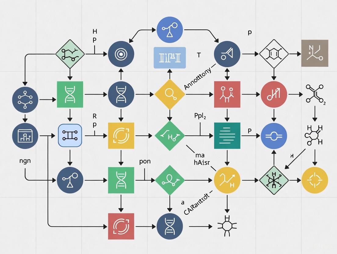
Single-Cell Analysis Workflow for Low-Heterogeneity Datasets
Workflow for analyzing low-heterogeneity datasets, highlighting the critical subclustering and validation steps.
Credibility Evaluation Strategy for Cell Annotation
Decision workflow for the Objective Credibility Evaluation strategy, which assesses annotation reliability based on marker gene expression [1].
The Scientist's Toolkit: Research Reagent Solutions
Table 3: Essential Reagents and Tools for scRNA-seq Heterogeneity Research
| Item Name | Function / Application | Example Use Case |
|---|---|---|
| 10x Genomics Chromium | High-throughput single-cell partitioning and barcoding. | Profiling thousands of cells from a tumor or PBMC sample [7] [4]. |
| UMI (Unique Molecular Identifier) Oligonucleotides | Molecular barcoding to correct for PCR amplification bias and enable accurate transcript counting. | Quantifying absolute transcript numbers in each cell [8]. |
| Ficoll-Paque Premium | Density gradient medium for isolation of viable PBMCs from whole blood. | Preparing samples for immune profiling studies [7]. |
| Anti-human CD14 Antibody | Cell surface marker for identification and isolation of classical monocytes. | Validating the expansion of the CD14+ monocyte population via FACS [7]. |
| Seurat R Toolkit | Comprehensive software package for single-cell genomics data analysis, including clustering, integration, and visualization. | Performing subclustering analysis on stromal cells and running UMAP [7] [4]. |
| LICT (LLM-based Identifier) | Software tool using multiple large language models for automated, reference-free cell type annotation with credibility scoring. | Improving annotation accuracy in low-heterogeneity datasets like embryos or stromal cells [1]. |
| FastMNN Algorithm | Computational method for integrating multiple scRNA-seq datasets and correcting for batch effects. | Combining data from different patients or studies into a unified analysis [2]. |
Frequently Asked Questions
FAQ 1: What is the "performance gap" in the context of cell type annotation? The "performance gap" refers to the significant drop in annotation accuracy that automated methods, including advanced AI and large language models (LLMs), experience when processing low-heterogeneity cellular datasets compared to highly heterogeneous ones. In highly diverse samples like Peripheral Blood Mononuclear Cells (PBMCs), LLMs can achieve high consistency with expert annotations. However, in low-heterogeneity environments like stromal cells or embryonic cells, the consistency of even top-performing LLMs can fall dramatically, with match rates to manual annotations dropping to as low as 33.3% to 39.4% [1]. This gap poses a major challenge for research in areas like developmental biology and specialized tissue studies.
FAQ 2: Why does annotation accuracy drop in low-heterogeneity environments? Accuracy drops primarily because the informational context in low-heterogeneity data is less rich, which can limit the model's ability to distinguish between subtly different cell types [1]. In highly heterogeneous data, the vast differences between cell populations provide strong signals for the model. In contrast, low-heterogeneity datasets feature cells that are more similar to one another, making it difficult for models to identify robust, distinguishing features without more sophisticated analysis strategies.
FAQ 3: How can I objectively verify the reliability of automated annotations for my low-heterogeneity dataset? You can implement an Objective Credibility Evaluation strategy. This involves:
- For each predicted cell type, query the model to retrieve a list of representative marker genes.
- Analyze the expression of these marker genes within the corresponding cell clusters in your input dataset.
- Classify an annotation as reliable if more than four marker genes are expressed in at least 80% of the cells within the cluster. This provides a reference-free method to validate results and can sometimes show that LLM-generated annotations are more credible than manual ones for challenging low-heterogeneity data [1].
FAQ 4: Our research relies on consistent annotations across multiple labs. How can we mitigate inconsistencies? Annotation inconsistencies often stem from inter-annotator variability, which is a well-documented challenge even among highly experienced experts [9]. To mitigate this:
- Establish clear and detailed annotation guidelines.
- Implement structured feedback loops and review processes.
- Utilize computational frameworks designed to harmonize heterogeneous data sources. For instance, approaches like the "talk-to-machine" strategy can iteratively refine annotations based on marker gene validation, improving alignment with manual annotations [1].
Quantitative Analysis of the Performance Gap
The following table summarizes the performance disparity of top LLMs in annotating different types of scRNA-seq datasets, highlighting the challenge of low-heterogeneity environments [1].
Table 1: Annotation Consistency of LLMs Across Dataset Types
| Dataset Type | Biological Example | Performance in High-Heterogeneity Data (e.g., PBMCs, Gastric Cancer) | Performance in Low-Heterogeneity Data (e.g., Embryo, Stromal Cells) |
|---|---|---|---|
| Normal Physiology | Peripheral Blood Mononuclear Cells (PBMCs) | High performance, low mismatch rates | --- |
| Disease State | Gastric Cancer | High performance, low mismatch rates | --- |
| Developmental Stage | Human Embryos | --- | Low consistency (e.g., 39.4% with Gemini 1.5 Pro) |
| Low-Heterogeneity Environment | Stromal Cells in Mouse Organs | --- | Low consistency (e.g., 33.3% with Claude 3) |
Table 2: Impact of Mitigation Strategies on Annotation Accuracy
| Mitigation Strategy | Key Mechanism | Effect on Low-Heterogeneity Datasets | Effect on High-Heterogeneity Datasets |
|---|---|---|---|
| Multi-Model Integration | Combines outputs from multiple LLMs (e.g., GPT-4, Claude 3) to leverage complementary strengths [1] | Increases match rates (e.g., to 48.5% for embryo data) | Reduces mismatch rates (e.g., to 9.7% for PBMCs) |
| "Talk-to-Machine" Interaction | Iterative human-computer feedback loop using marker gene expression for validation [1] | Boosts full match rate (e.g., 16-fold improvement for embryo data vs. GPT-4 alone) | Achieves high full match rates (e.g., 69.4% for gastric cancer) |
Troubleshooting Guides
Problem 1: Poor Automated Annotation of Subtle Cell Types
Symptoms: Your automated annotation tool runs without error, but the resulting cell types are too broad, miss rare populations, or have low confidence scores for clusters you know should be distinct.
Solutions:
- Implement a Multi-Model Strategy: Do not rely on a single LLM. Use a framework like LICT that integrates several top-performing models (e.g., GPT-4, Claude 3, Gemini) to generate a consensus annotation, which significantly improves accuracy in low-heterogeneity settings [1].
- Employ the "Talk-to-Machine" Protocol: Engage in an interactive validation loop.
- Step 1: Run the initial automated annotation.
- Step 2: For each predicted cell type, command the model to output a list of canonical marker genes.
- Step 3: Validate the expression of these markers in your dataset. If fewer than four markers are expressed in >80% of cells, the annotation is likely unreliable.
- Step 4: Feed this validation result, along with the top differentially expressed genes (DEGs) from your dataset, back to the model and request a revised annotation [1].
- Utilize Advanced Graph-Based Models: For a non-LLM approach, consider tools like scGraphformer. This method uses a graph transformer network to learn cell-cell relationships directly from the data without relying on predefined graphs, which can better capture subtle cellular heterogeneity [10].
Problem 2: Discrepancies Between Automated and Manual Annotations
Symptoms: You find significant disagreements between the labels generated by your automated pipeline and the annotations performed by your domain experts, causing uncertainty about which result to trust.
Solutions:
- Apply Objective Credibility Evaluation: Use the marker-gene-based credibility assessment described in FAQ #3. This provides a data-driven metric to determine which annotation—automated or manual—is more reliable for a given cluster. In some cases, the automated annotation may be more credible based on marker evidence [1].
- Audit for Inter-Annotator Variability: Recognize that expert manual annotation is not a perfect gold standard. Studies show that models trained on annotations from different experts can perform inconsistently on external validation sets, with low pairwise agreement (average Cohen’s κ = 0.255) [9]. If possible, use annotations from multiple experts and assess their consensus.
- Check for Data Heterogeneity: Use a tool like scGraphformer to visualize the learned cell-cell relationship network. This can help you understand if the model is failing to distinguish subpopulations that experts can identify, indicating a potential weakness in the model's learning for your specific data type [10].
Experimental Protocols
Protocol 1: Benchmarking Annotation Tools on a Low-Heterogeneity Dataset
This protocol is adapted from the validation methodology used in [1].
1. Objective: To quantitatively evaluate and compare the performance of different automated cell type annotation tools on a low-heterogeneity scRNA-seq dataset.
2. Materials:
- A well-annotated, public low-heterogeneity scRNA-seq dataset (e.g., stromal cells from mouse organs [1] or human embryo data [1]).
- Software Tools: The annotation tools to be benchmarked (e.g., LICT, scGraphformer, scBERT, CellTypist).
- Computing Environment: A server or computing cluster with sufficient memory and processing power to run the selected tools.
3. Procedure:
- Step 1 - Data Preprocessing: Download the chosen dataset and perform standard quality control and normalization using a pipeline like Seurat or Scanpy.
- Step 2 - Ground Truth Definition: Use the original manual annotations from the dataset publication as the ground truth for benchmarking.
- Step 3 - Tool Execution: Run each annotation tool according to its official documentation. For LLM-based tools like LICT, provide standardized prompts that include the top differentially expressed genes for each cell cluster.
- Step 4 - Performance Metric Calculation: For each tool, calculate the following:
- Annotation Consistency: The percentage of cells where the tool's label matches the manual label.
- Mismatch Rate: The percentage of cells with conflicting labels.
- Credibility Score: The percentage of annotations deemed reliable by the Objective Credibility Evaluation (see FAQ #3).
4. Analysis: Compare the metrics across all tested tools to identify the best-performing solution for your specific low-heterogeneity data context.
Benchmarking Experimental Workflow
Protocol 2: Implementing the "Talk-to-Machine" Refinement Loop
This protocol details the steps for the iterative refinement strategy proven to enhance annotation accuracy [1].
1. Objective: To iteratively improve the initial annotations of an LLM-based tool by incorporating marker gene expression validation from the dataset.
2. Materials:
- Your preprocessed scRNA-seq dataset (cell clusters and DEGs).
- Access to an LLM-based annotation tool (e.g., as implemented in LICT).
3. Procedure:
- Step 1 - Initial Annotation: Submit the top marker genes for each cell cluster to the LLM and request an initial cell type prediction.
- Step 2 - Marker Retrieval: For each LLM-predicted cell type, prompt the model to provide a list of known, representative marker genes.
- Step 3 - Expression Validation: Check the expression of these retrieved marker genes in the corresponding cell cluster of your dataset.
- Step 4 - Decision Point:
- PASS: If >4 marker genes are expressed in >80% of cells, accept the annotation.
- FAIL: If not, proceed to Step 5.
- Step 5 - Iterative Feedback: Generate a structured prompt for the LLM that includes: (i) the initial prediction, (ii) the list of marker genes that failed validation, and (iii) the top DEGs from your dataset. Request a new, refined annotation.
Talk-to-Machine Refinement Loop
The Scientist's Toolkit
Table 3: Essential Research Reagents and Computational Tools
| Item Name | Type | Function / Application | Relevant Context |
|---|---|---|---|
| LICT (LLM-based Identifier for Cell Types) | Software Tool | Integrates multiple LLMs for robust, reference-free cell type annotation. Crucial for low-heterogeneity data. | Core method for multi-model integration and "talk-to-machine" [1]. |
| scGraphformer | Software Tool | A graph transformer network that learns cell-cell relationships directly from data, capturing subtle heterogeneity. | An alternative to graph-based methods that avoids predefined kNN graphs [10]. |
| Objective Credibility Evaluation | Analytical Protocol | A method to assess annotation reliability by validating marker gene expression, providing an objective quality score. | Used to resolve conflicts between automated and manual annotations [1]. |
| Stromal Cell Dataset | Reference Data | A scRNA-seq dataset from mouse organs, used as a benchmark for low-heterogeneity environments. | Used to quantify the performance gap of LLMs [1]. |
| Human Embryo Dataset | Reference Data | A scRNA-seq dataset representing developmental stages, characterized by low heterogeneity. | Used to validate annotation tools on developmental biology questions [1]. |
The table below summarizes the key quantitative findings from the evaluation of Large Language Models (LLMs) on low-heterogeneity cell type annotation tasks, including embryo data.
Table 1: LLM Performance on Low-Heterogeneity Annotation Tasks
| Model/Dataset | Performance Metric | Score | Context |
|---|---|---|---|
| Gemini 1.5 Pro on Embryo Data | Consistency with Manual Annotations | 39.4% | Initial performance on low-heterogeneity human embryo dataset [1] |
| Claude 3 on Fibroblast Data | Consistency with Manual Annotations | 33.3% | Performance on low-heterogeneity mouse stromal cells [1] |
| Multi-Model Integration on Embryo Data | Match Rate (Full + Partial) | 48.5% | Performance after applying Strategy I [1] |
| "Talk-to-Machine" on Embryo Data | Full Match Rate | 48.5% | Performance after applying Strategy II [1] |
| LLM-generated Annotations on Embryo Data | Credible Annotations in Mismatches | 50.0% | Proportion of LLM annotations deemed reliable per Strategy III [1] |
| Expert Annotations on Embryo Data | Credible Annotations in Mismatches | 21.3% | Proportion of manual annotations deemed reliable per Strategy III [1] |
Frequently Asked Questions (FAQs)
Q1: Why does LLM performance drop significantly on low-heterogeneity datasets like embryo cells? LLMs struggle with low-heterogeneity data due to limited informational context and subtle distinguishing features. These models are trained on highly diverse data and excel at identifying clear, distinct patterns. In low-heterogeneity environments—where cell subpopulations share many characteristics—the models lack sufficient signal to make accurate differentiations, leading to performance drops as severe as 39.4% compared to manual annotations [1].
Q2: What is the evidence that the problem is with the data rather than the models? Objective credibility evaluations reveal that LLM-generated annotations for embryo data show higher reliability (50% credible) than expert manual annotations (21.3% credible) when validated against marker gene expression patterns. This suggests that discrepancies often reflect inherent ambiguities in the biological data itself rather than purely model deficiencies [1].
Q3: How can researchers determine if their dataset suffers from low heterogeneity? Low-heterogeneity datasets typically exhibit: minimal variance in gene expression profiles, high cellular similarity, poor clustering separation in dimensional reduction (UMAP/t-SNE), and consistent failure of multiple algorithms to achieve satisfactory annotation accuracy. Specifically, if multiple LLMs consistently achieve below 40% agreement with manual annotations on embryo data, low heterogeneity is likely a contributing factor [1].
Q4: What are the main sources of annotation inconsistency in biological data? Annotation inconsistencies arise from four primary sources: (1) insufficient information for reliable labeling, (2) insufficient domain expertise, (3) human error and cognitive slips, and (4) inherent subjectivity in the labeling task. Studies show even highly experienced clinical experts exhibit significant inter-rater variability (Fleiss' κ = 0.383, indicating only fair agreement) [9].
Troubleshooting Guides
Problem: Poor LLM Performance on Low-Heterogeneity Cell Annotation
Symptoms:
- Consistent annotation accuracy below 40% on embryo or stromal cell data
- High mismatch rates between LLM predictions and manual annotations
- Low inter-annotator agreement across multiple models
Solution: Implement a Three-Strategy Framework
Verification: After implementation, researchers should observe:
- Increase in embryo data annotation match rates from 39.4% to approximately 48.5%
- Reduction in mismatches for high-heterogeneity datasets to below 10%
- Improved reliability scores for LLM-generated annotations
Problem: Handling Discrepancies Between LLM and Expert Annotations
Symptoms:
- Contradictory annotations between LLMs and domain experts
- Uncertainty about which annotations to trust for downstream analysis
- Inconsistent validation results
Solution: Implement Objective Credibility Evaluation
Verification:
- Credibility assessment showing >50% of LLM annotations are reliable despite mismatches
- Identification of cases where both LLM and manual annotations are reliable but different (14% of cases)
- Clear prioritization of cell clusters for downstream analysis based on reliability scores
Experimental Protocols
Protocol 1: Multi-Model Integration for Enhanced Annotation
Purpose: Leverage complementary strengths of multiple LLMs to improve annotation accuracy on low-heterogeneity datasets.
Materials:
- Top-performing LLMs (GPT-4, LLaMA-3, Claude 3, Gemini, ERNIE 4.0)
- Standardized prompts incorporating top marker genes
- scRNA-seq dataset with preliminary clustering
Methodology:
- Model Selection: Evaluate 77 publicly available LLMs using benchmark PBMC dataset to identify top performers [1]
- Parallel Annotation: Submit standardized prompts with cluster-specific marker genes to all selected models
- Result Integration: Select best-performing annotations from each model rather than using majority voting
- Validation: Compare integrated annotations with manual benchmarks using consistency metrics
Expected Outcomes:
- Match rate improvement from 39.4% to 48.5% for embryo data
- Mismatch rate reduction from 21.5% to 9.7% for high-heterogeneity data
- More comprehensive coverage of diverse cell types
Protocol 2: "Talk-to-Machine" Iterative Optimization
Purpose: Enhance annotation precision through human-computer interaction and iterative feedback.
Materials:
- Pre-annotated dataset using multi-model integration
- Differentially expressed genes (DEGs) analysis pipeline
- Validation threshold parameters (80% expression in clusters)
Methodology:
- Initial Annotation: Generate preliminary annotations using multi-model integration
- Marker Gene Retrieval: Query LLM for representative marker genes for each predicted cell type
- Expression Validation: Validate marker gene expression in corresponding clusters
- Iterative Feedback: For validation failures, generate structured feedback prompts with expression results and additional DEGs
- Re-query LLM: Use feedback prompts to obtain revised annotations
- Repeat steps 2-5 until validation criteria are met or maximum iterations reached
Validation Criteria:
- Annotation considered valid if >4 marker genes expressed in ≥80% of cluster cells
- Maximum of 3 iteration cycles to prevent over-optimization
Expected Outcomes:
- Full match rate of 34.4% for PBMC and 69.4% for gastric cancer data
- Significant reduction in mismatches (7.5% for PBMC, 2.8% for gastric cancer)
- 16-fold improvement in full match rate for embryo data compared to single-model approach
The Scientist's Toolkit
Table 2: Essential Research Reagents and Solutions
| Tool/Reagent | Function | Application Note |
|---|---|---|
| LICT (LLM-based Identifier for Cell Types) | Integrates multiple LLMs with three core strategies for reliable cell annotation | Specifically designed to address low-heterogeneity challenges [1] |
| Benchmark scRNA-seq Dataset (PBMC) | Standardized evaluation of LLM performance using peripheral blood mononuclear cells | Serves as initial screening tool for model selection [1] |
| Standardized Prompt Templates | Ensure consistent query structure across different LLMs | Incorporates top ten marker genes for each cell subset [1] |
| Objective Credibility Evaluation Framework | Validates annotation reliability based on marker gene expression | Reference-free validation method [1] |
| Multi-gate Mixture-of-Experts (MMoE) | Coordinates co-optimization of shared and local tasks in distributed learning | Helps address data heterogeneity in collaborative settings [11] |
| HeteroSync Learning (HSL) Framework | Privacy-preserving distributed learning for heterogeneous medical data | Useful for multi-institutional collaborations [11] |
Troubleshooting Guide: Low Heterogeneity Dataset Annotation
Common Problems & Solutions
| Problem | Possible Cause | Solution | Reference |
|---|---|---|---|
| Low annotation match rate with manual labels | Inherent low cellular diversity; limited marker gene variety. | Implement a multi-model integration strategy to leverage complementary LLM strengths. | [1] |
| Ambiguous or biased cell type predictions | Standardized LLM data formats struggle with dynamic biological data. | Apply the iterative "talk-to-machine" strategy to enrich model input with contextual data. | [1] |
| Uncertainty in annotation reliability | Lack of an objective, reference-free method for validation. | Employ an objective credibility evaluation based on marker gene expression patterns. | [1] |
| Inconsistent data labeling across the project | Unclear annotation guidelines; subjective interpretations by different annotators. | Define precise annotation rules and implement a cross-validation process between annotators. | [12] |
| Bias in the annotated dataset | Homogeneous group of annotators; unbalanced dataset classes. | Diversify annotators and apply data rebalancing techniques for underrepresented classes. | [12] |
Frequently Asked Questions (FAQs)
Conceptual & Biological Basis
Q1: What defines a "low-heterogeneity" cellular environment in developmental biology? A low-heterogeneity environment consists of cells that are very similar to each other in terms of their state, function, and genetic expression profiles. This is common in early embryonic stages and within specialized tissues like certain stromal cell populations, where cells have not yet undergone extensive diversification or have converged on a highly specific function. In these contexts, the limited diversity makes it difficult to distinguish subtle differences between cell subpopulations using automated annotation tools [1].
Q2: How do fundamental developmental processes like cell differentiation contribute to heterogeneity? Cell differentiation is the process by which a less specialized cell becomes a specific, functional cell type (e.g., neuron, muscle fiber). This process is driven by specific transcription factors (like NeuroD for neurons) that activate unique sets of genes, giving the cell its characteristic appearance and function [13]. The progression of cells through different states of commitment toward these differentiated fates is a primary source of cellular heterogeneity within a tissue [14].
Technical & Computational Challenges
Q3: Why do automated annotation tools, including LLMs, perform poorly on low-heterogeneity data? These tools often rely on identifying distinct patterns in marker gene expression. In low-heterogeneity populations, the differences in gene expression between cell subtypes are subtler and less pronounced. The informational context is poorer, providing fewer robust signals for the models to latch onto, which leads to higher rates of discrepancy compared to expert manual annotation [1].
Q4: What is an objective credibility evaluation for cell type annotation? This is a reference-free method to assess the reliability of an annotation. After an LLM predicts a cell type, it is queried for a list of representative marker genes for that type. The annotation is deemed credible if more than four of these marker genes are expressed in at least 80% of the cells within the cluster. This provides a data-driven measure of confidence independent of manual labels [1].
Q5: How can semi-automated labeling improve our workflow for these difficult datasets? A hybrid AI/human approach is often most effective. An AI model can perform the initial "pre-annotation," handling the bulk of the data quickly. Human annotators then validate or correct these results, adding nuance and understanding that algorithms may miss. This combines speed with accuracy, ensuring reliable annotations for model training [12].
Experimental Protocols for Enhanced Annotation
Protocol 1: Multi-Model Integration Strategy
Purpose: To increase annotation accuracy and consistency by leveraging the complementary strengths of multiple large language models (LLMs), especially for low-heterogeneity datasets [1].
Methodology:
- Input Preparation: For each cell cluster, compile a list of top marker genes (e.g., the top 10 most differentially expressed genes).
- Model Selection & Query: Submit a standardized prompt containing the marker gene list to five top-performing LLMs (e.g., GPT-4, Claude 3, Gemini, LLaMA-3, ERNIE 4.0).
- Result Integration: Instead of using a simple majority vote, select the best-performing annotation result from among the five LLMs for each cluster. This approach capitalizes on the unique strengths of each model for different cell types.
Protocol 2: Iterative "Talk-to-Machine" Refinement
Purpose: To iteratively improve annotation precision for ambiguous or incorrect predictions through a structured human-computer feedback loop [1].
Methodology:
- Initial Annotation & Marker Retrieval: Obtain an initial cell type prediction from an LLM. Then, query the same LLM for a list of known marker genes for the predicted cell type.
- Expression Validation: Evaluate the expression of these retrieved marker genes in the original dataset's corresponding cell cluster.
- Validation Check:
- PASS: If >4 marker genes are expressed in ≥80% of cells in the cluster, accept the annotation.
- FAIL & REFINE: If the condition is not met, generate a feedback prompt for the LLM. This prompt includes the validation results and additional differentially expressed genes (DEGs) from the dataset. Use this prompt to re-query the LLM, asking it to revise or confirm its annotation.
- Iteration: Repeat steps 1-3 until a validated annotation is achieved or a maximum number of iterations is reached.
Protocol 3: Objective Credibility Evaluation
Purpose: To provide a reference-free, unbiased assessment of annotation reliability, distinguishing methodological limitations from intrinsic data ambiguity [1].
Methodology:
- For any given annotation (whether from an LLM or a manual expert), retrieve a set of representative marker genes for that cell type.
- Analyze the expression pattern of these markers within the annotated cell cluster in your scRNA-seq dataset.
- Apply Credibility Threshold: The annotation is classified as "reliable" if more than four marker genes are expressed in at least 80% of the cells in the cluster. Annotations not meeting this threshold are classified as "unreliable" for downstream analysis.
Experimental Workflow Visualization
LICT Annotation Workflow
Talk-to-Machine Refinement Loop
Research Reagent Solutions
Essential Materials for scRNA-seq Annotation Research
| Item | Function / Description | Application in Low-Heterogeneity Context |
|---|---|---|
| Peripheral Blood Mononuclear Cells (PBMCs) | A benchmark dataset of highly heterogeneous immune cells. | Serves as a positive control to validate annotation pipeline performance on well-defined cell types. [1] |
| Human Embryo scRNA-seq Data | Represents a lower-heterogeneity dataset from early developmental stages. | Used to test and optimize annotation strategies for challenging, less diverse cellular environments. [1] |
| Stromal Cell scRNA-seq Data | Data from specialized, low-heterogeneity tissues like mouse organ fibroblasts. | Provides a model for annotating dedicated tissue-specific cell populations with subtle differences. [1] |
| GPT-4, Claude 3, Gemini | Top-performing Large Language Models (LLMs) for biological inference. | Core engines for initial cell type prediction. A multi-model integration approach leverages their complementary strengths. [1] |
| LICT (LLM-based Identifier for Cell Types) | A software package integrating multiple LLMs and strategies. | The primary tool for implementing the multi-model, "talk-to-machine," and credibility evaluation protocols. [1] |
| Data Annotation Platforms (e.g., Labelbox, V7) | Tools for creating ergonomic interfaces for manual and semi-automated data labeling. | Facilitates the human-in-the-loop validation and correction essential for refining AI-generated annotations. [12] |
This technical support center provides troubleshooting guides for researchers addressing annotation errors in biological data analysis. Annotation—the process of labeling biological data such as cell types, genes, or genomic features—is a critical step in bioinformatics pipelines. When performed inaccurately, these errors propagate through downstream analyses, leading to flawed biological interpretations and reduced reproducibility. This guide focuses specifically on the challenges of low-heterogeneity datasets, where subtle annotation errors can have disproportionately large effects, and provides actionable solutions for researchers and drug development professionals.
Quantitative Impact of Annotation Errors
The tables below summarize key quantitative findings from recent studies on how annotation and segmentation errors distort downstream biological analyses.
Table 1: Impact of Segmentation Errors on Clustering and Phenotyping Consistency
| Perturbation Level | k-Means Clustering Consistency | Leiden Clustering Consistency | Cell Phenotyping Accuracy |
|---|---|---|---|
| Low Error | Minimal reduction | Minimal reduction (with larger neighborhood sizes) | >95% for distinct cell types |
| Moderate Error | Significant reduction | Significant reduction (with smaller neighborhood sizes) | 85-95% for distinct cell types |
| High Error | Severe reduction | Severe reduction | Notable misclassification between closely related cell types [15] [16] |
Table 2: Annotation Tool Performance Across Dataset Types
| Dataset Heterogeneity | Manual Annotation | Single LLM Tool (e.g., GPT-4) | Multi-Model Integration (LICT) |
|---|---|---|---|
| High Heterogeneity (e.g., PBMCs) | High accuracy, but subjective and time-consuming | 78.5% match rate | 90.3% match rate |
| Low Heterogeneity (e.g., Embryonic cells) | Considered benchmark, but potential for bias | 39.4% match rate | 48.5% match rate [1] |
Troubleshooting Guides & FAQs
FAQ 1: How do annotation errors specifically affect the analysis of low-heterogeneity datasets?
Answer: In low-heterogeneity datasets, where cell populations have similar molecular profiles, annotation errors cause more severe consequences than in highly heterogeneous data.
- Mechanism: The feature space—the mathematical representation of cellular characteristics—is inherently compressed in low-heterogeneity data. Minor errors in assigning cell boundaries or labels introduce noise that is large relative to the subtle biological differences between cell states. This noise directly obscures these critical distinctions [15] [1].
- Downstream Impact: The result is a significant drop in the performance of automated annotation tools. For example, one study showed that even top-performing Large Language Models (LLMs) like Gemini 1.5 Pro achieved only a 39.4% consistency with manual annotations on embryo data, a low-heterogeneity scenario [1]. This leads to unreliable cell type identification and flawed conclusions about cellular functions and relationships.
FAQ 2: My clustering results are unstable and change with different algorithm parameters. Could this be caused by annotation quality?
Answer: Yes, instability in clustering results is a classic symptom of underlying annotation or segmentation errors.
- Mechanism: Annotation errors distort the fundamental input to clustering algorithms: the single-cell expression profiles. As segmentation inaccuracies increase, they alter the computed protein expression levels for each cell. This "feature distortion" changes the distances between cells in the feature space, making the neighborhoods used by algorithms like k-Means and Leiden inherently unstable [15] [16].
- Diagnosis: If your clustering results are highly sensitive to small changes in parameters like the number of clusters (k) or the neighborhood size, you should first investigate the quality of your input data and annotations before further tuning the algorithms.
FAQ 3: What are the most effective strategies to improve annotation reliability for difficult datasets?
Answer: A multi-layered strategy that combines computational checks with expert knowledge is most effective.
- Implement a Multi-Model Integration Strategy: Instead of relying on a single annotation tool, leverage the complementary strengths of multiple models. One study used five different LLMs (including GPT-4, Claude 3, and Gemini) and selected the best-performing result for each cell type, which significantly reduced the mismatch rate in low-heterogeneity data [1].
- Adopt a "Talk-to-Machine" Feedback Loop: Create an interactive process where an initial annotation is validated against the dataset's own evidence.
- The tool suggests an annotation and provides a list of expected marker genes.
- The expression of these genes is automatically checked in the corresponding cell cluster.
- If validation fails (e.g., fewer than four markers are expressed in 80% of cells), the tool re-queries with the new evidence to refine its annotation [1].
- Apply Rigorous Quality Control Metrics: Use established metrics to quantify annotation quality.
- F1 Score: Balances precision (how many annotations are correct) and recall (how many correct annotations were found) [17].
- Inter-Annotator Agreement (IAA): Measures consistency between different annotators or tools. Use metrics like Fleiss' kappa (for multiple annotators) or Krippendorff's alpha (which can handle missing data and partial agreement) [17].
FAQ 4: What are the best practices for preparing data to minimize annotation errors from the start?
Answer: Preventing errors at the source is the most efficient troubleshooting strategy. Adhere to the following best practices:
- Define Clear Guidelines: Before annotation begins, create detailed, unambiguous instructions for annotators. Use simple language, provide visual examples of "do's" and "don'ts," and explicitly describe how to handle edge cases [18].
- Establish Golden Standards: Have domain experts create a small, "ground truth" dataset that reflects the ideal annotation. This serves as a benchmark for training annotators and evaluating the quality of all other annotations [17].
- Implement Systematic Review Cycles: Build quality control into your workflow. This includes periodic double-checks, having multiple annotators label the same data point to measure consistency, and holding regular meetings to resolve ambiguities [18] [19].
- Ensure Ongoing Training and Support: Annotation is not a one-time task. Provide continuous training for your team and maintain a clear channel for annotators to ask questions and get timely feedback [18] [19].
Experimental Protocols for Error Mitigation
Protocol 1: Benchmarking Segmentation Robustness
This methodology allows you to quantitatively evaluate how sensitive your analysis is to segmentation errors.
- Input Ground Truth Data: Start with a high-quality, manually validated segmentation mask.
- Apply Controlled Perturbations: Use the Affine Transform function from the Albumentations library to simulate realistic segmentation errors. Systematically apply combinations of translation, rotation, scaling, and shearing to each cell mask. Parameters for these transformations should be sampled from uniform distributions to create a range of perturbation strengths [15] [16].
- Generate Perturbed Masks:
- Initialize an output array matching the input mask size.
- For each cell, extract its mask, set non-cell pixels to zero, and apply padding.
- Apply the sampled affine transformations.
- Write the transformed non-zero pixels back to the output array.
- Use binary opening (erosion followed by dilation) to clean up the resulting fuzzy masks.
- Detect and resolve any overlapping masks by randomly removing border pixels to maintain a one-pixel separation [15].
- Run Downstream Analysis: Execute your standard clustering (e.g., k-Means, Leiden) and phenotyping (e.g., Gaussian Mixture Models) pipelines on both the ground truth and the series of perturbed datasets.
- Quantify Impact: Calculate the consistency between the results from the perturbed data and the ground truth. Use the F1 score to compare clustering outputs and track metrics like misclassification rates for cell phenotyping [15].
Protocol 2: Credibility Evaluation for Cell Type Annotations
This protocol provides an objective framework for assessing the reliability of automated or manual cell type annotations.
- Retrieve Marker Genes: For a given annotated cell type (e.g., "CD4+ T-cell"), query the annotation tool or a reference database to generate a list of representative marker genes (e.g., CD3D, CD4, IL7R).
- Evaluate Expression Patterns: In your single-cell dataset (e.g., scRNA-seq or multiplexed imaging), analyze the expression of these marker genes within the cluster of cells that received the annotation.
- Assess Credibility: Apply a predefined, objective threshold to determine reliability. For example, an annotation can be deemed "credible" if more than four of the suggested marker genes are expressed in at least 80% of the cells within the cluster. Annotations failing this threshold should be flagged for manual review or re-annotation [1].
Visualization of Error Propagation & Mitigation
Diagram 1: Annotation Error Propagation Pathway
Diagram 2: Strategy for Robust Annotation
The Scientist's Toolkit: Key Research Reagents & Solutions
Table 3: Essential Tools for Annotation and Quality Control
| Tool / Resource | Type | Primary Function | Application Context |
|---|---|---|---|
| CellSeg / Cellpose / Stardist | Segmentation Algorithm | Delineates individual cell boundaries in imaging data | Highly multiplexed tissue imaging (CODEX, MIBI, IMC) [15] [16] |
| LICT (LLM-based Identifier) | Annotation Tool | Automated cell type annotation for scRNA-seq data using multi-LLM integration | Single-cell RNA sequencing analysis, especially for low-heterogeneity data [1] |
| PubTator 3.0 | Database & NER Tool | Validates and normalizes biomedical entities (genes, chemicals) via canonical IDs | Grounding LLM outputs to reduce hallucinations in metadata annotation [20] |
| Albumentations Library | Python Library | Applies affine transformations (scale, rotate, shear) to simulate segmentation errors | Benchmarking segmentation robustness and pipeline error tolerance [15] [16] |
| FastQC / MultiQC | Quality Control Tool | Provides initial quality assessment of raw sequencing data (e.g., base quality, GC content) | First step in bioinformatics pipeline to identify issues before they propagate [21] [22] |
| F1 Score / Fleiss' Kappa | Quality Metric | Quantifies annotation precision/recall (F1) and inter-annotator agreement (Fleiss' Kappa) | Objectively measuring the consistency and accuracy of annotations [15] [17] |
Advanced Computational Frameworks for Low-Heterogeneity Annotation
Frequently Asked Questions (FAQs)
Q1: What are the main advantages of using multiple LLMs over a single model for annotating low-heterogeneity cell types? Using multiple LLMs leverages their complementary strengths, which is crucial for low-heterogeneity datasets where single models often struggle. For example, while Claude 3 might excel in annotating highly heterogeneous cell subpopulations, Gemini 1.5 Pro or GPT-4 could provide better results for specific low-heterogeneity contexts. Multi-model integration significantly improves match rates with manual annotations, reducing mismatch from over 50% to more manageable levels [1].
Q2: My multi-LLM pipeline is producing inconsistent annotations for similar cell clusters. How can I resolve this? Inconsistency often arises from ambiguous marker gene expression in low-heterogeneity environments. Implement the "talk-to-machine" strategy: query the LLM to provide representative marker genes for its predicted cell type, then validate if these genes are expressed in your dataset. If validation fails, provide this feedback with additional differentially expressed genes to the LLM for re-annotation. This iterative process significantly improves annotation consistency [1].
Q3: What methods can I use to objectively evaluate which LLM annotations are most reliable? Use an objective credibility evaluation strategy. For each LLM-predicted cell type, retrieve representative marker genes and assess their expression pattern in your dataset. An annotation is considered reliable if more than four marker genes are expressed in at least 80% of cells within the cluster. This reference-free validation provides quantitative assessment of annotation reliability independent of manual annotations [1].
Q4: How can I efficiently compare and integrate outputs from different LLMs without constantly switching interfaces? Use specialized systems like LLMartini that provide unified interfaces for comparing multiple LLM outputs. These systems automatically segment responses into semantically-aligned units, merge consensus content, and highlight discrepancies through color coding. This approach significantly reduces cognitive load and operational friction compared to manual multi-tab workflows [23].
Q5: What are the most effective technical frameworks for implementing multi-LLM pipelines in biomedical research? For entity recognition, consider cache-augmented generation approaches that integrate GPT-4o with specialized tools like PubTator 3.0. This combines LLM analysis with validated biomedical databases. For systematic evaluation, frameworks like DeepEval provide metrics specifically designed for LLM assessment, including faithfulness, contextual relevancy, and answer relevancy metrics [20] [24].
Troubleshooting Guides
Problem: High Discrepancy Between LLM and Manual Annotations
Symptoms:
- Over 50% inconsistency between LLM-generated and manual annotations for low-heterogeneity cell types
- LLM annotations flagged as unreliable by credibility evaluation
- Significant inter-model variability in annotation results
Resolution Steps:
- Implement Multi-Model Integration: Instead of relying on a single LLM, deploy a panel of complementary models (GPT-4, Claude 3, Gemini, LLaMA-3, ERNIE 4.0) and select the best-performing results for each cell type [1].
Apply "Talk-to-Machine" Strategy:
- Step 1: Obtain initial annotations from your LLM panel
- Step 2: Query each LLM for representative marker genes for its predicted cell types
- Step 3: Validate expression patterns in your dataset
- Step 4: For failed validations, provide structured feedback with additional DEGs
- Step 5: Iterate until convergence or maximum iterations reached [1]
Objective Credibility Assessment:
- Calculate the percentage of expressed marker genes for each annotation
- Apply the 4-gene/80% threshold for reliability classification
- Prioritize annotations meeting credibility criteria for downstream analysis [1]
Problem: LLM Hallucinations in Biomedical Entity Recognition
Symptoms:
- LLM generates plausible but incorrect biomedical entities
- Entities not validated in reference databases
- Inconsistent entity identification across similar datasets
Resolution Steps:
- Implement Cache-Augmented Generation:
- Step 1: GPT-4o-based full-text analysis for candidate entity generation
- Step 2: PubTator 3.0 validation of suggested terms
- Step 3: Schema-constrained full-text analysis using domain-specific metadata
- Step 4: Combined evaluation of validated and schema-related terms [20]
Domain Schema Integration:
- Develop dedicated metadata schema for your research area
- Constrain LLM output to schema-defined entities
- Combine universal entities (via PubTator) with project-specific concepts [20]
Validation Workflow:
- Use PubTator 3.0 for high-precision normalization with canonical IDs
- Maintain project-specific schema for in-house concepts
- Merge results with clear provenance tracking [20]
Experimental Protocols & Data
Quantitative Performance of Multi-LLM Strategies
Table 1: Annotation Performance Across Dataset Types Using Multi-Model Integration
| Dataset Type | Single Model Mismatch Rate | Multi-Model Mismatch Rate | Improvement | Key Performing Models |
|---|---|---|---|---|
| High Heterogeneity (PBMC) | 21.5% | 9.7% | 55% reduction | Claude 3, GPT-4 |
| High Heterogeneity (Gastric Cancer) | 11.1% | 8.3% | 25% reduction | Claude 3, Gemini 1.5 Pro |
| Low Heterogeneity (Embryo) | >50% inconsistency | 48.5% match rate | 16x improvement | Gemini 1.5 Pro, GPT-4 |
| Low Heterogeneity (Stromal Cells) | >50% inconsistency | 43.8% match rate | Significant improvement | Claude 3, LLaMA-3 |
Source: Validation across four scRNA-seq datasets representing diverse biological contexts [1]
Table 2: Credibility Assessment Results for LLM vs. Manual Annotations
| Dataset | LLM Annotations Deemed Reliable | Manual Annotations Deemed Reliable | Advantage |
|---|---|---|---|
| Gastric Cancer | Comparable to manual | Benchmark | Comparable reliability |
| PBMC | Higher than manual | Lower than LLM | LLM outperformed manual |
| Embryo (Low Heterogeneity) | 50% of mismatched annotations credible | 21.3% credible | 2.3x more credible |
| Stromal Cells (Low Heterogeneity) | 29.6% credible | 0% credible | Significant LLM advantage |
Source: Objective credibility evaluation based on marker gene expression patterns [1]
Detailed Methodological Protocols
Protocol 1: Multi-Model Integration for scRNA-seq Annotation
Model Selection: Identify top-performing LLMs for your specific domain through benchmarking (e.g., GPT-4, LLaMA-3, Claude 3, Gemini, ERNIE 4.0 for cell typing) [1].
Standardized Prompting:
- Format: Incorporate top ten marker genes for each cell subset
- Structure: Use consistent prompt templates across all models
- Context: Provide equivalent biological context for all queries
Output Integration:
- Method: Select best-performing results from each model rather than simple voting
- Validation: Compare against benchmark datasets with known annotations
- Metrics: Calculate consistency rates with manual annotations
Iterative Refinement:
- Identify low-performance scenarios (e.g., low-heterogeneity cells)
- Implement additional strategies for challenging cases
- Re-benchmark improved pipeline [1]
Protocol 2: Cache-Augmented Generation for Biomedical Entities
Initial Entity Generation:
- Tool: GPT-4o with full-text analysis capability
- Scope: Analyze complete manuscript text excluding discussion and bibliography
- Instruction: Generate relevant biomedical entities without restrictions
PubTator 3.0 Validation:
- Method: Custom GPT with PubTator 3.0 augmentation
- Process: Query PubTator for standardized entity IDs for each generated term
- Output: Retain only validated entities with canonical identifiers
Schema-Constrained Extraction:
- Input: Dedicated metadata schema in tree-like structure
- Task: Re-analyze full text identifying schema-defined entities
- Output: Project-specific entities not in universal databases
Combined Evaluation:
- Merge: Schema-related and PubTator-validated entities
- Deduplicate: Prioritize schema-derived entities
- Finalize: Comprehensive entity list with provenance tracking [20]
Workflow Diagrams
Multi-Model LLM Integration Workflow for Low-Heterogeneity Data
Objective Credibility Evaluation Protocol
The Scientist's Toolkit
Table 3: Essential Research Reagent Solutions for Multi-LLM Experiments
| Tool/Resource | Function | Application Context | Key Features |
|---|---|---|---|
| PubTator 3.0 | Biomedical entity validation and normalization | Step 2 validation in cache-augmented generation | Provides canonical IDs for entities, reduces hallucinations [20] |
| Domain-Specific Metadata Schema | Constrains LLM output to project-relevant concepts | Schema-constrained entity extraction | Captures in-house cell lines, endpoints not in universal databases [20] |
| LLMartini System | Visual comparison and fusion of multiple LLM outputs | Multi-model comparison and selection | Segments responses, merges consensus, highlights differences [23] |
| DeepEval Framework | LLM evaluation metrics and testing | Validation of multi-LLM pipeline performance | Provides hallucination, bias, relevance metrics [24] |
| Cache-Augmented Generation | Proprietary data integration without retrieval latency | Full-text analysis with extended context | Eliminates retrieval errors, handles large documents [20] |
| RAGAs Framework | Retrieval-Augmented Generation assessment | Evaluation of knowledge-grounded LLM systems | Measures faithfulness, contextual relevancy, answer relevancy [24] |
| Objective Credibility Evaluation | Reference-free annotation validation | Assessing reliability of LLM vs manual annotations | Uses marker gene expression patterns as ground truth [1] |
Frequently Asked Questions
Q1: My genetic algorithm fails when converting binary data back to float values, showing an "unpack requires a buffer of 4 bytes" error. What's wrong?
This error typically occurs when the binary data buffer size doesn't match the expected 4 bytes for a float conversion. The function binary_to_float might be receiving a binary list of incorrect length.
- Solution: Verify that every binary string representing a float is exactly 32 bits (4 bytes) long before unpacking. Debug by checking the exact value of
binary_listwhen the error occurs and ensure the byte conversion creates a buffer of precisely 4 bytes [25].
Q2: How can I prevent data leakage when preprocessing data for the ensemble model?
Data leakage causes overly optimistic performance estimates and models that fail on unseen data.
- Solution: Always split your data into training and test sets before applying any preprocessing steps. Use pipelines to ensure preprocessing steps (like imputation and scaling) are fitted only on the training data and then applied to the test data. Never preprocess the full dataset before splitting [26].
Q3: My feature selection process seems unstable—different runs select different features. How can I improve consistency?
Instability in feature selection can arise from high-dimensional data and correlated features, especially with limited samples.
- Solution: Implement a robust ensemble feature selection approach. Aggregate results from multiple feature selectors and use a pseudo-variables assisted tuning strategy. This method uses permuted copies of features as known irrelevant controls; only features consistently outperforming these pseudo-variables across multiple permutations are selected [27].
Q4: What is the most common mistake in machine learning projects that I should avoid?
A common mistake is insufficient data understanding and preprocessing. Real-world datasets are rarely usable in their native form and require extensive cleaning.
- Solution: Perform thorough exploratory data analysis (EDA) before modeling. Use summary statistics and visualizations to understand distributions, identify outliers, and handle missing values appropriately before proceeding to feature engineering and model building [26].
Q5: When should I use knowledge-based versus data-driven feature selection?
The choice depends on your data context and goals. Knowledge-based feature selection leverages prior biological knowledge, while data-driven methods rely on patterns in the experimental data.
- Solution: For drug response prediction with transcriptome data, knowledge-based methods (like using drug target pathways) often yield more interpretable models and can be highly predictive for drugs targeting specific genes and pathways. Data-driven methods may perform better for drugs affecting general cellular mechanisms [28] [29].
Troubleshooting Guides
Issue 1: Poor Annotation Accuracy on Low-Heterogeneity Datasets
Problem: Ensemble model with genetic feature selection performs poorly when annotating single-cell RNA sequencing data with low cellular heterogeneity.
Diagnosis Steps:
- Check if the genetic algorithm's feature selection is too aggressive, removing biologically relevant but low-expression markers.
- Verify whether batch effects or technical variations are confounding the genetic optimizer.
- Evaluate if the ensemble learners are overfitting to the majority cell types.
Resolution:
- Adjust Genetic Algorithm Parameters: Incorporate prior biological knowledge into the fitness function. Penalize feature sets that exclude genes from known, biologically relevant pathways [29].
- Implement Advanced Normalization: Apply techniques like SCTransform to handle technical noise before feature selection.
- Utilize Pseudo-Variables for Tuning: Integrate pseudo-variables (known irrelevant features) into the genetic algorithm's selection process. This helps ensure selected features show consistently stronger signals than noise [27].
Issue 2: Genetic Algorithm Convergence Problems
Problem: The genetic optimizer fails to converge or gets stuck in local minima during feature selection.
Diagnosis Steps:
- Check population diversity metrics across generations.
- Analyze fitness score progression over iterations.
- Verify mutation and crossover rates are appropriately set.
Resolution:
- Parameter Adjustment: Implement adaptive mutation rates that increase when population diversity drops below a threshold. For feature selection, typical mutation rates range from 0.001 to 0.1 [25] [30].
- Alternative Selection Methods: Experiment with different parent selection strategies:
- Implement Elitism: Preserve a small percentage of top-performing solutions unchanged in the next generation to ensure fitness doesn't decrease [30].
Issue 3: Handling High-Dimensional Data with Limited Samples
Problem: The ensemble model struggles with datasets where the number of features (genes) vastly exceeds the number of samples (cells), common in scRNA-seq studies.
Diagnosis Steps:
- Determine if the feature selection process is retaining too many variables.
- Check for overfitting by comparing training and validation performance.
- Evaluate if the chosen ML models are appropriate for high-dimensional data.
Resolution:
- Knowledge-Based Feature Pre-Filtering: Before applying genetic algorithm-based selection, reduce feature space using biological knowledge. For drug response prediction, start with features related to drug targets or their pathways [29].
- Consider Feature Transformation: Instead of selecting gene subsets, use methods like Pathway Activities or Transcription Factor Activities, which transform many gene expressions into fewer, biologically meaningful scores [28].
- Apply Regularization: Use models with built-in regularization like Ridge regression or Elastic Net, which have been shown to perform well on high-dimensional biological data [28].
Experimental Protocols & Data
Protocol 1: Benchmarking Ensemble-Genetic Framework Against Established Methods
Objective: Evaluate the performance of the Ensemble Machine Learning with Genetic Optimization framework against existing annotation tools like scMRA, ItClust, Scmap, and Seurat [31].
Methodology:
- Data Preparation: Obtain well-annotated scRNA-seq reference datasets and corresponding query datasets with known cell type labels.
- Performance Metrics: Measure annotation accuracy under varying conditions: different levels of data scarcity (mild, moderate, severe reduction in training data) and increasing number of cell type clusters [31].
- Experimental Runs: For each method and condition, execute multiple runs to ensure statistical significance of results.
Expected Outcome: The proposed ensemble-genetic framework is expected to demonstrate superior accuracy and generalization, particularly under conditions of limited reference data and increasing dataset complexity [31].
Protocol 2: Evaluating Feature Reduction Methods for Drug Response Prediction
Objective: Compare the performance of knowledge-based and data-driven feature reduction methods for predicting drug sensitivity from transcriptome data [28].
Methodology:
- Feature Reduction Methods: Apply nine different methods to cell line gene expression data:
- Machine Learning Models: Feed reduced features to multiple ML models (Ridge regression, Lasso, SVM, Random Forest, etc.) [28].
- Validation: Perform both cross-validation on cell lines and validation on clinical tumor data [28].
Key Results Summary: Table: Comparative Performance of Feature Reduction Methods for Drug Response Prediction
| Feature Reduction Method | Type | Typical Feature Count | Best-Performing ML Model | Key Strengths |
|---|---|---|---|---|
| Transcription Factor Activities | Knowledge-based | Varies | Ridge Regression | Effectively distinguishes sensitive/resistant tumors [28] |
| Pathway Activities | Knowledge-based | ~14 | Ridge Regression | High interpretability, minimal features [28] |
| Drug Pathway Genes | Knowledge-based | ~3,704 | Ridge Regression | Incorporates known biological mechanisms [28] |
| Autoencoder Embedding | Data-driven | User-defined | Ridge Regression | Captures non-linear patterns [28] |
| Principal Components | Data-driven | User-defined | Ridge Regression | Maximizes variance explained [28] |
Protocol 3: Robust Ensemble Feature Selection with Pseudo-Variables
Objective: Implement a robust ensemble feature selection approach integrated with group Lasso to identify impactful features from high-dimensional data with survival outcomes [27].
Methodology:
- Feature Aggregation: Apply multiple feature selectors to the dataset and aggregate their results to create a ranked feature set [27].
- Group Lasso Application: Fit a group Lasso model on the ranked features, where groups are defined based on correlation structure [27].
- Pseudo-Variable Tuning: Incorporate permuted copies of features (pseudo-variables) as known irrelevant controls. Select only features that consistently show stronger signals than the strongest pseudo-variable across multiple permutations [27].
Application: This method has been successfully applied to colorectal cancer data from TCGA, generating a composite score based on selected genes that correctly distinguishes patient subtypes [27].
The Scientist's Toolkit
Table: Essential Research Reagents and Computational Tools
| Item | Function/Application | Example/Notes |
|---|---|---|
| scRNA-seq Datasets | Provide single-cell resolution transcriptome data for model training and validation. | Human Cell Atlas, Mouse Cell Atlas [31] |
| Drug Sensitivity Databases | Source of drug response data for building predictive models. | GDSC, CCLE, PRISM [28] [29] |
| Pathway Databases | Provide biological knowledge for knowledge-based feature selection. | Reactome, KEGG, MSigDB [28] |
| Genetic Algorithm Framework | Optimizes feature selection by evolving solutions over generations. | Custom implementation in Python; key parameters: mutation rate (0.001-0.1), crossover type (one-point/two-point), selection method [25] [30] |
| Ensemble Machine Learning Models | Combines multiple models to improve prediction accuracy and robustness. | Gradient Boosting, Random Forest, Stacking of LSTM/BiLSTM/GRU [31] [32] |
| Pseudo-Variables | Act as negative controls during feature selection to reduce false discoveries. | Created by permuting original features; only features outperforming pseudo-variables are selected [27] |
Workflow and System Diagrams
Ensemble Genetic Feature Selection Workflow
Troubleshooting Process Flow
Troubleshooting Guides
Guide 1: Annotation Inconsistency in Low Heterogeneity Datasets
Issue or Problem Statement Researchers encounter inconsistent annotation results despite working with low heterogeneity datasets where data originates from similar sources, formats, and collection environments [6] [33].
Symptoms or Error Indicators
- High inter-annotator disagreement despite clear guidelines
- Model performance variance with different annotation batches
- Inconsistent ground truth labels for visually similar samples
- Poor model generalization despite high training accuracy
Environment Details
- Low heterogeneity datasets (structured/semi-structured formats: CSV, JSON, Parquet) [6]
- Multiple annotators working simultaneously
- Standardized annotation platforms (LabelBox, CVAT, Prodigy)
- Homogeneous data sources (single institution, consistent imaging protocols) [11]
Possible Causes
- Subtle Data Variations: Minor differences in data characteristics not captured in heterogeneity assessment [33]
- Annotation Fatigue: Repetitive labeling tasks leading to decreased attention [34]
- Guideline Ambiguity: Unclear boundaries for similar-looking classes
- Tooling Limitations: Annotation interface not optimized for fine-grained distinctions
Step-by-Step Resolution Process
- Data Quality Assessment: Verify dataset homogeneity using statistical tests (KS-test, χ²)
- Annotation Validation: Implement cross-annotation with expert review
- Guideline Refinement: Clarify edge cases with visual examples
- Tool Optimization: Configure interface to highlight distinguishing features
- Quality Metrics: Establish consistency metrics (Cohen's κ > 0.8)
Escalation Path or Next Steps If consistency metrics remain below threshold after two refinement cycles, escalate to data science lead for protocol revision and additional annotator training.
Validation or Confirmation Step Measure inter-annotator agreement scores across three consecutive annotation batches with κ ≥ 0.85.
Guide 2: Model Performance Discrepancies with Homogeneous Data
Issue or Problem Statement AI models show unexpected performance variations when trained on apparently homogeneous datasets, contradicting expectations of stable learning curves [11].
Symptoms or Error Indicators
- Fluctuating validation accuracy despite data consistency
- Overfitting on homogeneous training data
- Poor cross-validation performance
- Inconsistent model predictions across similar test samples
Environment Details
- Homogeneous data sources (single collection protocol) [11]
- Standardized preprocessing pipelines
- Consistent feature extraction methods
- Fixed model architectures and hyperparameters
Possible Causes
- Hidden Heterogeneity: Undetected variations in data subpopulations [33]
- Annotation Noise: Imperfect ground truth labels
- Feature Sensitivity: Model over-emphasizing minor data variations
- Evaluation Bias: Test set not representing true data distribution
Step-by-Step Resolution Process
- Data Auditing: Cluster analysis to identify hidden subpopulations
- Annotation Verification: Expert review of uncertain labels
- Feature Analysis: Ablation studies to identify sensitive features
- Cross-Validation: Implement stratified k-fold validation
- Regularization: Adjust dropout, weight decay to prevent overfitting
Escalation Path or Next Steps For persistent performance issues despite regularization, escalate to ML lead for architecture modification or data augmentation strategy development.
Frequently Asked Questions (FAQs)
Q1: What defines a truly low heterogeneity dataset in drug discovery research? A low heterogeneity dataset exhibits minimal variance across these dimensions: data sources (single institution), collection protocols (standardized equipment/settings), formats (consistent structured formats like Parquet, CSV), and annotation schemes (uniform labeling criteria). True homogeneity requires verification through statistical testing of feature distributions and label consistency metrics [6] [33] [11].
Q2: How can we maintain annotation consistency across multiple researchers? Implement these strategies: standardized training protocols with competency assessment, annotation software with built-in validation checks, regular calibration sessions using reference datasets, clear visual guides for edge cases, and continuous inter-annotator agreement monitoring with κ-score targets ≥0.8. Automated flagging of inconsistent labels enables rapid retraining [34].
Q3: What are the most effective quality control metrics for homogeneous data annotation? The essential metrics include: inter-annotator agreement (Cohen's κ, Fleiss' κ), inter-annotator agreement scores, label distribution consistency across batches, time-to-annotation stability, expert validation concordance, and intra-annotator consistency measured through repeated samples. Establish acceptable thresholds for each metric during protocol development [34] [11].
Q4: How does data homogeneity affect machine learning model selection? Homogeneous data often enables simpler model architectures with fewer regularization requirements. However, it increases overfitting risk to specific data characteristics. Recommended approaches include: linear models with moderate regularization, standard CNNs with dropout for imaging, and tree-based methods with pruning. Avoid overly complex architectures that may exploit dataset-specific artifacts [11].
Q5: What tools best support collaborative annotation for homogeneous datasets? Platforms with these features are optimal: real-time collaboration capabilities, version control for annotation guidelines, integrated quality metrics dashboard, automated inconsistency flagging, role-based access controls, and API connectivity with data storage systems. Specific solutions include LabelBox, CVAT, and Prodigy, configured for homogeneous data workflows [6] [35].
Experimental Protocols for Low Heterogeneity Research
Protocol 1: Homogeneity Verification Methodology
Purpose: Quantitatively verify dataset homogeneity before annotation initiation.
Materials:
- Dataset samples (minimum 1000 instances)
- Statistical analysis software (R, Python with scipy/statsmodels)
- Feature extraction tools relevant to data modality
Procedure:
- Feature Distribution Analysis
- Extract representative features from all data samples
- Apply Kolmogorov-Smirnov test for distribution consistency
- Perform cluster analysis to identify natural groupings
- Calculate intra-cluster vs inter-cluster variance ratios
Temporal Consistency Check
- Group data by collection date/batch
- Compute statistical significance between temporal groups
- Establish maximum acceptable p-value threshold (typically p>0.05)
Annotation Baseline Establishment
- Select reference subset (100 samples)
- Multiple expert annotations on reference set
- Calculate baseline inter-annotator agreement
- Set quality thresholds for full annotation project
Quality Control: Dataset homogeneity confirmed when ≥95% of feature comparisons show p>0.05 on KS-test and expert annotation agreement ≥0.85 κ-score.
Protocol 2: Iterative Annotation Refinement Process
Purpose: Systematically improve annotation quality through human-computer interaction cycles.
Materials:
- Initial annotated dataset (minimum 500 samples)
- Annotation platform with versioning capabilities
- Quality metrics tracking system
- Reference expert annotators (2-3 specialists)
Procedure:
- Initial Annotation Cycle
- Annotators complete first pass on assigned samples
- System calculates initial agreement metrics
- Flag samples with disagreement for review
Discrepancy Resolution Phase
- Expert reviewers assess flagged samples
- Establish consensus labels for disputed items
- Update annotation guidelines based on patterns
Guideline Refinement
- Document common ambiguity patterns
- Create visual examples for edge cases
- Update training materials with resolved discrepancies
Validation Cycle
- Annotators apply refined guidelines to new sample set
- Measure improvement in agreement metrics
- Repeat until quality thresholds achieved
Quality Control: Each cycle should demonstrate ≥5% improvement in agreement metrics until target κ≥0.85 achieved.
Table 1: Homogeneity Assessment Metrics
| Metric Category | Specific Measures | Target Values | Measurement Frequency | Tools/Methods |
|---|---|---|---|---|
| Feature Distribution | KS-test p-value, Cluster separation index | p > 0.95, Silhouette score > 0.7 | Pre-annotation, Post-processing | Scikit-learn, SciPy |
| Annotation Consistency | Cohen's κ, Fleiss' κ, Intra-class correlation | κ > 0.85, ICC > 0.9 | Each annotation batch, Weekly | Statsmodels, IRR package |
| Temporal Stability | Batch-to-batch variance, Drift detection p-value | CV < 0.15, p > 0.05 | Monthly, Quarterly | Custom monitoring scripts |
| Model Performance | Cross-validation variance, Generalization gap | CV < 0.05, Gap < 0.1 | Each model iteration | MLflow, Weights & Biases |
Table 2: Annotation Quality Benchmarking
| Quality Dimension | Beginner Performance | Expert Performance | Acceptable Threshold | Improvement Timeline |
|---|---|---|---|---|
| Inter-annotator Agreement | κ = 0.65-0.75 | κ = 0.85-0.95 | κ ≥ 0.80 | 4-6 weeks with training |
| Label Accuracy | 85-90% | 95-98% | ≥92% | 2-3 calibration cycles |
| Processing Speed | 20-30 samples/hour | 40-50 samples/hour | Maintain quality at speed | 8-10 weeks plateau |
| Edge Case Handling | 70-80% correct | 90-95% correct | ≥85% correct | 6-8 weeks with feedback |
Experimental Workflow Visualization
Low Heterogeneity Annotation Workflow
Systematic Troubleshooting Methodology
The Scientist's Toolkit: Research Reagent Solutions
Table 3: Essential Materials for Low Heterogeneity Research
| Reagent/Resource | Function | Specification Requirements | Quality Controls |
|---|---|---|---|
| Standardized Annotation Platforms | Provide consistent interface for data labeling | Version-controlled, API-enabled, audit trail capability | Uptime >99.5%, Response time <2s |
| Reference Datasets | Establish annotation benchmarks and training | Curated by domain experts, comprehensive coverage | Expert agreement ≥95%, Documentation completeness |
| Quality Metrics Software | Monitor annotation consistency and drift | Real-time calculation, customizable thresholds | Validation against manual calculations |
| Data Visualization Tools | Identify patterns and outliers in homogeneous data | Interactive plots, cluster visualization | Rendering accuracy, Export functionality |
| Statistical Analysis Packages | Verify homogeneity and measure agreement | Latest stable versions, peer-reviewed methods | Reproducibility of benchmark results |
| Version Control Systems | Track annotation guideline evolution | Branching capability, change tracking | Integrity checks, Backup frequency |
| Collaboration Frameworks | Enable researcher coordination and calibration | Integrated communication, role-based access | Availability metrics, User satisfaction |
Technical Support Center
Frequently Asked Questions (FAQs)
Q1: What is the primary purpose of MrVI and when should I use it? MrVI (Multi-resolution Variational Inference) is a deep generative model designed for the analysis of large-scale single-cell transcriptomics data from multi-sample, multi-batch experimental designs [36]. It is particularly suited for datasets with hundreds of samples where you want to understand sample-level heterogeneity—such as how clinical conditions, donors, or experimental perturbations relate to cellular and molecular composition—without relying on predefined cell clusters for the analysis [37] [36]. Use MrVI when your goal is to perform exploratory analysis (de novo grouping of samples) or comparative analysis (differential expression and abundance) at single-cell resolution.
Q2: What are the key latent variables in MrVI and what do they represent? MrVI infers two key low-dimensional latent variables for each cell [36]:
u_n(the "sample-unaware" representation): This captures the fundamental cell state (e.g., cell type or state) while being invariant to both sample-level target covariates (like donor ID) and technical nuisance covariates (like batch).z_n(the "sample-aware" representation): This augmentsu_nby incorporating the effects of the sample-level target covariates, while remaining corrected for the effects of nuisance covariates.
Q3: My model training seems unstable or the ELBO is not converging well. What should I check? Instability during training can often be mitigated by:
- Reproducibility Seed: Set the random seed for scvi-tools to ensure reproducible results, as demonstrated in the tutorial:
scvi.settings.seed = 0[38]. - Preprocessing: Ensure you have followed general scRNA-seq preprocessing steps and have correctly identified highly variable genes [38].
- Parameter Tuning: While not explicitly detailed in the results, consulting the official scvi-tools documentation for guidance on learning rates and other hyperparameters is recommended. The provided tutorial uses
max_epochs=400as a reference [38].
Q4: How does MrVI handle batch effects?
MrVI explicitly models and corrects for nuisance covariates, which typically include technical factors like batch, sequencing run, or processing site [36]. The model architecture is designed so that the latent variable z_n is invariant to these nuisance covariates, effectively integrating data from different batches while preserving biologically relevant sample-level effects [37] [36].
Q5: Can MrVI be applied to spatial transcriptomics data? The provided search results focus on MrVI's application to dissociated single-cell RNA sequencing data. A related method called SIMVI (Spatial Interaction Modeling using Variational Inference) is designed specifically for spatial omics data to disentangle cell-intrinsic properties from spatial-induced variations [39]. For spatial data with similar goals, investigating SIMVI would be more appropriate.
Troubleshooting Guides
Issue 1: Incorrect Setup of Anndata for MrVI
A common source of error is the incorrect preparation of the Anndata object before model initialization.
- Symptoms: Errors during
MRVI.setup_anndata()or model training regarding missing or incorrect covariates. - Solution:
- Ensure your
Anndataobject has a column in theobsdataframe that uniquely identifies each biological sample (e.g., donor ID). This will be yoursample_key. - Identify any technical batches you wish to correct for and ensure they are also in a column in
obs. This will be yourbatch_key. - Follow the setup procedure exactly as shown below. Note that the
batch_keyis optional, butsample_keyis required.
- Ensure your
Issue 2: Interpreting Differential Expression Results
Understanding the output of MrVI's differential expression (DE) analysis is crucial.
- Symptoms: Uncertainty about how to interpret the effect sizes and log-fold changes (LFCs) produced by the model.
- Solution:
- MrVI performs DE at the single-cell level by using a counterfactual framework. It essentially asks: "For this specific cell, how would its gene expression profile change if it came from a different sample with different covariates?" [37] [36].
- The
differential_expressionmethod returns a results object containing effect sizes and LFCs for each gene and cell, linked to the sample-level covariates you specify (e.g., 'Status_Covid'). - You can visualize the overall effect of a covariate by taking the effect size for each cell and projecting it onto an embedding, as shown in the tutorial. Cells with high effect sizes are those whose state is most influenced by that covariate [38].
- To find genes most affected by a covariate in a cell type, average the LFCs across all cells belonging to that cell type or cluster.
Experimental Protocols & Workflows
MrVI Standard Analysis Workflow
The following diagram illustrates the end-to-end workflow for a standard MrVI analysis, from data preparation to biological insights.
MrVI Model Architecture and Analysis Principle
This diagram outlines the core architecture of the MrVI model and how it enables its key analyses.
Key Research Reagents and Computational Materials
The table below details the essential "research reagents" or key components required to implement an MrVI analysis in a computational environment.
| Item Name | Function / Role in the Experiment | Specification / Notes |
|---|---|---|
| scvi-tools Library | Core software ecosystem providing the MrVI implementation. | Version 1.3.3 or later. Installed via pip install scvi-tools [38]. |
Anndata Object (adata) |
Standard container for single-cell data. Must be properly formatted. | Requires n_obs (cells) × n_vars (genes) matrix in adata.X [38]. |
Sample Key (sample_key) |
Primary target covariate defining sample entities for comparison. | A column in adata.obs (e.g., patient_id, donor_id) [38] [36]. |
Nuisance Covariate (batch_key) |
Technical factor to be corrected for (e.g., batch, site). | A column in adata.obs (e.g., Site). Optional but recommended for multi-batch data [38] [36]. |
| Highly Variable Genes | Gene subset used for model training to reduce noise and computational load. | Typically 5,000-10,000 genes. Identified via sc.pp.highly_variable_genes() [38]. |
| Cell State Annotations | (Optional) Predefined cell labels (e.g., initial_clustering) for guided analysis and result interpretation. |
Used for grouping cells when computing average sample distances or summarizing DE results [38]. |
Quantitative Data and Benchmarking
Key Metrics for Evaluating MrVI Model Training
After training the MrVI model, it is essential to monitor the following metrics to ensure successful convergence and model quality.
| Metric | Description | How to Access | Interpretation |
|---|---|---|---|
| Validation ELBO | Evidence Lower Bound on validation data. Primary metric for convergence. | model.history["elbo_validation"] [38] |
The curve should stabilize and converge over epochs, indicating successful training. |
| Training ELBO | Evidence Lower Bound on training data. | model.history["elbo_train"] [38] |
Should also stabilize. Comparing with validation ELBO helps check for overfitting. |
| Latent Representation | Low-dimensional embeddings u and z for cells. |
model.get_latent_representation() [38] |
u should separate cell states without sample/batch effects. Used for visualization (UMAP). |
Example: COVID-19 DE Results (Stephenson et al. Data)
The following table summarizes a hypothetical outcome from a MrVI differential expression analysis, illustrating the type of results one might obtain. The data is inspired by the tutorial analysis [38].
| Cell Type | Top Genes Associated with COVID-19 Status (Example) | Average | LFC | * | Biological Interpretation |
|---|---|---|---|---|---|
| CD16+ Monocytes | ISG15, IFIT3, RSAD2, MX1, OASL | > 1.5 | Strong interferon-stimulated gene (ISG) signature indicating antiviral response. | ||
| Dendritic Cells (DCs) | IFI44L, IFIT1, ISG15, OAS1, STAT1 | > 1.2 | Activated antiviral defense and signaling pathways. | ||
| CD14+ Monocytes | S100A8, S100A9, IL1RN, FCN1, VCAN | > 1.0 | Pro-inflammatory response and calprotectin upregulation. | ||
| B Cells | None significantly elevated | < 0.5 | Minimal specific transcriptional response detected in this population. |
*|LFC|: Absolute value of Log Fold Change
FAQs: Core Concepts and Problem Solving
Q1: What are the primary types of data heterogeneity in multi-center medical studies, and how do they impact distributed learning?
Data heterogeneity in multi-center studies typically manifests in three key forms, each posing distinct challenges to distributed learning models:
- Feature Distribution Skew: This occurs when data from different centers have varying feature distributions due to differences in data collection equipment, imaging protocols, or patient population characteristics. For example, radiographs from different anatomical regions (e.g., elbow vs. hand) represent a feature skew [11]. This skew can cause local models to diverge, making it difficult to aggregate them into a robust global model.
- Label Distribution Skew: This arises from inconsistencies in annotations or varying disease prevalence across clinical sites. An example is when one clinical site has a balanced dataset (e.g., normal:abnormal = 1:1) while another has a highly imbalanced one (e.g., 100:1) [11]. Models can become biased toward the label distributions of larger or more prevalent sites.
- Quantity Skew: This refers to significant disparities in the number of patient records or images available across different institutions, such as a large hospital versus a small clinic [11]. This can lead to the global model being dominated by centers with larger datasets, underperforming on data from smaller centers.
Q2: My distributed training job stalls during initialization or at the end of training. What could be the cause?
Training stalls can occur for several reasons, and troubleshooting depends on when the stall happens [40]:
- Stall During Initialization: If you are using EFA-enabled instances, this is often due to a misconfiguration in the security group of your VPC subnet. The security group must allow all traffic between the nodes participating in the training job.
- Stall at the End of Training: This is frequently caused by an uneven number of batches across different worker nodes (ranks). In synchronous training, all workers must synchronize gradients after each batch. If one group of workers finishes and exits while another group still has batches to process, the latter will wait indefinitely for gradients that will never arrive. Ensure each worker is configured to process the same number of data batches.
Q3: How can I ensure my synthetic data generated via distributed learning protects patient privacy?
The Distributed Synthetic Learning (DSL) architecture provides a privacy-preserving approach [41]. Instead of sharing raw patient data, each clinical site trains a local discriminator on its real, private data. A central generator learns to produce synthetic images by trying to fool all the local discriminators. The key is that the central generator never accesses the real patient data; it only learns from the feedback (gradients) of the discriminators. The resulting synthetic dataset, which mimics the statistical properties of the real data, can then be shared and used for downstream tasks like training segmentation models without exposing sensitive information [41].
Q4: What is a "Shared Anchor Task" and how does it help with heterogeneity?
A Shared Anchor Task (SAT) is a core component of the HeteroSync Learning (HSL) framework [11]. It is a homogeneous reference task, derived from a public dataset (e.g., CIFAR-10, RSNA), that is uniform across all nodes in a distributed network. Its primary function is to establish a cross-node representation alignment. By co-training local, heterogeneous primary tasks (e.g., cancer diagnosis) with this shared, homogeneous task, the model learns feature representations that are generalized and aligned across all participating centers. This process effectively "homogenizes" the heterogeneous feature spaces, leading to more robust and stable global models [11].
Troubleshooting Guides
Guide 1: Resolving SageMaker Distributed Training Stalls
Problem: Distributed training job in Amazon SageMaker stalls, either at startup or upon completion.
Diagnosis and Solution:
| Phase of Stall | Potential Root Cause | Solution |
|---|---|---|
| During Initialization | Misconfigured VPC Security Group for EFA-enabled instances. | 1. Navigate to the VPC Console and edit the inbound/outbound rules for your security group [40]. 2. Add a rule for "All traffic" and set the source (for inbound)/destination (for outbound) to the same Security Group ID [40]. |
| At the End of Training | Mismatch in the number of batches processed per epoch across worker nodes [40]. | Ensure your data loading and distribution logic assigns the same number of data samples (and thus batches) to each worker. This prevents some workers from finishing early and breaking the synchronous gradient synchronization. |
Guide 2: Addressing Performance Degradation in Distributed Learning
Problem: The final global model exhibits poor performance or high bias when applied to data from specific clinical sites, often due to unaddressed heterogeneity.
Diagnosis and Solution:
| Observed Symptom | Underlying Issue | Recommended Framework & Solution |
|---|---|---|
| Model fails to generalize to sites with different feature distributions (e.g., scanner types). | Feature distribution skew. | HeteroSync Learning (HSL): Implement the Shared Anchor Task (SAT) with an auxiliary learning architecture (e.g., MMoE) to align representations across nodes [11]. |
| Model is biased against sites with rare outcomes or low disease prevalence. | Label distribution skew. | Distributed Conditional Logistic Regression (dCLR): Use this distributed algorithm designed to account for between-site heterogeneity in event rates, providing robust estimation [42]. |
| Model performance is poor on smaller clinical sites. | Quantity skew and general data heterogeneity. | Distributed Synthetic Learning (DSL): Use DSL to generate a high-quality, homogeneous synthetic dataset from all centers. Then, train your model on this synthetic data, which often outperforms models trained on misaligned real data [41]. |
Experimental Protocols & Performance Data
Protocol 1: Implementing Distributed Synthetic Learning (DSL)
Objective: To learn from multi-center heterogeneous medical data without sharing patient-level information by generating a central synthetic dataset [41].
Methodology:
- Architecture Setup: Deploy one central generator and multiple distributed discriminators (one per clinical site/node).
- Input: The central generator takes task-specific inputs, such as segmentation masks outlining key anatomical structures.
- Distributed Adversarial Training:
- The generator creates synthetic images.
- Each local discriminator at a clinical site evaluates these synthetic images against its private, real data.
- The discriminators provide feedback to the generator.
- Through this process, the generator learns to produce synthetic images that follow the joint data distribution of all centers without ever accessing the real data.
- Output: A central generator capable of producing a large, public synthetic dataset for downstream tasks (e.g., segmentation, classification).
Key Performance Metrics (Cardiac CTA Segmentation): Table: Comparison of Segmentation Performance using Different Learning Methods on Multi-center Cardiac Data [41]
| Learning Method | Dice Score | 95% Hausdorff Distance (HD95) | Average Surface Distance (ASD) |
|---|---|---|---|
| Real-All (Centralized Baseline) | Baseline | Baseline | Baseline |
| Real-CAT08 (Single Center) | ~25% lower than Real-All | - | - |
| FLGAN | 0.709 | - | - |
| AsynDGAN | - | - | - |
| FedMed-GAN | - | - | - |
| DSL (Proposed) | 0.864 | Lowest | Lowest |
Protocol 2: Implementing HeteroSync Learning (HSL)
Objective: To mitigate data heterogeneity in distributed learning through collaborative representation alignment using a Shared Anchor Task (SAT) [11].
Methodology:
- SAT Selection: Choose a homogeneous public dataset (e.g., CIFAR-10 for natural images, RSNA for X-rays) to serve as the SAT. This dataset and its task are identical across all nodes.
- Model Architecture: Employ a Multi-gate Mixture-of-Experts (MMoE) architecture. This allows the model to learn shared and task-specific representations for both the local primary task and the global SAT.
- Local Training: Each node trains its local MMoE model on a combination of its private primary task data and the SAT dataset.
- Parameter Fusion: The model parameters related to the SAT are shared and aggregated across all nodes (e.g., via federated averaging).
- Iterative Synchronization: Repeat steps of local training and parameter fusion until the model converges.
Key Performance Metrics (Combined Heterogeneity Scenario): Table: Model Performance (AUC) in a Combined Heterogeneity Simulation [11]
| Learning Method | Large Screening Center | Large Specialty Hospital | Small Clinic 1 | Small Clinic 2 | Rare Disease Region |
|---|---|---|---|---|---|
| FedBN | - | - | - | - | - |
| FedProx | - | - | - | - | - |
| SplitAVG | - | - | - | - | - |
| HSL (Proposed) | 0.846 | 0.846 | 0.846 | 0.846 | 0.846 |
Note: HSL demonstrated superior and stable performance (AUC = 0.846) across all nodes, outperforming other methods by 5.1-28.2%, especially in the challenging rare disease region node [11].
Framework Architecture and Workflows
DSL for Multi-Modality and Continual Learning
Diagram: DSL Architecture with Central Generator and Distributed Discriminators.
HeteroSync Learning (HSL) Workflow
Diagram: HSL Workflow Coordinating Shared Anchor Task and Local Primary Tasks.
The Scientist's Toolkit: Research Reagent Solutions
Table: Essential Computational Tools for Distributed Learning on Heterogeneous Data
| Item / Framework | Function in Addressing Heterogeneity |
|---|---|
| Distributed Synthetic Learning (DSL) | A GAN-based architecture for generating a homogeneous synthetic dataset from multiple centers without sharing raw data, enabling high-quality downstream analysis [41]. |
| HeteroSync Learning (HSL) | A framework that uses a Shared Anchor Task (SAT) and auxiliary learning to align feature representations across nodes, mitigating feature, label, and quantity skew [11]. |
| Distributed Conditional Logistic Regression (dCLR) | A communication-efficient, one-shot distributed algorithm that accounts for between-site heterogeneity in event rates for robust estimation of binary outcomes [42]. |
| Shared Anchor Task (SAT) | A homogeneous public dataset and task used across all nodes in HSL to create a common representation space, forcing model alignment [11]. |
| Multi-gate Mixture-of-Experts (MMoE) | A neural network architecture used in HSL to efficiently learn both shared representations (for the SAT) and task-specific representations (for local primary tasks) [11]. |
Frequently Asked Questions (FAQs)
Q1: Our single-cell research involves stromal cells or early embryos, which have low heterogeneity. Automated annotation tools perform poorly. What specific strategies can we use? A1: Low-heterogeneity datasets (e.g., stromal cells, embryos) are a known challenge because traditional tools rely on clear, distinct molecular signatures. To address this, you should:
- Employ a Multi-Model Integration Strategy: Instead of relying on a single AI model, use a platform that leverages multiple large language models (LLMs) simultaneously. This approach selects the best-performing annotation from several models (e.g., GPT-4, Claude 3, LLaMA-3), significantly improving accuracy and consistency for low-heterogeneity cell types [43].
- Implement a "Talk-to-Machine" Iterative Feedback Loop: Modern platforms can interactively validate their own predictions. The system queries itself for marker genes of its predicted cell type, checks their expression in your dataset, and if validation fails, it uses that feedback to re-query and refine the annotation. This iterative process is crucial for annotating ambiguous cell populations [43].
Q2: We are getting conflicting annotations between our manual expert assessment and the AI platform. How should we interpret this? A2: Discrepancies do not automatically mean the AI is wrong. Manual annotations can be subjective and suffer from inter-expert variability.
- Use an Objective Credibility Evaluation: Leverage your platform's built-in credibility assessment. It automatically retrieves representative marker genes for the AI-predicted cell type and evaluates their expression patterns within your dataset. A high-confidence score from this objective check strongly supports the AI's annotation and should prompt a re-evaluation of the manual label [43].
- Check for Multifaceted Cell Populations: The AI might be identifying a cell population that expresses genes associated with multiple lineages, which an expert might subjectively assign to a single, dominant lineage. The platform's objective framework is designed to interpret such complex cases [43].
Q3: How can we ensure our data is truly "AI-ready" to get the best results from platforms like scUnified? A3: AI-ready data goes beyond just being in the correct file format. It requires a foundation of standardized management and rich metadata.
- Adopt a Unified Bioinformatics Platform: Use a platform that provides end-to-end data management, automating ingestion, quality control (e.g., FastQC), and capturing rich, structured metadata according to FAIR principles (Findable, Accessible, Interoperable, Reusable). This creates a structured, queryable research asset that is primed for AI analysis [44].
- Ensure Pipeline Reproducibility: AI models require consistency. Your bioinformatics pipelines should be version-controlled and containerized (e.g., using Docker/Singularity) to guarantee that the software environment is identical every time an analysis is run, which is critical for obtaining reliable, reproducible AI insights [44].
Q4: What are the top-performing AI models currently used for cell type annotation? A4: Based on benchmark studies using PBMC data, the top-performing models for cell annotation tasks are listed in the table below. Accessibility and performance should guide your choice or the configuration of a multi-model platform [43].
Table 1: Top-Performing Large Language Models for Cell Annotation
| Model | Provider | Key Characteristic | Number of Cell Types Matched (in benchmark) |
|---|---|---|---|
| Claude 3 opus | Anthropic | Highest overall performance in benchmark studies | 26 out of 31 |
| Llama 3 70B | Meta | High-performing, open-source model | 25 out of 31 |
| ERNIE-4.0 | Baidu | Leading Chinese-language model | 25 out of 31 |
| GPT4 | OpenAI | Widely accessible, strong performance | 24 out of 31 |
| Gemini 1.5 pro | DeepMind | Free access, good performance | 24 out of 31 |
Troubleshooting Guides
Problem: Poor Annotation Accuracy on Low-Heterogeneity Datasets
Issue: Your dataset, comprising cells with very similar gene expression profiles (e.g., different fibroblast subtypes), returns inconsistent or biologically implausible annotations.
Solution: Follow this detailed workflow to leverage the advanced features of AI-ready platforms.
Methodology & Commands:
- Execute Multi-Model Annotation:
- Action: In your platform's workflow configuration, ensure the setting for "Multi-Model Integration" or "Ensemble Method" is enabled. This will run the annotation task through the top models like Claude 3, GPT-4, and Gemini in parallel [43].
- Expected Output: A single, consolidated annotation list that selects the best result from the pool of models, which should show an immediate improvement in match rates for low-heterogeneity data.
Initiate "Talk-to-Machine" Validation:
- Action: This is often an advanced or iterative analysis mode. Trigger the "validation" or "refinement" workflow for your annotated clusters. The system will automatically:
- a. Retrieve marker genes for its predicted cell type.
- b. Check if >4 of these genes are expressed in ≥80% of cells in the cluster.
- c. If not, it uses the failed validation result and additional DEGs from your data to re-query the model and refine the annotation [43].
- Expected Output: A refined annotation list with a log of changed calls and the validation metrics that prompted the change.
- Action: This is often an advanced or iterative analysis mode. Trigger the "validation" or "refinement" workflow for your annotated clusters. The system will automatically:
Run Objective Credibility Evaluation:
- Action: For any remaining discrepancies, run the platform's "Credibility Report" or "Confidence Scoring" function on the annotated clusters. This generates an objective score based on the expression of model-predicted marker genes in your specific dataset [43].
- Expected Output: A confidence score (e.g., High, Medium, Low) for each cell type annotation, providing a data-driven metric to assess reliability.
Problem: Managing Data Heterogeneity and Bias in Multi-Institutional Studies
Issue: When combining or comparing datasets from different labs or sequencing centers, batch effects and heterogeneity (in features, labels, or data quantity) skew your AI model's performance and generalizability.
Solution: Implement a privacy-preserving distributed learning framework to harmonize data without centralizing it.
Methodology & Protocols: The HeteroSync Learning (HSL) framework is a state-of-the-art methodology for this purpose. The core experiment involves two components [11]:
- Shared Anchor Task (SAT): A homogeneous reference task (e.g., using a public dataset like CIFAR-10 or RSNA) that is uniform across all participating nodes/institutions. This task helps align the feature representations learned by the models at different sites [11].
- Auxiliary Learning Architecture: A multi-gate Mixture-of-Experts (MMoE) model that coordinates the training of the local primary task (e.g., your cell annotation) with the global SAT. This architecture allows knowledge from the SAT to improve the primary task without sharing raw data [11].
Table 2: HeteroSync Learning (HSL) Performance vs. Classical Methods
| Method | Feature Distribution Skew (AUC) | Label Distribution Skew (AUC) | Combined Heterogeneity (AUC) |
|---|---|---|---|
| HeteroSync Learning (HSL) | Consistently high and stable | Stable performance even at high skew | Superior efficacy and stability |
| FedAvg, FedProx | Moderate, variable | Performance declines as skew increases | Poor efficiency/stability in rare disease nodes |
| SplitAVG | Comparable in some nodes | Moderate | Moderate |
| Personalized Learning | High but unstable (high variance) | Comparable to HSL | Variable performance |
Validation Protocol: To validate the effectiveness of HSL in your context, you would:
- Simulate Heterogeneity: Split your data across multiple "nodes" to mimic different institutions, introducing controlled skews in feature distribution (e.g., by batch), label distribution (e.g., different cell type ratios), and data quantity [11].
- Benchmark: Train your model using HSL against other federated learning methods like FedAvg and FedProx.
- Evaluate: Use the Area Under the Curve (AUC) metric to compare model performance and stability across all nodes, especially those with the most extreme data skew or smallest quantities. HSL has been shown to outperform other methods by up to 40% in AUC and match the performance of a model trained on a perfectly centralized dataset [11].
The Scientist's Toolkit: Research Reagent Solutions
Table 3: Essential Resources for AI-Driven Single-Cell Analysis
| Item | Function/Benefit |
|---|---|
| LICT Software Package | An LLM-based identifier for cell types that uses multi-model integration and a "talk-to-machine" approach for reliable, interpretable annotations, especially on difficult datasets [43]. |
| Unified Bioinformatics Platform (e.g., Lifebit) | Provides a single pane of glass for data management, workflow orchestration, and analysis. Ensures data is AI-ready by enforcing FAIR principles, version control, and containerized pipelines for full reproducibility [44]. |
| HeteroSync Learning (HSL) Framework | A privacy-preserving distributed learning framework. Its Shared Anchor Task (SAT) and auxiliary architecture mitigate data heterogeneity across institutions, enabling robust collaborative AI model training without sharing raw data [11]. |
| Dubber AI Call Recording & Analytics | While primarily for UC, it exemplifies embedded AI for transcription and sentiment analysis. Analogously, seek out AI tools that provide automated, searchable transcripts and insights from every analytical run or data interrogation [45]. |
| Containerization Software (Docker/Singularity) | Creates isolated, consistent software environments. This is non-negotiable for ensuring that complex AI pipelines and their dependencies run identically across different computing environments, guaranteeing reproducible results [44]. |
Practical Solutions for Annotation Challenges and Performance Optimization
Troubleshooting Guides & FAQs
Frequently Asked Questions
Q1: What are the primary causes of high background or non-specific staining in flow cytometry, and how can I resolve them? High background is often caused by the presence of dead cells, too much antibody, or off-target binding to Fc receptors. To resolve this, use a viability dye to gate out dead cells, titrate your antibodies to determine the optimal concentration, and block Fc receptors with Bovine Serum Albumin or a commercial Fc receptor blocking reagent prior to staining [46].
Q2: My antibody worked in other applications but is not detecting the target in flow cytometry. What should I check? First, verify that the antibody is validated for flow cytometry on the product data sheet. If it is approved for immunofluorescence only, you may test it for flow by performing a titration series. Also, ensure your fixation and permeabilization steps (for intracellular targets) are appropriate and do not compromise the epitope recognized by the antibody [46].
Q3: I am getting weak or no fluorescence signal. What is the likely cause? Possible causes include insufficient induction of the target, inadequate fixation/permeabilization, pairing a low-density target with a dim fluorochrome, or incorrect laser and photomultiplier tube (PMT) settings on the cytometer. Ensure treatment conditions properly induce the target, use bright fluorochromes (e.g., PE) for low-density targets, and verify that your instrument settings match the fluorochrome's excitation and emission wavelengths [46].
Q4: How can I address high variability in results from day to day? Inconsistent sample preparation is a common culprit. Strictly follow standardized protocols for cell handling, staining, and fixation. Use fresh reagents and include the same control samples (e.g., quality control cells like Beckman Coulter IMMUNO-TROL Cells) in every run to monitor instrument performance and staining reproducibility [47] [48].
Q5: What computational tools can help identify specific marker genes from single-cell RNA-seq data for flow cytometry or imaging?
The sc2marker tool is designed specifically for this purpose. It uses a maximum margin index to rank marker genes based on their ability to distinguish a target cell type and can restrict its search to genes with commercially available antibodies for flow cytometry or imaging, stored in its integrated databases [49].
Table 1: Key Performance Metrics for Flow Cytometer Validation
This table summarizes the essential parameters and their acceptable criteria for validating a flow cytometer's performance, ensuring data accuracy and reproducibility [48].
| Performance Parameter | Measurement Method | Acceptance Criterion |
|---|---|---|
| Fluorescence Sensitivity | Sphero Rainbow Calibration Particles | Detection limit ≤ 200 MESF for FITC; ≤ 100 MESF for PE [48] |
| Fluorescence Linearity | Sphero Rainbow Calibration Particles | Linear regression fit of R² ≥ 0.98 [48] |
| Forward Scatter Sensitivity | Sphero Nano Fluorescent Particle Size Standard Kit | Detection limit ≤ 1 μm [48] |
| Signal Resolution (CV) | BD CS&T Research Beads | Coefficient of variation ≤ 3.00% [48] |
| Carry-over Contamination | BD Calibrate APC Beads | Contamination rate ≤ 0.5% [48] |
| Short-term Stability (8h) | BD CS&T Research Beads | Fluorescence intensity fluctuation ≤ 10% [48] |
| Reproducibility (Surface Markers) | Beckman Coulter IMMUNO-TROL Cells | CV ≤ 8% (cell percentage ≥30%); CV ≤ 15% (cell percentage <30%) [48] |
Table 2: Troubleshooting Common Flow Cytometry Issues
This table outlines specific problems, their potential causes, and recommended solutions to guide experimental optimization [47] [46].
| Problem | Possible Cause | Recommended Solution |
|---|---|---|
| High Background | Dead cells; excessive antibody; Fc receptor binding | Use viability dye; titrate antibody; block Fc receptors [46]. |
| Weak/No Signal | Low target expression; poor fixation/permeabilization; dim fluorochrome | Optimize induction/fixation; use bright fluorochrome (e.g., PE) for low-density targets [46]. |
| Suboptimal Scatter | Incorrect instrument settings; clogged flow cell; poor sample prep | Load correct settings; unclog with 10% bleach; follow standardized prep protocol [46]. |
| Day-to-Day Variability | Inconsistent sample processing or instrument calibration | Adhere to strict SOPs; run quality control cells (e.g., IMMUNO-TROL) with each experiment [47] [48]. |
| Poor Cell Cycle Resolution | High flow rate; insufficient DNA staining | Use lowest flow rate setting; ensure adequate incubation with DNA dye (e.g., PI) [46]. |
Experimental Protocols
Protocol 1: Analytical Validation of a Flow Cytometer
This detailed protocol is for verifying the performance of a flow cytometer to ensure the reliability of generated data [48].
1. Fluorescence Sensitivity and Linearity:
- Materials: Sphero Rainbow Calibration Particles (8 peaks).
- Method: Resuspend a drop of particles in 500 µl PBS. Acquire data and plot the Mean Fluorescence Intensity (MFI) against the Molecules of Equivalent Soluble Fluorochrome (MESF) for each peak.
- Validation: Calculate the linear regression equation. The fit (R²) should be ≥ 0.98, and the fluorescence detection limits must meet manufacturer specifications [48].
2. Forward Scatter (FSC) Sensitivity:
- Materials: Sphero Nano Fluorescent Particle Size Standard Kit (bead sizes: 1.35, 0.88, 0.45, 0.22 µm).
- Method: Run the beads and analyze the FSC histogram.
- Validation: The instrument must reliably detect the 0.22 µm beads, confirming an FSC limit of ≤ 1 µm [48].
3. Carry-over Contamination:
- Materials: BD Calibrate APC Beads in Trucount Tubes, deionized water.
- Method: Acquire three replicates of beads (H~i~), followed by three replicates of water (L~i~).
- Calculation: Use the formula: ( Ci = \frac{(L{i-1} - L{i-3})}{(H{i-3} - L_{i-3})} \times 100\% )
- Validation: The carry-over rate (C~i~) should be ≤ 0.5% [48].
4. Reproducibility of Surface Marker Determination:
- Materials: Beckman Coulter IMMUNO-TROL Cells, a licensed lymphocyte subset kit (e.g., BD Multitest 6-color TBNK).
- Method: Stain and acquire the control cells 10 times under identical conditions.
- Validation: Calculate the Coefficient of Variation (CV) for percentages of CD3, CD4, CD8, CD19, CD16/56, and CD45. CV should be < 8% for markers ≥30% and < 15% for markers <30% [48].
Protocol 2: Identifying Macrophage and Dendritic Cell Subsets in Mouse Lung by Flow Cytometry
This protocol provides a systematic approach for the accurate identification of complex innate immune cell populations in lung tissue [50].
1. Sample Preparation:
- Perfuse mouse lungs via the right ventricle with PBS.
- Dissect peripheral lung tissue, chop into small pieces, and transfer to C-tubes.
- Digest tissue in 1 mg/ml Collagenase D and 0.1 mg/ml DNase I solution using a GentleMACS dissociator.
- Pass the homogenate through a 40-µm mesh and lyse red blood cells.
2. Cell Staining:
- Count cells and exclude dead cells using trypan blue.
- Resuspend cells in staining buffer. First, incubate with a viability dye (e.g., Aqua or eFluor 506).
- Block Fc receptors with an anti-CD16/32 antibody (FcBlock).
- Stain cells with a pre-titrated cocktail of fluorochrome-conjugated antibodies (see "Research Reagent Solutions" table below).
3. Data Acquisition and Analysis:
- Acquire data on a flow cytometer configured for 10+ colors.
- Use a sequential gating strategy to identify populations:
- Exclude doublets and debris.
- Gate on live, CD45+ hematopoietic cells.
- Identify alveolar macrophages as Siglec-F+ CD11c+ CD64+ F4/80+ CD11b- cells with high autofluorescence.
- Identify other populations (e.g., CD103+ DCs, CD11b+ DCs, interstitial macrophages) using the marker combinations detailed in the referenced study [50].
Workflow Visualization
Marker Gene Validation Workflow
Strategies for Low Heterogeneity Data
The Scientist's Toolkit: Research Reagent Solutions
Table 3: Essential Reagents for Flow Cytometry-based Validation
| Reagent / Material | Function / Application | Specific Example |
|---|---|---|
| Rainbow Calibration Particles | Validates fluorescence sensitivity and linearity of the flow cytometer. | Sphero RCP-30-20A (8 peaks) [48] |
| Nano Fluorescent Particle Kit | Determines the forward scatter (FSC) sensitivity and detection limit of the instrument. | Sphero NFPPS-52-4K (0.22-1.35 µm beads) [48] |
| Quality Control Cells | Monitors the accuracy and reproducibility (inter-assay CV) of surface marker detection. | Beckman Coulter IMMUNO-TROL Cells [48] |
| CS&T / Calibration Beads | Assesses signal resolution (CV) and instrument stability over time. | BD CS&T Research Beads; BD Calibrate APC Beads [48] |
| Viability Dyes | Distinguishes live from dead cells to reduce background from non-specific staining. | Fixable Viability Dye eFluor 506; Aqua Viability Dye [46] [50] |
| Fc Receptor Block | Reduces non-specific antibody binding to Fc receptors on immune cells. | Purified anti-mouse CD16/32 antibody [50] |
| Cell Dissociation Kit | Prepares single-cell suspensions from solid tissues for flow analysis. | GentleMACS Dissociator with Collagenase D/DNase I [50] |
| Computational Tool (sc2marker) | Identifies and ranks specific marker genes from scRNA-seq data for antibody-based validation. | R package with integrated antibody databases for flow cytometry and imaging [49] |
FAQs: Core Concepts and Methodology
Q1: What is the fundamental advantage of using ensemble methods for scarce data, as opposed to a single complex model?
Ensemble methods mitigate the high variance and overfitting that simple models are prone to on small datasets by combining multiple learners. The core advantage lies in leveraging diversity. By integrating predictions from various models, or from models trained on different data perspectives, the ensemble stabilizes predictions and often achieves more robust performance than any single constituent model. For instance, an adaptive ensemble combining Neural Networks, Support Vector Regression, and Random Forest was shown to maximize information extraction from limited experimental data, effectively compensating for the weaknesses of individual algorithms [51].
Q2: How can I effectively handle imbalanced medical datasets where the condition of interest is rare?
Addressing class imbalance requires specialized strategies at both the data and algorithmic levels. A comprehensive review of medical data suggests a multi-pronged approach:
- Data-Level Methods: Use techniques like undersampling the majority class or oversampling (creating synthetic instances) the minority class to adjust the data distribution.
- Algorithmic-Level Methods: Modify learning algorithms to increase the cost of misclassifying minority class instances, a technique known as cost-sensitive learning.
- Combined Techniques: Hybrid approaches that use both data adjustment and algorithmic modifications are often most effective. The key is to select evaluation metrics, like F-measure, that are robust to imbalance and focus on the predictive power for the rare, but critical, minority cases [52].
Q3: Our research involves complex, multi-relational biological data (e.g., drug-gene-disease interactions) that is also sparse. What ensemble approach is suitable?
For sparse, heterogeneous data, a powerful strategy is to combine graph-based learning with ensemble classifiers. One effective framework involves:
- Constructing a Heterogeneous Graph: Model your entities (e.g., drugs, genes, diseases) as nodes and their complex relationships as edges in a graph.
- Generating Node Embeddings: Use a Relational Graph Convolutional Network (R-GCN) to learn high-quality vector representations (embeddings) for each node, which capture the complex relational structure.
- Ensemble Classification: Input the generated feature vectors into a powerful ensemble classifier like XGBoost for the final association prediction. This hybrid method has been demonstrated to achieve an Area Under the Curve (AUC) of 0.92 on sparse biological association tasks [53].
Q4: Are there modern ensemble strategies designed specifically to handle datasets with heterogeneous levels of difficulty?
Yes, newer frameworks like "Hellsemble" explicitly address data heterogeneity by dynamically specializing models. Its training workflow is based on "circles of difficulty":
- The dataset is incrementally partitioned. A first model is trained on the initial data.
- Instances it misclassifies are considered "more difficult" and passed to a subsequent model.
- This process continues, creating a committee of specialists, each focused on a distinct subset of data complexity.
- A router model is trained to learn which base model is most competent for a given new instance. This approach maintains high accuracy while improving computational efficiency [54].
Troubleshooting Guides
Issue 1: Poor Ensemble Performance on Highly Imbalanced Multiclass Data
Problem: Your ensemble model shows high overall accuracy but fails to predict minority classes effectively in a multiclass setting.
Solution: Implement a decomposition strategy to break down the multiclass problem into binary sub-problems, making it easier to handle imbalance.
- Step 1: Apply a Decomposition Technique. Use the Error Correcting Output Code (ECOC) framework. ECOC decomposes the multiclass problem into multiple binary classification tasks, which allows the use of powerful binary classifiers and imbalanced data tactics directly.
- Step 2: Integrate Cost-Sensitive Learning. For each binary classifier within the ECOC framework, employ cost-sensitive learning. This increases the penalty for misclassifying instances from the minority class in each binary task.
- Step 3: Construct a Weighted Ensemble. Combine strong baseline classifiers (e.g., Random Forest, SVM) using an enhanced weighted average ensemble. Weights should be optimized to favor models with better performance on minority classes. This workflow has proven effective for complex, imbalanced multiclass problems like lithology log generation [55].
Issue 2: Ensemble Model Overfitting on Small Training Sets
Problem: Despite using ensemble methods, your model performance drops significantly on the validation set, indicating overfitting.
Solution: Prioritize simplicity, regularization, and data-efficient base learners.
- Step 1: Select Robust Base Learners. Choose algorithms known for their generalization capability with limited samples. Support Vector Machines (SVR/SVC) are a good choice for their robustness in high-dimensional spaces, and Random Forests provide stability through ensemble averaging [51].
- Step 2: Leverage Hybrid Training Strategies. Adopt frameworks like Hellsemble that incorporate regularization by design. During its iterative training, it adds a portion of correctly classified instances to the next "difficulty circle" to prevent the model from over-specializing on a narrow, hard set of data points [54].
- Step 3: Reduce Problem Complexity. If features are high-dimensional, consider reducing the problem to a bipartite ranking task instead of direct risk estimation. The "Smooth Rank" algorithm, which uses unsupervised aggregation of predictors, has been shown to have a critical advantage and not suffer from overfitting where other methods do on scarce data [56].
Issue 3: Inefficient Model Training and High Computational Cost
Problem: Training a large ensemble is computationally prohibitive given your resources.
Solution: Implement dynamic ensemble selection or efficient routing frameworks.
- Step 1: Adopt a Dynamic Selection Framework. Use methods that only engage a subset of models for each prediction. The Hellsemble framework, for example, trains a router model that assigns each new instance to the single most suitable base model from its committee, drastically reducing inference time [54].
- Step 2: Use Greedy Model Selection. During training, use a greedy strategy that, in each iteration, only adds the model that provides the greatest improvement to the validation score. This builds a performant but lean committee without training all possible models exhaustively [54].
Experimental Protocols and Performance Data
Protocol 1: R-GCN and XGBoost for Biological Association Prediction
This protocol is designed for predicting sparse associations in a heterogeneous biological network [53].
- Heterogeneous Graph Construction: Construct a graph with nodes representing your entities (e.g., drugs, genes, diseases). Define and encode the different types of relationships between them as distinct edge types.
- Node Embedding Generation: Train a Relational Graph Convolutional Network (R-GCN) on the constructed graph. The R-GCN uses a message-passing mechanism to aggregate features from neighbors, generating high-dimensional embedding vectors for each node that capture their relational context.
- Feature Vector Formation: For each potential association triple (e.g., Drug-Gene-Disease), concatenate the embedding vectors of the corresponding nodes to form a single feature vector.
- Model Training: Input the formatted feature vectors into an XGBoost classifier for training and prediction.
Table 1: Performance Metrics of R-GCN + XGBoost Ensemble on Sparse Biological Data
| Metric | Reported Performance |
|---|---|
| Area Under the Curve (AUC) | 0.92 |
| F1 Score | 0.85 |
Protocol 2: Weighted Average Ensemble for Imbalanced Multiclass Data
This protocol outlines the workflow for generating high-resolution lithology logs from an imbalanced multiclass dataset [55].
- Data Preparation and Baseline Training: Split your data, ensuring to leave out one subset (e.g., a "blind well") for final testing. Train multiple baseline classifiers (e.g., SVM, Random Forest, XGBoost) on the remaining data.
- Model Evaluation and Selection: Evaluate all baseline models on a validation set. Identify the top performers (e.g., SVM and Random Forest were found to be superior in the original study).
- Handle Class Imbalance with ECOC and CSL: Integrate the top-performing models with the Error Correcting Output Code (ECOC) framework to handle multiple classes. Apply Cost-Sensitive Learning (CSL) within this framework to address class imbalance.
- Build Weighted Ensemble: Create a final ensemble model by combining the predictions of the top models (after ECOC/CSL) using a weighted average. The weights should be optimized to maximize performance on the validation set.
Table 2: Performance of Weighted Ensemble on Imbalanced Multiclass Lithology Data
| Metric | Reported Performance |
|---|---|
| Average Kappa Statistic | 84.50% |
| Mean F-measure | 91.04% |
The Scientist's Toolkit: Research Reagent Solutions
Table 3: Essential Computational Materials for Ensemble Learning on Scarce Data
| Item / Algorithm | Function in the Context of Data Scarcity |
|---|---|
| XGBoost (Extreme Gradient Boosting) | A highly efficient and effective tree-based ensemble algorithm often used as a final classifier or booster. It incorporates regularization to prevent overfitting, which is crucial for small datasets. |
| R-GCN (Relational Graph Convolutional Network) | Used to generate informative node embeddings from a heterogeneous knowledge graph. It effectively models multi-relational data, uncovering latent associations even when explicit data is sparse. |
| SVM (Support Vector Machines) | Valued for its robustness and strong generalization capabilities with limited samples, making it a stable base learner in ensembles for high-dimensional spaces. |
| ECOC (Error Correcting Output Codes) | A meta-technique that decomposes a complex multiclass classification problem into several simpler binary problems, enabling the use of binary imbalance-handling methods. |
| Cost-Sensitive Learning (CSL) | An algorithmic-level method that assigns a higher misclassification cost to minority class instances, directly steering the model's focus towards the rare classes without resampling data. |
Workflow and Conceptual Diagrams
Diagram 1: R-GCN to XGBoost Ensemble Workflow
Diagram 2: Hellsemble's "Circles of Difficulty" Training
Addressing Batch Effects and Platform Variability in Multi-Center Studies
Troubleshooting Guides
How can I detect if my dataset has significant batch effects?
Problem: You observe unexpected clustering or statistical results in your multi-center data and suspect technical artifacts.
Solution: Use a combination of qualitative visualization and quantitative metrics to diagnose batch effects.
Experimental Protocol:
- Perform Principal Component Analysis (PCA): Color the data points by their center or batch of origin. Visual inspection that shows clustering by batch rather than biological condition strongly suggests batch effects [57] [58].
- Calculate Quantitative Metrics: Use specialized tools that provide quantitative scores for batch effect severity:
- For medical images, use the open-source tool BEEx (Batch Effect Explorer), which provides metrics based on intensity, gradient, and texture features to distinguish datasets from different sites in an unsupervised manner [59].
- For single-cell RNA sequencing, calculate quality control metrics like the number of counts per barcode, genes per barcode, and fraction of mitochondrial counts, then use Median Absolute Deviation (MAD) to automatically identify outlier cells that may indicate batch-related issues [60].
- Assess Goodness of Fit: In deconvolution studies, compute the goodness of fit when reconstituting mixed-tissue sample expression. Significant heterogeneity in goodness of fit across platforms indicates technical bias [57].
Which normalization method should I choose for my multi-omics data?
Problem: Different data types (transcriptomics, proteomics, metabolomics) require specific normalization approaches to avoid removing biological signal.
Solution: Select normalization methods based on your primary data type and experimental design, particularly for time-course studies.
Experimental Protocol: For mass spectrometry-based multi-omics (metabolomics, lipidomics, proteomics) in time-course studies:
- Prepare Data: Process raw data using standard software (Compound Discoverer for metabolomics, MS-DIAL for lipidomics, Proteome Discoverer for proteomics) [61].
- Apply Type-Specific Normalization:
- Evaluate Effectiveness: Assess normalization by checking improvement in QC feature consistency and preservation of treatment/time-related variance [61].
Table: Normalization Method Performance in Multi-Omics Time-Course Studies
| Omics Type | Recommended Methods | Preserves Biological Variance | Reduces Technical Variation |
|---|---|---|---|
| Metabolomics | PQN, LOESS-QC | Effective for time-related variance | Consistently enhances QC consistency |
| Lipidomics | PQN, LOESS-QC | Effective for time-related variance | Consistently enhances QC consistency |
| Proteomics | PQN, Median, LOESS | Preserves treatment-related variance | Effective for technical variation |
How can I integrate data while protecting patient privacy?
Problem: Regulatory restrictions (HIPAA, GDPR) prevent sharing raw patient data across institutions, limiting multi-center study capabilities.
Solution: Implement privacy-preserving distributed learning architectures that generate synthetic data.
Experimental Protocol: Distributed Synthetic Learning (DSL)
- Setup Architecture: Deploy one central generator with multiple distributed discriminators at different data centers. No private data leaves individual centers [41].
- Train Model: The central generator learns to create synthetic images from task-specific inputs (e.g., segmentation masks). Distributed discriminators at each center ensure synthetic data matches their local data distribution [41].
- Generate Synthetic Dataset: Use the trained generator to produce a public synthetic database for downstream tasks [41].
- Validate Quality: Assess synthetic data quality using distributed metrics like Dist-FID, which outperforms traditional FID in multi-center settings [41].
How do I correct for strongly confounded batch effects?
Problem: Batch effects are completely confounded with biological factors of interest (e.g., all cases processed in one batch, all controls in another).
Solution: Use a reference-material-based ratio method, which outperforms other approaches in confounded scenarios.
Experimental Protocol: Ratio-Based Batch Effect Correction
- Include Reference Materials: Concurrently profile one or more reference materials (e.g., Quartet Project reference materials) along with study samples in each batch [58].
- Transform Data: Convert absolute feature values to ratios using the expression data of reference sample(s) as denominator [58].
- Apply Scaling: Use ratio-based scaling (Ratio-G) to normalize study samples relative to reference materials [58].
- Validate Correction: Assess performance using metrics like signal-to-noise ratio (SNR) and relative correlation (RC) coefficient compared to reference datasets [58].
Table: Batch Effect Correction Algorithm Performance Comparison
| Algorithm | Balanced Scenario | Confounded Scenario | Multi-Omics Applicability |
|---|---|---|---|
| Ratio-Based (Ratio-G) | Effective | Most Effective | Broadly applicable |
| ComBat | Effective | Limited | Moderate |
| Harmony | Effective | Limited | Moderate |
| BMC | Effective | Limited | Moderate |
| SVA | Effective | Limited | Moderate |
Frequently Asked Questions
Batch effects arise from multiple technical sources:
- Instrument variations: Different scanner models, manufacturers, or performance drift over time [59] [62]
- Protocol differences: Variations in sample preparation, acquisition parameters, or analytical protocols across sites [41]
- Reagent lots: Different batches of antibodies, kits, or other reagents [62] [58]
- Operator handling: Technician-to-technician variability in sample processing [62]
- Environmental factors: Laboratory-specific conditions affecting measurements [58]
Can batch effects ever be beneficial for analysis?
Yes, when properly accounted for, the heterogeneity across multiple datasets can actually improve robustness. One study demonstrated that deliberately incorporating biological and technical heterogeneity from 6160 samples across 42 platforms created a basis matrix (immunoStates) that significantly reduced biological and technical biases compared to single-platform matrices [57]. The key is leveraging this heterogeneity through appropriate statistical frameworks rather than simply eliminating it.
How do I handle missing modalities in multi-center data?
Use architectures specifically designed for missing modality completion:
- Implement multi-modality distributed synthetic learning (MM-DSL) where the central generator learns to synthesize missing modalities from available ones [41]
- The generator can complete missing data by learning joint distributions across centers that have different modality combinations [41]
- This approach has been shown to outperform real misaligned modalities segmentation by 55% in validation studies [41]
What metrics should I use to evaluate batch effect correction success?
Evaluate using multiple complementary metrics:
- Quantitative scores: Dist-FID for synthetic medical image quality [41], MAD for single-cell data [60]
- Biological preservation: Signal-to-noise ratio (SNR) and preservation of known biological variances [58]
- Consistency measures: Relative correlation coefficient between batches or with reference standards [58]
- Downstream performance: Dice scores in segmentation tasks or classification accuracy [41]
The Scientist's Toolkit: Research Reagent Solutions
Table: Essential Reference Materials and Tools for Multi-Center Studies
| Resource | Function | Application Context |
|---|---|---|
| BEEx (Batch Effect Explorer) | Open-source tool for qualitative & quantitative batch effect assessment in medical images [59] | Multicenter medical imaging studies |
| Quartet Project Reference Materials | Multiomics reference materials (DNA, RNA, protein, metabolites) from same source [58] | Cross-platform, cross-batch multiomics studies |
| ImmunoStates Basis Matrix | Reference matrix built from 6160 samples across 42 platforms for deconvolution [57] | Blood transcriptomics deconvolution studies |
| DSL (Distributed Synthetic Learning) | Architecture for generating synthetic data across centers without sharing raw data [41] | Privacy-preserving multi-center collaborations |
| Normalization Algorithms (PQN, LOESS, Median) | Statistical methods to remove technical variation while preserving biological signal [61] | Mass spectrometry-based omics studies |
Frequently Asked Questions (FAQs)
Q1: Our model's performance has plateaued after the first round of annotation. What should we do? This is a common sign that your iterative protocol requires adjustment. First, ensure your feedback mechanism is extracting meaningful discrepancy signals, not just superficial errors. The refinement step should use this feedback to drive targeted upgrades to the current solution [63]. If the model is overfitting to the initial low-heterogeneity data, introduce a Shared Anchor Task (SAT). This is a homogeneous reference task that establishes cross-node representation alignment, helping to homogenize the feature distribution and improve generalization, even with limited data variety [11].
Q2: How many iterative rounds are typically sufficient before diminishing returns set in? The optimal number varies, but empirical results suggest relatively few rounds are needed. In chart-to-code generation, 2-3 refinement steps sufficed for near-maximum performance [63]. For medical image segmentation, significant performance gains were achieved within 3-5 iterations, with a four- to tenfold increase in annotation speed [64]. A good practice is to implement a stopping rule that halts the process after no improvement is seen for K consecutive attempts (e.g., K=2–3) [63].
Q3: We are concerned about annotation consistency and quality when using a human-in-the-loop system. How can this be managed? Implement a two-stage segmentation approach. A first network identifies regions of interest at a low resolution, while a second network segments them at high resolution. This multi-pass method trades some sensitivity for significantly higher precision and a lower false-positive rate, making corrections easier and more reliable for human experts [64]. Furthermore, the iterative process itself helps qualify network performance, as experts can visualize and correct network biases in each round [64].
Q4: How can we leverage Large Language Models (LLMs) for iterative refinement without encountering "hallucinations" or degraded quality? Standard LLMs aligned with methods like DPO often have weak innate self-refinement capabilities. To address this, use a framework like ARIES (Adaptive Refinement and Iterative Enhancement Structure), which uses iterative preference training to instill self-refinement capacity into the model [65]. For tasks like biomedical entity recognition, mitigate hallucinations by combining the LLM's initial output with a validation step using a trusted database like PubTator 3.0 and constraining the final output to a domain-specific metadata schema [66].
Troubleshooting Guides
Problem: Refinement fails to converge; model performance fluctuates or degrades with subsequent rounds.
- Potential Cause 1: Noisy or uninformative feedback. The feedback extracted from the discrepancy between the current output and the target may not be a useful signal for the refinement step.
- Solution: Redesign your feedback mechanism. Instead of a simple correct/incorrect signal, provide structured, language-based feedback. For example, generate a "description" of the current output and a targeted "difference" from the ideal output to inform the next refinement step [63].
- Potential Cause 2: The refinement step is too drastic. Large updates based on limited feedback can destabilize the model.
- Solution: Implement an experience refinement heuristic. Only accept the candidate solution if it reduces a defined discrepancy measure. If not, increment a failure counter and revert to the previous best solution, eventually breaking the loop after a set number of failures [63].
Problem: The annotation process remains slow and labor-intensive despite automation.
- Potential Cause: Inefficient human-in-the-loop workflow. The interface between the human annotator and the model predictions is not optimized.
- Solution: Integrate your annotation pipeline directly into the domain expert's native software (e.g., a digital pathology slide viewer). This allows experts to correct predictions (deleting false positives, annotating false negatives) seamlessly within their normal workflow, drastically reducing the time per annotation in subsequent rounds [64].
Problem: Model performs poorly on rare classes or novel cell types in low-heterogeneity data.
- Potential Cause: The model is biased toward dominant classes in the data.
- Solution: Adopt a framework like MINGLE, which uses a masking-based class balancing strategy. It applies downsampling to major cell types and oversampling to rare cell types. It then leverages contrastive learning and graph convolutional networks to annotate based on cellular similarities and topological structures, significantly improving performance on rare and novel cell types [67].
The following table summarizes empirical results from implementing iterative refinement protocols across various domains.
| Domain / Application | Protocol / Method | Key Quantitative Outcome | Source |
|---|---|---|---|
| Multimodal Code Generation | ChartIR (Iterative Refinement) | Improved GPT-4o score from 5.61 → 6.95 (+1.34) on Plot2Code benchmark [63]. | |
| Medical Image Segmentation | H-AI-L (Human-in-the-loop) | Achieved 4-10x increase in average annotation speed over 5 iterations. Best performance: 0.92 sensitivity, 0.93 precision [64]. | |
| LLM Alignment & Training | ARIES (Self-Refinement) | Achieved 62.3% length-controlled win rate on AlpacaEval 2, surpassing GPT-4o and Iterative DPO by over 27% [65]. | |
| Cell Type Annotation (scCAS) | MINGLE (Interpretable Framework) | Significantly outperformed baseline methods (SANGO, EpiAnno) on metrics like Macro-F1, crucial for evaluating performance on rare cell types [67]. | |
| Distributed Medical AI | HeteroSync Learning (HSL) | Matched central learning performance on heterogeneous data; achieved 0.846 AUC on pediatric thyroid cancer data (outperforming others by 5.1-28.2%) [11]. |
Detailed Experimental Protocols
Protocol 1: Human-in-the-Loop Iterative Annotation for Medical Image Segmentation [64]
This protocol, termed H-AI-L, was used for segmenting glomeruli in kidney tissue WSIs.
- Initial Annotation: A domain expert manually annotates regions of interest (e.g., glomeruli) on one or more WSIs using an annotation tool. These are stored in XML format.
- Mask Creation: The annotated XML regions are converted into image region masks for training.
- Model Training: A semantic segmentation network (e.g., DeepLab v2) is trained on the current set of masks.
- Prediction & Visualization: The trained network makes predictions on new or holdout WSIs. These predictions are converted back to XML and overlaid on the original images in the WSI viewer for expert review.
- Correction and Iteration: The expert corrects the network's predictions directly in the viewer interface by deleting false positives and adding annotations for false negatives.
- Data Aggregation: The newly corrected annotations are added to the training set.
- Re-training: Steps 3-6 are repeated for multiple iterations (e.g., 3-5 rounds). Performance and annotation speed are monitored until convergence.
Human-in-the-Loop Workflow
Protocol 2: Cache-Augmented Generation for Biomedical Entity Recognition [66]
This 4-step protocol uses an LLM (GPT-4o) to automate the annotation of biomedical datasets while mitigating hallucinations.
- Candidate Generation: The full text of a scientific article (PDF) is analyzed by the LLM (GPT-4o) with the instruction to generate a list of relevant biomedical entities, ignoring the discussion and bibliography.
- External Validation: Each entity from Step 1 is validated by querying the PubTator 3.0 database. The goal is to retrieve a standardized entity ID, ensuring the entity is recognized in a authoritative biomedical database.
- Schema-Constrained Extraction: The LLM re-analyzes the full text, but is now instructed to identify only those entities defined in a pre-specified, domain-specific metadata schema. The schema is provided to the LLM in a tree-like structure within the prompt.
- Combined Evaluation: The final list of annotated entities is a combination of:
- All entities identified in Step 3 (schema-related).
- Any PubTator-validated entities from Step 2 that were not already captured in Step 3.
Cache-Augmented Generation Protocol
The Scientist's Toolkit: Research Reagent Solutions
| Tool / Resource | Function in Iterative Refinement | Relevant Context |
|---|---|---|
| Shared Anchor Task (SAT) | A homogeneous reference task used to align representations across different data nodes, mitigating the effects of feature distribution skew in heterogeneous or low-heterogeneity datasets [11]. | Distributed Learning, Federated AI |
| PubTator 3.0 Database | A tool for validating biomedical entities mentioned in text. It provides canonical IDs for entities, grounding LLM outputs in a trusted source and reducing hallucinations [66]. | Biomedical Text Mining, LLM Validation |
| Human AI Loop (H-AI-L) | An integrated interface that connects a segmentation network (DeepLab v2) with whole-slide image viewing software (Aperio ImageScope), creating a seamless human-in-the-loop annotation pipeline [64]. | Digital Pathology, Medical Imaging |
| Multi-gate Mixture-of-Experts (MMoE) | An auxiliary learning architecture that coordinates the simultaneous optimization of a primary task (e.g., cancer diagnosis) and a Shared Anchor Task (SAT), improving model generalization [11]. | Multi-Task Learning, Distributed AI |
| ARIES Framework | A training and inference framework that cultivates self-refinement capability in LLMs through iterative preference optimization, enabling them to generate progressively improved responses [65]. | Large Language Model (LLM) Training |
Core Quality Control Metrics for Homogeneous Data
The following metrics are essential for establishing reliability thresholds in low-heterogeneity dataset annotation.
Table 1: Core Data Quality Metrics and Thresholds
| Metric | Definition | Measurement Method | Target Threshold for Homogeneous Data |
|---|---|---|---|
| Accuracy [68] | Conformity of labels to ground truth and ontology. | Item-level comparison to verified ground truth; Class-specific IoU (Computer Vision) or token-level F1 (NLP) [68]. | > 98% agreement with gold set; IoU > 0.95 for defined classes. |
| Consistency [68] [69] | Likelihood that trained annotators reach the same decision on the same item. | Inter-Annotator Agreement (IAA) using Cohen's Kappa or Fleiss' Kappa [68]. | Kappa > 0.9 (Almost Perfect Agreement). |
| Completeness [69] | Presence of all necessary data fields and labels. | Percentage of populated required fields across the dataset [69]. | > 99.5% of required fields populated. |
| Coverage [68] | Representation of all required classes or categories in the dataset. | Analysis of class balance and representation against project specifications [68]. | No missing classes; < 1% deviation from target class distribution. |
Experimental Protocols for Metric Validation
Protocol 1: Establishing a Gold Set Benchmark
Purpose: To create an objective ground truth for measuring annotation accuracy and consistency. Materials: Curated subset of data (50-100 samples) representing the homogeneous dataset's scope. Methodology:
- Gold Set Creation: A panel of 3+ senior annotators or domain experts independently labels the selected samples.
- Adjudication: The panel meets to resolve any labeling disagreements, establishing a single, consensus version for each sample—the final Gold Set.
- Accuracy Measurement: Regularly task annotators with labeling samples from this Gold Set. Calculate accuracy as the percentage of their labels that match the adjudicated ground truth [68].
- Threshold Application: If an annotator's accuracy on the Gold Set falls below the 98% threshold, they must undergo recalibration training.
Protocol 2: Quantifying Inter-Annotator Agreement (IAA)
Purpose: To measure the uniformity and reproducibility of labels across the annotation team. Materials: A batch of data (20-30 samples) randomly selected from the project pipeline. Methodology:
- Multiple Annotations: Have each sample in the batch independently labeled by multiple annotators (typically 3 or more).
- Statistical Analysis: Calculate a reliability statistic, such as Cohen's Kappa (for 2 annotators) or Fleiss' Kappa (for 3+ annotators). Kappa corrects for agreement by chance, providing a more robust measure than simple percent agreement [68].
- Threshold Monitoring: Monitor the calculated Kappa value against the target threshold ( > 0.9). A drop below this threshold signals a need for guideline refinement or team retraining.
Troubleshooting Guides & FAQs
FAQ 1: Accuracy is High, but Model Performance is Poor. Why?
- Problem: High accuracy on a Gold Set does not guarantee the model has learned robust features, especially in homogeneous data where superficial patterns can dominate.
- Solution:
- Audit for Labeling Consistency: Even with high accuracy, subtle inconsistencies in labeling can confuse the model. Re-run IAA studies focusing on edge cases within your homogeneous set.
- Check for Completeness & Coverage: Ensure that the data, while homogeneous, still has complete coverage of all the subtle variations present in the real-world scenario you are modeling. A gap in coverage can cause failure in production [68].
- Analyze Feature Diversity: Homogeneous data can suffer from "feature bias," where non-causal correlations are learned. Use model interpretability tools (e.g., saliency maps) to confirm the model is focusing on the correct features.
FAQ 2: How to Maintain Consistency with a Large Annotation Team?
- Problem: As team size grows, labeling decisions can drift, introducing silent errors into the dataset.
- Solution:
- Implement a Maker-Checker Workflow: Separate the roles of initial annotator ("Maker") and reviewer ("Checker") to add a layer of validation, which is crucial for high-stakes domains [68].
- Use Honeypot Tasks: Seeded tasks with known answers are randomly inserted into the workflow to detect annotator fatigue or shortcutting in real-time [68].
- Establish a Feedback Loop: Create a centralized log for ambiguous cases and their adjudicated resolutions. This log becomes a living extension of your annotation guidelines, ensuring consistent future decisions.
FAQ 3: Our Data is Homogeneous but Sparse. How to Ensure Reliability?
- Problem: In contexts like rare disease research, data is homogeneous by nature but extremely limited, making standard quality checks difficult.
- Solution:
- Adopt Advanced Frameworks: Utilize privacy-preserving distributed learning frameworks like HeteroSync Learning (HSL). HSL uses a Shared Anchor Task (SAT) from a public dataset to align representations and improve model stability and generalization, even with severely limited local data [11].
- Increase Annotation Rigor: In sparse data conditions, every data point is critical. Implement a 100% review policy (Maker-Checker) and consider expert-level adjudication for every sample.
Research Reagent Solutions
Table 2: Essential Materials for Data Annotation Experiments
| Item | Function | Example/Tool |
|---|---|---|
| Gold Set | Serves as the objective ground truth for measuring annotator accuracy and calibrating the team [68]. | Curated, adjudicated dataset subset. |
| Annotation Platform with QC Features | Provides the workflow infrastructure for labeling, incorporating quality gates, IAA calculation, and honeypot deployment [68]. | Taskmonk, Labelbox, Scale AI. |
| Inter-Annotator Agreement (IAA) Calculator | Quantifies the consistency of labeling across multiple human annotators [68]. | Scripts for Cohen's Kappa, Fleiss' Kappa (e.g., in Python using statsmodels or sklearn). |
| Shared Anchor Task (SAT) Dataset | A homogeneous public dataset used in distributed learning to align model representations across nodes and mitigate the effects of local data heterogeneity or sparsity [11]. | Public datasets like CIFAR-10, RSNA. |
Experimental Workflow Visualization
Quality Control Protocol
HeteroSync Learning for Sparse Data
Frequently Asked Questions
FAQ 1: What defines a "low-heterogeneity" dataset, and why does it pose a challenge for automated annotation? A low-heterogeneity dataset contains cell populations that are very similar to each other, with subtle differences in gene expression [1]. While automated tools, including LLMs, excel with diverse, high-heterogeneity data, their performance can significantly drop with low-heterogeneity data because the minimal variation provides less distinct signal for the model to learn from, leading to higher uncertainty and error rates [1].
FAQ 2: Our analysis is constrained by limited computational resources. What is the most efficient way to improve annotation accuracy without a major hardware upgrade? Implementing a multi-model integration strategy is a computationally efficient solution [1]. Instead of running a single model or many models in parallel, you can selectively run a few top-performing LLMs (e.g., GPT-4, Claude 3, Gemini) and integrate their best-performing results. This leverages complementary model strengths without the full processing burden of running dozens of models, significantly improving accuracy and consistency for a modest computational cost [1].
FAQ 3: We are getting inconsistent or low-confidence annotations from the LLM. How can we improve them without starting over? Employ a "talk-to-machine" strategy, an iterative feedback process that enhances precision without requiring a new model [1]. If an initial annotation fails a validation check (e.g., fewer than four marker genes are expressed), the system automatically generates a new prompt for the LLM that includes the failed validation results and additional differentially expressed genes from your dataset, prompting the model to revise its annotation [1].
FAQ 4: How can we objectively determine if an automated annotation is reliable, especially when it conflicts with expert judgment? Use an objective credibility evaluation strategy that assesses reliability based on the input data itself [1]. For a given LLM annotation, the system queries the model for representative marker genes and then checks their expression within the corresponding cell cluster in your dataset. An annotation is deemed reliable if more than four marker genes are expressed in at least 80% of the cells, providing a reference-free, data-driven measure of confidence [1].
FAQ 5: What are the key metrics for benchmarking the computational efficiency of an annotation tool? Key metrics include processing time per million cells, memory (RAM) consumption, scalability with dataset size, and the cost associated with API calls for cloud-based LLMs. The optimal tool balances these efficiency metrics with annotation accuracy and consistency scores [1].
Troubleshooting Guides
Issue 1: Poor Annotation Accuracy on Low-Heterogeneity Datasets
Problem: Your automated cell type annotation tool (especially an LLM) is producing a high rate of errors or inconsistencies when analyzing datasets with very similar cell subpopulations.
Solution: A combined strategy of model integration and iterative validation.
- Step 1: Implement Multi-Model Integration. Do not rely on a single LLM. Identify and run 3-5 top-performing models and use a method to select the best result from among them, leveraging their complementary strengths [1].
- Step 2: Apply the "Talk-to-Machine" Strategy. For each initial annotation, perform a validation check. The workflow for this strategy is detailed in the diagram below.
Verification: After implementing this workflow, re-benchmark the tool's performance. The match rate with manual annotations for low-heterogeneity data should show significant improvement, with a documented reduction in mismatch rates [1].
Issue 2: High Computational Cost and Slow Processing Times
Problem: The annotation process is consuming excessive time and computational resources, making it impractical for large-scale studies.
Solution: Optimize the workflow by focusing on strategic model use and pre-filtering.
- Step 1: Adopt a Multi-Model Integration Strategy. This approach reduces the need to run all available models. By using a curated set of top-performing LLMs, you avoid the computational expense of larger, less effective model ensembles while still gaining accuracy [1].
- Step 2: Pre-filter and Pre-process Data. Before annotation, rigorously filter low-quality cells and genes. Use standard preprocessing steps (normalization, scaling) to improve data quality, which can reduce noise and lead to faster, more accurate model convergence.
- Step 3: Leverage Caching. For repeated analyses or similar datasets, cache the results of marker gene retrieval and initial model queries to avoid redundant, costly computations.
Verification: Monitor processing time per 10,000 cells and total memory usage before and after optimization. A successful implementation will show a decrease in both metrics without a loss in annotation quality.
Experimental Protocols & Data
Protocol: Benchmarking LLM Performance for Cell Type Annotation
Objective: To systematically evaluate and identify the most effective Large Language Models (LLMs) for annotating a given single-cell RNA sequencing dataset.
Methodology:
- Dataset Preparation: Use a benchmark scRNA-seq dataset (e.g., Peripheral Blood Mononuclear Cells - PBMCs) [1].
- Model Selection: Select a range of publicly accessible LLMs (e.g., GPT-4, LLaMA-3, Claude 3, Gemini, ERNIE) [1].
- Prompting: Use standardized prompts that incorporate the top ten marker genes for each cell subset to query the LLMs for annotations.
- Benchmarking: Assess the agreement between the LLM-generated annotations and manual expert annotations as the ground truth [1].
Quantitative Comparison of Annotation Strategies
The table below summarizes the performance of different annotation strategies across various dataset types, based on real validation studies [1].
- Performance Key:
++= Major Improvement or High Performance+= Moderate Improvement or Good Performance~= Minimal or No Change-= Performance Decline
| Strategy | Core Principle | PBMCs (High-Heterogeneity) | Gastric Cancer (High-Heterogeneity) | Human Embryo (Low-Heterogeneity) | Stromal Cells (Low-Heterogeneity) |
|---|---|---|---|---|---|
| Single Top LLM | Uses one best-performing model. | + | + | - | - |
| Multi-Model Integration | Selects best results from multiple top LLMs. | ++ | + | + | + |
| "Talk-to-Machine" | Iterative feedback with marker gene validation. | ++ | ++ | ++ | + |
| Objective Credibility | Data-driven reliability score for each annotation. | ++ | + | ++ | ++ |
The Scientist's Toolkit
Research Reagent Solutions for scRNA-seq Annotation
| Item | Function in the Experiment |
|---|---|
| Benchmark scRNA-seq Dataset (e.g., PBMCs) | A well-annotated, public dataset used as a standardized benchmark to evaluate and compare the performance of different automated annotation tools and strategies [1]. |
| Top-Performing LLMs (e.g., GPT-4, Claude 3) | The core computational "reagents" that perform the cell type annotation based on input marker gene lists and structured prompts [1]. |
| Standardized Prompt Template | A pre-defined text format used to consistently query LLMs, ensuring that all models are given the same information (e.g., marker genes) for a fair performance comparison [1]. |
| Marker Gene Validation Script | A custom computational script that checks the expression levels of LLM-suggested marker genes in the target dataset, which is central to the "talk-to-machine" and objective credibility strategies [1]. |
Strategic Workflow for Efficient Annotation
The following diagram outlines the complete integrated workflow, from data input to final reliable annotation, designed to maximize both accuracy and computational efficiency.
Benchmarking Performance and Validating Biological Relevance
Troubleshooting Guides
Graph Visualization and Diagramming Issues
Problem 1: Node fill color does not appear in the rendered graph.
- Question: I am using the
fillcolorattribute on a node, but it renders with a default white or grey fill. Why isn't my specified color appearing? - Solution: The
fillcolorattribute requires the node'sstyleto be set tofilled. Without this, thefillcolor(orcolor) attribute is not applied to the node's interior [70]. - Resolution Protocol: Add
style=filledto the node's attributes.- Incorrect Code:
- Corrected Code:
Problem 2: I need different colored text within a single node label.
- Question: How can I have one word in a node label be red and the rest black, or change font sizes within the same label?
- Solution: Use Graphviz's HTML-like labels for fine-grained control over text formatting within a single node [71] [72]. These labels allow you to use HTML tags such as
<FONT>to specify color, point size, and face for portions of text. - Resolution Protocol: Enclose the entire label specification with
<...>instead of the usual quotation marks. Use the<FONT>tag with its attributes.- Example Code:
Problem 3: Text inside a colored node is difficult to read.
- Question: The text color on my filled node has poor contrast, making it hard to read.
- Solution: Explicitly set the
fontcolorattribute for the node. Thecolorattribute controls the border color of graphics, whilefontcoloris used for text [73]. - Resolution Protocol: Always define
fontcolorwhen usingfillcolorto ensure readability.- Example Code:
Problem 4: Adding a caption or secondary text to a node.
- Question: How can I add supplementary information, like a reference note, to a node without it being part of the main label?
- Solution: Two primary methods are available:
- Resolution Protocol: Choose the method based on the need for caption placement.
- xlabel Example:
- HTML-like Label Example:
- xlabel Example:
Experimental Data Annotation Challenges
Problem 5: Handling inconsistent biomarker expression in low heterogeneity datasets.
- Question: In samples with low heterogeneity, how should we annotate sporadic, low-prevalence biomarker signals to avoid them being statistically drowned out by null signals?
- Solution: Implement a tiered annotation system that captures both signal intensity and prevalence. Use a minimum threshold for population-wide expression while flagging rare, high-intensity signals in a separate metadata layer.
- Resolution Protocol:
- Calculate the coefficient of variation (CV) for the biomarker across the sample population.
- For biomarkers with CV < 0.2 (low heterogeneity), apply a two-tiered annotation:
- Primary Annotation: Standard, continuous expression value.
- Secondary Annotation: Binary flag for signals exceeding 3 standard deviations from the mean, recorded in the experiment's metadata.
Problem 6: Standardizing manual annotation across multiple researchers.
- Question: How can we minimize inter-annotator variability when multiple researchers are manually labeling the same low-heterogeneity dataset?
- Solution: Utilize a structured annotation rubric with clear, discrete decision boundaries and a mandatory training session with a pre-annotated gold-standard set.
- Resolution Protocol:
- Develop a decision tree or flow chart for common ambiguous scenarios.
- Require all annotators to independently label a standardized set of 100 samples.
- Calculate inter-annotator agreement (Fleiss' Kappa). Proceed only if Kappa > 0.8. Recalibrate using the rubric if the score is lower.
Frequently Asked Questions
FAQ 1: What is the difference between the color and fillcolor attributes?
- The
colorattribute typically defines the color of a node's border or an edge's line. Thefillcolorattribute specifies the color used to fill the interior of a node or cluster, but this only takes effect ifstyle=filledis set [73] [75] [76].
FAQ 2: When should I use HTML-like labels versus standard labels?
- Use standard labels for simple, uniformly formatted text. Use HTML-like labels when you need multiple lines with different alignments, varied fonts, colors, or sizes within a single node, or when constructing table-like structures within a node [72].
FAQ 3: How can I ensure my diagrams adhere to accessibility color contrast standards?
- Always explicitly set the
fontcolorandfillcolorto have high contrast. Use online color contrast checkers to verify the contrast ratio between foreground (text) and background (node fill) colors. The provided color palette (#4285F4,#EA4335,#FBBC05,#34A853,#FFFFFF,#F1F3F4,#202124,#5F6368) is designed with this in mind. For example, use#202124text on a#FBBC05background.
FAQ 4: What defines a "low heterogeneity dataset" in the context of biomarker discovery?
- A low heterogeneity dataset is characterized by a low coefficient of variation (typically <0.3) in biomarker expression levels across the sample population. This often occurs in highly purified cell lines, inbred animal models, or samples collected under extremely standardized conditions, and poses a challenge for identifying statistically significant subpopulations or correlative patterns.
FAQ 5: What is the minimum recommended sample size for annotation tasks in low-heterogeneity studies?
- While power analysis is always study-specific, a general rule of thumb for low-heterogeneity transcriptomic studies is a minimum of 8-12 biological replicates per group to reliably detect expression differences with an effect size of 1.5 at 80% power.
The table below summarizes key quantitative data and thresholds from the troubleshooting guides and FAQs.
| Protocol / Metric | Parameter Measured | Threshold / Value | Application Context |
|---|---|---|---|
| Biomarker Heterogeneity | Coefficient of Variation (CV) | CV < 0.2 [71] | Threshold for low-heterogeneity classification |
| Rare Signal Detection | Standard Deviation from Mean | > 3 [71] | Threshold for flagging rare, high-intensity signals |
| Annotator Standardization | Fleiss' Kappa (κ) | κ > 0.8 [71] | Minimum acceptable inter-annotator agreement |
| Sample Size Guidance | Biological Replicates | 8 - 12 [71] | Minimum per group for low-heterogeneity transcriptomics |
Graphviz Workflow Diagrams
Experimental Annotation Workflow
Inter-Annotator Agreement Protocol
Research Reagent Solutions
Essential materials and tools for experiments in handling low-heterogeneity datasets.
| Reagent / Tool | Function / Description | Application Note |
|---|---|---|
| Graphviz (DOT language) | Open-source graph visualization software for generating standardized, reproducible diagrams of workflows and signaling pathways. | Essential for creating clear visual protocols and decision trees for annotator guidance. |
| Structured Annotation Rubric | A predefined set of rules and decision boundaries for manual data labeling. | Critical for minimizing inter-annotator variability, especially with subtle phenotypes in low-heterogeneity data. |
| Gold-Standard Sample Set | A pre-annotated subset of data where the "true" labels have been established by expert consensus. | Serves as a benchmark for training new annotators and quantifying inter-annotator agreement. |
| Coefficient of Variation (CV) | A statistical measure of the dispersion of data points in a series around the mean. | The primary metric for quantifying and defining the level of heterogeneity within a dataset. |
Technical Support Center
Frequently Asked Questions (FAQs)
Q1: What are the most common causes of low annotation accuracy in low-heterogeneity datasets, and how can I address them?
Low-heterogeneity datasets, such as stromal cells or embryonic cells, often lack distinct transcriptional differences between cell types. This is the primary challenge. To address it:
- Implement a multi-model integration strategy: Combine the strengths of multiple AI models (e.g., GPT-4, Claude 3, Gemini) to leverage their complementary strengths, which has been shown to significantly reduce mismatch rates [1].
- Utilize a "talk-to-machine" iterative feedback loop: If an initial annotation fails a validation check, re-query the model with additional data like marker gene expression results and new differentially expressed genes (DEGs) to refine the prediction [1].
- Apply an objective credibility evaluation: For any annotation (whether AI-generated or manual), retrieve representative marker genes for the predicted cell type and validate that more than four are expressed in at least 80% of the cells in the cluster. This step objectively identifies which annotations are reliable for downstream analysis [1].
Q2: My AI model performs well on internal validation but fails in independent, real-world clinical settings. How can I improve its generalizability?
This is a common issue related to reproducibility and clinical applicability. Solutions include:
- Address the "reproducibility crisis": Be aware that performance can drop significantly when models are tested on external data. For instance, the ThyNet model's accuracy dropped from 89.1% to 64% upon independent validation [77]. Standardized image storage and preprocessing protocols are urgently needed.
- Seek diverse, multi-center data for training and validation: Models trained on data from a single hospital or protocol may not generalize well. Prioritize models developed and validated on large, multicenter datasets [77].
- Validate on population-representative cohorts: Be cautious of performance metrics derived only from hospital-confirmed data, as they may distort positive/negative predictive values. Large-scale screening validation is critical for real-world applicability [77].
Q3: How can I effectively validate AI-generated annotations against traditional expert methods, especially when they disagree?
Disagreement does not automatically mean the AI is wrong. It is essential to have an objective framework for evaluation.
- Do not rely solely on expert judgment as the absolute gold standard: Manual annotations can have inter-rater variability and systematic biases [1].
- Use an objective credibility evaluation strategy: As described in FAQ 1, this method assesses the annotation based on the underlying gene expression data in your dataset. Research has shown that in some low-heterogeneity datasets, over 50% of mismatched AI annotations were deemed credible by this objective measure, compared to only 21.3% of the expert annotations [1]. This helps you focus on biologically plausible results.
Troubleshooting Guides
Issue: Poor Performance in Low-Heterogeneity Cell Type Annotation
| Observed Problem | Potential Root Cause | Resolution Steps | Validation Method |
|---|---|---|---|
| High mismatch rate between AI and manual annotations in low-heterogeneity data (e.g., stromal cells). | Standard AI models lack sufficient context or training on subtly differentiated cell populations. | 1. Activate Multi-Model Integration.2. Initiate the "Talk-to-Machine" strategy. Provide the AI with initial results for validation and feed back DEGs upon failure.3. Run Credibility Evaluation. Objectively assess both AI and manual annotations to determine which has stronger support from your data. | Check for an increase in the "full match" rate with manual labels and a higher percentage of annotations passing the objective credibility check. |
| Inconsistent or conflicting annotations from different AI models. | Individual models have unique strengths, weaknesses, and training data biases. | 1. Implement a selection or voting system. Choose the best-performing result from a panel of models (e.g., GPT-4, LLaMA-3, Claude 3) for each cell type, rather than relying on a single model. [1]. | Measure the overall annotation consistency and accuracy against a manually curated, high-confidence benchmark dataset. |
Issue: Technical and Reproducibility Challenges in Clinical AI Validation
| Observed Problem | Potential Root Cause | Resolution Steps | Validation Method |
|---|---|---|---|
| An AI model for thyroid nodule classification shows high accuracy in the original study but performs poorly on your local data. | Differences in data acquisition (e.g., ultrasound machine settings), preprocessing, or patient population demographics. | 1. Audit Preprocessing Pipelines. Ensure consistency in image normalization, segmentation, and feature extraction. The lack of disclosed preprocessing codes is a major hurdle [77].2. Benchmark on a Local Gold Standard. Validate the model against your institution's histopathology data.3. Advocate for Standardization. Follow and promote standardized reporting and image storage protocols like those being developed to address the reproducibility crisis. | Re-calibrate the model using a subset of local data. Monitor performance metrics like AUC and specificity/sensitivity on a held-out local test set. |
Table 1: Performance of AI Strategies in Single-Cell Annotation Across Datasets [1]
| Dataset Type | Baseline Mismatch (GPTCelltype) | After Multi-Model Integration | After "Talk-to-Machine" Strategy | Key Insight |
|---|---|---|---|---|
| High-Heterogeneity (PBMC) | 21.5% | 9.7% | 7.5% | Multi-model integration alone significantly improves accuracy. |
| High-Heterogeneity (Gastric Cancer) | 11.1% | 8.3% | 2.8% | The iterative feedback strategy is highly effective. |
| Low-Heterogeneity (Human Embryo) | N/A | Match Rate: 48.5% | Match Rate: 48.5% (16x improvement vs. GPT-4) | Highlights the profound challenge and the critical need for advanced strategies in low-heterogeneity contexts. |
| Low-Heterogeneity (Stromal Cells) | N/A | Match Rate: 43.8% | Match Rate: 43.8% |
Table 2: Quantitative Performance of AI in Thyroid Cancer Diagnosis [77]
| Diagnostic Method | Reported Accuracy | Reported Sensitivity | Reported Specificity | Clinical Impact |
|---|---|---|---|---|
| Average Expert Cytopathologist | 88.91% | 87.26% | 90.58% | Baseline for human performance. |
| AI Model (Specific Cytopathology) | 99.71% | 99.81% | 99.61% | Outperformed human experts by >2 standard deviations. |
| Conventional ACR TI-RADS | N/A | 86.7% | 49.2% | Lower specificity leads to more unnecessary procedures. |
| AI-TI-RADS | N/A | 82.2% | 70.2% | Superior specificity; could avoid 42.3% of unnecessary biopsies. |
| AI with Radiomics | N/A | N/A | N/A | Reduced unnecessary FNA biopsies from ~30-37% to ~4.5%. |
The Scientist's Toolkit: Research Reagent Solutions
Table 3: Essential Tools for Advanced Cell Annotation and Clinical AI Validation
| Item / Tool Name | Function | Application Context |
|---|---|---|
| LICT (LLM-based Identifier for Cell Types) | A software tool that uses multiple LLMs and a "talk-to-machine" approach for reliable, reference-free cell type annotation. [1] | Single-cell RNA sequencing (scRNA-seq) analysis, particularly for low-heterogeneity datasets. |
| ScEMLA (Ensemble ML-Based Pre-Trained Framework) | An ensemble machine learning framework that uses genetic optimization for feature selection to improve annotation under data scarcity. [78] | scRNA-seq data annotation, especially with limited reference data or significant batch effects. |
| AI-TI-RADS Classification Model | An AI-based system for classifying thyroid nodules from ultrasound images, offering higher specificity than conventional TI-RADS. [77] | Medical image analysis for thyroid cancer, reducing unnecessary fine-needle aspiration (FNA) biopsies. |
| Radiomics Models | Extracts quantitative features from medical images to predict disease characteristics beyond what the human eye can see. [77] | Predicting lymph node metastasis in thyroid cancer (AUC of 0.90) and assessing disease-free survival. |
| Multi-Model Integration Strategy | A methodology, not a single tool, that involves leveraging a panel of top-performing AI models (e.g., GPT-4, Claude 3) and selecting the best result. [1] | Improving accuracy and consistency in any AI-driven annotation task, from scRNA-seq to image analysis. |
Workflow Diagrams
Diagram 1: A workflow for handling low-heterogeneity datasets, integrating three core strategies to improve annotation reliability.
Diagram 2: The objective credibility evaluation process, which validates any cell type annotation against the actual gene expression data.
➤ Troubleshooting Guide: FAQs on Low-Heterogeneity Dataset Annotation
FAQ 1: Why does my automated cell type annotation perform poorly on low-heterogeneity datasets, and how can I improve it?
Automated annotation tools, including those based on Large Language Models (LLMs), often experience a significant performance drop with low-heterogeneity data because the subtle distinctions between similar cell types provide fewer strong, unique marker genes for the model to leverage [79]. You can improve performance by implementing these strategies:
- Implement a Multi-Model Integration Strategy: Instead of relying on a single LLM, use a strategy that selects the best-performing results from multiple models (e.g., GPT-4, Claude 3, Gemini). This leverages their complementary strengths and has been shown to increase the match rate with manual annotations for low-heterogeneity data, such as embryo and fibroblast cells, to nearly 50% [79].
- Adopt an Interactive "Talk-to-Machine" Approach: Create a feedback loop where the model's initial annotations are validated based on marker gene expression within your dataset. If validation fails, the model is re-queried with additional differentially expressed genes (DEGs). This iterative process can improve the full match rate for low-heterogeneity data by 16-fold compared to using a single model like GPT-4 alone [79].
- Apply an Objective Credibility Evaluation: Assess the reliability of any annotation (automated or manual) by checking if the purported marker genes are actually expressed in your dataset. An annotation is considered reliable if more than four marker genes are expressed in at least 80% of the cells within a cluster. This reference-free method provides an unbiased measure of annotation confidence [79].
FAQ 2: What metrics should I use to objectively measure annotation reliability when a gold-standard reference is unavailable?
When a verified reference dataset is not available, you can use these objective metrics to quantify reliability:
- Marker Gene Expression Concordance: This is a core credibility metric. For a given annotated cluster, retrieve a set of representative marker genes for the predicted cell type and calculate the percentage of cells within the cluster that express these genes. A reliable annotation should have more than four marker genes expressed in at least 80% of the cluster's cells [79].
- Inter-Annotator Agreement (IAA) / LLM Consensus: If using multiple automated models or human annotators, use statistics like the Kappa coefficient to measure agreement. A high Kappa score (e.g., 0.92 was achieved in one NLP project) indicates consistent and reliable annotations [80] [81]. For multiple LLMs, the consensus or integration of their outputs serves a similar purpose [79].
The following table summarizes the quantitative improvements achievable by applying these advanced strategies to low-heterogeneity datasets.
Table 1: Performance Improvement of Advanced Annotation Strategies on Low-Heterogeneity Data
| Strategy | Key Metric | Performance on Low-Heterogeneity Data (e.g., Embryo, Stromal cells) | Comparison Baseline |
|---|---|---|---|
| Multi-Model Integration | Match Rate (Full & Partial) | Increased to 48.5% (embryo) and 43.8% (fibroblast) [79] | Single LLM performance (e.g., Gemini: 39.4%) [79] |
| "Talk-to-Machine" Iteration | Full Match Rate | Improved by 16-fold for embryo data [79] | Using GPT-4 without interactive feedback [79] |
| Objective Credibility Evaluation | Credibility Rate of Mismatched Annotations | 50% of LLM-generated mismatches were deemed credible vs. 21.3% for expert annotations (embryo data) [79] | Subjective manual expert judgment [79] |
FAQ 3: How can I design an effective experimental protocol to benchmark a new annotation tool against existing methods?
A robust benchmarking protocol should be designed to evaluate performance across datasets with varying levels of cellular heterogeneity.
- Dataset Curation: Select a diverse set of public scRNA-seq datasets. This must include:
- Baseline Establishment: Run established annotation tools (e.g., SingleR, scType, CellTypist, GPTCelltype) on these datasets using standardized prompts or parameters. Use manual expert annotations as a benchmark where available [79] [82].
- Metric Calculation: For all tools, calculate key performance metrics, including:
- Annotation Accuracy (Match Rate): The percentage of cells or clusters where the tool's annotation matches the manual or consensus annotation. Differentiate between "full match" and "partial match" [79].
- Mismatch Rate: The percentage of incorrect annotations [79].
- Credibility Score: The percentage of a tool's annotations that pass the objective marker gene expression concordance check [79].
- Heterogeneity Analysis: Compare the performance metrics between high- and low-heterogeneity datasets to quantitatively assess the tool's robustness. The goal is to minimize the performance gap between these conditions.
The workflow below visualizes the key steps and decision points in this benchmarking protocol.
➤ The Scientist's Toolkit: Research Reagent Solutions
Table 2: Essential Tools and Software for Advanced scRNA-seq Annotation
| Item | Function in Annotation Research |
|---|---|
| LICT (LLM-based Identifier) | A specialized tool that uses multi-model integration and a "talk-to-machine" strategy to improve annotation accuracy and provide objective reliability scores, particularly for challenging low-heterogeneity datasets [79]. |
| scExtract | A framework that leverages LLMs to fully automate the processing and annotation of scRNA-seq data by extracting critical parameters and methodological details directly from research articles, ensuring alignment with original study contexts [82]. |
| CellTypist & SingleR | Established, reference-based automated cell type annotation tools. They are often used as benchmarks for comparing the performance of novel annotation methods [82]. |
| scanpy | The standard Python toolkit for single-cell data analysis. It provides the foundational infrastructure for data preprocessing, clustering, and visualization, upon which many custom annotation pipelines are built [82]. |
| Energy Distance Metric | A quantitative measure used to assess feature heterogeneity across different datasets or clients in distributed learning systems. It helps diagnose data-related challenges that could impact model performance and annotation consistency [83]. |
➤ Advanced Annotation and Integration Workflow
For researchers aiming to build large-scale integrated atlases from multiple annotated datasets, the following workflow, implemented by tools like scExtract, ensures consistency and preserves biological diversity.
Frequently Asked Questions (FAQs)
Q1: What is a benchmark dataset and why is it critical for my research? A benchmark dataset is a standardized, well-characterized resource used to rigorously compare the performance of different computational methods on a level playing field [84]. For research on low heterogeneity datasets, they are essential because they provide a controlled and consistent foundation. This allows you to isolate the performance of your annotation method or model, ensuring that any performance differences you observe are due to the method itself and not uncontrolled variations in the data [84].
Q2: I am working with low heterogeneity medical image data. My federated learning model performs poorly. What could be wrong? Poor performance in federated learning often stems from unaddressed data heterogeneity, even if your dataset has low heterogeneity in one aspect (e.g., a single imaging device). Your data may still have skews in label distribution or data quantity across different client nodes [11]. A framework like HeteroSync Learning (HSL) has been proposed to mitigate this by using a Shared Anchor Task (SAT) to align representations across nodes and an auxiliary learning architecture to coordinate this task with your primary local task, significantly improving model stability and AUC performance [11].
Q3: My AI model's performance is inconsistent and I suspect my "gold-standard" clinical annotations are to blame. What is the best practice for creating a reliable ground truth? Your suspicion is valid. Studies show that even highly experienced clinical experts exhibit significant annotation inconsistencies due to inherent bias, judgment, and "slips" [9]. Simply using a majority vote for consensus can lead to suboptimal models [9]. Best Practice: Instead of assuming a single "super expert," assess the learnability of each expert's annotations. Build individual models from datasets labeled by each expert, then evaluate their performance on an external validation set. Use only the annotations from experts whose models demonstrate learnable patterns to determine the final consensus, as this approach has been shown to produce more optimal models [9].
Q4: Where can I find high-quality, fit-for-purpose benchmark datasets for AI in drug discovery? The field is addressing the historical lack of high-quality public datasets. You can access modern, purpose-built benchmarks through platforms like:
- Polaris: A cross-industry benchmarking platform for drug discovery that provides access to datasets and benchmarks, such as the RxRx3-core phenomics dataset [85] [86].
- TDC (Therapeutics Data Commons): Features the ADMET Benchmark Group, which formulates 22 datasets for predicting the absorption, distribution, metabolism, excretion, and toxicity of small molecules [87].
- Recursion Pharmaceuticals: Releases open-source datasets like RxRx3-core, a large-scale collection of cellular screening images designed for benchmarking microscopy vision and drug-target interaction models [86].
Q5: For biomedical NLP tasks, should I use a fine-tuned traditional model like BioBERT or a large language model (LLM) like GPT-4? Your choice should be guided by the specific task and your available resources [88]. The following table summarizes a systematic comparison:
| Model Type | Best For | Performance Note | Setting |
|---|---|---|---|
| Fine-tuned BERT/BART (e.g., BioBERT) | Most BioNLP tasks, especially information extraction (NER, Relation Extraction) [88] | Outperforms zero/few-shot LLMs by a large margin (e.g., >40% higher in relation extraction) [88] | Requires a labeled training dataset. |
| Closed-source LLMs (e.g., GPT-4) | Reasoning-related tasks (Medical QA) and some generation tasks (summarization) [88] | Can outperform fine-tuned models in QA; shows competitive results in summarization [88] | Effective in zero-shot/few-shot settings. |
| Open-source LLMs (e.g., LLaMA 2, PMC-LLaMA) | Scenarios where data privacy is paramount and you can perform fine-tuning [88] | Typically requires fine-tuning to close the performance gap with closed-source LLMs [88] | Zero-shot/Few-shot or Fine-tuning. |
The table below lists essential resources for conducting rigorous benchmarking experiments.
| Resource | Function & Application |
|---|---|
| BLUE Benchmark [89] | A suite of 5 biomedical NLP tasks (e.g., NER, relation extraction) across 10 corpora to evaluate model performance on diverse text genres (literature, clinical notes). |
| ADMET Benchmark Group [87] | A collection of 22 standardized datasets for predicting critical drug properties (absorption, distribution, metabolism, excretion, and toxicity), using scaffold splitting for realistic evaluation. |
| Polaris Platform [85] | A central hub for accessing and sharing machine learning datasets and benchmarks for drug discovery, promoting a single source of truth for the community. |
| ExplainBench [90] | An open-source benchmarking suite for the systematic evaluation of local model explanation methods (e.g., SHAP, LIME) on fairness-critical datasets (e.g., COMPAS, Adult Income). |
| HeteroSync Learning (HSL) [11] | A privacy-preserving distributed learning framework that uses a Shared Anchor Task (SAT) to mitigate data heterogeneity across institutions in medical imaging. |
| RxRx3-core Dataset [85] [86] | A managed-sized, publicly available benchmark dataset of 222,601 cellular microscopy images for evaluating zero-shot drug-target interaction prediction and representation learning. |
Structured Data for Experimental Design
Table 1: Summary of the ADMET Benchmark Group Datasets [87]
| Property | Dataset Example | Unit | Size | Task | Metric |
|---|---|---|---|---|---|
| Absorption | Caco2_Wang |
cm/s | 906 | Regression | MAE |
HIA |
% | 578 | Binary Classification | AUROC | |
| Distribution | BBB |
% | 1,975 | Binary Classification | AUROC |
VDss |
L/kg | 1,130 | Regression | Spearman | |
| Metabolism | CYP2C9 Inhibition |
% | 12,092 | Binary Classification | AUPRC |
| Toxicity | hERG |
% | 648 | Binary Classification | AUROC |
DILI |
% | 475 | Binary Classification | AUROC |
Table 2: Systematic Evaluation of LLMs on BioNLP Tasks (Macro-Average Performance) [88]
| Model Category | Example Models | Information Extraction (e.g., NER) | Reasoning (e.g., QA) | Text Generation (e.g., Summarization) |
|---|---|---|---|---|
| SOTA Fine-Tuning | BioBERT, BioBART | ~0.79 | Varies | Varies |
| Zero/Few-shot LLMs (Closed) | GPT-3.5, GPT-4 | ~0.33 | Outperforms SOTA | Competitive |
| Zero/Few-shot LLMs (Open) | LLaMA 2, PMC-LLaMA | Lower than closed-source | Lower than closed-source | Lower than closed-source |
Detailed Experimental Protocols
Protocol 1: Designing a Neutral Benchmarking Study [84] This protocol is crucial for producing unbiased comparisons, especially when evaluating new annotation methods on low-heterogeneity datasets.
- Define Purpose and Scope: Clearly state whether the benchmark is a "neutral" comparison of existing methods or for demonstrating a new method's merits. A neutral benchmark should be as comprehensive as possible.
- Select Methods: For a neutral benchmark, include all available methods. Define clear, unbiased inclusion criteria (e.g., software availability, installability). Justify the exclusion of any widely used method.
- Select/Design Datasets: Use a variety of datasets (both simulated and real). For simulated data, ensure it accurately reflects properties of real data to be relevant.
- Standardize Evaluation: Apply the same parameter-tuning strategy and software versions to all methods. Do not extensively tune your new method while using defaults for others.
- Choose Performance Metrics: Select key quantitative metrics (e.g., AUC, F1-score) that reflect real-world performance. Use rankings to identify top-performing methods and highlight their different trade-offs.
Protocol 2: Establishing a Reliable Consensus from Heterogeneous Annotations [9] This protocol addresses the core challenge of working with inconsistent expert labels in low-heterogeneity data.
- Individual Model Training: Have each of your M clinical experts annotate the same dataset. Build M separate classifier models, one for each expert's annotations.
- External Validation: Instead of internal validation, evaluate all M classifiers on a held-out external validation dataset (e.g., from a different institution).
- Assess Learnability and Agreement: Measure the agreement between the models' classifications on the external data using metrics like Fleiss' κ or average pairwise Cohen's κ. This reveals the consistency of the learned patterns.
- Form an Optimal Consensus: Rather than a simple majority vote, use the performance of the individual models on the external set as a proxy for annotation quality. Give more weight to, or build a consensus only from, the annotations of experts whose models show high and learnable performance.
Protocol 3: Implementing a Distributed Learning Benchmark with HeteroSync Learning [11] Use this protocol to benchmark federated learning methods on your distributed, low-heterogeneity data.
- Framework Setup: Implement the HeteroSync Learning (HSL) framework, which consists of a Shared Anchor Task (SAT) and an Auxiliary Learning Architecture (e.g., Multi-gate Mixture-of-Experts).
- Local Training: At each node (e.g., a hospital), train the local model on its private data and the homogeneous SAT dataset for a set number of epochs.
- Parameter Fusion: Each node sends its model parameters to a central server for aggregation (e.g., via federated averaging).
- Iterative Synchronization: Repeat the local training and parameter fusion steps until the model converges. The SAT helps align the feature representations across heterogeneous nodes.
Workflow and Process Diagrams
Optimal Consensus from Expert Annotations
Neutral Benchmarking Design Process
FAQs: Core Concepts in Ground-Truthing
What is the difference between 'experimental validation' and 'experimental corroboration'? The term "experimental validation" can be misleading, as it implies that computation alone is insufficient and requires wet-lab experiments to "prove" or "authenticate" its findings [91]. A more appropriate term is "experimental corroboration" or "calibration," which better reflects that orthogonal experimental methods provide additional, supporting evidence for computational results, rather than serving as the sole source of truth [91]. This is especially critical when working with low-heterogeneity datasets, where subtle biological signals can be difficult to distinguish.
Why are low-heterogeneity datasets particularly challenging for annotation and ground-truthing? In low-heterogeneity environments, such as specific stromal cell populations or early developmental stages, cell subpopulations exhibit very similar molecular profiles [1]. This makes it difficult for both computational and manual annotation methods to reliably distinguish between closely related cell types. One study found that even advanced large language model-based identifiers showed significant discrepancies compared to manual annotations when applied to low-heterogeneity data, with consistency scores for fibroblast annotations as low as 33.3% [1].
When should I use simulated data versus experimental data for method assessment? Simulated and experimental data serve complementary roles and should be used together for rigorous assessment [92]. The table below summarizes the core strengths of each data type for ground-truthing workflows.
| Data Type | Primary Strength | Role in Assessment |
|---|---|---|
| Simulated Data | Unconstrained size; full control over ground truth signals [92] | Ensures assessment reliability; confirms method works as intended under known parameters [92] |
| Experimental Data | Handles real-world signal complexity and noise profiles [92] | Ensures assessment validity; confirms method recovers biologically relevant signals [91] [92] |
How can I objectively assess the reliability of a computational annotation? An objective credibility evaluation can be performed by checking the expression of marker genes. For a specific cell cluster annotation, retrieve a list of representative marker genes for the predicted cell type. The annotation is considered reliable if more than four of these marker genes are expressed in at least 80% of the cells within the cluster [1]. This provides a reference-free, quantitative measure of confidence.
Troubleshooting Guides
Guide 1: Handling Discrepancies Between Computational and Experimental Results
Problem: Your computational analysis (e.g., from an scRNA-seq pipeline) identifies a cell type or signal, but initial experimental results (e.g., immunohistochemistry) do not visually confirm its presence.
Solution: Follow this structured troubleshooting workflow.
Steps:
- Repeat the Experiment: Before investigating complex causes, simply repeat the experiment. It is common to have made a simple mistake, such as adding an incorrect volume of a reagent or adding extra wash steps by accident [93].
- Re-evaluate the Biological Plausibility: Critically assess the scientific premise. A negative experimental result could mean the computational prediction is wrong, but it could also mean the biology is different than expected. For example, a dim fluorescent signal might indicate a protocol problem, or it could correctly show that the protein is expressed at very low, undetectable levels in that specific tissue [93].
- Verify Your Controls: Scrutinize your control experiments. A valid positive control (e.g., staining a protein known to be highly expressed in the tissue) is essential. If the positive control also fails, the problem likely lies with the protocol or reagents, not the computational prediction [93].
- Inspect Reagents and Equipment: Methodically check all materials.
- Reagents: Ensure they have been stored at the correct temperature and have not expired. Visually inspect solutions; cloudiness in a normally clear solution can indicate contamination or degradation. Confirm that primary and secondary antibodies are compatible [93].
- Equipment: Verify the functionality of all instruments, especially microscope light sources and settings [93].
- Change One Variable at a Time: If the problem persists, systematically test variables. Generate a list of potential failure points (e.g., fixation time, antibody concentration, number of rinses) and alter them one at a time. Start with the easiest variable to change, such as microscope light settings, before moving to more time-consuming tests like antibody concentration gradients [93].
- Document Everything: Keep detailed notes in a lab notebook. Record exactly how variables were changed and what the outcomes were. This creates a reliable record for you and your team [93].
Guide 2: Improving Annotation Reliability for Low-Heterogeneity Data
Problem: Your automated cell type annotation tool performs poorly on a low-heterogeneity dataset, producing inconsistent or unreliable labels.
Solution: Implement a multi-model integration and interactive feedback strategy to enhance reliability [1].
Steps:
- Apply a Multi-Model Integration Strategy: Do not rely on a single annotation model. Instead, use multiple top-performing large language models (LLMs) like Claude 3, Gemini, and GPT-4, and integrate their results. This leverages their complementary strengths and has been shown to significantly reduce mismatch rates in challenging datasets [1].
- Implement a "Talk-to-Machine" Feedback Loop: Create an interactive process to refine annotations [1].
- From the initial annotation, task the LLM with providing a list of representative marker genes for the predicted cell type.
- Evaluate the expression of these genes in the corresponding cell cluster from your dataset.
- If more than four marker genes are expressed in at least 80% of the cells, the annotation is validated.
- If validation fails, generate a structured prompt for the LLM that includes the validation results and additional differentially expressed genes from your dataset. Use this prompt to ask the LLM to revise or confirm its annotation [1].
- Perform an Objective Credibility Evaluation: Use the marker gene expression check described above as a final, objective filter for all your annotations (both LLM-generated and manual). This helps identify which cell clusters have strong molecular evidence supporting their label, allowing you to focus downstream analysis on the most reliable annotations [1].
Experimental Protocols for Corroboration
Protocol 1: Orthogonal Corroboration of Copy Number Aberrations
Objective: To corroborate genome-wide copy number aberration (CNA) calls from Whole Genome Sequencing (WGS) using an orthogonal method.
Background: While WGS-based CNA calling provides high resolution, using fluorescent in-situ hybridisation (FISH) for "validation" has limitations. FISH typically analyzes only 20-100 cells, uses a few probes, and involves some subjective interpretation, whereas WGS uses quantitative signals from thousands of SNPs [91]. Therefore, FISH is better viewed as a corroborative technique.
Methodology:
- Sample Preparation: Use the same tumour sample and matched normal pair used for WGS.
- FISH Probe Selection: Select locus-specific FISH probes targeting genomic regions identified as aberrant in the WGS analysis.
- Hybridisation and Imaging: Follow standard FISH protocols for hybridization, washing, and counterstaining. Image a sufficient number of cells (e.g., 100-200) using a fluorescence microscope.
- Analysis:
- Count the number of fluorescent signals per nucleus for each probe.
- Compare the FISH-derived copy number counts with the allele-specific copy numbers called from the WGS data for the same genomic regions.
- Interpretation: Concordance between the two methods increases confidence. Note that WGS may detect smaller, subclonal CNAs that FISH cannot resolve due to its lower resolution and smaller cell count [91]. A powerful alternative corroborative method is low-depth whole-genome sequencing of thousands of single cells [91].
Protocol 2: Credibility Evaluation for Cell Type Annotations
Objective: To objectively assess the reliability of a cell type annotation, whether generated computationally or manually, based on marker gene expression.
Background: This protocol provides a reference-free method to score annotation confidence, which is particularly valuable when manual and computational annotations disagree [1].
Methodology:
- Marker Gene Retrieval: For a given cell type annotation (e.g., "CD4+ T-cell"), query a knowledge base or LLM to obtain a list of representative marker genes (e.g., CD3D, CD4, IL7R).
- Expression Analysis: Using the scRNA-seq dataset, analyze the expression of these marker genes in the cell cluster associated with the annotation.
- Quantification: Calculate the percentage of cells within the cluster that express each marker gene.
- Credibility Assessment: Apply a predefined threshold. The annotation is deemed reliable if more than four of the suggested marker genes are expressed in at least 80% of the cells in the cluster. Otherwise, it is classified as unreliable [1]. This helps prioritize downstream analyses on the most confident annotations.
The Scientist's Toolkit: Research Reagent Solutions
| Reagent / Material | Function in Ground-Truthing |
|---|---|
| Matched Normal/Tumor Sample Pairs | Essential for accurate somatic variant and CNA calling in cancer genomics, serving as the baseline for identifying tumour-specific alterations [91]. |
| Locus-Specific FISH Probes | Used for the orthogonal corroboration of specific copy number alterations or genomic rearrangements identified computationally [91]. |
| Validated Antibodies (for Western Blot/IHC) | Allow for the detection and semi-quantification of specific proteins to corroborate computational predictions from proteomic or transcriptomic data [91] [93]. |
| Positive Control Samples/Knockdown Cell Lines | Critical for confirming that an experimental protocol is working correctly, especially when faced with a negative result that may contradict a computational finding [93]. |
| PubTator 3.0 Database | Provides a curated source of biomedical entities (genes, chemicals, etc.) used to validate terms identified by LLMs, mitigating the risk of "hallucinations" in automated metadata annotation [20]. |
Frequently Asked Questions (FAQs)
1. What is the primary advantage of the LICT framework over traditional deconvolution methods like IRIS or LM22? The LICT framework's primary advantage is its significant reduction in technical and biological bias, achieved by constructing its basis matrix from a vast collection of 6,160 samples across 42 different microarray platforms and including data from various disease states [57]. This incorporation of heterogeneity reduces platform-specific bias and improves accuracy when analyzing data from diverse experimental conditions.
2. My dataset comes from a specific microarray platform not used in traditional methods. Will LICT still be effective? Yes. Traditional matrices like IRIS and LM22, built solely on data from Affymetrix platforms, show significant platform-dependent technical bias, leading to higher mismatch rates [57]. The LICT framework was specifically designed to overcome this by integrating data from 42 platforms, which our results show eliminates significant heterogeneity in goodness-of-fit across different technologies [57].
3. How does LICT achieve better performance with low-heterogeneity datasets? For low-heterogeneity datasets, the key is the selection of signature genes. The LICT framework's basis matrix, "immunoStates," was built from biologically and technologically heterogeneous data, and a large fraction (76%) of its 317 cell-type-specific genes are not shared with traditional matrices [57]. This curated gene set is more robust, improving deconvolution accuracy even when the target dataset itself has low heterogeneity.
4. Does the choice of deconvolution algorithm (e.g., linear regression, support vector regression) matter when using the LICT framework? Our analyses indicate that once an appropriate basis matrix is selected, the choice of deconvolution method has virtually no or minimal effect on the correlation of the results [57]. The accuracy of cellular proportion estimates is more strongly dependent on the basis matrix itself rather than the statistical model used for deconvolution.
5. We are studying a specific disease state. Can a basis matrix built from healthy samples accurately deconvolve our data? No, using a basis matrix created only from healthy samples (a source of biological bias) will likely lead to lower deconvolution accuracy and higher mismatch rates for disease samples [57]. The LICT framework's basis matrix includes data from both healthy and diseased subjects, which reduces this biological bias and makes it broadly applicable across various disease conditions.
Troubleshooting Guides
Issue 1: High Mismatch Rates in Cell Type Proportion Estimation
- Problem Statement: Estimated cell proportions from your bulk expression data do not match validation data (e.g., flow cytometry), showing a mismatch rate similar to the 21.5% observed with traditional methods.
- Symptoms & Error Indicators:
- Goodness-of-fit metrics (e.g., R²) are low when reconstituting the original mixed-tissue sample expression [57].
- Significant discrepancies exist between computationally estimated proportions and physically measured counts.
- Estimates vary widely when analyzing the same biological sample profiled on different platforms.
- Possible Causes:
- Technical Bias: The reference basis matrix (e.g., IRIS, LM22) was built on a single microarray platform, and your data is from a different platform [57].
- Biological Bias: The basis matrix was constructed using only healthy donor samples, while your samples are from a disease cohort [57].
- Incorrect Basis Matrix: The basis matrix does not contain expression profiles for all relevant cell types in your experiment.
- Step-by-Step Resolution Process:
- Confirm the Bias: Check the origin of your basis matrix. If it was built using a single platform (e.g., only Affymetrix) and only healthy samples, technical and biological bias is likely [57].
- Switch Basis Matrix: Implement the LICT framework using a basis matrix built on heterogeneous data, such as "immunoStates," which incorporates multiple platforms and disease states [57].
- Validate with Ground Truth: Compare your new estimates against a validation dataset with known proportions. The correlation should improve significantly.
- Check Signature Genes: Ensure the new basis matrix contains a robust set of signature genes relevant to your cell types of interest.
- Validation Step: Recalculate the goodness-of-fit for your samples. The mean goodness of fit should be significantly higher and show no significant heterogeneity across different platforms in your dataset [57].
Issue 2: Poor Goodness-of-Fit Across Different Experimental Platforms
- Problem Statement: When deconvolving a dataset that contains samples run on multiple microarray or sequencing platforms, the goodness-of-fit for the expression model varies dramatically between these platforms.
- Symptoms & Error Indicators:
- A high Median Absolute Deviation (MAD) in goodness-of-fit across platforms [57].
- Statistically significant differences in fit between data from different manufacturers (e.g., Affymetrix vs. Illumina).
- Possible Causes:
- The basis matrix has inherent technical bias toward the specific platform on which it was built [57].
- Step-by-Step Resolution Process:
- Quantify Heterogeneity: Calculate the MAD of the goodness-of-fit across all platforms in your dataset. A significant MAD value (e.g., IRIS: MAD=0.21, p=2.71e-8) confirms platform bias [57].
- Utilize a Heterogeneous Basis: Replace the single-platform basis matrix with one constructed from data across dozens of platforms, like the one used in the LICT framework [57].
- Re-run Deconvolution: Deconvolve your multi-platform dataset using the new basis matrix.
- Re-evaluate Fit Heterogeneity: Re-calculate the MAD. The platform-specific bias should be eliminated, resulting in a non-significant MAD value (e.g., immunoStates: MAD=0.07, p=0.16) [57].
- Escalation Path: If platform bias persists, investigate and document the specific platforms that are outliers. This information can be used to further refine future versions of the basis matrix.
Experimental Protocols & Data
The following table summarizes the core quantitative findings from the case study, comparing the traditional methods (IRIS, LM22) with the LICT framework.
| Metric | Traditional Methods (IRIS/LM22) | LICT Framework (immunoStates) |
|---|---|---|
| Overall Mismatch Rate | 21.5% | 9.7% |
| Technical Bias (MAD of Goodness-of-Fit) | IRIS: 0.21 (p=2.71e-8)LM22: 0.09 (p=4.4e-2) [57] | 0.07 (p=0.16) [57] |
| Basis of Basis Matrix | Healthy samples from a single microarray platform (Affymetrix) [57] | 6,160 samples across 42 platforms, including multiple disease states [57] |
| Number of Signature Genes | Not specified in results | 317 cell-type-specific genes [57] |
| Dependence on Deconvolution Algorithm | Significant variation between methods [57] | Virtually no or minimal effect once the basis matrix is selected [57] |
Detailed Methodology for Basis Matrix Construction (immunoStates)
Objective: To create a basis matrix for cell mixture deconvolution that minimizes technical (platform-specific) and biological (disease-state) bias.
Data Collection:
Gene Selection:
- Perform a multi-cohort analysis to identify a robust set of 317 cell-type-specific signature genes [57].
- This process leverages biological and technical heterogeneity to select genes that are stable markers across platforms and conditions.
Matrix Assembly:
- Construct the final basis matrix ("immunoStates") where rows represent the 317 signature genes and columns represent the 20 sorted cell types [57].
- The expression value for each gene in each cell type is computed from the aggregated heterogeneous dataset.
Detailed Methodology for Technical Bias Evaluation
Objective: To quantitatively assess the platform-specific technical bias in a given basis matrix.
Cohort Definition:
Deconvolution Execution:
- Deconvolve the entire cohort using the basis matrix under evaluation (e.g., IRIS, LM22, immunoStates).
- Repeat the deconvolution using five different algorithms: linear regression, PERT, quadratic programming, robust regression, and support vector regression [57].
Bias Quantification:
- For each sample, calculate the goodness-of-fit, which measures how well the original mixed-tissue expression can be reconstituted from the estimated proportions and the basis matrix [57].
- Calculate the Median Absolute Deviation (MAD) of the goodness-of-fit across samples from different platforms [57].
- Estimate the statistical significance of the observed MAD against the null hypothesis of no technical variation [57]. A significant p-value indicates presence of platform-specific bias.
The Scientist's Toolkit: Research Reagent Solutions
| Item | Function in Context |
|---|---|
| Reference Basis Matrix | A matrix containing cell-type-specific gene expression profiles, essential for estimating cell proportions from bulk data. The choice (e.g., IRIS vs. immunoStates) critically impacts accuracy [57]. |
| Sorted Cell Expression Datasets | Purified cell type expression data from public repositories (e.g., GEO) used to construct or validate a basis matrix. Heterogeneity in these datasets is key to reducing bias [57]. |
| Deconvolution Algorithms | Computational methods (e.g., linear regression, support vector regression) that use the basis matrix to solve the mathematical inverse problem of estimating proportions from bulk data [57]. |
| Goodness-of-Fit Metric | A statistical measure (e.g., R²) used to evaluate how well the deconvolution model reconstructs the original bulk expression data, serving as a proxy for accuracy [57]. |
| Technical Bias Evaluation Cohort | A carefully curated dataset containing samples run on multiple platforms, used to benchmark and quantify the platform-independence of a basis matrix [57]. |
Workflow and Relationship Diagrams
LICT Framework Construction and Evaluation Workflow
Source of Bias in Traditional vs. LICT Matrices
Conclusion
The annotation of low-heterogeneity datasets remains challenging but surmountable through integrated computational strategies. The convergence of multi-model LLM frameworks, ensemble machine learning, and innovative validation approaches demonstrates significant improvements in annotation accuracy and reliability. Future directions include developing specialized algorithms for homogeneous cellular environments, creating more comprehensive benchmark datasets, and enhancing human-AI collaborative frameworks. These advances will crucially support drug development and precision medicine by enabling more accurate cellular characterization in developmentally synchronized, tissue-specific, and disease-progression contexts. As single-cell technologies evolve, robust annotation of low-heterogeneity samples will become increasingly vital for uncovering subtle but biologically significant cellular states and transitions.
References
- 1. Evaluation of cell type annotation reliability using a large ... [https://www.nature.com/articles/s42003-025-08745-x]
- 2. A comprehensive human embryo reference tool using ... [https://www.nature.com/articles/s41592-024-02493-2]
- 3. Examining heterogeneity of stromal cells in tumor ... [https://pmc.ncbi.nlm.nih.gov/articles/PMC8763007/]
- 4. Machine learning analysis reveals tumor heterogeneity ... [https://www.nature.com/articles/s41746-025-01967-7]
- 5. Single-cell RNA-seq variant analysis for exploration of ... [https://www.nature.com/articles/s41598-019-45934-1]
- 6. Heterogeneous Data: Use Cases, Tools & Best Practices [https://lakefs.io/blog/heterogeneous-data/]
- 7. Single-Cell Transcriptomic Analysis Reveals Cell ... [https://pmc.ncbi.nlm.nih.gov/articles/PMC12472115/]
- 8. Dissecting Cellular Heterogeneity Using Single-Cell RNA ... [https://pmc.ncbi.nlm.nih.gov/articles/PMC6449718/]
- 9. The impact of inconsistent human annotations on AI driven ... [https://pmc.ncbi.nlm.nih.gov/articles/PMC9944930/]
- 10. scGraphformer: unveiling cellular heterogeneity and ... [https://www.nature.com/articles/s42003-024-07154-w]
- 11. Addressing data heterogeneity in distributed medical ... [https://www.nature.com/articles/s41467-025-64459-y]
- 12. Data Labeling FAQ – Everything About AI Data Annotation [https://www.innovatiana.com/en/faq]
- 13. Developmental biology [https://en.wikipedia.org/wiki/Developmental_biology]
- 14. Biological development | Definition, Stages, Examples ... [https://www.britannica.com/science/biological-development]
- 15. Effects of segmentation errors on downstream-analysis in ... [https://pmc.ncbi.nlm.nih.gov/articles/PMC12456762/]
- 16. Effects of segmentation errors on downstream-analysis in ... [https://journals.plos.org/ploscompbiol/article?id=10.1371/journal.pcbi.1013350]
- 17. Four Key Metrics for Ensuring Data Annotation Accuracy [https://www.telusdigital.com/insights/data-and-ai/article/data-annotation-metrics]
- 18. Best Practices for Ensuring Accurate Annotations [https://kotwel.com/best-practices-for-ensuring-accurate-annotations/]
- 19. Boost Your Annotation Accuracy with These Pro Tips [https://keymakr.com/blog/boost-your-annotation-accuracy-with-these-pro-tips/]
- 20. Identifying Biomedical Entities for Datasets in Scientific Articles [https://pmc.ncbi.nlm.nih.gov/articles/PMC12633840/]
- 21. Bioinformatics Pipeline Troubleshooting [https://www.meegle.com/en_us/topics/bioinformatics-pipeline/bioinformatics-pipeline-troubleshooting]
- 22. How to Troubleshoot Sequencing Preparation Errors (NGS ... [https://www.cd-genomics.com/resource-sequencing-preparation-troubleshooting.html]
- 23. LLMartini: Seamless and Interactive Leveraging of Multiple ... [https://arxiv.org/html/2510.19252v1]
- 24. LLM Testing Guide: Modern QA Strategies in 2025 [https://testquality.com/llm-testing-and-evaluation-qa-guide/]
- 25. Error in Selection/Mutation/Population Formation Steps [https://stackoverflow.com/questions/79062646/genetic-algorithm-implementation-error-in-selection-mutation-population-format]
- 26. 5 Common Mistakes in Machine Learning and How to ... [https://machinelearningmastery.com/5-common-mistakes-in-machine-learning-and-how-to-avoid-them/]
- 27. A robust ensemble feature selection approach to prioritize ... [https://pmc.ncbi.nlm.nih.gov/articles/PMC11786965/]
- 28. Comparative evaluation of feature reduction methods for ... [https://www.nature.com/articles/s41598-024-81866-1]
- 29. Feature selection strategies for drug sensitivity prediction [https://pmc.ncbi.nlm.nih.gov/articles/PMC7287073/]
- 30. Hyperparameter Optimization with Genetic Algorithms [https://towardsdatascience.com/hyperparameter-optimization-with-genetic-algorithms-a-hands-on-tutorial-ef17e337eaad/]
- 31. Ensemble machine learning-based pre-trained annotation approach for scRNA-seq data using gradient boosting with genetic optimizer | BMC Bioinformatics | Full Text [https://bmcbioinformatics.biomedcentral.com/articles/10.1186/s12859-025-06151-y]
- 32. An ensemble deep learning framework for energy demand forecasting using genetic algorithm-based feature selection - PMC [https://pmc.ncbi.nlm.nih.gov/articles/PMC11734974/]
- 33. Data Transformation Strategies to Remove Heterogeneity [https://arxiv.org/html/2507.12677v1]
- 34. Utilizing a Human–Computer Interaction Approach to ... [https://pmc.ncbi.nlm.nih.gov/articles/PMC8618963/]
- 35. 5 Strategies to Improve Workflow Efficiency in Drug Discovery [https://genemod.net/blog/5-strategies-to-improve-workflow-efficiency-in-drug-discovery]
- 36. MrVI [https://docs.scvi-tools.org/en/1.3.3/user_guide/models/mrvi.html]
- 37. Deep generative modeling of sample-level heterogeneity ... [https://www.nature.com/articles/s41592-025-02808-x]
- 38. MrVI Quick Start Tutorial [https://docs.scvi-tools.org/en/latest/tutorials/notebooks/scrna/MrVI_tutorial.html]
- 39. SIMVI disentangles intrinsic and spatial-induced cellular ... [https://pmc.ncbi.nlm.nih.gov/articles/PMC11950362/]
- 40. Troubleshooting for distributed training in Amazon SageMaker ... [https://docs.aws.amazon.com/sagemaker/latest/dg/distributed-troubleshooting-data-parallel.html]
- 41. Mining multi-center heterogeneous medical data with ... [https://www.nature.com/articles/s41467-023-40687-y]
- 42. Distributed learning for heterogeneous clinical data with ... [https://pmc.ncbi.nlm.nih.gov/articles/PMC9198031/]
- 43. Evaluation of cell type annotation reliability using a large ... [https://pmc.ncbi.nlm.nih.gov/articles/PMC12462508/]
- 44. Bioinformatics Platform: Unlock Insights 2025 [https://lifebit.ai/blog/bioinformatics-platform-best-practices-2025/]
- 45. The UC Market in 2025: What's Changing and Why It Matters [https://redsquid.co.uk/blog/the-uc-market-in-2025-whats-changing-and-why-it-matters/]
- 46. Flow Cytometry Troubleshooting Guide [https://www.cellsignal.com/learn-and-support/troubleshooting/flow-cytometry-troubleshooting-guide?srsltid=AfmBOop7SHOQ4ZOzBrXTCmbBWPhGP6ySwguRP1hdqkkRmUBDsB_lIX_w]
- 47. Flow Cytometry Troubleshooting Guide [https://www.sigmaaldrich.com/US/en/technical-documents/technical-article/protein-biology/flow-cytometry/flow-cytometry-troubleshooting-guide?srsltid=AfmBOood-jdxsyAM8Cl4egIE9U29DR2QLfxEKdCnsTa28JGj58lFfH8c]
- 48. Evaluation and validation of a novel 10‐color flow cytometer [https://pmc.ncbi.nlm.nih.gov/articles/PMC8605138/]
- 49. Detection of cell markers from single cell RNA-seq with sc2marker | BMC Bioinformatics | Full Text [https://bmcbioinformatics.biomedcentral.com/articles/10.1186/s12859-022-04817-5]
- 50. Flow Cytometric Analysis of Macrophages and Dendritic Cell Subsets in the Mouse Lung - PMC [https://pmc.ncbi.nlm.nih.gov/articles/PMC3824047/]
- 51. Machine Learning Ensemble Strategy for HTC Reactor ... [https://www.cetjournal.it/index.php/cet/article/view/CET25119028]
- 52. Handling imbalanced medical datasets: review of a decade ... [https://link.springer.com/article/10.1007/s10462-024-10884-2]
- 53. Ensemble learning methods and heterogeneous graph ... [https://pmc.ncbi.nlm.nih.gov/articles/PMC12286780/]
- 54. A New Approach to Efficient Ensemble Learning [https://arxiv.org/html/2506.20814v1]
- 55. An ensemble-based machine learning solution for ... [https://www.nature.com/articles/s41598-023-49080-7]
- 56. Ensemble Risk Modeling Method for Robust Learning on ... [https://arxiv.org/abs/1108.2820]
- 57. Leveraging heterogeneity across multiple datasets increases ... [https://pmc.ncbi.nlm.nih.gov/articles/PMC6226523/]
- 58. Correcting batch effects in large-scale multiomics studies ... [https://genomebiology.biomedcentral.com/articles/10.1186/s13059-023-03047-z]
- 59. BEEx Is an Open-Source Tool That Evaluates Batch Effects in ... [https://pubmed.ncbi.nlm.nih.gov/39661030/]
- 60. 6. Quality Control [https://www.sc-best-practices.org/preprocessing_visualization/quality_control.html]
- 61. Evaluation of normalization strategies for mass spectrometry ... [https://pmc.ncbi.nlm.nih.gov/articles/PMC12214035/]
- 62. MCP Data Normalization: Techniques & Best Practices 2025 [https://www.byteplus.com/en/topic/542085]
- 63. Iterative Refinement Protocol [https://www.emergentmind.com/topics/iterative-refinement-protocol]
- 64. An integrated iterative annotation technique for easing neural ... [https://pmc.ncbi.nlm.nih.gov/articles/PMC6557463/]
- 65. ARIES: Stimulating Self-Refinement of Large Language ... [https://arxiv.org/html/2502.05605v1]
- 66. Identifying Biomedical Entities for Datasets in Scientific Articles [https://formative.jmir.org/2025/1/e73822]
- 67. MINGLE: a mutual information-based interpretable ... [https://pmc.ncbi.nlm.nih.gov/articles/PMC12153207/]
- 68. Data labeling quality [https://www.taskmonk.ai/blogs/guide-to-data-labeling-quality-2025]
- 69. A Beginner's Guide to Data Reliability Metrics [https://www.qualdo.ai/blog/a-beginners-guide-to-data-reliability-metrics-best-practices-for-2025/]
- 70. why doesn't fillcolor work with graphviz? [https://stackoverflow.com/questions/17252630/why-doesnt-fillcolor-work-with-graphviz]
- 71. Change font attributes within a (node) label's text? - Help [https://forum.graphviz.org/t/change-font-attributes-within-a-node-labels-text/779]
- 72. Node Shapes [https://graphviz.org/doc/info/shapes.html]
- 73. color [https://graphviz.org/docs/attrs/color/]
- 74. Graphviz: Putting a Caption on a Node In Addition to a Label [https://stackoverflow.com/questions/19280229/graphviz-putting-a-caption-on-a-node-in-addition-to-a-label]
- 75. fillcolor [https://graphviz.org/docs/attrs/fillcolor/]
- 76. Node Attributes [https://graphviz.org/docs/nodes/]
- 77. Application progress of artificial intelligence in managing ... [https://pmc.ncbi.nlm.nih.gov/articles/PMC12208828/]
- 78. Ensemble machine learning-based pre-trained annotation ... [https://pmc.ncbi.nlm.nih.gov/articles/PMC12220795/]
- 79. Evaluation of cell type annotation reliability using a large language... [https://www.nature.com/articles/s42003-025-08745-x?error=cookies_not_supported&code=3041de39-145c-44ae-bb98-e7212a821698]
- 80. Techniques for improving accuracy of data annotation for AI [https://www.deasylabs.com/blog/techniques-for-improving-accuracy-of-data-annotation-for-ai]
- 81. Top Challenges in Data Annotation And How To Overcome ... [https://www.labellerr.com/blog/how-to-overcome-challenges-in-data-annotation/]
- 82. scExtract: leveraging large language models for fully automated... [https://genomebiology.biomedcentral.com/articles/10.1186/s13059-025-03639-x]
- 83. Measuring Heterogeneity in Machine Learning with ... [https://arxiv.org/html/2501.16174v1]
- 84. Essential guidelines for computational method benchmarking [https://pmc.ncbi.nlm.nih.gov/articles/PMC6584985/]
- 85. Polaris - The benchmarking platform for drug discovery [https://polarishub.io/]
- 86. Accelerating AI Drug Discovery with Open Source Datasets [https://www.recursion.com/news/accelerating-ai-drug-discovery-with-open-source-datasets]
- 87. ADMET Benchmark Group - TDC [https://tdcommons.ai/benchmark/admet_group/overview/]
- 88. Benchmarking large language models for biomedical ... [https://www.nature.com/articles/s41467-025-56989-2]
- 89. ncbi-nlp/BLUE_Benchmark: BLUE benchmark consists of ... [https://github.com/ncbi-nlp/BLUE_Benchmark]
- 90. ExplainBench: A Benchmark Framework for Local Model ... [https://arxiv.org/html/2506.06330v1]
- 91. Re-evaluating experimental validation in the Big Data Era [https://pmc.ncbi.nlm.nih.gov/articles/PMC7903713/]
- 92. Access to ground truth at unconstrained size makes simulated ... [https://pmc.ncbi.nlm.nih.gov/articles/PMC9620827/]
- 93. Troubleshooting Protocols - Biological Sciences: Summer ... [https://guides.library.cmu.edu/biologicalsciencesSRI/troubleshooting]