Preventing Osmotic Shock During Cell Thawing: A Scientist's Guide to Maximizing Viability and Recovery
This article provides a comprehensive guide for researchers and drug development professionals on preventing osmotic shock during the thawing of cryopreserved cells, a critical step that directly impacts cell viability,...
Preventing Osmotic Shock During Cell Thawing: A Scientist's Guide to Maximizing Viability and Recovery
Abstract
This article provides a comprehensive guide for researchers and drug development professionals on preventing osmotic shock during the thawing of cryopreserved cells, a critical step that directly impacts cell viability, functionality, and experimental reproducibility. Covering foundational principles to advanced applications, it details the biophysical mechanisms of cryo-injury and the specific threat of osmotic shock. The content delivers optimized, step-by-step thawing protocols for diverse cell types, including iPSCs and therapeutic cells like CAR-T cells. It further offers practical troubleshooting strategies for common post-thaw recovery problems and presents a comparative analysis of emerging cryoprotectants, such as sugar-based solutions and macromolecular agents, against traditional methods. The goal is to equip scientists with the knowledge to standardize thawing processes, enhance cell recovery, and ensure the reliability of downstream research and clinical applications.
Understanding the Enemy: The Biophysics of Osmotic Shock and Cryo-Injury
FAQ: Core Concepts
What is osmotic shock in the context of cryopreservation and thawing? Osmotic shock, or osmotic stress, is a physiological dysfunction caused by a sudden change in the solute concentration around a cell, which drives a rapid movement of water across its cell membrane. During thawing, as extracellular ice melts, the environment becomes hypoosmotic (lower solute concentration) compared to the cell's cytosol. This causes water to rush into the cell, leading to swelling and potential lysis (bursting) or the triggering of apoptosis (programmed cell death) [1] [2].
Why is preventing osmotic shock critical for cell recovery post-thaw? Preventing osmotic shock is essential for maintaining high cell viability and functionality after thawing. Rapid water influx can cause cells to swell and lyse, leading to immediate cell death. Even if cells do not burst, the stress can disrupt vital cellular processes, damage macromolecules like proteins, and severely delay or prevent cell recovery, complicating subsequent experiments or applications [3] [2].
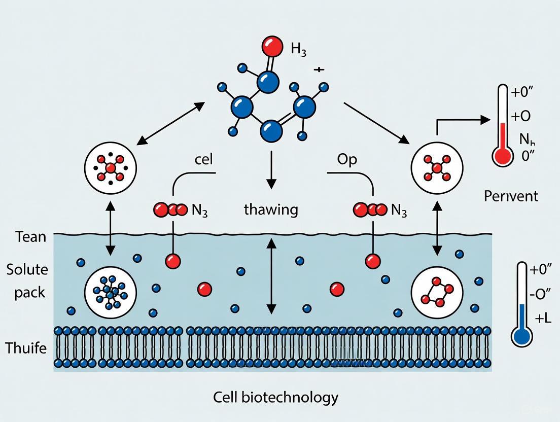
How does the thawing process itself induce osmotic shock? During the freezing process, extracellular ice forms, concentrating solutes in the remaining liquid water and creating a hyperosmotic environment. This draws water out of cells, dehydrating them and increasing their internal solute concentration. During thawing, the rapid melting of this extracellular ice suddenly dilutes the external environment, creating a hypoosmotic shock. Water then rapidly enters the dehydrated cells, causing them to swell [2]. The following diagram illustrates this cycle:
Troubleshooting Guide: Post-Thaw Cell Recovery
A systematic approach is required to troubleshoot poor cell recovery after thawing. The flowchart below outlines key investigation steps and solutions, with a focus on osmotic shock.
Quantitative Data: Thermodynamic Properties and Protocol Parameters
Table 1: Key Thermodynamic Parameters of a Freezing Medium for Ovarian Tissue Cryopreservation
This table summarizes critical temperatures for a formulated freezing medium, which guide the development of optimized freezing and thawing protocols [4].
| Parameter | Symbol | Value | Significance in Protocol Design |
|---|---|---|---|
| Glass Transition Temperature | Tg' | -120.49 °C | Storage temperature must remain below this point to prevent damaging molecular processes. |
| Crystallization Temperature | Tc | -20 °C | Temperature at which ice crystallization occurs during cooling at 2.5 °C/min. |
| Melting Temperature | Tm | -4.11 °C | Ice begins to melt at this temperature during warming. |
Table 2: Impact of Storage Temperature Stress on Cell Viability
Storing cryopreserved cells at inappropriate temperatures can induce stress and ice crystal formation, severely impacting viability [3] [4].
| Storage Temperature | Potential Consequence | Impact on Cell Viability |
|---|---|---|
| Warmer than -123°C | Extracellular medium devitrifies, inflicting stress. | High cell mortality risk. |
| Warmer than -47°C | Intracellular glass transition temperature is crossed. | Significant reduction in viability. |
| Warmer than -25°C | High risk of intracellular ice crystal formation. | Particularly high cell mortality observed. |
Experimental Protocols
Protocol 1: Optimized Thawing to Prevent Osmotic Shock
This protocol is designed to minimize osmotic shock during the thawing of sensitive cells, such as iPSCs and ovarian tissue [4] [3].
- Rapid Warming to Melting Point (Tm): Immediately transfer the cryovial from storage to a 37°C water bath. Gently agitate until only a small ice clump remains. This step quickly passes through the dangerous temperature zone where ice recrystallization can cause mechanical damage.
- Slow Warming Through Glass Transition (Tg'): Some advanced protocols for tissues may incorporate a brief incubation in a cold chamber (e.g., 3.5 minutes) to slowly reach the glass transition temperature (Tg' ≈ -120°C), limiting thermal and mechanical shocks before the final rapid warm-up to 37°C [4].
- Gradual Dilution of Cryoprotectant:
- Immediately after thawing, transfer the cell suspension to a conical tube containing pre-warmed culture medium.
- Do not add the medium directly to the cryovial. This starts the dilution process of the cryoprotectant (e.g., DMSO) outside the vial.
- Slowly add fresh medium dropwise to the cell suspension over several minutes while gently swirling the tube. This gradual dilution prevents a sudden osmotic imbalance that would cause a massive and damaging influx of water into the cells.
- Centrifugation and Seeding: Pellet the cells via gentle centrifugation, aspirate the supernatant containing the diluted cryoprotectant, and resuspend the cell pellet in fresh, pre-warmed complete culture medium. Seed the cells at an appropriate density onto culture plates.
Protocol 2: Measuring Osmotic Stress on Protein Stability in Cells
This methodology, adapted from a study on E. coli, details how to quantify the destabilizing effect of osmotic shock on intracellular proteins using 19F NMR spectroscopy [5].
- Objective: To quantify the change in stability of a test protein inside living cells subjected to hyperosmotic shock.
- Test Protein: The 7-kDa N-terminal SH3 domain from the Drosophila protein drk, labeled with a fluorine atom on its sole tryptophan residue.
- Procedure:
- Hyperosmotic Shock: Induce shock by adding 0.3 M NaCl to the cell culture media. This causes water efflux, reducing cell volume by ~35% and increasing macromolecular crowding [5].
- In-Cell NMR: Acquire a 19F NMR spectrum of the shocked cells. The fluorine label allows the folded (F) and unfolded (U) populations of SH3 to be distinguished by their distinct chemical shifts.
- Lysate Spectrum: Lyse the cells and acquire a 19F NMR spectrum of the clarified lysate. The dilution of cellular contents in the buffer attenuates crowding-induced attractive interactions, shifting the equilibrium toward the folded state.
- Data Analysis: The stability (modified standard-state free energy of unfolding, ΔG°′U) is calculated by comparing the population ratios of folded and unfolded protein from the in-cell and lysate spectra, correcting for background fluorine signals.
- Expected Outcome: Hyperosmotic shock decreases SH3 stability by approximately 1 kcal/mol, demonstrating that increased crowding from volume reduction can destabilize proteins via transient attractive interactions, contrary to traditional hard-core repulsion theory [5].
The Scientist's Toolkit: Essential Reagents for Osmotic Shock Research
Table 3: Key Research Reagents and Their Functions
| Reagent | Function / Role in Osmotic Shock Research | Example Application |
|---|---|---|
| Dimethyl Sulfoxide (DMSO) | Penetrating cryoprotectant. Reduces ice crystal formation by dehydrating cells before freezing and modulating water influx during thawing [3]. | Standard component (typically 10%) in cryopreservation solutions for many cell types. |
| Sucrose / Trehalose | Non-penetrating cryoprotectants and compatible osmolytes. Increase solution viscosity, reduce ice formation, and help stabilize proteins and membranes during osmotic stress [4] [2]. | Added to freezing media (e.g., 0.1M sucrose) as an osmotic buffer [4]. |
| Glycine Betaine | A compatible organic osmolyte. Accumulates in cells to counteract hyperosmotic stress and restore protein stability without disrupting function [5] [6]. | Added (e.g., 1 mM in media) to study osmoprotection; cells accumulate it internally to high concentrations [5]. |
| Sorbitol / NaCl | Osmotic stressing agents. Used to experimentally create hyperosmotic conditions to study cellular responses or, in the case of sorbitol, to potentially improve protein expression [6] [1]. | Adding 500-1000 mM sorbitol to growth media to impose osmotic stress on bacteria [6]. |
| Polyethylene Glycol (PEG) / Dextran | High molecular weight neutral polymers. Used in osmotic shock methods for cell disruption and to simulate molecular crowding conditions in vitro [7]. | Inducing cell rupture in algal cells for intracellular component release [7]. |
Core Concepts FAQ
What are the dual threats of intracellular ice formation and cell dehydration during cryopreservation? During slow freezing, the extracellular solution freezes first, concentrating solutes outside the cell. This creates an osmotic gradient that draws water out of the cell, leading to cell dehydration. If cooling is too rapid, water does not have sufficient time to exit the cell and freezes internally, causing intracellular ice formation. Both mechanisms can cause fatal cell damage [3].
How does the freezing process balance these two threats? Successful cryopreservation protocols balance cooling rates to minimize both hazards. A cooling rate that is too slow leads to excessive dehydration and solute damage, while a rate that is too fast results in lethal intracellular ice formation [3]. For human induced pluripotent stem cells (iPSCs), research indicates that a fast-slow-fast cooling pattern across different temperature zones may optimize survival [3].
What is the connection between these freezing threats and osmotic shock during thawing? The osmotic stress experienced during freezing is compounded during thawing. As the frozen solution melts, cells are suddenly exposed to a sharp change in solute concentration. If not managed correctly by controlling the reintroduction of water, this can cause a rapid influx of water, leading to osmotic shock and cell membrane rupture [3] [8].
Troubleshooting Guide
Problem: Low Post-Thaw Viability
| Potential Cause | Investigation | Solution |
|---|---|---|
| Suboptimal Cooling Rate | Review controlled-rate freezer settings or passive freezing container method. | For iPSCs, a cooling rate of -1 °C/min is often effective. Test rates between -0.3 °C/min and -3 °C/min [3]. |
| Inadequate Cryoprotectant | Verify concentration and type of cryoprotectant agent (CPA). | Ensure a sufficient concentration of a penetrating CPA like DMSO (e.g., 10%) is used. For sensitive cells, consider combinations with non-penetrating CPAs [3] [9]. |
| Improper Cell State at Freezing | Check culture logs for confluency and health before freezing. | Freeze cells during the logarithmic growth phase for optimal recovery [3]. |
Problem: Cell Rupture or Membrane Damage Post-Thaw
| Potential Cause | Investigation | Solution |
|---|---|---|
| Osmotic Shock During Thawing | Observe thawing technique: is dilution of CPA too rapid? | Add pre-warmed medium dropwise to the thawed cell suspension to gradually reduce CPA concentration and prevent rapid water influx [8] [10]. |
| Intracellular Ice Formation | Check storage temperature logs for fluctuations. | Ensure cells are stored below the extracellular glass transition temperature (-123°C for DMSO) to prevent devitrification and ice crystal growth [3]. |
| Fast/Uncontrolled Thawing | Review thawing protocol. | Thaw cells rapidly in a 37°C water bath to avoid the dangerous temperature zone where ice recrystallization occurs [8]. |
Experimental Protocols for Optimization
Protocol 1: Standardized Thawing to Prevent Osmotic Shock
This protocol is designed to minimize osmotic stress during the critical thawing phase [8] [10].
- Preparation: Warm complete culture medium to 37°C. Pre-warm and ethanol-sterilize a water bath to 37°C.
- Rapid Thaw: Remove cryovial from liquid nitrogen and immediately place it in the 37°C water bath. Gently swirl until only a small ice crystal remains (approximately 1-2 minutes).
- Controlled Dilution: Wipe the cryovial with ethanol and transfer the thawed cell suspension to a conical tube. Slowly and dropwise, add 5-10 mL of pre-warmed medium to the cells. This gradual dilution reduces the osmotic gradient.
- CPA Removal: Centrifuge the cell suspension at 200 x g for 5 minutes to pellet the cells. Carefully aspirate the supernatant containing the CPA.
- Reseeding: Resuspend the cell pellet in fresh, pre-warmed medium. Count cells and assess viability, then seed at the recommended density.
Protocol 2: Assessing the Impact of Cooling Rates on iPSC Recovery
This methodology helps identify the optimal cooling rate for a specific cell line, balancing dehydration and ice formation [3].
- Cell Preparation: Culture and harvest iPSCs at the logarithmic growth phase. Prepare identical cell aliquots in cryopreservation medium.
- Variable Cooling: Using a controlled-rate freezer, subject aliquots to different cooling rates (e.g., -0.5 °C/min, -1 °C/min, -3 °C/min, -10 °C/min).
- Storage and Thawing: After cooling, transfer all vials to liquid nitrogen for storage for at least 24 hours. Thaw all samples using the standardized protocol from Protocol 1.
- Viability Analysis: Quantify post-thaw viability using a method like trypan blue exclusion or an automated cell counter.
- Functional Recovery: Monitor cell attachment, morphology, and time to reach confluency over 4-7 days. The cooling rate yielding the highest viability and fastest functional recovery is optimal.
Research Reagent Solutions
The following table lists key reagents used in cryopreservation and their specific functions in combating the dual threats.
| Reagent | Function & Mechanism |
|---|---|
| Dimethyl Sulfoxide (DMSO) | A penetrating cryoprotectant. Lowers the freezing point, reduces ice crystal formation, and modulates electrolyte concentration. A 10% solution is hypertonic, promoting cell dehydration before freezing [3]. |
| CryoStor CS10 | A commercial, serum-free freezing medium containing 10% DMSO. Formulated to offer stability and reduce the stress associated with ice formation [10]. |
| Hyaluronic Acid (HA) | A biomaterial used in 3D constructs. Acts as a non-penetrating macromolecular cryoprotectant, improves uniform CPA diffusion, and can modulate intracellular stress pathways (e.g., RhoA/ROCK) to improve viability [9]. |
| Y-27632 (ROCK Inhibitor) | A small molecule inhibitor. Added to culture medium post-thaw for single cells or sensitive aggregates. Enhances cell survival by inhibiting apoptosis induced by cytoskeletal stress during the freeze-thaw cycle [10]. |
| Trehalose | A non-penetrating sugar. Often used in DMSO-free or low-DMSO formulations. Stabilizes cell membranes and proteins in a dry state through water substitution, reducing dehydration damage [9]. |
Process Visualization
Freezing Process and Cell Damage Pathways
This diagram illustrates the critical branching point during freezing where the cooling rate determines the primary pathway of cell damage, leading to either dehydration or intracellular ice formation.
Frequently Asked Questions (FAQs)
Q1: What does it mean that DMSO creates a hypertonic environment, and why is it a "double-edged sword" for my cells?
DMSO solutions are hypertonic, meaning they have a higher solute concentration than the inside of your cells. For instance, a 10% DMSO solution has an osmolarity of approximately 1.4 osm/L [3]. When cells are introduced to this environment, water rapidly moves out of the cells to equilibrate the osmotic imbalance, causing cell dehydration [3]. This is a crucial protective mechanism, as it reduces the amount of intracellular water available to form damaging ice crystals during freezing [3].
The "double-edged sword" is that this essential hypertonic environment, combined with DMSO's chemical properties, also introduces risks. The beneficial dehydration is accompanied by two major damaging phenomena:
- Osmotic Injury: Extreme and rapid volume changes can cause mechanical stress and rupture cell membranes, a process known as expansion lysis [11].
- Cytotoxicity: DMSO itself is chemically toxic to cells. This toxicity increases with higher DMSO concentration, elevated temperature, and longer exposure time [11] [12]. The delicate balance is that you need a high enough DMSO concentration to protect against ice formation, but this same concentration can cause chemical damage to the cells [13].
Q2: During the thawing process, what specific steps can I take to prevent osmotic shock?
Preventing osmotic shock during thawing is critical for cell recovery. The key is to avoid a rapid influx of water into cells that are still in a hypertonic state. The following protocol outlines a standard method for thawing cryopreserved cells, with special emphasis on steps that prevent osmotic shock.
The most critical step for preventing osmotic shock is the initial dilution (Step 2 in the diagram). Adding the thawed cell suspension directly into a large volume of pre-warmed, isotonic culture medium rapidly reduces the extracellular concentration of DMSO. This creates a gentler osmotic gradient, preventing a sudden and excessive influx of water that would cause the cells to swell and lyse [3]. The subsequent centrifugation and resuspension step (Steps 4 & 5) is then used to remove the cryoprotectant almost completely.
Q3: I see a lot of cell death after thawing. Is this due to DMSO toxicity or osmotic shock?
Distinguishing between these two causes is a common troubleshooting challenge. The timing and nature of the observed damage can help you identify the primary culprit. The table below summarizes the key differences.
| Feature | DMSO Toxicity | Osmotic Shock during Thawing |
|---|---|---|
| Primary Cause | Chemical damage from DMSO interaction with cell membranes and internal structures [11] [12]. | Physical damage from rapid water influx causing excessive cell swelling and rupture (expansion lysis) [11] [3]. |
| Typical Manifestation | Gradual reduction in cell viability over hours to days; impaired cellular function and metabolism [11] [14]. | Immediate, massive cell death and loss of membrane integrity upon or shortly after dilution/thawing [3]. |
| Key Influencing Factors | High DMSO concentration, high temperature, long exposure time [11] [15]. | Overly rapid dilution, omission of dilution step, using hypotonic solutions [3]. |
To diagnose your specific issue, review your thawing protocol against the workflow in the diagram above, paying close attention to the speed and ratio of the initial dilution step.
Troubleshooting Guides
Problem: Consistently Low Post-Thaw Cell Viability
This is one of the most common problems in cryopreservation. The solutions often involve optimizing the entire workflow, from freezing to thawing.
Potential Cause 1: Excessive DMSO toxicity due to suboptimal freezing protocol.
- Solution: Ensure you are using a controlled-rate freezer or an appropriate "Mr. Frosty"-type container that achieves a cooling rate of approximately -1°C/min, which is optimal for many cell types including stem cells [3]. A cooling rate that is too slow can expose cells to toxic solute concentrations for too long, while a rate that is too fast leads to deadly intracellular ice formation [3].
Potential Cause 2: Intracellular ice formation causing mechanical damage.
Potential Cause 3: Osmotic shock during the addition of DMSO before freezing.
- Solution: Implement a step-wise addition of the DMSO-containing freezing medium. Adding the cryoprotectant in several steps, with gentle mixing between each step, allows cells to equilibrate more gradually, minimizing the initial osmotic shrinkage and subsequent swelling [18].
Problem: Poor Cell Attachment and Proliferation After Thawing
Even if viability is acceptable immediately after thawing, cells may fail to recover and function normally.
Potential Cause 1: Cytotoxic effects on cell metabolism and membranes.
- Solution: Reduce the post-thaw exposure time to DMSO. After the initial 1:10 dilution, centrifuge the cells and resuspend them in fresh medium promptly. Prolonged contact with even low concentrations of DMSO can hinder cell attachment and proliferation [15].
Potential Cause 2: Chilling injury or damage during the freezing process.
- Solution: Assess the health and passage number of the culture before freezing. Always freeze cells from a healthy, logarithmically growing culture [3]. Cells frozen from an over-confluent or stressed culture will not recover well.
Potential Cause 3: Inadequate removal of DMSO post-thaw.
- Solution: Ensure the centrifugation step is performed correctly. The pellet should be resuspended in a sufficient volume of fresh medium to effectively dilute and remove the DMSO. For highly sensitive cells, consider a second wash step.
Problem: High Variability in Recovery Between Cell Batches
Inconsistency can stem from subtle differences in handling or cell state.
Potential Cause 1: Inconsistent handling times during protocol steps.
- Solution: Standardize the "room temperature" or "on-ice" exposure times for all users. The toxicity of DMSO is time- and temperature-dependent [11]. Create a detailed, step-by-step Standard Operating Procedure (SOP) that all lab members must follow.
Potential Cause 2: Variation in aggregate size for iPSC or MSC cultures.
- Solution: When freezing cells as aggregates (clumps), aim for uniform size. Large aggregates can limit the penetration of DMSO into the core and create pockets of high osmotic stress, leading to variable viability within the sample [3].
The Scientist's Toolkit: Essential Reagents & Materials
The table below lists key reagents used in cryopreservation protocols and their specific functions in managing the hypertonic environment.
| Reagent/Material | Function in Cryopreservation |
|---|---|
| Dimethyl Sulfoxide (DMSO) | A penetrating cryoprotectant. It enters the cell, depresses the freezing point, and reduces ice crystal formation by disrupting the water hydrogen-bonding network [3] [13] [14]. |
| Trehalose | A non-penetrating disaccharide. It remains outside the cell, creating a stable hypertonic environment that promotes gentle dehydration. It also stabilizes membrane phospholipids and proteins [14]. |
| Fetal Bovine Serum (FBS) | A common component of freezing media. Proteins in FBS can provide additional membrane stabilization and mitigate some of the stresses of the hypertonic environment [15]. |
| Programmable Freezer / Cryo-container | Essential for achieving the slow, controlled cooling rate (~-1°C/min) necessary to balance cell dehydration with intracellular ice formation [3]. |
| Pre-warmed Culture Medium | Used for the critical first dilution step upon thawing. Its large volume and isotonicity rapidly reduce extracellular DMSO concentration, preventing osmotic shock [3]. |
Advanced Topic: Mathematical Optimization to Eliminate Osmotic Stress
For researchers requiring the highest possible cell recovery, advanced mathematical modeling can be used to design optimized CPA loading and unloading protocols. The "Two-Parameter Formalism" model, based on the Kedem-Katchalsky equations, can be constrained to maintain a constant cell volume during the addition of DMSO [18]. This approach eliminates osmotic stress by dynamically controlling the extracellular concentration of permeable (DMSO) and non-permeable (e.g., trehalose) solutes. The solution to these equations provides exact, analytical methods to design a perfusion protocol that loads the CPA into the cell without causing it to shrink or swell beyond its natural volume, thereby preventing mechanical damage [18]. While this method may be unnecessarily precise for some applications, it offers a powerful tool for cryopreserving particularly sensitive or high-value cell lines.
Cell Membrane Permeability and the Critical Temperature Zones During Thawing
FAQs: Navigating the Thawing Process
1. What are the critical temperature zones during thawing, and why do they matter? During thawing, cells pass through critical temperature zones that pose risks like intracellular ice recrystallization and osmotic shock [3]. Rapid warming through the -123°C to -47°C range is crucial to avoid these stressful thermal events, which can mechanically damage cells from ice crystals or cause excessive water influx that ruptures the cell membrane [3] [19].
2. How does the thawing rate specifically affect cell membrane permeability? The thawing rate directly influences the physical state of membrane lipids. During cooling, membrane lipids undergo a transition from a liquid crystalline to a gel phase [19]. A slow thawing rate can prolong the time the membrane spends in a disordered gel state, increasing its permeability and potential for leakage upon rehydration. Rapid thawing helps the membrane transition back to its stable, functional liquid crystalline state with minimal damage [19].
3. What is the single most important step to prevent osmotic shock during thawing? The most critical step is the slow, dropwise dilution of the thawed cell suspension into pre-warmed culture medium [10] [8] [20]. This gradual dilution allows the intracellular concentration of cryoprotectants like DMSO to equilibrate slowly with the extracellular environment, preventing a sudden and massive influx of water that would cause the cell to swell and burst [3].
4. My cells have low viability post-thaw, but I followed the protocol. What could be wrong? Low viability can stem from issues prior to thawing. Key factors to check include:
- Pre-freeze Cell Health: Ensure cells were frozen during the logarithmic growth phase for maximum recovery [3].
- Freezing Rate: An inappropriate freezing rate can cause excessive dehydration or intracellular ice formation, causing damage that becomes apparent only upon thawing [3] [19].
- Storage Temperature Fluctuations: If stored cells warmed above critical glass transition temperatures (e.g., -123°C), intracellular ice crystals may have formed, causing mechanical damage [3].
Troubleshooting Guide
| Problem | Potential Cause | Recommended Solution |
|---|---|---|
| Low Cell Viability | Osmotic shock during dilution | Dilute thawed cells dropwise into pre-warmed medium while gently rocking the tube [10] [20]. |
| Intracellular ice crystal damage from slow thawing | Thaw cells rapidly (<1 minute) in a 37°C water bath until only a small ice pellet remains [21] [8]. | |
| Cell damage from toxic DMSO exposure | Promptly remove cryoprotectant (DMSO) by centrifuging and resuspending cells in fresh medium after thawing [8] [22]. | |
| Poor Cell Attachment & Growth | Cells were not in log growth phase pre-freeze | Freeze cells only when they are in a state of active, logarithmic growth [3]. |
| Inaccurate or low seeding density | Plate thawed cells at a high density to optimize cell-cell contact and recovery [21] [10]. | |
| Inconsistent thawing | Use a controlled, automated thawing system to standardize the warming rate and minimize human error [23]. |
Experimental Protocols
Protocol 1: Standardized Thawing for Preventing Osmotic Shock
This protocol is designed to minimize osmotic stress and is applicable to many adherent and suspension cell lines [21] [8] [20].
Materials:
- Cryovial of frozen cells
- Pre-warmed complete growth medium (37°C)
- 70% ethanol
- 37°C water bath or automated thawing system
- 15 mL or 50 mL sterile centrifuge tubes
Method:
- Rapid Thawing: Remove the cryovial from liquid nitrogen and immediately place it in a 37°C water bath. Gently swirl the vial for approximately 60 seconds or until only a small ice pellet remains [21] [20].
- Sterile Transfer: Wipe the cryovial with 70% ethanol and transfer the entire contents to an empty sterile centrifuge tube.
- Slow Dilution: Slowly and dropwise, add 10 mL of pre-warmed growth medium to the thawed cells while gently rocking the tube. This step is critical for the gradual equilibration of solutes and prevents osmotic shock [20].
- Cryoprotectant Removal: Centrifuge the cell suspension at 200 × g for 5-10 minutes [21] [8].
- Resuspension and Plating: Aspirate the supernatant containing DMSO. Gently resuspend the cell pellet in fresh, pre-warmed growth medium and transfer to a culture vessel. Plate at a high density to support recovery [21] [10].
Protocol 2: Using FTIR Spectroscopy to Analyze Membrane Phase Behavior During Thawing
This methodology allows for the direct investigation of membrane lipid and protein stability during a freeze-thaw cycle [19].
Materials:
- Cell pellet (e.g., LNCaP prostate tumor cells)
- Fourier Transform Infrared Spectrometer (FTIR)
- CaF2 IR windows and mylar spacer
- Variable temperature cell and controller
- Liquid nitrogen coolant
Method:
- Sample Preparation: Sandwich approximately 10 μL of a cell pellet between two CaF2 IR windows separated by a 6 μm mylar spacer. Mount the assembly into the variable temperature cell [19].
- Controlled Freezing and Thawing: Cool the sample from ambient temperature to -80°C at a controlled rate (e.g., 2°C/min). Initiate the thawing phase by warming the sample from -80°C to +90°C at a rate of 2°C/min, recording spectra at regular temperature intervals [19].
- Data Analysis:
- Membrane Phase Transition: Monitor the wavenumber of the CH2 symmetric stretching band (~2850 cm⁻¹). Plot the wavenumber against temperature; the phase transition temperature (Tm) is determined from the maximum in the first derivative of this plot. A steeper slope at Tm indicates a more cooperative phase transition [19].
- Protein Denaturation: Analyze the amide-I and amide-II bands (1700-1500 cm⁻¹). Heat-induced denaturation is marked by an increase in β-sheet structures (visible as a band at ~1625 cm⁻¹) and a decrease in α-helical structures [19].
The following table consolidates key quantitative findings on cellular responses to freezing and thawing parameters.
Table 1: Biophysical and Experimental Parameters in Cryopreservation
| Parameter / Observation | Experimental Value / Condition | Model System / Context |
|---|---|---|
| Optimal Cooling Rate (Freezing) | -1 °C/min [3] | Human iPSC |
| Extracellular Glass Transition Temp. | -123 °C [3] | DMSO-based freezing medium |
| Intracellular Glass Transition Temp. | ≈ -47 °C [3] | Jurkat T cells (example) |
| High Cell Mortality Zone | > -25 °C [3] | General cell thawing |
| Membrane Phase Behavior | Liquid crystalline to gel transition coincides with extracellular ice nucleation temperature [19] | LNCaP cells |
| DMSO Concentration | 10% (standard) [22] | PBMC Cryopreservation |
| Centrifugation Post-Thaw | 200 × g for 5-10 minutes [21] [8] | General cell culture protocol |
Visualizing the Thawing Workflow and Membrane Dynamics
The following diagram illustrates the critical pathway and molecular events during the cell thawing process.
The Scientist's Toolkit: Essential Reagents & Materials
Table 2: Key Research Reagents and Solutions
| Item | Function / Application |
|---|---|
| DMSO (Dimethyl Sulfoxide) | A common cryoprotective agent (CPA) that penetrates cells, reduces ice crystal formation, and protects from dehydration damage during freezing [3] [22]. |
| CryoStor CS10 | A commercial, serum-free cryopreservation medium specifically optimized for stem cells and other sensitive cell types [10]. |
| ROCK Inhibitor (Y-27632) | Significantly improves the survival and attachment of single pluripotent stem cells (PSCs) after thawing by inhibiting apoptosis [10]. |
| Ficoll 70 | An additive that can enable long-term storage of iPSCs at -80°C without compromising viability and pluripotency [3]. |
| Pre-warmed Complete Growth Medium | Used for diluting thawed cells; pre-warming prevents thermal shock and supports immediate metabolic reactivation [21] [8]. |
| Fetal Bovine Serum (FBS) | Often used as a component in cryopreservation media (e.g., 90% FCS + 10% DMSO) to provide extracellular protection and support cell membranes [22]. |
Frequently Asked Questions (FAQs)
FAQ 1: What makes iPSCs particularly sensitive to the cryopreservation and thawing process? iPSCs are inherently more vulnerable to intracellular ice formation than many other human or animal cells [24]. This sensitivity is compounded by their large surface area-to-volume ratio and the permeability of their plasma membrane. During freezing, the delicate balance between preventing intracellular ice formation and preventing cell dehydration is critical; an imbalance in this process significantly reduces post-thaw cell recovery [24].
FAQ 2: Why is controlling the cooling rate so crucial for iPSC survival? The cooling rate must be strictly controlled because it directly impacts two key damaging factors: intracellular ice formation and cell dehydration [24]. A rate that is too fast promotes intracellular ice, while a rate that is too slow leads to excessive dehydration. Research indicates that a controlled freezing rate between -0.3 °C/min and -1.8 °C/min is optimal for human embryonic stem cells (closely related to iPSCs), with -1 °C/min being a frequently used and successful rate [24].
FAQ 3: How does the method of passaging (as single cells vs. aggregates) affect post-thaw recovery of iPSCs? The choice between passaging and freezing iPSCs as single cells or as cell aggregates (clumps) involves a trade-off.
- Freezing as Aggregates: This method benefits from preserved cell-cell contacts, which support survival. Recovery is often faster because the cells do not need to reform these connections from scratch after thawing [24].
- Freezing as Single Cells: This allows for better quality control and more consistent cell quantification. However, post-thaw recovery can be slower as single cells need time to proliferate and re-establish aggregate structures [24].
FAQ 4: What are the primary causes of osmotic shock during thawing, and how can it be prevented? Osmotic shock occurs when cells are exposed to rapid changes in solute concentration. During thawing, this happens if the cryoprotectant (e.g., DMSO) is not diluted out carefully. DMSO is highly permeable at 0–4 °C but becomes increasingly cytotoxic at higher temperatures [25]. To prevent osmotic shock, it is vital to use rapid thawing to 0–4 °C to outpace DMSO toxicity, followed by a slow and controlled dilution and removal of the cryoprotectant-containing medium [24] [25].
FAQ 5: Are there alternatives to FBS-based freezing media for immune cells like PBMCs, and how effective are they? Yes, serum-free, animal-protein-free alternatives are available and effective. A comprehensive study showed that serum-free media like CryoStor CS10 and NutriFreez D10 (both containing 10% DMSO) maintained high PBMC viability, phenotype, and functionality over a 2-year period, performing comparably to traditional FBS-supplemented media [25]. Media with DMSO concentrations below 7.5% showed significantly lower viability, highlighting the essential protective role of DMSO despite its cytotoxicity [25].
Troubleshooting Guides
Problem: Low Cell Viability After Thawing iPSCs
Potential Causes and Solutions:
| Problem Area | Specific Issue | Recommended Solution |
|---|---|---|
| Freezing Process | Suboptimal cooling rate [24] | Use a controlled-rate freezer or isopropanol-filled container to ensure a consistent cooling rate of approximately -1 °C/min |
| Inadequate cryoprotectant [24] | Use a freezing medium containing 10% DMSO. Ensure it is hypertonic to draw water out of cells and prevent ice crystals | |
| Thawing Process | Osmotic shock during cryoprotectant removal [24] | Slowly dilute thawed cells drop-wise with fresh, pre-warmed medium before centrifugation |
| DMSO cytotoxicity [25] | Thaw cells quickly, then immediately start dilution process; minimize DMSO exposure time at room temperature | |
| Cell Handling | Freezing cells from the wrong growth phase [24] | Freeze iPSCs during the logarithmic growth phase for maximum health and recovery potential |
| Microbial contamination (e.g., Mycoplasma) [24] | Confirm cells are free of contamination before freezing; wear a face mask during handling to prevent oral microbe transfer |
Problem: Poor Recovery of Immune Cell Functionality Post-Thaw
Potential Causes and Solutions:
| Problem Area | Specific Issue | Recommended Solution |
|---|---|---|
| Freezing Medium | Use of FBS with high batch-to-batch variability [25] | Switch to a qualified, commercially available serum-free, animal-protein-free freezing medium |
| Inadequate or excessive DMSO concentration [25] | For PBMCs, a 10% DMSO concentration is recommended for long-term (up to 2 years) functional preservation | |
| Storage & Handling | Temperature fluctuations during storage [24] | Store cells in vapor-phase liquid nitrogen or a -150°C freezer; avoid warming above -123°C (extracellular glass transition temperature) |
| Improper thawing technique [25] | Thaw cells rapidly in a 37°C water bath, then dilute immediately with pre-warmed culture medium to mitigate DMSO toxicity |
Comparative Cell Vulnerability & Cryopreservation Data
The table below summarizes quantitative data on the sensitivity and optimal handling of different cell types.
| Cell Type | Key Vulnerability Factor | Optimal Freezing Rate | Optimal DMSO Concentration | Post-Thaw Viability Benchmark |
|---|---|---|---|---|
| iPSCs | High susceptibility to intracellular ice formation; sensitive plasma membrane [24] | -1 °C/min [24] | ~10% [24] | Ready for experiments in 4-7 days under optimized conditions [24] |
| PBMCs | Sensitivity to DMSO cytotoxicity; functionality depends on medium composition [25] | Not specified; use of passive cooling devices | 10% (in serum-free media like CryoStor CS10) [25] | High viability and functionality maintained for up to 2 years with CS10 [25] |
| Human Oocytes | Large surface area/volume ratio; high plasma membrane permeability [24] | -0.3 °C/min to -30°C, then -50 °C/min to -150°C [24] | Varies (often with sucrose) | Varies; protocol specifically designed to minimize ice crystal formation [24] |
| Spermatozoa | Membrane lipid composition and fluidity; cholesterol loss [26] | Species-specific (e.g., -20 to -50 °C/min) [26] | Varies (often glycerol-based) | Cattle: ~40% functional; Horses: <30% functional [26] |
The Scientist's Toolkit: Essential Reagents & Materials
| Item | Function | Application Note |
|---|---|---|
| Dimethyl Sulfoxide (DMSO) | Penetrating cryoprotectant agent; reduces ice crystal formation by dehydrating cells [24] [25] | Use at ~10% concentration. It is cytotoxic at room temperature; use pre-chilled and minimize exposure [25]. |
| Programmable Freezer | Allows for precise, controlled-rate freezing critical for sensitive cells like iPSCs [24] | Enables implementation of complex freezing profiles (e.g., fast-slow-fast) for optimal survival [24]. |
| Serum-Free Freezing Media | Xeno-free, chemically defined alternative to FBS; reduces variability and safety risks [25] | Products like CryoStor CS10 are validated for long-term preservation of PBMC form and function [25]. |
| Liquid Nitrogen (Vapor Phase) | Provides stable, ultra-low temperature environment for long-term cell storage [24] | Prevents stressful temperature fluctuations; storage temperature should not rise above -123°C [24]. |
| Matrigel / Laminin-521 | Extracellular matrix coating for feeder-free iPSC culture [24] | Provides crucial adhesion and survival signals to iPSCs during the critical post-thaw recovery period [24]. |
Experimental Protocols for Key Methodologies
Protocol 1: Optimized Thawing of iPSCs to Prevent Osmotic Shock
Principle: This protocol emphasizes rapid thawing to minimize DMSO cytotoxicity, followed by slow, controlled dilution to prevent osmotic shock, which can rupture cells due to rapid water influx [24].
Procedure:
- Rapid Thaw: Remove the cryovial from liquid nitrogen and immediately place it in a 37°C water bath. Gently agitate until only a small ice crystal remains.
- Controlled Dilution: Transfer the thawed cell suspension to a sterile centrifuge tube. Slowly add pre-warmed culture medium drop-wise (e.g., 1 mL over 1 minute) while gently swirling the tube. This gradually reduces the extracellular DMSO concentration, allowing water to enter the cell at a non-lethal rate.
- Centrifugation: Centrifuge the cell suspension at a low speed (e.g., 200 x g for 5 minutes) to pellet the cells.
- Reseeding: Aspirate the supernatant containing the DMSO and resuspend the cell pellet in fresh, pre-warmed complete culture medium. Seed the cells onto a Matrigel-coated plate at the recommended density.
Protocol 2: Assessing PBMC Viability and Functionality Post-Thaw
Principle: This procedure evaluates the success of cryopreservation by measuring not just cell survival, but also the critical immune functions of T-cells and B-cells, which are essential for downstream assays [25].
Procedure:
- Thawing and Washing: Rapidly thaw PBMCs and dilute in pre-warmed RPMI-1640 medium supplemented with 10% FBS. Centrifuge to remove the cryoprotectant.
- Viability and Yield Assessment: Resuspend the cell pellet and count using an automated cell counter or trypan blue exclusion to determine total cell count and viability percentage.
- Functional Assay (T-cell Proliferation):
- Seed PBMCs in a culture plate.
- Stimulate with anti-CD2/CD3/CD28 coated beads or a similar mitogen.
- After 5-7 days, use a flow cytometry-based assay to measure the percentage of proliferated T-cells.
- Functional Assay (Cytokine Secretion):
- Culture PBMCs with or without specific antigens.
- Collect supernatant after 24-48 hours.
- Use ELISA or a multiplex bead array to quantify secreted cytokines (e.g., IFN-γ, IL-2, TNFα) as a measure of immune cell activation [25].
Signaling and Workflow Visualizations
Diagram 1: iPSC Thawing and Recovery Workflow
Diagram 2: Key Stress Pathways in Cell Cryopreservation
Procedural Safeguards: Step-by-Step Thawing Protocols to Minimize Osmotic Stress
Core Scientific Rationale
Why Rapid Thawing is Critical
The principle of rapid thawing is fundamental to successful cell recovery after cryopreservation. Unlike the slow, controlled cooling required during freezing, thawing must be rapid to avoid the damaging effects of ice recrystallization.
- Ice Recrystallization: During slow warming, small intracellular ice crystals can melt and refreeze into larger, more damaging crystals. This process of recrystallization causes mechanical damage to cellular structures and membranes [27] [28].
- Thermal Transition: Rapid heating of the cell suspension prevents localized recrystallization during cell thawing, which preserves cellular integrity [28]. For a standard 1mL cryovial, all ice should disappear within a few minutes to maximize cell viability.
The Mechanism of Osmotic Shock
Osmotic shock occurs when cells are exposed to rapid changes in the solute concentration around them, leading to potentially lethal water flux.
- Cryoprotectant Exposure: Cells thawed in concentrated cryoprotectant solutions like dimethyl sulfoxide (DMSO) face immediate osmotic stress. As the cells warm, the high extracellular concentration of cryoprotectants creates an osmotic imbalance that can draw water out of cells, causing dehydration [3] [29].
- Dilution Stress: Conversely, if thawed cells are transferred directly into a standard culture medium without proper handling, the rapid influx of water into cells can cause them to swell and potentially burst [3] [20].
- Prevention Strategy: The use of controlled, dropwise dilution or specialized thawing media helps equilibrate osmotic gradients gradually, protecting cells from these damaging fluid shifts [20] [30].
Table: Key Threats During the Thawing Process and Their Cellular Consequences
| Thawing Phase | Primary Threat | Cellular Consequence | Preventive Measure |
|---|---|---|---|
| Ice Melting | Recrystallization | Mechanical damage to membranes and organelles | Rapid warming in a 37°C water bath [20] [28] |
| Initial Dilution | Osmotic Shock (Water influx) | Cell swelling and potential lysis | Slow, dropwise addition of warm medium [20] [30] |
| Post-Thaw Handling | Cryoprotectant Toxicity | Altered metabolism and reduced viability | Prompt centrifugation to remove DMSO [22] [30] |
Standardized Thawing Protocol for Optimal Recovery
General Thawing Procedure
This protocol synthesizes best practices for recovering adherent and suspension cells, with particular attention to preventing osmotic shock [20] [30].
- Quick Thawing: Remove the cryovial from storage and immediately place it in a 37°C water bath. Gently swirl the vial until only a small ice crystal remains (approximately 1-2 minutes). Do not allow the cells to thaw completely in the water bath, as prolonged exposure to 37°C in the presence of DMSO is harmful [20] [30].
- Sterilization: Wipe the exterior of the vial with 70% ethanol before transferring it to a biosafety cabinet.
- Gentle Dilution: Two recommended approaches exist for this critical step to mitigate osmotic shock:
- Method A (Direct Transfer): Transfer the entire contents of the vial to a sterile conical tube containing 10 mL of pre-warmed thaw medium dropwise, while gently swirling the tube to ensure gradual mixing [20].
- Method B (Gradual Dilution): For more sensitive cells, transfer the thawed cell suspension to an empty tube and then slowly add 10 mL of pre-warmed medium dropwise, with gentle rocking [20].
- Centrifugation: Immediately spin down the cells at 200-300 x g for 5-10 minutes to pellet the cells and remove the cryoprotectant-containing supernatant [20] [30].
- Resuspension and Culture: Carefully resuspend the cell pellet in fresh, pre-warmed culture medium (without selective antibiotics initially) and transfer to an appropriate culture vessel [20].
Special Considerations for Primary Cells and PBMCs
Peripheral Blood Mononuclear Cells (PBMCs) and other primary cells require additional care due to their heightened sensitivity [22] [30].
- Controlled Thawing: Thaw the vial in a 37°C water bath until about 90% ice has melted [20].
- DNase Treatment: If cell clumping is observed after resuspension, add DNase I (e.g., 100 µg per mL of cell suspension) and incubate at room temperature for 15 minutes to digest DNA released from dead cells, which can trap viable cells in aggregates [30].
- Viability Expectations: A cell loss of up to 20-30% during the washing steps is normal. A viable cell count must be performed immediately after thawing to establish a baseline [20] [30].
Troubleshooting Guide: Common Thawing Problems and Solutions
Table: Frequently Encountered Issues During Cell Thawing
| Problem | Potential Cause | Solution | Reference |
|---|---|---|---|
| Low Cell Viability Post-Thaw | Slow thawing allowing ice recrystallization; toxic cryoprotectant exposure. | Ensure rapid thawing; dilute and remove DMSO promptly after thawing. | [3] [28] |
| Excessive Cell Clumping | Release of cellular DNA from dead cells; inadequate dispersion. | Use DNase I treatment; ensure gentle but thorough resuspension without vortexing. | [30] |
| Poor Cell Attachment (Adherent Cells) | Osmotic shock during dilution; insufficient resting period. | Use slow, dropwise dilution; allow cells 24 hours in culture before first medium change. | [3] [20] |
| Low Recovery of PBMCs/ Primary Cells | Overly aggressive pipetting; incorrect medium composition. | Use wide-bore pipettes; use recommended thaw media like PBS with 2% FBS. | [22] [30] |
| Contamination | Non-sterile technique during the thawing process. | Thoroughly disinfect vial exterior; perform all fluid handling in a biosafety cabinet. | [30] |
Frequently Asked Questions (FAQs)
Q1: Why is "rapid thawing" emphasized when "slow freezing" is the standard? The physical stresses differ between the two processes. Slow freezing is necessary to allow water to exit the cell gradually, minimizing lethal intracellular ice crystal formation. Rapid thawing is crucial to swiftly pass through the temperature zone where small, otherwise harmless ice crystals can melt and recrystallize into larger, damaging forms, and to limit the time cells are exposed to toxic cryoprotectants at higher temperatures [3] [27] [28].
Q2: What is the single most critical step to prevent osmotic shock? The most critical step is the initial dilution of the thawed cell suspension. Adding pre-warmed medium dropwise while gently agitating the tube allows for a gradual equilibration of solutes across the cell membrane, preventing the rapid water influx that causes cells to swell and burst [20].
Q3: Can I use the cells immediately for experiments after thawing? It is generally not recommended. Most cell populations require a recovery period of 24-48 hours in culture to regain normal metabolism, repair any sublethal damage, and re-establish adherence (for adherent cells). Performing functional assays immediately post-thaw can yield unreliable results due to reduced viability and functionality [3] [22].
Q4: Is it acceptable to thaw cells directly in complete culture medium instead of a specialized "thaw medium"? While complete culture medium can be used, a specialized thaw medium (often containing a higher serum percentage or other osmotic stabilizers) can provide better protection. The key is that the medium must not contain selective antibiotics initially, as cells are more vulnerable post-thaw. Antibiotics can be added at the first medium change [20].
Visual Guide: Optimized Thawing Workflow
The following diagram outlines the logical decision-making process for thawing cells, integrating steps to prevent osmotic shock and ensure high viability.
Research Reagent Solutions
Table: Essential Materials for Cell Thawing
| Reagent / Material | Function / Purpose | Application Notes |
|---|---|---|
| DMSO (Dimethyl Sulfoxide) | Penetrating cryoprotectant that prevents intracellular ice formation during freezing. | Must be promptly removed post-thaw due to cytotoxicity at higher temperatures [3] [22]. |
| Fetal Bovine Serum (FBS) | Provides proteins and growth factors that stabilize cell membranes and mitigate osmotic stress. | Often used at 10% in thawing and washing media [22] [30]. |
| DNase I Solution | Enzyme that degrades DNA released from dead cells, preventing viable cells from being trapped in clumps. | Critical for thawing sensitive primary cells like PBMCs [30]. |
| Specialized Thaw Media | Pre-formulated solutions designed to provide optimal osmotic support and nutrients during recovery. | Can be cell-type specific; often lacks antibiotics initially [20]. |
| Programmed Thawing Device (e.g., ThawSTAR) | Provides consistent, automated thawing at the correct rate, improving reproducibility and sterility. | Reduces operator-dependent variability [30]. |
Theoretical Foundation: The Physics of Osmotic Shock During Thawing
During thawing, cells are acutely vulnerable to osmotic shock, a primary cause of reduced viability and functionality. Understanding the underlying physics is crucial for prevention.
Cryoprotectants like Dimethyl Sulfoxide (DMSO) are essential for preventing intracellular ice crystal formation during freezing [3]. However, upon thawing, cells are suspended in a high-concentration of DMSO. If this environment is not rapidly diluted, a damaging osmotic gradient is created. Water rapidly moves into the cells to equilibrate the high internal solute concentration, causing them to swell and potentially lyse [3] [31]. A slow dilution process exacerbates this stress and prolongs exposure to DMSO, which can be toxic to cells at temperatures above 4°C [8] [31]. Therefore, the core principle of this protocol is rapid dilution with pre-warmed medium to quickly reduce the DMSO concentration and minimize osmotic stress [32].
Standard Operating Procedure: Thawing and Diluting Cell Suspensions
Materials and Equipment (The Scientist's Toolkit)
| Item | Function & Specification |
|---|---|
| Complete Growth Medium | Pre-warmed to 37°C. Provides essential nutrients and correct osmotic environment for cells post-thaw [32] [33]. |
| Cryovial containing cells | Retrieved directly from liquid nitrogen or <-150°C storage. Ensure vial integrity [32]. |
| 37°C Water Bath | For rapid, uniform thawing. Disinfect with 70% ethanol to maintain sterility [8] [32]. |
| Personal Protective Equipment (PPE) | Lab coat, gloves, and face shield/goggles. Protects user from potential vial explosion and DMSO exposure [32]. |
| Centrifuge | Swinging bucket type, capable of 200 x g. Gently pellets cells for cryoprotectant removal [32] [33]. |
| Sterile Centrifuge Tubes | For diluting and washing the cell suspension. |
| Pipettes and Tips | For accurate and sterile liquid handling. |
Step-by-Step Protocol
Preparation
Rapid Thawing
- Retrieve the cryovial from storage, handling it with extreme care due to explosion risks [32].
- Immediately and fully immerse the vial in the 37°C water bath. Keep the cap above the waterline to prevent contamination [8].
- Gently swirl the vial for approximately 1-2 minutes until only a small ice crystal remains. The goal is a swift thaw to minimize damaging ice crystal formation [8] [32].
Immediate Dilution and Cryoprotectant Removal
- Transfer the vial to the laminar flow hood and disinfect its exterior with 70% ethanol [32].
- Transfer the thawed cell suspension dropwise into a centrifuge tube containing at least 10 mL of pre-warmed growth medium. This slow addition gradually reduces the DMSO concentration, preventing osmotic shock [8].
- Gently mix the cell suspension.
Washing and Resuspension
- Centrifuge the cell suspension at 200 x g for 5-10 minutes at 4°C to pellet the cells [32] [33].
- After centrifugation, carefully aspirate and discard the supernatant, which contains the diluted DMSO, without disturbing the cell pellet [32].
- Gently resuspend the cell pellet in a fresh, pre-warmed complete growth medium. Use a pipette to break up clumps gently, avoiding vigorous pipetting that can shear fragile cells [8] [34].
Cell Count, Viability Assessment, and Seeding
Workflow for Thawing and Preventing Osmotic Shock
Table 1: Key Parameters for Thawing and Centrifugation
| Parameter | Optimal Specification | Rationale & Impact on Osmotic Shock |
|---|---|---|
| Thawing Temperature | 37°C [32] | Ensures rapid phase change, minimizing time for small ice crystals to recrystallize and damage cells. |
| Thawing Duration | 1-2 minutes [8] [32] | Prevents prolonged exposure to high solute concentrations as DMSO warms, reducing toxicity. |
| Dilution Medium Volume | 10x vial volume (minimum) [33] | Provides sufficient volume for immediate and significant reduction of DMSO concentration upon dilution. |
| Centrifugation Speed | 200 x g [32] | Gentle enough to pellet cells without causing mechanical damage or compounding stress from osmotic shock. |
| Centrifugation Duration | 5-10 minutes [32] | Balances the need for a firm pellet against the risk of keeping cells in a hypoxic, stressed state. |
Frequently Asked Questions (FAQs)
Why is it critical to dilute the thawed cell suspension immediately and dropwise?
Rapid dilution with pre-warmed medium is the most effective action to prevent osmotic shock. The freeze medium contains a high concentration of cryoprotectant (e.g., 10% DMSO). When thawed cells are introduced into a much larger volume of isotonic medium, the osmotic gradient is reversed more gently than if they were added all at once. Adding the cells dropwise allows for a gradual decrease in extracellular DMSO, preventing a massive and rapid influx of water into the cells which would cause them to swell and burst [8] [31].
My cell viability post-thaw is low. What are the primary troubleshooting steps?
Low viability can stem from several points in the process. Systematically check the following:
- Thawing Process: Was the thaw rapid enough? Prolonged thawing increases DMSO toxicity [8] [31].
- Dilution: Was the dilution immediate and performed with pre-warmed medium? Using cold medium or delaying dilution subjects cells to toxic DMSO and temperature stress [32].
- Centrifugation: Was the centrifugation speed too high? Excessive g-force can damage already stressed cells [32] [34].
- Cell Stock Quality: Were the cells frozen at a high viability and proper density (e.g., in the late logarithmic growth phase)? Using low-quality stocks is a common source of failure [31] [34].
What is the consequence of skipping the centrifugation and wash step after dilution?
Skipping the wash step leaves cryoprotectant in the culture medium. While the initial dilution reduces the concentration, the residual DMSO can be toxic to cells over time, inhibiting cell attachment, spreading, and proliferation. For sensitive cell types like iPSCs, this can be particularly detrimental to recovery and pluripotency [8] [31].
Can I use a cell viability dye other than trypan blue?
Yes, several dyes are available for viability assessment. Trypan blue is a common and cost-effective exclusion dye where non-viable cells with compromised membranes take up the blue color. Alternative methods include automated cell counters using similar principles or fluorescent viability stains like propidium iodide, which may offer greater accuracy for some cell types [23].
Frequently Asked Questions (FAQs)
Q1: What is osmotic shock during cell thawing, and why is it detrimental? Osmotic shock occurs during thawing when cells are rapidly transferred from a hypertonic freezing solution (containing cryoprotectants like DMSO) into an isotonic culture medium. This creates a sudden, large osmotic difference, causing water to rush into the cells too quickly. This rapid influx can cause excessive swelling, stress, and rupture of the cell membrane, leading to significant cell death and poor recovery post-thaw [24] [35]. Preventing this is crucial for maintaining high cell viability.
Q2: How does the dropwise dilution method prevent osmotic shock? The dropwise dilution method works by gradually changing the osmotic environment around the cell. Instead of a single, abrupt transfer, the thawed cell suspension is added slowly (drop-by-drop) to a larger volume of warm, isotonic buffer or medium while gently mixing. This stepwise dilution allows cryoprotectants like DMSO to slowly and safely diffuse out of the cell, while water enters at a controlled rate. This minimizes drastic volume changes and protects membrane integrity [24].
Q3: What is the recommended rate for adding the thawed cells to the dilution medium? A slow, controlled addition is key. A common and effective protocol is to dilute the thawed cell suspension dropwise into a volume of medium that is at least 10 times larger than the volume of the cell suspension. The addition should be performed gradually over 1-2 minutes with gentle agitation to ensure immediate mixing of each drop [24].
Q4: After dropwise dilution, should the cells be immediately centrifuged? No, it is often beneficial to include a short incubation period. After the dropwise dilution is complete, allow the cell suspension to incubate for approximately 5-10 minutes at room temperature. This gives the cells additional time to equilibrate osmotically before the centrifugation and resuspension steps, further reducing stress [24].
Q5: Can the composition of the dilution medium be optimized? Yes. Using a medium supplemented with compounds that help counteract osmotic stress can improve outcomes. For instance, adding 50 mM glucose to the dilution medium has been shown to improve cell recovery and reduce apoptosis in sensitive cells like T-cells by providing extracellular osmotic support [35]. Alternatively, using a specialized buffer like Normosol-R as a basal solution has also been reported for certain cell types [36].
Troubleshooting Guide
| Problem | Potential Cause | Solution |
|---|---|---|
| Low cell viability after thawing and dilution | Excessive osmotic shock from overly rapid dilution. | Ensure the dropwise addition is slow enough (1-2 minutes) and into a sufficiently large volume (10x) of pre-warmed medium. Gently swirl the medium while adding cells. |
| Incorrect temperature of the dilution medium. | Use medium pre-warmed to 37°C to support cell metabolism and recovery. Avoid using cold medium. | |
| Cells appear swollen or ruptured | The osmotic difference was too great, too fast. | Verify that the culture medium is isotonic. Consider a two-step dilution or using an osmotic buffer like Normosol-R for the first dilution step [36]. |
| Poor cell attachment and growth post-thaw | Delayed damage from osmotic stress or cryoprotectant toxicity. | After dilution and centrifugation, resuspend the cell pellet in fresh, complete culture medium containing 5-10% serum or a ROCK inhibitor (for pluripotent stem cells) to support recovery and attachment [24]. |
| High levels of apoptosis 18-24 hours post-thaw | The cryopreservation or thawing process induced delayed-onset cell death. | Optimize the entire process. Ensure controlled-rate freezing, use cryoprotectants like 50 mM glucose to reduce apoptosis, and always use the dropwise dilution method upon thawing [35]. |
Key Experimental Protocols and Data
Protocol: Standard Dropwise Dilution for Thawed Cells
This protocol is adapted from established methods for thawing sensitive cells like iPSCs and T-cells [24] [35].
- Preparation: Warm a sufficient volume of complete culture medium or a specialized dilution buffer (e.g., containing 50 mM glucose) to 37°C. The volume should be at least 10 times the volume of the thawed cell suspension.
- Thawing: Rapidly thaw the cryovial in a 37°C water bath (approximately 1-2 minutes). Do not submerge the cap. Gently swirl until only a small ice crystal remains.
- Decontamination: Wipe the outside of the vial with 70% ethanol and transfer it to a biosafety cabinet.
- Initial Transfer: Gently transfer the thawed cell suspension into a sterile 15 mL conical tube.
- Dropwise Dilution: Using a sterile pipette, slowly add the cell suspension drop by drop to the pre-warmed 10x volume of medium/buffer over the course of 1-2 minutes. Gently swirl or agitate the tube continuously during the addition to ensure immediate mixing.
- Incubation: After all cells have been added, let the tube sit at room temperature for 5-10 minutes to allow for full osmotic equilibrium.
- Centrifugation and Resuspension: Centrifuge the cell suspension at a low, cell-appropriate speed (e.g., 200-300 x g for 5 minutes). Carefully aspirate the supernatant, which now contains the diluted cryoprotectant. Resuspend the cell pellet in fresh, pre-warmed complete culture medium for counting and subsequent plating.
Quantitative Data on Osmotic Support Agents
Research has quantified the effects of various additives that provide osmotic support during and after the thawing process. The table below summarizes key findings.
Table 1: Efficacy of Osmotic Support Agents in Post-Thaw Cell Recovery
| Agent | Mechanism of Action | Effective Concentration | Demonstrated Effect (Cell Type) | Source |
|---|---|---|---|---|
| Glucose | Increases extracellular osmolarity, limits intracellular water retention, reduces cell shrinkage and membrane damage. | 50 mM | Significantly improved cell recovery and reduced apoptosis (52.58% to 39.50%) in hCAR-T cells. | [35] |
| Sucrose | Suppresses intracellular ice formation, modulates extracellular osmotic pressure, alleviates osmotic shock during thawing. | Varies by protocol; often 0.1-0.2 M | Protects membrane integrity by complementing intracellular cryoprotectants. | [35] |
| Trehalose | Stabilizes membrane structures by forming hydrogen bonds with phospholipid bilayers. | Varies by protocol; often 0.1-0.2 M | Serves as a non-penetrating cryoprotectant to mitigate osmotic stress. | [35] |
| Ficoll 70 | High molecular weight polymer; mechanism not fully elucidated but enables long-term storage at -80°C. | 5-10% (w/v) | Enabled iPSC storage at -80°C for one year without compromising viability and pluripotency. | [24] |
Protocol: Evaluating Post-Thaw Osmotic Behavior
Understanding how cells respond osmotically after thawing can guide protocol optimization. The following methodology is used to characterize this behavior [36].
- Cell Preparation: Thaw and process cells using your standard protocol, including the dropwise dilution method.
- Resuspension: Resuspend the final cell pellet in an isotonic culture medium.
- Measurement: Immediately place a sample of the cell suspension on a microscope stage with a time-lapse imaging capability.
- Data Collection: Measure the diameter of a population of cells over time (e.g., every 30 seconds for 10-15 minutes).
- Analysis: Plot the mean cell diameter against time. Normal osmotic behavior would show a stable or slightly fluctuating diameter. A sharp, continuous drop in volume indicates excessive dehydration and anomalous osmotic behavior, suggesting the need for further optimization of the cryopreservation or thawing solution [36].
The Scientist's Toolkit: Essential Reagents for Osmotic Equilibrium
Table 2: Key Research Reagent Solutions
| Item | Function in Osmotic Equilibrium | Brief Explanation | |
|---|---|---|---|
| Dimethyl Sulfoxide (DMSO) | Penetrating Cryoprotectant | A standard intracellular cryoprotectant that prevents ice crystal formation. Its hypertonic nature (~1.4 osm/L for 10% solution) causes initial cell dehydration, making controlled dilution upon thawing critical. | [24] |
| Glucose Solution | Extracellular Osmotic Buffer | When added to dilution medium at ~50 mM, it increases extracellular osmolarity, providing a gentler osmotic gradient during DMSO removal, thereby improving recovery and reducing apoptosis. | [35] |
| Sucrose/Trehalose Solutions | Non-Penetrating Cryoprotectants | These disaccharides cannot cross the cell membrane. They function as osmotic buffers, drawing water out of cells in a controlled manner before freezing and mitigating swelling during thawing. | [35] |
| Normosol-R / Isotonic Buffers | Dilution Base Solution | A balanced, isotonic electrolyte solution that can be used as a base for the dropwise dilution or for cryoprotectant formulation, providing a physiologically stable ionic environment. | [36] |
| ROCK Inhibitor (Y27632) | Post-Thaw Survival Enhancer | A small molecule that significantly improves the survival and attachment of single pluripotent stem cells after thawing by inhibiting apoptosis, often added to the culture medium after osmotic equilibrium is achieved. | [24] [36] |
Workflow and Mechanism Diagrams
Diagram 1: Dropwise Dilution Workflow
Diagram 2: Osmotic Shock vs. Controlled Equilibrium
Core Mechanisms: How DMSO and Sucrose Work in Tandem
What is the primary function of DMSO and sucrose in a cryoprotectant mixture?
DMSO and sucrose work synergistically through different yet complementary mechanisms to protect cells from the two main causes of freezing damage: ice crystal formation and osmotic shock. DMSO, a penetrating cryoprotectant, replaces intracellular water to prevent lethal intracellular ice formation. Sucrose, a non-penetrating cryoprotectant, creates a hypertonic extracellular environment that promotes gentle cellular dehydration, thereby reducing the amount of freezable intracellular water and minimizing mechanical damage from ice crystals [37] [38]. Furthermore, sucrose helps to stabilize cell membranes and proteins by interacting with phospholipids and replacing water molecules, which maintains structural integrity during freezing and thawing [38].
How does this combination specifically prevent osmotic shock during thawing?
During thawing, a rapid influx of water can cause cells to swell and burst, a phenomenon known as osmotic shock. The presence of sucrose in the extracellular space during the thaw process counteracts this. Sucrose increases the osmolarity outside the cell, which slows down the rate of water influx and allows for a more gradual rehydration of the cell, giving it time to expel intracellular cryoprotectants like DMSO without experiencing damaging volumetric changes [3] [38]. This controlled water balance is critical for cell survival post-thaw.
Table 1: Key Characteristics of DMSO and Sucrose
| Characteristic | DMSO (Dimethyl Sulfoxide) | Sucrose |
|---|---|---|
| Classification | Penetrating Cryoprotectant (PA) | Non-Penetrating Cryoprotectant (NPA) |
| Primary Mechanism | Enters the cell, depresses freezing point, prevents intracellular ice | Remains outside cell, creates osmotic gradient, promotes controlled dehydration |
| Role in Thawing | Is gradually removed as the cell rehydrates | Buffers osmotic shock by slowing water influx during rehydration |
| Common Working Concentration | ~10% (often with 2 M final concentration in medium) [37] | 0.1 M - 0.2 M (common in vitrification solutions) [4] [39] |
| Toxicity Concern | Concentration and temperature-dependent; can be toxic at high levels/time [12] | Generally low toxicity; allows reduction of penetrating CPA concentration [37] |
Troubleshooting Guide: Common Issues and Solutions
Problem: Low Cell Viability Post-Thaw
- Possible Cause 1: Excessive osmotic shock during thawing.
- Possible Cause 2: Toxic effects from DMSO overexposure.
- Solution: Reduce DMSO concentration by using a vitrification mixture. Combining a lower concentration of DMSO (e.g., 1.5 M) with 0.1 M sucrose has been shown to be effective for cryopreserving human ovarian tissue, reducing toxicity while maintaining protection [4] [39]. Also, minimize the time cells are exposed to DMSO at elevated temperatures by using pre-chilled solutions and working quickly at 0-4°C when possible [37] [12].
- Possible Cause 3: Suboptimal cooling rate.
- Solution: Optimize the freezing protocol. While a cooling rate of -1°C/min is standard for many cell types, some, like oocytes and pancreatic islets, require rapid cooling, while others, like hepatocytes and mesenchymal stem cells, require slow cooling [37] [3]. Use a controlled-rate freezer for consistency.
Problem: High Apoptosis Rates in Recovered Cultures
- Possible Cause: Cellular stress from cryoprotectant toxicity or osmotic imbalance.
- Solution: Consider alternative non-penetrating agents. Research on intact rat ovaries showed that while both sucrose and trehalose preserved ovarian histology well, the trehalose group exhibited a significantly lower percentage of apoptotic cells, suggesting it may offer superior protection against apoptosis for some sensitive cell systems [39].
Problem: Inconsistent Results Between Vials
- Possible Cause 1: Inconsistent seeding during freezing.
- Solution: Implement a manual or automated seeding step. For a protocol on human ovarian tissue, seeding was triggered at -7°C to ensure uniform, controlled ice nucleation in the extracellular space, which is critical for reproducible results [4].
- Possible Cause 2: Variable thawing rates.
- Solution: Standardize the thawing procedure. Use a 37°C water bath with gentle agitation for rapid and uniform thawing until only a small ice crystal remains, then immediately proceed to dilute out the cryoprotectants in a sucrose-buffered solution [3].
Frequently Asked Questions (FAQs)
Q1: Can I use sucrose alone without DMSO for cryopreservation? No, sucrose alone is not sufficient for most cell types. As a non-penetrating agent, sucrose cannot enter the cell and therefore cannot prevent the formation of intracellular ice, which is lethal. It must be combined with a penetrating cryoprotectant like DMSO, ethylene glycol, or glycerol to provide comprehensive protection [37] [38].
Q2: What is the ideal concentration of DMSO and sucrose for my cells? The optimal concentration is cell-type specific. A common starting point is 10% DMSO (approx. 2 M) with 0.1 M sucrose [37] [4]. However, for sensitive cells like induced pluripotent stem cells (iPSCs) or primary tissues, it is crucial to consult literature specific to your cell type. Using vitrification mixtures with multiple CPAs at lower individual concentrations can help mitigate toxicity [3] [39].
Q3: Why is the cooling and thawing rate so critical? Cooling and thawing rates are critical because they dictate the balance between two damaging factors: intracellular ice formation and osmotic stress. A slow cooling rate allows water to leave the cell gradually, preventing intracellular ice but risking excessive dehydration. A rapid cooling rate traps water inside, leading to deadly intracellular ice. Similarly, rapid thawing minimizes the damaging growth of ice crystals (recrystallization) during the warming phase [37] [3]. The optimal rate is a compromise that minimizes both hazards.
Q4: Are there safer alternatives to DMSO? Research is ongoing for alternatives due to DMSO's toxicity concerns. Other penetrating agents like ethylene glycol and propylene glycol are used, particularly for embryos and oocytes [37] [12]. Furthermore, non-penetrating sugars like trehalose are being investigated for their robust stabilizing properties and potential to reduce the required concentration of toxic penetrating agents [37] [39]. However, DMSO remains the most widely used and effective penetrating cryoprotectant for a vast range of cell types.
Experimental Protocol: A Standardized Workflow
Below is a generalized workflow for a slow-freezing protocol utilizing DMSO and sucrose, adaptable for many mammalian cell types.
Table 2: Step-by-Step Freezing and Thawing Protocol
| Step | Procedure | Critical Parameters & Rationale |
|---|---|---|
| Freezing Medium Preparation | Prepare a solution of your base culture medium, supplemented with 10-20% Fetal Bovine Serum (FBS), 10% DMSO, and 0.1 M sucrose. | Keep the medium cold (2-4°C) to reduce CPA toxicity. Filter sterilize. |
| Cell Harvest & Mixing | Harvest cells in their logarithmic growth phase. Gently mix the cell pellet with the cold freezing medium to achieve the desired final cell concentration. | Use pre-chilled equipment and work quickly on ice. Mix gently to avoid osmotic shock. |
| Freezing | Transfer aliquots to cryovials. Place vials in a controlled-rate freezer, cooling at approximately -1°C/min to at least -40°C to -80°C. | A consistent, slow cooling rate is vital for cell dehydration and survival [3]. |
| Storage | After programmed freezing, immediately transfer vials to long-term storage in the vapor phase of liquid nitrogen or a -150°C freezer. | Prevents warming above critical glass transition temperatures (e.g., -123°C), which can cause ice crystal growth and damage [3]. |
| Thawing | Rapidly warm the vial by gently swirling it in a 37°C water bath until only a small ice crystal remains. | Rapid thawing prevents damaging ice recrystallization [37] [3]. |
| Dilution | Immediately upon thawing, transfer the cell suspension drop-wise into a large volume (e.g., 10x) of pre-warmed culture medium containing 0.2 M sucrose. | The sucrose in the dilution medium acts as an osmotic buffer, preventing a rapid water influx and swelling that causes osmotic shock [3] [38]. |
| Washing & Plating | Centrifuge the cell suspension to remove the cryoprotectant-containing supernatant. Resuspend the pellet in fresh, complete culture medium and plate. | Removes the now-toxic levels of DMSO and sucrose from the extracellular environment, allowing normal cell metabolism to resume. |
The Scientist's Toolkit: Essential Reagents and Materials
Table 3: Key Research Reagent Solutions
| Item | Function/Application in Cryopreservation |
|---|---|
| Dimethyl Sulfoxide (DMSO) | Penetrating cryoprotectant; primary role is to enter the cell and depress the freezing point of intracellular water, preventing lethal intracellular ice formation [37] [38]. |
| Sucrose | Non-penetrating cryoprotectant and osmotic buffer; critical for mitigating osmotic shock during thawing by moderating water influx into the rehydrating cell [3] [38]. |
| Trehalose | Alternative non-penetrating disaccharide; can stabilize membranes and proteins more effectively than sucrose in some systems, leading to reduced apoptosis post-thaw [37] [39]. |
| Ethylene Glycol (EG) | A smaller, rapidly penetrating cryoprotectant; often used in vitrification protocols for oocytes and embryos due to its low toxicity at high concentrations [37] [12]. |
| Controlled-Rate Freezer | Equipment that provides a consistent, reproducible, and optimized cooling profile, which is essential for high cell viability and protocol standardization [3]. |
| Serum (e.g., FBS) | Common component of freezing media; provides proteins and other macromolecules that can help stabilize cell membranes and reduce cryo-injury [39]. |
Advanced Concepts: Visualizing Osmotic Balance
The following diagram illustrates the critical osmotic dynamics managed by DMSO and sucrose during the thawing process.
Frequently Asked Questions (FAQs)
Q1: What is the most critical step to prevent osmotic shock during thawing? The universal recommendation across cell types is to gradually dilute the thawed cell suspension. Adding warm culture medium dropwise to the cells, rather than adding the cells directly to a large volume of medium, allows for a gradual change in osmolarity, preventing water from rushing into the cells and causing them to lyse [3] [10].
Q2: Why is post-thaw viability sometimes good, but my cells still fail to expand or function? Viability assays immediately post-thaw primarily measure membrane integrity. Key functionalities like attachment, proliferation, and differentiation can be compromised by cryo-injury that is not immediately lethal. This is often linked to intracellular ice crystal formation during freezing or apoptosis triggered by the thawing process itself. Allowing cells a "recovery" period of 24-48 hours in culture before assaying function is crucial [3] [40].
Q3: Can I use the same controlled-rate freezer program for all my cell therapies? While a cooling rate of -1 °C/min is a common starting point, it is not universally optimal. Human iPSCs, for example, are particularly vulnerable to intracellular ice formation and may require more nuanced cooling profiles. Always validate the cooling rate for your specific cell type [3] [41].
Q4: Is a 37°C water bath the only acceptable thawing method? While a 37°C water bath is the traditional and most widely used method, automated thawing devices are now available. These systems provide consistent, controlled warming and reduce the risk of contamination, offering a viable and potentially more reproducible alternative [41].
Troubleshooting Guides
Problem: Poor Cell Recovery and Viability
| Cell Type | Symptom | Potential Cause | Solution |
|---|---|---|---|
| iPSCs | Low attachment post-thaw; cells fail to form colonies. | Osmotic shock during thawing; intracellular ice damage during freezing; suboptimal seeding density. | Thaw quickly to 37°C, then dilute cryoprotectant dropwise with warm medium [3] [10]. Use a validated controlled-rate freezer. For single cells, use a ROCK inhibitor for the first 24 hours [10]. |
| CAR-T Cells | Low recovery of live cells; reduced tumor-killing potency in vitro. | DMSO toxicity due to prolonged post-thaw exposure; apoptosis; damage during uncontrolled ice formation. | Thaw rapidly and wash cells to remove DMSO promptly [42] [41]. Consider post-thaw "resting" in culture for several hours before functional assays [42]. |
| Monocytes (THP-1) | High rates of apoptosis; poor differentiation into macrophages. | High supercooling in small volumes (e.g., 96-well plates); intrinsic sensitivity to cryopreservation. | For assay-ready formats, use macromolecular cryoprotectants (e.g., polyampholytes) and ice nucleators to control ice formation and reduce intracellular ice [43]. |
Problem: Suboptimal Post-Thaw Functionality
| Cell Type | Functional Assay | Potential Cause | Solution |
|---|---|---|---|
| iPSCs | Spontaneous differentiation or failure to maintain pluripotency markers. | Cryo-injury to key signaling pathways; overgrowth of differentially surviving cell subtypes. | Ensure cells are frozen in a healthy, logarithmic growth phase [3]. After thawing, manually pick and replate high-quality undifferentiated colonies [10]. |
| CAR-T Cells | Reduced cytokine secretion or target cell killing. | Altered phenotype (e.g., increased exhausted T-cell markers); cryopreservation-induced metabolic shock. | Use fresh CAR-T products when highest potency is critical [42]. If using frozen, ensure in vitro potency is confirmed post-thaw. Phenotype thawed cells to check for critical surface markers [42]. |
| Monocytes (THP-1) | Inconsistent CD14/CD11b marker expression after PMA differentiation. | Loss of differentiation capacity due to cryopreservation stress. | Use optimized cryopreservation formulations containing polyampholytes, which have been shown to improve post-thaw differentiation capacity, making it comparable to non-frozen controls [43]. |
Table 1: Key Parameters for Thawing and Recovery of Different Cell Types
| Parameter | iPSCs (Aggregates) | CAR-T Cells | Monocytes (THP-1) |
|---|---|---|---|
| Recommended Thawing Method | 37°C water bath, swift thaw until small ice crystal remains [10] | 37°C water bath, rapid thaw [41] [44] | 37°C water bath, rapid thaw [43] |
| Post-Thaw Dilution | Dropwise addition of warm medium to cells [3] [10] | Direct transfer to warm medium, or slow dilution [41] | Dilution in warm medium containing 20% serum [43] |
| Post-Thaw Wash? | Typically, yes (centrifugation) | Yes, to remove DMSO [41] [44] | Yes, centrifugation to remove cryoprotectant [43] |
| Critical Additives | ROCK inhibitor (for single cells) [10] | Serum (FBS/Human), IL-2 in culture medium [42] | Serum (FBS/Human) in wash medium [43] [44] |
| Typical Recovery Time | 4-7 days for experiments [3] | Several hours to days for functional assays [42] | 24 hours for recovery; several days for differentiation [43] |
Table 2: Impact of Optimized vs. Standard Cryopreservation on Monocytes (THP-1) [43]
| Metric | Standard (5% DMSO) | Optimized (5% DMSO + Polyampholyte) |
|---|---|---|
| Post-Thaw Recovery | Baseline | ~2x improvement |
| Apoptosis | Higher | Significantly Reduced |
| Macrophage Phenotype Post-Differentiation | Suboptimal | Comparable to non-frozen controls |
| Mechanistic Insight (via Cryo-Raman) | Higher intracellular ice formation | Reduced intracellular ice formation |
- Preparation: Pre-warm culture medium to 37°C. Ensure coated culture plates are ready.
- Thawing: Transfer cryovial from liquid nitrogen to a 37°C water bath. Gently swirl until only a small ice pellet remains.
- Decontamination: Wipe the vial with 70% ethanol before opening.
- Transfer and Dilution: Using a serological pipette, gently transfer the cell suspension to a conical tube. Slowly and dropwise, add pre-warmed medium to the cells while gently swirling the tube. This gradual dilution is critical to prevent osmotic shock.
- Centrifugation: Centrifuge the cell suspension at a low speed to pellet the cells.
- Seeding: Aspirate the supernatant and resuspend the cell pellet in fresh, warm culture medium. Seed the cells onto the prepared plate.
- Recovery: For iPSCs frozen as single cells, include a ROCK inhibitor (Y-27632) in the culture medium for the first 24 hours post-thaw.
- Thawing: Rapidly thaw the CAR-T cell cryovial in a 37°C water bath.
- Washing: Transfer the cells to a tube containing warm RPMI-1640 medium supplemented with 10% Fetal Bovine Serum (FBS). Centrifuge to wash and remove the DMSO-containing cryopreservation medium.
- Resting: Resuspend the cell pellet in complete culture medium (e.g., AIM-V with serum and IL-2) and place in a culture incubator (37°C, 5% CO2) for several hours or overnight to recover.
- Viability & Count: Use Trypan Blue exclusion or an automated cell counter to determine live cell count and viability.
- Phenotyping: Stain the cells with fluorescently labeled antibodies against critical surface markers (e.g., CD3, CD4, CD8, CAR-specific marker, exhaustion markers like TIM-3) and analyze by flow cytometry.
- Potency Assay: Co-culture the thawed and rested CAR-T cells with target cells expressing the relevant antigen. Measure specific lysis or cytokine (e.g., IFN-γ) release after 18-24 hours.
Workflow Diagrams
The Scientist's Toolkit: Essential Reagents and Materials
Table 3: Key Research Reagent Solutions
| Item | Function | Example Application |
|---|---|---|
| Dimethyl Sulfoxide (DMSO) | Penetrating cryoprotectant; prevents intracellular ice formation. | Standard component of freezing media for most cell types (typically 5-10%) [3] [41]. |
| Polyampholyte Macromolecules | Non-penetrating extracellular cryoprotectant; reduces intracellular ice and osmotic shock. | Significantly improves recovery of sensitive cells like monocytes (THP-1) [43]. |
| ROCK Inhibitor (Y-27632) | Inhibits Rho-associated kinase; reduces apoptosis in dissociated single cells. | Greatly improves survival and attachment of iPSCs thawed as single cells [10]. |
| Ice Nucleators | Macromolecules that control ice formation at high subzero temperatures; reduce well-to-well variability. | Essential for cryopreservation in "assay-ready" multi-well plates [43]. |
| Serum (FBS/Human) | Provides proteins, growth factors, and lipids; mitigates stress during thawing and washing. | Key component of thawing and wash media to support cell membrane repair [44]. |
| CryoStor CS10 | A proprietary, serum-free, GMP-compliant cryopreservation medium. | Used for freezing iPSCs and other clinical-grade cell therapies to ensure optimized, defined conditions [10] [41]. |
Core Principles and Key Controversies
The removal of cryoprotectants, particularly Dimethyl Sulfoxide (DMSO), is a critical step following the thawing of cryopreserved cells. While essential for preventing ice crystal formation during freezing, DMSO becomes cytotoxic upon warming and can adversely affect cell viability, function, and the accuracy of downstream assays if not adequately removed. The central challenge lies in balancing the complete removal of this toxic agent against the avoidance of additional cellular stress, primarily osmotic shock and mechanical damage during processing. The core principle is to execute protocols that maximize cryoprotectant removal while minimizing collateral damage to the already fragile post-thaw cells.
A significant controversy in standard laboratory practice revolves around the timing of cryoprotectant removal. Some laboratories advocate for the immediate centrifugation of thawed cells to remove DMSO-containing supernatant, followed by resuspension in fresh culture medium. This approach aims to minimize the duration of DMSO exposure. However, a substantial body of evidence suggests that cells are extremely fragile immediately after thawing and that this centrifugation step may itself increase cell death [45] [46]. Consequently, an alternative school of thought recommends direct plating of the thawed cell suspension, allowing cells to adhere and recover before replacing the medium to remove the DMSO at a later time [46]. The optimal pathway often depends on the specific cell type and its inherent sensitivity.
Detailed Methodologies and Experimental Protocols
Standard Centrifugation-Based Washing Protocol
This is a widely used method for processing larger volumes of cells, such as those used in cell therapy products like hematopoietic progenitor cells (HPCs).
- Step 1: Rapid Thawing. Remove the cryovial from liquid nitrogen storage and immediately place it in a 37°C water bath. Gently agitate until only a small ice crystal remains (typically 2-3 minutes). The thawing process should be as rapid as possible [46] [47].
- Step 2: Dilution. Immediately transfer the 1 mL thawed cell suspension into a conical tube containing a large volume (e.g., 10 mL) of pre-warmed complete culture medium or a specialized washing solution. This rapid dilution is the first critical step to reduce DMSO concentration and mitigate osmotic shock [46].
- Step 3: Centrifugation. Centrifuge the cell suspension at a low relative centrifugal force (RCF). For example, a protocol for HPCs uses 400 x g for 20 minutes at 4°C [47]. It is crucial to optimize the speed and duration for each cell type to balance cell recovery against the formation of a firm pellet.
- Step 4: Supernatant Removal. Carefully decant or aspirate the supernatant without disturbing the cell pellet. The supernatant will contain the majority of the diluted DMSO.
- Step 5: Resuspension. Gently resuspend the cell pellet in fresh, pre-warmed culture medium. Use of wide-bore pipette tips is recommended for sensitive cells to reduce shear stress [48].
- Step 6: Final Plating or Analysis. Perform a cell count and viability assessment, then plate the cells for culture or proceed with downstream applications.
Direct Plating and Delayed Washing Protocol
This gentler alternative is often recommended for adherent, sensitive, or research-scale cell cultures.
- Step 1: Rapid Thawing. As described in the standard protocol, thaw the cells quickly in a 37°C water bath [46].
- Step 2: Direct Transfer. Without a centrifugation or extensive dilution step, directly transfer the entire contents of the cryovial into a culture vessel containing a small volume of pre-warmed complete medium. The existing DMSO in the freezing medium is sufficiently diluted by the culture medium to be tolerated by the cells for a short period.
- Step 3: Initial Adhesion/Recovery. Place the culture vessel in the incubator and allow the cells to adhere (for adherent cells) or recover (for suspension cells) for a period, typically 4 to 24 hours [46].
- Step 4: Medium Exchange. After the recovery period, carefully remove the old medium, which now contains the residual DMSO, and replace it with fresh, pre-warmed complete medium. This delayed wash effectively removes the cryoprotectant after the cells have stabilized.
The workflow below contrasts these two primary approaches to post-thaw processing.
Troubleshooting Guide: Post-Thaw Washing and Centrifugation
Table 1: Common problems, causes, and solutions during post-thaw processing.
| Problem | Possible Cause | Recommendation |
|---|---|---|
| Low cell viability after washing | - Excessive centrifugal force or duration- Overly aggressive pipetting during resuspension- Insufficient dilution prior to centrifugation | - Optimize centrifugation speed and time for the specific cell type [47].- Use wide-bore pipette tips for gentle mixing and resuspension [48].- Ensure a 1:10 dilution of thawed cells in warm medium before spinning [46]. |
| Poor cell recovery (low yield) | - Cells left in toxic DMSO for too long before processing- Apoptosis triggered by cryopreservation stress- Cell loss due to clumping or adherence to tube walls | - Work quickly and efficiently after thawing [49].- Consider adding caspase inhibitors to washing media for apoptosis-prone cells [45].- Use tubes treated for cell culture and ensure a homogeneous cell mixture before counting [48]. |
| Inefficient DMSO removal | - Inadequate supernatant removal after centrifugation- Insufficient medium volume for dilution | - Carefully aspirate supernatant without disturbing the pellet. For clinical applications, use automated closed systems (e.g., COBE 2991, Sepax) for efficient washing [47]. |
| Cellular osmotic shock | - Direct exposure to hypotonic solutions- Rapid addition/removal of solutions without temperature control | - Use isotonic washing solutions (e.g., Normosol-R, Plasma-Lyte 148) supplemented with human serum albumin or dextran [47].- Ensure all solutions are pre-warmed to 37°C before use. |
Frequently Asked Questions (FAQs)
Q1: Why is it necessary to remove DMSO after thawing, and what are the risks if it's not removed? DMSO is an efficient cryoprotectant but becomes toxic to cells at 37°C, affecting cell viability, differentiation capacity, and gene expression. For cell therapies, high doses of DMSO in patients can cause adverse reactions like nausea, cardiac arrhythmias, and respiratory distress. Therefore, its removal or significant reduction is critical for both experimental accuracy and patient safety [47] [13].
Q2: What is the recommended centrifugal force for washing different cell types? The optimal force is cell type-dependent. For example, human hepatocytes are centrifuged at 100 x g for 10 minutes at room temperature [48], while a protocol for hematopoietic progenitor cells uses 400 x g for 20 minutes [47]. A general starting point for many mammalian cells is 200-300 x g for 5-10 minutes, but this should be optimized empirically.
Q3: Are there alternatives to centrifugation for removing DMSO? Yes, for sensitive cells or specific applications, the direct plating and delayed washing method is an effective alternative that avoids the stresses of immediate centrifugation [46]. Furthermore, in clinical settings, automated closed systems like the COBE 2991 or Sepax S-100 use a combination of dilution and centrifugation or filtration for standardized, GMP-compliant DMSO reduction [47].
Q4: How does the composition of the washing medium impact cell recovery? The washing medium should be isotonic to prevent osmotic shock. Common solutions include 0.9% NaCl, Normosol-R, or Plasma-Lyte 148. These are often supplemented with agents like 1-5% human serum albumin, dextran-40, or hydroxyethyl starch (HES) to help stabilize cell membranes and reduce aggregation during the washing process [47].
Q5: What are the key quality control metrics after post-thaw washing? The two most critical metrics are viability (the percentage of live cells) and total cell recovery (the total number of live cells recovered post-thaw versus the number frozen). Relying on viability alone can be misleading, as it may appear high even if total recovery is low. It is also essential to culture cells for at least 24 hours post-thaw, as apoptosis can manifest during this time, providing a more accurate picture of long-term survival and functionality [45].
The Scientist's Toolkit: Essential Reagents and Equipment
Table 2: Key materials and reagents for effective post-thaw centrifugation and washing.
| Item | Function | Example/Brief Explanation |
|---|---|---|
| Programmable Centrifuge | Provides controlled, reproducible spinning conditions. | Allows precise setting of RCF, time, and temperature (e.g., 4°C) to minimize cell stress [47]. |
| Isotonic Washing Solutions | Dilutes DMSO without causing osmotic shock. | Plasma-Lyte 148, Normosol-R, or saline (0.9% NaCl) form the base of the washing solution [47]. |
| Biocompatible Supplements | Protects cells during washing and resuspension. | Human Serum Albumin (HSA) or dextran-40 is added to washing solutions to stabilize cell membranes and reduce clumping [47]. |
| Automated Cell Processors | For standardized, closed-system DMSO removal in GMP. | Systems like COBE 2991 or Sepax S-100 automate dilution and centrifugation, reducing operator variability and contamination risk [47]. |
| Wide-Bore Pipette Tips | For gentle handling of fragile cells. | Reduces shear stress and physical damage during resuspension and transfer steps [48]. |
Beyond the Basics: Troubleshooting Poor Recovery and Optimizing Thawing Efficacy
Troubleshooting Guide: Key Post-Thaw Cell Recovery Issues
This guide addresses the most common post-thaw problems, their root causes, and evidence-based solutions to improve cell recovery and function.
Q1: Why do my cells show high death rates immediately after thawing?
Low immediate post-thaw viability typically results from fundamental physical damage during the freezing or thawing process.
- Primary Cause: Intracellular Ice Formation (IIF) during freezing. This occurs when cooling rates are too rapid for water to exit the cells, leading to lethal ice crystal formation that physically ruptures organelles and membranes [50] [51].
- Secondary Cause: Slow Thawing. Slow thawing increases the time cells spend in a transitional state where damaging ice recrystallization can occur [51] [52].
- Osmotic Shock Link: During slow thawing, cells are exposed to increasingly concentrated solute environments as small ice crystals melt, leading to severe osmotic stress [50] [24].
Solution: Implement rapid, controlled thawing. Thaw vials in a 37°C water bath until just ice-free (approximately 1-2 minutes), ensuring the vial cap remains dry to prevent contamination. Immediately dilute the cell suspension in a large volume of pre-warmed culture medium to quickly reduce the concentration of cryoprotectants like DMSO [31] [52].
Q2: Why do my adherent cells fail to attach after thawing?
Poor attachment indicates damage to cell membranes, adhesion proteins, or energy pathways necessary for spreading.
- Primary Cause: Cryoprotectant Toxicity and Osmotic Stress. Prolonged exposure to DMSO at room temperature or during post-thaw handling is toxic and can damage membrane integrity and adhesion molecules [31] [51].
- Secondary Cause: Insufficient Post-Thaw Care. Thawed cells are fragile. Transferring them to cold media, using overly aggressive centrifugation, or seeding at too low a density can prevent recovery and attachment [53] [24].
Solution: Optimize post-thaw processing. After rapid thawing and dilution, pellet cells via gentle centrifugation (e.g., 100-200 RCF for 5 minutes). Resuspend the pellet in pre-warmed, protein-rich culture medium and seed them immediately at an optimal density. Using a thawing medium containing proteins like Human Serum Albumin (HSA) has been proven essential to prevent massive cell loss during this vulnerable stage [53].
Q3: Why do my cells die days after a seemingly successful thaw?
Delayed apoptosis, or apoptosis occurring 24-72 hours post-thaw, is often driven by biochemical and metabolic stresses incurred during the freeze-thaw cycle.
- Primary Cause: Activation of Apoptotic Pathways and Oxidative Stress. The freeze-thaw process can trigger mitochondrial permeabilization, leading to the release of pro-apoptotic factors. It also generates reactive oxygen species (ROS) that cause oxidative damage [43] [54] [51].
- Secondary Cause: Residual Cryoinjury. While not immediately lethal, damage to cellular machinery and the actin cytoskeleton can compromise long-term function and lead to apoptosis [50].
Solution: Mitigate biochemical stress. For sensitive cell types like immune cells and stem cells, consider adding a Rho-associated protein kinase (ROCK) inhibitor to the culture medium for the first 24-48 hours post-thaw to suppress apoptosis [24]. Research also shows promise for mitochondrial-protective agents like Elamipretide, which can reduce ROS production and support membrane integrity [54].
Table 1: Summary of Post-Thaw Problems and Corrective Actions
| Problem | Primary Cause | Key Corrective Action |
|---|---|---|
| Low Immediate Viability | Intracellular ice formation; Slow thawing [50] [51] | Implement rapid thawing at 37°C and immediate dilution of cryoprotectant [31] [52]. |
| Poor Cell Attachment | Cryoprotectant toxicity; Membrane damage; Insufficient protein in thaw medium [31] [53] | Use protein-rich (e.g., HSA) thawing medium; gentle centrifugation; seed at optimal density [53]. |
| Delayed Apoptosis | Oxidative stress & mitochondrial dysfunction; Activation of apoptotic pathways [43] [54] | Use ROCK inhibitors (for stem cells); explore antioxidant supplements [54] [24]. |
Experimental Protocols for Diagnosing Thawing Issues
The following methodologies are essential for quantitatively assessing post-thaw cell health and identifying specific failure points.
Protocol 1: Assessing the Impact of Thawing Rate on Osmotic Shock
This protocol tests whether your thawing method is minimizing osmotic damage.
Cell Thawing: Divide cryopreserved cells into two groups.
- Test Group: Thaw rapidly in a 37°C water bath with gentle swirling until only a small ice clump remains (≈1.5 min). Immediately transfer to pre-warmed medium [52].
- Control Group: Thaw slowly at room temperature (≈10 min).
Viability Measurement: Use the Trypan Blue Exclusion Assay immediately post-thaw for both groups.
Analysis: A significantly higher viability in the rapid-thaw group indicates that slow thawing is causing osmotic shock and ice recrystallization damage.
Protocol 2: Evaluating Post-Thaw Membrane Integrity and Function
This protocol goes beyond simple viability to assess cell health and adhesion potential.
Hypo-Osmotic Swelling Test (HOST): To assess membrane function and integrity.
- Incubate a sample of thawed cells in a hypo-osmotic solution (e.g., 100 mOsm) at 37°C for 30-60 minutes.
- Under a microscope, count the percentage of sperm cells with coiled and swollen tails, indicating a functionally intact membrane [54].
Flow Cytometry for Apoptosis: To detect early and late-stage apoptotic events 24-48 hours post-thaw.
- Stain cells with Annexin V (binds to phosphatidylserine, exposed on the outer leaflet of early apoptotic cells) and a viability dye like 7-AAD or Propidium Iodide (PI) (penetrates late apoptotic and necrotic cells).
- Analyze on a flow cytometer to distinguish healthy (Annexin V-/PI-), early apoptotic (Annexin V+/PI-), late apoptotic (Annexin V+/PI+), and necrotic (Annexin V-/PI+) populations [53].
Table 2: Key Reagents for Post-Thaw Analysis
| Reagent / Assay | Function | Application in Diagnosis |
|---|---|---|
| Trypan Blue | Viability dye excluded by intact membranes [43]. | Quantifies immediate post-thaw viability. |
| Annexin V / 7-AAD | Flags apoptotic and necrotic cells via flow cytometry [53]. | Detects delayed apoptosis 24-48 hours post-thaw. |
| HOST Solution | Challenges membrane integrity and function [54]. | Assesses functional health of membranes beyond simple viability. |
| ROCK Inhibitor (Y-27632) | Suppresses apoptosis in stressed single cells [24]. | Used as a rescue agent to improve recovery of sensitive cells. |
The Scientist's Toolkit: Essential Reagents and Materials
- Dimethyl Sulfoxide (DMSO): A penetrating cryoprotectant that reduces ice crystal formation. It must be used at optimized concentrations (typically 5-10%) and quickly removed post-thaw due to its cytotoxicity [31] [51].
- Human Serum Albumin (HSA) / Fetal Bovine Serum (FBS): Proteins added to thawing and culture media. They provide osmotic support, reduce mechanical shear, and neutralize toxicants, drastically reducing cell loss during post-thaw processing [53].
- Polyampholytes: Synthetic macromolecular cryoprotectants. They function as non-penetrating extracellular agents that reduce intracellular ice formation and mitigate osmotic shock, shown to double post-thaw recovery in sensitive THP-1 cells compared to DMSO alone [43].
- Elamipretide: A mitochondria-targeted peptide. It mitigates oxidative stress by stabilizing mitochondrial membranes and reducing reactive oxygen species (ROS) production during cryopreservation, improving post-thaw sperm motility and membrane integrity [54].
- Ice Nucleators: Macromolecules (e.g., from pollen) that raise the temperature at which extracellular ice forms. This controls the freezing process, reduces supercooling, and decreases well-to-well variability, which is critical for cryopreservation in multi-well plates [43].
Visualizing Post-Thaw Failure Pathways and Solutions
The following diagram illustrates the cascade of events from common thawing errors to specific cellular outcomes, and the corresponding solutions.
Post-Thaw Problem Pathways
This workflow outlines the optimized post-thaw procedure to prevent osmotic shock and maximize cell recovery.
Optimal Post-Thaw Workflow
Frequently Asked Questions (FAQs)
Q: My protocol says to thaw cells slowly to prevent osmotic shock. Is this incorrect? A: Yes, this is a common misconception. Rapid thawing is universally recommended to minimize the time cells spend in a hypertonic, potentially damaging environment where small ice crystals can recrystallize into larger, more destructive ones. Slow thawing exacerbates osmotic stress and ice damage [51] [52].
Q: Can I use phosphate-buffered saline (PBS) to thaw and wash my cells? A: No. Using protein-free solutions like PBS for thawing and washing can cause significant cell loss. Research on Mesenchymal Stromal Cells (MSCs) showed up to 50% cell loss when thawed in protein-free solutions. Always use a medium containing protein (e.g., HSA or serum) for thawing and reconstitution to ensure high yield and viability [53].
Q: Are there alternatives to DMSO to avoid toxicity? A: Yes, research is actively exploring DMSO-free options. Macromolecular cryoprotectants like polyampholytes show great promise. They are synthetic polymers that work extracellularly to reduce intracellular ice formation and osmotic shock, effectively doubling post-thaw recovery for some immune cell types compared to DMSO alone [43] [51].
Frequently Asked Questions (FAQs)
FAQ 1: What is the primary mechanical role of non-permeating sugars like sucrose in preventing osmotic shock during thawing?
Non-permeating sugars protect cells primarily by increasing the osmotic pressure of the extracellular solution. During thawing, this creates a gradient that prevents a rapid influx of water into the cells, thereby minimizing the risk of cell swelling and lysis. They act as osmotic buffers, stabilizing the cell membrane by promoting a more controlled rehydration process [39]. Furthermore, studies suggest that sugars like sucrose can interact directly with the phospholipids of the cell membrane, forming hydrogen bonds that help maintain membrane integrity under low-water-activity conditions [39].
FAQ 2: How do I determine the optimal concentration of a sugar like sucrose for my specific cell type?
The optimal concentration is a balance between providing sufficient osmotic support and avoiding excessive solute toxicity or hypertonic stress. A common starting range for sucrose in cryopreservation protocols is 0.1 M to 0.2 M when used in combination with penetrating CPAs like DMSO [39]. However, the ideal concentration can vary. The table below summarizes experimental data from various studies. It is strongly recommended to perform a dose-response viability assay, testing a range of concentrations on your specific cell type to identify the optimum.
Table: Experimentally Determined Concentrations of Non-Permeating Sugars
| Sugar | Typical Concentration Range | Cell/Tissue Type | Key Findings | Source |
|---|---|---|---|---|
| Sucrose | 0.1 M - 0.2 M | Intact Rat Ovary | Preserved ovarian histological structure effectively. | [39] |
| Trehalose | 0.1 M | Intact Rat Ovary | Showed superior reduction of apoptotic changes compared to sucrose. | [39] |
| Sucrose | Not specified (used in calibration) | Jurkat Cells | Concentration in non-frozen solution at -10°C was estimated at ~3.65 M. | [55] |
| Fructose | 0.1 M | Intact Rat Ovary | Preserved ovarian histology less effectively than sucrose or trehalose. | [39] |
FAQ 3: Can I replace penetrating cryoprotectants (e.g., DMSO) entirely with non-permeating sugars for cell cryopreservation?
Generally, no. Non-permeating sugars cannot protect against intracellular ice formation because they do not enter the cell [56]. They are typically used in combination with penetrating CPAs. The combination allows for a reduction in the concentration of the more toxic penetrating CPA (e.g., DMSO) while maintaining high cryoprotective efficacy. The non-permeating sugar handles extracellular ice and osmotic balance, while the penetrating CPA protects the intracellular space [39] [56].
FAQ 4: What is a common sign that my cryoprotectant formulation is causing excessive osmotic stress during thawing?
A significant and rapid drop in post-thaw cell viability, particularly if accompanied by observable cell swelling or lysis under a microscope, is a key indicator of osmotic shock during thawing. This often occurs if the thawing medium is too hypotonic, causing water to rush into the dehydrated cells too quickly. Implementing a controlled, multi-step dilution process for CPA removal is crucial to mitigate this [57].
Troubleshooting Guides
Problem: Low Post-Thaw Cell Viability (Suspected Osmotic Shock)
- Symptoms: Cell rupture (lysis) immediately after thawing; low membrane integrity.
Investigation Path:
- Review Thawing Protocol: Are you adding the thawed cell suspension directly to a large volume of culture medium? This creates a massive osmotic gradient.
- Check Sugar Concentration: Is the concentration of non-permeating sugar (e.g., sucrose) sufficient to provide an osmotic buffer? Refer to the table above for guidance.
- Verify Combination with Penetrating CPA: Ensure you are using a balanced combination of a penetrating CPA (e.g., DMSO at 1.0-1.5 M) and a non-permeating sugar. Relying on sugar alone may lead to intracellular ice damage.
Solution: Implement a sequential dilution/thawing protocol.
- Thaw cells quickly.
- Gradually dilute the thawed cell suspension in a stepwise manner using a solution containing an isotonic concentration of a non-permeating sugar (e.g., 0.2 M sucrose) to allow for controlled efflux of CPAs and influx of water.
- After several dilution steps, pellet the cells and resuspend in the final culture medium.
Problem: Inconsistent Results Between Batches
- Symptoms: Varying recovery rates between different experiments or cell batches.
Investigation Path:
- Check CPA Sourcing and Quality: Ensure you are using high-purity, pharmaceutical-grade cryoprotectants from a reliable supplier. Variability between batches can affect performance [15].
- Audit Storage Conditions: Are your stock solutions of sugars and CPAs stored correctly? Repeated freeze-thaw cycles or improper storage temperature can lead to degradation and inconsistent performance [15].
- Standardize Exposure Times: Precisely control the time cells are exposed to the cryoprotectant solution before freezing. Prolonged exposure, especially at room temperature, can increase chemical toxicity.
Solution:
- Source cryoprotectants from suppliers that provide compendial-grade materials (e.g., USP, NF) with full documentation [56] [15].
- Aliquot stock solutions to avoid repeated freeze-thaw cycles.
- Create and strictly adhere to a Standard Operating Procedure (SOP) that defines exact mixing steps, exposure times, and temperatures.
Experimental Protocol: Evaluating Sugar Efficacy
This protocol provides a methodology to compare the protective effect of different non-permeating sugars in a cryopreservation formulation.
Aim: To assess the post-thaw viability and membrane integrity of cells cryopreserved with different non-permeating sugars.
Workflow Diagram:
Materials (The Scientist's Toolkit):
Table: Essential Research Reagents and Materials
| Item | Function / Description |
|---|---|
| Penetrating CPA (e.g., DMSO) | Protects the intracellular compartment from ice crystal formation. A common base component. |
| Non-Permeating Sugars (e.g., Sucrose, Trehalose, Glucose) | The variable being tested. Provides extracellular cryoprotection and mitigates osmotic shock. |
| Base Culture Medium (e.g., DMEM, RPMI) | The solvent for preparing CPA solutions, often supplemented with serum. |
| Fetal Bovine Serum (FBS) | Adds proteins and growth factors that can provide additional membrane stabilization. |
| Viability Stain (e.g., Trypan Blue, Propidium Iodide) | Distinguishes live cells (exclude dye) from dead cells (take up dye). |
| Apoptosis Detection Kit (e.g., TUNEL, Annexin V) | Detects programmed cell death, a common consequence of cryopreservation stress. |
| Controlled-Rate Freezer | Provides a reproducible, optimal cooling rate (e.g., -1°C/min) to minimize ice crystal damage. |
| Liquid Nitrogen Storage Tank | For long-term storage of frozen samples at cryogenic temperatures. |
Detailed Methodology:
- Cell Preparation: Harvest and concentrate your cell line (e.g., Jurkat cells, mesenchymal stem cells) to a standardized density (e.g., 1x10^7 cells/mL) in your base culture medium [55].
- CPA Solution Preparation: Prepare several cryopreservation solutions. All solutions should contain a fixed concentration of a penetrating CPA (e.g., 1.5 M DMSO). To these, add the non-permeating sugar you are testing (e.g., 0.1 M sucrose, 0.1 M trehalose, 0.1 M fructose). Include a control solution with DMSO only [39].
- CPA Loading & Freezing: Add the CPA solutions to the cell suspension in a stepwise manner (e.g., 0.5 M, 1.0 M, then final 1.5 M DMSO with sugar) on ice to reduce chemical toxicity. Allow for equilibration (10-15 minutes). Transfer the cell-CPA mixture to cryovials and freeze using a controlled-rate freezer, following an optimal cooling curve for your cell type (e.g., -1°C/min to -80°C). Finally, transfer vials to liquid nitrogen for storage [58] [39].
- Thawing & Dilution: After a minimum storage period (e.g., 1 week), rapidly thaw the vials in a 37°C water bath. Immediately perform a stepwise dilution of the CPA by slowly adding a pre-warmed solution containing a lower concentration of CPA and sugar (e.g., 1.0 M, then 0.5 M, then base medium) to prevent osmotic shock [57].
- Viability & Function Assessment:
- Cell Viability: Perform a cell count with a viability stain (e.g., Trypan Blue) to determine immediate post-thaw membrane integrity.
- Apoptosis Assay: Culture the recovered cells for 24-48 hours and then assess apoptosis levels using a TUNEL assay or Annexin V/propidium iodide staining to quantify delayed cell death [39].
- Functional Assay: Perform a cell-specific functional assay (e.g., metabolic activity assay like MTT, clonogenic assay, or differentiation assay) to ensure full functional recovery.
Mechanism of Action Visualization
The following diagram illustrates the synergistic protective mechanism of combined penetrating and non-permeating cryoprotectants during the freezing and thawing processes.
Mechanism Diagram:
Frequently Asked Questions (FAQs)
1. What are macromolecular cryoprotectants and how do they differ from traditional cryoprotectants like DMSO? Macromolecular cryoprotectants are large molecules, such as polyampholytes, that typically do not penetrate the cell. They function by mitigating ice crystal formation and reducing osmotic shock extracellularly. In contrast, traditional cryoprotectants like Dimethyl Sulfoxide (DMSO) are small molecules that permeate the cell, protecting the intracellular environment but potentially introducing toxicity and facilitating undesirable intracellular ice formation upon thawing [43] [13].
2. How do ice nucleators improve cryopreservation outcomes? Ice nucleators are substances that induce controlled ice formation in the extracellular solution at warmer sub-zero temperatures (e.g., -7°C to -8°C). This controlled nucleation allows cells more time to dehydrate properly during the freezing process, which significantly reduces the lethal formation of intracellular ice, a primary cause of cell death during cryopreservation [59] [60].
3. Why is controlled ice nucleation particularly important for cryopreservation in multi-well plates? Small liquid volumes, like those in 96-well plates, have a high tendency to supercool deeply (to around -15°C or lower) before freezing spontaneously. This deep supercooling leads to uncontrolled, rapid ice formation which promotes intracellular ice and results in low cell viability and high well-to-well variability. Ice nucleators solve this by ensuring uniform, controlled freezing across all wells, making "assay-ready" formats feasible [43] [59].
4. Can these advanced cryoprotectants prevent osmotic shock during the thawing process? While their primary mechanism acts during the freezing phase, these advanced cryoprotectants indirectly reduce factors that lead to osmotic shock during thawing. By minimizing intracellular ice formation, they help maintain membrane integrity. This results in healthier cells post-thaw that are better equipped to handle the osmotic stresses of DMSO dilution and removal [43] [61].
5. Are these cryoprotectants suitable for complex cell models like spheroids and organoids? Yes, evidence suggests they are particularly beneficial for 3D cell models. Spheroids and organoids are highly vulnerable to cryo-damage due to ice propagation through cell-cell contacts. Chemically triggered extracellular ice nucleation has been shown to dramatically improve post-thaw viability in such models by preventing the fatal propagation of ice between adjacent cells [60].
Troubleshooting Guides
Common Challenges and Solutions
| Challenge | Possible Cause | Recommended Solution |
|---|---|---|
| Low Post-Thaw Cell Recovery | Uncontrolled intracellular ice formation. | Supplement cryopreservation medium with an ice nucleator (e.g., PWW) to control extracellular ice formation [43] [60]. |
| High Well-to-Well Variability in 96-Well Plates | Stochastic deep supercooling and uncontrolled nucleation. | Use a soluble ice nucleator like Pollen Washing Water (PWW) to ensure consistent, warm-temperature nucleation across all wells [59]. |
| Reduced Cell Functionality Post-Thaw | Sublethal damage from conventional cryoprotectants. | Use a macromolecular polyampholyte in combination with a low concentration of DMSO to better preserve cell function, such as differentiation capacity in THP-1 cells [43] [62]. |
| Osmotic Shock During Thawing | Rapid influx of water into damaged cells upon DMSO dilution. | Ensure a controlled freezing process using macromolecular cryoprotectants to minimize membrane damage. Dilute thawed cells drop-wise with pre-warmed medium to gradually reduce DMSO concentration [21] [61]. |
Quantitative Data on Performance Enhancement
Table 1: Efficacy of Advanced Cryoprotectants in Various Cell Models
| Cell Model | Cryopreservation Format | Base Cryoprotectant | Advanced Supplement | Key Improvement |
|---|---|---|---|---|
| THP-1 Monocytes [43] | Cryovials | 5% DMSO | Polyampholyte | Doubled post-thaw recovery compared to DMSO-alone. |
| THP-1 Monocytes [43] | 96-well plates | 5% DMSO | Polyampholyte + Ice Nucleator (PWW) | Achieved "assay-ready" format with reduced well-to-well variability. |
| Jurkat T-Cells [59] | 96-well plates | Not Specified | Ice Nucleator (PWW) | Post-thaw metabolic activity increased to 97.4% of unfrozen control. |
| A549 Lung Carcinoma [59] | 96-well plates | Not Specified | Ice Nucleator (PWW) | Post-thaw metabolic activity dramatically increased from 1.6% to 55.0%. |
| A549 Monolayers [60] | 96-well plates | 10% DMSO | Ice Nucleator (PWW) | Significantly increased post-thaw viability (p<0.001); reduced intracellular ice formation from ~40-50% to <10% of cells. |
Experimental Protocols
Protocol 1: Synthesis of Polyampholyte Cryoprotectant
This protocol is adapted from methods used for cryopreserving THP-1 cells [43].
- Dissolution: Dissolve 10 g of poly(methyl vinyl ether-alt-maleic anhydride) (average M_n ≈ 80 kDa) in 100 mL of tetrahydrofuran (THF) with heating to 50°C.
- Reaction: Add an excess (∼10 g) of dimethylamino ethanol to the solution. The mixture will turn into a pink waxy solid. Stir for 30 minutes.
- Hydrolysis: Dissolve the resulting solid in 100 mL of water and leave to stir overnight.
- Purification: Remove residual THF under vacuum. Transfer the aqueous solution to dialysis tubing (12–14 kDa MWCO) and dialyze against water for 72 hours, changing the water at least six times.
- Final Product: Recover the purified polyampholyte by freeze-drying to obtain an off-white powder. The product should be characterized by NMR and IR spectroscopy.
- Application: For cryopreservation, sterile filter the polyampholyte and add to the freezing medium at a concentration of 40 mg mL⁻¹.
Protocol 2: Preparation and Application of Pollen Washing Water (PWW) as an Ice Nucleator
This protocol is adapted from methods used for cryopreserving cells in 96-well plates [43] [59].
- Extraction: Suspend 0.8 g of European Hornbeam (Carpinus betulus) pollen in 10 mL of sterile, cold water (4°C). Leave the suspension to incubate at 4°C overnight.
- Sterilization: The following day, sterilize the solution by passing it through a 0.22 μm filter. This filtered solution is the PWW stock.
- Cryopreservation Medium Preparation: Prepare a 2X concentrated cryoprotectant solution. For example, a final formulation targeting 5% DMSO, 20% FBS, and 40 mg mL⁻¹ polyampholyte would require an 80 mg mL⁻¹ polyampholyte stock.
- Mixing: Just before aliquoting cells, mix the PWW stock 1:1 with the 2X cryoprotectant solution. This ensures the final working concentration of all components is correct and that the ice nucleator is present throughout the solution.
- Freezing: Aliquot the cell suspension into 96-well plates and proceed with controlled-rate freezing.
Mechanism and Workflow Visualization
Diagram 1: Cryopreservation pathway showing how controlled nucleation and macromolecular cryoprotectants prevent cell death by reducing intracellular ice formation (IIF).
The Scientist's Toolkit: Research Reagent Solutions
Table 2: Essential Reagents for Advanced Cryopreservation
| Reagent | Function & Mechanism | Example Application |
|---|---|---|
| Synthetic Polyampholyte [43] | Acts as a macromolecular cryoprotectant. Reduces intracellular ice formation by promoting cellular dehydration and mitigating osmotic shock. | Added at 40 mg mL⁻¹ to DMSO-containing freezing media for THP-1 cells. |
| Pollen Washing Water (PWW) [43] [59] | Serves as a soluble, sterile ice nucleator. Contains polysaccharides that induce extracellular ice formation at warm temperatures (~ -7°C), reducing supercooling. | Mixed 1:1 with cryoprotectant for freezing in 96-well plates to ensure uniform nucleation. |
| Dimethyl Sulfoxide (DMSO) [63] [61] | Traditional permeating cryoprotectant. Penetrates the cell, lowers the freezing point, and reduces ice crystal size. Often used at a reduced concentration with advanced supplements. | Typically used at 5-10% (v/v) in combination with macromolecular cryoprotectants. |
| Controlled-Rate Freezer [63] [61] | Equipment that ensures a consistent, optimal cooling rate (typically -1°C/min). Crucial for allowing cellular dehydration to occur effectively. | Used for all complex cell models to ensure reproducibility and high viability. |
Technical Support Center
Troubleshooting Guide: Thawing Process
Problem: Low Post-Thaw Cell Viability
- Potential Cause 1: Uncontrolled (Too Slow) Thawing Rate
- Explanation: Slow, passive thawing allows for the growth of damaging ice crystals and extends the time cells are exposed to high solute concentrations, leading to osmotic shock and mechanical damage [3] [63].
- Solution: Implement rapid thawing in a 37°C water bath until only a small ice pellet remains, typically for 60-90 seconds [20] [10]. For greater reproducibility, use an automated thawing system that can standardize the warming rate.
Potential Cause 2: Osmotic Shock During Cryoprotectant Removal
- Explanation: The sudden dilution of the cryoprotectant (e.g., DMSO) outside the cell after thawing creates a large osmotic difference, causing a rapid influx of water that can lyse the cells [3] [64].
- Solution: After rapid thawing, gradually dilute the cell suspension. Slowly add pre-warmed culture medium dropwise to the thawed cells while gently rocking the tube to ensure gentle mixing [20] [10].
Potential Cause 3: Incorrect Cell State at Time of Freezing
- Explanation: Cells frozen when they are over-confluent or not in a healthy, logarithmic growth phase are more susceptible to freezing and thawing stress, leading to poor recovery [63].
- Solution: Ensure cells are harvested for cryopreservation during their logarithmic growth phase, typically 2-4 days after passaging, and are at optimal density [63].
Problem: High Variability Between Vials
- Potential Cause: Inconsistent Thawing Protocols
- Explanation: Manual thawing methods, such as water baths, can introduce user-to-user variability in thawing speed and subsequent handling, leading to inconsistent results [65].
- Solution: Implement a standardized, programmable thawing protocol. Automated thawing systems can apply precise and reproducible warming ramps (e.g., up to +10°C/min with ±0.5°C accuracy), minimizing vial-to-vial and user-to-user variability [65].
Frequently Asked Questions (FAQs)
Q1: Why is controlling the thawing ramp rate as important as controlling the freezing rate? The thawing process is critically important because it must be managed to minimize the damage from two main sources: the growth of small, intracellular ice crystals as the sample warms through specific temperature zones, and the osmotic stress that occurs as cryoprotectants are diluted out. An optimized, controlled thawing rate helps navigate these hazards to maximize cell survival [3] [65].
Q2: My lab uses a 37°C water bath for thawing. What are the main limitations of this method? While a water bath is common, it has several limitations:
- Contamination Risk: The outside of the vial must be thoroughly wiped with 70% ethanol after thawing to prevent contaminating the culture [10].
- Lack of Control and Data Logging: The process offers no control over the warming rate and provides no audit trail or data for regulatory compliance [66].
- User Variability: The exact time in the bath and subsequent handling steps can vary between users, leading to inconsistent cell recovery [65].
Q3: For highly sensitive cells like iPSCs, what are the key considerations for thawing? Induced pluripotent stem cells (iPSCs) are particularly vulnerable. Key considerations include:
- Precision: Using a controlled warming process to prevent intracellular ice crystal formation [3] [65].
- Osmotic Protection: Diluting the thawed cell suspension dropwise to prevent osmotic shock [3] [10].
- Handling: Thawing as cell aggregates can sometimes aid recovery, as cell-cell contacts support survival [3] [10].
- Culture Conditions: Seeding cells onto Matrigel-coated plates and potentially using ROCK inhibitor for the first 24 hours post-thaw, especially if frozen as single cells, to enhance attachment and survival [10].
Quantitative Data on Freeze-Thaw Systems
Table 1: Performance Specifications of Programmable Freeze-Thaw Systems
| System/Feature | CRFT System [65] | Kryo 370 [67] |
|---|---|---|
| Temperature Range | -100 °C to +30 °C | +40 °C to -180 °C |
| Max Thawing Rate | Up to +10 °C/min | Up to +10 °C/min |
| Accuracy | ±0.5 °C | Information missing |
| Key Innovation | Liquid-nitrogen-free operation for both freezing and thawing; water-bath-free thaw | Menu-driven controls; independent data storage |
| Typical Applications | iPSCs, organoids, cell lines | Embryo, sperm, oocyte, cell line, tissue preservation |
Table 2: Market Context and Key Player Characteristics (Data for 2025-2033 Forecast Period) [68]
| Metric | Detail |
|---|---|
| Projected Global Market Value (2025) | \$2.5 Billion |
| Projected Compound Annual Growth Rate (CAGR) | 7% |
| Key Market Drivers | Rising demand for biologics and cell/gene therapies; need for automated solutions; regulatory compliance requirements |
| Leading Market Players | Sartorius, Thermo Fisher Scientific, Zeta, Integrated Biosystems |
| Market Concentration | Moderately concentrated, with significant players holding major share but specialized companies also contributing. |
Experimental Protocols
Detailed Methodology: Thawing iPSCs with Controlled Ramp Rates
This protocol is designed for use with a programmable freeze-thaw system and is optimized to prevent osmotic shock.
System Preparation:
- Pre-warm the programmable thawing system to a holding temperature just below the sample's melting point (e.g., -5°C to -2°C) if the protocol requires it.
- Pre-equilibrate the required culture vessels (e.g., 6-well plates) coated with Matrigel or other substrate with appropriate culture medium and incubate at 37°C, 5% CO₂.
- Prepare a 50 mL conical tube with 10 mL of pre-warmed thaw medium (lacking selection antibiotics).
Programmable Thawing:
- Retrieve the cryovial from long-term storage (liquid nitrogen vapor phase or ≤ -150°C freezer) and immediately place it into the pre-cooled chamber of the programmable thawing system.
- Execute the pre-validated thawing protocol. An advanced profile may follow a "fast-slow-fast" pattern [3]:
- Fast Ramp: Rapidly warm the sample through the "intracellular ice formation zone" (e.g., from -80°C to near the melting point).
- Slow Ramp: Apply a slower, controlled warming rate (e.g., -1°C/min) through a critical temperature zone to mitigate osmotic stress.
- Fast Ramp: Once ice is fully melted, quickly bring the sample to a temperature suitable for handling (e.g., +4°C to +10°C).
- The system's data logging function will record the entire process for traceability [65].
Post-Thaw Handling and Seeding:
- Quickly remove the vial from the system and wipe its exterior with 70% ethanol.
- Transfer the entire contents of the vial to the empty 50 mL conical tube.
- Critically, to prevent osmotic shock, slowly add the 10 mL of pre-warmed thaw medium to the cell suspension dropwise over 1-2 minutes while gently rocking the tube [20] [10].
- Mix the suspension gently and centrifuge at 200-300 x g for 5 minutes.
- Carefully aspirate the supernatant, resuspend the cell pellet gently in 5 mL of fresh, pre-warmed culture medium, and seed onto the prepared culture vessel.
Workflow and Process Diagrams
The Scientist's Toolkit: Research Reagent Solutions
Table 3: Essential Reagents for Optimized Cell Thawing
| Reagent/Medium | Function/Benefit |
|---|---|
| DMSO-based Cryopreservation Media (e.g., CryoStor CS10) | A well-established intracellular cryoprotectant that penetrates cells, reduces ice crystal formation, and is a standard for many cell types, including PSCs [10]. |
| Specialized PSC Cryopreservation Media (e.g., mFreSR, FreSR-S) | Formulated specifically for pluripotent stem cells, either frozen as aggregates or single cells, to enhance recovery and maintain pluripotency [10]. |
| ROCK Inhibitor (Y-27632) | Significantly improves the survival and attachment of single pluripotent stem cells after thawing when added to the culture medium for the first 24 hours [10]. |
| Thaw Medium (without antibiotics) | A medium used specifically for the initial dilution and washing steps post-thaw. The absence of antibiotics helps maximize recovery of sensitive cells [20]. |
| Extracellular Cryoprotectants (e.g., Sucrose, Ficoll 70) | Large molecules that do not enter the cell. They help control osmotic balance outside the cell during freezing and thawing, reducing osmotic shock and, in some cases, enabling storage at -80°C [3] [63]. |
## Troubleshooting Guides
Poor Post-Thaw Cell Recovery
- Problem: Low cell viability or attachment after thawing.
- Potential Cause: Cells were not in the log-phase of growth at the time of freezing.
- Solution: Ensure cells are harvested at 80-90% confluency and are actively dividing. Do not use over-confluent or senescent cultures [3] [69].
Microbial Contamination in Frozen Stocks
- Problem: Cryovials show signs of microbial (e.g., bacterial, fungal, mycoplasma) contamination upon thawing.
- Potential Cause: Non-sterile technique or freezing of an already contaminated culture.
- Solution: Always use proper aseptic technique. Test cells for mycoplasma and other contaminants before freezing. Wearing a face mask can prevent the transfer of Mycoplasma orale from the experimenter [3] [69].
Excessive Cell Clumping Post-Thaw
- Problem: Thawed cell suspensions contain large clumps, making accurate seeding difficult.
- Potential Cause: Freezing cells at an excessively high concentration.
- Solution: Optimize the freezing cell concentration. Typical ranges are 1x10^3 to 1x10^6 cells/mL, but this should be empirically determined for your cell type. Freezing at multiple concentrations can help identify the optimal one [69].
## Frequently Asked Questions (FAQs)
Q1: Why is the log-phase (exponential growth phase) so critical for cryopreservation?
A1: Cells in the log-phase are metabolically active and robust, which allows them to better withstand the stresses of the freezing process. Using cells in this phase leads to faster post-thaw recovery, higher viability, and more reproducible results [3] [70] [69]. Freezing confluent or senescent cells can increase the time needed for recovery after thawing from a few days to several weeks [3].
Q2: What quantitative methods can I use to confirm cell health before freezing?
A2: You should determine both total cell count and percent viability before freezing. The table below compares common viability assessment methods suitable for pre-freezing quality control.
Table 1: Viability Assays for Pre-Thaw Quality Control
| Assay Name | Principle | Key Advantages | Key Disadvantages | Optimal Readout |
|---|---|---|---|---|
| Trypan Blue Exclusion | Dye exclusion by viable cells (membrane integrity) [70]. | Fast, simple, cost-effective; allows cell counting [70]. | Does not measure metabolic activity; can misidentify early apoptotic cells. | Hemocytometer or automated cell counter [70]. |
| WST-1 Assay | Mitochondrial dehydrogenase activity reduces tetrazolium salt to water-soluble formazan [71]. | Higher sensitivity than MTT; one-step, non-radioactive; no solubilization step [71]. | Requires optimization for each cell line; can be more expensive than alternatives [71]. | Absorbance at 440-450 nm [71]. |
| MTT Assay | Mitochondrial activity reduces MTT to insoluble purple formazan [72]. | Widely adopted, thousands of published references [72]. | Requires solubilization step; formazan crystals can be cytotoxic [72]. | Absorbance at 570 nm [72]. |
Q3: How does pre-thaw quality control relate to preventing osmotic shock during the thawing process?
A3: Healthy, log-phase cells with intact membranes are fundamentally more resilient to osmotic stress. During thawing, cells are exposed to rapid changes in solute concentration as the cryoprotectant (e.g., DMSO) mixes with the culture medium. Cells compromised before freezing have a reduced capacity to regulate volume and are more susceptible to the osmotic shock that can occur during this dilution phase [3]. Therefore, starting with the healthiest possible cells is the first and most critical step in a comprehensive strategy to minimize osmotic injury.
## Experimental Protocols for Pre-Thaw Assessment
Protocol 1: Assessing Confluency and Morphology
- Visual Inspection: Observe cells under a phase-contrast microscope before passaging for freezing.
- Target Confluency: Harvest cells for cryopreservation when they are 80-90% confluent and display characteristic, healthy morphology (e.g., defined borders, uniform size for iPSCs) [3] [69].
- Do Not Freeze: Avoid freezing if cells are under-confluent (<60%), over-confluent (100%, contact-inhibited), or show signs of differentiation, granulation, or detachment.
Protocol 2: Determining Viable Cell Count via Trypan Blue Exclusion
- Harvest Cells: Gently detach adherent cells using a standard dissociation reagent like trypsin [70].
- Prepare Sample: Mix 10-20 µL of cell suspension with an equal volume of 0.4% Trypan Blue solution [70].
- Load and Count: Immediately transfer ~10 µL to a hemocytometer and count under a microscope. Viable cells will exclude the dye and appear clear/white, while non-viable cells will uptake the dye and appear blue [70].
- Calculate Viability: Use the following formula: Cell Viability (%) = (Number of viable cells / Total number of cells) x 100. Only proceed with freezing if viability exceeds 90% [70] [69].
Pre-Thaw Quality Control Workflow
The following diagram illustrates the logical workflow for pre-thaw quality control, from cell culture to final freezing decision.
## The Scientist's Toolkit: Key Research Reagent Solutions
Table 2: Essential Materials for Pre-Thaw Quality Control and Freezing
| Item | Function | Example Products / Components |
|---|---|---|
| Cryoprotectant | Penetrates cells to prevent intracellular ice crystal formation during freezing [3] [70]. | Dimethyl sulfoxide (DMSO), Glycerol [70] [69]. |
| Serum / Protein Source | Provides a protective environment for cells during freeze-thaw process; can help stabilize cell membranes [70]. | Fetal Bovine Serum (FBS), Bovine Serum Albumin (BSA) [70]. |
| Defined Freezing Medium | Ready-to-use, serum-free formulations that provide a consistent and optimized environment for freezing specific cell types [69]. | CryoStor CS10, mFreSR, Synth-a-Freeze [70] [69]. |
| Viability Stain | Distinguishes live cells (exclude dye) from dead cells (take up dye) based on membrane integrity [70]. | Trypan Blue [70]. |
| Metabolic Viability Assay | Measures cellular metabolic activity as a marker for viable cell number [72] [71]. | WST-1, MTT, MTS assay reagents [72] [71]. |
| Cryogenic Vials | Sterile vials designed for safe storage in liquid nitrogen [70] [69]. | Internal-threaded cryogenic vials (e.g., Corning) [69]. |
Evidence and Innovation: Validating Thawing Success and Evaluating New Cryoprotectants
For researchers in drug development and cellular therapeutics, the post-thaw recovery of cryopreserved cells is a critical juncture that can determine experimental success or failure. A significant threat to cell viability and function during this phase is osmotic shock, a physical stress caused by rapid changes in solute concentration as cryoprotectants like Dimethyl Sulfoxide (DMSO) equilibrate across the cell membrane [73] [51]. This technical support center provides targeted guidance to help you accurately assess key post-thaw metrics and troubleshoot common issues, with a specific focus on methodologies that mitigate osmotic damage to ensure the reliability of your downstream applications.
FAQs: Post-Thaw Cell Recovery and Osmotic Shock
1. What is osmotic shock and how does it damage cells during thawing? Osmotic shock occurs during thawing when cells are rapidly exposed to solutions of differing osmolarity, causing water to rush into the cells faster than solutes like DMSO can diffuse out [51]. This sudden influx of water can cause the cell membrane to swell and rupture, leading to immediate cell death [73]. The process is a major contributor to the significant cell death often observed post-thaw, even when other procedures are correctly followed.
2. Why is a high viability count post-thaw not always indicative of a successful experiment? While viability assays (e.g., dye exclusion) measure membrane integrity immediately after thawing, they do not assess a cell's functional capacity [74]. Cells can be "cryo-stunned"—they may remain intact but exhibit compromised adhesion, proliferation, or specific effector functions [74]. A viability count above 90% is excellent, but it should be coupled with functional assays to confirm the cells are performing as expected for your application.
3. How does the choice of cryoprotectant influence osmotic stress? DMSO, the most common cryoprotectant, is highly effective at penetrating cells to prevent intracellular ice formation. However, its toxicity at warmer temperatures and the osmotic stress during its addition and removal are significant drawbacks [51]. The high concentration of DMSO (typically 10%) in freezing medium creates a hypertonic solution with an osmolarity of approximately 1.4 osm/L, causing rapid cell dehydration upon exposure [24]. During thawing, the rapid dilution of this extracellular DMSO creates a strong osmotic gradient that drives water into the cells. Research into DMSO-free alternatives often focuses on combinations of penetrating and non-penetrating cryoprotectants that can reduce this osmotic stress [51].
4. What is the single most critical step to prevent osmotic shock during thawing? The most critical step is the slow, dropwise dilution of the thawed cell suspension into a large volume (e.g., 10x) of pre-warmed culture medium [73] [75]. This gradual dilution slowly reduces the concentration of DMSO outside the cells, allowing water to enter at a controlled rate that the membrane can accommodate, thereby preventing rupture.
Troubleshooting Guide: Post-Thaw Cell Recovery
| Problem | Potential Causes | Recommended Solutions |
|---|---|---|
| Low Cell Viability | Rapid thawing causing ice recrystallization [73]; Prolonged DMSO exposure at 37°C [49] [73]; Osmotic shock during dilution [51]. | Thaw rapidly in a 37°C water bath until a small ice crystal remains [21] [75]; Immediately dilute cells dropwise into pre-warmed medium and centrifuge to remove DMSO [73]. |
| Poor Cell Attachment & Spreading | Osmotic or thermal shock [73]; Incorrect seeding density; Unstable culture conditions post-thaw. | Handle cells gently; avoid vigorous pipetting [73]; Ensure stable CO₂ and temperature in the incubator; do not change medium for the first 24 hours [73]. |
| Low Recovery & High Clumping | Cell death causing DNA release and sticky clumps [49]; Contaminating granulocytes in PBMC fractions [49]. | Use a controlled-rate freezer or isopropanol chamber (e.g., Mr. Frosty) at -1°C/min for freezing [49] [76]; Use DNase to digest extracellular DNA [75]; For PBMCs, deplete granulocytes with CD15/CD16 MicroBeads [49]. |
| Inconsistent Results Between Vials | Variable freezing rates; Fluctuations during storage or transport [49]; Donor-to-donor variability [49]. | Use validated freezing containers; store cells in the vapor phase of liquid nitrogen or ≤ -150°C freezers [49] [24]; Adopt standardized donor programs [49]. |
Key Metrics and Assessment Methods
A comprehensive post-thaw assessment should include the following key metrics. The quantitative targets in the table below serve as general guidelines; optimal values are often cell-type specific.
Table 1: Key Post-Thaw Metrics and Assessment Methods
| Metric | Description | Assessment Method | Typical Target |
|---|---|---|---|
| Viability | Percentage of live cells with intact membranes. | Dye exclusion (Trypan Blue, Eosin-nigrosine) [77]; Automated cell counters. | >80-90% [75] |
| Recovery | Percentage of the original, viable cells recovered post-thaw. | (Viable cell count post-thaw / Viable cell count pre-freeze) x 100. | Cell-type dependent |
| Functional Capacity | Measurement of cell-specific biological activities. | *Proliferation assays (CFSE, ATP); *Adhesion assays (for adherent cells); *Flow cytometry for surface markers [51] [74]; *Secretion of cytokines or other functional products [74]. | Comparable to fresh or pre-freeze controls |
Detailed Protocol: Thawing to Minimize Osmotic Shock
This protocol is adapted for general use with cryopreserved cells like PBMCs or stem cells and emphasizes steps critical for preventing osmotic shock [21] [75].
Materials:
- Pre-warmed complete growth medium (37°C)
- 50 mL conical tube
- Cryovial of frozen cells
- Water bath or bead bath (37°C)
- Centrifuge
- Pipettes and serological pipettes
Workflow:
Steps:
- Preparation: Warm a sufficient volume of complete growth medium in a 37°C water bath. You will need enough medium to dilute the thawed cells by at least 10 times.
- Rapid Thaw: Remove the cryovial from storage and immediately place it in a 37°C water bath. Gently swirl the vial until only a small pellet of ice remains (approximately 1-2 minutes) [21] [75].
- Decontaminate: Quickly move the vial to a biosafety cabinet and wipe the outside thoroughly with 70% ethanol.
- Transfer and Dilute: Using a pipette, transfer the cell suspension from the cryovial to a 50 mL conical tube. Critical Step: Slowly add the pre-warmed medium dropwise (about 1 mL at a time) to the cell suspension while gently swirling the tube. This slow dilution is the most effective action to prevent osmotic shock [73].
- Wash and Pellet: Centrifuge the cell suspension at 200-300 × g for 5-10 minutes at room temperature [21] [75].
- Resuspend: Carefully aspirate the supernatant without disturbing the cell pellet. Gently resuspend the cells in fresh, pre-warmed complete growth medium.
- Plate and Recover: Plate the cells at a high density in an appropriate culture vessel to optimize recovery [21]. For sensitive cells like iPSCs, adding a ROCK inhibitor (Y-27632) for the first 24 hours can improve survival and attachment [10].
Research Reagent Solutions
Table 2: Essential Materials for Post-Thaw Recovery Experiments
| Reagent/Material | Function | Example Use Case |
|---|---|---|
| DMSO-based Cryoprotectant | Penetrating cryoprotectant to prevent intracellular ice formation. | General-purpose freezing of PBMCs, T cells, and other immune cells [49] [75]. |
| CryoStor CS10 | A ready-to-use, serum-free freezing medium. Provides optimized cryoprotection while minimizing formulation variability [10]. | Standardized cryopreservation of sensitive cell types. |
| ROCK Inhibitor (Y-27632) | Improves viability and attachment of single cells post-thaw by inhibiting apoptosis [10]. | Recovery of human pluripotent stem cells (iPSCs/ESCs) thawed as single cells [10]. |
| DNase | Enzyme that digests extracellular DNA released by dead cells, preventing clumping [75]. | Improving recovery of PBMC samples with significant cell death. |
| Pre-warmed Complete Growth Medium | Provides nutrients and a physiologically compatible environment for cell recovery. The pre-warmed state prevents thermal shock. | Essential for all thawing protocols [21] [73]. |
This technical resource center provides a detailed comparison between defined sugar-based cryoprotectants and commercial solutions like CellBanker. Focusing on the critical context of preventing osmotic shock during thawing processes, this guide equips researchers with the protocols and troubleshooting knowledge necessary for optimizing cell recovery and function in advanced therapeutic applications.
The following table summarizes key performance metrics for sugar-based and commercial cryoprotectants from recent studies.
Table 1: Comparative Performance of Cryoprotectant Solutions
| Cryoprotectant Type | Post-Thaw Viability | Cell Recovery (x10⁶ cells) | Apoptosis Rate (at 18h) | Key Functional Outcomes |
|---|---|---|---|---|
| Glucose (50 mM) + DMSO | Comparable to CellBanker [35] | 1.59 ± 0.20 [35] | 39.50 ± 2.16% [35] | ~1.9x higher proliferation after 3 days; stable T-cell phenotype [35] |
| CellBanker (Commercial) | ~90% (manufacturer claim) [78] | 1.03 ± 0.29 [35] | 52.58 ± 7.31% [35] | Reliable, broad-spectrum performance; used with various cell lines and organoids [78] |
| DMSO Alone | Lower than optimized solutions [35] | Not Specified | Higher than optimized solutions [35] | Standard but can be suboptimal for sensitive cells like hCAR-T [35] |
| Sucrose + DMSO (in OTC) | Not Specified | Not Specified | Not Specified | Used in clinical ovarian tissue cryopreservation (OTC) protocols; reduces osmotic stress [4] |
Experimental Protocols for Comparative Analysis
Protocol: Evaluating Sugar-Based Cryoprotectants for hCAR-T Cells
This methodology is adapted from a 2025 study investigating glucose, sucrose, and trehalose [35].
1. Cryoprotectant (CPA) Preparation:
- Prepare base freezing medium (e.g., RPMI-1640 with 10-20% FBS).
- Add DMSO at a standard concentration (e.g., 10%).
- Supplement with the test sugar: 50 mM glucose, or equivalent molar concentrations of trehalose or sucrose. Filter-sterilize the final solution.
- Positive Control: Use a commercial solution like CellBanker 1 or 2 [78].
- Negative Control: Use base freezing medium with DMSO only.
2. Cell Freezing Process:
- Resuspend the harvested hCAR-T cells in the prepared CPA solutions at a target concentration (e.g., 5-10 x 10^6 cells/mL).
- Aliquot the cell suspension into cryovials.
- Use a controlled-rate freezer or a -80°C isopropanol chamber for initial freezing.
- Freezing Curve: A typical slow-freezing rate of -1°C/min is a good starting point for many cell types, including T-cells [3]. For more complex protocols, a multi-step curve (e.g., 1°C/min to -7°C, followed by 0.3°C/min to -40°C, then rapid cooling to -140°C) can be optimized [4].
- Transfer frozen vials to long-term storage in liquid nitrogen vapor phase (< -150°C).
3. Thawing and Osmotic Shock Prevention:
- Rapidly thaw cryovials in a 37°C water bath until only a small ice crystal remains.
- Critical Step - Dilution: To prevent osmotic shock during CPA removal, immediately add pre-warmed culture medium drop-wise to the cell suspension while gently agitating. A 1:10 dilution is common.
- Alternatively, for greater control, centrifuge the diluted cells and resuspend in fresh culture medium.
4. Post-Thaw Assessment (Multi-Time Point):
- Viability & Recovery: Measure immediately post-thaw using trypan blue exclusion or an automated cell counter.
- Apoptosis: Assess at 18-24 hours post-thaw using Annexin V/PI flow cytometry, as cryo-damage can be delayed [35].
- Functionality:
- Proliferation: Track cell counts over 3-7 days in culture.
- Phenotype: Use flow cytometry to confirm the stability of key markers (e.g., CD4+/CD8+ ratio, memory T-cell markers) several days post-thaw [35].
Workflow Visualization
The following diagram illustrates the logical workflow and critical decision points for the comparative experimental protocol.
The Scientist's Toolkit: Essential Research Reagents
Table 2: Key Reagents for Cryopreservation Research
| Reagent / Material | Function / Application | Examples / Notes |
|---|---|---|
| Permeating CPAs | Enter cells, depress freezing point, inhibit intracellular ice formation [37]. | DMSO, Glycerol, Ethylene Glycol. Use at 5-10% (v/v). Pre-cool for reduced toxicity [15] [12]. |
| Non-Permeating CPAs (Sugars) | Extracellular stabilization, modulate osmotic pressure, reduce CPA toxicity in mixtures [35] [37]. | Glucose, Sucrose, Trehalose. Use at 50-100 mM. Sucrose is common in clinical OTC protocols [4]. |
| Commercial Media | Ready-to-use, standardized formulations for reliable, broad-spectrum cryopreservation [78]. | CELLBANKER 1 (serum-based), CELLBANKER 2 (serum-free). Proprietary compositions can limit optimization [35] [78]. |
| Controlled-Rate Freezer | Ensures reproducible cooling rates, critical for protocol optimization and high-value samples. | N/A |
| Programmable Water Bath | Ensures rapid, consistent thawing at 37°C, a key variable in recovery. | N/A |
Troubleshooting Guides & FAQs
FAQ: General Cryoprotectant Questions
Q: What are the primary mechanisms by which sugars like glucose provide cryoprotection? A: Sugars are primarily non-penetrating cryoprotectants. They work by:
- Increasing extracellular osmolarity, promoting gentle cell dehydration before freezing, which reduces intracellular ice formation [35].
- Stabilizing membrane phospholipids by forming hydrogen bonds, protecting against phase transitions and leakage [37].
- Immobilizing free water molecules, thereby suppressing ice crystal growth during freezing and thawing [79].
Q: Why is the commercial formulation CELLBANKER so widely used despite its proprietary composition? A: CELLBANKER offers significant practical advantages:
- Proven Efficacy: It consistently demonstrates high (e.g., >90%) post-thaw viability across a wide range of cell types, including stem cells and organoids [78].
- Convenience: It is a ready-to-use solution that often does not require a programmable freezer, simplifying workflows [78].
- Reliability: It reduces batch-to-batch variability compared to "home-brew" media, providing consistency in research and manufacturing [35] [78].
Troubleshooting Guide: Common Experimental Issues
Problem: Low Cell Viability Immediately After Thawing
- Potential Cause 1: Intracellular Ice Crystal Formation.
- Solution: Optimize the cooling rate. A rate that is too fast prevents water from exiting the cell. Implement a slow, controlled freezing rate (e.g., -1°C/min) [3].
- Potential Cause 2: Cryoprotectant Toxicity.
Problem: High Levels of Delayed-Onset Apoptosis (18-24 Hours Post-Thaw)
- Potential Cause: Activation of Biochemical Death Pathways.
Problem: Osmotic Shock During Thawing and CPA Removal
- Potential Cause: Rapid influx of water into cells as extracellular solutes are diluted too quickly.
- Solution: This is a central focus for preventing osmotic shock. Always perform drop-wise dilution of the thawed cell suspension into a large volume of pre-warmed, isotonic medium. This allows for gradual equilibration and prevents membrane stress [3]. Using non-penetrating sugars like sucrose in the thawing medium can also help stabilize the extracellular environment [4].
Problem: Poor Proliferation or Altered Phenotype After Recovery
- Potential Cause: Sublethal cryo-damage that impairs function without causing immediate death.
- Solution: Extend post-thaw assessments beyond simple viability. Conduct multi-day proliferation assays and immunophenotyping to ensure functional and phenotypic integrity is maintained. Optimize the CPA combination, as sugar-based formulas have shown benefits in preserving the central memory T-cell (TCM) profile, for instance [35].
Experimental Protocols & Workflows
Core Protocol: Cryopreservation of hCAR-T Cells with Glucose-Enhanced Formulation
This protocol details the process for cryopreserving human Chimeric Antigen Receptor T (hCAR-T) cells using a defined glucose-enhanced formulation to improve post-thaw recovery and function [80] [35].
Materials:
- Cells: Expanded hCAR-T cells.
- Basal Solution: Appropriate cell culture medium (e.g., RPMI-1640) or an isotonic buffer.
- Cryoprotective Agent (CPA): Dimethyl sulfoxide (DMSO).
- Sugar Additive: D-Glucose.
- Equipment: Controlled-Rate Freezer (CRF), cryogenic vials, -80°C or liquid nitrogen storage.
Procedure:
- Prepare Cryopreservation Formulation: Create the freezing medium by supplementing the basal solution with 10% DMSO and 50 mM D-Glucose [80] [35]. Filter-sterilize the formulation.
- Harvest and Concentrate Cells: Collect the hCAR-T cells and centrifuge them. Gently resuspend the cell pellet in the pre-chilled (4°C) glucose-enhanced cryopreservation formulation to achieve a final concentration of 5-20 x 10^6 cells/mL [35].
- Aliquot and Freeze: Dispense the cell suspension into cryovials. Place the vials in a Controlled-Rate Freezer and initiate the freezing program. A standard cooling rate of -1 °C/min is commonly used for T lymphocytes [81] [41].
- Transfer to Long-Term Storage: Once the freezing cycle is complete (typically upon reaching -80°C or lower), immediately transfer the cryovials to a vapor-phase liquid nitrogen freezer or a -150°C ultra-low freezer for long-term storage.
Protocol: Post-Thaw Assessment of hCAR-T Cell Recovery and Function
This methodology outlines the key assays for evaluating the success of the cryopreservation protocol, with assessments recommended at 18 hours post-thaw to capture delayed apoptosis [80] [35].
Cell Recovery and Viability Assay:
- Thaw Cells: Rapidly thaw cryovials in a 37°C water bath until a small ice crystal remains [20].
- Dilute and Wash: Immediately transfer the cell suspension to a tube containing 10 mL of pre-warmed thaw medium. Centrifuge at 200-300 x g for 5-10 minutes to remove the cryoprotectant [20].
- Count and Assess: Resuspend the cell pellet in fresh culture medium. Use an automated cell counter or trypan blue exclusion to determine post-thaw cell count and viability [80].
Apoptosis Analysis (at 18 hours post-thaw):
- Culture Thawed Cells: Culture the washed cells in complete growth medium for 18 hours [80] [35].
- Stain for Apoptosis: Use an Annexin V/propidium iodide (PI) staining kit according to the manufacturer's instructions.
- Analyze by Flow Cytometry: Quantify the percentages of viable (Annexin V-/PI-), early apoptotic (Annexin V+/PI-), and late apoptotic/necrotic (Annexin V+/PI+) cells [80].
Proliferation Assay:
- Plate Cells: Seed thawed and washed cells in culture plates.
- Monitor Growth: Count cells every 24-72 hours over several days using an automated cell counter or similar method.
- Calculate Fold Expansion: Determine the cumulative population doublings or the fold-increase in cell number over the culture period [80].
Immunophenotyping:
- Stain Surface Markers: Aliquot cells and stain with fluorescently labeled antibodies against CD4, CD8, and central memory T cell (TCM) markers like CD45RO and CCR7 [80].
- Acquire and Analyze: Perform flow cytometry analysis to confirm the preservation of key T cell subsets and memory phenotypes post-thaw [80].
Data Presentation: Quantitative Results
Table 1: Post-Thaw Recovery and Viability of hCAR-T Cells
This table compares key quantitative metrics of hCAR-T cells at 18 hours after thawing, using different cryopreservation formulations [80].
| Cryopreservation Formulation | Cell Recovery (x10^6 cells) | Apoptosis Rate (%) | Proliferation (Fold-increase after 3 days) |
|---|---|---|---|
| DMSO Alone | 1.03 ± 0.29 | 52.58 ± 7.31 | Data Not Provided |
| Commercial CellBanker | Data Not Provided | Data Not Provided | 1.0 (Reference) |
| 50 mM Glucose + DMSO | 1.59 ± 0.20 | 39.50 ± 2.16 | ~1.9 |
Table 2: Comparison of Cryopreservation Methods
This table summarizes the primary characteristics of different freezing methods used in cell therapy [81].
| Feature | Controlled-Rate Freezing | Passive Freezing |
|---|---|---|
| Cooling Control | Precise control over cooling rate (e.g., -1°C/min) | Uncontrolled, relies on insulated container |
| Process Consistency | High, allows for detailed documentation | Lower, more variable |
| Cost & Infrastructure | High cost, requires specialized equipment | Low cost, low technical barrier |
| Scalability | Can be a bottleneck for large batches | Easier to scale |
| Best Use Case | Late-stage clinical & commercial products; sensitive cells | Early-stage research and development |
Visualization of Workflows and Mechanisms
Glucose-Enhanced Cryopreservation Workflow
Mechanism of Glucose Cryoprotection
The Scientist's Toolkit: Essential Research Reagents
Table 3: Key Reagents for Glucose-Enhanced hCAR-T Cell Cryopreservation
| Reagent / Solution | Function in the Protocol | Key Consideration |
|---|---|---|
| D-Glucose | Primary non-penetrating cryoprotectant; modulates extracellular osmolarity to reduce osmotic shock and ice crystal damage [80] [35]. | Use at 50 mM concentration for optimal effect [80]. |
| Dimethyl Sulfoxide (DMSO) | Penetrating cryoprotectant; reduces intracellular ice formation by lowering the freezing point [41]. | Standardly used at 10%; known for cytotoxicity with prolonged exposure [41]. |
| CellBanker (Commercial) | Proprietary, undefined cryopreservation solution; serves as a commercial benchmark [80]. | High cost and undisclosed composition limit optimization [80]. |
| Thaw Medium | Isotonic solution (often serum-free) used to gently dilute cells post-thaw; minimizes osmotic shock during cryoprotectant removal [20]. | Must be pre-warmed to 37°C. |
| Annexin V / PI Apoptosis Kit | Flow cytometry-based assay to quantify early and late apoptosis/necrosis at 18 hours post-thaw [80] [35]. | Critical for assessing delayed-onset cell death. |
| Anti-CD4/CD8/CCR7 Antibodies | Fluorescently conjugated antibodies for immunophenotyping by flow cytometry to confirm T cell subset and memory phenotype stability [80]. | Ensures cryopreservation does not alter therapeutic cell characteristics. |
Troubleshooting Guides & FAQs
FAQ: Why is a defined glucose-based formulation preferable to a commercial product like CellBanker? Defined formulations offer transparency, lower cost, and flexibility for optimization. The composition of commercial media is often proprietary, which hinders mechanistic understanding and process control [80].
FAQ: Why assess apoptosis at 18 hours post-thaw instead of immediately? Cryopreservation-induced damage can trigger delayed cell death pathways. Viability measured immediately post-thaw often overestimates true recovery, as many cells are committed to apoptosis. Assessment at 18 hours provides a more accurate and functionally relevant measure of survival [80] [35].
FAQ: What is the single most critical step to prevent osmotic shock during thawing? The immediate and gentle dilution of the thawed cell suspension in a large volume of pre-warmed medium. This rapidly reduces the concentration of toxic DMSO and extracellular solutes, preventing a massive and damaging influx of water into the cells [20] [41].
Troubleshooting Guide
| Problem | Potential Cause | Solution |
|---|---|---|
| Low Post-Thaw Viability | 1. Suboptimal cooling rate.2. Excessive DMSO toxicity.3. Intracellular ice formation. | 1. Validate controlled-rate freezer profile (-1°C/min).2. Limit exposure to DMSO pre-freeze and post-thaw to <30 mins [41].3. Ensure glucose is at 50 mM to stabilize extracellular environment [80]. |
| Poor Cell Recovery & High Apoptosis | 1. Delayed apoptosis not mitigated.2. Osmotic shock during thawing. | 1. Include 50 mM glucose in formulation to reduce apoptosis [80].2. Use gentle, dropwise dilution upon thawing and do not directly inject cells into diluent [20]. |
| Inadequate Proliferation Post-Thaw | 1. Loss of central memory T (TCM) cells.2. Metabolic incompetence after thaw. | 1. Confirm stable CD4+/CD8+ and TCM profile via flow cytometry [80].2. Ensure culture medium provides adequate nutrients for metabolic recovery. |
| Inconsistent Results Between Batches | 1. Unqualified freezing process.2. Variability in thawing technique. | 1. Qualify the controlled-rate freezer with a range of container types and loads [81].2. Standardize the thawing protocol and train all staff consistently [81]. |
FAQ: Core Concepts and Mechanisms
1. What are the primary mechanisms of cell damage during freezing and thawing that these technologies address? The primary mechanisms are intracellular ice formation (IIF) and osmotic shock. During freezing, ice formation excludes solute molecules, leading to "freeze concentration"—a dramatic increase in electrolyte concentration in the extracellular solution. This creates an osmotic pressure gradient that causes cell dehydration to avoid IIF. During thawing, rapid water influx into dehydrated cells can cause swelling and rupture (osmotic shock), while the growth of ice crystals (recrystallization) causes mechanical damage to cell structures [82] [83] [84].
2. How do Ice-Binding Proteins (IBPs) function differently from traditional cryoprotectants like DMSO? Traditional cryoprotectants like DMSO are small, penetrating molecules that work colligatively to reduce ice formation and moderate salt concentration increases. In contrast, IBPs are large, non-penetrating proteins that function non-colligatively. They adsorb to specific planes of ice crystals, inhibiting their growth and recrystallization, a property known as Ice Recrystallization Inhibition (IRI). This prevents the mechanical damage caused by large, sharp ice crystals without significantly increasing the solute concentration inside the cell [85] [86] [87].
3. What is the proposed mechanism of action for polyampholyte cryoprotectants? Research suggests that polyampholytes, such as carboxylated poly-ʟ-lysine (COOH-PLL), do not primarily function through strong ice binding. Instead, they form a highly viscous, glassy state upon cooling due to strong intermolecular interactions. This matrix traps water and salts, preventing their reorganization into large ice crystals and mitigating the drastic osmotic shifts that lead to cell dehydration and shock. Essentially, they control dehydration from outside the cell membrane during the freeze-concentration process [82] [83].
4. Why is "bio-inspiration" crucial for the next generation of cryoprotectants? Nature has evolved highly efficient and non-toxic mechanisms for surviving freezing temperatures, such as antifreeze proteins in polar fish and dehydration tolerance in certain organisms. Bio-inspiration allows scientists to move beyond traditional, often cytotoxic, chemicals like DMSO. By learning from and mimicking these natural strategies—be it the ice-binding site of an IBP or the charge distribution of a disordered protein—researchers can develop disruptive technologies that are more effective, safer, and applicable to a wider range of sensitive cell types, including those used in advanced therapies [88] [83] [87].
Troubleshooting Guide: Experimental Challenges
Issue 1: Low Cell Viability Post-Thaw with Macromolecular Cryoprotectants
| Potential Cause | Diagnostic Steps | Recommended Solution |
|---|---|---|
| Insufficient Dehydration Control | Measure solution viscosity at low temperatures; observe IIF with cryomicroscopy. | Optimize polymer concentration to strengthen the glassy matrix. Combine with a low, non-toxic concentration of a penetrating CPA (e.g., 1-2% DMSO) to aid intracellular dehydration [82] [83]. |
| Ice Recrystallization during Thawing | Use sucrose diffusion assay to check for Ice Recrystallization Inhibition (IRI) activity. | Incorporate a potent IRI-active molecule (e.g., a validated IBP or synthetic mimic) into your cryopreservation solution to suppress ice growth during the critical thawing phase [83] [87]. |
| Osmotic Shock upon CPA Removal | Measure cell volume changes upon dilution; check for low post-thaw membrane integrity. | Use a multi-step, gradual dilution protocol for CPA removal. Alternatively, include non-penetrating osmolytes like trehalose in the wash medium to buffer osmotic pressure changes [5]. |
Issue 2: Inconsistent Performance of Bio-Inspired Polymers
| Potential Cause | Diagnostic Steps | Recommended Solution |
|---|---|---|
| Poorly Defined Polymer Structure | Characterize polymer charge density (e.g., TNBS assay, NMR) and molecular weight distribution. | Standardize synthesis and purification protocols. Aim for a consistent carboxylation ratio (e.g., ~65% for COOH-PLL) and narrow polydispersity to ensure reproducible behavior [82] [83]. |
| Solution pH and Ionic Strength | Measure pH and osmolarity of the final cryopreservation solution. | Buffer the solution to physiological pH (7.4) and adjust osmotic pressure to be isotonic (~600 mOsm) to prevent pH- or osmolarity-induced stress independent of freezing [82]. |
| Incompatibility with Cell Type | Test viability and phenotype post-thaw for your specific cell line (e.g., stem cells, macrophages). | Polyampholytes are not a universal solution. Screen a panel of cryoprotectants. For DMSO-sensitive cells (e.g., certain immune cells), polyampholytes may be a superior primary CPA [83]. |
Table 1: Comparison of Cryoprotective Agent (CPA) Efficacy in L929 Cell Line [82]
| Cryoprotective Agent | Concentration | Cell Viability Post-Thaw | Observed Intracellular Ice Formation (IIF) |
|---|---|---|---|
| Saline (Control) | - | 0% | Not Observed |
| DMSO (Standard) | 10% | High | Not Observed |
| PLL-(0.65) Polyampholyte | 7.5% | High | Not Observed |
| Bovine Serum Albumin (BSA) | 7.5% | Low-Moderate | Not Observed |
| Polyethylene Glycol (PEG) | 7.5% | Low | Observed (Majority of cells) |
Table 2: Impact of Osmotic Stress on Protein Stability in E. coli [5]
| Condition | Modified Standard-State Free Energy of Unfolding (ΔG°′) for SH3 Domain |
|---|---|
| Buffer (Reference) | 1.5 ± 0.1 kcal/mol |
| Hyperosmotic Shock ( +0.3 M NaCl) | ~0.5 kcal/mol (decrease of ~1.0 kcal/mol) |
| Hyperosmotic Shock with Glycine Betaine | ~1.5 kcal/mol (restored to reference stability) |
Experimental Protocols
Protocol 1: Investigating Cryoprotectant Mechanism via Solid-State NMR
This protocol is adapted from studies investigating the molecular mobility of cryoprotectant solutions at low temperatures [82].
Objective: To characterize the polymer-chain dynamics and water/ion mobilities in a cryoprotectant solution under freezing conditions to elucidate vitrification and matrix formation.
Key Materials:
- Solid-state NMR spectrometer with magic angle spinning (MAS) capability and low-temperature probe.
- Cryoprotectant solution of interest (e.g., 7.5% COOH-PLL in isotonic buffer).
- Reference solutions (e.g., 10% DMSO, saline, other polymers).
Methodology:
- Sample Preparation: Prepare the CPA solutions, ensuring precise pH and osmolarity adjustment. Load the solution into a suitable NMR rotor.
- Data Acquisition: Cool the sample to the target low temperature (e.g., -35°C to mimic cryopreservation conditions). Acquire 1H NMR spectra under MAS conditions.
- Signal Analysis: Analyze the linewidth (broadening) of the water, ion (e.g., Sodium-23), and polymer-chain signals. Signal broadening indicates restricted mobility and increased solution viscosity.
- Interpretation: Compare the signal broadening across different CPA solutions. A solution that forms a strong glassy matrix (like COOH-PLL) will show significant broadening of all signals, indicating the trapping of water and salts, which is linked to its cryoprotective efficacy.
Protocol 2: Visualizing Intracellular Ice Formation (IIF) with a Cryomicroscope
This protocol is used to directly observe one of the primary causes of freezing damage [82].
Objective: To visually confirm the presence or absence of lethal intracellular ice formation in cells during a controlled freezing cycle.
Key Materials:
- Cryomicroscope stage with precise temperature control.
- Cell culture (e.g., L929 cells).
- Test cryoprotectant solutions.
- Cooled needle for ice seeding.
Methodology:
- Sample Loading: Place a small drop of cell suspension in the test CPA on the cryomicroscope cover glass.
- Controlled Freezing: Initiate a slow cooling ramp (e.g., 1°C/min). At -2°C, perform ice seeding by briefly touching the surface with a cooled needle to initiate controlled extracellular ice formation and avoid supercooling.
- Observation: Continue cooling while monitoring the cells. Intracellular ice formation is identified by a sudden darkening or "flashing" of the cell due to light scattering by the formed ice crystals.
- Data Collection: Record the temperature at which IIF occurs and the proportion of cells affected for each CPA.
Diagram 1: IIF Observation Workflow
The Scientist's Toolkit: Research Reagent Solutions
Table 3: Essential Reagents for Investigating Novel Cryoprotectants
| Reagent / Material | Function / Application | Key Considerations |
|---|---|---|
| Carboxylated Poly-ʟ-lysine (COOH-PLL) | A model polyampholyte cryoprotectant. Provides excellent post-thaw viability for many mammalian cell types. | The carboxylation ratio (e.g., PLL-(0.65)) is critical for efficacy. Requires pH and osmotic pressure adjustment before use [82] [83]. |
| Type III Antifreeze Protein | A well-studied IBP from polar fish. Used as a positive control for IRI activity and to study the mechanism of ice growth inhibition. | Can cause spicular (needle-like) ice growth which may be damaging. Potency is highly dependent on solution conditions [85] [86] [87]. |
| Glycine Betaine | A natural osmolyte. Used to study and mitigate osmotic stress. Can stabilize proteins and restore stability lost due to hyperosmotic shock. | Accumulated by cells to counter dehydration. Effective at high (mM) concentrations in the medium [5]. |
| Hydroxyethyl Starch (HES) | A traditional non-penetrating cryoprotectant. Serves as a benchmark for comparing new macromolecular CPAs. | Generally weaker cryoprotective properties compared to modern polyampholytes and not suitable for all cell types [82]. |
| Synthetic Polyampholytes | Custom-designed polymers (e.g., based on 2-vinylpyridine and methacrylic acid) to explore structure-function relationships. | Allows systematic variation of charge density, backbone hydrophobicity, and molecular weight to optimize cryoprotection [83]. |
Diagram 2: Freeze-Thaw Damage & Protection Mechanisms
Troubleshooting Guide: Freeze-Thaw Cycles for LNPs
The table below outlines common issues encountered during the freeze-thawing of Lipid Nanoparticles (LNPs), their potential causes, and recommended solutions.
| Problem | Possible Causes | Recommended Solutions |
|---|---|---|
| LNP Aggregation [89] [90] | Ice crystal formation causing physical damage; Insufficient or ineffective cryoprotectant [3]. | Incorporate cryoprotectants like sucrose (e.g., 87 mg/mL) or betaine/trehalose mixtures (25 mg/mL each) [89]; Use controlled-rate freezing protocols [3]. |
| mRNA Leakage [89] [90] | Osmotic or physical stress disrupting the lipid bilayer during freezing or thawing [89]. | Optimize cryoprotectant formulation (e.g., BT-CPA) [89]; Avoid multiple freeze-thaw cycles; Consider plate-based freezing for faster, controlled freezing [90]. |
| Reduced Transfection Efficiency | Cryoprotectant toxicity or damage to LNP structure impacting cellular uptake and endosomal escape [89]. | Leverage functional cryoprotectants like betaine, which can be incorporated into LNPs during freezing to enhance endosomal escape [89]. |
| Incomplete or Slow Thawing | Improper thawing technique leading to prolonged exposure to damaging conditions [8]. | Rapidly thaw LNPs in a 37°C water bath until only a small ice crystal remains (typically 1-2 minutes) [8] [64]. |
Frequently Asked Questions (FAQs)
What is freeze concentration and how can it be leveraged to improve LNPs?
Freeze concentration is a phenomenon where ice formation during freezing excludes solutes and LNPs, concentrating them in the remaining liquid phase. This creates a steep concentration gradient across the LNP membrane [89]. Researchers can leverage this by formulating cryoprotectant agents (CPAs) that are driven into the LNPs during freeze-thaw. For instance, incorporating betaine this way has been shown to enhance endosomal escape and boost mRNA delivery efficacy both in vitro and in vivo [89].
How can I prevent osmotic shock during the thawing of LNP formulations?
While direct studies on LNP osmotic shock are limited, principles from cell biology can be applied. The key is to minimize abrupt changes in solute concentration [3]. For thawed cells, it is recommended to rapidly dilute the suspension dropwise into a larger volume of pre-warmed buffer or medium to gradually reduce the concentration of cryoprotectants like DMSO [8] [64]. Applying this concept, thawed LNP suspensions could be gently diluted or dialyzed against a suitable buffer to mitigate osmotic stress.
Are there cryoprotectants that do more than just stabilize LNPs during freezing?
Yes, recent research has identified "functional" cryoprotectants. A prime example is betaine. When used in a combined formulation with trehalose (BT-CPA), it not only preserves LNP stability during freeze-thaw but also gets incorporated into the LNP. The incorporated betaine protonates in the acidic environment of endosomes, promoting membrane disruption and significantly enhancing mRNA delivery and immune responses in animal models [89].
My LNPs aggregate after lyophilization. What could be the issue?
Lyophilization (freeze-drying) introduces multiple stresses. Research indicates that while certain cryoprotectants like specific PEGs can stabilize LNPs during frozen storage at -20°C, they may not prevent aggregation during the lyophilization process itself [91]. The damage often occurs during the freezing or drying steps. Optimization should focus on the formulation of the lyophilization buffer, including the use of disaccharides like sucrose or trehalose, and controlling critical process parameters like freezing rate and primary drying conditions [92].
Experimental Protocol: Incorporating Betaine via Freeze Concentration
This protocol details the methodology for leveraging freeze concentration to incorporate betaine into LNPs, based on research demonstrating enhanced mRNA delivery efficacy [89].
Materials and Reagents
- Lipid Nanoparticles (LNPs): Synthesized using a microfluidic device with an ionizable lipid (e.g., SM102 for the mRNA-1273 formulation), phospholipid, cholesterol, and PEG-lipid [89].
- Cryoprotectant Solution (BT-CPA): 25 mg/mL betaine and 25 mg/mL trehalose in an appropriate buffer (e.g., PBS) [89].
- Control Solutions: Sucrose (87 mg/mL in buffer) and buffer alone (e.g., PBS) [89].
- Equipment: Microfluidic mixer, cryovials, -80°C freezer, 37°C water bath, dynamic light scattering (DLS) instrument, cryogenic transmission electron microscope (Cryo-TEM), dialysis tubing, and instrumentation for proton nuclear magnetic resonance (
1H-NMR) or high-resolution mass spectrometry.
Step-by-Step Procedure
- LNP Formulation: Synthesize mRNA-loaded LNPs using a microfluidic device. Characterize the initial LNP size, polydispersity index (PDI), and encapsulation efficiency using DLS and an appropriate assay [89].
- Mixing with Cryoprotectants: Mix the LNP solution with the BT-CPA solution, ensuring homogenous distribution. Prepare control samples with sucrose and buffer only [89].
- Freeze-Thaw Cycling: Aliquot the LNP mixtures into cryovials. Subject the vials to two complete freeze-thaw cycles. A typical cycle involves:
- Post-Thaw Dialysis: To remove non-incorporated cryoprotectants from the solution outside the LNPs, dialyze the thawed LNP+BT-CPA sample against a large volume of PBS at room temperature [89].
- Analysis of Incorporation:
Validation and Functional Assays
- Stability Assessment: Post-thaw, measure the hydrodynamic diameter, PDI, and mRNA encapsulation efficiency of the LNPs. Compare these values to fresh LNPs and controls to ensure stability was maintained [89] [90].
- Efficacy Testing:
- In vitro: Transfert cells (e.g., DC2.4 cells) with the dialyzed LNPs and measure mRNA expression (e.g., luciferase activity). LNPs with incorporated betaine should show a significant increase (e.g., ~2.4-fold) in delivery efficiency compared to controls [89].
- In vivo: Administer the LNPs intramuscularly to animal models (e.g., C57BL/6 mice) and assess protein expression levels at various time points to confirm enhanced delivery [89].
Diagram 1: Experimental workflow for functional cryoprotectant incorporation into LNPs via freeze concentration.
The Scientist's Toolkit: Key Research Reagents & Materials
The table below lists essential materials used in the featured freeze-concentration experiment for LNP reformulation.
| Reagent/Material | Function in the Protocol |
|---|---|
| Ionizable Lipids(e.g., SM102, ALC-0315) | Core structural component of LNPs; encapsulates and protects mRNA; critical for endosomal escape [89] [92]. |
| Betaine | Zwitterionic, functional cryoprotectant. Incorporated into LNPs during freezing to enhance endosomal escape and boost mRNA delivery efficacy [89]. |
| Trehalose | A disaccharide cryoprotectant. Works synergistically with betaine to improve LNP stability during freeze-thaw cycles and prevent aggregation [89]. |
| Polyethylene Glycol (PEG)-Lipid | Lipid component that regulates LNP particle size, reduces aggregation by forming a steric barrier, and influences pharmacokinetics [92] [91]. |
| Microfluidic Device | Enables precise, reproducible mixing of lipid and aqueous phases to form monodisperse, nano-sized LNPs [89] [91]. |
Diagram 2: Proposed mechanism for enhanced mRNA delivery from betaine-loaded LNPs.
Conclusion
Preventing osmotic shock is not a single step but a comprehensive strategy integral to successful cell thawing. It requires a deep understanding of biophysical principles, meticulous execution of optimized protocols, and proactive troubleshooting. The move towards defined, non-toxic cryoprotectants, such as sugar-based solutions and advanced macromolecules, shows great promise in enhancing post-thaw recovery and function, particularly for sensitive therapeutic cells like iPSCs and CAR-T cells. As cell-based therapies and advanced research models continue to evolve, future directions must include the development of standardized, cell-type-specific thawing workflows and the integration of novel cryoprotective technologies. By systematically addressing osmotic shock, researchers and clinicians can significantly improve cell viability, ensure data reproducibility, and accelerate the translation of cell-based discoveries into clinical applications.
References
- 1. Osmotic shock [https://en.wikipedia.org/wiki/Osmotic_shock]
- 2. 13.4 Osmotic Stress [in progress] [https://pressbooks.atlanticoer-relatlantique.ca/advcellbiol/chapter/13-4-osmotic-stress/]
- 3. Improving Cell Recovery: Freezing and Thawing ... [https://pmc.ncbi.nlm.nih.gov/articles/PMC8909336/]
- 4. Optimization of freezing and thawing protocols for human ... [https://www.sciencedirect.com/science/article/pii/S0011224025000513]
- 5. Osmotic Shock Induced Protein Destabilization in Living ... [https://pmc.ncbi.nlm.nih.gov/articles/PMC5985519/]
- 6. Osmotic shock for better protein expression [https://bitesizebio.com/52943/protein-expression-using-osmotic-shock/]
- 7. Osmotic Shock - an overview [https://www.sciencedirect.com/topics/agricultural-and-biological-sciences/osmotic-shock]
- 8. Best Practices for Cell Thaw and Recovery in Mammalian ... [https://cellculturecompany.com/best-practices-for-cell-thaw-and-recovery-in-mammalian-cell-culture/]
- 9. Advances in Cryopreservation Strategies for 3D Biofabricated ... [https://pmc.ncbi.nlm.nih.gov/articles/PMC12295925/]
- 10. Cryopreservation and Thawing of Pluripotent Stem Cells [https://www.stemcell.com/cryopreservation-and-thawing-of-es-ips-cells.html]
- 11. hMSCs in contact with DMSO for cryopreservation [https://pmc.ncbi.nlm.nih.gov/articles/PMC9546233/]
- 12. Cryoprotectant Toxicity: Facts, Issues, and Questions - PMC [https://pmc.ncbi.nlm.nih.gov/articles/PMC4620521/]
- 13. Chemical approaches to cryopreservation [https://www.nature.com/articles/s41570-022-00407-4]
- 14. Effect of trehalose as an additive to dimethyl sulfoxide ... [https://www.sciencedirect.com/science/article/abs/pii/S0011224016303455]
- 15. Cryoprotectant Use: Common Limitations [https://allanchem.com/cryoprotectant-use-limitations/]
- 16. Dimethyl sulfoxide in cryopreserved mesenchymal stromal cell ... [https://translational-medicine.biomedcentral.com/articles/10.1186/s12967-025-06807-6]
- 17. Reassessing DMSO–lipid interactions: Improved AMBER ... [https://www.sciencedirect.com/science/article/abs/pii/S0005273625000756]
- 18. Eliminating osmotic stress during cryoprotectant loading [https://www.sciencedirect.com/science/article/abs/pii/S0011224025000045]
- 19. Effects of freezing on membranes and proteins in LNCaP ... [https://pmc.ncbi.nlm.nih.gov/articles/PMC1994664/]
- 20. Cell Thawing Protocol [https://bpsbioscience.com/cell-thawing-protocol]
- 21. Thawing Cells | Thermo Fisher Scientific - US [https://www.thermofisher.com/us/en/home/references/gibco-cell-culture-basics/cell-culture-protocols/thawing-cells.html]
- 22. Technical pitfalls when collecting, cryopreserving, thawing ... [https://pmc.ncbi.nlm.nih.gov/articles/PMC11133553/]
- 23. Thawing cells – process, difficulties & recommendations [https://www.susupport.com/blogs/knowledge/thawing-cells-process-difficulties-recommendations]
- 24. Improving Cell Recovery: Freezing and Thawing ... [https://www.mdpi.com/2073-4409/11/5/799]
- 25. Evaluation of the viability and functionality of human ... [https://www.frontiersin.org/journals/immunology/articles/10.3389/fimmu.2025.1627973/full]
- 26. Sperm Membrane: Molecular Implications and Strategies ... [https://pmc.ncbi.nlm.nih.gov/articles/PMC12189332/]
- 27. A review of best practices of rapid-cooling vitrification for ... [https://www.asrm.org/practice-guidance/practice-committee-documents/a-review-of-best-practices-of-rapid-cooling-vitrication-for-oocytes-and-embryos-a-committee-opinion-2021/]
- 28. How to Thaw Cells - Guide for More Reproducible ... [https://www.eppendorf.com/us-en/lab-academy/life-science/cell-biology/how-to-thaw-cells-guide-for-more-reproducible-cryopreservation-results/]
- 29. Walking on thin ice: controlled freezing & thawing of ... [https://www.insights.bio/cell-and-gene-therapy-insights/journal/article/2677/Walking-on-thin-ice-controlled-freezing-thawing-of-pharmaceutical-active-molecules]
- 30. How to Thaw Frozen Primary Cells [https://www.stemcell.com/how-to-thaw-frozen-primary-cells.html]
- 31. Why Aren't My Cells Surviving Cell Thawing? [https://www.corning.com/worldwide/en/products/life-sciences/resources/stories/at-the-bench/why-arent-my-cells-surviving-cell-thawing.html]
- 32. Thawing Cells | Thermo Fisher Scientific - US [http://www.thermofisher.com/us/en/home/references/gibco-cell-culture-basics/cell-culture-protocols/thawing-cells.reg.tw.html]
- 33. FAQ - Cell Systems [https://cell-systems.com/apps/help-center?srsltid=AfmBOoqIDJAD2BPMi4n8ku69Q93PlaVnU2YJfgE5Q5pTMff1im0Uv4UB]
- 34. Poor Cell Growth Troubleshooting [https://www.sigmaaldrich.com/US/en/technical-documents/technical-article/cell-culture-and-cell-culture-analysis/mammalian-cell-culture/poor-cell-growth?srsltid=AfmBOooV72J0_nigWIQxWss5Vejs1pmxkr7msdqXg6tcbHXUipSKKyF3]
- 35. Glucose-Enhanced Cryopreservation of hCAR-T Cells [https://pmc.ncbi.nlm.nih.gov/articles/PMC12580645/]
- 36. DMSO-free cryopreservation of hiPSC-derived cardiomyocytes [https://stemcellres.biomedcentral.com/articles/10.1186/s13287-025-04384-5]
- 37. Cryopreservation: An Overview of Principles and Cell- ... [https://pmc.ncbi.nlm.nih.gov/articles/PMC7995302/]
- 38. How Cryoprotectants Prevent Cell Damage [https://allanchem.com/cryoprotectants-prevent-cell-damage/]
- 39. Effects of Sucrose, Trehalose, and Fructose on Whole ... [https://www.imrpress.com/journal/CEOG/51/4/10.31083/j.ceog5104090/htm]
- 40. Impact of Cryopreservation and Freeze-Thawing on ... [https://link.springer.com/article/10.1007/s40778-022-00212-1]
- 41. Preservation of cell-based immunotherapies for clinical trials [https://pmc.ncbi.nlm.nih.gov/articles/PMC6746578/]
- 42. Impact of cryopreservation on CAR T production and clinical ... [https://pmc.ncbi.nlm.nih.gov/articles/PMC9582437/]
- 43. Cryopreservation and post-thaw differentiation of monocytes ... [https://pmc.ncbi.nlm.nih.gov/articles/PMC12147442/]
- 44. Optimizing recovery of frozen human peripheral blood ... [https://journals.plos.org/plosone/article?id=10.1371/journal.pone.0187440]
- 45. Post-Thaw Culture and Measurement of Total Cell ... [https://pmc.ncbi.nlm.nih.gov/articles/PMC7362331/]
- 46. Freezing and Thawing Cells in the Lab: 8 Tips and Tricks [https://bitesizebio.com/13684/freezing-and-thawing-mammalian-cells-tutorial/]
- 47. Post-thaw dimethyl sulfoxide reduction in autologous ... [https://pmc.ncbi.nlm.nih.gov/articles/PMC12398813/]
- 48. Hepatocytes Troubleshooting Tips [https://www.thermofisher.com/us/en/home/references/protocols/drug-discovery/adme-tox-protocols/hepatocytes-troubleshooting-tips.html]
- 49. Optimizing PBMC Viability and Recovery: Updated Best ... [https://cgt.global/pbmc-viability-and-recovery/]
- 50. Cryopreservation and Recovery of Primary Cells [https://www.intechopen.com/online-first/1231876]
- 51. Cryopreservation of NK and T cells without DMSO for adoptive ... [https://pmc.ncbi.nlm.nih.gov/articles/PMC12376086/]
- 52. How to Thaw Cells: Best Practices for Cell Culture... - Assay Genie [https://www.assaygenie.com/blog/how-to-thaw-cells]
- 53. Reconstitution and post - thaw storage of cryopreserved human... [https://pmc.ncbi.nlm.nih.gov/articles/PMC10154666/]
- 54. Elamipretide enhances post-thaw rooster sperm quality by ... [https://www.nature.com/articles/s41598-025-09943-7]
- 55. Interfacial Interactions of Sucrose during Cryopreservation ... [https://pmc.ncbi.nlm.nih.gov/articles/PMC8023323/]
- 56. 5 Key Differences Between Cryoprotectant Types [https://allanchem.com/key-differences-cryoprotectant-types/]
- 57. Eliminating osmotic stress during cryoprotectant loading [https://pubmed.ncbi.nlm.nih.gov/39824240/]
- 58. Insights on the role of cryoprotectants in enhancing the ... [https://pmc.ncbi.nlm.nih.gov/articles/PMC11729100/]
- 59. Pollen derived macromolecules serve as a new class of ice ... [https://www.nature.com/articles/s41598-022-15545-4]
- 60. Chemically Induced Extracellular Ice Nucleation Reduces ... [https://pmc.ncbi.nlm.nih.gov/articles/PMC10207112/]
- 61. FAQ Series: The Science Behind Cryopreservation [https://allcells.com/faq-series-the-science-behind-cryopreservation/]
- 62. Cryopreservation and post-thaw differentiation of ... [https://pubmed.ncbi.nlm.nih.gov/40503111/]
- 63. Cryopreservation of Cells: Dos and Don'ts [https://www.corning.com/worldwide/en/products/life-sciences/resources/stories/at-the-bench/the-dos-and-donts-of-cryopreservation.html]
- 64. How can thawed cell viability be improved? [https://milecell-bio.com/faq/167.html]
- 65. CRFT (Controlled Rate Freeze Thaw) System [https://www.grantinstruments.com/products/crft-system-precision-cryopreservation-for-ipscs-organoids]
- 66. Ensuring Safe and Efficient Transportation of Biologics [https://cryometrix.com/2025/06/30/ensuring-safe-and-efficient-transportation-of-biologics/]
- 67. Planer - Cryogenic Freezing Solutions [https://bionordika.dk/planer-cryogenic-freezing-solutions]
- 68. Freeze and Thaw Systems 2025-2033 Trends and ... [https://www.datainsightsmarket.com/reports/freeze-and-thaw-systems-670843]
- 69. Protocols and Best Practices for Freezing Cells [https://www.stemcell.com/cryopreservation-basics-protocols-and-best-practices-for-freezing-cells]
- 70. Cell Freezing Protocols | Thermo Fisher Scientific - US [https://www.thermofisher.com/us/en/home/references/gibco-cell-culture-basics/cell-culture-protocols/freezing-cells.html]
- 71. WST-1 Assay: principles, protocol & best practices for cell ... [https://www.abcam.com/en-us/knowledge-center/cell-biology/wst-1-assay-principles-protocols-and-best-practices-for-cell-viability]
- 72. Cell Viability Assays - Assay Guidance Manual - NCBI Bookshelf [https://www.ncbi.nlm.nih.gov/books/NBK144065/]
- 73. Why Do My Cells Die After Thawing? - Blogs - News [https://www.pekybio.com/news/why-do-my-cells-die-after-thawing-85292247.html]
- 74. Impact of Cryopreservation and Freeze- Thawing on Therapeutic... [https://pmc.ncbi.nlm.nih.gov/articles/PMC9045030/]
- 75. PBMC Thawing Protocol: How to Optimize Cell Recovery [https://cytologicsbio.com/pbmc-thawing-protocol-how-to-optimize-cell-recovery/]
- 76. Cell culture and maintenance protocol [https://www.abcam.com/en-us/technical-resources/protocols/cell-culture-and-maintenance-protocol]
- 77. Frontiers | Which is more effective in thawing frozen rooster sperm... [https://www.frontiersin.org/journals/veterinary-science/articles/10.3389/fvets.2025.1534638/full]
- 78. CELLBANKER® Cell Freezing Media [https://www.amsbio.com/cellbanker-cell-freezing-media/]
- 79. The Role of Cryoprotectants in ATMP Cryopreservation [https://www.susupport.com/blogs/knowledge/stressing-cells-role-cryoprotectants-atmp-cryopreservation]
- 80. Glucose-Enhanced Cryopreservation of hCAR-T Cells [https://pubmed.ncbi.nlm.nih.gov/41084238/]
- 81. Cryopreservation - Key Survey Findings from the ISCT ... [https://www.isctglobal.org/telegrafthub/blogs/ken-ip1/2025/05/28/cryopreservation-key-survey-findings-from-the-isct]
- 82. Molecular mechanisms of cell cryopreservation with ... [https://www.nature.com/articles/s43246-021-00118-1]
- 83. Polyampholytes as Emerging Macromolecular ... [https://pmc.ncbi.nlm.nih.gov/articles/PMC6960013/]
- 84. Effects of Freezing, Frozen Storage and Thawing on the ... [https://pmc.ncbi.nlm.nih.gov/articles/PMC12331523/]
- 85. Ice-Binding Proteins and Their Function [https://pubmed.ncbi.nlm.nih.gov/27145844/]
- 86. Ice-binding proteins: a remarkable diversity of structures for ... [https://pubmed.ncbi.nlm.nih.gov/25440715/]
- 87. Ice-binding proteins and bioinspired synthetic mimics in ... [https://www.sciencedirect.com/science/article/pii/S2589004222005569]
- 88. 50 of the World's Best Biomimicry Examples (so far) [https://www.learnbiomimicry.com/blog/best-biomimicry-examples?srsltid=AfmBOoqbho-yeacMxmR0YwBST-xFxqGIHLmOY_FWVDSf0MJqe2XpAGC3]
- 89. Freezing induced incorporation of betaine in lipid ... [https://www.nature.com/articles/s41467-025-60040-9]
- 90. Freezing LNPs & the impact on their long-term stability [https://www.susupport.com/blogs/knowledge/freezing-lnps-impact-long-term-stability]
- 91. Influence of polyethylene glycol on the mucus penetration ... [https://pmc.ncbi.nlm.nih.gov/articles/PMC12542897/]
- 92. Freeze-Drying of mRNA-LNPs Vaccines: A Review - PMC [https://pmc.ncbi.nlm.nih.gov/articles/PMC12389932/]