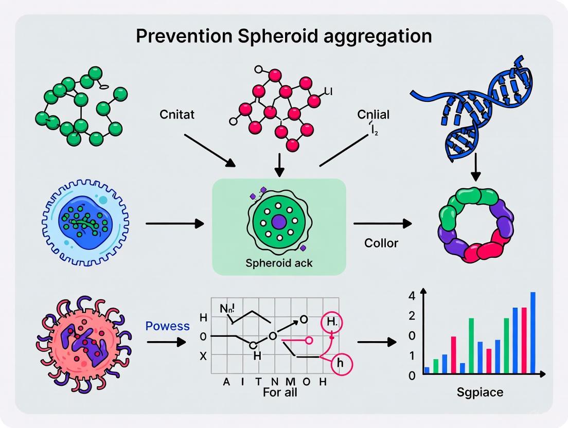
Preventing Spheroid Aggregation in Culture: A Complete Guide for Consistent 3D Models
This article provides researchers, scientists, and drug development professionals with a comprehensive guide to understanding, preventing, and managing unwanted spheroid aggregation in 3D cell culture.
Preventing Spheroid Aggregation in Culture: A Complete Guide for Consistent 3D Models
Abstract
This article provides researchers, scientists, and drug development professionals with a comprehensive guide to understanding, preventing, and managing unwanted spheroid aggregation in 3D cell culture. It covers the fundamental mechanisms driving aggregation, compares established and novel culture methodologies to minimize fusion, offers practical troubleshooting for common challenges, and outlines validation techniques to ensure model consistency for high-throughput screening and preclinical research.
Understanding Spheroid Aggregation: The Science Behind Cell-Cell Adhesion
Spheroid fusion is a fundamental process in tissue engineering where cellular aggregates self-assemble to form larger, more complex three-dimensional (3D) structures. This phenomenon replicates key aspects of tissue formation in development and regeneration. The process is primarily governed by cell adhesion molecules, particularly cadherins and integrins, and their interactions with the extracellular matrix (ECM). While essential for creating tissue-like constructs, uncontrolled spheroid aggregation in culture can lead to experimental variability, inconsistent morphology, and difficulties in reproducing results. A detailed understanding of the molecular mechanisms driving spheroid fusion is therefore critical for both harnessing its potential in tissue engineering and preventing undesirable aggregation in experimental cultures. This guide addresses the key molecular players and provides troubleshooting advice for managing spheroid aggregation in research settings.
Key Molecular Mechanisms in Spheroid Fusion
The Dynamic Roles of E-cadherin and β1-Integrin
Research has demonstrated that spheroid formation and fusion occur in distinct, sequential stages mediated by specific cell adhesion molecules. A dynamic analysis of hepatoma spheroid formation revealed a three-stage process [1]:
- Stage 1: Loose Aggregation via Integrins. In the initial phase, ECM fibers act as long-chain linkers for the attachment of dispersed single cells. This loose aggregation is primarily mediated by β1-integrins, which bind to ECM components [1].
- Stage 2: Delay and Cadherin Accumulation. Following initial aggregation, the cell clusters enter a delay period where compaction pauses. This stage is characterized by the accumulation of sufficient amounts of E-cadherin on the cell surfaces [1].
- Stage 3: Compaction via E-cadherin. The final stage involves a morphological transition from loose aggregates to compact, dense spheroids. This compaction is driven by strong homophilic (like-to-like) interactions between E-cadherin molecules on adjacent cells [1].
The functional roles of these molecules can be summarized as follows:
- E-cadherin: This calcium-dependent glycoprotein is the principal mediator of homotypic cell-cell adhesion. Its interactions are essential for establishing tight, compact spheroids and are a major force in the fusion process where two spheroids merge into one [1] [2].
- β1-Integrin: These molecules facilitate cell-matrix interactions by binding to proteins in the ECM, such as collagen and fibronectin. This initial anchorage is crucial for bringing cells into close proximity, enabling subsequent cadherin-mediated interactions [1].
The following diagram illustrates this sequential process and the distinct roles of E-cadherin and β1-integrin.
Figure 1: The Three-Stage Dynamics of Spheroid Formation and Fusion.
Extracellular Matrix (ECM) as a Structural and Signaling Platform
The ECM is not merely a passive scaffold but an active component that regulates spheroid fusion. Its roles include:
- Providing a Structural Framework: The ECM offers mechanical support and a substrate for integrin binding, which is the first step in cell aggregation [1] [2].
- Influencing Fusion Kinetics: The composition and concentration of the ECM directly impact the fusion process. Studies manipulating collagen content in magnetic cellular spheroids found that spheroids with low ECM concentrations exhibited more fusion and cellular intermixing over time compared to those with high ECM concentrations. Conversely, high ECM content promoted tissue contraction but limited the extent of fusion [3].
- Stimulating ECM Production: The integration of certain components, such as iron oxide magnetic nanoparticles, has been shown to increase collagen production over time, thereby influencing the spheroid's internal structure and its potential to fuse with others [3].
Troubleshooting Guides & FAQs
Frequently Asked Questions
Q1: My cell lines do not form compact spheroids and instead remain as loose aggregates. What can I do? A1: This is often due to insufficient E-cadherin-mediated compaction. You can try the following:
- Centrifugation: After seeding cells in a low-attachment plate, centrifuge the plate at a low speed (e.g., 150 x g for 5 minutes) to help cells quickly settle and initiate contact [4].
- Patience and Media Care: Some cell types require several days to form compact spheroids. For long-term culture, replace half of the media volume with fresh media every 2-3 days to maintain health without disturbing the aggregating cells [4].
- Use of Methylcellulose: Adding methylcellulose to the culture medium can increase viscosity and promote cell aggregation, leading to denser, more stable spheroids, as demonstrated in challenging lines like MiaPaCa-2 pancreatic cancer cells [5].
Q2: How can I prevent unwanted spheroid aggregation in my culture wells? A2: Unwanted aggregation typically occurs when multiple spheroids form per well instead of a single, uniform one.
- Use Confined Physical Spaces: Avoid culture vessels with large surface areas like T-flasks. Instead, use round-bottom (U-bottom) microplates or micro-patterned plates. These tools provide a confined space that promotes the formation of a single spheroid per well [4].
- Select Quality Low-Attachment Plates: Use reliable low cell attachment plates with superior surface modifications. These surfaces inhibit cell and protein attachment, forcing cells to aggregate into a single spheroid and reducing the formation of satellite colonies [4].
Q3: What are the best practices for handling spheroids to avoid disintegration during media changes or transfer? A3: Spheroids, especially loose ones, are fragile.
- Gentle Media Changes: Carefully tilt the microplate and aspirate half of the supernatant without touching the bottom. Gently dispense fresh media along the well wall to avoid shear stress [4].
- Use Wide-Bore Tips: When transferring spheroids, use wide-orifice pipette tips. These tips have a larger diameter that accommodates the spheroid, preventing damage, deformation, or disintegration during aspiration [4] [6].
Q4: Which cell lines are notoriously difficult for spheroid formation? A4: Several cell types present unique challenges [7]:
- Primary cells often struggle due to their limited lifespan and specific microenvironmental needs.
- Highly adherent cells may have difficulty detaching and forming proper 3D aggregates.
- Cells with complex signaling requirements or those that are slow-growing can fail to establish the necessary cell-cell interactions.
- A specific example is the MiaPaCa-2 pancreatic cancer cell line, which is known for forming unstable and weak spheroids that are difficult to manipulate [5].
Advanced Troubleshooting: Optimizing Experimental Variables
Several experimental parameters critically influence spheroid attributes and fusion behavior. Systematic analysis has quantified the impact of key variables, which are summarized in the table below [8].
Table 1: Impact of Key Experimental Variables on Spheroid Attributes
| Experimental Variable | Key Effect on Spheroids | Recommended Range for Stability | Mechanistic Insight |
|---|---|---|---|
| Serum Concentration | Dictates spheroid architecture, density, and viability. Lower concentrations (<5%) lead to shrinkage, reduced density, and increased cell death. | 10-20% FBS | Serum above 10% promotes the formation of dense spheroids with distinct necrotic, quiescent, and proliferative zones [8]. |
| Oxygen Level | Significantly affects size and necrosis. Hypoxia (3% O₂) reduces spheroid dimensions and cell viability while increasing necrotic signals. | Physioxia (e.g., 5-10% O₂) may better model in vivo conditions. | Lower oxygen tension creates a harsh microenvironment that limits proliferation in the core and promotes necrosis [8]. |
| Initial Seeding Cell Number | Directly controls final spheroid size. However, very high cell numbers can lead to structural instability and rupture. | Cell line dependent; must be optimized (e.g., 2,000-6,000 for some lines). | High cell numbers can exceed the spheroid's structural integrity, leading to rupture as necrotic and proliferative zones are expelled [8]. |
| Media Composition | Influences growth kinetics, viability, and death signals. Varying glucose, calcium, and other components can lead to statistically significant differences. | Must be optimized for cell type; DMEM/F12 showed lowest viability in one study. | Media components directly fuel metabolism and provide ions (e.g., Ca²⁺) that are essential for cadherin function and signaling [8]. |
The Scientist's Toolkit: Essential Reagents and Materials
Success in spheroid culture and fusion experiments relies on using the appropriate tools. The following table lists key materials and their functions.
Table 2: Essential Research Reagent Solutions for Spheroid Studies
| Tool Category | Specific Example | Function in Spheroid Research |
|---|---|---|
| Low-Attachment Plates | Nunclon Sphera plates; Corning Ultra-Low Attachment Plates | Provides a hydrophilic, electrostatically charged surface that inhibits cell attachment, promoting 3D aggregation into a single spheroid per well [4] [9]. |
| Wide-Bore Pipette Tips | Finntip Wide Orifice Tips | Enables gentle aspiration and transfer of fragile spheroids without causing damage or disintegration, preserving structural integrity [4] [6]. |
| Methylcellulose | Viscosity-enhancing agent (e.g., from Sigma-Aldrich) | Increases medium viscosity to limit cell movement, promote aggregation, and enhance the compactness and stability of forming spheroids [5]. |
| Extracellular Matrix (ECM) | Bovine Type I Collagen; Matrigel | Provides a biochemical and structural scaffold that supports cell-matrix interactions via integrins, influencing spheroid formation, compaction, and fusion kinetics [6] [3]. |
| Magnetic Nanoparticles | Iron Oxide (Fe₃O₄) Nanoparticles | When incorporated into spheroids, allows for non-invasive manipulation using magnetic fields to pattern and promote active fusion. Can also stimulate endogenous ECM production [3]. |
Experimental Protocols for Managing Spheroid Fusion
Protocol: Generating Uniform Spheroids to Minimize Variable Aggregation
This protocol leverages round-bottom, low-attachment plates for reproducible, single-spheroid formation [4].
- Cell Preparation: Harvest cells using standard trypsinization methods. Count and resuspend the cells in the appropriate growth medium, potentially supplemented with methylcellulose for problematic cell lines [5].
- Seeding: Calculate the volume needed for the desired cell seeding number (e.g., 2,000-6,000 cells/well for a 96-well plate). Pipette the cell suspension into each well of a round-bottom, low-attachment plate.
- Centrifugation: Seal the plate and centrifuge at 150 x g for 5 minutes. This step ensures all cells collect at the bottom of the well, initiating contact.
- Incubation: Place the plate in a 37°C, 5% CO₂ incubator. Spheroid formation can take from a few hours to several days.
- Media Maintenance: For long-term culture (>3 days), perform half-media changes every 2-3 days. Tilt the plate, carefully remove half the supernatant, and gently add fresh pre-warmed medium along the well wall.
Protocol: Modulating Spheroid Composition to Control Fusion
This protocol, adapted from research on magnetic spheroids, describes how manipulating ECM and cell number can directly influence fusion kinetics [3].
- Spheroid Fabrication: Use the hanging drop method. Combine equal volumes of:
- Cell suspension (e.g., Primary rat aortic SMCs)
- Iron oxide MNP suspension (for magnetic manipulation, optional)
- Collagen solution (e.g., Bovine Type I) at varying concentrations (e.g., 0.017 mg/ml for low ECM, 0.24 mg/ml for high ECM)
- Droplet Formation: Dispense 15 µl drops of the mixture onto the lid of a culture dish.
- Inversion and Incubation: Invert the lid and place it over a dish filled with PBS to maintain humidity. Incubate for 3 days to allow spheroid formation.
- Fusion Assay: Carefully collect the formed spheroids using wide-bore tips. For fusion analysis, place multiple spheroids in a capillary tube or pattern them in a ring formation on a magnetic plate.
- Observation: Monitor the fusion process over 24-48 hours using time-lapse microscopy or by fixing samples at set time points for morphological analysis.
The workflow for these protocols and the factors they control can be visualized as follows:
Figure 2: Experimental Workflows for Generating Uniform Spheroids and Controlling Fusion.
Frequently Asked Questions (FAQs)
1. What is the difference between normal spheroid formation and problematic uncontrolled aggregation? Normal spheroid formation is a controlled, self-assembly process driven by specific biological interactions, such as E-cadherin and integrin function, which results in consistent, reproducible 3D structures that mimic in vivo conditions [10]. Uncontrolled aggregation, in contrast, is a non-specific and unpredictable clumping of cells or particles. This can be caused by factors like improper cell suspension, suboptimal seeding density, or inconsistent matrix conditions, leading to high variability in size and shape, which compromises experimental reproducibility and data interpretation [11] [12].
2. How does uncontrolled aggregation specifically impact drug screening results? Uncontrolled aggregation can significantly increase apparent drug resistance in 3D cell culture models compared to conventional 2D models. This can lead to misleading efficacy data during high-throughput screening (HTS). For instance, drugs like sorafenib show different efficacy profiles in well-controlled 3D-aggregated spheroid models (3D-ASM) versus traditional 2D-HTS or poorly controlled 3D models, potentially causing researchers to overlook effective compounds [11].
3. What are the main biological drivers of controlled cell aggregation in spheroids? The primary mechanism involves cell adhesion molecules, especially E-cadherin (CDH1), which creates strong homophilic bonds between adjacent cells, initiating and stabilizing the spheroid [10] [13]. β1-integrin also plays a crucial early role by facilitating cell-extracellular matrix (ECM) interactions that support the aggregation process [10].
4. Beyond biology, what physical forces influence aggregation? The physical confinement provided by the matrix or culture environment is a critical factor. High matrix confinement can promote cell sorting within a heterogeneous spheroid, while reducing confinement can trigger a collective "unjamming" and burst-like migration of cells into the surrounding matrix. The balance between cell-generated forces and matrix resistance governs this behavior [13].
Troubleshooting Guide: Uncontrolled Aggregation
Problem: Inconsistent Spheroid Size and Shape
| Potential Cause | Diagnostic Check | Corrective Action |
|---|---|---|
| Non-uniform cell suspension [12] | Check if cells settle in the source tube during dispensing, leading to uneven seeding. | Ensure a homogeneous cell suspension by gently but consistently mixing the cell-hydrogel mixture during the entire seeding process [12]. |
| Suboptimal seeding density [12] | Observe a wide variation in final spheroid diameters across the culture platform. | Optimize and strictly control the initial cell seeding density. Higher densities lead to larger spheroids with greater nutrient needs [12]. |
| Improper ECM gelation [11] | Note inconsistent spheroid formation locations or irregular shapes. | Establish and meticulously control the temperature and timing for hydrogel gelation (e.g., Matrigel) to ensure all spheroids form under identical conditions [11]. |
Problem: Hypoxia and Necrosis in Spheroid Core
| Potential Cause | Diagnostic Check | Corrective Action |
|---|---|---|
| Excessive spheroid size [10] | Identify a core of dead cells using viability staining. | Control the initial cell number to limit spheroid size, preventing the formation of a diffusion-limited core that lacks nutrients and oxygen [10] [12]. |
| Infrequent media changes [12] | Measure low glucose/high waste levels in the culture medium. | Increase the frequency of media changes to ensure adequate nutrient delivery and waste removal, especially for larger spheroids [12]. |
Problem: Poor Experimental Reproducibility
| Potential Cause | Diagnostic Check | Corrective Action |
|---|---|---|
| Lot-to-lot reagent variation [14] | Note experimental drift when a new bottle of matrix or serum is introduced. | Document all reagent lot numbers. When a new lot must be introduced, perform a side-by-side comparison experiment to validate its performance before full-scale use [14]. |
| Insufficient protocol detail [15] | Lab members cannot reproduce each other's results. | Use Electronic Lab Notebooks (ELNs) to maintain detailed, version-controlled Standard Operating Procedures (SOPs). Deposit full protocols on repositories like Protocols.io for unambiguous sharing [15]. |
| Inconsistent environmental parameters [14] | Seasonal variations in room temperature affect "room temperature" incubation steps. | Use incubated environments for all steps requiring specific temperatures instead of relying on ambient lab conditions [14]. |
Research Reagent Solutions
The following table details key materials essential for controlling aggregation in spheroid culture.
| Item | Function in Controlling Aggregation |
|---|---|
| Ultra-Low Attachment (ULA) Surfaces [12] | Prevents cell attachment to the culture vessel surface, forcing cells to aggregate with each other to form spheroids in a controlled manner. |
| Matrigel/ECM Hydrogels [11] [12] | Provides a biologically relevant scaffold that entraps cells, promotes consistent cell-ECM interactions, and fixes spheroid position for reproducible analysis. |
| Hyaluronic Acid-based Matrices [10] | Serves as an alternative to agarose, particularly in cancer research, as it can interact with specific cell surface receptors to activate relevant signaling pathways. |
| Collagen-Alginate Hybrid Hydrogels [13] | Allows for independent tuning of mechanical stiffness (via calcium crosslinking of alginate) and biological adhesion (via collagen), offering precise control over matrix confinement. |
Experimental Workflows for Controlled Spheroid Formation
This protocol is designed for high reproducibility and high-throughput screening.
- Cell Preparation: Harvest cells (e.g., Hep3B or HepG2) using standard trypsinization and resuspend in culture medium.
- Hydrogel Mixing: Mix the cell suspension with an ECM hydrogel, such as Matrigel, on ice to prevent premature polymerization.
- Automated Dispensing: Use an automated 3D-cell spotter (e.g., ASFA Spotter) to dispense a precise, nanoliter-volume cell-hydrogel mixture uniformly onto a 384-pillar plate. This ensures a low coefficient of variation (CV <6%) between samples [11].
- Icing and Aggregation: Place the spotted pillar plate into a specially designed wet chamber. Perform an "icing step" to aggregate the cells in one spot through gravity before gelation.
- Controlled Gelation: Transfer the chamber to a 37°C incubator to initiate the "gelation step," solidifying the ECM and fixing the formed spheroid in a defined location.
- Drug Screening: Combine the pillar plate with a matching 384-well plate containing drug solutions for treatment and subsequent analysis.
This protocol allows precise control over matrix stiffness to study its effect on spheroid behavior.
- Spheroid Formation: Use inverse pyramidal PDMS microwells (e.g., AggreWell) treated with an anti-adherence rinse. Seed cells (~1000 cells/microwell) and centrifuge at 300 g for 5 minutes to aggregate cells at the bottom. Incubate overnight to form spheroids.
- Hydrogel Preparation: Prepare a hydrogel solution consisting of 3 mg/ml Type I rat tail collagen and 0.25% alginate. Keep on ice.
- Encapsulation: Harvest the pre-formed spheroids and mix them gently into the collagen-alginate solution. Pipet the mixture into a culture plate and incubate at 37°C for 1 hour to allow collagen polymerization.
- Stiffness Tuning: After imaging (Day 0), independently modulate the alginate crosslinking density and final hydrogel stiffness by adding specific concentrations of CaCl₂ to the cell culture medium.
- Monitoring and Analysis: Culture the spheroids and image them regularly (e.g., by Day 4) to monitor cell sorting and invasion behaviors in response to the tuned matrix confinement.
Signaling Pathways and Logical Workflows
Diagram: Mechanisms of Spheroid Formation and Disruption
The diagram below illustrates the key drivers of controlled spheroid formation and how common experimental errors can lead to uncontrolled aggregation.
Diagram: Experimental Workflow for a Robust 3D-ASM
This flowchart outlines the optimized protocol for creating a highly reproducible 3D-Aggregated Spheroid Model (3D-ASM) for high-throughput drug screening [11].
Core Concepts: SCDS vs. MCS
Single-Cell-Derived Spheroids (SCDS) are initiated from isolated single cells and form through clonal expansion. Multicellular Spheroids (MCS), also known as multicellular tumor spheroids (MCTS), are generated from pre-aggregated clusters of cells [16]. The choice between them fundamentally shapes your experimental outcomes.
The table below summarizes the core characteristics, mechanisms, and primary applications of each method.
| Feature | Single-Cell-Derived Spheroid (SCDS) | Multicellular Spheroid (MCS) |
|---|---|---|
| Starting Material | Isolated single cells [16] | Pre-formed cellular aggregates [16] |
| Formation Mechanism | Clonal expansion and self-renewal [16] | Cell aggregation and self-assembly [10] |
| Key Readouts | Clonogenicity, stem cell potential, self-renewal [16] | Cell-cell interaction, drug penetration, gradient formation [10] |
| Primary Applications | Cancer stem cell (CSC) enrichment, potency assays, studying tumor initiation [16] | Tumor biology modeling, drug efficacy and penetration studies [10] [17] |
Method Selection Guide
Your research question should guide the selection of a spheroid culture method. The following table outlines appropriate choices based on common experimental goals.
| Research Goal | Recommended Method | Rationale |
|---|---|---|
| Cancer Stem Cell (CSC) Enrichment | Single-Cell-Derived Spheroid (SCDS) | SCDS culture selectively promotes the expansion of cells with self-renewing capability, leading to higher expression of CSC markers and pluripotent genes compared to MCS [16]. |
| Drug Sensitivity & Resistance Testing | Context-Dependent | For bulk tumor response, use MCS. To specifically target the resistant CSC subpopulation, use SCDS. One study showed 5637 SCDS exhibited increased cisplatin resistance and upregulation of the ABCG2 drug efflux gene [16]. |
| Tissue Engineering & Transplantation | Multicellular Spheroid (MCS) | MCS provide advantageous 3D cell-cell and cell-matrix interactions that enhance differentiation potential and improve tissue formation upon transplantation (e.g., for cartilage, bone, nerve) [10] [17]. |
| Basic Tumor Biology & Microenvironment | Multicellular Spheroid (MCS) | The structure of MCS naturally develops physiological gradients (nutrients, oxygen, waste) and mimics in vivo cell-cell interactions, making them ideal for studying necrosis, hypoxia, and proliferation gradients [10]. |
Detailed Experimental Protocols
Protocol 1: Multicellular Spheroid (MCS) Culture via Hanging Drop
This method is well-suited for generating uniform spheroids from a defined number of cells [16].
- Step 1: Cell Suspension Preparation. Trypsinize your monolayer culture (e.g., 5637 or HT-1376 bladder cancer cell lines) to obtain a single-cell suspension. Prepare a suspension of 200,000 cells/ml in your spheroid culture medium [16].
- Step 2: Hanging Drop Setup. Using a multichannel pipette, dispense 25 µL droplets (containing ~5,000 cells) onto the lid of a sterile Petri dish. Carefully invert the lid and place it over the bottom of the dish, which can be filled with PBS to maintain humidity. Culture for 2 days to allow for initial spheroid formation [16].
- Step 3: Transfer and Long-Term Culture. After 2 days, transfer the formed MCSs to a 24-well ultralow attachment (ULA) plate containing 500 µL of fresh spheroid media. Culture for an additional 8 days, replenishing the media every 3 days [16].
Protocol 2: Single-Cell-Derived Spheroid (SCDS) Culture in ULA Plates
This protocol is designed for CSC enrichment by promoting the clonal expansion of individual cells [16].
- Step 1: Low-Density Seeding. Prepare a single-cell suspension as in Protocol 1. Seed cells at a low density (e.g., 1,000 - 5,000 cells per well) into a 96-well U-bottom ultralow attachment (ULA) plate. The low density is critical to ensure spheroids originate from a single cell [16] [4].
- Step 2: Centrifugation. Centrifuge the plate at a low speed (e.g., 150 x g for 5 minutes) to gently pellet the cells at the bottom of the U-bottom well, promoting initial contact and aggregation [4].
- Step 3: Spheroid Formation and Maintenance. Culture the plate for 7-14 days. For slow-forming spheroids, replace half of the media volume with fresh, pre-warmed media every 2-3 days, taking care not to disturb the forming spheroids [4].
Troubleshooting Common Spheroid Culture Challenges
FAQ 1: How can I consistently grow uniform spheroids for repeatable results?
The most reliable way to control spheroid size and uniformity is by using low cell attachment (LCA) plates with U-bottom wells and standardizing the initial cell seeding density [4]. These plates inhibit cell attachment to the plastic surface, forcing cells to aggregate into a single spheroid per well. The U-bottom geometry naturally guides cells to the center of the well. Consistency is key: ensure your single-cell suspension is homogeneous before seeding and consider low-speed centrifugation (e.g., 150 x g for 5 minutes) after seeding to gently pellet all cells to the bottom of the well, initiating uniform contact [4].
FAQ 2: What should I do if my cell lines do not form compact spheroids?
Not all cell types readily form tight spheroids. If you encounter this issue:
- Verify Surface Quality: Ensure you are using high-quality, ultralow attachment plates. Imperfections in the surface coating can allow cells to attach and spread [4].
- Optimize Media: For stubborn cell types, try supplementing the media with growth factors or reducing the serum concentration to discourage adhesion and encourage cell-cell interactions.
- Allow More Time: Some cell lines require several days to form compact spheroids. Be patient and replace half the media volume with fresh media every 2-3 days to maintain culture health during this period [4].
FAQ 3: How do I handle and perform media changes without damaging spheroids?
Manual handling requires care to prevent spheroid disintegration.
- For Media Changes: Tilt the microplate at a slight angle. Slowly aspirate the supernatant from the meniscus, ensuring the pipette tip does not touch the bottom of the well or the spheroid. Gently dispense fresh media along the side of the well wall [4].
- For Spheroid Transfer: Use wide-bore or wide-orifice pipette tips. These tips have a larger diameter that accommodates the spheroid without causing shear stress or physical damage during aspiration and dispensing [4].
FAQ 4: Can I use my standard 2D cell viability and staining assays on spheroids?
Yes, but protocols require significant optimization to account for the 3D structure's limited reagent penetration [4]. The table below provides general guidance for adapting common assays.
| Assay Type | Example Reagent | 2D Protocol | 3D Protocol Adjustment |
|---|---|---|---|
| Cell Viability | PrestoBlue HS / alamarBlue HS | Standard concentration, 30-60 min incubation | Increased incubation time (e.g., 2-4 hours); may require rotation for penetration [4]. |
| Immunostaining | Antibodies | Standard concentration, 30-60 min incubation | Higher antibody concentration (e.g., 2-5X), longer incubation (overnight), and use of tissue clearing reagents [4]. |
| Apoptosis | CellEvent Caspase-3/7 | 1X, 30 min | Lower reagent concentration (e.g., 1/3X) with longer incubation (e.g., 2 hours) [4]. |
The Scientist's Toolkit: Essential Research Reagents
The following table lists key materials and reagents essential for successful spheroid culture, based on protocols from the search results.
| Item | Function & Description |
|---|---|
| Ultra-Low Attachment (ULA) Plates | Cultureware with a coated surface that inhibits cell attachment, forcing cells to aggregate and form spheroids. Crucial for both SCDS and MCS methods [16] [4]. |
| Serum-Free Spheroid Media | Defined media (e.g., RPMI 1640 supplemented with B27, EGF, bFGF) that supports stemness and proliferation without inducing differentiation, essential for CSC enrichment [16]. |
| Wide-Bore Pipette Tips | Pipette tips with a larger orifice to prevent physical damage and shear stress when transferring intact spheroids [4]. |
| Tissue Clearing Reagents | Chemical solutions that reduce light scattering within the spheroid, enabling deeper penetration of antibodies and dyes for high-quality 3D imaging [4]. |
| Extracellular Matrix (ECM) Proteins | Proteins like agarose or hyaluronic acid used in liquid overlay techniques to create a non-adhesive surface for spheroid formation [10]. |
Methodologies to Control Aggregation: From Standard Plates to Advanced Techniques
Core Principles: How ULA Surfaces Prevent Unwanted Fusion
What is the fundamental mechanism by which ULA surfaces prevent spheroid fusion? ULA plates are engineered with a specialized ultra-hydrophilic polymer coating that creates a surface where cell-substrate adhesion is minimized. This forces cells to rely on cell-cell adhesion for survival, promoting spontaneous self-assembly into single, discrete spheroids. When the adhesive forces between cells are stronger than the forces between the cells and the plate surface, cells aggregate freely. The consistent, non-adhesive surface ensures that once formed, spheroids remain as separate entities and do not fuse together randomly, which is crucial for experimental reproducibility [18] [19].
How do different ULA surface chemistries achieve this effect? While the core principle is minimizing adhesion, different proprietary chemistries are used to create the ULA surface. The table below summarizes the mechanisms of several key surface types.
Table 1: Comparison of ULA Surface Chemistries and Their Properties
| Surface Chemistry/Coating | Core Mechanism | Key Characteristics | Reported Outcomes |
|---|---|---|---|
| Standard ULA Polymer [18] [19] | Ultra-hydrophilic polymer creates a hydration layer, preventing protein adsorption and cell attachment. | Pre-coated, ready-to-use; available in U-bottom (looser) and V-bottom (tighter) well shapes. | Enables uniform single spheroid formation per well; prevents random fusion. |
| N-hexanoyl glycol chitosan (HGC) [20] | Thermosensitive hydrogel layer providing an ultra-low attachment (ULA) surface. | Can be incorporated with a micromesh lattice structure to control spheroid uniformity and prevent fusion. | Supports formation of regular-sized 3D spheroids and enables spatial cell reorganization. |
| Polypeptide Polyelectrolyte Multilayer (PEM) [21] | Layer-by-layer deposition of poly-L-lysine (PLL) and poly-L-glutamic acid (PLGA). | Promotes cell attachment but restricts cell spreading and aggregate fusion; allows real-time monitoring. | Enhances spheroid formation from single cells and upregulates stemness markers. |
| Chitosan Nano-deposit [22] | Polysaccharide coating derived from chitin, providing a highly biocompatible, non-adhesive surface. | Used to induce 3D sphere formation of stem cells, enhancing mitochondrial function and stemness. | Promotes formation of compact spheroids with unique metabolic and functional properties. |
Troubleshooting Guide: FAQs on Preventing Spheroid Fusion
FAQ 1: My spheroids are still fusing together in ULA plates. What are the main causes? Unwanted fusion typically results from two factors: excessive well size or high cell seeding density. If the well is too large for the number of cells seeded, multiple, smaller aggregates can form and later fuse. Conversely, a high cell density in a standard well can lead to the formation of a single, but overly large and unstable spheroid that may fuse with neighbors if it breaks apart. Furthermore, the use of enzyme-based cell detachment (e.g., trypsin) can damage cell surface proteins like cadherins and integrins that are critical for proper aggregation, leading to irregular fusion patterns [23].
FAQ 2: How can I optimize my protocol to prevent fusion from the start? To prevent fusion, a multi-faceted approach is recommended:
- Select the Appropriate Well Bottom Shape: V-bottom plates are designed to guide all cells to a single point of contact, forcing the formation of one tight, compact spheroid per well and is the preferred choice for cell types prone to forming multiple aggregates [18].
- Optimize Cell Seeding Density: This is the most critical parameter. Refer to the table below for general guidance based on common applications.
- Use Enzyme-Free Cell Detachment: Where possible, use ultrasound-based detachment methods. Studies show this preserves cell surface proteins (e.g., integrin α5, M-cadherin), leading to faster and more robust aggregation, reducing the window for irregular fusion [23].
Table 2: Recommended Seeding Densities for Common ULA Applications
| Application / Spheroid Type | Example Cell Line / System | Recommended Seeding Density | Platform / Format |
|---|---|---|---|
| Cardiac Spheroids [19] | iPSC-derived Cardiomyocytes, Fibroblasts, Endothelial Cells | Optimized ratios (e.g., 2:1:1 or 4:1); thousands of cells per spheroid. | U-bottom ULA plates, AggreWell |
| Epithelial Spheroids [24] | HaCaT Keratinocytes | • 5.0×10⁴ cells/well (96-well microcavity)• 5.0×10³ cells/well (96-well U-bottom)• 8.0×10³ cells/well (6-well ULA plate) | Elplasia 96-well, BIOFLOAT 96-well, 6-well ULA plates |
| Cancer Spheroids (MCTS) [25] | Colorectal Cancer (CRC) Cell Lines (e.g., HCT116, SW480) | Cell line-dependent; requires optimization for compactness. | U-bottom ULA plates, methylcellulose-supplemented media |
| Immune Organoid Mimicry [20] | Human B cells & MS5-CD40L Stromal Cells | Co-culture system on HGC-coated ULA lattice plates. | Custom HGC-coated ULA lattice plates |
| Mesenchymal Stem Cell (MSC) Aggregates [26] | Human MSCs (hMSCs) | 500 - 5,000 cells/well | U-bottom 96-well ULA plate |
FAQ 3: Beyond basic plates, what advanced tools can help control fusion and improve uniformity? For applications requiring extreme uniformity and a guarantee of a single spheroid per well, consider plates with microcavities or micromesh structures. The Elplasia plate contains hundreds of micro-wells within a single standard well, producing a large number of highly uniform spheroids simultaneously [24]. Similarly, incorporating a micromesh lattice onto an HGC-coated surface has been shown to reduce intrinsic heterogeneity and prevent the random fusion of spheroids by physically separating them during formation [20]. For scaffold-based approaches, adding viscosity-enhancing agents like methylcellulose to the medium can improve spheroid circularity and compaction, reducing the tendency for loose aggregates to fuse [19].
Experimental Protocols & Methodologies
This protocol outlines a comparative approach for generating spheroids in high-throughput and low-throughput formats.
Key Reagents:
- Cell Line: Immortalized human keratinocytes (HaCaT).
- Culture Medium: Dulbecco’s Modified Eagle’s Medium (DMEM) supplemented with 10% Fetal Bovine Serum (FBS), penicillin/streptomycin, and amphotericin B.
- ULA Plates: 96-well U-bottom BIOFLOAT plate, 96-well black round-bottom microcavity Elplasia plate, and 6-well ULA plates.
Methodology:
- Cell Preparation: Culture HaCaT cells to 70-80% confluence. Detach using 0.05% trypsin-EDTA and resuspend in complete medium.
- High-Throughput Formation (for uniform spheroids):
- Elplasia Plate: Seed 5.0×10⁴ cells in 50 µL per well.
- BIOFLOAT Plate: Seed 5.0×10³ cells in 50 µL per well.
- Incubate plates undisturbed for 48 hours at 37°C and 5% CO₂.
- Low-Throughput Formation (for heterogeneous populations):
- Seed 8.0×10³ cells in 2 mL per well of a 6-well ULA plate.
- To enhance stemness and holosphere formation, add 5 µM ROCK1 inhibitor (Y-27632) to the medium.
- Incubate for 5 days without a medium change.
- Analysis: Image spheroids using an automated high-content imager. Quantify spheroid number, diameter, and circularity using analysis software (e.g., MetaXpress). Classify spheroids by size and morphology (holospheres, merospheres, paraspheres).
This protocol leverages ultrasound detachment to preserve surface proteins and accelerate aggregation.
Key Reagents:
- Cell Line: C2C12 myoblasts or other relevant lines.
- Device: Ultrasound detachment device (e.g., from Canon Inc.).
- ULA Plates: Standard 96-well U-bottom ULA plates.
Methodology:
- Cell Detachment: Culture cells until ready for passage. Instead of trypsin, use the ultrasound detachment device at the optimized voltage (e.g., 150 V for the cited device) to detach cells in their native medium.
- Cell Seeding: Count the detached cells and seed them into the ULA plates at the desired density.
- Spheroid Formation: Incubate the plates undisturbed. Monitor the aggregation process. Studies show that ultrasound-detached cells have a shorter aggregation phase (9.5 hours vs. 12.3 hours for trypsin) due to higher concentrations of adhesion-related proteins (fibronectin, integrin α5, M-cadherin).
- Validation: Compare the size, morphology, and protein expression of the resulting spheroids against those formed from enzyme-detached cells using Western blotting or functional assays.
The Scientist's Toolkit: Key Research Reagent Solutions
Table 3: Essential Materials for ULA Spheroid Culture
| Reagent / Material | Function / Application | Example Product / Component |
|---|---|---|
| ULA Plates (U-bottom) | Promotes formation of a single, central spheroid per well; ideal for high-throughput screening. | PrimeSurface 96U, Nunclon Sphera [18] [19] |
| ULA Plates (V-bottom) | Forces tighter cell aggregation for more compact spheroid formation. | PrimeSurface 96V [18] |
| ULA Plates (Microcavity) | Generates hundreds of uniform spheroids per well for high-content analysis. | Elplasia plates [24] |
| ROCK Inhibitor (Y-27632) | Enhances cell survival after passaging, promotes stemness, and increases holosphere formation in scaffold-free cultures. | Tocris, others [24] [27] |
| Methylcellulose | Increases medium viscosity to enhance spheroid compaction and circularity; reduces image blur. | Sigma-Aldrich, others [19] |
| Extracellular Matrix (ECM) | Provides a scaffold for embedded 3D culture or for assessing spheroid outgrowth and invasion. | Matrigel, Collagen Type I [24] [25] |
| Chitosan Coating | Biocompatible polysaccharide coating used to induce 3D sphere formation and enhance stem cell properties. | Synthesized from glycol chitosan and hexanoic anhydride [20] [22] |
Visualizing Spheroid Formation and Fusion Prevention
The following diagrams illustrate the core concepts and workflows for successful, fusion-free spheroid culture.
Diagram 1: ULA Surface Chemistry Mechanism
Diagram 2: Optimized Workflow for Fusion-Free Spheroids
Troubleshooting Guide: Addressing Common Experimental Challenges
Spheroid Formation Issues
Problem: Failure to Form Compact Spheroids
- Potential Cause 1: Insufficient cell seeding density or incorrect centrifugation.
- Solution: Optimize the initial cell seeding number. For plates, centrifuge the cell-seeded plate at a low speed (e.g., 150 x g for 5 minutes) to help cells settle quickly at the bottom of the wells and initiate aggregation [4].
- Potential Cause 2: Cell type requires longer aggregation time.
- Solution: Certain cell types may need several days to form compact spheroids. During this period, replace half of the culture media with fresh media every 2-3 days to maintain culture health without fully disrupting the forming aggregates [4].
Problem: Inconsistent Spheroid Size and Shape
- Potential Cause 1: Inhomogeneous cell suspension or uneven seeding.
- Solution: Ensure a single, well-dispersed cell suspension before seeding. For hanging drop methods, use pipetting aids or specialized matrices like the SpheroMold to standardize droplet volume and placement [28].
- Potential Cause 2: Variable culture conditions across the platform.
- Solution: Use commercially available low-cell-attachment plates with proven surface modifications to ensure uniform inhibition of cell adhesion across all wells, minimizing satellite colonies and improving homogeneity [4].
Spheroid Handling and Manipulation Issues
Problem: Spheroids Are Aspirated or Disrupted During Media Changes
- Potential Cause: Manual pipetting is too forceful or the tip is placed too close to the spheroid.
- Solution:
- Manual Method: Carefully tilt the microplate and slowly aspirate the supernatant from the upper part of the well, avoiding the bottom where the spheroid rests. Dispense fresh media gently along the well wall [4].
- Automated Method: Employ an automated liquid handling system with optimized aspiration rates. Smaller spheroids require slower aspiration rates to prevent accidental removal [29].
- Solution:
Problem: Spheroids Break Apart During Transfer
- Potential Cause: Using standard pipette tips with small orifices.
- Solution: Always use wide-bore or wide-orifice pipette tips, which have a larger diameter to accommodate the spheroid without causing shear stress or physical damage during transfer [4].
Spheroid Coalescence in Hanging Drop Arrays
Problem: Droplets Merge During Plate Handling or Inversion
- Potential Cause: Droplets are too close together or the plate is handled roughly.
- Solution: Implement a physical barrier between droplets. The SpheroMold, a PDMS-based matrix attached to the Petri dish lid, features precisely positioned holes that confine individual droplets. This design prevents droplet fusion during inversion and simplifies manipulation, enabling higher density spheroid production [28].
Problem: High Evaporation Rate in Hanging Drops
- Potential Cause: Insufficient humidity in the incubation chamber.
- Solution: Create a humidified chamber. When using a hanging drop plate, place it over a reservoir, such as a 6-well plate filled with sterile autoclaved water (4-5 mL per well). Adding 800–1000 μL of sterile water around the rim of the hanging drop plate further minimizes evaporation [30].
Analysis and Assay Complications
Problem: Poor Penetration of Assay Reagents (e.g., Viability Dyes, Antibodies)
- Potential Cause: The dense, thick nature of 3D spheroids creates a diffusion barrier.
- Solution: Optimize protocols for 3D cultures. This typically involves:
- Increasing Concentration: Using a higher concentration of the probe (e.g., 2X for MitoTracker Orange) [4].
- Prolonging Incubation: Extending incubation times to allow for deeper penetration (e.g., 2 hours for caspase 3/7 reagent instead of 30 minutes) [4].
- Using Clearing Reagents: Employing tissue-clearing reagents specifically designed for 3D cultures to enhance reagent penetration and imaging quality [4].
- Solution: Optimize protocols for 3D cultures. This typically involves:
Problem: Spheroids Move Out of Imaging Field of View
- Potential Cause: Liquid flow during plate handling moves unattached spheroids.
- Solution: During imaging, capture a montage of images in the x- and y-axes. Combining this with z-stacking (imaging multiple focal planes) ensures the entire spheroid is captured and can be accurately analyzed [29].
Frequently Asked Questions (FAQs)
Q1: How can I consistently grow uniform spheroids to get repeatable results? The easiest way is to control the initial cell seeding density within a confined physical space that promotes the formation of a single spheroid per well [4]. While adjusting cell number is key, the choice of platform is critical for reproducibility. Low-cell-attachment plates with round bottoms are highly effective for generating uniform, single spheroids in a user-friendly manner. The hanging drop method also produces spheroids of relatively uniform size and shape, and modernizations like the SpheroMold enhance this consistency [28] [4].
Q2: What are the main advantages of the hanging drop method over other techniques? The hanging drop method is a scaffold-free, cost-effective technique that minimizes mechanical stress on cells, allowing for natural self-assembly [28]. It facilitates the formation of compact spheroids with relatively uniform size and is particularly noted for creating hypoxic cores, making it a suitable model for cancer research [31]. Its theoretical underpinning relies on gravity-enforced self-assembly [31].
Q3: My spheroids need long-term culture. How can I manage media exchanges without damaging them? Frequent, gentle media exchange is essential. For high-throughput workflows, automated systems like the AMX (Automated Media Exchange) module are ideal. They control aspiration rates and dispense liquid slowly to protect spheroids [29]. For manual protocols, perform half-media changes carefully by tilting the plate and pipetting along the wall to avoid disturbing the spheroid [4].
Q4: Can I use my standard 2D cell culture assays (viability, immunostaining) with 3D spheroids? Yes, but protocols require modification. Assays designed for 2D monolayers do not penetrate 3D structures effectively. You will typically need to increase reagent concentrations and extend incubation times (see Table 2). For immunostaining, the use of tissue-clearing reagents is highly recommended to improve antibody penetration and image resolution [4].
Q5: How do low-cell-attachment plates support spheroid formation? These plates are coated with a hydrogel or have a covalently modified surface that inhibits the attachment of cells and ECM proteins. This forces the cells to aggregate and interact with each other in three dimensions, leading to spheroid formation. The round-bottom geometry of the wells further assists in the formation of a single, central spheroid [4].
Experimental Workflow Visualization
The following diagram illustrates the core procedural pathways for creating spheroids using the two primary methods discussed in this guide.
Diagram 1: Comparative Workflow for Spheroid Formation Methods.
Research Reagent and Material Solutions
The table below lists key materials and reagents essential for successfully conducting spheroid formation experiments via hanging drop and micro-patterned methods.
Table 1: Essential Research Reagents and Materials
| Item | Function/Application | Key Considerations |
|---|---|---|
| Hanging Drop Plates | Provides a structured platform for creating multiple suspended droplets for spheroid formation [30]. | Available in 96- and 384-well formats. Requires pre-treatment with Pluronic acid or similar to prevent spheroid adherence [30]. |
| Low-Cell-Attachment Microplates (e.g., Nunclon Sphera) | Inhibits cell attachment, promoting 3D aggregation into a single spheroid per well. User-friendly and compatible with HTS [4]. | Choose round (U) bottom wells for single spheroid formation. Surface modification quality is critical for performance and reproducibility [4]. |
| AggreWell Microwell Plates | Microwell culture plates designed for easy, reproducible production of large numbers of embryoid bodies and spheroids [32]. | Useful for high-throughput, standardized spheroid generation. |
| SpheroMold | A 3D-printed PDMS support for Petri dish lids that prevents droplet coalescence in hanging drop methods, increasing throughput and simplifying handling [28]. | Allows for a larger medium volume per drop, reducing the frequency of medium exchange [28]. |
| Wide-Bore Pipette Tips | Transfer of formed spheroids without causing damage or shearing [4]. | Essential for harvesting spheroids from wells or droplets without disrupting their structure. |
| 3D-Clearing Reagents (e.g., CytoVista) | Enhances penetration of dyes and antibodies into the spheroid core for improved imaging and analysis [4]. | Crucial for obtaining high-quality fluorescence images from the interior of large, dense spheroids. |
| Pluronic F-127 / F-68 | A surfactant used to coat surfaces, preventing protein adsorption and cell attachment [30]. | Used to treat hanging drop plates and other surfaces to create a non-adhesive environment. |
| Automated Media Exchange System (e.g., AMX Module) | Performs gentle, automated media changes and dosing for long-term spheroid assays, minimizing human error and spheroid loss [29]. | Ideal for high-throughput labs; allows optimization of aspiration rates for different spheroid sizes [29]. |
Protocol Modification Guide for Enhanced Assays
Table 2: Protocol Adjustment Guide for 3D Spheroid Assays
This table summarizes common adjustments needed when adapting 2D cell culture protocols for 3D spheroids.
| Assay / Reagent | Typical 2D Protocol | Recommended 3D Protocol Adjustment | Rationale |
|---|---|---|---|
| Cell Viability (PrestoBlue/alamarBlue) | 30-60 min incubation | Incubation Time: 2-4 hours (or longer). Consider rotation. [4] | Longer diffusion time required for reagents to penetrate dense core. |
| Apoptosis (Caspase 3/7) | 1X concentration, 30 min [4] | Concentration: 1/3X. Time: 2 hours [4] | Optimized for better signal-to-noise ratio in 3D. |
| Mitochondrial Health (MitoTracker) | 1X concentration, 30 min [4] | Concentration: 2X. Time: 1 hour [4] | Increased dye concentration to ensure sufficient labeling throughout spheroid. |
| Immunofluorescence | Standard protocol (few hours) | Incubation Time: Extend significantly (overnight for antibodies). Use clearing reagents. [4] | Antibodies require extended time to diffuse into the spheroid. Clearing reduces light scattering. |
| Imaging | Single focal plane image | Z-stacking and X-Y montage imaging [29] | Captures the entire 3D structure and accounts for spheroid movement in the well. |
Troubleshooting Guides & FAQs
FAQ: General Concepts
Q1: What is the primary advantage of using scaffold-based systems over suspension cultures for spheroid formation? A1: Scaffold-based systems provide a physical, extracellular matrix (ECM)-mimetic structure that prevents uncontrolled spheroid aggregation and fusion. This leads to more uniform spheroid size and shape, enhances reproducibility for drug screening, and allows for better nutrient/waste diffusion compared to large, fused aggregates in suspension.
Q2: How does the choice between natural (e.g., Matrigel, collagen) and synthetic (e.g., PEG, PLA) hydrogels impact my experiment? A2:
- Natural Hydrogels: (e.g., Matrigel, Collagen I, Alginate) Provide bioactive motifs that support cell adhesion, proliferation, and signaling. However, they can have batch-to-batch variability and may contain undefined growth factors.
- Synthetic Hydrogels: (e.g., Polyethylene Glycol (PEG), Polylactic Acid (PLA)) Offer high reproducibility, tunable mechanical properties, and defined chemistry. They often require functionalization with adhesion peptides (e.g., RGD) to support cell attachment.
Q3: My spheroids are not forming. What are the most common causes? A3:
- Incorrect Cell Seeding Density: Too few cells will not aggregate; too many will form large, necrotic clumps.
- Unsuitable ECM Concentration: A soft hydrogel may not provide sufficient support, while a very stiff one can impede cell migration and aggregation.
- Lack of Cell-ECM Adhesion: Certain cells require specific adhesion ligands (like RGD) to be incorporated into synthetic hydrogels to initiate aggregation.
- Poor Cell Viability: Low viability at seeding will prevent the cell-cell interactions necessary for spheroid formation.
Troubleshooting: Specific Experimental Issues
Q4: I am observing a high degree of size variability in my spheroids within a hydrogel-embedded culture. How can I improve uniformity? A4: High size variability is often a result of uneven cell distribution during hydrogel polymerization.
- Solution 1: Optimize the mixing and polymerization protocol. Ensure cells are in a single-cell suspension and thoroughly mixed with the hydrogel precursor solution before gelation. Avoid introducing air bubbles.
- Solution 2: Use a microwell scaffold system. Platforms like AggreWell or micro-molded hydrogels provide physically distinct compartments, forcing a defined number of cells per well to form spheroids of highly uniform size.
- Solution 3: Titrate the cell seeding density. Perform a density gradient experiment to identify the optimal number of cells per unit volume of hydrogel for your specific cell type.
Q5: My spheroids are contracting and degrading the surrounding hydrogel over time. Is this a problem? A5: This indicates active cell-mediated remodeling of the ECM, which can be a feature or a problem depending on your research goal.
- If you need stable, isolated spheroids: This is a problem. To mitigate it:
- Increase the crosslinking density of your hydrogel to make it more resistant to degradation.
- Use a protease-resistant hydrogel material (e.g., some PEG-based hydrogels).
- Incorporate protease inhibitors (e.g., GM6001, a broad-spectrum MMP inhibitor) into the culture medium.
- If you are studying invasion or metastasis: This is a desired feature, mimicking the process of cells breaking down the basement membrane.
Q6: How can I efficiently and safely extract spheroids from a scaffold or hydrogel for downstream analysis (e.g., sequencing, histology)? A6: Extraction is a critical and delicate step.
For natural hydrogels (e.g., Collagen, Matrigel):
- Enzymatic Degradation: Incubate the hydrogel with a solution of collagenase (for collagen) or dispase (for Matrigel). Use the lowest effective concentration and shortest incubation time to preserve spheroid integrity and cell surface markers.
- Procedure: Wash spheroids with PBS, then add the pre-warmed enzyme solution. Gently agitate at 37°C. Monitor under a microscope every 5-10 minutes. Once the hydrogel is dissolved, carefully collect spheroids by gentle centrifugation and wash with culture medium to neutralize the enzyme.
For synthetic hydrogels (e.g., PEG with degradable linkers):
- Specific Degradation: Use a hydrogel designed with cleavable crosslinkers (e.g., peptides sensitive to MMPs, or crosslinkers that degrade in response to light or a specific chemical like dithiothreitol (DTT)). This allows for highly specific and gentle release.
Table 1: Troubleshooting Common Spheroid Culture Problems
| Problem | Potential Cause | Solution |
|---|---|---|
| No Spheroid Formation | - Cell density too low- Excessively stiff hydrogel- Lack of adhesion ligands | - Titrate cell seeding density (e.g., 1,000-10,000 cells/spheroid).- Use a softer hydrogel (e.g., reduce PEG-DA % from 10% to 5%).- Functionalize hydrogel with RGD peptide. |
| High Size Variability | - Uneven cell distribution- Aggregation before gelation | - Use a microwell scaffold system.- Work quickly and use cold precursors to delay gelation until plated. |
| Necrotic Core | - Spheroids too large- Hydrogel too dense, impeding diffusion | - Form smaller spheroids.- Use a more porous hydrogel or reduce hydrogel concentration. |
| Hydrogel Degradation | - High protease activity from cells | - Use a protease-resistant polymer.- Add MMP inhibitors to the culture medium. |
| Poor Viability Post-Extraction | - Harsh enzymatic treatment | - Optimize enzyme type, concentration, and duration.- Use a degradable synthetic hydrogel for gentler release. |
Experimental Protocols
Protocol 1: Forming Spheroids in a Collagen I Hydrogel
Objective: To create uniform, isolated spheroids embedded within a 3D collagen I matrix.
Materials:
- Rat tail Collagen I, high concentration (e.g., ~8-10 mg/mL)
- Sterile 0.1M Acetic Acid
- 10x Phosphate Buffered Saline (PBS)
- 1M Sodium Hydroxide (NaOH)
- Single-cell suspension of your cell type
- Complete culture medium
- Cell culture plates (e.g., 24-well plate)
Method:
- Preparation: Chill all components and tubes on ice. Prepare a neutralization solution by mixing 10x PBS and 1M NaOH in a ratio of 8:1 (v/v).
- Calculate the Mix: Determine the final volume and collagen concentration (typically 2-4 mg/mL). For 1 mL of 2 mg/mL collagen gel in one well of a 24-well plate:
- Collagen I Stock (8 mg/mL): 250 µL
- 10x PBS: 100 µL
- Neutralization Solution: ~25 µL (volume may need optimization)
- Cell Suspension in Medium: 625 µL (containing desired number of cells)
- Mixing and Seeding:
- In a cold tube, combine the collagen stock and 10x PBS.
- Slowly add the calculated volume of neutralization solution and mix gently. The solution should turn pink/orange, indicating a neutral pH.
- Quickly add the cell suspension and mix thoroughly by pipetting gently. Avoid bubbles.
- Immediately pipet the cell-collagen mixture into the pre-chilled culture plate (500 µL/well for a 24-well plate).
- Transfer the plate to a 37°C incubator for 30 minutes to allow polymerization.
- Culture: After polymerization, carefully overlay each gel with 500 µL of pre-warmed complete culture medium. Change the medium every 2-3 days.
Protocol 2: Retrieving Spheroids from a Matrigel Embedment for Flow Cytometry
Objective: To isolate viable, single cells from spheroids for downstream flow cytometric analysis without the ECM.
Materials:
- Matrigel-embedded spheroid culture
- Dispase solution (e.g., 5 mg/mL in PBS)
- Cell Recovery Solution (Corning) or PBS (for ice method)
- Accutase or Trypsin/EDTA
- Flow cytometry staining buffer (PBS with 1% BSA)
Method:
- Hydrogel Dissolution:
- Option A (Enzymatic): Aspirate the culture medium. Add pre-warmed dispase solution (1 mL/well for a 24-well plate). Incubate at 37°C for 30-60 minutes, gently pipetting up and down every 15 minutes to aid dissolution.
- Option B (Chemical/Cold): Aspirate medium. Add chilled Cell Recovery Solution or PBS. Incubate on ice for 30-60 minutes, pipetting occasionally.
- Spheroid Collection: Once the gel is dissolved/disrupted, transfer the suspension containing spheroids to a 15 mL conical tube. Rinse the well with PBS and pool the washes.
- Spheroid Washing: Let spheroids settle by gravity or gentle centrifugation (100-200 x g for 2-3 min). Carefully aspirate the supernatant.
- Spheroid Dissociation: Resuspend the spheroid pellet in Accutase or Trypsin/EDTA. Incubate at 37°C for 10-20 minutes, triturating every 5 minutes with a P200 pipette until a single-cell suspension is achieved.
- Quenching and Filtering: Neutralize the enzyme with a large volume of flow cytometry buffer. Pass the cell suspension through a 40 µm cell strainer to remove any remaining aggregates.
- Staining and Analysis: Proceed with your standard flow cytometry staining protocol.
Visualizations
Title: Hydrogel Spheroid Culture Workflow
Title: ECM Signaling in Spheroid Stabilization
The Scientist's Toolkit
Table 2: Essential Reagents for ECM-Based Spheroid Cultures
| Reagent | Function & Rationale |
|---|---|
| Matrigel | A basement membrane extract from murine tumors. Rich in ECM proteins (laminin, collagen IV) and growth factors. Ideal for organoid and stem cell-derived spheroid cultures, but has batch variability. |
| Collagen I | The most abundant protein in the body's ECM. Forms a fibrous gel that supports cell adhesion and migration. Highly tunable stiffness. The gold standard for many stromal and epithelial cell co-cultures. |
| Polyethylene Glycol (PEG) | A synthetic, bio-inert polymer. Must be functionalized with peptides (RGD for adhesion, MMP-sensitive for degradability). Provides a highly defined and reproducible microenvironment. |
| Hyaluronic Acid (HA) | A major component of the native ECM. Can be modified to form hydrogels. Particularly relevant for modeling cancer and stem cell niches. |
| RGD Peptide | A tri-peptide (Arginine-Glycine-Aspartic acid) that mimics cell adhesion sites in fibronectin and other ECM proteins. Crucial for functionalizing synthetic hydrogels to permit cell attachment. |
| Dispase / Collagenase | Enzymes used to degrade Matrigel and Collagen I hydrogels, respectively, for the gentle retrieval of intact spheroids. |
| MMP Inhibitor (e.g., GM6001) | A broad-spectrum matrix metalloproteinase inhibitor. Added to culture medium to prevent cell-mediated degradation of the surrounding hydrogel, maintaining spheroid isolation. |
Ultrasound detachment is an enzyme-free method for harvesting cells, offering a significant advantage for 3D spheroid formation. Conventional enzyme-based methods, like trypsinization, damage cell surface proteins critical for cell-cell interactions. Ultrasound detachment preserves these proteins, leading to faster spheroid formation, reduced variability, and more robust aggregates, ultimately enhancing the reliability of your research in drug screening and tissue engineering. [23]
Frequently Asked Questions (FAQs)
Q1: What is the primary advantage of using ultrasound detachment over trypsin for spheroid formation? The primary advantage is the preservation of cell surface proteins. Enzymes like trypsin digest these proteins, which are essential for initial cell aggregation. Ultrasound detachment mechanically loosens cells without this enzymatic damage, resulting in cells that are intrinsically more capable of aggregating. This leads to a reduction in aggregation time and decreased variability in the resulting spheroids. [23]
Q2: Does ultrasound detachment affect the final properties or quality of the formed spheroids? Research indicates that the final properties of spheroids formed from ultrasound-detached cells are not degraded. While the initial aggregation is faster, studies show that after 48 hours of culture, spheroids from both ultrasound- and enzyme-detached cells showed no statistically significant difference in size. Furthermore, transplantation experiments showed equally successful engraftment properties. [23]
Q3: What specific cell surface proteins are better preserved with this method? Western blotting analyses have demonstrated that ultrasound-detached cells show significantly higher concentrations of adhesion-related proteins compared to trypsin-detached cells. These key proteins include:
- Fibronectin: An ECM protein that regulates α5β1 integrin-mediated cell cohesion.
- Integrin α5: Promotes cell aggregation with β1 integrin.
- M-cadherin: A critical adhesion molecule for cell-cell interaction. [23]
Q4: Can this method be used for co-cultured spheroid applications? Yes, the benefits extend to co-cultured spheroids. Using ultrasound-detached cells in co-cultures has been shown to result in more localized cell groups inside the spheroids. This improved spatial organization can potentially enhance therapeutic effects and vascularization in tissue engineering applications. [23]
Troubleshooting Guides
Issue 1: Low Cell Detachment Efficiency
Problem: The ultrasound device fails to detach a sufficient number of cells from the culture surface.
| Possible Cause | Recommended Solution |
|---|---|
| Insufficient ultrasound power | Optimize the voltage setting for your specific device and cell type. A process of systematic testing is required; one study found a peak detachment rate of 42% at 150 V. [23] |
| Incorrect resonance frequency | Ensure the ultrasound device is operating at the correct resonant frequency for your cultureware. Use a device specifically designed for this purpose that can generate the necessary resonance vibrations. [23] |
| Excessively strong cell adhesion | For very adherent cell lines, consider slightly reducing the initial seeding density to prevent overly strong adhesion, or optimize the culture time before harvesting. |
Issue 2: Poor Spheroid Formation After Ultrasound Detachment
Problem: Even after successful detachment, the cells do not form compact, uniform spheroids.
| Possible Cause | Recommended Solution |
|---|---|
| Low cell viability post-detachment | Check the living cell ratio after detachment. While ultrasound is gentler on proteins, it can still cause cell death. One study reported a living ratio of around 70%; optimize parameters to maximize this. [23] |
| Inadequate culture conditions | Ensure your spheroid formation protocol is optimized. This includes using a supportive hydrogel (e.g., Matrigel), maintaining proper humidity in a wet chamber, and correct gelation timing. [11] |
| Cell type-specific limitations | Validate the protocol for your specific cell line. The efficacy of aggregation, while generally improved, may vary between different cell types. |
Issue 3: High Variability in Spheroid Size
Problem: The formed spheroids are inconsistent in size, despite using ultrasound detachment.
| Possible Cause | Recommended Solution |
|---|---|
| Inconsistent cell dispensing | Use an automated 3D-cell spotter to ensure each spheroid is formed from an identical number of cells. Manual pipetting can introduce significant variation. [11] |
| Fluctuations in media composition | Closely monitor and control media components like serum concentration. Studies show that serum levels (0-20%) critically regulate cell viability and structural integrity. [8] |
| Uncontrolled oxygen tension | Maintain consistent oxygen levels in the incubator. Oxygen significantly affects spheroid size and necrosis; even small fluctuations can increase variability. [8] |
Experimental Data & Protocols
Key Quantitative Findings
The following table summarizes core experimental data demonstrating the impact of ultrasound detachment on spheroid formation. [23]
Table 1: Comparative Spheroid Formation Metrics: Ultrasound vs. Enzyme Detachment
| Metric | Trypsin/Enzyme Detachment | Ultrasound Detachment | Significance |
|---|---|---|---|
| Average Aggregation Time | 12.33 hours | 9.5 hours | Faster initiation of spheroid formation. |
| Average Compaction Time | 5.33 hours | 8 hours | Longer compaction phase may indicate different ECM remodeling. |
| Spheroid Diameter (after 48h) | 292.6 ± 22.9 µm | 296.5 ± 20.7 µm | No statistically significant difference in final size. |
| Key Preserved Proteins | Lower concentration | Significantly higher Fibronectin, Integrin α5, and M-cadherin | Enhanced cell-cell and cell-ECM interaction potential. |
Detailed Experimental Protocol
Methodology for Spheroid Formation Using Ultrasound-Detached Cells [23]
- Cell Culture: Culture your cells (e.g., C2C12 myoblasts) in standard conditions until ~70-80% confluence in a 60-mm dish.
- Ultrasound Detachment:
- Use a dedicated ultrasound detachment device (e.g., fabricated by Canon Inc. based on resonant vibration principles).
- Aspirate the culture medium and add the appropriate enzyme-free medium.
- Apply ultrasound resonance vibrations at the optimized voltage (e.g., 150 V) to detach the cells. Note: The detachment rate may be partial (e.g., 42%).
- Cell Collection: Gently collect the detached cell suspension.
- Spheroid Formation:
- Mix the collected cells with a bio-hydrogel like Matrigel.
- Using an automated 3D-cell spotter (e.g., ASFA Spotter DZ), dispense the cell-hydrogel mixture uniformly onto a target plate (e.g., a 384-pillar plate).
- Transfer the plate to a specially designed wet chamber.
- Perform a critical icing step to aggregate the cells into one spot via gravity.
- Incubate to initiate gelation of the hydrogel, fixing the cells in place for 3D culture.
- Culture and Monitoring: Culture the spheroids for the desired duration (e.g., 2-7 days), monitoring aggregation and compaction kinetics.
The Scientist's Toolkit
Table 2: Essential Research Reagents and Materials
| Item | Function in the Protocol |
|---|---|
| Ultrasound Detachment Device | A device capable of generating resonant ultrasonic vibrations to loosen cells from the culture surface without enzymes. [23] |
| High-Frequency Linear Transducer | For application in ocular ultrasound, though not directly used in the cell detachment process described above. [33] |
| Enzyme-Free Cell Culture Medium | A medium without trypsin or other proteases, used during and after ultrasound detachment to maintain surface protein integrity. [23] |
| Bio-Hydrogel (e.g., Matrigel) | An extracellular matrix (ECM) substitute that provides a 3D scaffold for cells, facilitating spheroid formation and mimicking the in vivo environment. [11] |
| Automated 3D-Cell Spotter | Ensures highly uniform and reproducible dispensing of the cell-hydrogel mixture onto array plates (e.g., 384-pillar plates), which is crucial for high-throughput screening. [11] |
| 384-Pillar/Well Plate System | A platform designed for high-throughput 3D cell culture and drug screening, allowing for easy media changes and compound application. [11] |
| Wet Chamber | Provides a humidified environment during the initial icing and gelation steps to prevent evaporation and ensure consistent spheroid formation. [11] |
Signaling Pathways and Workflow Diagrams
Troubleshooting Guides
Common Agitation Culture Challenges and Solutions
| Problem Phenomenon | Potential Causes | Recommended Solutions & Troubleshooting Steps |
|---|---|---|
| Excessive Cell Clumping or Aggregation | • Agitation speed too low [34]• Cell seeding density too high [4]• Inadequate gas exchange [35] | • Optimize and increase agitation speed to keep cells/MCs in suspension [34] [36].• Reduce cell seeding density; for spheroids, optimize initial cell number per well [4].• Loosen flask caps to ensure proper gas exchange [35]. |
| Low Cell Viability or Cell Death | • Agitation speed too high, causing shear damage [34] [37]• Impeller or stir bar damaging cells [35]• Critical media components depleted | • Optimize agitation speed to the minimum required for suspension [34] [37].• Use surfactants like Pluronic F-68 (0.1%) to protect cell membranes from shear [35] [34].• Ensure impeller is correctly positioned and rotates freely [35]. |
| Poor Cell Attachment to Microcarriers (MCs) | • Incorrect initial seeding protocol [37]• Agitation during seeding is too aggressive or insufficient• MC surface not optimal for cell line | • Use an intermittent agitation cycle during seeding (e.g., 4 min on, 16 min off) [37].• Reduce working volume during seeding to increase cell-MC contact [37].• Pre-coat MCs with ECM proteins (e.g., collagen, fibronectin) if necessary. |
| Inconsistent Spheroid Size and Shape | • Seeding cell number not optimized [4]• Aggregation method does not yield single spheroid per well [4]• Agitation is not uniform within the vessel [34] | • Control initial seeding density to directly influence spheroid size [4].• Use low cell attachment plates with U- or V-bottom wells to promote single spheroid formation [4].• Centrifuge plate after seeding to settle cells [4]. |
| Formation of Disruptive Air Bubbles | • Incorrect humidification in incubator [38]• Gas exchange through permeable membrane causes bubble nucleation [38] | • Ensure incubator humidity is maintained at or near 100% [38].• Manually remove bubbles by tilting vessel or using syringe ports [38].• Consider using a bubble-capturing bioreactor (BCB) design [38]. |
Agitation Speed Optimization Guide
Agitation speed is a critical parameter. The table below summarizes optimal ranges for different culture setups, but the exact speed must be determined empirically for your specific system [36].
| Culture System / Cell Type | Typical Agitation Speed Range | Key Considerations & Goals |
|---|---|---|
| Standard Suspension Cells (e.g., Hybridomas) | 75 - 250 rpm [34] | • Speed depends on cell type and vessel geometry [34].• Maintains homogeneity and adequate gas transfer [34]. |
| Microcarrier Cultures (General) | 40 - 80 rpm [36] [37] | • Use the lowest speed that keeps all MCs in suspension [37].• Higher speeds can cause bead breakage and cell damage [36]. |
| hMSCs on Microcarriers | ~50 rpm [37] | • Minimum speed for complete suspension of Plastic Plus microcarriers in a 125 mL flask [37]. |
| iPSCs on Cytodex 3 MCs | Very narrow, optimized range [36] | • Speed is critical for attachment and expansion [36].• A few rpm outside the ideal range can lead to bead breakage or poor suspension [36]. |
Comparison of Agitation-Based Bioreactor Systems
Selecting the right system is fundamental to preventing aggregation. The table below compares the two primary agitation-based methods.
| Parameter | Spinner Flask | Rotating Wall Vessel (RWV) |
|---|---|---|
| Fluid Dynamics & Shear | Turbulent flow; higher shear stress, especially near impeller [34]. | Laminar, solid-body rotation; very low shear stress [38] [34]. |
| Mechanism of Action | Magnetic stir bar creates mixing and fluid flow [34]. | The entire vessel rotates, gently dragging fluid and cells [38]. |
| Primary Clumping Prevention Method | Constant mixing disrupts cell-cell attachments [34]. | Simulated microgravity keeps cells in suspension without forceful mixing [38]. |
| Ideal For | • Scaling up suspension cells [34]• Microcarrier culture [36] [37] | • Generating complex 3D structures (spheroids, organoids) [38]• Delicate cells sensitive to shear [38] |
| Key Limitation | Shear stress can damage cells and alter phenotype [37]. | Susceptible to disruption from air bubbles, which ruin the low-shear environment [38]. |
Frequently Asked Questions (FAQs)
Q1: My cells are clumping despite agitation. What should I check first?
First, verify your agitation speed is sufficient. The minimum speed required to keep microcarriers in suspension is a good benchmark; if they settle, cells will definitely clump [34] [37]. Second, check your seeding density. If it's too high, cells will agglomerate faster than agitation can separate them; reduce density to mitigate this [4]. Finally, ensure all parameters that affect cell health are optimal, including pH (by loosening caps for gas exchange) and temperature [35].
Q2: How can I prevent shear stress from damaging my cells in a spinner flask?
- Optimize Agitation: Use the lowest possible stir speed that keeps cells or microcarriers uniformly suspended [37].
- Use Protective Additives: Supplement media with Pluronic F-68, a surfactant that coats cells and protects membranes from fluid shear forces [35] [34].
- Ensure Proper Hardware: Verify the impeller is correctly aligned and doesn't contact the vessel walls, and use stir bars with low-shear designs where possible [35] [34].
Q3: Why are my spheroids not uniform in size, and how can I fix it?
Inconsistent spheroid size is often due to uneven initial cell distribution. To fix this:
- Use Confined Spaces: Culture cells in round-bottom/low-attachment plates which physically guide cells to aggregate into a single, uniform spheroid per well [4].
- Standardize Seeding: Precisely control the initial cell seeding number per well, as this directly correlates with final spheroid size [4].
- Centrifuge: After seeding, centrifuge the plate at a low speed (e.g., 150 x g for 5 min) to gather all cells at the bottom of the well, initiating a single, synchronous aggregation event [4].
Q4: Air bubbles keep forming in my RWV, disrupting the culture. What can I do?
Air bubbles are a common failure point for RWVs because they disrupt the low-shear, solid-body rotation [38]. To combat this:
- Maximize Humidity: Maintain the incubator at 100% humidity to prevent water evaporation from the media, which creates negative pressure and draws out dissolved gases [38].
- Manual Removal: Bubbles can be removed manually by tilting the vessel or using syringe ports, but this interrupts the culture and risks contamination [38].
- Consider a Novel Design: Emerging RWV designs, known as bubble-capturing bioreactors (BCBs), incorporate a dedicated channel to continuously remove bubbles as they form, significantly improving culture consistency [38].
The Scientist's Toolkit: Essential Research Reagents & Materials
| Item | Function / Application in Agitation Culture |
|---|---|
| Pluronic F-68 | A non-ionic surfactant added to culture medium to reduce cell membrane shearing caused by agitation and bubble rupture [35] [34]. |
| Cytodex 3 | A popular microcarrier for scaling up adherent cell types. Consists of a dextran matrix coated with a thin layer of denatured collagen to promote cell attachment [36]. |
| Low-Attachment Plates (e.g., Nunclon Sphera) | Cultureware with a proprietary hydrophilic coating that inhibits cell attachment, promoting the formation of a single, central spheroid in each well [4]. |
| Trypan Blue | A vital dye used in cell counting with a hemocytometer or automated counter to distinguish between live (unstained) and dead (blue) cells, crucial for monitoring culture health [35]. |
| Wide-Bore Pipette Tips | Tips with a larger orifice designed for aspirating and transferring delicate 3D structures like spheroids without causing mechanical damage or disintegration [4]. |
| Gas-Permeable Membrane | A critical component of sealed vessels like RWVs, allowing for the exchange of O₂ and CO₂ to maintain pH and respiration while preventing contamination [38]. |
Experimental Protocol Visualizations
Agitation Culture Setup and Troubleshooting Workflow
Agitation Method Selection Logic
Troubleshooting Aggregation: Practical Solutions for Common Lab Challenges
Optimizing Initial Seeding Density and Centrifugation for Compact, Single Spheroids
Frequently Asked Questions
Q1: Why is controlling the initial seeding density critical for forming a single, compact spheroid?
The initial seeding density directly determines the size, homogeneity, and final structure of the spheroid. Using a defined number of cells promotes the formation of a single aggregate per well, preventing the formation of multiple, irregular loose aggregates. Research shows that varying the initial seeded cell number from 2,000 to 7,000 cells results in significant, cell line-dependent differences in spheroid size, growth kinetics, and structural stability [8]. A screen of twenty tumor cell lines successfully generated compact, single spheroids with homogeneous sizes by using defined cell numbers ranging from 1,000 to 20,000 per well [39].
Q2: What is the purpose of the centrifugation step, and how can I optimize it?
Centrifugation rapidly pellets cells into the bottom of a round- or conical-bottom well, initiating close cell-cell contact and promoting efficient aggregation into a single spheroid within a short time (e.g., 24 hours) [39]. A standardized protocol recommends centrifuging a 150 µL cell suspension in a 96-well round-bottom plate at 300-1000 x g for 4-10 minutes at room temperature [39] [40]. For fragile cells, use caution and optimize the speed to avoid damage [4].
Q3: My cells only form loose aggregates instead of compact spheroids. What can I do?
Some cell lines, particularly certain colorectal cancer lines, require additional matrix support to form compact structures. A highly effective solution is to add a small quantity of basement membrane extract (e.g., Matrigel) to the culture medium prior to centrifugation [39] [25]. This switch from aggregate to spheroid morphology is evident as early as 24 hours after centrifugation [39]. For the SW48 CRC cell line, which typically forms loose aggregates, novel culture conditions involving specific medium additives were essential to develop a compact spheroid model [25].
Q4: How do low cell attachment plates work, and why are they preferred?
Low cell attachment plates feature a hydrophilic and neutrally charged surface modification that inhibits cell attachment to the plastic and the deposition of attachment proteins from the serum [4] [21]. This forces cells to aggregate with each other. Compared to non-treated plates or other methods, they provide a more reliable and reproducible environment for forming a single, uniform spheroid per well with fewer satellite colonies, making them ideal for high-throughput screening [4].
Q5: What are the best practices for handling spheroids after they are formed?
Careful handling is essential to avoid disrupting the spheroid structure.
- Media Changes: Tilt the plate and slowly aspirate half the supernatant without touching the spheroid. Gently dispense fresh media along the well wall. For a complete change, performing repeated half-media changes is recommended [4].
- Transferring Spheroids: Use wide-bore or wide-orifice pipette tips to prevent mechanical stress and damage when aspirating spheroids [4].
Troubleshooting Guide
| Problem | Potential Cause | Solution |
|---|---|---|
| Multiple, loose aggregates per well | Seeding density too low; insufficient centrifugation | Increase cell seeding number; ensure centrifugation force and time are adequate [39] [4]. |
| Irregular, non-compact spheroid morphology | Cell line requires extracellular matrix support; serum concentration suboptimal | Add a small quantity of Matrigel (e.g., 2-5%) to culture medium [39]. Test higher serum concentrations (e.g., 10-20%) to promote density [8]. |
| Spheroid size is too large/small | Initial seeding cell number is inappropriate | Titrate the seeding density. Standardized protocols often use 1,000-20,000 cells/well for 96-well plates [39] [8]. |
| Spheroids rupture or lose structure over time | Prolonged culture leads to excessive growth and central necrosis | Reduce initial seeding density or shorten the culture duration. Some spheroids may show self-repair capabilities, but protocol adjustment is needed for consistency [8]. |
| Low cell viability in spheroids | Inadequate nutrient penetration; incorrect media | Replace half the media every 2-3 days for long-term culture. Optimize media composition, as different media (e.g., RPMI vs. DMEM) significantly affect viability [4] [8]. |
Experimental Parameters & Data
The table below summarizes key parameters identified from large-scale studies for generating consistent spheroids.
Table 1: Key Experimental Variables for Spheroid Optimization
| Variable | Optimal / Influential Range | Impact on Spheroid Attributes |
|---|---|---|
| Seeding Density | 1,000 - 20,000 cells/well (96-well plate) [39] | Determines final spheroid size. Lower densities may form loose aggregates; very high densities can cause structural instability [8]. |
| Centrifugation Force | 300 - 1,000 x g [39] [40] | Initiates aggregation. Lower forces may be used for fragile cells [4]. |
| Centrifugation Time | 4 - 10 minutes [39] [40] | Ensures cells are pelleted into a single aggregate. |
| Serum Concentration (FBS) | 10% - 20% [8] | Promotes dense spheroid formation with distinct necrotic and proliferative zones. Concentrations below 5% can reduce ATP content and compactness [8]. |
| Oxygen Level | Physioxia (e.g., 3% O₂) [8] | Reduces spheroid dimensions and increases necrosis, better mimicking the in vivo tumor microenvironment [8]. |
| Matrix Additive (for recalcitrant lines) | 2-5% Matrigel [39] | Switches morphology from loose aggregates to compact spheroids. |
Detailed Protocol: Spheroid Formation by Centrifugation
This protocol is adapted from published methodologies for generating single spheroids in 96-well round-bottom plates [39] [40].
Materials:
- Cell Line: Your cell of interest (e.g., MCF-7, HCT 116, or co-cultures).
- Plate: 96-well round-bottom plate with low cell attachment surface (e.g., Nunclon Sphera, Corning ULA).
- Culture Medium: Appropriate medium, potentially supplemented with Matrigel for stubborn cell lines [39].
- Centrifuge: With a plate-swinging bucket rotor.
Steps:
- Cell Suspension Preparation: Harvest and count cells. Prepare a cell suspension at a concentration that will deliver the desired number of cells (e.g., 2,000 - 10,000) in a volume of 150 µL per well [8] [40]. For co-cultures, prepare a mixed suspension (e.g., 10,000 KPC + 20,000 iMEF in 150 µL total) [40].
- Dispensing: Dispense 150 µL of the cell suspension into each well of the 96-well round-bottom plate.
- Centrifugation:
- Incubation: Carefully transfer the plate to a 37°C, 5% CO₂ incubator. Avoid disturbing the pellets.
- Culture Maintenance: Within 24 hours, a single spheroid should form in each well. For long-term culture, perform half-media changes every 2-3 days as needed [4]. Treatments or further analyses can typically be performed 4 days post-seeding [40].
Research Reagent Solutions
Table 2: Essential Materials for Spheroid Formation
| Reagent / Material | Function | Example Product |
|---|---|---|
| Low Attachment Round-Bottom Plates | Confines cells to promote formation of a single, central spheroid. | Nunclon Sphera Plates, Corning Ultra-Low Attachment (ULA) Plates [4] |
| Basement Membrane Extract | Induces compact spheroid formation in cell lines prone to loose aggregation. | Matrigel [39] |
| Wide-Orifice Pipette Tips | Enables safe handling and transfer of formed spheroids without damage. | Finntip Wide Orifice Tips [4] |
| TGF-β Solution | Used in specific co-culture protocols to pre-treat feeder cells. | PEPROTECH TGFβ [40] |
The Scientist's Workflow
The diagram below outlines the logical workflow and decision points for establishing a robust spheroid formation protocol.
My cells are not forming a single, compact spheroid. What can I do?
Several strategies can promote effective aggregation for stubborn cell lines.
- Optimize Centrifugation: For cells that are slow to aggregate, centrifuging the culture plate after seeding can help. Centrifuge the plate at 150 x g for 5 minutes to gently pellet the cells together at the bottom of the well, which encourages them to aggregate into a single spheroid. Use caution with fragile cell lines to avoid damage [4].
- Adjust Media Composition: The concentrations of methyl cellulose and serum are critical. Methyl cellulose acts as an inert suspending agent that enhances cell-cell adhesion and prevents the formation of a monolayer. If your cells form a monolayer or multiple satellite spheroids, you should titrate the concentrations of both methyl cellulose (typically 1-5 mg/mL) and serum to find the optimal condition for your specific cell line [41].
- Allow More Time: The rate of spheroid formation is cell-type dependent. While some cells aggregate within hours, others may require several days to form a compact spheroid. For slower-forming cell lines, you can maintain culture health by replacing half of the media volume with fresh media every 2-3 days during the aggregation phase [4].
How can I troubleshoot a cell line that consistently forms multiple small aggregates instead of one spheroid?
The formation of multiple satellite aggregates instead of a single spheroid often points to issues with the culture surface or the initial cell settling.
- Verify Plate Quality and Type: Ensure you are using a high-quality, U-bottom, cell-repellent plate. The low cell attachment surface works by inhibiting the attachment of ECM proteins, forcing cells to adhere to each other. Imperfections in this surface modification can cause cells to attach to the plastic instead, disrupting single spheroid formation. Always choose a reputable manufacturer for these plates [4].
- Eliminate Contaminants: The presence of dust, fibers, or other insoluble particles in your solutions can act as foci for multiple aggregates to form. To prevent this, pass all culture medium and solutions through a 0.45 µm or 0.22 µm filter before use. Avoid using cotton-filtered pipettes, as they can introduce fibers [41].
- Ensure a Single-Cell Suspension: The initial cell suspension used for seeding must be a free of clumps. After trypsinization, ensure the cell pellet is properly triturated to create a single-cell suspension before counting and diluting in the spheroid formation medium [41].
What should I do if my spheroids are too loose or fragile?
Loosely aggregated spheroids can be difficult to handle and may not be suitable for all assays.
- Fine-Tune Adhesion Promoters: Increasing the concentration of methyl cellulose in your spheroid formation medium can enhance cell-cell adhesion, leading to tighter, more robust spheroids. Test concentrations within the 1-5 mg/mL range to find the optimum without compromising cell viability [41].
- Confirm Cell Health and Viability: The quality of the starting cells is paramount. Use cells with high viability (>90%) and ensure they are in the exponential growth phase. Poor viability can lead to a high percentage of dead cells in the spheroid, which release DNA and can increase viscosity and prevent tight packing. If viability is low, consider cleaning the cell sample using density gradient centrifugation or other methods before beginning spheroid formation [42].
- Use a Scaffold or ECM: For cell lines that persistently refuse to form tight aggregates in suspension, consider using a scaffold-based method. Embedding cells in a hydrogel like Matrigel or collagen can provide a supporting extracellular matrix that promotes and maintains 3D structure [11].
What are the best practices for handling spheroids without breaking them?
Spheroids are delicate structures and require careful handling, especially during media changes and transfers.
- Perform Gentle Media Changes: For long-term culture, perform half-media changes instead of full changes. Carefully tilt the microplate and slowly aspirate the supernatant from the side of the well without touching the bottom where the spheroid has settled. When adding fresh media, dispense it gently along the wall of the well to avoid shearing forces that can break the spheroid apart [4].
- Use Wide-Bore Tips for Transfer: When you need to move spheroids from one vessel to another, always use wide-orifice (wide-bore) pipette tips. These tips have a larger diameter that accommodates the spheroid without causing damage or shearing as it passes through [4].
Research Reagent Solutions
The following table lists key reagents and materials used in 3D spheroid culture protocols to address aggregation issues.
| Item | Function/Application |
|---|---|
| U-bottom Cell-Repellent Plates | Prevents cell attachment to the plastic well bottom, forcing cell-cell contact and promoting the formation of a single spheroid per well [41] [4]. |
| Methyl Cellulose | An inert, viscous polymer that discourages monolayer formation and promotes cell aggregation in suspension [41]. |
| Cell Dissociation Buffer (Non-enzymatic) | A gentle method for dissociating adherent cells, helping to preserve cell surface proteins important for cell-cell adhesion during spheroid formation [43]. |
| TrypLE Express Enzyme | An animal-origin-free enzymatic dissociation reagent that can be used as a direct substitute for trypsin to create single-cell suspensions for seeding [43]. |
| Collagenase | An enzyme used for the disaggregation of primary tissues to obtain single-cell suspensions for 3D culture [43]. |
| Extracellular Matrix (ECM) Hydrogels (e.g., Matrigel, Collagen) | Used as a scaffold to support 3D cell growth and aggregation for cell lines that do not form spheroids well in suspension [11]. |
| Wide-Bore Pipette Tips | Essential for transferring formed spheroids without causing damage or disintegration [4]. |
Experimental Optimization Workflow
The diagram below outlines a logical, step-by-step workflow for troubleshooting and optimizing conditions for stubborn or non-aggregating cells.
Best Practices for Media Changes and Spheroid Handling to Prevent Mechanical Disruption
Frequently Asked Questions
1. Why are my spheroids fragmenting during media changes? Spheroid fragmentation is most commonly caused by improper pipetting techniques and the use of standard culture media that lack viscosity-enhancing agents. The shear force from pipetting can physically tear the spheroid apart, especially if it is not fully compacted. Using methylcellulose-containing media significantly reduces this risk by increasing viscosity and providing protective macromolecules that support spheroid integrity [44] [41].
2. How can I improve the consistency of my spheroid experiments? Pre-select spheroids based on morphological parameters before experimentation. Data variability is significantly reduced when using spheroids of homogeneous volume and shape. Automated image analysis tools like AnaSP can help quantify and select spheroids with consistent circularity and compactness, which respond more predictably to treatments [45].
3. What is the optimal time for performing the first media change on newly formed spheroids? Allow at least 24-48 hours for initial spheroid formation and compaction before the first media change. Cells typically aggregate into a single spheroid within this timeframe. To confirm successful formation, gently pipette medium over the spheroid while observing under a microscope—properly formed spheroids will loosen and roll, confirming their 3D structure [41].
4. How does media composition affect spheroid stability? Media composition critically regulates spheroid structural integrity. Serum concentrations above 10% promote the formation of denser spheroids with distinct necrotic and proliferative zones. Additionally, media components like calcium concentration influence cell adhesion molecules essential for maintaining spheroid compactness [8].
Troubleshooting Guide
Problem: Spheroid Disruption During Media Handling
| Observed Issue | Potential Causes | Recommended Solutions |
|---|---|---|
| Spheroids fragment when pipetting | High shear forces from standard pipetting techniques; Low media viscosity | - Use reverse pipetting technique- Add 1-5 mg/mL methylcellulose to culture media- Use wide-bore or filtered pipette tips |
| Spheroids adhere to pipette tips | Static electricity; Tip surface properties | - Use low-retention or cell-repellent filter tips- Pre-rinse tips with conditioned media |
| Irregular spheroid shape after media change | Insufficient compaction time; Mechanical disruption | - Extend pre-media change formation period to 48 hours- Pre-select only spheroids with Sphericity Index ≥0.90 [45] |
| Satellite spheroids form | Excessive media turbulence; Suboptimal adhesion | - Optimize methylcellulose and serum concentrations for your cell line- Avoid bubbling during media changes |
Problem: Spheroid Morphology and Integrity Issues
| Observation | Quantitative Parameters | Corrective Actions |
|---|---|---|
| Spheroid disintegration | Decreasing compactness & solidity over time | - Titrate serum concentration (typically 10-20% for dense spheroids)- Verify E-cadherin function in cell line [10] |
| Excessive size variation | High coefficient of variation in equivalent diameter | - Standardize initial cell seeding number- Use U-bottom cell-repellent plates for uniform aggregation [41] |
| Necrotic core expansion | Increasing PI signal in spheroid center | - Reduce spheroid size by lowering seeding density- Ensure oxygen diffusion by maintaining spheroids <200μm [46] |
| Failure to form single spheroid | Multiple aggregates per well | - Increase methylcellulose concentration (1-5 mg/mL)- Optimize serum concentration for specific cell line [41] |
Experimental Protocols
Standardized Media Change Procedure for Spheroids
This protocol minimizes mechanical disruption during media changes in 96-well U-bottom plates:
Preparation: Pre-warm fresh culture media supplemented with appropriate concentration of methylcellulose (1-5 mg/mL) to 37°C [41].
Tip Selection: Use wide-bore or low-retention filter tips for all fluid handling. Avoid standard pipette tips which create higher shear forces.
Media Removal:
- Position pipette tip against the side of the well, away from the spheroid
- Set pipette to 50-75% of the well volume
- Aspirate slowly at a consistent rate, keeping tip away from the spheroid
- Leave approximately 20% of conditioned media to avoid disturbing the spheroid
Media Addition:
- Use reverse pipetting technique to avoid introducing bubbles
- Dispense fresh media slowly down the side of the well opposite the spheroid
- Allow media to flow gently into the well without direct impingement on the spheroid
Quality Control: After media change, visually inspect spheroids under microscope for integrity. Document any morphological changes [45].
Spheroid Pre-Selection Protocol for Experimental Reproducibility
Image Acquisition: Capture brightfield images of spheroids using standardized magnification and lighting.
Morphological Analysis: Use automated image analysis software (e.g., AnaSP) to calculate:
- Equivalent diameter (target 150-500μm for most applications)
- Sphericity Index (SI ≥ 0.90 for high-quality spheroids)
- Compactness and solidity values [45]
Selection: Include only spheroids meeting pre-defined morphological criteria in experiments to minimize data variability.
The Scientist's Toolkit: Research Reagent Solutions
| Reagent/Material | Function | Application Notes |
|---|---|---|
| Methylcellulose | Increases media viscosity; reduces shear stress during handling; promotes compaction | Use at 1-5 mg/mL; not simply a viscosity agent—actively improves spheroid morphology [44] |
| U-bottom Cell-Repellent Plates | Promotes spontaneous spheroid formation by preventing adhesion | Enables consistent spheroid formation without forced aggregation [41] |
| Low-Retention Filter Tips | Minimizes spheroid adhesion and material loss during pipetting | Essential for consistent media changes without spheroid attachment [41] |
| Type I Collagen | Provides ECM support for spheroid embedding in invasion assays | Neutralize to pH 7.4 before use; final concentration of 2.5 mg/mL [41] |
Workflow Visualization
Spheroid Media Change Protocol
Spheroid Formation and Selection
Protocol Adjustments for Long-Term Culture and Co-culture Systems
This guide provides targeted troubleshooting for researchers facing the challenge of spheroid aggregation in long-term and co-culture systems. Spheroids, which are three-dimensional (3D) cell aggregates, are invaluable for creating preclinical models that better recapitulate the in vivo microenvironment, including critical cell-cell and cell-matrix interactions [10]. However, maintaining their structural integrity and preventing unwanted aggregation or necrosis over time is a common hurdle. The following FAQs and guides are designed to help you identify and correct specific protocol issues, ensuring the consistency and reliability of your experiments.
Troubleshooting Guide: Common Spheroid & Co-culture Issues
Problem: Spheroids are unstable and disaggregate during handling or medium changes.
- Potential Causes & Solutions:
- Cause: Insufficient cell-cell adhesion, often due to low expression of adhesion molecules like E-cadherin, or excessive mechanical stress [10].
- Solution: Enhance spheroid compactness by incorporating low-concentration methylcellulose (e.g., 0.5-1.25%) into the culture medium. This polymer increases viscosity, reduces hydrodynamic stress, and promotes stronger cell-cell contacts, leading to more stable and denser spheroids [5].
- Protocol Adjustment: When using hanging drop plates, avoid full medium changes, which can shear fragile spheroids. Instead, replace only half of the medium every 2-3 days to minimize disturbance [5].
Problem: Spheroids form irregular shapes and show high size variability.
- Potential Causes & Solutions:
- Cause: Inconsistent cell seeding numbers or uneven distribution of forces in the culture system [47].
- Solution: Standardize the initial cell seeding number. For methods like pellet culture, ensure centrifugal force is applied consistently to maximize initial cell-cell contact [10].
- Protocol Adjustment: Employ a slow rotation around a horizontal axis (using a clinostat). This method reduces gravitational settling and clumping, leading to spheroids with more consistent size and shape [47].
Problem: Necrotic core develops in spheroids over time.
- Potential Causes & Solutions:
- Cause: A diffusion gradient forms as the spheroid grows, leading to a lack of nutrients and oxygen in the core [10]. This is a common limitation when spheroids exceed approximately 500 µm in diameter.
- Solution: Actively monitor spheroid size and limit the culture period once a critical size is reached to ensure all cells remain viable. This phenomenon can also be leveraged to study hypoxic and necrotic regions akin to in vivo tumors [10] [5].
Problem: Co-culture effect is low; minimal factor exchange between cell populations.
- Potential Causes & Solutions:
- Cause: Insufficient culture volume or air trapped in the pores of trans-well filters, blocking the passage of soluble factors [48].
- Solution: Ensure an adequate volume of culture medium is used to fully cover the filter surface area. Prior to use, "prime" or degas the filter membrane by pre-treating it with 100% ethanol for one minute, followed by washes with pure water and PBS to remove air bubbles [48].
Problem: Cells in long-term culture show reduced growth or viability.
- Potential Causes & Solutions:
- Cause: Evaporation of medium, leading to increased osmolarity and concentration of toxic metabolites [49].
- Solution: Ensure incubator water reservoirs are kept full to maintain high humidity. For open systems like hanging drops, work quickly to minimize exposure to dry air, and consider using humidified chambers [49].
- General Practice: Adhere strictly to aseptic techniques to prevent contamination, which can devastate long-term cultures. Regularly check cells for signs of contamination like turbid medium or unexpected pH shifts [50] [51].
Quantitative Data on Spheroid Formation Methods
The choice of spheroid formation method significantly impacts the outcome. The table below summarizes key techniques and their properties to help you select the most appropriate one for your application [10].
Table 1: Comparison of Common Spheroid Formation Techniques
| Technical Method | Key Principle | Key Advantages | Common Challenges |
|---|---|---|---|
| Pellet Culture | Uses centrifugal force to concentrate cells [10]. | Simple; good for chondrogenesis studies [10]. | Can be harsh on cells; may induce unwanted differentiation. |
| Hanging Drop | Uses surface tension and gravity to aggregate cells [10]. | Forms uniform, well-organized spheroids; no specialized coatings needed [5]. | Low-throughput; delicate handling required; medium evaporation [5]. |
| Liquid Overlay | Uses non-adhesive materials (e.g., agarose) to inhibit cell attachment, forcing aggregation [10]. | Simple setup; suitable for high-throughput screening in plates. | Agarose does not interact with tumor cell signaling pathways; can be less physiologically relevant [10]. |
| Spinner Culture | Uses convectional force from a stirring bar to keep cells in suspension [10]. | Highly scalable for large spheroid production. | Shear stress can damage cells; spheroid size can be heterogeneous. |
| Rotating Wall Vessel | Uses constant circular rotation of a vessel to create low-shear conditions [47]. | Low-shear environment; promotes strong cell-cell interactions. | Requires specialized equipment. |
| Magnetic Levitation | Uses magnetic force and nanoparticles to levitate and aggregate cells [10]. | Allows precise spatial control of spheroids. | Requires labeling cells with magnetic particles. |
Detailed Experimental Protocol: Generating Robust Spheroids via Hanging Drop with Methylcellulose
This protocol is optimized for challenging cell lines (e.g., MiaPaCa-2 pancreatic cancer cells) that are prone to forming unstable aggregates [5].
1. Prepare Methylcellulose (MC) Stock Solution:
- Autoclave a stir bar and an appropriate amount of ultra-pure water.
- While the water is still hot, slowly sprinkle methylcellulose powder onto the surface while stirring vigorously to avoid clumping.
- Continue stirring until the solution is fully dissolved and clear. Aliquot and store at 4°C.
2. Prepare Cell Suspension in MC-Medium:
- Trypsinize and count your cells as for standard subculture. Centrifuge (200 x g, 5 minutes) and resuspend the cell pellet in your complete growth medium.
- Add the required volume of MC stock solution to the cell suspension to achieve a final working concentration of 1.25% methylcellulose. Mix gently by pipetting to avoid bubbles [5].
3. Set Up Hanging Drops:
- Pipette a 20-30 µL drop of the cell suspension in MC-medium onto the inside of a Petri dish lid. The number of cells per drop will need optimization (e.g., 500 - 5,000 cells/drop).
- Carefully invert the lid and place it over the bottom of the dish, which contains PBS to maintain humidity.
- Culture the drops for 3-10 days. Spheroids will form and compact within this "spheroidization time" [5].
4. Maintain and Harvest Spheroids:
- For medium exchange: To prevent disaggregation, do not replace the entire drop. Carefully remove only 50% of the medium (e.g., 10 µl from a 20 µl drop) and replace it with fresh, pre-warmed MC-medium every 2-3 days [5].
- For harvesting: Use a wide-bore pipette tip to gently collect the spheroids from the hanging drops.
The Scientist's Toolkit: Essential Reagents & Materials
Table 2: Key Research Reagent Solutions for Spheroid and Co-culture Work
| Reagent / Material | Function in Spheroid/Co-culture | Key Considerations |
|---|---|---|
| Methylcellulose | Increases medium viscosity to promote cell aggregation and spheroid stability; reduces hydrodynamic stress [5]. | Optimize concentration (often 0.5-1.25%); high-quality, sterile powder is essential. |
| DMSO (Dimethyl Sulfoxide) | Cryoprotective agent for freezing down cell stocks or finished spheroids [51]. | Can be toxic to some cells at room temperature; use controlled-rate freezing. |
| Agarose/Agar | Used in liquid overlay techniques to create a non-adhesive surface, forcing cells to aggregate into spheroids [10]. | Biologically inert; does not engage with cell surface receptors, which may limit physiological relevance for some cancer studies [10]. |
| Matrigel / Hydrogels | Provides a 3D scaffold that mimics the extracellular matrix (ECM); supports spheroid growth and complex organoid co-cultures [10] [5]. | Lot-to-lot variability; requires cold handling. Can be used for "bottom-up" spheroid formation. |
| Poly-L-Lysine / Collagen-I | Coating agents to promote cell attachment for 2D culture of adherent cells before trypsinization for 3D assays [47]. | Essential for culturing certain primary cells and cell lines that require a coated surface for 2D expansion. |
Workflow and Signaling Pathway Diagrams
Experimental Workflow for Spheroid Formation
Key Signaling in Spheroid Self-Assembly
The formation of stable spheroids relies on specific molecular interactions that initiate aggregation.
Validating Your Models: Ensuring Spheroid Quality and Functional Relevance
Frequently Asked Questions (FAQs)
Q1: What are the most critical morphometric parameters for assessing spheroid quality and how are they linked to biological relevance? The most critical parameters are size, circularity (or sphericity), and structure (often indicated by compactness or solidity). These are biologically relevant because they directly influence the internal spheroid microenvironment. Size determines the presence of a necrotic core; as spheroids grow beyond approximately 150-200 µm in diameter, diffusion limitations can lead to hypoxia and necrosis in the center, mimicking the core of solid tumors [52]. Circularity indicates even nutrient and oxygen diffusion gradients; more circular spheroids are associated with more predictable growth rates and drug responses [52]. Structural compactness reflects the strength of cell-cell interactions, which can impact drug penetration and the expression of cell adhesion molecules [53] [52].
Q2: My spheroid images have uneven backgrounds or debris, which interferes with automated analysis. How can I improve image segmentation? This is a common challenge, especially after drug treatments that cause cell death and debris [54]. You can take several steps:
- Pre-processing Filtering: Use open-source platforms like Fiji/ImageJ or Icy to apply background subtraction and smoothing filters to your images before segmentation [55].
- Software with Advanced Features: Utilize open-source tools like AnaSP and ReViSP (for MATLAB), which are specifically designed for spheroid image analysis and can help handle such variations [54].
- Exclude Border Objects: During analysis, configure your software (e.g., using the Custom Module Editor in MetaXpress software) to exclude objects touching the image border to improve accuracy [56].
- Clean Images Pre-Analysis: If debris is substantial, a manual or semi-automated cleaning step may be necessary before running automated segmentation to ensure only intact spheroids are quantified [54].
Q3: For high-throughput screening, is it better to use fluorescent labels or label-free imaging for spheroid morphometry? Both methods have their place, and the choice depends on your assay needs.
- Label-free (Transmitted Light) Analysis is faster, less expensive, and avoids potential biological interference from stains. It is excellent for quickly quantifying gross morphological characteristics like area, diameter, and a shape factor over time in live cells [56]. The workflow is simpler as it does not require fixation or staining.
- Fluorescence-based Analysis (e.g., with a nuclear stain like Hoechst) provides higher contrast, which can lead to more robust segmentation, especially for smaller or less compact spheroids. It allows you to confidently count spheroid numbers and analyze nuclear features [56]. For complex mechanistic studies involving 3D volumetric analysis or specific cellular markers, confocal imaging with fluorescent labels is essential [55] [56].
- Recommendation: A common strategy is to use label-free analysis for initial, rapid screening and growth monitoring, followed by more detailed fluorescence-based confocal imaging for hit validation [56].
Q4: How does the choice of cell detachment method during spheroid formation influence subsequent morphometric analysis? The cell detachment method can significantly impact initial aggregation and the resulting spheroid morphology. Conventional enzyme-based detachment (e.g., trypsin) can damage cell surface proteins (integrins and cadherins) that are critical for cell-cell adhesion [23]. This may lead to longer aggregation times and potentially higher variability in spheroid size and compactness. Emerging evidence shows that enzyme-free detachment methods, such as ultrasound detachment, preserve these surface proteins. This results in faster aggregation times (as shown in the table below) and may produce more consistent spheroids, thereby reducing variability in morphometric parameters and improving experimental reproducibility [23].
Table 1: Impact of Cell Detachment Method on Spheroid Formation Kinetics
| Detachment Method | Average Aggregation Phase Duration | Key Effect on Cells | Impact on Morphometry |
|---|---|---|---|
| Enzyme-based (Trypsin) | 12.33 hours [23] | Damages cell surface proteins (integrins, cadherins) [23] | Can increase variability in initial spheroid size and compactness [23] |
| Enzyme-free (Ultrasound) | 9.5 hours [23] | Preserves cell surface adhesion proteins [23] | Faster aggregation; may yield more consistent spheroids [23] |
Troubleshooting Guide
Table 2: Common Issues in Spheroid Imaging and Morphometric Analysis
| Problem | Potential Causes | Solutions & Recommendations |
|---|---|---|
| High variability in spheroid size and shape within a plate | • Inconsistent cell seeding density [53].• Edge effects from uneven agarose surfaces or evaporation [54].• Inhomogeneous ECM coating in liquid overlay method [57]. | • Standardize seeding protocol and use cell counting aids for accuracy.• Use plates with hydrophilic polymer coatings (e.g., Nunclon Sphera) to minimize adsorption variability and ensure single spheroid per well [57].• Pre-select spheroids based on size and sphericity before an experiment to create uniform treatment groups [54]. |
| Failure to segment spheroids accurately | • Low contrast between spheroid and background in transmitted light [56].• Out-of-focus images or incorrect Z-plane selection [56].• Presence of cell debris or satellite colonies [54] [56]. | • Acquire Z-stacks and use maximum or best-focus projections for analysis [56].• Use a nuclear stain (e.g., Hoechst) for clearer object identification [56].• Apply post-acquisition filters in analysis software to exclude objects below a size threshold (debris) or touching image borders [56]. |
| Morphometric parameters do not correlate with viability assays | • Using inappropriate parameters; some (e.g., sphericity) may be poor standalone viability indicators [54].• The assay endpoint does not match the morphological change timeline. | • Focus on area, volume, and diameter, which have shown a strong linear correlation with decreased viability in treatments like PDT [54].• Establish a time-course to link morphological changes with biochemical assay results for your specific cell line and treatment. |
| Inability to distinguish individual spheroids in a well | • Formation of multiple or satellite spheroids due to non-optimized surface adhesion [57].• Cell seeding density is too high or too low. | • Use U-bottom ultra-low attachment (ULA) plates with proven non-adhesive coatings to promote the formation of a single, central spheroid per well [53] [57].• Optimize seeding density for each cell line; consult resources like the SLiMIA atlas which provides data across 47 cell lines [53]. |
Detailed Experimental Protocols
Protocol 1: Basic Label-free Morphometric Analysis of Spheroids
This protocol allows for the quick, non-destructive quantification of spheroid growth and gross morphology, ideal for live-cell time-course experiments [56].
Key Research Reagent Solutions:
- ULA Plates: Ultra-Low Attachment plates with U-bottom (e.g., Corning or Nunclon Sphera) to facilitate single spheroid formation [53] [57].
- Cell Culture Media: Standard media for your cell line, supplemented with FBS and Penicillin/Streptomycin [53].
Methodology:
- Spheroid Formation: Seed a single-cell suspension of your chosen cell line (e.g., HCT 116 at 4,000 cells/well in 96-well ULA plates). Centrifuge plates briefly (e.g., 500 x g for 3 minutes) to concentrate cells at the well bottom and promote consistent aggregation [53] [10].
- Culture: Culture spheroids at 37°C and 5% CO₂ for the desired duration, changing media carefully every 2-3 days.
- Image Acquisition:
- Use an automated cell imager (e.g., ImageXpress Pico) with a transmitted light channel.
- Use a 4x or 10x objective to capture multiple spheroids per field of view.
- Enable Z-stacking: Acquire multiple Z-planes (e.g., 2 planes with a 10 µm step) to ensure the entire spheroid is in focus.
- Use hardware-based autofocus set to the well bottom.
- Image Analysis (using MetaXpress Custom Module Editor or similar):
- Import transmitted light images.
- Create a analysis mask to identify spheroid boundaries based on contrast.
- Set constraints to exclude objects touching the image border.
- Extract morphological parameters: Area, Diameter (Width/Height), and Shape Factor (a measure of circularity where 1.0 is a perfect circle).
Protocol 2: Fluorescence-Based Analysis of Spheroid Size and Number
This protocol provides higher contrast for more robust segmentation, suitable for endpoint assays or when spheroid boundaries are faint in transmitted light [56].
Key Research Reagent Solutions:
- Fixative: Methanol-free, ultra-pure Formaldehyde (4% final concentration) [56].
- Nuclear Stain: Hoechst 33342 (e.g., 15 µg/mL final well concentration) [56].
- Staining Buffer: Phosphate-Buffered Saline (PBS).
Methodology:
- Spheroid Formation & Treatment: Form spheroids as in Protocol 1 and apply experimental treatments.
- Fixation: Aspirate media and carefully add 4% formaldehyde to each well. Fix for 8 hours at room temperature [56].
- Staining: Wash spheroids twice with PBS. Add Hoechst 33342 solution in PBS and stain for 6 hours at room temperature. Perform final wash with PBS [56].
- Image Acquisition:
- Use an automated imager with a DAPI filter set.
- Acquire Z-stacks as described in Protocol 1.
- Use a 2D projection (e.g., Maximum Projection) for analysis to reduce data size and processing time.
- Image Analysis (using CellReporterXpress or similar):
- Use a "Cell Count" or "Object Identification" module.
- Set intensity and size thresholds (e.g., Min Width = 30 µm, Max Width = 100 µm) to identify spheroids and exclude debris [56].
- Export data for average spheroid area, spheroid count per well, and object-by-object data for population distribution analysis.
Workflow and Data Interpretation
Experimental Workflow for Spheroid Morphometric Analysis
The diagram below outlines a standard workflow for the preparation, imaging, and analysis of spheroids.
Interpreting Morphometric Parameter Changes
Understanding what changes in key parameters indicate is crucial for drawing correct biological conclusions.
Table 3: Guide to Interpreting Key Morphometric Parameters
| Parameter | What an INCREASE Indicates | What a DECREASE Indicates |
|---|---|---|
| Area / Volume / Diameter | Spheroid growth and proliferation; potentially developing necrotic core if excessive [52]. | Treatment efficacy (cytotoxicity); inhibition of proliferation; loss of spheroid integrity/cell death [54] [56]. |
| Circularity / Sphericity | Even, isotropic growth; stable, uniform microenvironment; often preferred for reproducible drug screening [52]. | Irregular, anisotropic growth; potentially invasive phenotype; response to treatment causing disintegration [56]. |
| Solidity / Compactness | Stronger cell-cell adhesion; denser spheroid structure that may impede drug penetration [53]. | Weaker cell-cell contacts; looser spheroid structure; may be a sign of dissociation or cell death [53]. |
FAQ and Troubleshooting Guide
Q1: My spheroids show a large necrotic core, affecting viability assays. How can I control this?
A large necrotic core is often a result of spheroids becoming too large, leading to diffusion limitations of nutrients and oxygen into the center [10]. Key parameters to control are the initial seeding cell number and culture duration [8].
- Actionable Protocol:
- Optimize Seeding Density: Systematically test a range of initial cell numbers. The table below summarizes findings from a large-scale study on MCF-7 and HCT 116 cells [8].
- Monitor Culture Time: Limit the time in culture to prevent spheroids from exceeding an optimal size where a necrotic core develops.
- Modulate Oxygen Tension: Culturing spheroids at physiologically relevant oxygen levels (e.g., 3% O2) can reduce dimensions and the associated necrotic core, though it may also decrease overall cell viability and ATP content [8].
Table 1: Impact of Initial Seeding Cell Number on Spheroid Attributes [8]
| Initial Seeded Cell Number | Impact on Spheroid Size | Impact on Structure & Viability | Key Considerations |
|---|---|---|---|
| 2000 | Smaller, more controllable size | Higher cell viability; more uniform structure | Ideal for high-throughput screening; minimal necrosis. |
| 4000 | Moderate size | Balanced growth and viability | A common starting point for many protocols. |
| 6000 | Larger size | Lowest compactness and sphericity; potential for structural instability and rupture | High risk of large necrotic core; may require frequent media changes. |
| 7000 | Size may be smaller than 6000 due to pre-necrotic effects | High cell death; structural instability observed in some cell lines | Not recommended for standard culture; requires extensive optimization. |
Q2: When I try to adapt my 2D viability assay (e.g., MTT, ATP) for 3D spheroids, the signal is weak or inconsistent. What is the cause?
This is a common challenge due to poor penetration of reagents and dyes into the 3D structure [12]. The compact nature of spheroids prevents dyes and lysis reagents from reaching all cells uniformly.
- Actionable Protocol:
- Use Stronger Lysis Reagents: For assays like ATP content, use commercially available cell lysis kits specifically formulated for 3D models, as they contain stronger lytic compounds to break down the entire spheroid [12].
- Increase Incubation Times: Allow significantly more time for dyes to penetrate and for fixation to occur compared to 2D cultures [12].
- Validate with Imaging: Correlate your biochemical assay with a live/dead imaging stain (e.g., Calcein-AM for live cells, Propidium Iodide for dead cells) to visually confirm viability throughout the spheroid structure [47].
Q3: My spheroids are not uniform in size and shape, leading to high variability in my functional assay data. How can I improve reproducibility?
Non-uniform spheroids often stem from a non-uniform initial cell suspension or inconsistent culture conditions [12]. Serum concentration and the choice of culture method are critical factors [8].
- Actionable Protocol:
- Ensure a Homogeneous Cell Suspension: "For all spheroid protocols, it's really important to start with a uniform suspension," advises Hilary Sherman, a Senior Applications Scientist at Corning. Always mix your cell suspension thoroughly before seeding to prevent cell clumps [12].
- Optimize Serum Concentration: Higher serum concentrations (e.g., 10-20%) generally promote the formation of denser, more regular spheroids with distinct zones. Serum-free conditions can lead to shrinkage and increased cell detachment [8].
- Select the Right Culture Method: Using ultra-low attachment (ULA) plates provides a consistent environment that forces cells to aggregate, improving uniformity over methods like hanging drop [12].
Table 2: Impact of Serum Concentration on Spheroid Architecture [8]
| Serum Concentration | Spheroid Growth & Morphology | Cell Viability & Function |
|---|---|---|
| 0% (Serum-Free) | Spheroids shrink (~200 μm); reduced density; increased cell detachment. | Low ATP content; high cell death signal. |
| 0.5% - 5% | Intermediate size and density. | ATP content drops over 60% compared to high serum; high cell death. |
| 10% - 20% | Dense, regular spheroids with distinct necrotic, quiescent, and proliferative zones. | Highest ATP content and cell viability; stable growth. |
Q4: I am establishing a new patient-derived organoid line. What are the critical parameters to optimize for preventing aggregation and ensuring viability?
Unlike cell lines, patient-derived organoids have unique growth requirements that must be empirically determined. The optimal seeding density and passaging size are critical for preventing undesirable aggregation and maintaining health [12].
- Actionable Protocol:
- Optimize Seeding Density: "Every organoid line from a patient is going to behave slightly differently, even if it is the same organ," notes Hilary Sherman. Test a range of cell densities to find the one that supports robust growth without excessive aggregation [12].
- Optimize Passaging Size: Determine the ideal size to break organoids into during passaging. If pieces are too small, viability may drop; if they are too large, necrosis can occur [12].
- Consider Advanced Culture Techniques: For organoids requiring specific polarity, the "sandwich culture" method (coating a plate with ECM and seeding cells on top in a dilute ECM mixture) or using ULA surfaces with ECM supplements can help control structure and reduce aberrant aggregation [12].
The Scientist's Toolkit: Key Research Reagent Solutions
Table 3: Essential Materials for 3D Spheroid Culture
| Reagent / Material | Function in 3D Culture | Example |
|---|---|---|
| Ultra-Low Attachment (ULA) Surfaces | Prevents cell attachment to the plate surface, forcing cells to aggregate and form spheroids. | Corning ULA plates [12] |
| Extracellular Matrix (ECM) Hydrogels | Provides a bioactive 3D scaffold that mimics the in vivo environment, supporting complex growth and polarity (essential for organoids). | Corning Matrigel matrix [58] [12] |
| Serum | Promotes dense, regular spheroid formation; concentration must be optimized for reproducibility [8]. | Fetal Bovine Serum (FBS) |
| Defined Media Kits | Provides organ-specific growth factors and supplements for demanding cultures like patient-derived organoids. | Cell-type specific media kits (e.g., Hepatocyte Medium Kit [47]) |
| 3D-Optimized Lysis Buffers | Stronger lytic compounds are required to fully break down dense 3D structures for downstream biochemical assays. | Commercial 3D cell lysis kits [12] |
| Viability Stains | For assessing live/dead cells in 3D models; requires validation of penetration into the spheroid core. | Calcein-AM (live) & Propidium Iodide (dead) [47] |
Experimental Workflow for Adapting 2D Viability Assays to 3D Spheroids
The following diagram outlines a systematic workflow to transition from 2D culture to robust and reproducible 3D spheroid viability and functional assays, incorporating key troubleshooting steps.
In modern oncology research, three-dimensional (3D) spheroid cultures have emerged as a critical bridge between traditional two-dimensional (2D) monolayers and in vivo tumor models. A fundamental aspect of generating reliable spheroids is aggregation control—the ability to direct cells to form uniform, reproducible 3D structures. The degree of control over this aggregation process significantly influences experimental outcomes in chemosensitivity testing.
When spheroids form inconsistently, research data can become unreliable. Inadequate aggregation control may result in spheroids of varying sizes and densities, which directly impacts nutrient diffusion, oxygen gradient formation, and drug penetration. Understanding and managing this process is therefore not merely a technical concern but a fundamental prerequisite for obtaining biologically relevant and reproducible drug efficacy data [11] [57].
Key Questions on Aggregation Control
FAQ: How does poor aggregation control specifically affect my chemosensitivity results?
Poor aggregation control introduces significant variability that directly compromises data reliability. The consequences manifest in several critical areas:
- Size and Density Variability: Inconsistent spheroid size creates differential drug diffusion kinetics. Larger, denser spheroids exhibit greater drug resistance due to limited penetration and enhanced cell-cell interactions, while smaller spheroids may show false sensitivity [59] [57].
- Gradient Formation: Uncontrolled aggregation leads to irregular hypoxia and nutrient gradient development. This variability affects proliferation rates and cellular metabolism across experiments, altering drug response profiles.
- Experimental Reproducibility: Without standardized aggregation, results become difficult to reproduce between technical replicates, experimental runs, and especially across different laboratories.
FAQ: What are the most effective methods to control spheroid aggregation for drug screening?
Several well-established methods can significantly improve aggregation control, each with distinct advantages and implementation considerations:
- Liquid Overlay Technique: Utilizes non-adherent surfaces coated with agarose or specialized hydrophilic polymers (e.g., Nunclon Sphera) to prevent cell attachment, forcing cell-cell interactions and spheroid formation. This method is particularly suitable for medium to high-throughput screening [10] [57].
- Hanging Drop Method: Creates highly uniform spheroids by suspending cell droplets from plate lids or specialized plates. This approach leverages gravity to concentrate cells at the air-liquid interface, promoting consistent aggregation. Modern 384-well hanging drop plates have significantly improved throughput capabilities [10] [59].
- Pellet Culture: Uses centrifugal force to concentrate cells at the bottom of tubes or plates. While effective for aggregation, this method may expose cells to mechanical stress that could influence subsequent drug responses [10].
FAQ: Why do drugs show different efficacy patterns in controlled 3D spheroids compared to 2D monolayers?
The differential drug responses stem from fundamental biological differences between the model systems:
- Microenvironment Complexity: Controlled 3D spheroids recreate critical tumor features including cell-ECM interactions, gradient formations, and cell-cell signaling networks—all absent in 2D monolayers [57].
- Proliferation and Metabolism Gradients: 3D spheroids develop heterogeneous microenvironments with proliferating cells at the periphery and quiescent or necrotic cells in the core, closely mimicking in vivo tumor dynamics [59] [57].
- Drug Penetration Barriers: In consolidated spheroids, drugs must penetrate multiple cell layers and ECM components, creating physical barriers that more accurately reflect the in vivo delivery challenges [11].
Table 1: Impact of Spheroid Aggregation Quality on Drug Response Data
| Aggregation Quality | Spheroid Uniformity | Drug Response Variability | Key Challenges |
|---|---|---|---|
| High | Consistent size and shape (low CV < 10%) | Low inter-spheroid variability | Requires optimized protocols and specialized equipment |
| Moderate | Moderate size variation (CV 10-20%) | Moderate data scatter | May require increased replication |
| Poor | High size variation (CV > 20%) | High variability, unreliable IC50 values | Difficult to distinguish true drug effects from artifacts |
Troubleshooting Aggregation Problems
Common Aggregation Issues and Solutions
Table 2: Troubleshooting Guide for Spheroid Aggregation Problems
| Problem | Possible Causes | Recommended Solutions |
|---|---|---|
| Multiple satellite spheroids | Incomplete cell aggregation | Use surface coatings that minimize protein adsorption (e.g., Nunclon Sphera) [57] |
| High size variability between spheroids | Inconsistent cell seeding | Implement automated dispensers (e.g., ASFA Spotter DZ with CV < 6%) [11] |
| Irregular spheroid shape | Premature ECM protein adsorption | Utilize U-bottom plates to promote symmetrical cell gathering [57] |
| Poor spheroid integrity | Insufficient cell-cell contacts | Optimize cell seeding density and include short pre-aggregation step |
| Excessive cell death in core | Overly large spheroids | Adjust initial cell number to control final spheroid size [10] |
Advanced Protocol: Optimized 3D-Aggregated Spheroid Model (3D-ASM)
For researchers requiring high reproducibility in drug screening applications, the following optimized protocol has demonstrated excellent performance:
Materials Required:
- Automated cell spotter (e.g., ASFA Spotter DZ)
- 384-pillar/well plate system
- ECM solution (e.g., Matrigel at appropriate concentration)
- Cell suspension in complete medium
- Wet chamber for humidity control
Step-by-Step Workflow:
- Cell-ECM Mixture Preparation: Combine cells with ECM solution at optimized ratio (protocol-dependent on cell type)
- Automated Dispensing: Use spotter to dispense cell-ECM mixture onto pillar plate (5.66% CV achieved in validation studies) [11]
- Icing Step: Incubate on chilled platform to aggregate cells in one location through gravity
- Gelation: Transfer to 37°C incubator for ECM polymerization
- Culture Maintenance: Combine pillar plate with well plate containing culture medium for feeding
This method specifically addresses aggregation control through the combination of automated dispensing, geometrical confinement, and precise regulation of the ECM gelation process [11].
Essential Research Reagent Solutions
Table 3: Key Reagents and Equipment for Controlled Spheroid Aggregation
| Product Category | Specific Examples | Primary Function |
|---|---|---|
| Specialized Surfaces | Nunclon Sphera plates [57] | Minimize protein adsorption to promote consistent spheroid formation |
| Automated Dispensing Systems | ASFA Spotter DZ [11] | Ensure precise, reproducible cell seeding with minimal variability |
| ECM Materials | Matrigel, collagen matrices [11] | Provide physiological 3D microenvironment for embedded culture |
| High-Throughput Platforms | 384-pillar/well plate systems [11] | Enable parallel processing for drug screening applications |
| Viability Assays | PrestoBlue, LIVE/DEAD staining, alamarBlue [59] [57] | Assess spheroid health and drug response in 3D format |
Experimental Workflow and Data Interpretation
The relationship between aggregation control and chemosensitivity outcomes can be visualized through the following experimental workflow:
Figure 1: Experimental workflow demonstrating how aggregation method selection directly impacts data reliability in chemosensitivity screening.
The relationship between aggregation quality and drug response mechanisms can be understood through this pathway analysis:
Figure 2: How aggregation control quality influences the biological relevance of chemosensitivity data through multiple mechanistic pathways.
Effective aggregation control is not merely a technical consideration but a fundamental determinant of data quality in spheroid-based chemosensitivity testing. Researchers should prioritize the following evidence-based practices:
- Implement Quality Control Metrics: Establish quantitative size and uniformity thresholds (e.g., coefficient of variation < 10-15%) for spheroid inclusion in screening studies [11].
- Select Appropriate Aggregation Methods: Match the aggregation technique to your screening format, prioritizing methods with demonstrated reproducibility for your cell type of interest.
- Validate Drug Response Patterns: Confirm that your 3D models recapitulate expected differential responses between 2D and 3D systems, such as increased resistance to certain chemotherapeutic agents in properly formed spheroids [59] [57].
Through careful attention to aggregation control parameters, researchers can significantly enhance the predictive power of in vitro drug screening platforms, ultimately contributing to more efficient translation of therapeutic candidates into clinical application.
Troubleshooting Guides
Guide 1: Troubleshooting Stromal Cell Co-culture for Spheroid Integrity
Problem: Spheroid Dissociation or Uncontrolled Aggregation when Co-cultured with Stromal Cells
Unwanted spheroid aggregation or dissociation often stems from incompatible media, improper physical setup, or cytotoxic factors. The table below outlines common issues and data-driven solutions.
Table 1: Troubleshooting Spheroid-Stromal Co-culture Integrity
| Problem | Possible Cause | Solution | Supporting Data/Protocol |
|---|---|---|---|
| Spheroid Dissociation | Incompatible or suboptimal culture medium; mechanical stress. | Implement a serum-free, chemically defined medium. Use gentle washing procedures [60]. | Viability increased significantly after switching to defined media; careful washing preserved fragile adipocyte structures [60]. |
| Uncontrolled Spheroid Fusion | Lack of physical constraints allowing spheroids to move and fuse randomly. | Utilize a Membrane Holding Device (MHD) with mesh scaffolds to confine spheroids into a thin layer, controlling fusion [61]. | MHD restrains spheroids between two meshes, enabling fusion into a membrane-like tissue instead of a large mass [61]. |
| Poor Spheroid Viability in Co-culture | Cytotoxic factors in differentiation media (e.g., dexamethasone); poor nutrient diffusion in large spheroids. | Wash co-culture partners to remove residual cytotoxic factors post-differentiation. Optimize spheroid size [60]. | Reducing adipogenic factor concentration by one-tenth significantly increased leukemia cell viability. Smaller (5,000-cell) spheroids showed better differentiation and viability than larger (25,000-cell) ones [60] [61]. |
| Inconsistent Experimental Results | Variable spheroid size and quality before co-culture. | Use standardized agarose molds to generate uniform spheroids. Avoid prolonged culture in molds to prevent spontaneous fusion [61]. | Agarose molds yield 1000 uniform spheroids per well of a 6-well plate. Spheroids left in molds too long float out and fuse heterogeneously [61]. |
Guide 2: Troubleshooting Contamination in 3D Cultures
Contamination can be more challenging to detect and resolve in 3D cultures. The table below summarizes common contaminants and recommended actions.
Table 2: Identifying and Addressing Cell Culture Contamination [62]
| Contamination Type | Visible Signs | Recommended Action |
|---|---|---|
| Bacterial | Medium turns yellowish; microscopic view shows numerous moving particles. | Mild: Wash with PBS and treat with 10x penicillin/streptomycin (temporary). Heavy: Discard culture and disinfect area [62]. |
| Yeast (Fungal) | Medium initially clear, turns yellow over time; round or oval budding cells under microscope. | Best practice: Discard the culture. Possible rescue: Wash with PBS, replace media, and add antifungals like amphotericin B [62]. |
| Mold (Fungal) | Medium becomes cloudy or fuzzy; filamentous hyphae visible. | Discard contaminated cells immediately. Clean incubator with 70% ethanol and a strong disinfectant like benzalkonium chloride [62]. |
| Mycoplasma | No obvious medium color change; slow cell growth, abnormal morphology; tiny black dots under microscope. | Confirm with a detection kit. Treat with mycoplasma removal reagents and use prevention kits for long-term protection [62]. |
Frequently Asked Questions (FAQs)
Q1: What is the fundamental difference between a spheroid and an organoid in co-culture research? A: While both are 3D models, spheroids are simpler, free-floating aggregates that can form from a single cell type or a limited number of cell types. They are valuable for studying cell-cell interactions, nutrient gradients, and drug penetration [63] [64] [65]. Organoids are more complex, self-organizing structures derived from stem cells that contain multiple cell types and exhibit tissue-specific architecture and function, making them suitable for modeling organ-level biology and diseases [63] [66]. The choice depends on your research question: use spheroids for high-throughput screening of drug response or basic cellular behaviors, and organoids for modeling complex diseases or for personalized medicine [63].
Q2: Why should I consider using a serum-free, chemically defined medium for my co-culture experiments? A: Serum is a major source of undefined variables and can significantly impact cell viability and proliferation, complicating data interpretation [60]. Switching to a chemically defined medium ensures reproducibility and eliminates the confounding effects of serum. It also allows you to precisely control the factors your co-cultured cells are exposed to, which is critical for understanding specific cellular interactions [60].
Q3: How does chronic inflammation in the bone marrow microenvironment affect co-culture outcomes? A: Recent research shows that chronic inflammation can fundamentally "rewire" the bone marrow niche long before diseases like leukemia develop. Inflammatory stromal cells (iMSCs) replace normal, supportive mesenchymal stromal cells, creating a self-reinforcing inflammatory loop with T cells [67]. This remodelling suppresses healthy blood formation and can disrupt critical signals, such as CXCL12, that anchor blood cells in the marrow [67]. When designing co-cultures, especially for hematological studies, accounting for this inflammatory state may be necessary for accurate disease modeling.
Q4: What are the best practices for preventing contamination in long-term 3D cultures? A: Prevention is key. Always master aseptic technique and work in a sterile biosafety cabinet. Use quality reagents from trusted suppliers and aliquot them to minimize freeze-thaw cycles. Regularly disinfect incubators, water pans, and work surfaces. Quarantine and test new cell lines for mycoplasma before introducing them to your lab. Most importantly, do not hesitate to discard a contaminated culture, as trying to save it often costs more in the long run [62].
The Scientist's Toolkit: Research Reagent Solutions
Table 3: Essential Reagents and Materials for Advanced Co-culture Models
| Item | Function/Application | Example/Note |
|---|---|---|
| Ultra-Low Attachment (ULA) Plates | Promotes cell aggregation and formation of single, free-floating spheroids in a scaffold-free manner [63] [65]. | A standard tool for matrix-independent spheroid generation. |
| Extracellular Matrix (ECM) Scaffolds | Provides a 3D structural and biochemical support matrix for more complex models, including organoids and matrix-based spheroids [63] [66]. | Matrigel is commonly used; synthetic alternatives are also available. |
| Chemically Defined Media | Eliminates the variability of serum, ensuring reproducible and interpretable co-culture conditions [60]. | Essential for isolating specific interaction mechanisms between cell types. |
| CellTrace Violet (CT) | A fluorescent cell dye for stable, long-term tracking and discrimination of one cell population from another in a co-culture system [60]. | At 1 µM concentration, it allowed discrimination of ALL cells from stromal cells for up to 14 days without affecting viability [60]. |
| Membrane Holding Device (MHD) | A custom device using mesh scaffolds and a frame to assemble spheroids into a thin, membrane-like tissue, preventing uncontrolled fusion into a large mass [61]. | Enhances nutrient diffusion and allows control over tissue shape and thickness. |
| Mycoplasma Detection & Removal Kits | For regular monitoring and eradication of mycoplasma contamination, which is common and can alter cell behavior without obvious signs [62]. | Regular testing every 1-2 months is recommended in shared lab environments [62]. |
Experimental Workflow: Establishing a Defined Co-culture Platform
The following diagram outlines the key steps for establishing a serum-free, chemically defined co-culture system, as demonstrated in a study investigating leukemia interactions with bone marrow stromal cells and adipocytes [60].
Conclusion
Effectively preventing spheroid aggregation is not merely a technical concern but a fundamental prerequisite for generating reproducible, physiologically relevant, and predictive 3D models. By integrating a foundational understanding of adhesion mechanisms with robust methodological choices, proactive troubleshooting, and rigorous validation, researchers can significantly enhance the quality of their preclinical data. Mastering these techniques paves the way for more reliable high-throughput drug screening, accurate disease modeling, and the successful development of novel therapeutics, ultimately bridging the gap between in vitro experiments and clinical outcomes.
References
- 1. Dynamic analysis of hepatoma spheroid formation [https://pubmed.ncbi.nlm.nih.gov/16489443/]
- 2. The utility of biomedical scaffolds laden with spheroids in ... [https://www.thno.org/v11p6818.htm]
- 3. Manipulation of cellular spheroid composition and the ... [https://pmc.ncbi.nlm.nih.gov/articles/PMC4737648/]
- 4. Spheroid Cell Culture – Practical solutions for frequently ... [https://cellculturedish.com/spheroid-cell-culture-practical-solutions-for-frequently-asked-questions/]
- 5. A synergic approach to enhance long-term culture and ... [https://www.nature.com/articles/s41598-020-66908-8]
- 6. Spheroid preparation and immunofluorescence protocol. [https://www.abcam.com/en-us/technical-resources/protocols/spheroid-preparation-and-immunofluorescence-protocol]
- 7. Challenging Cell Lines for Successful Spheroid Formation [https://visikol.com/blog/2023/08/14/challenging-cell-lines-for-successful-spheroid-formation/]
- 8. Influence of experimental variables on spheroid attributes [https://www.nature.com/articles/s41598-025-92037-1]
- 9. Protocol for 3D tumor spheroid generation, immunostaining ... [https://pmc.ncbi.nlm.nih.gov/articles/PMC12615730/]
- 10. Spheroid Culture System Methods and Applications for ... [https://pmc.ncbi.nlm.nih.gov/articles/PMC6953111/]
- 11. Optimization of 3D-aggregated spheroid model (3D-ASM) ... [https://www.nature.com/articles/s41598-022-23474-5]
- 12. Cell Culture Protocol: Best Practice in Spheroid and ... [https://www.corning.com/worldwide/en/products/life-sciences/resources/stories/at-the-bench/cell-culture-protocol-best-practices-in-spheroid-and-organoid-cultures.html]
- 13. Matrix confinement modulates 3D spheroid sorting and burst ... [https://pmc.ncbi.nlm.nih.gov/articles/PMC11263001/]
- 14. Experimental Reproducibility: How to Get the Most "Bang" ... [https://bitesizebio.com/40862/getting-reproducible-data/]
- 15. 5 Techniques For Dramatic Improvements In Reproducibility [https://expertcytometry.com/5-techniques-for-dramatic-improvements-in-reproducibility/]
- 16. Comparison of cancer stem cell enrichment between ... [https://bmrat.biomedpress.org/index.php/BMRAT/article/view/823]
- 17. Therapeutic strategies of three-dimensional stem cell ... [https://pmc.ncbi.nlm.nih.gov/articles/PMC8987319/]
- 18. PrimeSurface - S-Bio [https://s-bio.com/primesurface/]
- 19. Generation and applications of cardiac spheroids [https://www.nature.com/articles/s44385-025-00024-y]
- 20. Ultra-low attachment surface enabling 3D co-culture of human ... [https://pubs.rsc.org/en/content/articlehtml/2025/bm/d5bm01039j]
- 21. Spontaneous Upregulation of Stemness and 3D Spheroid ... [https://www.sciencedirect.com/science/article/abs/pii/S0142961225006210]
- 22. Enhanced mitochondrial function and delivery from adipose ... [https://stemcellres.biomedcentral.com/articles/10.1186/s13287-025-04164-1]
- 23. Spheroid Cell Aggregation Enhanced by Enzyme‐Free ... [https://pmc.ncbi.nlm.nih.gov/articles/PMC12365725/]
- 24. Standardized Methodology of Scaffold-Free ... - PubMed Central [https://pmc.ncbi.nlm.nih.gov/articles/PMC12566299/]
- 25. Comparative analysis of 3D-culture techniques for ... [https://www.nature.com/articles/s41598-025-13588-x]
- 26. Compaction, Fusion, and Functional Activation of Three- ... [https://pmc.ncbi.nlm.nih.gov/articles/PMC4426301/]
- 27. An improved multicellular human organoid model for the ... [https://pmc.ncbi.nlm.nih.gov/articles/PMC11253831/]
- 28. modernizing the hanging drop method for spheroid culture [https://www.frontiersin.org/journals/drug-delivery/articles/10.3389/fddev.2024.1397153/full]
- 29. Master media exchange for long-term spheroid assays [https://www.selectscience.net/article/master-media-exchange-for-long-term-spheroid-assays-solutions-and-troubleshooting-top-tips]
- 30. Physiologic Patient Derived 3D Spheroids for Anti ... [https://pmc.ncbi.nlm.nih.gov/articles/PMC9894675/]
- 31. Principles of Hanging Drop Method (Spheroid Formation) ... [https://pubmed.ncbi.nlm.nih.gov/38411887/]
- 32. AggreWell™400 Microwell Culture Plates [https://www.stemcell.com/products/aggrewell400.html]
- 33. Intern Ultrasound of the Month: Retinal Detachment [https://www.thelandofem.com/blog/2024/7/25/iusotm-retinal-detachment]
- 34. Spinner Flask - an overview [https://www.sciencedirect.com/topics/engineering/spinner-flask]
- 35. Suspension Cell Culture Protocol [https://www.thermofisher.com/us/en/home/references/gibco-cell-culture-basics/cell-culture-protocols/subculturing-suspension-cells.html]
- 36. Optimization of agitation speed in spinner flask for ... [https://pmc.ncbi.nlm.nih.gov/articles/PMC4698264/]
- 37. Agitation in a Microcarrier-based Spinner Flask Bioreactor ... [https://pmc.ncbi.nlm.nih.gov/articles/PMC8104359/]
- 38. An Air Bubble-Isolating Rotating Wall Vessel Bioreactor for ... [https://pmc.ncbi.nlm.nih.gov/articles/PMC6686703/]
- 39. Rapid Generation of Single-Tumor Spheroids for High- ... [https://www.sciencedirect.com/science/article/pii/S2472555222085367]
- 40. Standard Protocol for Spheroid Formation in the 96-Well ... [https://www.protocols.io/view/standard-protocol-for-spheroid-formation-in-the-96-d7dn9i5e.html]
- 41. An Efficient and Flexible Cell Aggregation Method for 3D ... [https://pmc.ncbi.nlm.nih.gov/articles/PMC5564434/]
- 42. Cell Preparation Considerations and Troubleshooting [https://www.aatbio.com/resources/application-notes/cell-preparation-considerations-and-troubleshooting]
- 43. Cell Culture Troubleshooting Guide [https://www.thermofisher.com/us/en/home/references/gibco-cell-culture-basics/cell-culture-troubleshooting/cell-culture-troubleshooting-guide]
- 44. Media additives to promote spheroid circularity and ... - PubMed [https://pubmed.ncbi.nlm.nih.gov/26218124/]
- 45. 3D tumor spheroid models for in vitro therapeutic screening [https://www.nature.com/articles/srep19103]
- 46. The mechanical properties of individual cell spheroids [https://www.nature.com/articles/s41598-017-07813-5]
- 47. Protocol for generation of multicellular spheroids through ... [https://pmc.ncbi.nlm.nih.gov/articles/PMC10323117/]
- 48. Troubleshooting | Horizontal co-culture plate [https://i-coculture.com/troubleshooting/]
- 49. What's Killing My Cell Cultures? Troubleshooting ... [https://www.corning.com/worldwide/en/products/life-sciences/resources/stories/at-the-bench/what-is-killing-my-cell-cultures-troubleshooting-cell-growth-issues.html]
- 50. A Beginner's Guide to Cell Culture: Practical Advice for ... [https://pmc.ncbi.nlm.nih.gov/articles/PMC10000895/]
- 51. Cell culture and maintenance protocol [https://www.abcam.com/en-us/technical-resources/protocols/cell-culture-and-maintenance-protocol]
- 52. Character And Morphology Of Spheroids [https://facellitate.com/character-and-morphology-of-spheroids/]
- 53. The Spheroid Light Microscopy Image Atlas for ... [https://www.nature.com/articles/s41597-025-04441-x]
- 54. Morphological Response in Cancer Spheroids for ... [https://pmc.ncbi.nlm.nih.gov/articles/PMC8637197/]
- 55. An Introduction to Image-Based Systems Biology of ... - NCBI [https://www.ncbi.nlm.nih.gov/books/NBK550340/]
- 56. Quantification of Spheroid Growth and Morphology [https://www.moleculardevices.com/en/assets/app-note/dd/img/quantification-of-spheroid-growth-and-morphology-a-fast-and-simple-approach-for-compound-screening]
- 57. Advanced Research Using Spheroid Culture [https://www.thermofisher.com/us/en/home/life-science/cell-culture/cell-culture-learning-center/cell-culture-resource-library/cell-culture-transfection-application-notes/harnessing-new-dimensions-research-coming-round-spheroid-culture.html]
- 58. 3D Cell Culture Summit Roadshow 2025 [https://www.corning.com/worldwide/en/products/life-sciences/news-events/events/2025/09/3d-summit-2025.html]
- 59. High-throughput 3D spheroid culture and drug testing ... [https://pmc.ncbi.nlm.nih.gov/articles/PMC7454010/]
- 60. Bridging the marrow: a co-culture-platform of leukemia ... [https://www.nature.com/articles/s41420-025-02631-5]
- 61. A platform for Bioengineering Tissue Membranes from cell ... [https://pmc.ncbi.nlm.nih.gov/articles/PMC11869014/]
- 62. Troubleshooting Cell Culture Contamination: A Practical ... [https://www.yeasenbio.com/blogs/cell/troubleshooting-cell-culture-contamination-a-practical-solution-guide]
- 63. What Are Organoids and Spheroids? [https://www.licorbio.com/blog/what-are-organoids-and-spheroids]
- 64. All About Spheroids (Podcast) | Eureka blog [https://www.criver.com/eureka/all-about-spheroids]
- 65. Mimicking the Complexity of Solid Tumors: How Spheroids ... [https://pmc.ncbi.nlm.nih.gov/articles/PMC11987746/]
- 66. Tumor organoid-immune co-culture models - PubMed Central [https://pmc.ncbi.nlm.nih.gov/articles/PMC12019369/]
- 67. Inflammation rewires bone marrow microenvironment long ... [https://www.embl.org/news/science-technology/inflammation-rewires-bone-marrow-microenvironment-long-before-leukaemias-develop/]