SEC-HPLC Comparability in Biopharmaceutical Development: A Comprehensive Guide to Methods, Validation, and Troubleshooting
This article provides a comprehensive guide to establishing SEC-HPLC comparability for researchers, scientists, and drug development professionals.
SEC-HPLC Comparability in Biopharmaceutical Development: A Comprehensive Guide to Methods, Validation, and Troubleshooting
Abstract
This article provides a comprehensive guide to establishing SEC-HPLC comparability for researchers, scientists, and drug development professionals. It covers foundational principles of Size Exclusion Chromatography, advanced methodological applications for biologics and novel modalities, systematic troubleshooting and optimization strategies, and rigorous validation frameworks. By integrating the latest technological innovations, case studies, and regulatory considerations, this resource supports robust analytical workflows essential for successful biopharmaceutical development, quality control, and regulatory submissions.
Understanding SEC-HPLC: Core Principles and Its Critical Role in Biopharmaceutical Comparability
What is SEC-HPLC? Defining the Separation Mechanism by Hydrodynamic Volume
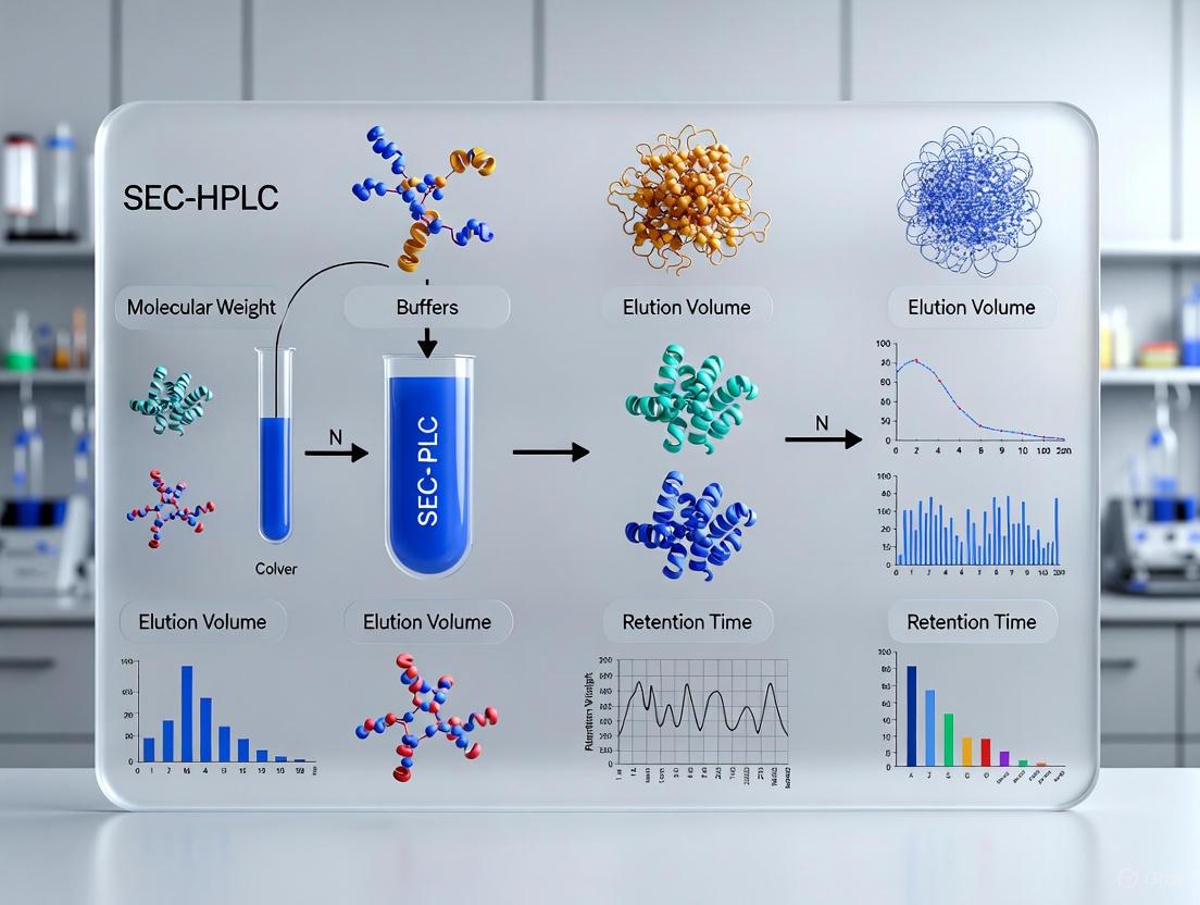
Size-Exclusion Chromatography High-Performance Liquid Chromatography (SEC-HPLC) is a powerful analytical technique that separates molecules in solution based on their hydrodynamic volume—their effective size in solution—rather than their chemical properties or molecular weight [1]. Unlike other chromatographic methods that rely on chemical interactions between the analyte and the stationary phase, SEC-HPLC functions as a molecular sieve, separating components by their ability to access the porous network of the chromatographic packing material [2] [3]. This mechanism makes it uniquely suited for characterizing macromolecules such as proteins, polymers, and nucleic acids while preserving their biological activity and native state [4].
The foundation of SEC-HPLC lies in its entropic separation process. Under ideal conditions, the mobile phase, packing, and column temperature are selected to ensure no enthalpic interaction occurs between the solute and the packing material (ΔH = 0) [2]. The separation is governed solely by the conformational entropy change (ΔS) when a macromolecule diffuses into and out of the stagnant mobile phase within the pores of the packing [2]. The driving force is simply the sample concentration gradient, resulting in a predictable elution order where larger molecules elute first, followed by progressively smaller molecules [4] [1]. This predictable, non-interactive nature simplifies instrumentation, as gradient elution is not required, and enables the technique's application in sensitive biopharmaceutical characterization where maintaining molecular integrity is paramount [2] [1].
The Separation Mechanism Based on Hydrodynamic Volume
Theoretical Foundation
The separation mechanism in SEC-HPLC is fundamentally described by a thermodynamic model. The experimental SEC distribution coefficient, KSEC, is defined as:
KSEC =
The Practical Separation Process
In practice, an SEC-HPLC column is packed with fine, porous beads of materials such as cross-linked dextran, agarose, or silica-based polymers with carefully controlled pore size distributions [3]. As a sample mixture flows through the column isocratically (with a constant mobile phase composition), molecules are separated based on their accessibility to the pore network [1]:
- Large Molecules: Molecules with a hydrodynamic volume larger than the pore size are completely excluded and cannot enter any pores. They are confined to the interstitial volume between beads and elute first at the column's void volume (Vo) [2] [3].
- Intermediate Molecules: Molecules of an appropriate size can partially access the pore network. Their path through the column is prolonged based on the extent to which they can penetrate the pores, resulting in an elution volume between Vo and Vt (the total permeation volume) [2].
- Small Molecules: Molecules significantly smaller than the pore sizes can freely access the entire pore volume. They travel the longest path through the column and elute last at the total permeation volume (Vt), which is the sum of the interstitial and pore volumes (Vt = Vi + Vo) [2] [4].
Table 1: Relationship between Molecular Size, KSEC, and Elution Behavior in SEC-HPLC
| Molecular Size | KSEC Value | Access to Pores | Elution Volume | Elution Order |
|---|---|---|---|---|
| Large | KSEC ≈ 0 | No access | VR ≈ Vo | First |
| Intermediate | 0 < KSEC < 1 | Partial access | Vo < VR < Vt | Middle |
| Small | KSEC ≈ 1 | Full access | VR ≈ Vt | Last |
This process is visualized in the following diagram, which illustrates the differential access of molecules to the porous stationary phase based on their hydrodynamic volume.
Key Instrumentation and Optimization Parameters
Core System Components
A modern SEC-HPLC system integrates several key components to achieve accurate and reproducible separations [4]:
- Stationary Phase: The heart of the system, consisting of porous beads (typically 3-5 µm in diameter for high-performance applications) packed into a column. Materials range from silica-based polymers to cross-linked agarose, selected for their inertness and defined pore size distribution, which dictates the separation range [5] [4].
- Mobile Phase: A buffer or solvent that maintains the native state of the analytes and prevents unwanted interactions with the stationary phase. For aqueous SEC (Gel Filtration), common solvents include phosphate-buffered saline (PBS) or Tris buffers, while organic solvents like tetrahydrofuran (THF) are used for Gel Permeation Chromatography of synthetic polymers [4] [1].
- Pump: Provides a precise, pulseless, isocratic flow of the mobile phase. High-pressure capabilities are essential for UHPLC-SEC systems utilizing sub-2 µm particles [4].
- Injection System: An automated or manual system for introducing the sample onto the column with high precision and minimal dispersion. Typical analytical sample volumes range from 5-100 µL [4].
- Detection Systems: Various detectors monitor the eluting analytes. Common configurations include UV absorbance (for proteins/nucleic acids), refractive index (RI, universal detection), and advanced detectors like Multi-Angle Light Scattering (MALS) for absolute molecular weight determination or viscometers for structural information [4].
Critical Optimization Parameters for Resolution
Optimizing SEC-HPLC methods is crucial for achieving high-resolution separations. Key parameters and their effects are summarized in the table below.
Table 2: Key Optimization Parameters for SEC-HPLC Resolution
| Parameter | Impact on Separation | Recommended Optimization Strategy |
|---|---|---|
| Flow Rate | Slower flow rates generally improve resolution but increase analysis time and may cause peak broadening [4]. | Optimize based on application: 0.5-1.0 mL/min for standard analytical columns. Use lower flows for maximum resolution, higher for speed [4]. |
| Sample Volume | Overloading (>10% of column volume) causes peak broadening and loss of resolution [4]. | Keep injection volume between 5-10% of the total column volume [4]. |
| Column Dimensions | Resolution increases with column length but also extends run time. Advances in small-particle columns (sub-2 µm) allow high resolution at faster flow rates [4]. | Use longer columns or coupled columns for complex mixtures. Consider UHPLC-SEC columns for high throughput and resolution [4]. |
| Mobile Phase Composition | Electrostatic or hydrophobic interactions with the stationary phase cause skewed peaks, tailing, and inaccurate molecular weight estimation [4]. | Adjust ionic strength (e.g., 100 mM NaCl) to shield charge; use additives like arginine to minimize hydrophobic interactions [4]. |
| Column Temperature | Fluctuations can cause baseline noise/drift and affect flow rate reproducibility. Ideally, ΔH=0, so temperature should not directly impact the mechanism [2]. | Thermostat the pump, column, and mobile phase flow lines for maximum reproducibility [2]. |
The interplay of these parameters and their impact on the final chromatographic output is summarized in the following workflow.
Essential Research Reagent Solutions
Successful execution of SEC-HPLC experiments, particularly within a biopharmaceutical context, requires a suite of specialized reagents and materials. The following table details key components of the "Researcher's Toolkit" for SEC-HPLC.
Table 3: Essential Research Reagent Solutions for SEC-HPLC Analysis
| Reagent/Material | Function/Purpose | Key Considerations |
|---|---|---|
| SEC Columns (e.g., AdvanceBio SEC, XBridge Premier) | The stationary phase for size-based separation; the core of the technique [6]. | Select based on pore size (e.g., 500-1000 Å for mAbs, 550-700 Å for rAAVs [5]), particle size (e.g., 2.7 µm for high resolution [6]), and surface chemistry to minimize secondary interactions. |
| Mobile Phase Buffers (PBS, Tris, etc.) | Dissolves the sample and carries it through the column without inducing interactions [4]. | Must be compatible with the sample and stationary phase. Ionic strength often needs optimization (e.g., ~100 mM NaCl) to suppress electrostatic interactions [4]. |
| Molecular Weight Standards | Used for system calibration to convert elution volume to molecular weight [2]. | Should be of known molecular weight and, ideally, similar structure to the analyte (e.g., protein standards for biologics). Not required if using absolute detection methods like MALS [2] [4]. |
| Mobile Phase Additives (e.g., NaCl, Arginine) | Minimize secondary (electrostatic/hydrophobic) interactions between the analyte and stationary phase [4]. | NaCl shields charged interactions; arginine can reduce hydrophobic adsorption, improving peak shape and recovery of aggregates and monomers [4]. |
| Column Storage & Cleaning Solutions | Preserve column integrity and performance over its lifetime. | Specific formulations recommended by the column manufacturer, often containing antimicrobial agents (e.g., 0.05% sodium azide) for aqueous SEC columns. |
Advanced Applications and Protocol for Biopharmaceuticals
Key Applications in Drug Development and Comparability Studies
SEC-HPLC is indispensable in the development and characterization of biopharmaceuticals, where it is primarily used to monitor Critical Quality Attributes (CQAs) related to size variants [5] [1]:
- Aggregate and Fragment Analysis: Quantifying high molecular weight (HMW) aggregates and low molecular weight (LMW) fragments of therapeutic proteins like monoclonal antibodies (mAbs) is a regulatory requirement. Aggregates can impact product safety (immunogenicity) and efficacy [1].
- Gene Therapy Product Characterization: New-generation wide-pore SEC columns are systematically evaluated for characterizing messenger RNA (mRNA) and recombinant adeno-associated virus (rAAV) serotypes used in gene therapies. Optimal selectivity for rAAVs is found with columns having larger pore sizes (550–700 Å) [5].
- Biosimilarity Assessment: Demonstrating comparability between a biosimilar and its reference product requires showing similarity in size variant profiles, for which SEC-HPLC is a benchmark method [6].
- Stability and Forced Degradation Studies: SEC-HPLC tracks changes in size variants over time or under stress conditions (e.g., temperature, pH) to establish product shelf-life and understand degradation pathways [5].
Detailed Experimental Protocol: Analysis of Monoclonal Antibody Aggregates
This protocol provides a detailed methodology for the separation and quantification of aggregates in a monoclonal antibody sample using SEC-HPLC, a standard analysis in biopharmaceutical development.
I. Objectives To separate, identify, and quantify high molecular weight (HMW) aggregates and low molecular weight (LMW) fragments from the monomeric peak of a monoclonal antibody using SEC-HPLC.
II. Materials and Equipment
- SEC-HPLC System: HPLC system with isocratic pump, autosampler, thermostatted column compartment, and UV-Vis detector.
- SEC Column: AdvanceBio SEC 300Å, 2.7µm, 7.8 x 300mm or equivalent [6].
- Mobile Phase: 100 mM Sodium Phosphate, 150 mM Sodium Chloride, pH 6.8. Filter through a 0.22 µm membrane and degas.
- Standards and Samples: mAb sample (1-2 mg/mL); System suitability standard (e.g., mixture of known proteins for plate count determination).
III. Step-by-Step Procedure
System Preparation
- Install the SEC column in the thermostatted compartment and set the temperature to 25°C.
- Prime the system with the filtered and degassed mobile phase.
- Set the mobile phase flow rate to 0.5 mL/min and allow the system to equilibrate until a stable baseline is achieved (typically 30-60 minutes).
System Suitability Test
- Inject the system suitability standard.
- Calculate the number of theoretical plates (N) for the main peak. The column should typically deliver >10,000 plates/meter. Asymmetry factor (As) should be between 0.8-1.8 [5].
Sample Preparation
- Dilute the mAb sample to a concentration of 1-2 mg/mL using the mobile phase.
- Centrifuge the sample at 10,000-14,000 x g for 5-10 minutes to remove any insoluble particles.
Chromatographic Run
- Set the UV detector to 280 nm.
- Program the autosampler to inject 10 µL of the prepared sample.
- Run the isocratic method for 30 minutes.
Data Analysis
- Identify peaks based on retention time: HMW aggregates (first eluting), mAb monomer, and LMW fragments (last eluting).
- Integrate all peaks and report the percentage of each species relative to the total peak area using the formula:
% Species = (Peak Area of Species / Total Integrated Peak Area) * 100
IV. Troubleshooting and Notes
- Tailing Peaks: Can indicate secondary interactions with the stationary phase. Increase ionic strength of the mobile phase or consider additives like arginine [4].
- Poor Resolution: Ensure the sample load is ≤10% of the column volume and that the flow rate is optimized. Linking two columns in series can improve resolution [4].
- High Backpressure: May indicate a clogged column frit. Filter all samples and mobile phase, and use an in-line guard column [1].
SEC-HPLC remains a cornerstone analytical technique for the characterization of macromolecules based on their hydrodynamic volume. Its unique, non-interactive separation mechanism provides a robust means of assessing molecular size distribution, making it invaluable for comparability studies in biopharmaceutical development. As therapeutic modalities evolve to include complex products like mRNA, rAAVs, and novel antibody formats, SEC-HPLC technology continues to advance with columns offering higher efficiency, wider pore sizes, and greater inertness. When coupled with advanced detection methods and rigorous optimization, SEC-HPLC delivers the precise and reproducible data required to ensure the safety, efficacy, and quality of modern biologic drugs.
Market Data: Quantifying the Parallel Growth of Biologics and SEC-HPLC
The expansion of the biologics market is a primary engine driving the adoption and development of Size Exclusion Chromatography High-Performance Liquid Chromatography (SEC-HPLC) columns. The quantitative data below illustrates this synergistic growth.
Table 1: Comparative Market Growth: Biologics and SEC-HPLC Columns
| Market Segment | Market Size (2024/2025) | Projected Market Size | Compound Annual Growth Rate (CAGR) | Key Growth Drivers |
|---|---|---|---|---|
| Biologics Market [7] | USD 577.5 Million (2025) | USD 1,169.8 Million by 2032 | 10.6% (2025-2032) | Rising demand for monoclonal antibodies (mAbs) and recombinant proteins; increasing prevalence of chronic diseases [7]. |
| SEC-HPLC Column Market [8] | USD 0.46 Billion (2024) | USD 0.73 Billion by 2029 | 9.5% (2024-2029) | Demand for protein purification in biopharmaceuticals; growth in biologics and biosimilars development; rising R&D in life sciences [8]. |
Table 2: SEC-HPLC Column Market Segmentation (2024)
| Segmentation Type | Key Segments | Application Notes |
|---|---|---|
| By Type [8] | Standard SEC, High-Resolution SEC, Ultra-High-Performance SEC (UHPSEC), 2D SEC | UHPSEC using sub-2µm particles offers higher resolution and faster analysis [4]. |
| By Application [8] [9] | Biopharmaceutical Analysis, Protein Purification, Nucleic Acid Analysis, Polymer Characterization | The pharmaceutical and biotech segment generates the highest revenue [9]. |
| By End-User [8] | Pharmaceutical Industry, Academic & Research Institutions, Biotechnology Firms, Environmental Testing Labs | North America was the dominant region in the market in 2024 [8]. |
Experimental Protocols: Core Applications of SEC-HPLC in Biologics Development
Protocol: Aggregate Analysis of Monoclonal Antibodies (mAbs) by SEC-HPLC
1. Purpose and Principle This protocol describes the quantitative analysis of high molecular weight (HMW) aggregates and fragments in a monoclonal antibody (mAb) sample. SEC-HPLC separates molecules based on their hydrodynamic volume, with larger aggregates eluting before the main monomer peak and smaller fragments eluting after [4] [10]. Aggregate levels are a Critical Quality Attribute (CQA) for biologics [10].
2. Research Reagent Solutions and Materials Table 3: Essential Materials for mAb Aggregate Analysis
| Item | Function | Example/Specification |
|---|---|---|
| SEC-HPLC Column | Size-based separation matrix. | Columns packed with silica- or polymer-based porous beads (e.g., 1.7µm to 5µm particle size, 150-300Å pore size). Bio-inert (metal-free) hardware is recommended to minimize protein adsorption and improve recovery [11] [12]. |
| Mobile Phase Buffer | Dissolves sample and controls elution. | Phosphate or phosphate-saline buffer, pH ~6.8. Contains 100-200 mM sodium chloride to minimize electrostatic interactions with the stationary phase [4]. |
| mAb Sample | The analyte for characterization. | Purified mAb, formulated at 1-5 mg/mL. Must be compatible with the mobile phase. |
| HPLC System | Instrumentation for precise separation. | UHPLC or HPLC system with auto-sampler, column oven, and UV detector. |
3. Method
- Column Equilibration: Equilibrate the selected SEC column with the mobile phase at a flow rate of 0.2-0.5 mL/min (analytical scale) until a stable baseline is achieved (typically 30-60 minutes) [4].
- Sample Preparation: Dilute or buffer-exchange the mAb sample into the mobile phase. Centrifuge at >10,000 x g for 5-10 minutes to remove any particulate matter. Injection volume should be 5-10% of the total column volume to avoid overloading and peak broadening [4].
- Chromatographic Separation:
- Flow Rate: 0.2 - 0.5 mL/min (scale accordingly for column dimensions).
- Temperature: Maintain column temperature at 20-25°C.
- Detection: Monitor UV absorbance at 280 nm.
- Run Time: Typically 15-30 minutes, sufficient for elution of fragments, monomer, and aggregates.
- Data Analysis: Integrate the chromatogram peaks. Calculate the percentage of each species relative to the total peak area:
- % HMW Aggregate = (Area of HMW peaks / Total Area) x 100
- % Monomer = (Area of monomer peak / Total Area) x 100
- % Fragments = (Area of fragment peaks / Total Area) x 100
Protocol: Purity Analysis of Adeno-Associated Virus (AAV) Vectors
1. Purpose To separate and quantify full, partially full (empty), and aggregated AAV capsids, which is critical for ensuring the safety and efficacy of gene therapies [12] [10].
2. Method Modifications for AAV Analysis
- Column Selection: Use SEC columns with pore sizes and separation ranges optimized for very large macromolecules (like viruses, ~20-30 nm). Ultra-high-performance SEC (UHPSEC) columns with sub-2µm particles are advantageous for high resolution [4] [12].
- Mobile Phase: Use a buffer formulation that maintains capsid integrity (e.g., PBS with 200-400 mM NaCl, potentially with additives like 1-5% glycerol or arginine to minimize non-specific interactions) [4].
- Chromatographic Conditions:
- Use a slower flow rate (e.g., 0.1-0.3 mL/min) to improve resolution of these large species [4].
- Detection can be performed using UV 260/280 nm ratios, which help distinguish between nucleic acid-containing (full) and empty capsids.
Workflow and Market Driver Visualization
The following diagrams illustrate the experimental workflow for SEC-HPLC analysis and the logical relationship between biologics market growth and SEC-HPLC adoption.
The Scientist's Toolkit: Key Materials for SEC-HPLC Analysis
Table 4: Essential Research Reagent Solutions for SEC-HPLC
| Item | Function / Rationale | Key Considerations |
|---|---|---|
| Bio-inert SEC Columns | Specialized columns with minimized metal surfaces to prevent analyte adsorption and improve recovery for sensitive biomolecules (proteins, mAbs, AAVs) [11] [12]. | Critical for analyzing metal-sensitive compounds like phosphorylated proteins and for achieving accurate quantification in AAV full/empty capsid ratio analysis [11]. |
| Ultra-High-Performance SEC (UHPSEC) Columns | Columns packed with sub-2µm particles for enhanced resolution and faster analysis times, compatible with UHPLC systems [8] [4]. | Enables higher throughput and more detailed characterization of complex samples, such as separating oligomers or species with very similar sizes [4]. |
| Mobile Phase Additives | Chemical modifiers added to the buffer to minimize secondary interactions. Common examples include salts (e.g., NaCl) and amino acids (e.g., arginine) [4]. | Ionic Strength (NaCl): Shields electrostatic interactions between the analyte and stationary phase. Arginine: Disrupts hydrophobic interactions, improving peak shape and recovery of both monomers and aggregates [4]. |
| Advanced Detection Systems | Detectors coupled in-line with SEC for absolute characterization without relying on column calibration. | Multi-Angle Light Scattering (MALS): Directly measures absolute molecular weight and size [4]. This is crucial for confirming the identity of aggregates and characterizing new biologic modalities like LNPs and nucleic acids [4] [12]. |
In the biopharmaceutical industry, ensuring the quality, safety, and efficacy of therapeutic products is paramount. For monoclonal antibodies (mAbs) and other protein-based therapeutics, size variants are considered critical quality attributes (CQAs)—properties that must be within appropriate limits to ensure desired product quality. Size-exclusion chromatography high-performance liquid chromatography (SEC-HPLC) serves as a primary analytical technique for monitoring these size variants under native conditions. This application note details the essential terminology of monomers, high molecular weight (HMW) and low molecular weight (LMW) species, and CQAs within the context of SEC-HPLC comparability research. We provide a detailed experimental protocol for a platform SE-HPLC method, complete with validation data and a case study on trastuzumab, to support scientists in drug development.
Essential Terminology and Definitions
Critical Quality Attributes (CQAs)
A Critical Quality Attribute (CQA) is a physical, chemical, biological, or microbiological property or characteristic that should be within an appropriate limit, range, or distribution to ensure the desired product quality [13]. For biopharmaceuticals, CQAs are closely monitored throughout development and manufacturing. Common CQAs for monoclonal antibodies include variants in size, charge, and glycosylation, as well as process-related impurities [13].
Monomer and Size Variants in SEC-HPLC
In SEC-HPLC, proteins are separated based on their hydrodynamic radius using a column packed with porous particles [14]. The separation principle is based on the differential access of molecules to the pore volume of the chromatographic media.
- Monomer: The monomer is the single, intact protein molecule and is the desired product species. In mAbs, the monomer has a molecular weight of approximately 150 kDa and typically appears as the main peak in the chromatogram. The relative peak area of the monomer is reported as percent purity [14].
- High Molecular Weight (HMW) Species: HMW species are size variants larger than the monomer, typically consisting of dimers, trimers, and larger aggregates of the antibody [14]. These species are partially excluded from the pores of the chromatographic media and thus elute earlier than the monomer peak [14]. HMW species are a CQA because they can correlate with undesired immunogenic effects and decreased product efficacy [15].
- Low Molecular Weight (LMW) Species: LMW species are size variants smaller than the monomer, often comprising fragments of the antibody, such as those generated by hinge region hydrolysis (e.g., Fc-Fab and Fab fragments) [14] [16]. These smaller species can permeate more pores in the stationary phase and elute later than the monomer peak [14]. LMW species can reduce serum half-life and lower therapeutic efficacy [16].
Table 1: Definitions of Key Species in SEC-HPLC Analysis of mAbs
| Term | Definition | Typical Composition | Elution Order in SEC | Impact on Product Quality |
|---|---|---|---|---|
| Monomer | Single, intact protein molecule; the main product species. | Intact mAb (~150 kDa). | Main Peak | Desired product; target species. |
| HMW Species | Size variants larger than the monomer. | Dimers, trimers, and larger aggregates. | Before Monomer | May increase immunogenicity and decrease efficacy [15]. |
| LMW Species | Size variants smaller than the monomer. | Fragments (e.g., Fab, Fc-Fab) generated by hydrolysis or cleavage. | After Monomer | May reduce serum half-life and lower efficacy [16]. |
The following diagram illustrates the separation principle and the elution profile of these species in an SEC-HPLC chromatogram.
Figure 1: SEC Separation Principle and Elution Profile. Larger HMW species are excluded from pores and elute first, followed by the monomer. Smaller LMW species penetrate deeper into pores and elute last.
Experimental Protocol: Platform SE-HPLC Method for mAb Analysis
This section provides a detailed methodology for a platform SE-HPLC method, adapted from published work [14], suitable for analyzing a wide range of therapeutic mAbs.
Materials and Equipment
Table 2: Research Reagent Solutions and Essential Materials
| Category | Item | Specification / Function |
|---|---|---|
| Chromatography System | HPLC System | Thermo Scientific U3000 or equivalent, with UV detection. |
| Data System | Chromatography Software | Thermo Scientific Chromeleon (v7.2 SR4) or equivalent for data acquisition and analysis. |
| SEC Column | TSKgel G3000SWxl | 7.8 mm x 30 cm, 5 µm particle size, 25 nm pore size. Separates molecules based on hydrodynamic radius. |
| Mobile Phase | Potassium Chloride | 0.2 M in 0.25 mM phosphate buffer, pH 7.0. Reduces secondary ionic interactions with the column matrix. |
| Samples | Therapeutic mAbs | Reconstitute lyophilized mAbs per manufacturer's instructions. Aliquot and store at -70°C. |
Chromatographic Conditions
- Mobile Phase: 0.2 M potassium chloride in 0.25 mM phosphate buffer, pH 7.0.
- Flow Rate: 0.5 mL/min.
- Column Temperature: 30°C.
- Detection: UV at 280 nm.
- Injection Volume: To achieve an on-column protein load of 50 µg (neat injection).
- Run Time: Approximately 21 minutes (integration window from 5 to 21 minutes).
Sample Preparation
- Reconstitute lyophilized mAb samples in water for injection according to the manufacturer's instructions.
- For mAbs in solution form, aliquot directly.
- Store all aliquots at -70°C prior to analysis.
- Centrifuge samples prior to injection to remove any particulate matter.
Data Analysis and Calculations
Peaks are integrated using a fixed baseline with a perpendicular delimiter drop. The percentages of the main peak (monomer), HMW, and LMW species are calculated as follows:
- % Main Peak = (Area of Main Peak / Total Peak Area) × 100
- % HMW Species = (Area of HMW Peaks / Total Peak Area) × 100
- % LMW Species = (Area of LMW Peaks / Total Peak Area) × 100
Results and Discussion
Method Performance and Validation
The platform SE-HPLC method was rigorously validated to ensure its suitability for its intended purpose [14]. The validation criteria, including repeatability, linearity, and robustness, are summarized in the table below.
Table 3: Summary of Platform SE-HPLC Method Validation Data [14]
| Validation Parameter | Experimental Conditions | Results | Acceptance Criteria |
|---|---|---|---|
| Repeatability | Two analysts; two HPLC systems; six preparations of IgG1 mAb. | Low %RSD for % area of main, HMW, and LMW species. | High reproducibility demonstrated. |
| Linearity | Protein load: 25, 37.5, 50, 62.5, and 75 µg (50% to 150% of nominal load). | R² > 0.99 for main, HMW, and LMW species. | Demonstrates direct proportionality over the range. |
| Robustness | Flow rate: ±0.05 mL/min; Temperature: ±5°C; Mobile phase pH: ±0.2. | Low percent difference in % species compared to nominal conditions. | Method is reliable under small, deliberate variations. |
Case Study: SEC Method Transfer and Trastuzumab Analysis
A key application of SEC in comparability research is method transfer between instruments. A study successfully transferred an SEC method for the mAb trastuzumab from an industry-standard HPLC system to a Waters Arc HPLC System [15]. The results demonstrated comparability, with retention time shifts of approximately 0.02 min and differences in the critical %HMW species within 0.01% between the two systems [15]. The repeatability of the percent area for all species was within 2% RSD across both systems, confirming the method's ruggedness [15].
The same platform method was applied to analyze 35 commercial lots of trastuzumab (14 from the US, 21 from the EU) to understand lot-to-lot variability [14]. The results showed consistent chromatographic profiles across all lots. The % main species (monomer) ranged from 99.0 to 99.4%, while %HMW species ranged from 0.3 to 0.7% [14]. A distinct dimer peak was well-resolved from the monomer, with a resolution greater than 2.6, far exceeding the typical acceptance criterion of 1.5 for baseline resolution [14].
Advanced Method Development: Design of Experiments (DoE)
Resolving the monomer peak from closely eluting fragments, such as the 100 kDa Fab-Fc fragment, can be challenging. A study employed a Design of Experiments (DoE) approach to systematically optimize an SEC procedure for enhanced resolution of LMW species [17] [16]. The study evaluated the impact of mobile phase composition and different SEC columns. Key findings included:
- The Waters BioResolve SEC column showed the best performance for resolving mAb size variants among the columns tested [16].
- The addition of L-arginine as a mobile phase additive helped reduce secondary interactions, improving peak shape and increasing the recovery of HMW species [17] [16].
This AQbD approach provides a more efficient and robust framework for SEC method development compared to traditional one-factor-at-a-time approaches [16].
A deep understanding of the terminology surrounding monomers, HMW/LMW species, and CQAs is fundamental for developing and applying SEC-HPLC methods in biopharmaceutical comparability research. The platform SE-HPLC method detailed herein, proven to be reproducible, linear, and robust, offers a reliable tool for the analysis of therapeutic mAbs. The successful case studies on method transfer and multi-lot analysis of trastuzumab underscore the critical role of SEC-HPLC in ensuring product quality and consistency throughout the drug development lifecycle. Furthermore, the adoption of advanced development strategies like DoE can significantly enhance method capability, particularly for resolving challenging species like LMW fragments.
The Strategic Importance of Comparability Studies for Process Changes and Cell Line Updates
In the research, development, and post-approval lifecycle of biological products, changes to the manufacturing process are inevitable. These changes, which can range from scaling up production to updating the production cell line, are often driven by the need to improve process efficiency, increase scale, enhance product stability, or meet new regulatory requirements [18]. Such modifications carry the inherent risk of impacting the critical quality attributes (CQAs) of the product, which may subsequently affect its safety and efficacy profile [19]. Consequently, demonstrating comparability between the pre-change and post-change product becomes a critical strategic imperative for biopharmaceutical organizations.
Comparability studies serve as the foundational element for successful evaluation of pharmaceutical changes in biological products [18]. These comprehensive assessments determine whether previously conducted non-clinical and clinical studies remain relevant to the product after manufacturing changes by evaluating quality differences that might affect safety and efficacy [18]. Within this framework, Size Exclusion Chromatography (SEC-HPLC) emerges as a pivotal analytical technique, providing essential data on size variants and aggregation that directly informs decisions about product comparability.
The global regulatory landscape for comparability assessments is defined by several key guidelines, including ICH Q5E "Comparability of Biotechnological/Biological Products Subject to Changes in their Manufacturing Process," FDA guidance on "Comparability Protocols for Post-approval Changes," and EMA's "Guideline on comparability of biotechnology-derived medicinal products after a change in the manufacturing process" [18]. These frameworks establish the scientific and regulatory expectations for demonstrating comparability following manufacturing changes.
This application note explores the strategic importance of comparability studies, with particular emphasis on the role of SEC-HPLC within a comprehensive analytical toolkit. We present detailed protocols and data interpretation frameworks that support robust comparability assessments for process changes and cell line updates, contextualized within the expanding biologics market where precise molecular characterization is increasingly crucial.
Regulatory Framework and Risk-Based Approach
Regulatory Foundation
Global regulatory authorities recognize that manufacturing changes are inevitable throughout a biologic product's lifecycle. The ICH Q5E guideline forms the cornerstone of the comparability paradigm, establishing that the goal of comparability assessment is to ensure that quality attributes of the post-change product are highly similar to those of the pre-change product, without adverse impact on safety or efficacy [18]. This framework does not necessitate identical quality attributes but requires demonstration that any differences fall within acceptable limits and do not adversely affect the product's safety profile.
The fundamental principle underlying all regulatory guidance is that the burden of proof for demonstrating comparability rests with the manufacturer. As outlined in ICH Q5E, this requires a comprehensive comparison of relevant quality attributes through extensive analytical characterization, with additional nonclinical or clinical studies when analytical studies alone cannot demonstrate comparability [19]. The depth of required evidence is directly proportional to the manufacturing change's complexity and its potential impact on CQAs.
Risk Assessment for Process Changes
A scientifically sound risk assessment is fundamental to determining the appropriate scope and depth of comparability studies. ICH Q9 quality risk management principles provide the framework for evaluating the potential impact of different categories of process changes [18]. The risk assessment should focus on the product and its characteristics, considering the knowledge gained throughout the product's development lifecycle.
Table 1: Risk Classification and Study Requirements for Different Process Changes
| Process Changes | Comparability Risk | Comparability Study Content |
|---|---|---|
| Production site transfer | Low | Release testing, including activity, structural characterization, and accelerated stability studies |
| Production site transfer with minor process changes | Low-Medium | Transfer all assays to the workshop, add receptor affinity analysis, ADCC or other functional assays |
| Changes in culture methods or purification processes | Medium | All of the above tests may also require animal PK or PD testing |
| Cell line changes | Medium-High | All of the above tests may also require GLP toxicology studies and human bridging studies |
Cell line changes represent one of the most complex post-approval changes due to their potential impact on multiple quality attributes, including glycosylation patterns, charge variants, and higher-order structures [19]. As evidenced by the IBI305 case study (a bevacizumab biosimilar), a comprehensive approach incorporating orthogonal analytical techniques, followed by confirmatory nonclinical and clinical PK studies, may be necessary to demonstrate comparability for such significant changes [19].
The Central Role of SEC-HPLC in Comparability Assessments
SEC-HPLC Fundamentals and Market Context
Size Exclusion Chromatography (SEC-HPLC) is a critical analytical technique that separates molecules based on their hydrodynamic volume in solution under non-denaturing conditions. The stationary phase consists of porous particles that allow smaller molecules to enter the pores and thus traverse a longer path, while larger molecules are excluded from the pores and elute first. For monoclonal antibodies and other therapeutic proteins, SEC-HPLC is primarily employed to quantify monomers, aggregates, and fragments, all of which are CQAs with potential implications for immunogenicity and efficacy [6].
The global SEC-HPLC column market, valued at USD 477 million in 2024 and projected to reach USD 880 million by 2032, reflects the technique's growing importance in biopharmaceutical characterization [6]. This growth is largely driven by increasing demand for biopharmaceuticals, where SEC-HPLC plays an essential role in quality control and regulatory compliance, particularly for aggregate and fragment analysis [6]. Technological advancements continue to enhance SEC performance, with improvements in column packing materials, hardware inertness, and resolution capabilities leading to faster separations with improved sensitivity [11].
Strategic Applications in Comparability Studies
Within comparability studies, SEC-HPLC data provides critical evidence regarding product purity and integrity. The technique's ability to detect subtle changes in size variant profiles between pre-change and post-change products makes it indispensable for assessing the impact of process modifications. For cell line changes specifically, where alterations in cellular machinery may affect protein folding and assembly, SEC-HPLC can detect unwanted shifts in aggregation levels that might compromise product quality [19].
The strategic value of SEC-HPLC extends beyond routine analysis. When integrated with advanced detection systems like multi-angle light scattering (MALS) or mass spectrometry, it provides absolute molecular weight determination and additional structural information beyond traditional SEC analysis [6]. These hyphenated approaches offer more comprehensive characterization capabilities for complex biologics, including the novel modalities increasingly entering development pipelines.
Experimental Protocols for Comprehensive Comparability Assessment
SEC-HPLC Methodology for Monoclonal Antibody Analysis
Principle: This method separates monoclonal antibody monomers, aggregates, and fragments based on their hydrodynamic size under native conditions to assess product purity and stability.
Materials and Equipment:
- SEC Column: AdvanceBio SEC 300Å, 2.7µm (Agilent) or equivalent [6]
- Mobile Phase: 100 mM sodium phosphate, 100 mM sodium sulfate, 0.05% sodium azide, pH 6.8
- HPLC System: UHPLC system capable of maintaining 4-40°C
- Detection: UV detector at 214 nm and 280 nm
- Autosampler: Temperature maintained at 4-8°C
- Standards: Molecular weight standards for system suitability
Sample Preparation:
- Dilute protein samples to 1-2 mg/mL in mobile phase
- Centrifuge at 14,000 × g for 10 minutes to remove particulates
- Transfer supernatant to HPLC vials
Chromatographic Conditions:
- Column Temperature: 25°C ± 0.5°C
- Flow Rate: 0.35 mL/min (for 4.6 × 300 mm column)
- Injection Volume: 10 µL
- Run Time: 15 minutes
- Detection Wavelengths: 214 nm (primary), 280 nm (secondary)
System Suitability Testing:
- Theoretical Plates: >15,000 per column
- Asymmetry Factor (Tailing): 0.8-1.8
- %RSD for Retention Time: <1% over six injections
- Resolution: >2.0 between monomer and dimer peaks
Data Analysis:
- Integrate peaks for high molecular weight species (aggregates), monomer, and low molecular weight species (fragments)
- Calculate percentage of each species relative to total peak area
- Compare pre-change and post-change profiles for significant differences
Extended Characterization Protocol for Cell Line Changes
Principle: Comprehensive structural and functional characterization using orthogonal techniques to detect subtle differences in products from different cell lines.
Materials:
- Reference Standard: Well-characterized pre-change material
- Test Samples: Multiple lots of post-change product
- SEC-HPLC System: As described in Section 4.1
- Mass Spectrometry: LC-MS system for intact mass and peptide mapping
- Circular Dichroism: Spectropolarimeter with temperature control
- Binding Affinity Assay: Surface Plasmon Resonance (SPR) or ELISA reagents
Procedure:
- Size Variant Analysis: Perform SEC-HPLC as described in Section 4.1
- Charge Variant Analysis: Using cation exchange chromatography (CEX-HPLC) or capillary isoelectric focusing (cIEF)
- Intact Mass Analysis:
- Desalt samples using reversed-phase cartridges
- Inject onto LC-MS system with C4 column
- Use mobile phase A: 0.1% formic acid in water; B: 0.1% formic acid in acetonitrile
- Deconvolute mass spectra using appropriate software
- Peptide Mapping:
- Denature, reduce, and alkylate samples
- Digest with trypsin at 37°C for 4 hours
- Analyze digests by LC-MS/MS
- Compare modification sites and levels between pre- and post-change products
- Higher Order Structure Analysis:
- Far-UV and Near-UV Circular Dichroism
- Differential Scanning Calorimetry (DSC) for thermal stability
- Binding Affinity:
- Determine binding kinetics to target antigen by SPR
- Perform cell-based potency assays
Acceptance Criteria:
- Comparable SEC profiles with no new aggregate or fragment species
- Similar post-translational modification profiles
- Equivalent higher order structure by CD and DSC
- Binding affinity within 1.5-fold difference
Case Study: SEC-HPLC in Cell Line Change Comparability
The strategic importance of a thorough comparability assessment is well-illustrated by a reported case involving IBI305, a bevacizumab biosimilar marketed in China [19]. Following initial approval, the manufacturer implemented a post-approval cell line change from lower-titer CHO-K1S to higher-titer CHO-K1SV GS-KO host cells, resulting in an approximately three-fold increase in expression titer [19].
In this comprehensive comparability exercise, SEC-HPLC played a critical role in the analytical comparison. The study employed a three-way comparison approach analyzing pre-change IBI305, post-change IBI305, and the reference product Avastin [19]. The SEC-HPLC analysis focused specifically on detecting differences in size variants that might result from the cell line change.
The comparability study design followed a hierarchical approach, beginning with extensive analytical characterization, then proceeding to nonclinical and clinical studies only as needed based on initial findings [19]. SEC-HPLC data contributed to the foundation of analytical evidence demonstrating that the post-change product was highly comparable to the pre-change product.
Additional orthogonal techniques employed in this assessment included:
- Nuclear magnetic resonance (NMR) for higher-order structure analysis
- High-resolution mass spectrometry for sequence variant identification
- Peptide mapping for primary structure confirmation
- Glycan analysis for post-translational modification profiling
- Forced degradation studies to compare degradation pathways
The successful demonstration of comparability for IBI305, which included confirmation through clinical PK studies, establishes a valuable precedent for post-approval cell line changes of commercialized biosimilars [19]. It further highlights how a well-designed comparability study incorporating techniques like SEC-HPLC can potentially reduce the need for extensive clinical trials.
Visualizing Comparability Study Workflows
SEC-HPLC Comparability Assessment Workflow
SEC-HPLC Comparability Workflow
SEC-HPLC Analytical Methodology
SEC-HPLC Analytical Methodology
Essential Research Reagent Solutions
Table 2: Key Research Reagents and Materials for Comparability Studies
| Category | Product/Technique | Function in Comparability | Example Vendors |
|---|---|---|---|
| SEC-HPLC Columns | AdvanceBio SEC 300Å, 2.7µm | Separation of monomers, aggregates, and fragments based on size | Agilent Technologies [6] |
| Bioinert Columns | YMC Accura BioPro IEX | Reduced surface adsorption for accurate quantification | YMC [11] |
| Mass Spec Standards | Intact mass standards | System calibration and mass accuracy verification | Multiple vendors |
| Binding Assay Reagents | SPR chips and buffers | Quantitative binding affinity measurements | Multiple vendors |
| Reference Standards | Well-characterized biologics | Benchmark for comparability assessment | In-house or commercial |
| Sample Prep Kits | Peptide mapping kits | Standardized digestion for sequence analysis | Multiple vendors |
Data Interpretation and Acceptance Criteria
Establishing Acceptance Criteria
Setting scientifically justified acceptance criteria is critical for meaningful comparability assessment. The criteria should be established prospectively based on historical data and process capability, not merely on statistical significance [18]. For SEC-HPLC and other analytical techniques, acceptance criteria can be categorized as quantitative or qualitative, with quantitative criteria requiring specification of ranges and qualitative criteria relying on comparative assessment of patterns or profiles [18].
Table 3: SEC-HPLC Acceptance Criteria for Comparability Studies
| Parameter | Acceptance Criteria | Basis for Setting Criteria |
|---|---|---|
| Main Peak (Monomer) | Within acceptance criteria based on statistical analysis of historical data | Process capability and validation data |
| Aggregate Peaks | No increase beyond historical levels; within specified limits | Product knowledge and stability data |
| Fragment Peaks | No increase beyond historical levels; within specified limits | Product knowledge and degradation studies |
| Elution Time | Consistent retention time for main species | Method validation data |
| Peak Shape | Comparable peak symmetry and resolution | System suitability standards |
For extended characterization methods where historical data may be limited, head-to-head comparative analysis is typically employed [18]. In such cases, similarity rather than identity is the goal, with the objective of demonstrating that any differences detected do not adversely impact safety or efficacy.
Statistical Considerations
The statistical approach to comparability should reflect the study design and analytical method variability. For well-controlled processes with established historical data, statistical tolerance intervals based on process capability are often appropriate. For techniques with higher variability or when comparing limited numbers of batches, equivalence testing with pre-specified margins may be more suitable.
The sample size (number of batches) for comparability studies should be justified based on the change's magnitude and risk level. For major changes such as cell line changes, ≥3 batches of commercial-scale samples are generally recommended, while minor changes may be adequately assessed with fewer batches [18].
Comparability studies for process changes and cell line updates represent a strategic necessity in the biopharmaceutical industry, ensuring that manufacturing improvements can be implemented without compromising product quality, safety, or efficacy. As demonstrated throughout this application note, SEC-HPLC serves as an indispensable tool within the comprehensive analytical framework required for robust comparability assessment.
The case study of IBI305 illustrates how a science-driven, risk-based approach incorporating orthogonal analytical techniques like SEC-HPLC can successfully demonstrate comparability even for complex changes such as production cell line updates [19]. This approach, conducted within the framework of established regulatory guidelines, provides a pathway for implementing manufacturing changes while maintaining product consistency.
As the biopharmaceutical landscape continues to evolve with novel modalities and increasing complexity, the principles of comparability assessment remain constant. SEC-HPLC, particularly when integrated with advanced detection systems and orthogonal methods, will continue to provide critical data supporting the manufacturing evolution of biological products throughout their lifecycle.
Size exclusion chromatography (SEC) is an indispensable high-performance liquid chromatography (HPLC) technique for the biopharmaceutical industry, primarily used to separate biomolecules based on their hydrodynamic volume. Within the context of gene therapy and biologic drug development, SEC-HPLC provides critical data on aggregate formation, purity, and stability of large, complex molecules like recombinant adeno-associated viruses (rAAVs), messenger RNA (mRNA), and therapeutic proteins [5]. The growing complexity of these drug modalities, coupled with stringent regulatory requirements for characterization, has driven significant innovation in SEC column technology and a competitive market landscape. This application note details the latest advancements, key industry players, and provides standardized protocols for column performance comparison, supporting robust SEC-HPLC comparability research.
Key Industry Players and Product Innovations
The SEC-HPLC column market features a dynamic ecosystem of established multinational corporations and specialized technology companies. Continuous innovation focuses on improving separation efficiency, resolution, and inertness to accommodate the analysis of increasingly challenging biotherapeutics.
Table 1: Key SEC-HPLC Column Manufacturers and Representative Products
| Manufacturer | Representative SEC Product(s) | Key Technology/Innovation | Reported Application |
|---|---|---|---|
| Phenomenex (Part of Danaher) | Biozen dSEC-1, Biozen dSEC-7 LC [5] [20] | Inert silica-based matrix for minimal analyte interaction; high mechanical strength [20]. | Midsize biotherapeutics (peptides, oligonucleotides, siRNA); small mRNA (~1000 nts) [5] [20]. |
| Tosoh Corporation | DNACore AAV-SEC [5] | Monodisperse 3 µm silica particles for high efficiency (e.g., 11,000 plates) [5]. | Optimal selectivity for various rAAV serotypes [5]. |
| SRT (Sinopak) | SRT SEC-500 [5] | 5 µm particle packing. | rAAV analysis (noted for lower efficiency: <1000 plates) [5]. |
| Sepax Technologies | Not Specified in Search Results | Specialized SEC columns for specific applications [8]. | Polymer characterization, biopharmaceutical analysis [8]. |
| Agilent Technologies | Not Specified in Search Results | Broad portfolio of SEC columns and systems [21] [8]. | Protein purification, biopharmaceutical analysis [8]. |
| Waters Corporation | Not Specified in Search Results | Extensive product portfolio for liquid chromatography [8]. | Protein characterization, biopharmaceuticals [8]. |
| Thermo Fisher Scientific | Not Specified in Search Results | Advanced chromatography and mass spectrometry systems [22]. | General HPLC and SEC applications. |
Recent product launches highlight specific market trends. For instance, the Biozen dSEC-1 column from Phenomenex is specifically engineered to address the challenge of non-specific binding of analytes like nucleic acids, which can cause unexpected chromatographic peaks. Its highly inert surface preserves analyte conformations, enabling accurate profiling for peptides and oligonucleotides, as demonstrated by its successful use in resolving duplex, sense, and antisense strands of a target siRNA with minimal column interactions [20].
Furthermore, a systematic 2025 study led by the University of Geneva compared a new generation of wide-pore SEC columns (pore sizes ranging from 450 to 1000 Å) for characterizing gene therapy products [5]. Key findings indicate that:
- For rAAV analysis, optimal selectivity was generally found with columns possessing larger pore sizes (550–700 Å), such as the DNACore AAV-SEC column [5].
- For mRNA analysis, the Biozen dSEC-7 LC column (700 Å) systematically achieved the highest efficiency for small mRNA (~1000 nucleotides), while columns with even larger pore sizes were more appropriate for larger mRNA molecules (>1000 nucleotides) [5].
- A significant challenge remains across all tested columns: the separation of low and high molecular weight species (LMWS and HMWS) of mRNA is limited, making their accurate quantification difficult [5].
Market Analysis and Quantitative Data
The SEC-HPLC column market is experiencing robust growth, fueled by rising demand in the pharmaceutical and biotechnology sectors.
Table 2: SEC-HPLC Column and System Market Overview
| Metric | Data | Source/Timeframe |
|---|---|---|
| Global SEC-HPLC Column Market Size (2024) | $0.46 Billion | [8] |
| Projected Market Size (2025) | $0.51 Billion | [8] |
| Projected Market Size (2029) | $0.73 Billion | [8] |
| Compound Annual Growth Rate (CAGR 2025-2029) | 9.5% | [8] |
| Global SEC System Market Projection (2025) | $1,500 Million | [21] |
| SEC System CAGR (2025-2033) | 9.5% | [21] |
| Dominant Geographic Region (2024) | North America | [8] |
This growth is primarily driven by the increasing demand for protein purification in biopharmaceuticals, the growth of the biotechnology and pharmaceutical industries, and rising research activities in life sciences [8]. Emerging trends include advancements in ultrahigh-pressure SEC columns and the adoption of multi-dimensional chromatography techniques that incorporate SEC [8].
Experimental Protocols for SEC Column Evaluation
A standardized experimental approach is critical for performing direct, head-to-head comparisons of different SEC-HPLC columns. The following protocol is adapted from recent research to ensure reliable and reproducible results [5].
Protocol: Comparative Evaluation of SEC Columns for rAAV and mRNA Analysis
1. Objective To systematically evaluate and compare the performance of various wide-pore SEC-HPLC columns for the separation and characterization of rAAV serotypes and mRNA molecules of varying lengths.
2. Materials and Reagents
- Analytical SEC-HPLC System: Equipped with autosampler, column oven, and UV/VIS detector. For example, a Thermo Scientific Vanquish Neo System [22].
- SEC Columns for Evaluation: A minimum of five columns with pore sizes ranging from 450 Å to 1000 Å. Examples include: Biozen dSEC-7 (700 Å), DNACore AAV-SEC (pore size not specified, 3 µm), and other columns with 550 Å, 700 Å, and 1000 Å pores [5].
- Mobile Phase: Appropriate phosphate or ammonium-based buffer, pH 7.0-7.4, filtered and degassed. A common choice is 1x PBS.
- Test Samples:
- rAAV Samples: Multiple serotypes (e.g., rAAV5, rAAV8, rAAV9) at a known concentration (e.g., ~1x10^12 vg/mL).
- mRNA Samples: A range of transcripts from ~1000 to ~5000 nucleotides.
- Stressed Samples: rAAV and mRNA samples subjected to accelerated stress conditions (e.g., elevated temperature) to induce degradation and aggregate formation [5].
3. Method Parameters Table 3: Standard HPLC Operating Conditions for Column Evaluation
| Parameter | Setting |
|---|---|
| Flow Rate | 0.2 - 0.8 mL/min (optimize for each column to maintain pressure <150 bar) |
| Column Temperature | 25 - 30 °C |
| Detection Wavelength | 260 nm (for nucleic acids, mRNA) and 280 nm (for proteins, rAAVs) |
| Injection Volume | 10 - 50 µL |
| Run Time | 15 - 20 minutes |
4. Experimental Procedure
- Column Equilibration: Condition each new column with at least 5-10 column volumes of mobile phase until a stable baseline is achieved.
- System Suitability Test: Inject a standard mixture or a well-characterized control sample to verify system performance.
- Analysis of Native Samples:
- Inject each rAAV serotype and mRNA sample onto each equilibrated SEC column.
- Record chromatograms and note retention times, peak symmetry, and resolution.
- Analysis of Stressed Samples:
- Inject the thermally stressed rAAV and mRNA samples.
- Compare the chromatographic profiles to native samples to assess the column's ability to resolve and quantify degradation products and aggregates.
- Data Collection: For each injection, record the following data:
- Retention time of the main peak.
- Peak width at half height.
- Theoretical plate count (N).
- Resolution (Rs) between any observable peaks (e.g., full/empty capsids for rAAV).
- Peak asymmetry factor (As).
5. Data Analysis and Interpretation
- Efficiency: Calculate theoretical plates (N) for the main peak. Higher values indicate greater column efficiency [5].
- Selectivity: Compare the elution profiles and resolution between species (e.g., full vs. empty capsids) across different columns.
- Recovery: Compare the peak areas of the main species across columns to identify any significant analyte adsorption.
- Capability for Impurity Quantification: Assess the baseline separation between the main peak and any LMWS or HMWS. Note the limitations in accurately quantifying these species if resolution is poor [5].
Diagram: SEC Column Evaluation Workflow. The process involves systematic column preparation, sample analysis, and data evaluation to generate a comparability report [5].
The Scientist's Toolkit: Essential Research Reagent Solutions
Successful SEC-HPLC characterization relies on a suite of specialized materials and consumables.
Table 4: Essential Reagents and Materials for SEC-HPLC Characterization
| Item | Function/Purpose | Example/Best Practice |
|---|---|---|
| Wide-Pore SEC Columns | Separation of large biomolecules based on size. | Select pore size based on application: 550-700 Å for rAAVs; 700+ Å for larger mRNA [5]. |
| Inert/Silica-Based Columns | Minimize non-specific binding of analytes like nucleic acids. | Biozen dSEC-1 for peptides/oligonucleotides to preserve conformations [20]. |
| Bioinert Guard Cartridges | Protect analytical column from contaminants and particulates; improve column lifetime. | YMC Accura BioPro IEX or similar bioinert guards for oligonucleotide/protein analysis [11]. |
| HPLC-Grade Buffers & Salts | Form the mobile phase; maintain pH and ionic strength. | Use phosphate or ammonium-based buffers (e.g., 1x PBS), filtered and degassed. |
| Stressed/Stability Samples | Challenge the column's ability to separate degraded species and aggregates. | rAAV/mRNA samples subjected to accelerated heat stress [5]. |
| Column Efficiency Standard | A well-characterized sample for calculating theoretical plates (N). | Used to verify and compare the performance of different columns. |
The SEC-HPLC column landscape is evolving rapidly, with key players driving innovations in particle technology, pore architecture, and surface chemistry to meet the demanding characterization needs of modern gene therapies and biologics. The successful application of this technique for comparability studies requires a meticulous approach to experimental design, as outlined in the provided protocols. By leveraging the latest column technologies and standardized evaluation methods, scientists can generate robust, high-quality data to ensure product quality, safety, and efficacy throughout the drug development lifecycle.
Advanced SEC-HPLC Method Development for Complex Biologics and Novel Modalities
Size Exclusion Chromatography (SEC) is an indispensable technique for the separation and analysis of synthetic polymers and biopolymers based on their hydrodynamic volume [23]. The selection of an appropriate stationary phase is a critical parameter in method development, directly impacting the accuracy and reproducibility of separations within SEC-HPLC comparability studies. The stationary phase dictates the separation mechanism, which in true SEC is purely an entropy-driven process where molecules are sorted by their ability to access the porous network of the column packing material [24] [25]. Among the available options, silica-based, polymeric, and diol-modified stationary phases represent the most prevalent choices, each possessing distinct chemical and physical characteristics that define their application scope.
The core separation mechanism in SEC relies on the differential access of analyte molecules to the pore volume of the stationary phase. Larger molecules that are excluded from the pores elute first, while smaller molecules that can penetrate the porous structure experience a longer path and elute later [25]. A fundamental requirement for this mechanism is the absence of non-size exclusion effects, such as adsorption or ionic interactions, between the analyte and the stationary phase surface [23]. The chemical nature of the packing material is therefore paramount, as it must be inert towards the analytes under the chosen mobile phase conditions to ensure a separation based solely on size.
Comparative Analysis of Stationary Phases
Silica-Based Stationary Phases
Silica-based packings are widely used in high-performance liquid chromatography (HPLC) due to their excellent mechanical stability and high surface area [26]. These materials consist of rigid, porous silica particles that can be synthesized with controlled pore sizes and particle diameters to optimize chromatographic performance [27] [26]. A key characteristic of silica is the presence of surface silanol groups, which can be acidic and lead to unwanted secondary interactions with basic or charged analytes, causing issues such as peak tailing, irreversible adsorption, or altered retention times [27] [23]. To mitigate this, silica surfaces are often modified with various bonded phases.
- Bare Silica: Useful for analyzing nonaqueous polar or nonpolar organic mobile phases, especially for high-temperature applications with nonionic polymers. It is generally not recommended for aqueous mobile phases due to the presence of active silanol adsorptive sites and the finite solubility of silica in aqueous buffers [24].
- Surface-Modified Silica: Silica particles can be bonded with hydrophobic ligands (e.g., C18, C8) for reversed-phase chromatography or with other functional groups to create normal-phase or ion-exchange columns [26]. This modification helps control selectivity and reduce unwanted interactions.
Diol-Modified Silica Phases
Diol-modified silica is a common and important hydrophilic stationary phase specifically designed to address the limitations of bare silica. The surface is typically modified with 1,2-propanediol functional groups, which render the surface hydrophilic and block or react with many of the acidic silanol groups [24]. This neutralization of the surface makes it ideal for the SEC separation of biopolymers and synthetic water-soluble polymers, as it significantly reduces ion-exchange interactions [24] [23]. Recent advancements include the development of ultra-wide pore size exclusion chromatography (SEC) columns based on diol-modified silica, which are particularly suited for large biomolecules like mRNA, adeno-associated viruses (AAVs), and lipid nanoparticles (LNPs) [28]. Despite diol modification, some residual silanol activity may remain, which can still interact with cationic polyelectrolytes or amino-containing polymers [24] [23].
Polymeric Stationary Phases
Polymer-based packing materials, most commonly cross-linked poly(styrene-co-divinylbenzene) or polymethacrylates, offer a distinct set of advantages, primarily centered on their enhanced chemical stability [27] [26]. These materials are popular for the analysis of macromolecules.
- Cross-linked Polymeric Phases: These are semi-rigid, highly cross-linked organic polymer particles. The degree of cross-linking can be adjusted to control the pore size, and their polarities can be optimized to match the polarities of samples and solvents [27]. They are delivered pre-packed in columns, often with metal bodies that may be coated to prevent metal ions from influencing the separation or detection [27].
- Hydrophilic Polymeric Phases: For aqueous SEC, most polymeric packings are proprietary hydroxylated derivatives of cross-linked polymethacrylates. Other types include sulfonated cross-linked polystyrene, polydivinylbenzene derivatized with glucose, and high-performance crossed-linked agarose [24].
A summary of the comparative properties is provided in Table 1.
Table 1: Comparative Properties of Silica, Diol-Modified, and Polymeric SEC Stationary Phases
| Property | Silica-Based | Diol-Modified Silica | Polymeric (Cross-linked) |
|---|---|---|---|
| Pressure Stability | High (up to 1200 bar in UHPLC) [26] [23] | High [24] | Moderate (200-350 bar) [26] |
| Typical pH Range | Limited (2-8) [26] | Limited (2-8, potentially wider with new tech) [24] | Wide (0-14) [27] [26] |
| Temperature Stability | Good | Good | Excellent (stable >90°C, some >160°C) [27] |
| Risk of Sample Interactions | High (residual silanols) [27] [23] | Low (reduced silanol activity) [24] [23] | Very Low (inert) [27] |
| Separation Range | High resolution in a narrow molar mass range [27] | Suitable for a variety of biomolecules [25] | Broad, easily combined for wide range [27] |
| Solvent Compatibility | High, fast solvent exchange [27] | High [23] | Variable, slow equilibration between solvents [27] |
| Typical Applications | Small molecules, proteins (with modification) | Biomolecules, water-soluble polymers [24] [25] | Synthetic polymers, biopolymers, extreme pH analyses [27] [24] |
Experimental Protocols for SEC-HPLC Comparability
Protocol 1: Method Development for Polymer Analysis
This protocol outlines a systematic approach for selecting a stationary phase and mobile phase conditions for the analysis of synthetic polymers, based on the principles of maximizing pore volume and eliminating non-size exclusion effects [23].
- Step 1: Column Selection. Based on the expected molecular size range of the polymer analyte, select a column with an appropriate pore size. For a broad distribution, combine columns of different pore sizes. Note that polymer-based columns are easier to combine for a wide molar mass range than silica columns, which often have a steeper calibration curve [27].
- Step 2: Mobile Phase Optimization. The mobile phase must be a strong solvent for the polymer to prevent adsorption and enthalpic interactions. For synthetic polymers like polystyrene (PS) and poly(methyl methacrylate) (PMMA), tetrahydrofuran (THF) is commonly used. The mobile phase composition is critical to achieve a pure size-exclusion mechanism [23].
- Step 3: System Suitability Test. Inject a narrow dispersity polymer standard to evaluate the column performance. Check for symmetric peak shapes, as tailing can indicate unwanted adsorption. Verify that the elution volume is consistent with a size-based separation and does not shift with small changes in injection concentration or volume [23].
- Step 4: Calibration. Construct a calibration curve using certified polymer standards of known molecular weight. Plot the logarithm of molecular weight against the elution volume to define the separation range and resolution of the column [23].
Protocol 2: Column Cleaning and Maintenance for Diol-Modified Columns
Proper maintenance is essential for the longevity and performance of SEC columns, especially when analyzing complex biological samples.
- Step 1: Routine Washing. After analysis with aqueous buffers, flush the diol-modified column with 3-5 column volumes of a mixture of water and a water-miscible organic solvent (e.g., 50:50 water/acetonitrile) to remove salts and residual proteins.
- Step 2: Deep Cleaning. If a decrease in performance is observed (e.g., increased backpressure or peak broadening), flush the column with 5-10 column volumes of a 20-50% isopropanol or acetonitrile solution. For more stubborn contaminants, a series of flushes with buffers of high and low pH may be used, but the manufacturer's specified pH limits (typically 2-8 for silica-based columns) must be strictly observed [24] [26].
- Step 3: Storage. For long-term storage, flush the column thoroughly with a water/organic solvent mixture (e.g., 30:70 water/acetonitrile) to prevent microbial growth and seal the column according to the manufacturer's instructions.
Protocol 3: Assessing Secondary Interactions
This protocol describes a simple test to verify the absence of non-size exclusion interactions, which is critical for accurate SEC analysis.
- Procedure: Inject a small amount of a compound that is known to potentially interact with the stationary phase. For a diol-modified column, this could be a basic protein or a cationic polyelectrolyte. Similarly, for a polymeric column, test with a polar compound.
- Data Analysis: Monitor the elution profile. The presence of secondary interactions is indicated by one or more of the following: no elution, late elution after the system peak, unexpected very high resolution, peak tailing, shifting retention times, or low reproducibility of retention times or areas [27].
- Troubleshooting: If interactions are detected, adjust the mobile phase conditions. This may include increasing the ionic strength (e.g., adding 50-150 mM salt to shield electrostatic forces), modifying the pH, or adding an organic moderator (e.g., <5% methanol) to suppress hydrophobic interactions [24].
Visualization of Column Selection Strategy
The following workflow provides a logical pathway for selecting the most appropriate stationary phase for a given SEC-HPLC application, incorporating key decision points based on analyte and method requirements.
SEC Column Selection Workflow
The Scientist's Toolkit: Essential Research Reagents and Materials
Successful execution of SEC-HPLC comparability research requires not only the correct column but also a suite of high-quality reagents and materials. The following table details key items for a typical laboratory working in this field.
Table 2: Essential Research Reagents and Materials for SEC-HPLC
| Item | Function/Application | Example/Note |
|---|---|---|
| Diol-Modified SEC Column | High-resolution separation of biomolecules (proteins, mAbs) in aqueous buffers with minimal secondary interactions [25]. | YMC-Pack Diol SEC columns with multiple pore sizes (e.g., 60, 120, 200, 300 Å) [25]. |
| Polymer-Based SEC Column | Separation of synthetic polymers or analyses requiring extreme pH or high temperature conditions [27] [24]. | Cross-linked poly(styrene-co-divinylbenzene) columns for organic phases; polymethacrylate for aqueous phases. |
| Narrow Dispersity Polymer Standards | Column calibration and determination of molecular weight distributions for synthetic polymers [23]. | Certified polystyrene (PS) or poly(methyl methacrylate) (PMMA) standards. |
| Protein Molecular Weight Markers | Column calibration and system suitability testing for biopolymer separations. | A mix of stable proteins covering a broad molecular weight range (e.g., thyroglobulin, BSA, ovalbumin). |
| HPLC-Grade Organic Solvents | Mobile phase preparation; sample dissolution. | Tetrahydrofuran (THF) for synthetic polymers; acetonitrile for cleaning and modifier addition [23]. |
| High-Purity Buffering Salts | Mobile phase preparation for aqueous SEC to control ionic strength and pH, shielding electrostatic interactions. | Sodium phosphate, Tris, or ammonium salts. Use MS-compatible salts if hyphenating with mass spectrometry. |
| ULC/MS-Grade Water & Acids | Mobile phase preparation, especially when using sensitive detection methods like mass spectrometry. | Minimizes background noise and system contamination. |
| In-Line Filter or Guard Column | Protects the analytical column from particulate matter and contaminants, extending its lifetime. | A guard column with the same packing material as the analytical column is ideal. |
The choice between silica, diol-modified silica, and polymeric stationary phases is fundamental to designing a robust and comparable SEC-HPLC method. Silica-based phases offer superior mechanical strength and high resolution in narrow mass ranges, while polymeric phases provide unmatched chemical stability across extreme pH and temperature conditions. Diol-modified silica phases strike an effective balance, providing a hydrophilic, low-interaction surface ideal for sensitive biomolecule analysis. This guide provides a structured framework, including comparative data, experimental protocols, and a clear selection workflow, to empower researchers and drug development professionals in making an informed decision that ensures the accuracy, reproducibility, and longevity of their SEC-HPLC analyses.
In the field of biopharmaceutical analysis, Size Exclusion Chromatography (SEC-HPLC) stands as a critical technique for monitoring the stability, purity, and quality of therapeutic products. For sophisticated modalities like monoclonal antibodies (mAbs) and gene therapy vectors such as adeno-associated viruses (AAVs), achieving optimal SEC separations requires meticulous mobile phase optimization. The mobile phase is not merely a carrier but an active component that influences critical quality attributes by modulating secondary interactions with the stationary phase. This application note details systematic approaches for mobile phase optimization, focusing on the roles of buffers, salts, and arginine additives, framed within comparability research for biotherapeutic development.
Thorough analytical characterization is required by health agencies to ensure product quality, safety, and efficacy, as these complex therapies can undergo various changes during preparation, formulation, and storage [5]. Undesirable electrostatic or hydrophobic interactions with the column hardware or stationary phase can lead to poor peak shapes, reduced resolution, and low analyte recovery, compromising the accuracy of size variant quantification [16]. Mobile phase optimization is therefore essential to mitigate these secondary interactions, and a systematic Quality by Design (QbD) approach is recommended for developing robust, regulatory-compliant methods [16].
Theoretical Foundations of Mobile Phase Interactions
In SEC, the primary separation mechanism is based on the differential partitioning of analytes into the pore volume of the stationary phase, governed by their hydrodynamic radius. However, the ideal separation is often compromised by secondary interactions.
- Electrostatic Interactions: These occur when charged groups on the analyte interact with ionized silanol groups on the stationary phase. If the protein and stationary phase have similar surface charges, an ion-exclusion effect can occur, leading to decreased elution time. Conversely, opposite charges can cause ion-exchange interactions, leading to adsorption and increased elution times [16].
- Hydrophobic Interactions: These involve non-polar interactions between the analyte and hydrophobic sites on the stationary phase, which can lead to increased elution times and low recoveries [16].
The isoelectric point (pI) of the analyte plays a significant role in the nature and extent of these secondary interactions, which can negatively impact peak shape and contribute to peak tailing. The strategic formulation of the mobile phase, using specific additives, is designed to suppress these non-ideal interactions without disrupting the primary size-based separation mechanism [16].
Key Mobile Phase Components and Their Functions
The following table summarizes the roles and common concentrations of key mobile phase additives used in SEC-HPLC for biopharmaceutical analysis.
Table 1: Key Mobile Phase Components for SEC-HPLC Optimization
| Component | Primary Function | Common Types | Typical Concentration Range | Considerations |
|---|---|---|---|---|
| Buffers | Maintains pH stability, affecting the charge state of the analyte and stationary phase. | Phosphate, acetate, histidine | 10-100 mM | Buffer capacity and compatibility with detection (e.g., UV, MS) must be evaluated. |
| Salts | Mitigates electrostatic interactions by increasing ionic strength, shielding opposite charges. | Sodium chloride, arginine hydrochloride | 0-250 mM | High concentrations can induce hydrophobic interactions or promote aggregation [16]. |
| Amino Acid Additives | Multi-functional: disrupts both hydrophobic and electrostatic protein-stationary phase interactions. | L-arginine | 0-150 mM | Effective at reducing non-specific interactions and improving peak shape and recovery [16]. |
| Organic Modifiers | Weakens hydrophobic interactions. | Acetonitrile, isopropanol | 0-5% (v/v) | Can strengthen ionic interactions; generally used at low concentrations [16]. |
The Multifunctional Role of Arginine
The amino acid L-arginine has emerged as a particularly effective mobile phase additive. Its efficacy stems from its ability to interact with amino acid side chains and peptide bonds, leading to a reduction in both electrostatic and hydrophobic interactions [16]. Historically, arginine has been used for molecule solubilization, refolding, stabilization, and aiding in the elution of antibodies from Protein A. In the SEC mobile phase, including arginine helps overcome secondary interaction effects and can significantly improve analyte recovery and peak shape [16].
Systematic Optimization Using Design of Experiments (DoE)
A one-factor-at-a-time (OFAT) approach to method development is inefficient and may fail to reveal critical parameter interactions. The Design of Experiments (DoE) methodology, aligned with Analytical Quality by Design (AQbD) principles, allows for a systematic understanding of multivariate relationships and leads to a more robust and optimized method [16].
A recent study on SEC method development for monoclonal antibodies exemplifies a two-stage DoE process: an initial screening design to identify critical factors, followed by an optimization design focused on those factors [16]. The primary response measured was the USP resolution between the monomer peak and the low molecular weight (LMW) species, a key critical quality attribute.
Diagram 1: DoE workflow for SEC method development. The process begins with goal definition and proceeds through screening and optimization phases to establish a robust design space.
Case Study: Protocol for SEC Method Development Using DoE
The following protocol outlines the key experimental steps for optimizing a mobile phase using a DoE approach, as applied in the cited research [16].
Objective: To develop a robust SEC-HPLC procedure for the quantification of mAb size variants (HMW, monomer, LMW).
Materials and Equipment:
- SEC Columns: Columns with diol-modified silica or bridged ethyl hybrid (BEH) particles are recommended. Example: Waters BioResolve SEC column (supports high efficiency for mAbs) [16].
- Chemicals: Water (HPLC grade), sodium phosphate monobasic monohydrate, sodium phosphate dibasic heptahydrate, L-arginine hydrochloride, sodium chloride.
- Instrumentation: HPLC or UHPLC system with autosampler and UV/VIS or PDA detector.
- Software: DoE software platform (e.g., JMP, Fusion Method Development Software).
Experimental Procedure:
Define Critical Method Parameters and Responses:
- Parameters (Factors): These are the variables to be tested. In this study, they included:
- Arginine Concentration: e.g., 0 - 150 mM.
- Salt Concentration: e.g., 0 - 200 mM sodium chloride.
- Buffer pH: e.g., pH 6.0 - 7.0.
- Column Type: A categorical factor, if comparing different columns.
- Responses: These are the measurable outcomes that define method quality. The primary response was the USP resolution between the monomer and LMW species. Secondary responses can include peak symmetry, retention time, and peak capacity [16].
- Parameters (Factors): These are the variables to be tested. In this study, they included:
Screening Phase:
- Use a screening design (e.g., a fractional factorial or Plackett-Burman design) to evaluate the main effects of all identified parameters with a minimal number of experimental runs.
- Perform the chromatographic runs according to the randomized order generated by the software. Use a fixed injection volume and flow rate appropriate for the column dimensions.
- Analyze the data to build a linear regression model and identify which parameters have a statistically significant effect on the resolution.
Optimization Phase:
- Focus on the critical parameters (e.g., arginine and salt concentration) identified in the screening phase.
- Employ a response surface methodology (RSM) design, such as a Central Composite Design, to model quadratic effects and interactions between these key parameters.
- Execute the experiments and analyze the data to generate a predictive model and contour plots.
Define the Design Space and Verify Robustness:
- Using the predictive model, identify the region of the operational parameter space where the method meets all performance criteria (e.g., Resolution > 1.5). This is the "design space."
- Confirm the model's predictions by performing verification runs at the set-point conditions within the design space.
- Finally, perform a small robustness test around the set-point to ensure the method is insensitive to minor, expected variations.
Research Reagent Solutions
The following table lists key materials and tools essential for executing the described SEC-HPLC optimization.
Table 2: Essential Research Reagents and Tools for SEC-HPLC Optimization
| Item | Function/Description | Example Products / Notes |
|---|---|---|
| SEC Columns with Inert Hardware | Minimizes non-specific adsorption of metal-sensitive analytes (e.g., phosphorylated compounds, proteins) to column hardware, improving recovery and peak shape. | Halo Inert [11], Restek Inert HPLC Columns [11], Evosphere Max [11] |
| Advanced SEC Stationary Phases | Provides high efficiency separations with reduced secondary interactions. Diol-modified surfaces are common for biomolecules. | Waters BioResolve SEC [16], TOSOH TSKgel UP-SW3000 [16], Agilent Bio SEC-3 [16] |
| Wide-Pore SEC Columns | Essential for separating large biomolecules and gene therapy products like rAAVs and mRNA. Larger pore sizes (e.g., 550–700 Å) offer optimal selectivity for rAAVs. | DNACore AAV-SEC column, Biozen dSEC-7 LC column [5] |
| DoE Software | Facilitates the design of experiments, statistical analysis of data, and generation of predictive models and contour plots. | JMP, Fusion Method Development Software [16] |
| Buffers and Salts | Provides the ionic foundation of the mobile phase to control pH and ionic strength. | Sodium phosphate, sodium chloride [16] |
| Amino Acid Additives | Used as a multi-functional mobile phase additive to suppress multiple types of secondary interactions. | L-arginine hydrochloride [16] |
The optimization of the mobile phase is a critical step in developing a reliable and robust SEC-HPLC method for biopharmaceutical comparability studies. A systematic approach that leverages Design of Experiments is far superior to traditional OFAT, as it efficiently uncovers interactions between critical parameters such as buffer pH, salt concentration, and arginine levels. The use of arginine as a multi-functional additive has proven highly effective in mitigating both electrostatic and hydrophobic secondary interactions, leading to improved resolution, peak shape, and analyte recovery. By adopting these structured protocols and utilizing modern, inert column technologies, scientists can accelerate method development timelines and establish robust, regulatory-compliant analytical procedures that ensure the safety and efficacy of complex therapeutic products.
Method Development Using Analytical Quality by Design (AQbD) and Design of Experiments (DoE)
This application note provides a detailed protocol for implementing Analytical Quality by Design (AQbD) principles and Design of Experiments (DoE) in the development of a Size Exclusion Chromatography-High Performance Liquid Chromatography (SEC-HPLC) method for monoclonal antibody (mAb) purity analysis. The systematic, risk-based approach outlined herein enhances method robustness, reliability, and transferability while supporting regulatory compliance. A case study demonstrates the development of a platform procedure using Waters XBridge Premier Protein SEC column technology, achieving high-performance separations without mobile phase additives. The protocol includes comprehensive experimental designs, validation data, and visualization tools to guide scientists in pharmaceutical development.
In the biopharmaceutical industry, monitoring critical quality attributes (CQAs) of therapeutic proteins, particularly size variants of monoclonal antibodies (mAbs), is essential for ensuring product safety and efficacy [29]. Size-exclusion chromatography separates biomolecules based on their hydrodynamic radius as they diffuse through a porous stationary phase, enabling quantification of high molecular weight (HMW) species, low molecular weight (LMW) species, and the monomeric main peak [29] [30].
Analytical Quality by Design represents a systematic, science-based framework for analytical method development that emphasizes predefined objectives, risk assessment, and statistical methodologies [29] [31]. Unlike traditional one-factor-at-a-time (OFAT) approaches, AQbD incorporates multivariate experiments to understand parameter interactions and establish a method operable design region (MODR) where method performance remains robust [16] [31]. This paradigm enhances method reliability, reduces the risk of failure, and supports continuous improvement throughout the analytical procedure lifecycle [29].
Theoretical Background
SEC-HPLC for mAb Analysis
SEC-HPLC operates on the principle of separating molecules based on their effective molecular size differences as they migrate through a column packed with porous particles [29] [30]. For mAb products, this technique resolves HMW species (aggregates), LMW species (fragments), and the monomeric main peak [29]. The thermodynamic retention factor (KD) in SEC is defined as:
KD = (VR - V0)/Vi
where VR is the analyte retention volume, V0 is the interstitial volume, and Vi is the intraparticle pore volume [30]. Ideal SEC separations are driven primarily by entropic processes, with minimal enthalpic contributions from secondary interactions [30].
AQbD Framework and DoE
The AQbD framework comprises four key stages: (1) defining the Analytical Target Profile (ATP), (2) risk assessment, (3) screening and optimization studies using DoE, and (4) establishing control strategies [29] [31]. The ATP is a critical document that outlines the method's purpose and defines required performance characteristics such as accuracy, precision, specificity, and working range [29].
DoE provides a structured approach for investigating multiple factors simultaneously, enabling efficient identification of critical method parameters and their interactions [29] [16]. Screening designs (e.g., fractional factorial) identify influential factors, while optimization designs (e.g., Central Composite Design, Box-Behnken) characterize response surfaces and define the MODR [16] [31].
Materials and Equipment
Research Reagent Solutions
Table 1: Essential materials and reagents for SEC-HPLC method development
| Item | Function | Examples |
|---|---|---|
| SEC Columns | Stationary phase for size-based separation | Waters XBridge Premier Protein SEC [29], Tosoh TSKgel UP-SW3000 [16], Agilent Bio SEC-3 [16] |
| Buffers | Mobile phase components to control ionic strength and pH | Sodium phosphate, potassium chloride [14] |
| Additives | Minimize secondary interactions | Arginine [16], salts (NaCl) [16] |
| mAb Samples | Analytics for method development | In-house IgG1 mAb [14], commercial mAbs (e.g., trastuzumab) [14] |
| Standards | System suitability and calibration | Monodisperse protein standards [32] |
Instrumentation
- HPLC/UHPLC system with binary pump, autosampler, and column oven
- UV-Visible detector (280 nm typical for proteins)
- Columns as specified in Table 1
- Data acquisition software (e.g., Chromeleon, Empower)
Experimental Protocol
Define Analytical Target Profile (ATP)
The ATP specifies the method's purpose and defines criteria for method performance characteristics. For mAb purity analysis by SEC-HPLC, the ATP should include:
- Analytical Procedure Intermediate Precision: ≤ 5% RSD for main peak, ≤ 35% RSD for HMW and LMW species [29]
- Relative Accuracy: 95-105% for main peak [29]
- Specificity: Resolution ≥ 1.5 between critical pairs [14]
- Working Range: 50-150% of target concentration with R2 ≥ 0.99 [14]
Risk Assessment
Conduct a systematic risk assessment to identify factors potentially affecting method performance:
- Material Factors: Column chemistry (silica, hybrid, polymeric), mobile phase composition (buffer type, pH, ionic strength, additives) [29] [16]
- Instrument Factors: Flow rate, column temperature, detection wavelength [29]
- Sample Factors: Concentration, injection volume, sample solvent [29]
Tools such as Ishikawa (fishbone) diagrams and CNX (Controlled, Noise, eXperimental) analysis can categorize factors based on their risk priority [29].
Screening Studies
Initial screening experiments explore wide parameter ranges to identify critical factors:
- Factors to Screen: Column type, buffer pH (e.g., 6.0-7.5), ionic strength (e.g., 0-200 mM), temperature (e.g., 25-35°C), flow rate (e.g., 0.4-0.6 mL/min) [16]
- Experimental Design: Fractional factorial or Plackett-Burman designs
- Critical Responses: Resolution between monomer and LMW species, peak asymmetry, retention time [16]
Optimization Studies
Optimize critical factors identified during screening using response surface methodologies:
- Experimental Design: Central Composite Design (CCD) or Box-Behnken Design (BBD) [16] [31]
- Factors: Typically 2-4 continuous factors (e.g., buffer concentration, pH, temperature) [16]
- Responses: Resolution, tailing factor, theoretical plates, precision [16]
- Analysis: Fit mathematical models to experimental data and perform analysis of variance (ANOVA) to assess model significance [31]
Table 2: Example experimental design for SEC-HPLC optimization
| Factor | Low Level | Center Point | High Level |
|---|---|---|---|
| Buffer Concentration (mM) | 20 | 40 | 60 |
| pH | 6.2 | 6.6 | 7.0 |
| Column Temperature (°C) | 20 | 25 | 30 |
| Flow Rate (mL/min) | 0.4 | 0.5 | 0.6 |
Robustness and Ruggedness Testing
Evaluate method robustness using DoE principles with narrow parameter variations approximating normal operational fluctuations:
- Parameter Variations: Flow rate (±0.05 mL/min), temperature (±5°C), mobile phase pH (±0.2 units) [14]
- Ruggedness Testing: Assess intermediate precision across different analysts, days, and instruments [29]
- Acceptance Criteria: % difference ≤ 2% for variant quantification compared to nominal conditions [14]
Figure 1: AQbD Workflow for SEC-HPLC Method Development. MODR: Method Operable Design Region
Case Study: Platform SEC-HPLC Procedure for mAb Purity
Method Parameters
- Column: Waters XBridge Premier Protein SEC, 250 Å, 2.5 µm, 7.8 × 300 mm [29]
- Mobile Phase: 100 mM sodium phosphate, 150 mM sodium chloride, pH 6.8 [29]
- Flow Rate: 0.7 mL/min [29]
- Temperature: 30°C [29]
- Detection: UV at 280 nm [29]
- Injection Volume: 10 µL [29]
Experimental Results
Table 3: Method validation results for platform SEC-HPLC procedure [29]
| Parameter | Main Peak | HMW Species | LMW Species |
|---|---|---|---|
| Intermediate Precision (% RSD) | ≤ 1.5% | ≤ 10% | ≤ 15% |
| Relative Accuracy | 98-102% | 90-110% | 90-110% |
| Specificity (Resolution) | ≥ 1.5 | ≥ 1.5 | ≥ 1.5 |
| Linearity (R²) | ≥ 0.999 | ≥ 0.990 | ≥ 0.990 |
Ruggedness Testing
The platform method was transferred between two laboratories within the Catalent Biologics network. Results demonstrated comparable performance across sites, with % RSD for the main peak remaining below 2% and meeting ATP criteria for total analytical error [29].
Figure 2: Factor-Attribute Relationships in SEC-HPLC Development
Data Analysis and MODR Establishment
Statistical Analysis
Analyze DoE data using appropriate software (e.g., Fusion QbD, Design Expert, Minitab):
- Perform ANOVA to identify significant factors and interactions [31]
- Evaluate model adequacy through residual analysis and lack-of-fit testing [31]
- Develop mathematical models (e.g., quadratic polynomials) describing relationships between factors and responses [31]
MODR Definition
The Method Operable Design Region represents the multidimensional combination of analytical procedure parameter ranges within which method performance meets ATP criteria [31]. Establish MODR boundaries using:
- Overlay Plots: Graphical representation of regions where all responses meet acceptance criteria [31]
- Desirability Functions: Numerical optimization combining multiple responses into a single metric [31]
- Uncertainty Boundaries: Incorporate prediction or tolerance intervals to ensure robustness [31]
Method Control Strategy
Implement a control strategy to ensure method performance throughout its lifecycle:
- System Suitability Tests: Resolution, tailing factor, theoretical plates, precision [29]
- Procedural Controls: Column qualification, mobile phase preparation specifications [29]
- Monitoring Plan: Regular performance verification against ATP criteria [29]
The application of AQbD and DoE principles to SEC-HPLC method development provides a systematic, science-based framework for establishing robust, transferable analytical procedures. The case study demonstrates successful development of a platform SEC-HPLC method for mAb purity analysis that meets predefined ATP criteria and functions effectively across multiple laboratories. This approach enhances method understanding, reduces development timelines, and supports regulatory compliance through documented scientific rationale for method parameters and controls.
In the development and quality control of monoclonal antibody (mAb) therapeutics, purity and aggregate analysis is a critical requirement for ensuring product safety and efficacy. Among the various analytical techniques available, Size Exclusion Chromatography-High Performance Liquid Chromatography (SEC-HPLC) has established itself as a cornerstone technology for monitoring size variants, particularly aggregates and fragments, in purified antibody samples [33] [34].
The growing dominance of mAbs and other biologics in the pharmaceutical market underscores the importance of this technique. The global SEC HPLC column market, a key indicator of the technology's adoption, is projected to grow from $0.46 billion in 2024 to $0.73 billion by 2029, with a compound annual growth rate (CAGR) of 9.5% [8] [35]. This growth is largely driven by the increasing demand for protein purification and characterization within the expanding biopharmaceutical industry [8] [36]. This application note details the use of SEC-HPLC for mAB analysis within the context of comparability studies, providing standardized protocols and key considerations for researchers and drug development professionals.
Table: SEC-HPLC Market Context and Key Drivers
| Aspect | Detail | Relevance to mAb Analysis |
|---|---|---|
| Market Size (2024) | $0.46 billion [8] [35] | Indicates widespread adoption and reliability of the technology. |
| Projected Market (2029) | $0.73 billion [8] | Reflects growing future demand for analytical characterization of biologics. |
| Key Growth Driver | Demand for protein purification in biopharmaceuticals [8] [36] | Directly linked to the need for robust mAb purity and aggregate testing. |
| Emerging Trend | Technological innovations (e.g., UHPLC, multi-dimensional chromatography) [8] [11] | Promises higher resolution, speed, and precision for complex analyses. |
Current Techniques and Trends in mAb Characterization
A comprehensive analysis of mAb purity requires a suite of orthogonal techniques, with SEC-HPLC being a central component for assessing size variants. Electrophoretic methods like CE-SDS provide high-resolution separation of aggregates and fragments, while cIEF precisely maps charge heterogeneity [33]. SEC-HPLC complements these by operating under native conditions, allowing for the direct quantification of soluble aggregates and fragments based on their hydrodynamic volume without denaturing the protein [33] [34].
Recent innovations are enhancing the capabilities of SEC-HPLC. The development of columns with superficially porous particles or sub-2 µm particle sizes has significantly improved resolution and reduced analysis time [33] [11] [35]. There is also a strong trend toward using inert (biocompatible) hardware to minimize metal-protein interactions, thereby improving analyte recovery and peak shape for metal-sensitive biomolecules like mAbs [11]. Furthermore, the coupling of SEC with Mass Spectrometry (SEC-HPLC-MS) is an emerging approach that provides simultaneous separation and detailed structural characterization, offering deeper insights into the nature of impurities [37] [38].
SEC-HPLC Protocol for mAb Purity and Aggregate Analysis
This section provides a detailed methodology for determining the aggregate and fragment content of a purified monoclonal antibody using SEC-HPLC.
Principle
SEC-HPLC separates molecules in solution based on their hydrodynamic volume [34]. Larger molecules (such as mAb aggregates) are excluded from the pores of the stationary phase and elute first, while smaller molecules (such as fragments) penetrate deeper into the pores and have a longer path through the column, resulting in later elution [33]. The monomeric antibody elutes at an intermediate retention time.
Materials and Equipment
- SEC-HPLC System: An HPLC system with an isocratic pump, autosampler (maintained at 4-8°C), and UV detector.
- SEC Column: A high-resolution, silica-based SEC column, such as a TSKgel G3000SWXL or equivalent. Columns with 2-5 µm particle sizes and pore sizes of 150-300 Å are typically suitable for mAbs [33] [11].
- Mobile Phase: 0.1 M Sodium phosphate, 0.1 M Sodium sulfate, pH 6.7. Filter through a 0.22 µm membrane and degas.
- Samples: Purified mAb sample and an appropriate system suitability standard.
Experimental Workflow
The logical flow of the analytical process, from sample preparation to data analysis, is outlined in the following diagram.
Step-by-Step Procedure
- Mobile Phase Preparation: Prepare the phosphate-sulfate buffer solution accurately. Filter (0.22 µm) and degas the mobile phase before use to prevent system blockages and baseline noise.
- System Equilibration: Install the SEC column and equilibrate with mobile phase at the operational flow rate (e.g., 0.5-1.0 mL/min) until a stable baseline is achieved. This may require 30-60 minutes and 10-15 column volumes of mobile phase.
- Sample Preparation: Dilute the purified mAb sample with mobile phase to a final concentration of 1-2 mg/mL. Centrifuge the diluted sample at ≥14,000 x g for 10 minutes to remove any particulate matter that could clog the column.
- Chromatographic Analysis:
- Set the UV detector to 280 nm.
- Maintain the autosampler temperature at 4-8°C.
- Inject an appropriate volume of the clarified supernatant (typically 10-20 µL).
- Perform isocratic elution with the mobile phase for a run time sufficient for all components to elute (e.g., 15-30 minutes).
Data Analysis and Interpretation
- Integration: Integrate the chromatogram to define the areas for the high-molecular-weight (HMW) aggregate peak, the main monomer peak, and the low-molecular-weight (LMW) fragment peak(s).
- Calculation: The percentage of each species is calculated using the formula:
% Species = (Peak Area of Species / Total Integrated Peak Area) x 100 - System Suitability: The analysis is considered valid if the resolution between the monomer and aggregate peaks is >1.5, and the %RSD of the monomer retention time from replicate injections is <1.0%.
Table: SEC-HPLC Method Parameters for mAb Analysis
| Parameter | Specification | Purpose & Notes |
|---|---|---|
| Column Type | Silica-based SEC (e.g., TSKgel G3000SWXL) | Industry standard for protein separation [33]. |
| Mobile Phase | 0.1 M Sodium phosphate, 0.1 M Sodium sulfate, pH 6.7 | Provides necessary ionic strength to minimize non-size exclusion interactions [33]. |
| Flow Rate | 0.5 - 1.0 mL/min | Optimized for resolution and analysis time on standard columns. |
| Detection | UV at 280 nm | Standard for protein detection based on aromatic amino acids. |
| Injection Volume | 10 - 20 µL | Balances detection sensitivity with column capacity. |
| Sample Concentration | 1 - 2 mg/mL | Ensures detection within linear range without overloading. |
The Scientist's Toolkit: Essential Research Reagents and Materials
Successful and reproducible SEC-HPLC analysis relies on a set of well-defined materials and reagents.
Table: Key Research Reagent Solutions for SEC-HPLC mAb Analysis
| Item | Function/Description | Example/Criteria |
|---|---|---|
| SEC-HPLC Column | The stationary phase that separates molecules by size. | High-resolution columns (e.g., Tosoh Bioscience TSK-GEL, Waters XBridge Premier) with appropriate pore size [33] [35]. |
| Buffers & Salts | Form the mobile phase, controlling pH and ionic strength. | High-purity salts (e.g., Sodium phosphate, Sodium sulfate) to prepare defined, filtered, and degassed mobile phase [33]. |
| mAb Standard | Used for system suitability testing and method qualification. | A well-characterized mAb sample with known aggregate profile to validate column performance and method setup. |
| Stabilizing Excipients | Added to reference standards or samples to maintain stability. | Excipients like Histidine (buffer), Sucrose (lyoprotectant), and Arginine (viscosity reducer/suppressor) are common in marketed formulations [39]. |
| Inert System Components | Tubing, frits, and connectors designed to be bio-inert. | Minimizes non-specific adsorption of mAbs to metal surfaces, improving recovery and peak shape [11]. |
Method Development and Optimization Logic
Developing a robust SEC-HPLC method requires a systematic approach to optimize critical parameters. The following logic path guides this process.
Key Optimization Steps:
- Column Selection: The choice of column is paramount. The pore size must be appropriate for the molecular weight range of the mAb monomer, aggregates, and fragments. Smaller particle sizes (e.g., sub-2 µm) offer higher efficiency but may require higher system pressure [37] [11].
- Mobile Phase Optimization: The buffer composition is critical not only for separation but also for maintaining mAb stability and suppressing non-size exclusion interactions (e.g., electrostatic or hydrophobic interactions with the stationary phase). A trend in commercial mAb formulations is the convergence of pH to a range of 5.75–6.0, which can serve as a useful starting point [39]. Adequate ionic strength (e.g., 100-300 mM salt) is often necessary to shield electrostatic interactions.
- Troubleshooting Interactions: If poor recovery or peak tailing is observed, it may indicate undesirable interactions with the column. Strategies to mitigate this include increasing ionic strength or switching to a column with inert hardware to minimize metal-protein interactions [11].
SEC-HPLC remains an indispensable, robust, and regulatory-accepted technique for monitoring the size-based heterogeneity of monoclonal antibodies. Its primary application in quantifying aggregates and fragments is crucial for demonstrating product comparability, especially after process changes. As the biopharmaceutical landscape evolves with more complex modalities like bispecifics and antibody-drug conjugates (ADCs), the principles and protocols outlined here provide a foundational approach. Continued innovation in column technology, system inertness, and hyphenated techniques (like SEC-HPLC-MS) will further solidify the role of SEC-HPLC in ensuring the safety and efficacy of next-generation therapeutic antibodies.
Within the field of gene therapy, adeno-associated viruses (AAVs) have emerged as a leading vehicle for gene delivery. A critical quality attribute (CQA) for any AAV-based therapeutic is the proportion of capsids containing the full intended transgene (full) versus those lacking it (empty) [40] [41]. The presence of empty capsids is considered a product-related impurity that can impact therapeutic efficacy and patient safety by increasing capsid load and potentially triggering immune responses, without providing therapeutic benefit [40] [42] [41].
Table 1: Impact of AAV Capsid Subpopulations
| Capsid Type | Description | Impact on Product Quality |
|---|---|---|
| Full | Capsids containing the full, intended transgene [40] | Therapeutically active species; desired product. |
| Empty | Capsids completely lacking genetic material [42] [41] | Product-related impurity; contributes to viral load without efficacy; potential immunogenicity risk [40] [41]. |
| Intermediate/Partially Filled | Capsids containing truncated genomes, plasmid fragments, or host cell DNA [40] [41] | Product-related impurity; do not contribute to potency; safety and efficacy profile not fully characterized [40]. |
Among the orthogonal techniques used for this analysis—including analytical ultracentrifugation (AUC), charge-detection mass spectrometry (CDMS), and transmission electron microscopy (TEM)—Size Exclusion Chromatography (SEC-HPLC) is gaining prominence for its ability to be leveraged as a multi-attribute monitoring (MAM) method within a comparability framework [43]. This application note details the use of SEC-HPLC for the determination of AAV full/empty capsid ratios and other CQAs.
The Scientist's Toolkit: Research Reagent Solutions
Table 2: Essential Materials for AAV Characterization by SEC-HPLC
| Item Category | Specific Examples / Properties | Function in the Workflow |
|---|---|---|
| SEC Column | Thermo Scientific SurePac Bio 550 SEC MDi; 3-µm monodisperse particles; 500-600 Å pore size; diol chemistry [44] | Separates AAV monomers (20-25 nm) from aggregates and other impurities based on hydrodynamic size [44]. |
| Inert Hardware | Restek Raptor Inert HPLC Columns; Passivated or bioinert hardware [11] | Minimizes metal-sensitive analyte interactions with stainless-steel surfaces, improving recovery and peak shape [11]. |
| Mobile Phase | Phosphate-buffered saline (PBS)-like solution [40] | Provides a physiologically compatible solvent for separating AAV capsids while maintaining their stability. |
| Detectors | Ultraviolet (UV), Fluorescence (FLD), Multi-Angle Light Scattering (MALS), Refractive Index (RI) [44] [43] [45] | UV260/280 for empty/full ratio; FLD for sensitive aggregate detection; MALS/RI for absolute capsid and genome titer [44] [45]. |
Comparative Analysis of Orthogonal Techniques
Multiple analytical techniques are available for quantifying AAV capsid content, each with distinct advantages and limitations. A comprehensive study comparing these methods found that SEC-MALS and AUC showed excellent agreement with theoretical values for empty capsid percentage, while other methods like UV absorbance and cryo-EM showed greater deviation [41] [45].
Table 3: Quantitative Comparison of Techniques for Capsid Ratio Determination (Data adapted from [41])
| Theoretical % Empty | % Empty by AUC | % Empty by CDMS | % Empty by cryo-EM | % Empty by SEC-MALS | % Empty by Titer Ratio |
|---|---|---|---|---|---|
| 100% | 100% | 100% | 99% | 100% | 100% |
| 83% | 85% | 84% | 95% | 90% | 87% |
| 67% | 69% | 68% | 85% | 75% | 76% |
| 50% | 55% | 49% | 74% | 58% | 64% |
| 33% | 41% | 38% | 67% | 40% | 47% |
| 9% | 10% | 8% | 34% | 9% | 25% |
| 0% | 3% | 0% | 14% | 0% | 15% |
Table 4: Qualitative Comparison of Key Analytical Methods
| Method | Key Principle | Advantages | Disadvantages |
|---|---|---|---|
| SEC-MALS | Size separation + absolute mass measurement [45] | Multi-attribute; label-free; high throughput; suitable for cGMP [43] [45]. | Limited resolution of intermediate species under standard conditions. |
| AUC | Buoyant density separation [40] [41] | High-resolution; separates empty, full, and intermediate capsids; considered an orthogonal gold standard [40] [41]. | Low sample throughput; large sample volume; complex data analysis [42] [41]. |
| Mass Photometry | Single-molecule light scattering interferometry [42] | Very fast; minimal sample volume; no special preparation; quantifies multiple subspecies [42]. | Emerging technology; requires careful sample dilution [42]. |
| CDMS | Simultaneous measurement of mass-to-charge (m/z) and charge (z) of single ions [40] [41] | Direct mass measurement; identifies empty, full, and intermediate capsids [41]. | Specialized equipment; low throughput; complex data interpretation [42] [41]. |
| UV A260/A280 | Nucleic acid-to-protein absorbance ratio [41] [46] | Fast; simple; low sample volume [46]. | Indirect measurement; cannot resolve intermediate species; accuracy can be compromised by free nucleic acids/proteins [41] [46]. |
Experimental Protocol: AAV Full/Empty Capsid Analysis by SEC-UV-MALS-dRI
This protocol describes the detailed procedure for determining the full/empty capsid ratio and other CQAs of an AAV sample using a multi-detector SEC setup, as referenced in studies demonstrating its use as a multi-attribute method [43] [45].
Materials and Equipment
- Chromatography System: UHPLC or HPLC system capable of maintaining 4°C and precise gradient formation.
- SEC Column: Thermo Scientific SurePac Bio 550 SEC MDi, 4.6 x 150 mm (or equivalent), with 3 µm monodisperse particles and 550 Å pore size [44].
- Detectors: In-line connected UV/Vis detector, MALS detector (e.g., Wyatt DAWN), and differential Refractive Index detector (e.g., Wyatt Optilab) [45].
- Mobile Phase: Filtered (0.1 µm) and degassed PBS-like formulation buffer, pH 7.4 [40].
- AAV Sample: Purified AAV drug substance, pre-filtered through a 0.22 µm centrifugal filter.
Method Parameters
- Column Temperature: 4°C
- Mobile Phase Flow Rate: 0.2 mL/min (isocratic)
- Injection Volume: 10 µL (for titer ~1x10^13 cp/mL) [44]
- UV Detection: 260 nm and 280 nm (for empty/full ratio estimation) [44]
- Data Analysis Software: ASTRA software (Wyatt) or equivalent for MALS/dRI data processing and viral vector analysis [45].
Step-by-Step Procedure
- System Equilibration: Install the SEC column and equilibrate the system with the mobile phase for at least 30 minutes at the operational flow rate until a stable baseline is achieved on all detectors.
- Sample Preparation: Thaw the AAV sample on ice and dilute with the formulation buffer to a target capsid titer of approximately 1 x 10^13 cp/mL. Gently mix and centrifuge briefly before loading into the vial.
- Sample Injection: Inject the 10 µL sample volume using the autosampler.
- Data Collection: Initiate the 30-minute data collection run, simultaneously acquiring signals from UV (260 nm, 280 nm), MALS, and dRI detectors.
- System Suitability Check: Visually inspect the chromatogram for a well-resolved monomer peak and the absence of significant aggregate peaks (>50 nm) eluting at the void volume.
- Data Processing:
- The ASTRA Viral Vector Analysis package is used to calculate the following from the combined detector signals [45]:
- Capsid Titer (Cp): Derived from the MALS signal, which is proportional to the concentration of AAV capsids (empty and full) [45].
- Genome Titer (Vg): Derived from the dRI signal, which is sensitive to the nucleic acid content of full capsids [45].
- Full/Total Ratio (Vg/Cp): Calculated for the entire peak or specific slices, providing the ratio of full capsids [45].
- The Empty/Full Ratio can also be estimated from the UV chromatogram by integrating the peak areas at 260 nm and 280 nm, though this is less precise than the MALS/dRI method [44].
- The ASTRA Viral Vector Analysis package is used to calculate the following from the combined detector signals [45]:
- Reporting: The software generates a final report detailing the total AAV concentration, concentration of empty and full capsids, and the full-to-total ratio.
Figure 1: AAV Full/Empty Capsid Analysis Workflow. This diagram outlines the key steps for determining the full/empty capsid ratio using SEC-HPLC with multi-detector analysis.
Discussion and Concluding Remarks
The data generated by SEC-HPLC, particularly when coupled with MALS and dRI detectors, provides a robust basis for comparability studies in AAV process development and manufacturing [43]. The technique simultaneously monitors multiple CQAs—including full/empty ratio, aggregation, and titers—in a single, automated assay with minimal sample handling, making it highly suitable for supporting product characterization, release, and stability testing [43] [45].
The precision of the full-to-total ratio determined by SEC-MALS is reported to be within ±3%, with the ability to detect a minimum quantifiable change of ±5% [45]. This performance, combined with its compatibility with cGMP environments and 21 CFR Part 11 compliant software, positions SEC-HPLC as a powerful and reliable analytical tool for ensuring the quality, safety, and efficacy of AAV-based gene therapies [45].
Multi-Attribute Monitoring (MAM) with In-Line Detectors (UV, MALS, RI)
Size exclusion chromatography (SEC) is a cornerstone technique for the analysis of biopharmaceuticals, separating molecules based on their hydrodynamic volume [47]. For complex therapeutics like monoclonal antibodies (mAbs) and adeno-associated viruses (AAVs), monitoring multiple Critical Quality Attributes (CQAs)—such as aggregation, fragmentation, and capsid content—is essential for ensuring product safety and efficacy [48] [16]. Traditionally, this has required multiple, orthogonal analytical methods, which are time-consuming, sample-intensive, and can be difficult to correlate [49].
The integration of multiple in-line detectors with SEC—including Ultraviolet (UV), Multi-Angle Light Scattering (MALS), and Refractive Index (RI)—creates a powerful Multi-Attribute Method (MAM). This approach enables the simultaneous quantification of multiple product quality attributes from a single, label-free assay [48]. This Application Note details the protocol and application of a SEC-based MAM, providing a framework for its use in comparability studies within biopharmaceutical development.
Principles of SEC-MAM
In a standard analytical SEC run, retention time is used to infer molecular size based on a column calibration curve. This approach relies on assumptions about the molecule's conformation and density and assumes a lack of enthalpic interactions with the column matrix [50]. These assumptions can be invalid for complex or non-globular molecules, leading to inaccurate characterization.
The power of the SEC-MAM lies in its absolute and independent measurement of molar mass and concentration. The SEC column serves solely to separate molecules by size, while the suite of in-line detectors provides complementary data streams that, when analyzed together, yield a comprehensive product profile [50]:
- MALS measures absolute molar mass and size (radius of gyration, Rg) at each elution volume, independent of retention time [50].
- UV and RI detectors provide concentration measurements for different analyte types [48] [50].
- The combination of these signals allows for the determination of aggregation, fragmentation, full/empty capsid ratios, and conjugation states without reference to standards [48] [50].
This methodology overcomes the limitations of column calibration and provides a more robust and information-rich dataset for comparability assessments [48] [50].
Workflow Diagram
The following diagram illustrates the logical workflow and data integration of the SEC-MAM approach:
Research Reagent Solutions
The successful implementation of a SEC-MAM relies on a carefully selected set of reagents and materials. The table below lists the key components and their critical functions in the analytical workflow.
Table 1: Essential Research Reagents and Materials for SEC-MAM
| Item Category | Specific Example(s) | Function & Importance |
|---|---|---|
| SEC Columns | Waters BioResolve SEC [16], Agilent Bio SEC-3 [16], TOSOH TSKgel UP-SW3000 [16] | Separates molecules based on hydrodynamic radius. Columns with diol-modified surfaces (e.g., 1,2-propanediol) minimize secondary electrostatic and hydrophobic interactions with biomolecules [16]. |
| Mobile Phase Buffers | Phosphate buffers [16], Arginine-containing buffers [16] | Elutes the sample while maintaining molecular integrity. Additives like arginine are highly effective at mitigating non-specific interactions between the analyte and column stationary phase, improving peak shape and recovery [16]. |
| In-line Detectors | MALS (e.g., DAWN, miniDAWN) [50], RI (e.g., Optilab) [50], UV/FLD HPLC detector [48] | The core of the MAM. MALS provides absolute molar mass; UV/RI provide concentration data for different analyte types. Their combined data is essential for absolute characterization [48] [50]. |
| Data Analysis Software | ASTRA Software [50] | Acquires and analyzes data from all detectors simultaneously. It performs the first-principles calculations to determine molar mass, size, and other attributes across the entire chromatogram [50]. |
Application Note: SEC-MAM for AAV and mAb Characterization
Experimental Protocol
This protocol outlines the steps for developing and executing a SEC-MAM for the characterization of AAV products or mAbs, summarizing a methodology validated in recent literature [48].
Materials and Equipment
- HPLC/FPLC System: Standard system capable of isocratic or low-gradient elution.
- Detectors: In-line UV, MALS, and RI detectors. A fluorescence (FLD) detector can be added for enhanced sensitivity [48].
- SEC Column: Select a column with a pore size suitable for the target molecular weight range (e.g., 200-300 Å for biomolecules from 10-500 kDa) [16]. The Waters BioResolve SEC column has demonstrated strong performance for mAb analysis [16].
- Mobile Phase: A typical mobile phase is 50-100 mM phosphate or ammonium acetate buffer, pH 6.8-7.2 [16]. For problematic separations, include 100-250 mM arginine to reduce secondary interactions [16].
- Samples: Purified AAVs or mAbs, formulated in a buffer compatible with the mobile phase.
Method Parameters
Table 2: Detailed SEC-MAM Method Parameters for AAV/mAb Analysis
| Parameter | Specification | Rationale & Consideration |
|---|---|---|
| Column Temperature | 20-25 °C (ambient) | Maintains consistent retention times and detector stability. |
| Flow Rate | 0.5 - 1.0 mL/min (analytical scale) | Optimized for the specific column to achieve sufficient resolution without excessive backpressure. |
| Injection Volume | 10 - 100 µL (dependent on sample concentration) | Aims to load 10-50 µg of protein or 1E12 viral genomes for optimal detector response [48]. |
| Run Time | 10 - 15 minutes | Must be long enough to elute all species of interest, from high molecular weight aggregates to small fragments. |
| UV Wavelength | 260 nm & 280 nm | The A260/A280 ratio is used to determine the full/empty capsid ratio for AAVs [48]. For mAbs, 280 nm is standard. |
| MALS Measurement | Data collected per second | Measures absolute molar mass and radius of gyration at every elution slice, independent of retention time [50]. |
| RI Measurement | Data collected per second | Provides a universal concentration measurement, crucial for determining molar mass with MALS and for analyzing polymers or conjugated species [50]. |
Procedure
- System Equilibration: Flush the SEC column with at least 3-5 column volumes of the filtered and degassed mobile phase until a stable MALS and RI baseline is achieved.
- Detector Calibration/Normalization: Perform according to the manufacturer's instructions. MALS detectors require annual calibration using a solvent such as toluene [50].
- Sample Preparation: Dilute the sample into the mobile phase to minimize viscosity effects. Centrifuge at high speed (e.g., 10,000-15,000 × g) for 10 minutes to remove any insoluble particulates.
- Sample Injection & Data Acquisition: Inject the prepared sample and initiate the method. The software (e.g., ASTRA) should synchronously collect data from all detectors (UV, MALS, RI).
- Data Analysis: In the analysis software (e.g., ASTRA):
- Define the peaks corresponding to HMW species, monomer, and LMW species from the UV or RI chromatogram.
- The software will use the combined MALS and concentration data to calculate the absolute molar mass across each peak.
- Quantify the percentage of HMW and LMW species based on the area percentages from the concentration chromatogram.
- For AAVs, calculate the full/empty capsid ratio using the A260/A280 ratio [48].
Results and Data Interpretation
The application of this SEC-MAM protocol yields a rich, quantitative dataset. The following table summarizes the key quality attributes that can be measured and how the data from each detector contributes.
Table 3: Key Quality Attributes Measured by SEC-MAM and Their Data Sources
| Quality Attribute | Measurement | Primary Data Source(s) | Representative Value |
|---|---|---|---|
| Capsid Titer (AAV) | Total capsid concentration | UV (A280) or RI | Varies by sample [48] |
| Genome Titer (AAV) | Full capsid concentration | UV (A260) | Varies by sample [48] |
| Full/Empty Capsid Ratio | Ratio of genome-containing capsids | UV (A260/A280) | Quantified in a single assay [48] |
| Aggregation (HMW Species) | Percentage of high molar mass species | MALS + Concentration (UV/RI) | < 2% (for stable mAbs) [16] |
| Fragmentation (LMW Species) | Percentage of low molar mass species | MALS + Concentration (UV/RI) | Quantified with high resolution [16] |
| Absolute Molar Mass | Molar mass of monomer and variants | MALS | ~150 kDa for IgG1 [16] |
Discussion
Advantages in Comparability Studies
For comparability research, where the goal is to demonstrate the similarity of products before and after a process change, the SEC-MAM offers distinct advantages over conventional methods:
- Comprehensive Data Profile: It consolidates multiple tests (e.g., qPCR, ELISA, AEX for AAVs) into a single, high-throughput assay, reducing analytical variability and providing a unified data picture [48].
- Absolute Measurement: The independence from column calibration standards makes the method highly robust and transferable, as results are based on first principles [50]. This is crucial for confidently assessing subtle product changes.
- Enhanced Sensitivity to Changes: The multi-parameter nature of the data makes it highly sensitive to subtle changes in product quality that might be missed by single-attribute methods.
Method Robustness and AQbD
To ensure the SEC-MAM is reliable and fit-for-purpose, its development should follow Analytical Quality by Design (AQbD) principles [16]. This involves:
- Using Design of Experiments (DoE) to systematically evaluate the impact of Critical Method Parameters (e.g., mobile phase ionic strength, pH, additive concentration) on Critical Method Attributes (e.g., resolution between monomer and fragment) [16].
- This multivariate approach is more efficient than one-factor-at-a-time (OFAT) experimentation and provides a deeper understanding of the method's robustness, defining a controlled "method operable design region" [16].
The integration of UV, MALS, and RI detectors with SEC chromatography creates a powerful Multi-Attribute Method that is ideally suited for the detailed characterization and comparability assessment of complex biopharmaceuticals like mAbs and AAVs. The protocol outlined here provides a framework for researchers to implement this technique, enabling the simultaneous quantification of key quality attributes—including aggregation, fragmentation, and full/empty capsid ratios—in a single, label-free, and absolute assay. By providing a more comprehensive and robust dataset than traditional orthogonal methods, SEC-MAM supports enhanced process understanding and accelerates confident decision-making in drug development.
Solving Common SEC-HPLC Challenges: A Troubleshooting and Performance Optimization Guide
Diagnosing and Correcting Common Peak Shape Issues
Within size exclusion chromatography (SEC-HPLC) comparability research for biopharmaceuticals, achieving and maintaining ideal peak shape is not merely a qualitative aesthetic goal but a fundamental quantitative requirement. Peak shape anomalies directly challenge the core purpose of SEC, which is the accurate separation and analysis of macromolecules like proteins, monoclonal antibodies, and other biologics based on their hydrodynamic volume [51]. Asymmetrical peaks can lead to incorrect molecular weight determinations, mask the presence of critical product-related impurities like aggregates or fragments, and ultimately compromise the assessment of drug product comparability [52] [8]. This application note details a systematic framework for diagnosing the root causes of common peak shape issues and provides validated corrective protocols to ensure data integrity in regulatory submissions.
Fundamental Peak Shape Anomalies and Diagnostic Workflows
A systematic diagnostic approach begins by observing whether the anomaly affects all peaks in the chromatogram or only specific analytes. This initial observation is a powerful tool for narrowing down the potential root cause [53] [54].
Universal Peak Tailing (Affecting All Peaks)
When all peaks in a chromatogram exhibit tailing, the issue is typically related to the chromatographic hardware or system flow path, rather than a specific chemical interaction.
Table 1: Diagnosis and Correction for Universal Peak Tailing
| Potential Cause | Diagnostic Experiments | Corrective Protocols |
|---|---|---|
| Extra-column Volume [55] [54] | Compare the observed tailing on a different instrument. Note if early eluting peaks are more severely affected. | Use the shortest, narrowest internal diameter tubing possible between the injector and detector. Ensure all connections are tight and use zero-dead-volume fittings. |
| Partially Blocked Inlet Frit [56] [55] | Observe if peaks are doubled or have a "double vision" appearance. Check for a significant increase in system pressure. | Protocol 1: Column Backflushing - Disconnect the column from the detector and reverse its direction. Flush with at least 10 column volumes of a strong solvent (e.g., 100% acetonitrile) at half the normal flow rate to waste. Consult the manufacturer's instructions to confirm if the column can be reversed [56] [54]. |
| Column Void Formation [56] [53] | A sudden onset of severe fronting or tailing on all peaks, potentially accompanied by a drop in pressure. | Protocol 2: Inlet Frit Replacement - If backflushing is ineffective, replace the inlet frit according to the manufacturer's guide. If a void has formed, the column must be replaced. |
The following workflow provides a logical sequence for troubleshooting universal peak shape issues:
Analyte-Specific Peak Tailing
Peak tailing that affects only basic or specific analytes is predominantly caused by unwanted secondary chemical interactions with the stationary phase.
Table 2: Diagnosis and Correction for Analyte-Specific Tailing
| Potential Cause | Diagnostic Experiments | Corrective Protocols |
|---|---|---|
| Silanol Interactions [53] [55] | Tailing is pronounced for basic compounds. Inject a smaller sample mass; if tailing improves, silanols are likely. | Protocol 3: Mobile Phase Optimization - Use a low-pH mobile phase (e.g., pH 3.0) to suppress silanol ionization. For silica-based columns, ensure pH is above 2 to prevent dissolution [55]. |
| Inadequate Buffering [54] | Peak shape deteriorates with sample load and is sensitive to mobile phase age. | Protocol 4: Buffer Preparation - Prepare a mobile phase buffer with a pKa within ±1.0 unit of the desired pH. Use a buffer concentration of 10-50 mM to ensure sufficient capacity. |
| Mass Overload [57] [54] | Dilute the sample 10-fold and re-inject. If peak shape improves significantly, the column is overloaded. | Reduce the injection volume or sample concentration. As a rule, inject 1-2% of the total column volume for sample concentrations of 1 µg/µL [57]. |
Essential Research Reagent and Material Solutions
The following reagents and materials are critical for diagnosing and resolving SEC-HPLC peak shape issues in a biopharmaceutical context.
Table 3: Key Research Reagent Solutions for Peak Shape Optimization
| Reagent/Material | Function & Rationale | Application Notes |
|---|---|---|
| High-Purity Buffers (e.g., phosphate, formate) [57] | Controls mobile phase pH and ionic strength to minimize analyte ionization and unwanted secondary interactions. | Essential for suppressing silanol effects and ensuring reproducible retention times. |
| Guard Column [53] | Protects the analytical column by trapping particulate matter and strongly absorbing sample matrix components (e.g., proteins, lipids). | A cost-effective consumable. Saturation often manifests as peak tailing for all analytes; replacement restores performance. |
| In-Line Filter [56] [55] | Placed between the injector and column to prevent frit blockage by particulates. | Crucial for extending column life, especially with complex biological samples. |
| Silanol-Suppressing Columns [53] [55] | Stationary phases designed with extensive end-capping or bidentate ligand bonding (e.g., "bridged ethylene hybrid" technology) to minimize interaction with basic analytes. | Recommended for methods analyzing basic compounds or when tailing cannot be resolved by mobile phase adjustment. |
| SEC Columns with Optimized Frits [56] | Columns designed with appropriate frit porosity (e.g., 0.5 µm for 3 µm particles) to retain packing material while minimizing clogging. | Prevents the "signature chromatogram" of double peaks or severe tailing from a blocked frit. |
Comprehensive Troubleshooting Protocols
Protocol 1: Systematic Column Investigation and Cleaning
Purpose: To diagnose and remediate physical issues related to the column, such as a blocked frit or accumulated contaminants [56] [53].
- Pressure Analysis: Record the system pressure with the column installed and mobile phase flowing. Compare it to the pressure of a new, similar column or the system pressure without the column.
- Column Reversal: a. Disconnect the column and reverse its direction, connecting the old outlet directly to the pump and the old inlet to the detector. b. Flush the column with at least 10 column volumes of a strong solvent (e.g., 100% acetonitrile or a 50:50 mixture of acetonitrile and water) at half the normal flow rate. Route the effluent to waste, not through the detector [56]. c. Return the column to its normal configuration and re-test with a standard.
- Guard Column Replacement: If a guard column is used, replace it with a new one. If peak shape is restored, the guard column was saturated with matrix components [53].
Protocol 2: Mobile Phase and Sample Solvent Optimization
Purpose: To eliminate peak distortions caused by chemical and mass overload effects [58] [57] [54].
- pH Adjustment: a. For basic analytes tailing due to silanol effects, lower the mobile phase pH to a value at least 2 units below the analyte's pKa. Use columns rated for low-pH operation [55]. b. Ensure the final pH is within the manufacturer's specified stability range for the column.
- Sample Solvent Strength: a. Prepare the sample in the initial mobile phase composition whenever possible. b. If the sample has poor solubility, use a solvent that is weaker than the mobile phase to prevent peak splitting and fronting. Avoid injecting samples in a solvent stronger than the mobile phase [58] [54].
- Mass Overload Test: a. Dilute the sample 10-fold and perform an injection. b. If peak shape improves (sharper, more symmetrical), reduce the injection volume or concentration for all subsequent analyses [55] [54].
In SEC-HPLC comparability studies, the integrity of peak shape is a direct reflection of data quality and reliability. A structured troubleshooting methodology that distinguishes between system-wide and analyte-specific anomalies allows scientists to efficiently identify root causes. The consistent application of the diagnostic workflows and corrective protocols outlined in this document ensures robust SEC-HPLC method performance. This is foundational to generating defensible data that meets the stringent requirements for demonstrating biopharmaceutical product comparability.
Within Size-Exclusion Chromatography (SEC-HPLC), the ideal separation mechanism is based solely on the hydrodynamic volume of analytes, with larger molecules eluting first. However, the practical reality is that secondary interactions, primarily electrostatic and hydrophobic effects, frequently complicate this idealized process. These interactions can significantly alter retention times, compromise resolution, and lead to inaccurate molecular weight determinations or aggregation profiling. For scientists engaged in SEC-HPLC comparability research, where detecting subtle differences between biotherapeutic batches is paramount, understanding and managing these effects is not merely beneficial—it is critical for ensuring the accuracy, reliability, and regulatory compliance of analytical data. This application note provides a detailed examination of these secondary interactions and offers standardized protocols for their control.
Electrostatic Effects in SEC-HPLC
Electrostatic interactions arise from attractive or repulsive forces between charged groups on the analyte and charged functional groups on the stationary phase surface. Although SEC is designed to be a size-based separation process, considerable evidence confirms the significant role of electrostatic interactions [59]. The effective size of a protein can appear to increase with decreasing ionic strength due to a reduction in electrostatic shielding, directly impacting its partitioning into the pore network of the SEC stationary phase [59].
The table below summarizes the core principles and control strategies for managing electrostatic interactions.
Table 1: Manifestations and Management of Electrostatic Interactions in SEC-HPLC
| Aspect | Description | Impact on Separation |
|---|---|---|
| Primary Mechanism | Non-specific ionic interactions between charged analytes and stationary phase. | Altered retention times (early or late elution), peak tailing, and inaccurate size attribution. |
| Ionic Strength Effect | Mobile phase ionic strength modulates charge shielding. Low ionic strength enhances interactions; high ionic strength screens them. | A systematic increase in ionic strength typically suppresses electrostatic effects, stabilizing elution times. |
| Analyte & Stationary Phase pH | The net charge of an analyte and the surface charge of the stationary phase are governed by their respective pKa values and the mobile phase pH. | At a pH below the analyte's pI, a net positive charge leads to interaction with negatively charged silica surfaces. |
| Practical Control Strategy | Use of buffered mobile phases with sufficient salt concentration (e.g., 150-250 mM NaCl or KCl) to screen charges without inducing hydrophobic effects. | Enables separation driven primarily by size, improving accuracy and reproducibility. |
Advanced Considerations for Gene Therapy Products
Recent research on characterizing advanced therapy medicinal products, such as recombinant adeno-associated viruses (rAAVs) and messenger RNA (mRNA), further highlights the importance of stationary phase selection. Studies comparing a new generation of wide-pore SEC columns found that optimal selectivity for different rAAV serotypes was highly sample-dependent and generally benefited from columns with larger pore sizes (550–700 Å) [5]. For complex analytes like these, the column's pore architecture and particle properties (e.g., monodisperse 3 µm silica vs. polydisperse 5 µm particles) become critical factors that can influence efficiency and resolution, sometimes overriding simple electrostatic models [5].
Hydrophobic Effects in SEC-HPLC
Hydrophobic interactions, while often less pronounced than in reversed-phase chromatography, can still significantly impact SEC separations. These effects occur when non-polar regions of an analyte interact with hydrophobic patches on the stationary phase matrix, leading to undesired retention.
Table 2: Manifestations and Management of Hydrophobic Interactions in SEC-HPLC
| Aspect | Description | Impact on Separation |
|---|---|---|
| Primary Mechanism | Interaction between hydrophobic residues on the analyte and hydrophobic sites on the stationary phase. | Delayed elution (retention times longer than predicted by size alone), peak broadening, and poor recovery. |
| Organic Modifier Effect | Adding small percentages of water-miscible organic solvents (e.g., acetonitrile, isopropanol) can disrupt hydrophobic interactions. | Reduces unwanted retention of hydrophobic analytes and improves peak shape. Caution is required to avoid protein denaturation or column damage. |
| Salt Type & Concentration | According to the Hofmeister series, high concentrations of kosmotropic salts (e.g., ammonium sulfate) can promote hydrophobic interactions. | Using chaotropic salts at low concentrations can help mitigate hydrophobic effects, unlike their role in managing electrostatic effects. |
| Stationary Phase Hydrophobicity | The base material of the column (e.g., silica vs. polymer) and the chemistry of its coating influence inherent hydrophobicity. | Highly hydrophilic, densely bonded columns are preferred for minimizing hydrophobic interactions with proteins. |
| Practical Control Strategy | Addition of 2-10% acetonitrile or 1-5% isopropanol to the mobile phase, provided column and analyte compatibility. | Suppresses hydrophobic retention, restoring a size-based elution order and improving analyte recovery. |
Experimental Protocols for Managing Secondary Interactions
The following protocols provide a systematic approach to diagnosing and mitigating secondary interactions in SEC-HPLC, which is essential for robust comparability studies.
Protocol 1: Diagnostic Scouting for Secondary Interactions
Objective: To identify the presence and type of secondary interactions (electrostatic or hydrophobic) affecting the separation of a target analyte.
Materials:
- SEC column appropriate for the analyte molecular weight range.
- HPLC or UHPLC system.
- Analytical standards or the sample of interest.
- Mobile Phase A: Standard buffer (e.g., 50-100 mM phosphate, pH 6.8).
- Mobile Phase B: Standard buffer + 250 mM NaCl.
- Mobile Phase C: Standard buffer + 10% (v/v) acetonitrile.
Method:
- Equilibrate the SEC column with Mobile Phase A for at least 5 column volumes.
- Inject the analyte and record the chromatogram, noting the retention time and peak shape.
- Switch to and equilibrate the system with Mobile Phase B.
- Repeat the injection and record the chromatogram.
- Switch to and equililbrate the system with Mobile Phase C.
- Repeat the injection and record the chromatogram.
Interpretation of Results:
- A significant shift in retention time when using Mobile Phase B (high salt) suggests electrostatic interactions are present.
- A significant shift in retention time when using Mobile Phase C (organic modifier) suggests hydrophobic interactions are present.
- Minimal change in retention time across all three conditions indicates a separation largely free from significant secondary interactions.
Protocol 2: Optimization of Mobile Phase Conditions
Objective: To develop a robust, size-dependent SEC-HPLC method for a target analyte by systematically optimizing mobile phase composition.
Materials:
- As listed in Protocol 1.
- Stock solutions for varying salt concentration (e.g., 4 M NaCl) and organic modifier (e.g., 100% acetonitrile).
Method:
- Define the Baseline: Use the retention time from the diagnostic run that showed the least evidence of secondary interactions as a baseline.
- Optimize Ionic Strength:
- If electrostatic interactions were diagnosed, prepare a series of mobile phases with NaCl concentrations ranging from 0 to 300 mM in 50 mM increments.
- Inject the analyte at each ionic strength and plot retention time vs. salt concentration. Select the lowest concentration at which retention time stabilizes.
- Optimize Organic Modifier:
- If hydrophobic interactions were diagnosed, prepare a series of mobile phases with acetonitrile concentrations from 0% to 8% in 2% increments.
- Inject the analyte at each concentration and plot retention time vs. %ACN. Select the lowest concentration that yields a stable, symmetrical peak and maximum recovery.
- Final Method Validation: Combine the optimized ionic strength and organic modifier conditions into a final mobile phase. Validate the method by demonstrating consistent retention times, symmetric peak shapes, and high recovery across multiple column lots and instruments.
Diagram 1: SEC method development workflow for managing secondary interactions.
The Scientist's Toolkit: Research Reagent Solutions
Successful management of secondary interactions relies on the appropriate selection of materials and reagents. The following table outlines key solutions for SEC-HPLC analysis.
Table 3: Essential Research Reagents and Materials for SEC-HPLC Analysis
| Item | Function/Description | Example Application |
|---|---|---|
| Wide-Pore SEC Columns | Columns with pore sizes typically between 300–1000 Å, designed for the separation of large biomolecules like proteins, mAbs, and gene therapy vectors. | rAAV Serotype Analysis: Columns with 550–700 Å pores show optimal selectivity [5]. mRNA Analysis: Columns with 700–1000 Å pores are needed for mRNA >1000 nucleotides [5]. |
| Buffering Salts | To maintain a constant pH, suppressing ionization-related variability and controlling the net charge of analytes and the stationary phase. | Phosphate Buffers (pH ~6.8): Common for protein SEC. Sodium Methylphosphonate/Ammonium Formate (pH ~2-3): Used in ERLIC and for phosphopeptide isolation [60]. |
| Inert Salts (Chaotropic) | To screen electrostatic interactions without promoting hydrophobic effects. | NaCl, KCl (50-300 mM): Standard for shielding charges. Perchlorate salts: More effective chaotropes for difficult cases. |
| Organic Modifiers | To disrupt hydrophobic interactions between analytes and the stationary phase. | Acetonitrile (2-10%): Most common modifier. Isopropanol (1-5%): Stronger eluting power, used for more hydrophobic analytes. |
| Stationary Phase Selector Kits | Commercial kits containing several different SEC columns for initial scouting of the most appropriate surface chemistry. | Rapid empirical identification of the best column for a specific analyte, minimizing secondary interactions. |
Diagram 2: Logical relationship between SEC issues and control mechanisms.
Addressing Retention Time Instability and Column Degradation
In the context of size-exclusion chromatography (SEC-HPLC) comparability research for biopharmaceuticals, the stability of retention time (RT) and the prevention of column degradation are paramount. Retention time, defined as the time interval from sample injection to compound detection, serves as a critical fingerprint for compound identification and method reproducibility [61]. Instability in RT and column degradation directly compromise data reliability in analytical similarity assessments, a cornerstone of biosimilar development [62]. This application note details the primary causes of these issues and provides standardized protocols for their mitigation, ensuring robust and comparable SEC-HPLC data.
Understanding Retention Time and Its Significance
Retention time (RT) is a fundamental parameter in HPLC analysis, measured as the time from injection until a compound elutes from the column and is detected [61]. Its consistency is a key indicator of system performance. In SEC-HPLC comparability studies, where the goal is to demonstrate analytical similarity between a biosimilar and its reference product, RT instability can lead to misidentification of peaks and invalidate the comparison of critical quality attributes (CQAs) such as aggregate and monomer levels [62] [43]. Furthermore, regulatory guidance for biosimilars, such as the FDA's quality range (QR) method, requires methods to be stable and in-control over extended periods while testing multiple reference lots [62]. Therefore, addressing RT instability is not merely a technical concern but a regulatory necessity.
Primary Causes of Retention Time Instability and Column Degradation
A systematic understanding of the factors affecting RT and column health is the first step in troubleshooting. The table below summarizes the primary causes and their impacts.
Table 1: Key Factors Causing Retention Time Instability and Column Degradation
| Factor Category | Specific Cause | Impact on Retention Time | Impact on Column Health |
|---|---|---|---|
| Temperature | Fluctuations in ambient or column temperature [61] [63] | Decrease of ~2% per 1°C increase [63] | Can accelerate chemical degradation of the stationary phase. |
| Mobile Phase | Inconsistent solvent composition or pH [61] | Drift or sudden shifts in RT | Can cause precipitation of buffers/salts, leading to clogging. |
| Improperly prepared or degraded buffers [61] | Altered ionization state of analytes, changing interaction | High or low pH outside column's stable range can dissolve silica. | |
| Flow & Pressure | Pump instability, worn seals, or air bubbles [61] | Inconsistent flow causes proportional RT changes [64] [61] | Pressure spikes can damage the column bed and frits. |
| Column Issues | Aging, contamination, or fouling [61] | Gradual drift as active sites are blocked or changed | Loss of efficiency, increased backpressure. |
| Inadequate equilibration [63] | Drift during initial injections until active sites are saturated | Not directly damaging, but leads to poor data quality. | |
| Sample Matrix | Incompatible solvent strength or contaminants [61] [63] | Early elution or peak splitting if sample solvent is stronger than mobile phase [63] | Irreversible adsorption of contaminants onto the stationary phase. |
A key relationship in understanding RT stability is the effect of flow rate and particle size on column efficiency. As shown in Equation 4, the mobile phase linear velocity (um) is a function of flow rate (F), column length (L), column inner radius (r), and total porosity (εT) [64]: um = L / tm = F / (π r² ε_T) [64] This relationship means that any inconsistency in flow rate will directly impact linear velocity and, consequently, retention time. Furthermore, the efficiency of a column, expressed as the plate number (N), is inversely related to the plate height (H) and is crucial for resolution [64]. Column degradation directly increases H, reducing N and compromising the ability to resolve critical species like full and empty capsids in AAV samples [43].
Experimental Protocols for Diagnosis and Mitigation
Protocol 1: Systematic Troubleshooting of RT Instability
Purpose: To methodically identify and correct the root cause of retention time drift. Materials: HPLC system with column oven, fresh mobile phase, standard sample, seal wash kit. Procedure:
- Verify Flow Rate Accuracy: Use a calibrated flow meter or measure the volumetric output at the detector waste line over a set time. Compare against the set point.
- Check for Mobile Phase Inconsistency: Prepare a fresh batch of mobile phase using high-purity solvents and buffers. Ensure pH is accurately measured and adjusted. Re-run the standard sample and compare RT to the original method.
- Stabilize Temperature: Place the column in a thermostatted oven set to the method's specified temperature. Allow sufficient equilibration time (≥30 min) after temperature is reached. Monitor for diurnal fluctuations if the oven is not used.
- Evaluate Sample Solvent: Ensure the sample is dissolved in a solvent that is weaker than or identical in strength to the initial mobile phase composition. For SEC, this typically means using the mobile phase itself as the diluent [63].
- Saturate Active Sites: If the column is new or has been regenerated, perform 5-10 rapid injections of a high-concentration sample to saturate active sites on the stationary phase. Discard the data from the first 1-2 injections [63].
- Inspect System for Dead Volume: Check all connections from the injector to the column and from the column to the detector for tightness. Loose fittings can cause peak broadening and RT shifts [63].
Diagram 1: Systematic Troubleshooting for RT Drift
Protocol 2: SEC Method Validation for Comparability Studies
Purpose: To validate the accuracy and precision of an SEC-HPLC method for quantifying protein content or molecular weight distribution, ensuring it remains in-control for multi-lot studies. Materials: SEC column (e.g., Protein KW-804), phosphate-buffered saline mobile phase (pH 7.0), bevacizumab (Avastin) or other therapeutic protein, internal standard [62]. Procedure:
- System Suitability Testing: Following ICH Q2(R1) guidelines, inject the standard solution (n=6) and calculate the relative standard deviation (RSD) of the retention time and peak area for the main monomer peak. The RSD should be ≤1.0% for a robust method [62] [32].
- Pre-Study Validation:
- Linearity: Prepare calibration standards at five concentrations (e.g., 5–30 µg/mL). Perform analytic runs on different days and pool data after confirming no significant difference between days (p > 0.05). A correlation coefficient R² > 0.999 is expected [62].
- Precision: Assess repeatability (intra-day, n=6) and reproducibility (inter-day, n=3 days). The method should demonstrate RSD < 1-2% for peak area and retention time [62].
- In-Study Validation (for Biosimilar Studies):
- Implement control charts for key system suitability parameters (e.g., RT, peak area, theoretical plates) during the analysis of the multiple reference and test lots [62].
- Use the quality range (QR) method, which requires knowledge of both the analytical method uncertainty (within-lot variation) and the between-lot variation of the reference product [62].
- The method must remain stable throughout the analysis of all lots to ensure the total variability is not artificially inflated by method error.
Table 2: Key Parameters for SEC-HPLC Method Validation in Comparability Studies
| Validation Parameter | Target Acceptance Criterion | Experimental Example from Literature |
|---|---|---|
| Retention Time Precision | RSD ≤ 1.0% | RSD of 0.35% for bevacizumab monomer peak [62] |
| Linearity | R² > 0.999 | R² > 0.9992 for HA concentration over 100–1000 mg/L range [65] |
| Repeatability | RSD ~98-99% | Repeatability of 97.33% at 70°C for HA analysis [65] |
| Reproducibility | RSD ~98-99% | Reproducibility of 97.13% at 70°C for HA analysis [65] |
| Column Temperature | Controlled and stable | 70°C shown to optimize repeatability/reproducibility in SEC [65] |
Diagram 2: SEC-HPLC Validation Workflow for Biosimilar Comparability
The Scientist's Toolkit: Essential Research Reagents and Materials
The following toolkit lists critical materials for conducting robust SEC-HPLC comparability studies, as identified in the literature.
Table 3: Essential Research Reagent Solutions for SEC-HPLC Comparability
| Item | Function/Application | Example from Literature |
|---|---|---|
| Bioinert SEC Columns | Minimize metal-sensitive analyte adsorption; improve recovery for proteins, oligonucleotides [11]. | Restek Raptor Inert HPLC Columns, Fortis Evosphere Max with inert hardware [11]. |
| Advanced SEC Stationary Phases | High-resolution separation of large biomolecules; optimized for specific applications like AAVs or mAbs [6]. | Agilent AdvanceBio SEC 500/1000 Å, Waters XBridge Premier GTx BEH SEC for AAVs [6]. |
| Guard Columns | Protect expensive analytical columns from contamination and fouling, extending their lifespan [11]. | Restek Raptor Inert Guard Cartridges, YMC Accura Triart bioinert guard cartridges [11]. |
| High-Purity Buffers & Salts | Ensure mobile phase consistency and prevent column contamination and degradation [61]. | Phosphate-buffered saline (300 mM NaCl, 25 mM phosphate, pH 7.0) for bevacizumab SEC [62]. |
| Characterized Reference Standards | Validate SEC method accuracy for molecular weight distribution and quantify column performance [32]. | Commercially available monodisperse polymer standards (e.g., from Agilent, Waters, Tosoh) [32]. |
| Internal Standards | Normalize retention time (as Relative Retention Time, RRT) and correct for minor system variations [61]. | A known, stable compound included in every run to calculate RRT = RTanalyte / RTstandard [61]. |
Addressing retention time instability and column degradation is a critical component of SEC-HPLC comparability research for biopharmaceuticals. By understanding the key factors outlined here—including temperature, mobile phase consistency, and sample properties—and implementing the standardized diagnostic and validation protocols, scientists can generate reliable, high-quality data. This robust analytical foundation is essential for demonstrating analytical similarity, a key requirement in the successful development of biosimilar products.
Strategies for Improving Resolution Between Monomer and Critical LMW Species
In the context of Size Exclusion Chromatography (SEC) comparability research for biotherapeutics, the separation of a monoclonal antibody (mAb) monomer from its critical low molecular weight (LMW) species, such as fragments missing one Fab domain (approximately 100 kDa), is a significant analytical challenge [16]. These LMW species often co-elute very close to the main monomer peak, typically presenting as a trailing shoulder, which complicates accurate quantification [16]. Given that the presence of these fragments can reduce serum half-life and lower the efficacy of a therapeutic product, achieving high resolution is essential for accurate characterization and ensuring product quality [16]. This Application Note details proven strategies to enhance resolution, leveraging advanced column characterization techniques, systematic method optimization, and innovative separation modes.
Critical Factors Influencing SEC Resolution
The resolution in SEC is fundamentally controlled by the quality of the column packing, the pore architecture of the stationary phase, and the mitigation of any non-size exclusion interactions between the analyte and the stationary phase [66]. Undesirable electrostatic or hydrophobic interactions can lead to peak tailing, reduced efficiency, and inaccurate quantification [16].
Identifying the Origin of Peak Tailing
Peak tailing of the mAb main peak can obscure the elution of low-abundance LMW species. The Flow Reversal (FR) technique can be used as a diagnostic tool to identify the cause of this tailing [66]. This method involves reversing the flow direction using a 4-port valve. If the column bed is uniform, flow reversal will not affect efficiency. However, if efficiency improves upon flow reversal, it indicates the presence of long-range flow velocity biases across the column diameter (e.g., faster flow in the center versus the wall), which can be a significant factor limiting resolution in modern SEC columns [66].
Overcoming Resolution Limits with Recycling Chromatography
When the intrinsic resolution of a single column is insufficient, Advanced Polymer Chromatography (APRLC), which employs twin-column recycling, can be applied [66]. In this technique, the effluent containing the partially separated monomer and LMW species is re-introduced to the column set for additional separation cycles. This multi-pass process increases the effective column length and number of theoretical plates, thereby enhancing the resolution between species that co-elute on a single column [66].
Experimental Protocols
Protocol: Flow Reversal to Diagnose Column Bed Heterogeneity
This protocol helps identify whether poor resolution is due to column packing quality or slow mass transfer kinetics [66].
- Equipment Setup: Configure the HPLC system with a 2-position, 4-port valve installed before the column inlet to allow for effortless reversal of the flow direction.
- Standard Analysis: Inject the mAb sample and run the method in the forward flow direction. Record the chromatogram and note the peak asymmetry and efficiency (plate count).
- Flow Reversal Analysis: Switch the 4-port valve to reverse the direction of the mobile phase flow through the column. Re-inject the same mAb sample and run the method under identical conditions. Record the chromatogram, peak asymmetry, and efficiency.
- Data Interpretation: Compare the chromatographic metrics (asymmetry, efficiency) from the forward and reversed flow runs. An improvement in efficiency upon flow reversal indicates the presence of significant long-range flow velocity biases within the column, identifying this as a key factor limiting resolution [66].
Protocol: DoE-Optimized Mobile Phase and Column Screening
A systematic approach using Design of Experiments (DoE) is highly effective for rapidly optimizing critical method parameters [16].
- Define Critical Process Parameters (CPPs): Select key variables for screening. These typically include:
- Mobile Phase Additives: Concentration of salts (e.g., phosphate, NaCl) or amino acids (e.g., arginine).
- pH: The pH of the mobile phase buffer.
- Column Temperature.
- Column Type: Different SEC columns from various vendors.
- Define Critical Quality Attributes (CQAs): The primary response for optimization should be the USP resolution between the monomer and the critical LMW species (e.g., the 100 kDa fragment) [16]. Secondary responses can include peak asymmetry and recovery.
- Experimental Execution:
- Utilize a screening design (e.g., a fractional factorial or custom design) to efficiently study the CPPs.
- Prepare mobile phases and samples according to the experimental design.
- Perform the SEC analyses and calculate the resolution for each run.
- Data Analysis and Optimization:
- Use statistical software to build a regression model linking the CPPs to the CQAs.
- Identify the significant factors and their interaction effects.
- Use the model to predict the optimal mobile phase composition and column type that maximizes resolution. One study found that a column with diol-modified bridged ethyl hybrid (BEH) particles (e.g., Waters BioResolve SEC) showed superior performance for separating mAbs from fragments [16].
Protocol: Platform SE-HPLC Method for Multiple mAbs
A robust, platform method can be established for analyzing size variants across multiple mAb products and subclasses [14].
- Column: TSKgel G3000SWxl, 7.8 mm x 30 cm (or equivalent with 5 µm particles and 25 nm pore size) [14].
- Mobile Phase: 0.2 M potassium chloride in 0.25 mM phosphate buffer, pH 7.0. The high salt concentration helps minimize electrostatic secondary interactions [14].
- Flow Rate: 0.5 mL/min [14].
- Column Temperature: 30 °C [14].
- Detection: UV at 280 nm [14].
- Sample Load: 50 µg of protein, injected neat [14].
- System Suitability: The method should yield a resolution of ≥1.5 between the monomer and dimer, with a main peak asymmetry factor of approximately 1.1-1.2, indicating minimal non-specific interaction [14].
Research Reagent Solutions
The following table details key materials and reagents essential for implementing the strategies described above.
Table 1: Essential Research Reagents and Materials for SEC Method Development
| Item | Function/Application | Example(s) |
|---|---|---|
| SEC Columns with Diol Chemistry | Hydrophilic, inert surface to minimize secondary interactions with proteins; crucial for achieving symmetric peaks. [16] | Waters BioResolve SEC, Agilent Bio SEC-3, TOSOH TSKgel UP-SW3000 [16] |
| BEH Particles | Provides high mechanical strength and pH stability; diol-modified BEH particles are particularly effective for mAb analysis. [16] | Diol-modified bridged ethyl hybrid (BEH) particles [16] |
| Arginine Hydrochloride | Mobile phase additive that disrupts non-specific protein-protein and protein-stationary phase interactions (both electrostatic and hydrophobic), improving recovery and peak shape. [16] | L-Arginine [16] |
| Phosphate Buffered Saline (PBS) | Common aqueous mobile phase buffer for maintaining native protein conditions. | Phosphate Buffered Saline (PBS) Tablets [66] |
| Potassium Chloride | Used at high concentrations (e.g., 0.2 M) in mobile phases to shield electrostatic interactions. [14] | Potassium chloride (KCl) [14] |
| Multi-Angle Light Scattering (MALS) Detector | Hyphenated detection for absolute determination of molar mass and size, independent of elution volume; used for peak characterization. [14] | microDawn MALS detector [14] |
Workflow and Strategy Diagrams
The following diagram illustrates the logical decision process for selecting and applying the appropriate resolution improvement strategy.
Figure 1: A strategic workflow for diagnosing the causes of poor SEC resolution and selecting appropriate improvement techniques.
The experimental protocol for systematic mobile phase optimization via Design of Experiments is detailed in the workflow below.
Figure 2: A step-by-step workflow for optimizing SEC mobile phase conditions using a Design of Experiments (DoE) approach.
Concluding Remarks
Implementing the strategies outlined in this document—ranging from diagnosing column heterogeneity with Flow Reversal to systematically optimizing the mobile phase using DoE, and finally employing twin-column recycling for the most challenging separations—enables researchers to significantly enhance the resolution between monomer and critical LMW species. This capability is fundamental for robust SEC-HPLC comparability assessments, ensuring the accurate quantification of product-related impurities and supporting the development of safe and effective biotherapeutic products.
The Impact of Inert Hardware on Analyte Recovery for Metal-Sensitive Molecules
In the field of biopharmaceutical analysis, particularly within size exclusion chromatography (SEC-HPLC) comparability research, the interaction between metal-sensitive analytes and the surfaces of chromatographic hardware presents a significant challenge. Conventional stainless steel columns and system components can cause non-specific adsorption of analytes, leading to compromised data quality, reduced analyte recovery, and poor peak shapes [67] [68]. This is particularly critical when characterizing complex biopharmaceuticals such as monoclonal antibodies, gene therapies like adeno-associated viruses (AAVs), and RNA-based therapeutics, where accurate quantification of aggregates, fragments, and impurities is essential for ensuring product quality, safety, and efficacy [5] [28].
The adoption of inert hardware technologies provides a robust solution to these challenges. This application note details the mechanisms of metal-analyte interactions, presents quantitative data demonstrating the performance benefits of inert hardware, and provides detailed protocols for scientists to implement these technologies in SEC-HPLC workflows for reliable characterization of metal-sensitive molecules.
Mechanisms of Metal Interference and Inert Solutions
The Problem of Metal Interactivity
Metal-sensitive molecules, particularly those containing phosphate groups, carboxylate groups, or other electron-rich moieties, are prone to irreversible adsorption on the metal surfaces of traditional stainless steel HPLC columns and systems [67] [68]. These interactions occur with the oxide layer present on metal surfaces and can result in several deleterious effects:
- Low analyte recovery due to irreversible adsorption [67]
- Peak tailing and broadening, reducing resolution [68] [69]
- Signal suppression, lowering analytical sensitivity [69]
- Formation of metal ion adducts, complicating mass spectrometry detection [68]
- Carryover between injections [67]
These issues are especially problematic in the analysis of phosphorylated nucleotides, acidic metabolites, oligonucleotides, phospholipids, and certain peptides and proteins [67] [68] [69]. For SEC-HPLC, which is critical for assessing aggregates and fragments of biologics, these interactions can lead to inaccurate quantification of critical quality attributes.
Inert Hardware Technologies
Several technological approaches have been developed to minimize metal-analyte interactions:
Bioinert Coatings: A hybrid organic/inorganic surface based on ethylene-bridged siloxane chemistry applied to stainless steel components creates a barrier that prevents contact between analytes and metal surfaces while maintaining the mechanical strength of stainless steel [68]. This approach, commercialized as Hybrid Surface Technology (HST) or Ultra Inert technology, is stable across a wide pH range (1-12) and does not require mobile phase additives [68] [69].
PEEK and PEEK-Lined Hardware: Polyether ether ketone (PEEK) provides an inert surface but has limitations including solvent incompatibility (e.g., with DMSO and THF), lower pressure tolerance, and potential for hydrophobic interactions with some analytes [67] [68].
Titanium Hardware: While offering biocompatibility, titanium surfaces can still adsorb phosphate-containing analytes and may corrode when exposed to certain solvents like methanol, releasing Ti4+ ions [67] [68].
The following diagram illustrates the experimental workflow for comparing conventional and inert hardware performance, as detailed in the protocols section:
Quantitative Performance Comparison
Enhanced Analyte Recovery with Inert Hardware
Comparative studies demonstrate significantly improved performance when using inert hardware versus conventional stainless steel columns. The following table summarizes quantitative improvements across multiple analyte classes:
Table 1: Quantitative Performance Improvement with Inert Hardware Across Various Analyte Classes
| Analyte Class | Specific Analytes | Performance Metric | Conventional Hardware | Inert Hardware | Improvement | Source |
|---|---|---|---|---|---|---|
| Phosphorylated Nucleotides | AMP, ADP, ATP | Relative Signal Intensity | Baseline (100%) | 200-300% | 2-3x increase | [69] |
| Synthetic Phosphopeptides | Peptide "b", "d" (multiple acidic residues) | Detectability | Undetectable | Clearly detected | Complete recovery | [69] |
| Acidic Metabolites | Glutamine, Glutamate, Malate | Relative Signal Intensity | Baseline (100%) | 150-200% | 1.5-2x increase | [69] |
| Organophosphorus Pesticides | Methamidophos | Peak Area Ratio (Inert/SS) | Baseline (1.0) | 1.68 | 68% increase | [70] |
| Organophosphorus Pesticides | Trichlorfon | Peak Area Ratio (Inert/SS) | Baseline (1.0) | 2.07 | 107% increase | [70] |
| Organophosphorus Pesticides | Acephate | Peak Height Ratio (Inert/SS) | Baseline (1.0) | 1.79 | 79% increase | [70] |
Peak Shape Improvements
In addition to enhanced sensitivity and recovery, inert hardware demonstrates substantial improvements in peak shape, as quantified by tailing factors:
Table 2: Peak Shape Improvement with Inert Hardware for Metal-Sensitive Analytes
| Analyte | Tailing Factor (Stainless Steel) | Tailing Factor (Inert Hardware) | ΔTF (Improvement) | Source |
|---|---|---|---|---|
| Peptide c (phosphorylated tyrosine) | 1.9 | 1.4 | -0.5 | [69] |
| Glutamine | 1.8 | 1.2 | -0.6 | [69] |
| AMP | 2.6 | 1.3 | -1.3 | [69] |
| ADP | 4.8 | 1.7 | -3.1 | [69] |
The following diagram illustrates the mechanism by which inert hardware prevents analyte adsorption and improves recovery:
Research Reagent Solutions
The following essential materials and reagents are critical for implementing robust SEC-HPLC methods with inert hardware for analyzing metal-sensitive molecules:
Table 3: Essential Research Reagents and Materials for Inert SEC-HPLC Analysis
| Category | Specific Product/Type | Key Features | Recommended Applications |
|---|---|---|---|
| Inert SEC Columns | AdvanceBio SEC 500 Å, 1000 Å [6] | Hydrophilic polymer coating, 2.7µm silica particles | Large biomolecules (AAVs, VLPs) |
| Inert SEC Columns | XBridge Premier GTx BEH SEC [6] | Low-adsorption BEH media | AAV analysis for gene therapy |
| Inert RP Columns | Agilent Altura with Ultra Inert Technology [69] | Coated stainless steel hardware | Phosphorylated nucleotides, metabolites |
| Inert RP Columns | Restek Raptor Inert ARC-18 [70] | Premium inert coating, 2.7µm SPP | Organophosphorus pesticides |
| Inert RP Columns | Restek Raptor Inert Biphenyl [70] | Premium inert coating | Mycotoxins analysis |
| Inert Guard Columns | Restek Inert Guard Cartridges [70] | Matching inert technology | System protection, peak shape preservation |
| Mobile Phase Additives | Ammonium acetate, ammonium formate [69] | MS-compatible, volatile | LC-MS applications |
| Mobile Phase Additives | Phosphoric acid, citric acid, EDTA [67] | Strong chelators | Non-MS applications (can corrode steel) |
Experimental Protocols
Protocol 1: Comparative Evaluation of Inert vs. Conventional Hardware
Objective: To quantitatively compare the performance of inert hardware versus conventional stainless steel hardware for analyzing metal-sensitive compounds.
Materials and Equipment:
- Inert HPLC column (e.g., Agilent Altura HILIC-Z, 2.1 × 150 mm, 2.7 μm) [69]
- Conventional stainless steel column (identical dimensions and stationary phase)
- UHPLC system with bio-inert flow path (optional but recommended)
- Mass spectrometer or UV detector
- Standard mixtures of metal-sensitive analytes (e.g., nucleotides, phosphopeptides, acidic metabolites)
Procedure:
- Column Conditioning:
- Condition both columns according to manufacturer recommendations.
- For conventional stainless steel columns, note any recommended passivation procedures (e.g., flushing with 0.5% phosphoric acid in 90:10 acetonitrile-water) [67].
System Setup:
- Use the same LC system for both columns to eliminate instrument variability.
- Ensure all system components (injector, tubing, detector) are clean and properly maintained.
Mobile Phase Preparation:
- Prepare mobile phases using high-purity water and solvents.
- Use volatile additives compatible with MS detection (e.g., 0.1% formic acid, ammonium acetate or formate) [69].
- Filter and degas all mobile phases.
Sample Preparation:
- Prepare standard solutions of target analytes at concentrations relevant to actual applications.
- Use a diluent that matches the initial mobile phase composition to avoid solvent effects.
- For phosphopeptides, consider sequences with varying degrees of phosphorylation (e.g., single and multiple phosphorylated residues) [69].
Chromatographic Method:
- Use identical gradient conditions for both columns.
- For HILIC separations: 65:35 v/v acetonitrile:aqueous 60 mM pH 6.8 ammonium acetate [68].
- Flow rate: 0.4-0.5 mL/min for 2.1 mm ID columns.
- Column temperature: 30-40°C.
- Injection volume: 1-5 μL.
Data Collection:
- Perform replicate injections (n=3-5) for statistical significance.
- Monitor key performance parameters:
- Peak area and height for recovery assessment
- Peak asymmetry/tailing factors
- Retention time reproducibility
- Signal-to-noise ratios
Data Analysis:
- Calculate mean values for all performance parameters.
- Determine percentage improvement for inert vs. conventional hardware.
- Statistical evaluation (e.g., t-tests) to confirm significance of differences.
Protocol 2: SEC-HPLC Analysis of AAVs with Inert Hardware
Objective: To characterize adeno-associated virus (AAV) serotypes and their size variants using wide-pore SEC columns with inert hardware.
Background: AAVs are crucial delivery vectors in gene therapy, requiring precise characterization of full vs. empty capsids and aggregates [5].
Materials and Equipment:
- Wide-pore SEC column with inert hardware (e.g., DNACore AAV-SEC, 450-700 Å pore size) [5]
- Bioinert UHPLC system
- UV and/or MALS detector
- AAV samples of various serotypes
Procedure:
- Column Selection:
- Select SEC columns with appropriate pore sizes (550-700 Å optimal for rAAV selectivity) [5].
- Prioritize columns with demonstrated high efficiency (e.g., >11,000 plates).
Mobile Phase Preparation:
- Use phosphate-buffered saline (PBS) or ammonium-based buffers at physiological pH.
- Include 100-200 mM salt to minimize non-size exclusion interactions.
- Filter through 0.1 μm membrane and degas thoroughly.
Chromatographic Conditions:
- Isocratic elution with selected buffer.
- Flow rate: 0.2-0.5 mL/min for analytical columns (lower flow rates enhance resolution).
- Column temperature: Maintain at 4°C or room temperature depending on sample stability.
- Injection volume: Optimize based on column dimensions and detection sensitivity.
Detection:
- UV detection at 260 nm (nucleic acid) and 280 nm (protein).
- Multi-angle light scattering (MALS) for absolute molecular weight determination.
- Refractive index (RI) detection for concentration measurement.
Data Analysis:
- Identify full (containing DNA) and empty capsid peaks based on UV ratios (A260/A280).
- Quantify percentage of full capsids and high molecular weight aggregates.
- Compare resolution between different AAV serotypes.
Notes:
- AAV selectivity is highly sample-dependent; no single column provides optimal separation for all serotypes [5].
- Consider using tandem SEC systems or gradient SEC columns for enhanced separations of complex AAV samples [28].
The implementation of inert hardware in SEC-HPLC and related chromatographic techniques provides substantial benefits for the analysis of metal-sensitive molecules in biopharmaceutical applications. Through the mechanisms of preventing non-specific adsorption, inert hardware technologies enable:
- Enhanced analyte recovery (1.5-3x improvement for sensitive compounds)
- Improved peak shapes with reduced tailing factors
- Increased detection sensitivity and lower limits of quantification
- More reproducible results with reduced carryover and conditioning requirements
For SEC-HPLC comparability studies of complex biologics including AAVs, monoclonal antibodies, and other novel modalities, inert hardware represents a critical advancement that supports the accurate characterization essential for regulatory compliance and product quality assurance. The protocols provided herein offer practical guidance for implementation, enabling scientists to achieve more reliable and robust analytical outcomes.
Mitigating High Cost of Ownership and Scalability Limitations
In the field of biopharmaceutical development, Size Exclusion Chromatography (SEC-HPLC) is an indispensable analytical technique for characterizing macromolecules, monitoring protein aggregation, and ensuring product quality. However, its application within comparability studies is often challenged by two significant constraints: the high cost of ownership and inherent scalability limitations. These challenges can impede crucial development workflows, from early research to commercial manufacturing. This application note details practical, evidence-based strategies and protocols designed to mitigate these constraints, enabling more efficient and scalable SEC-HPLC operations within a structured comparability research framework.
Quantifying the Challenges: Cost and Market Dynamics
A thorough understanding of cost structures and market trends is the first step in developing effective mitigation strategies. The following tables summarize the key financial and growth parameters of the SEC-HPLC market.
Table 1: SEC-HPLC Column Market Size and Growth Projections
| Metric | Value | Source/Timeframe |
|---|---|---|
| Market Size (2025) | USD 480.4 million | [52] |
| Projected Market Size (2033) | ~USD 960 million | [52] |
| Compound Annual Growth Rate (CAGR) | 8.8% | [52] |
Table 2: SEC-HPLC Operational Cost and Restraint Analysis
| Cost Factor / Restraint | Quantitative Impact | Context and Geographic Relevance |
|---|---|---|
| Complete UHPLC System Cost | Exceeds USD 50,000 | A complete package including instrumentation [71] |
| Annual Consumables & Service | 15-20% of initial system cost | Recurring annual operational expenditure [71] |
| Impact of High Capital Cost | -0.80% on CAGR | Global, with greater impact in emerging markets [71] |
| Shortage of Skilled Personnel | -0.70% on CAGR | Global, particularly acute in rapidly growing markets [71] |
The data indicates a robustly growing market, driven largely by the biopharmaceutical sector [52]. This growth, however, is tempered by the high capital and operational expenditures associated with advanced HPLC systems and a global shortage of skilled chromatographers, which together can stifle innovation and increase project timelines [71].
Strategic Approaches for Mitigation
To address these challenges, a multi-faceted strategy is recommended, focusing on technology adoption, operational efficiency, and strategic sourcing.
Embracing Green Analytical Chemistry and Miniaturization
The principles of Green Analytical Chemistry (GAC), particularly miniaturization, directly address both cost and scalability. Scaling down methods to use columns with smaller internal diameters or microfluidic systems reduces solvent consumption by up to 80%, thereby lowering procurement and waste disposal costs [71] [72]. This approach also aligns with corporate sustainability goals and facilitates method transfer to quality control environments by simplifying workflows.
Leveraging Process Analytical Technology (PAT)
Integrating SEC-HPLC as a Process Analytical Technology (PAT) tool enables real-time monitoring of critical quality attributes during biomanufacturing. For instance, a validated SEC-HPLC method can monitor the sialylation levels of an Fc-fusion protein directly from cell culture samples, establishing a high correlation (R² = 0.992) between retention time and sialic acid content [73]. This real-time data facilitates better process control, reduces the need for extensive offline testing, and accelerates batch release, thereby enhancing scalability and reducing the overall cost of quality.
Navigating the Refurbished Equipment and Consumables Market
For budget-constrained laboratories, the certified refurbished equipment market presents a viable alternative to new instrument purchases, offering significant capital savings without a substantial sacrifice in performance [71]. Furthermore, the consumables segment is a major cost driver. By selecting suppliers that offer high-quality, compatible columns and solvents—potentially from a growing market of third-party providers—labs can achieve considerable recurring cost savings.
Investing in Automation and Digital Tools
Automation and AI are powerful tools for mitigating the skill gap and improving operational efficiency. Automated sample preparation and analysis enhance reproducibility, reduce human error, and free up skilled scientists for more complex tasks [74] [72]. Meanwhile, AI and machine learning tools can assist in method development, optimize separation parameters, and perform predictive maintenance on instruments, maximizing uptime and extending equipment lifespan [74].
Experimental Protocol: A PAT Workflow for Bioprocess Monitoring
This protocol outlines the use of SEC-HPLC as a PAT tool for monitoring protein glycosylation, a critical quality attribute, demonstrating a direct application that mitigates scalability and cost challenges in bioprocessing.
Diagram: PAT Workflow for SEC-HPLC Bioprocess Monitoring. The process integrates analytical data directly into bioprocess control.
A rapid SEC-HPLC method is developed and validated to monitor the sialylation levels of an Fc-fusion protein during a production process. The method correlates the protein's retention time shift (Δt) from an internal standard with its sialic acid content, providing a high-throughput, real-time PAT tool [73].
Materials and Equipment
Table 3: Research Reagent Solutions for PAT SEC-HPLC
| Item | Function / Specification | Experimental Role |
|---|---|---|
| SEC-HPLC System | UHPLC system capable of >15,000 psi | High-pressure separation platform [71] |
| SEC Column | Size-exclusion column (e.g., Ultra-hydrogel DP) | Separates molecules based on hydrodynamic volume [75] |
| Internal Standard | A stable compound with a consistent retention time | Improves method precision by reducing systematic errors [73] |
| Mobile Phase | Aqueous buffer (e.g., water-acetonitrile 65:35) | Liquid phase for eluting analytes through the column [75] |
| Refractive Index Detector | Or alternative suitable detector | Detects and quantifies the separated analytes [75] |
Step-by-Step Procedure
- Sample Preparation: Clarify cell culture samples via centrifugation and perform buffer exchange into the mobile phase. Spike with a pre-determined internal standard.
- System Equilibration: Equilibrate the SEC-HPLC system with the mobile phase until a stable baseline is achieved. Ensure the system pressure is within the specified range.
- Chromatographic Analysis:
- Inject the prepared sample onto the SEC column.
- Run the method using an isocratic or gradient elution, as optimized.
- Record the retention times of both the internal standard and the target Fc-fusion protein peak.
- Data Processing and Analysis:
- Calculate the retention time shift (Δt) of the protein relative to the internal standard.
- Determine the sialic acid content of the sample by referencing the pre-established linear calibration curve (Δt vs. sialic acid content).
- Process Feedback: Report the results to the process development team. The real-time data on this Critical Quality Attribute (CQA) can be used to make informed decisions about continuing, adjusting, or halting the production process.
Validation Parameters
For the method to be robust as a PAT tool, it must be validated by assessing:
- Specificity: Confirm no interference from the sample matrix at the retention times of the analyte and internal standard.
- Linearity & Range: Establish a linear relationship (e.g., R² = 0.985) between Δt and sialic acid content over the expected concentration range [73].
- Precision: Demonstrate minimal variation in retention time measurements (e.g., RSD < 2%) through repeated analyses.
- Accuracy: Validate by comparing experimental results with theoretical values or results from an orthogonal method.
The high cost of ownership and scalability limitations of SEC-HPLC need not be insurmountable barriers in comparability research. By strategically adopting miniaturized and green chemistry principles, integrating SEC-HPLC as a PAT tool for real-time decision-making, leveraging cost-effective equipment and consumable options, and implementing automation, researchers can significantly enhance the efficiency and scalability of their analytical workflows. The experimental protocol provided offers a concrete example of how these strategies can be applied to streamline bioprocess development and control, ultimately accelerating the path to market for critical biopharmaceuticals.
Demonstrating Analytical Comparability: Validation Strategies and Case Studies
Accuracy Validation Procedures for Molecular Weight Distribution Analysis
Accuracy validation is a critical component of Size-Exclusion Chromatography (SEC) methods used for determining molecular weight (MW) distribution and average molecular weights (MWs) in biopharmaceutical and polymer characterization. Unlike HPLC methods for single-component quantitation, validating accuracy for MW distribution analysis presents unique challenges, primarily due to the polydisperse nature of samples and the need for well-characterized reference materials [32]. This application note details standardized procedures for accuracy validation within SEC-HPLC comparability research, supporting drug development and regulatory submissions.
The fundamental principle of accuracy validation in this context involves comparing experimentally determined MW values against known reference values. The percent accuracy is calculated from the difference between these values, providing a quantitative measure of method performance [32]. This process is essential for establishing confidence in analytical results used to support comparability studies for biotherapeutics, including biosimilars and gene therapy products [62] [5].
Theoretical Background
Accuracy Definitions in SEC Analysis
In SEC for MW distribution analysis, accuracy validation depends on the specific analytical application. When SEC is used for determining molecular weight distribution and average molecular weights, accuracy validation becomes more complex than for single-component quantitation [32]. Accuracy is mathematically defined as the difference between the true or expected value and the experimentally determined result, requiring reference standards of well-characterized polymers that are chemically and structurally identical to samples [32].
Two primary approaches exist for expressing accuracy errors:
- Absolute Errors (AE): Represent the direct difference between experimental and true values [32]
- Relative Errors (RE): Express the accuracy as a percentage of the true value [32]
Calibration Approaches
Different calibration methods require specific validation approaches:
- Conventional Calibration: Relies on polymer standards with known molecular weights. A significant limitation is that results are only truly accurate if the standard and sample share the same chemical structure, as different polymers of identical molecular weight can have different hydrodynamic volumes [76].
- Universal Calibration: Based on the principle that hydrodynamic volume determines elution behavior. This approach enables accurate molecular weight determination regardless of the chemical nature of the standards used for calibration when coupled with intrinsic viscosity measurements [76].
- Absolute Methods: Incorporate multi-angle laser light scattering (MALLS) detection, which measures molecular weight directly without relying on calibration curves. These methods provide the highest accuracy but require more complex instrumentation [65] [76].
Experimental Protocols
Preparation of Polydisperse Reference Standards
When well-characterized reference standards identical to the sample are unavailable, a validated alternative approach involves preparing polydisperse reference standards from monodisperse standards [32].
Procedure:
- Generate SEC calibration curve (log M versus Vr) using either primary or secondary monodisperse standards [32]
- Obtain Mn and Mw values of at least three representative samples using the SEC calibration specified by the method. Calculate the average of these results [32]
- Calculate required monodisperse standards using the equations below to generate the experimental Mn and Mw values of an average representative sample [32]
- Select two monodisperse standards that most closely match calculated M1 and M2 values [32]
- Prepare three reference standards with MW averages greater than, less than, and approximately equal to the target values for comprehensive validation [32]
Calculations: For a two-component mixture with equal weights (w1 = w2):
- Number average molecular weight (Mn) = 2 / (1/M1 + 1/M2) [32]
- Weight average molecular weight (Mw) = (M1 + M2) / 2 [32]
- Polydispersity index (PDI) = Mw / Mn = (M1 + M2)² / (4 × M1 × M2) [32]
Solving these simultaneous equations yields:
To match detector response, the weight (w) of each standard is calculated as: w = ws × (dn/dc)s / (dn/dc)std, where ws is the typical sample weight and dn/dc is the specific refractive index increment [32].
Accuracy Validation Methodology
Comprehensive Procedure:
- Analyze prepared standards with triplicate injections [32]
- Determine experimental values (Mn)exp and (Mw)exp for each reference mixture using the calibration procedure specified for samples [32]
- Calculate average values for (Mn)exp and (Mw)exp across all injections and mixtures [32]
- Compute accuracy metrics using the equations below [32]
Accuracy Calculations:
- Absolute Error: (Mn)AE = (Mn)exp - (Mn)t [32]
- Relative Error: %(Mn)RE = [(Mn)exp - (Mn)t] / (Mn)t × 100% [32]
- Similar calculations apply for Mw [32]
Method Validation for Specific Applications
For Biopharmaceutical Analysis (e.g., Bevacizumab):
- Pre-study validation following ICH Q2(R1) guidelines, evaluating system suitability parameters (retention time, tailing factors, theoretical plates), specificity, linearity, precision, and accuracy [62]
- In-study validation using control charts to ensure method remains stable and in-control over time, particularly important for biosimilarity assessment [62]
- Linearity assessment through calibration standards across the working range (e.g., 5-30 μg/mL for bevacizumab), with statistical evaluation of between-day variations [62]
For Polymer Analysis (e.g., Polystyrene):
- Column selection based on molecular weight range (e.g., connected Styragel HR 4, HR 2, and HR 1 columns for broad MW range) [77]
- Calibration using narrow polystyrene standards with third-order fit, requiring correlation coefficient (R²) >0.9997 [77]
- Precision evaluation through replicate injections (RSD <1% for MW parameters) [77]
- Accuracy confirmation by comparing calculated molecular weights to target values (98.9-100.6% accuracy demonstrated) [77]
Results and Data Analysis
Validation Parameters and Acceptance Criteria
Table 1: Typical Validation Parameters and Results for SEC Methods of Different Applications
| Validation Parameter | Bevacizumab [62] | Hyaluronic Acid [65] | Polystyrene [77] |
|---|---|---|---|
| Linearity Range | 5-30 μg/mL | 100-1000 mg/L | Up to 600,000 Da |
| Correlation Coefficient (R²) | >0.99 | >0.9992 | >0.9997 |
| Repeatability (RSD) | 0.35% (area) | 97.33-99.01% | 0.20-0.67% |
| Reproducibility | Between-lab variation <8% for Mn | 97.13-99.20% | Inter-day consistency confirmed |
| LOD | 2.14 μg/mL | 12.63-22.09 mg/L | N/R |
| LOQ | 6.49 μg/mL | 42.10-73.63 mg/L | N/R |
| Accuracy Range | N/R | N/R | 98.9-100.6% |
Table 2: Precision Data for Molecular Weight Parameters from SEC Analysis of Low Molecular Weight Heparin [78]
| Molecular Weight Parameter | Within-Run Precision (% RSD) | Between-Run Precision (% RSD) | Between-Laboratory Variation (%) |
|---|---|---|---|
| Peak Maximum (Mp) | <2% | <2% | <5% |
| Weight Average (Mw) | <2% | <2% | <3% |
| Number Average (Mn) | <2% | <2% | <8% |
Advanced SEC Applications
Gene Therapy Products: Recent studies with gene therapy products (mRNA, rAAVs) highlight the importance of column selection for accurate characterization [5]. For recombinant adeno-associated viruses (rAAVs), columns with larger pore sizes (550-700 Å) showed optimal selectivity, though resolution was highly sample-dependent [5]. For mRNA analysis, columns with larger pore sizes were more appropriate for larger mRNA (>1000 nucleotides), though accurate quantification of low and high molecular weight species remained challenging across all tested columns [5].
High Molecular Weight Proteoforms: The s3SEC-RPLC-MS/MS method enables analysis of high MW proteoforms from minimal sample amounts (1 mg tissue) [79]. This small-scale serial SEC approach significantly enhances sensitivity and reduces proteome complexity across fractions, enabling detection of high MW proteoforms previously undetected in one-dimensional RPLC analysis [79].
Workflow and Relationship Diagrams
Diagram 1: SEC Accuracy Validation Workflow. This diagram illustrates the comprehensive workflow for validating the accuracy of SEC methods for molecular weight distribution analysis, from initial calibration through final validation.
Diagram 2: Relationship Between Calibration Methods and Accuracy. This diagram shows how different calibration approaches in SEC provide varying levels of accuracy in molecular weight determination, from conventional relative methods to absolute measurements.
Research Reagent Solutions
Table 3: Essential Research Reagents and Materials for SEC Accuracy Validation
| Category | Specific Examples | Function/Application |
|---|---|---|
| SEC Columns | Ultrahydrogel 250 & 2000 [78] [65], Protein KW-804 [62], Styragel HR series [77], Wide-pore columns (450-1000 Å) [5] | Separation based on hydrodynamic size; different pore sizes target specific molecular weight ranges |
| Reference Standards | Polystyrene narrow standards [77], Heparin standards [78], Monodisperse polymer standards [32] | Calibration curve generation and accuracy assessment |
| Mobile Phases | Tetrahydrofuran (THF) [77], Phosphate-buffered saline [62] | Solvent system that maintains solute integrity and enables separation |
| Detection Systems | Refractive Index (RI) [77] [62], UV-Vis [62], Multi-angle Light Scattering (MALS) [65], Viscometry [76] | Concentration and molecular weight detection; different detectors provide complementary information |
| Specialized Hardware | Inert/biological HPLC systems [11], Strong solvent compatibility kits [77] | Minimize analyte-surface interactions and enable use of aggressive solvents |
Discussion
Interpretation of Validation Results
Successful accuracy validation for SEC molecular weight distribution analysis requires meeting predefined acceptance criteria across multiple parameters. The precision levels demonstrated in the referenced studies (typically <2% RSD for within-run and between-run precision for MW parameters) establish benchmarks for method acceptability [78]. The between-laboratory variation of <8% for number average molecular weight (Mn) highlights the importance of ruggedness testing, particularly for methods used in multi-site comparability studies [78].
The consistent demonstration of high linearity (R² > 0.99) across various applications and molecular weight ranges confirms the suitability of SEC for quantitative analysis [77] [62] [65]. Accuracy values approaching 100% for polystyrene characterization demonstrate the potential performance of well-validated methods [77].
Application in Comparability Studies
For biosimilar development, the quality range (QR) method recommended by FDA requires thorough understanding of method variability [62]. The analytical method must remain in control and stable throughout the extended period needed to test multiple reference product lots [62]. The between-lots variation of the reference product and the analytical method uncertainty both contribute to the total variability, emphasizing the need for highly accurate and precise methods [62].
Recent innovations in SEC continue to address analytical challenges. The development of wide-pore SEC columns (450-1000 Å) enables characterization of advanced modalities like gene therapy products [5]. The s3SEC method demonstrates how technical improvements can enhance sensitivity for high molecular weight proteoforms from minimal sample inputs [79].
Robust accuracy validation of SEC methods for molecular weight distribution analysis requires systematic approaches incorporating polydisperse reference standards, comprehensive statistical evaluation, and application-specific acceptance criteria. The procedures outlined in this application note provide a framework for demonstrating method suitability within SEC-HPLC comparability research for drug development.
As therapeutic modalities evolve to include more complex products like gene therapies and biosimilars, accuracy validation remains fundamental to ensuring product quality, safety, and efficacy. The integration of advanced detection technologies and specialized columns continues to expand SEC capabilities while maintaining the fundamental validation principles described herein.
Setting System Suitability Criteria for Robust Comparability Protocols
Within biopharmaceutical development, demonstrating analytical comparability is critical after process changes. Size exclusion chromatography (SEC-HPLC) is a cornerstone technique for monitoring size variants of biologics, such as monoclonal antibodies (mAbs) and gene therapy products, where changes in high molecular weight (HMW) aggregates or low molecular weight (LMW) fragments can impact safety and efficacy [80] [16]. A well-defined system suitability test (SST) protocol ensures that the chromatographic system is performing adequately to generate reliable and comparable data across different studies, instruments, and laboratories [81] [82]. This application note establishes a framework for setting system suitability criteria specifically for SEC-HPLC within robust comparability protocols, leveraging modern column technologies and a science-based approach.
Critical System Suitability Parameters for SEC-HPLC
System suitability for SEC-HPLC should verify that the method maintains the required resolution, precision, and sensitivity to accurately quantify size variants. The parameters listed in the table below are considered essential for SEC methods where the primary goal is the quantification of aggregates and fragments [81] [16].
Table 1: Key System Suitability Parameters and Acceptance Criteria for SEC-HPLC of Proteins
| Parameter | Recommended Acceptance Criteria | Scientific Rationale |
|---|---|---|
| Resolution (Rs) | Rs ≥ 1.5 between critical pair (e.g., monomer and closest LMW species) [16] | Ensures baseline separation for accurate quantification of closely eluting species, a primary goal in comparability [16]. |
| Peak Tailing Factor (Tf) | Tf ≤ 2.0 for the main monomer peak [81] | Indicates minimal secondary interactions with the stationary phase, which can affect retention and integration [16]. |
| Theoretical Plates (N) | Report column performance; trend over time [81] | A measure of column efficiency. A significant drop can indicate column degradation. |
| Precision / %RSD | %RSD ≤ 2.0% for retention time and peak area (n ≥ 3) [80] | Demonstrates the injection-to-injection reproducibility of the chromatographic system. |
| Sensitivity / LOQ Check | Confirm ability to detect impurities at the reporting threshold [81] | Verifies that the method can detect low-level impurities critical to patient safety. |
For comparability protocols, the resolution between the monomer and the closest-eluting LMW fragment is often the most critical parameter. As highlighted in recent research, a USP resolution of at least 1.5 is targeted to adequately separate the monomer from fragments, such as an antibody missing one Fab domain, which can appear as a shoulder on the main peak [16]. The use of a well-characterized system suitability sample containing the target analyte with a known level of HMW and LMW species is strongly recommended over generic standards to confirm that the required resolution and sensitivity are met for each analysis [81].
Experimental Protocol for SST Implementation
This protocol outlines the procedure for performing system suitability testing for an SEC-HPLC method used in the analysis of monoclonal antibodies.
Materials and Reagents
- Mobile Phase: 150 mM phosphate buffered saline (PBS), pH 7.0 ± 0.1. Filter through a 0.22 µm nylon membrane filter under vacuum to degas and remove particulates [83].
- System Suitability Sample: A reference standard of the product (e.g., mAb) that has been stressed to generate a defined, low level of HMW aggregates and/or LMW fragments. Alternatively, a mixture of the product with a characterized HMW/LMW impurity can be used [81].
- Columns: Bioinert SEC column with diol-modified hybrid surface. Example: Waters BioResolve SEC, Agilent Bio SEC-3, or TOSOH TSKgel UP-SW3000 [16].
- Instrumentation: A biocompatible or bioinert HPLC system is recommended to minimize protein adsorption and ensure analyte recovery [80] [83].
Step-by-Step Procedure
- Mobile Phase Preparation: Prepare the mobile phase as specified, filter, and degas via ultrasonication to prevent bubble formation in the system and detector [83].
- System Equilibration: Install the column and equilibrate with mobile phase at the method-specified flow rate (e.g., 0.5 mL/min for a 7.8 mm ID column) until a stable baseline is achieved (typically 30-60 minutes) [83].
- SST Sample Preparation: Adjust the concentration of the system suitability sample to the method-specific level (e.g., 1 mg/mL). Filter the sample using a 0.22 µm syringe filter [83].
- System Suitability Injection:
- Inject the system suitability sample in at least triplicate [80].
- Record chromatograms and evaluate the data against predefined acceptance criteria (Table 1).
- Data Analysis:
- Calculate Resolution: Determine the resolution between the monomer peak and its nearest neighbor (HMW or LMW).
- Calculate Tailing Factor: Measure the tailing factor for the monomer peak.
- Calculate Precision: Determine the %RSD for the retention time and peak area of the monomer and key impurities across the replicate injections.
- Verify Sensitivity: Confirm that the signal-to-noise for a low-level impurity in the SST sample meets the required threshold.
The experiment can only proceed if all system suitability parameters meet the established acceptance criteria.
Workflow Visualization
The following diagram illustrates the logical workflow for implementing system suitability within a comparability study.
The Scientist's Toolkit: Essential Research Reagent Solutions
The selection of appropriate columns and mobile phase additives is critical for developing a robust SEC-HPLC method. Modern columns designed with diol-modified hybrid surfaces and inert hardware help minimize secondary interactions, improving peak shape and recovery for metal-sensitive biomolecules [11] [16].
Table 2: Key Research Reagent Solutions for SEC-HPLC
| Item | Function / Key Characteristics | Example Products |
|---|---|---|
| SEC Columns | Diol-modified hybrid particles with inert hardware to reduce protein adsorption and improve recovery. | Waters BioResolve SEC [16], Agilent Bio SEC-3 [16], TOSOH TSKgel UP-SW3000 [16] |
| Wide-Pore SEC Columns | Designed for large biomolecules; pore sizes of 550–1000 Å for analyzing mRNA and viral vectors. | Biozen dSEC-7 (700 Å) [5], DNACore AAV-SEC [5] |
| Mobile Phase Additives | Salts shield electrostatic interactions; arginine mitigates both electrostatic and hydrophobic interactions. | Arginine [16], Sodium Phosphate [16], Sodium Chloride [16] |
| Bioinert HPLC System | Components with reduced metal surfaces to prevent analyte adsorption and improve recovery, especially for phosphorylated proteins. | Agilent 1260 Infinity Bio-inert LC System [83] |
Concluding Remarks
Setting scientifically sound system suitability criteria is a fundamental component of a robust SEC-HPLC comparability protocol. By focusing on critical resolution pairs, peak symmetry, and system precision, and by employing a well-characterized suitability sample, scientists can ensure their analytical systems are capable of detecting meaningful product changes. Adopting this rigorous approach, potentially supported by Analytical Quality by Design (AQbD) principles and Design of Experiments (DoE), provides high confidence in analytical data, ensuring that process comparability is assessed reliably for the continued quality of biopharmaceutical products [84] [16].
The implementation of a post-approval cell line change represents one of the most complex modifications in the lifecycle of a biological product, carrying significant potential implications for product quality, safety, and efficacy [19]. This case study examines the comprehensive analytical comparability exercise conducted for IBI305, a bevacizumab biosimilar, following a manufacturing change from a lower-titer (CHO-K1S) to a higher-titer (CHO-K1SV GS-KO) production cell line [19] [85]. The strategy and methodologies detailed herein, particularly the central role of size exclusion chromatography (SEC-HPLC) within the analytical framework, provide a model approach for demonstrating comparability after such a fundamental process change.
This transition was pursued to significantly increase production yield—approximately three-fold—thereby improving product affordability and availability without adversely affecting critical quality attributes (CQAs) [19]. As current regulatory guidelines do not specifically address the requirements for comparability studies for post-approval cell line changes, this case study serves as a valuable precedent, demonstrating a systematic approach grounded in Quality by Design (QbD) principles and rigorous risk assessment [19] [85].
Case Study Background
IBI305 (BYVASDA), a bevacizumab biosimilar developed by Innovent Biologics, initially received approval from China's National Medical Products Administration (NMPA) in 2020 [19]. To enhance manufacturing efficiency and reduce costs, the manufacturer undertook a major post-approval change to transition from the original CHO-K1S cell line to a higher-producing CHO-K1SV GS-KO cell line. This change represented a major manufacturing process modification with the potential to impact the product's structural and functional properties, thus necessitating an extensive comparability exercise [19].
The comparability study followed a tiered approach, beginning with extensive analytical characterization, followed by nonclinical and clinical pharmacokinetics (PK) studies to confirm the initial analytical findings [19] [85]. The overall analytical strategy employed a three-way comparison among the pre-change IBI305, post-change IBI305, and the reference product, Avastin [19]. This multi-faceted approach allowed for a comprehensive assessment of whether the cell line change had altered the product in any meaningful way relative to both the original biosimilar and the innovator product.
Experimental Design and Workflow
The comparability study was designed following QbD principles and incorporated a thorough risk assessment to evaluate potential impacts on CQAs [19]. The experimental workflow proceeded through several critical stages, from initial risk assessment through to final comparability confirmation, with SEC-HPLC serving as a pivotal technique throughout this process.
Figure 1. Comprehensive workflow for the comparability study, illustrating the sequential process from risk assessment through to clinical confirmation, with SEC-HPLC as a core analytical component.
Risk Assessment and Study Scope
Cell line changes are generally categorized as medium-to-high risk modifications, necessitating comprehensive comparability studies [18]. The risk assessment for the IBI305 cell line change focused on potential impacts to product safety, identity, strength, purity, and quality (SISPQ) [86]. This risk classification directly informed the scope and depth of the analytical and functional characterization required.
For this level of risk, the comparability study incorporated extended characterization analyses, including detailed structural and functional assessments, accelerated stability studies, forced degradation studies, and orthogonal methods to evaluate higher-order structures and process-related impurities [19] [18]. The study employed a panel of state-of-the-art analytical techniques, including nuclear magnetic resonance (NMR) spectroscopy and high-resolution mass spectrometry, to address potential uncertainties regarding higher-order structures and to exclude any new sequence variants, scrambled disulfide bonds, or alterations in glycan profiles [19].
Materials and Methods
Research Reagent Solutions
The successful execution of the comparability study relied on a comprehensive suite of specialized reagents, analytical columns, and instrumentation, as detailed in the table below.
Table 1: Essential Research Reagents and Materials for Comparability Studies
| Category | Specific Product/Instrument | Function in Comparability Study |
|---|---|---|
| Reference Standards | 22 lots of Avastin (reference product) [19] | Serves as primary comparator for analytical similarity assessment |
| In-House Reference Material | Pre-change IBI305 (18 lots) [19] | Calibration point for pre- vs. post-change comparison |
| Test Articles | Post-change IBI305 (4 lots) [19] | Evaluation of post-change product quality attributes |
| SEC-HPLC Columns | Specialized size exclusion columns [18] | Quantification of monomers, aggregates, and fragments |
| MS-Compatible SEC Columns | Bioinert SEC columns with inert hardware [11] | SEC analysis with minimal metal interaction for MS detection |
| HPLC System | Ultra-high performance liquid chromatography system [19] | High-resolution separation of size variants |
| Mass Spectrometer | Q Exactive HF-X Orbitrap MS [19] | High-resolution mass analysis for sequence variants |
| NMR Spectrometer | Bruker Avance III 900 MHz [19] | Higher-order structure assessment |
| HCP ELISA Kit | CHO HCP ELISA kit (Cygnus Technologies) [19] | Quantification of host cell protein impurities |
| Protein A ELISA Kit | ELISA kit (ADI-900-057, Enzo) [19] | Measurement of residual Protein A leaching |
Sample Preparation and Batch Selection
For the comparability study, multiple batches were selected to ensure statistical relevance and demonstrate process robustness. The study included 18 lots of pre-change IBI305 and 4 lots of post-change IBI305, all manufactured at commercial scale (100 mg/4 mL) by Innovent Biologics [19]. Additionally, 22 lots of the reference product Avastin were purchased over approximately five years to account for natural product heterogeneity [19].
For head-to-head comparisons, particularly for methods with limited historical data, cryopreserved samples were analyzed in parallel to minimize analytical variability [18]. This approach was especially critical for techniques such as peptide mapping, charge variant analysis, and potency assays, where direct comparison under identical conditions was essential for meaningful results.
SEC-HPLC Methodology for Size Variant Analysis
SEC-HPLC served as a pivotal analytical technique for quantifying size variants, particularly monomers, aggregates, and fragments, which represent critical quality attributes for monoclonal antibodies. The specific methodology employed in this comparability study is detailed below.
Figure 2. SEC-HPLC methodology workflow for size variant analysis in comparability assessment, detailing from sample preparation through data analysis.
The SEC-HPLC methodology was optimized to achieve optimal separation of size variants while maintaining protein integrity during analysis. The use of bioinert column hardware was particularly important to prevent metal-sensitive interactions and ensure accurate quantification of size variants [11]. The mobile phase composition included phosphate buffer with sodium chloride to mitigate non-size exclusion interactions that could compromise the accuracy of aggregate quantification.
For data analysis, the percentage of each species (aggregates, monomer, and fragments) was calculated based on peak area normalization. The acceptance criteria for comparability required that the percentage of the main peak (monomer) fall within statistically derived acceptance criteria based on historical data, and that aggregate and fragment peaks demonstrate comparable retention times between pre-change and post-change products [18].
Results and Data Analysis
Comprehensive Quality Attribute Comparison
The comparability assessment employed a wide array of analytical techniques to evaluate physicochemical properties, structural characteristics, biological activity, and purity attributes. The results of these analyses are summarized in the table below, demonstrating the extensive head-to-head comparison conducted between pre-change and post-change IBI305.
Table 2: Analytical Comparability Results for Pre-Change vs. Post-Change IBI305
| Quality Attribute | Analytical Method | Pre-Change IBI305 Results | Post-Change IBI305 Results | Acceptance Criteria |
|---|---|---|---|---|
| Size Variants | SEC-HPLC | Monomer: >98% [19] | Monomer: >98% [19] | Meet release criteria; main peak within statistical acceptance criteria [18] |
| Molecular Weight | LC-MS (intact/reduced) | Consistent with expected mass [19] | Consistent with expected mass [19] | Mass error within instrument accuracy range [18] |
| Primary Structure | Peptide Mapping (LC-MS) | Correct sequence confirmation [19] | Correct sequence confirmation [19] | Confirmation of primary structure; comparable peak shapes [18] |
| Charge Variants | iCIEF/CEX-HPLC | Consistent charge profile [19] | Consistent charge profile [19] | Meet release criteria; major peaks within statistical acceptance criteria [18] |
| Glycan Profile | Glycan Mapping | Consistent distribution [19] | Consistent distribution [19] | Meet release criteria; no new peaks [18] |
| Higher-Order Structure | NMR, CD, DSC | Native conformation [19] | Native conformation [19] | No significant difference in spectra [18] |
| VEGF Binding | Binding Affinity | Effective binding [19] | Effective binding [19] | Meet release criteria; within statistical acceptance criteria [18] |
| Biological Activity | Cell-based Assay | Potent inhibition [19] | Potent inhibition [19] | Potency within acceptance criteria [18] |
| HCP Level | ELISA | <100 ppm [19] | <100 ppm [19] | Historical data meeting comparability criteria [18] |
SEC-HPLC Data Interpretation
The SEC-HPLC analysis demonstrated that the size variant profile of post-change IBI305 was highly comparable to both the pre-change product and the reference material [19]. Specifically, the monomer content remained consistently above 98% in both pre-change and post-change products, with comparable retention times for the monomeric peak and consistent elution profiles for high-molecular-weight (HMW) and low-molecular-weight (LMW) species [18].
The quantitative data obtained from SEC-HPLC was further supported by orthogonal techniques, including reduced and non-reduced CE-SDS, which confirmed the consistency of fragment patterns and provided additional verification of size variant comparability [19]. This multi-technique approach strengthened the overall comparability conclusion by demonstrating consistent results across complementary analytical platforms.
Stability and Forced Degradation Studies
Accelerated stability studies conducted at 25°C ± 2°C for six months demonstrated comparable degradation profiles between pre-change and post-change IBI305 [19]. Forced degradation studies under stressed conditions (40°C and light exposure of 5000 ± 500 lux for up to 10 days) further confirmed that both products followed similar degradation pathways with comparable degradation kinetics [19].
SEC-HPLC played a critical role in these stability assessments by monitoring the formation of aggregates and fragments under stress conditions. The comparable behavior observed in these studies provided strong evidence that the cell line change did not alter the inherent stability profile or degradation pathways of the product, further supporting the demonstration of comparability.
Discussion
Regulatory Considerations for Post-Approval Changes
Post-approval manufacturing changes for biological products are categorized based on their potential impact on product safety and effectiveness. According to FDA guidance, changes are classified as major, moderate, or minor, with corresponding reporting categories [87]:
- Prior Approval Supplement (PAS): Required for major changes with substantial potential to adversely affect the product
- Changes Being Effected in 30 Days (CBE-30): For moderate changes with moderate potential adverse effects
- Annual Report: For minor changes with minimal potential adverse effects [87]
Cell line changes are typically considered major changes due to their potential to significantly impact product quality attributes, thus requiring a PAS and extensive comparability data [87]. The comparability exercise should follow principles outlined in ICH Q5E, evaluating the product before and after the change with data commensurate with the type and potential impact of the change [87].
SEC-HPLC as a Critical Tool in Comparability Assessment
SEC-HPLC emerged as a cornerstone analytical technique in this comparability study due to its ability to detect subtle changes in protein size and aggregation state that might result from cell line modifications. The technique's sensitivity to molecular size distribution makes it particularly valuable for monitoring CQAs such as aggregate and fragment levels, which can directly impact product immunogenicity and efficacy [18].
The implementation of bioinert SEC columns with minimized metal interactions was crucial for obtaining accurate and reproducible results, particularly for metal-sensitive analytes [11]. This technological advancement in column chemistry helps prevent analyte adsorption and recovery issues that could compromise data quality in traditional stainless-steel HPLC systems.
Strategic Approach to Comparability Study Design
The successful demonstration of comparability for the IBI305 cell line change underscores the importance of a systematic, risk-based approach to comparability study design [19] [86]. This approach should incorporate:
- Comprehensive analytical characterization using orthogonal methods
- Appropriate sample selection with sufficient numbers of pre-change and post-change batches
- Well-qualified reference standards including in-house reference material and reference product
- Stability studies under accelerated and stress conditions
- Tiered approach progressing from analytical comparison to nonclinical and clinical studies as needed [19]
This strategy aligns with emerging best practices for demonstrating comparability for clinical cell-based therapies, which emphasize planning comparability strategies in advance based on anticipated changes and focusing resources on the most impactful studies [86].
This case study demonstrates that a systematic and comprehensive comparability exercise, with SEC-HPLC as a central analytical component, can successfully demonstrate that a post-approval cell line change does not adversely impact the quality, safety, or efficacy of a biological product. The extensive analytical comparison, complemented by orthogonal techniques and confirmed through nonclinical and clinical studies, established that the post-change IBI305 maintained comparability with the pre-change product while achieving significant improvements in manufacturing efficiency [19] [85].
The methodologies and acceptance criteria detailed in this case study provide a valuable framework for assessing comparability following manufacturing changes for biological products, particularly when implementing advanced analytical techniques like SEC-HPLC within a rigorous quality-by-design framework. This approach supports the continuous improvement of biotherapeutic manufacturing processes while ensuring consistent product quality and performance throughout the product lifecycle.
In the development and comparability assessment of biopharmaceuticals, particularly monoclonal antibodies (mAbs), Size Exclusion Chromatography (SEC-HPLC) is a foundational technique for quantifying size variants and aggregates. However, SEC alone provides a limited view of a product's critical quality attributes. Orthogonal analytical techniques, such as Nuclear Magnetic Resonance (NMR), Mass Spectrometry (MS), and Cation-Exchange Chromatography (CEX), are essential for correlating size-based separation with other molecular properties like higher-order structure, precise mass, and charge heterogeneity [88]. This application note details protocols for integrating data from these techniques to provide a comprehensive characterization profile, which is vital for robust SEC-HPLC comparability research during drug development.
The following table summarizes the primary and complementary information provided by each orthogonal technique when correlated with SEC-HPLC data.
Table 1: Summary of Orthogonal Techniques for Correlating with SEC-HPLC
| Technique | Primary Measured Attribute | Complementary Information for SEC | Application in Comparability |
|---|---|---|---|
| Size Exclusion Chromatography (SEC) | Hydrodynamic radius, molecular size [89] | Base technique for quantifying monomeric purity, aggregates, and fragments. | Monitor stability and lot-to-lot consistency in size variants [88]. |
| Nuclear Magnetic Resonance (NMR) | Higher-order structure (HOS), conformational dynamics, atomic-level interactions | Detect subtle structural changes in aggregates or fragments isolated by SEC. | Confirm structural biosimilarity after process changes [88]. |
| Mass Spectrometry (MS) | Accurate molecular mass, post-translational modifications (PTMs) [88] | Identify and characterize chemical modifications (e.g., oxidation, glycosylation) associated with SEC-separated species. | Link specific PTMs to changes in aggregation propensity or stability. |
| Cation-Exchange Chromatography (CEX) | Surface charge heterogeneity [89] | Resolve charge variants (e.g., deamidation) that may not be size-resolved by SEC [89]. | Understand the role of charge in stability and aggregation under stress [89]. |
Experimental Protocols
Protocol for SEC-HPLC with CEX Correlation
This protocol is designed to study antibody aggregation, where CEX provides superior resolution for complex mixtures that SEC cannot separate by size alone [89].
Research Reagent Solutions:
- Mobile Phase A: 20 mM Sodium phosphate buffer, pH 6.8 [89].
- Mobile Phase B: 20 mM Sodium phosphate buffer, pH 6.8, with 1 M Sodium chloride (for CEX gradient elution).
- Sample Buffer: 20 mM Sodium phosphate buffer, pH 6.8.
- mAb Samples: Individual or mixed solutions of IgG1 and IgG2 antibodies at 1-2 mg/mL.
Procedure:
- Induce Aggregation: Subject mAb samples to a stress condition (e.g., incubation at 70°C for 10 minutes) [89].
- Remove Insoluble Aggregates: Centrifuge the stressed samples at high speed (e.g., 14,000 × g for 10 minutes) to pellet large, insoluble aggregates.
- Analyze Supernatant by CEX:
- Column: Use a strong cation-exchange column (e.g., PolyCATICO or similar).
- System: HPLC or UHPLC system with UV detection (e.g., 280 nm).
- Gradient: Elute with a linear gradient of 0% to 100% Mobile Phase B over 30-40 minutes.
- Flow Rate: 1 mL/min.
- Injection Volume: 10-50 µg of protein.
- Data Correlation: Quantify the remaining soluble monomer by integrating the corresponding peak area in the CEX chromatogram. The loss of monomer peak area directly correlates with the extent of aggregation, which may not be fully captured by SEC [89].
Protocol for SEC Fractionation with Off-Line MS and NMR
This protocol involves fractionating a protein sample via SEC for subsequent detailed analysis by MS and NMR.
Research Reagent Solutions:
- SEC Mobile Phase: 100 mM Sodium phosphate, 150 mM Sodium chloride, pH 7.0 (or formulation-compatible buffer).
- Mass Spec Buffer: 0.1% Formic acid in water (for electrospray ionization).
- NMR Buffer: 20 mM Sodium phosphate, 50 mM Sodium chloride, in D₂O (for locking), pD 7.0.
Procedure:
- SEC Fractionation:
- Column: Wide-pore SEC column (e.g., for mAbs or gene therapy products like rAAVs) [5].
- Isocratically elute the sample with SEC Mobile Phase.
- Using an automated fraction collector, collect separate peaks corresponding to the monomer, high molecular weight aggregates (HMWS), and low molecular weight species (LMWS).
- Sample Preparation for MS:
- Desalt the collected SEC fractions using a spin column or online desalting cartridge into Mass Spec Buffer.
- Analyze via LC-ESI-MS or direct infusion on a high-resolution mass spectrometer (e.g., Q-TOF or Orbitrap).
- Sample Preparation for NMR:
- Concentrate the collected SEC fractions using a centrifugal concentrator to a required volume (typically >300 µL) and a protein concentration of >10 µM.
- Transfer the sample into a 3 mm or 5 mm NMR tube.
- Data Acquisition & Correlation:
- MS: Deconvolute mass spectra to determine the precise molecular weight of the species in each SEC fraction.
- NMR: Acquire ¹H-¹⁵N HSQC spectra. Compare the peak patterns and chemical shifts of the monomeric fraction with the aggregate fraction to identify perturbations in the protein's structure.
- SEC Fractionation:
Workflow Visualization
The following diagram illustrates the logical workflow for integrating SEC with orthogonal techniques to characterize a biopharmaceutical product thoroughly.
The Scientist's Toolkit: Essential Research Reagent Solutions
The following table lists key reagents and materials critical for executing the protocols described in this application note.
Table 2: Key Research Reagent Solutions for Orthogonal Characterization
| Item | Function / Application | Key Considerations |
|---|---|---|
| Wide-Pore SEC Columns | Separation of large biomolecules like mAbs and viral vectors (rAAVs) based on size [5]. | Pore size (e.g., 450–1000 Å) must be selected based on the analyte. Larger pores (e.g., 550–700 Å) are optimal for rAAVs [5]. |
| Cation-Exchange Columns | High-resolution separation of charge variants (e.g., deamidated species) in protein mixtures [89]. | Superior to SEC for resolving complex mixtures of similar-sized mAbs [89]. |
| Stable Isotope-Labeled Nutrients | Production of ¹⁵N/¹³C-labeled proteins for NMR spectroscopy to enable structural studies. | Essential for multi-dimensional NMR experiments; requires expression in controlled bioreactors. |
| MS-Grade Solvents & Buffers | Sample preparation and mobile phases for mass spectrometry to minimize ion suppression and adduct formation. | High purity, low volatility, and compatibility with ESI-MS (e.g., 0.1% formic acid). |
| Formulation Buffers | Provides the native environment for the protein during SEC and stress studies [89]. | Buffer composition (e.g., 20 mM sodium phosphate) and pH are critical for maintaining protein stability and reproducibility [89]. |
Forced Degradation and Stability Studies to Predict Product Behavior
Forced degradation and stability studies are essential components of pharmaceutical development, providing critical data on the intrinsic stability of drug substances and products. When framed within size exclusion chromatography (SEC-HPLC) comparability research, these studies yield precise insights into the size variants and aggregation profiles of complex molecules, thereby ensuring product quality and regulatory compliance [90] [91]. This application note details the protocols for conducting these studies, specifically leveraging the high-resolution separation capabilities of modern SEC-HPLC to monitor changes in molecular size distribution under various stress conditions.
Background and Principles
The Role of Forced Degradation and Stability Studies
Forced degradation studies intentionally expose a drug substance or product to extreme stress conditions to identify likely degradation pathways and products, understand the molecule's intrinsic stability, and most importantly, develop and validate stability-indicating analytical methods [90] [91]. In contrast, long-term stability studies evaluate how a drug's quality attributes change under recommended storage conditions over months to years, with the primary goal of establishing product shelf life [90]. These studies are complementary; one predicts degradation behavior, while the other confirms real-world stability.
SEC-HPLC as a Stability-Indicating Tool
Size-exclusion chromatography is a high-throughput analytical method that, under isocratic conditions, separates molecules based on their hydrodynamic volume [34]. This makes it particularly suitable for quantifying aggregates and fragments of biologics such as purified antibodies, proteins, and newer modalities like gene therapy products [5] [34]. Its utility in comparability research stems from its ability to detect subtle changes in molecular size distribution that can indicate instability, degradation, or changes in product quality.
Table 1: Key Differences Between Forced Degradation and Long-Term Stability Studies
| Parameter | Forced Degradation Studies | Long-Term Stability Studies |
|---|---|---|
| Purpose | Identify degradation pathways & impurities; method validation [90] | Assess product stability & establish shelf life [90] |
| Conditions | Extreme (heat, light, pH, oxidation) [90] [91] | Controlled, as per ICH guidelines (e.g., 25°C ± 2°C / 60% RH ± 5% RH) [90] |
| Duration | Hours to days [90] | 12 to 36 months [90] |
| Stage | Early development [90] [91] | Late development & post-approval [90] |
| Primary Outcome | Understanding of degradation chemistry; validated analytical method [91] | Shelf-life assignment & storage recommendations [90] |
Application Note: SEC-HPLC in Forced Degradation Comparability
Experimental Design and Workflow
A systematic approach to forced degradation within an SEC-HPLC comparability framework involves subjecting the molecule of interest to defined stresses, followed by analysis using a calibrated SEC-HPLC system. The workflow is designed to generate relevant degradation products that can be resolved and quantified by the SEC method.
Research Reagent Solutions and Materials
The following table details essential materials and their specific functions in SEC-HPLC-based forced degradation studies, emphasizing recent column innovations.
Table 2: Essential Research Reagents and Materials for SEC-HPLC Forced Degradation Studies
| Item | Function/Description | Key Characteristics & Examples |
|---|---|---|
| Wide-Pore SEC Columns | Separates size variants of large biomolecules (e.g., mAbs, mRNA, rAAVs) by hydrodynamic volume [5]. | Pore sizes 450-1000 Å; e.g., DNACore AAV-SEC (3 µm monodisperse silica, 11,000 plates) for rAAVs; Biozen dSEC-7 (700 Å) for small mRNA [5]. |
| Mobile Phase Buffers | Dissolves and elutes samples under isocratic conditions; maintains protein stability and prevents non-size-based interactions. | Compatible salts (e.g., phosphate, sulfate); pH and ionic strength optimized for specific analyte [34]. |
| Chemical Stress Agents | Generates hydrolytic and oxidative degradants for pathway identification [90] [91]. | Acid (e.g., HCl), Base (e.g., NaOH), Oxidant (e.g., H₂O₂) [90] [91]. |
| Advanced Detection Systems | Provides absolute molecular weight and characterization of eluted species. | UV/PDA: Concentration and purity assessment [92]. MALS: Absolute molecular weight ±2-3%, detects aggregates and conjugate molar ratios [92]. |
Detailed Experimental Protocols
Protocol 1: Forced Degradation Stress Study
This protocol outlines the procedure for stressing a biologic sample (e.g., a monoclonal antibody) to generate degradants for subsequent SEC-HPLC analysis.
4.1.1 Materials and Equipment
- Drug substance (API) or drug product in final formulation
- Forced degradation reagents: 0.1 M HCl, 0.1 M NaOH, 3% H₂O₂, relevant pH buffers
- Thermostated oven and photo-stability chamber
- SEC-HPLC system with UV and MALS detectors
4.1.2 Procedure
- Sample Preparation: Prepare a solution of the drug substance at a concentration suitable for SEC-HPLC analysis (e.g., 1 mg/mL) in a compatible solvent.
- Stress Application (Typical Conditions):
- Acidic Hydrolysis: Mix sample with equal volume of 0.1 M HCl. Seal and incubate at 60°C for 1-7 days [91].
- Basic Hydrolysis: Mix sample with equal volume of 0.1 M NaOH. Seal and incubate at 60°C for 1-7 days [91].
- Oxidation: Add 3% H₂O₂ to the sample solution (final concentration 0.1-0.3%). Incubate at room temperature for 24 hours [91].
- Thermal Stress (Solid): Expose solid drug substance to 70°C for 1-2 weeks [91].
- Photolytic Stress: Expose solid drug substance and/or solution to ICH-specified light conditions (e.g., 1.2 million lux hours) [91].
- Reaction Quenching: After the stress period, neutralize acid/base hydrolysates immediately. Dilute all samples to stop further degradation.
- Control Sample: Maintain an unstressed sample under inert, refrigerated conditions for the duration of the stress studies.
4.1.4 Notes
- The target degradation is typically 5-20% of the main peak to ensure generation of relevant degradants without over-stressing [91].
- For drug products, a placebo should be stressed similarly to distinguish excipient-derived impurities from true degradation products [91].
Protocol 2: SEC-HPLC Analysis of Stressed Samples
This protocol describes the specific SEC-HPLC conditions for analyzing stressed samples to separate and quantify aggregates, monomers, and fragments.
4.2.1 Materials and Equipment
- Stressed and control samples from Protocol 1.
- SEC-HPLC system equipped with:
- Recommended Column: A wide-pore SEC column (e.g., Biozen dSEC-7 for mRNAs ~1000 nts; DNACore AAV-SEC for rAAVs) [5].
- Detectors: UV detector (e.g., 280 nm for proteins) and a MALS detector.
- Mobile phase: 0.1 M Sodium phosphate, 0.1 M Sodium sulfate, pH 6.8 (or buffer appropriate for the analyte) [34].
4.2.2 Chromatographic Conditions
- Column Temperature: Ambient to 30°C
- Mobile Phase: Isocratic elution with pre-filtered and degassed buffer.
- Flow Rate: 0.5 - 1.0 mL/min (adjust for column dimensions and pressure limits) [34].
- Injection Volume: 10 - 100 µL, adjusted based on sample concentration and detection sensitivity.
- Run Time: Sufficient to elute high molecular weight aggregates, monomers, and low molecular weight fragments (typically 20-30 minutes).
4.2.3 Data Analysis
- Integration: Integrate the chromatograms to identify and quantify the peak areas for the main monomeric species, high molecular weight species (HMWS - aggregates), and low molecular weight species (LMWS - fragments) [34].
- Mass Photometry Orthogonal Analysis (Optional): As an orthogonal technique, use mass photometry to characterize aggregation in antibody samples. This label-free technique measures the mass of single molecules in solution and can confirm monomer-to-dimer ratios in under a minute, providing results consistent with SEC-UV [93].
- Calculations:
% Monomer = (Peak Area Monomer / Total Peak Area) * 100% HMWS = (Total Peak Area of Aggregates / Total Peak Area) * 100% LMWS = (Total Peak Area of Fragments / Total Peak Area) * 100
- Comparability Assessment: Compare the chromatographic profiles and calculated percentages of the stressed samples against the unstressed control. A stable formulation will show minimal change in the aggregation and fragmentation profile after stress.
Results and Data Interpretation
The successful application of these protocols generates a comprehensive degradation profile. SEC-HPLC analysis of a forced degradation study for a monoclonal antibody might reveal an increase in HMWS (aggregates) under thermal stress and an increase in LMWS (fragments) under acidic hydrolytic conditions. The data is often summarized for easy comparison.
Table 3: Exemplified Data: SEC-HPLC Analysis of a Monoclonal Antibody Under Various Stress Conditions
| Stress Condition | % Monomer | % HMWS (Aggregates) | % LMWS (Fragments) | Key Observations |
|---|---|---|---|---|
| Control (Unstressed) | 98.5 | 1.2 | 0.3 | Baseline profile |
| Thermal (70°C, 1 week) | 92.1 | 7.5 | 0.4 | Significant aggregate formation |
| Acidic Hydrolysis (0.05 M HCl, 48h) | 95.3 | 1.5 | 3.2 | Prominent fragment formation |
| Oxidation (0.3% H₂O₂, 24h) | 97.8 | 1.8 | 0.4 | Slight increase in aggregates |
| Photo-stress (ICH) | 98.0 | 1.7 | 0.3 | Minimal change from control |
The relationship between the stressor and the primary degradation pathway can be visualized to guide formulation development.
Forced degradation studies, analyzed through the high-resolution lens of modern SEC-HPLC, provide an indispensable strategy for predicting the stability behavior of biopharmaceuticals. The detailed protocols outlined herein enable researchers to systematically identify degradation pathways, quantify critical quality attributes like aggregates and fragments, and develop robust, stability-indicating methods. This scientific foundation is crucial for guiding formulation strategies, selecting appropriate packaging, and ensuring the safety and efficacy of drug products throughout their shelf life, thereby directly supporting successful comparability research and regulatory submissions.
In the development of biopharmaceuticals, demonstrating comparability is crucial after implementing manufacturing process changes. Size Exclusion Chromatography-High Performance Liquid Chromatography (SEC-HPLC) serves as a pivotal analytical technique for assessing critical quality attributes (CQAs) related to size variants, particularly high molecular weight (HMW) aggregates and low molecular weight (LMW) fragments, which can impact product safety and efficacy. Regulatory authorities including the International Council for Harmonisation (ICH), U.S. Food and Drug Administration (FDA), and European Medicines Agency (EMA) require rigorous analytical characterization to ensure product quality, safety, and efficacy remains unaffected by process modifications. This application note provides a detailed framework for designing and executing SEC-HPLC comparability studies that meet current international regulatory expectations, with a focus on monoclonal antibodies (mAbs) and emerging modalities like gene therapy products.
Regulatory Landscape and Key Guidelines
Navigating the regulatory requirements for comparability studies requires understanding both the harmonized and divergent expectations of major authorities. The following table summarizes key regulatory guidelines and their implications for SEC-HPLC comparability assessment:
Table 1: Key Regulatory Guidelines for Comparability Assessment
| Regulatory Body | Guideline/Program | Key Focus Areas | Relevance to SEC-HPLC |
|---|---|---|---|
| ICH | ICH Q2(R2) - Validation of Analytical Procedures | Method validation parameters: specificity, linearity, accuracy, precision | Establishes validation requirements for SEC methods used in comparability [94] |
| FDA | Expedited Programs for Regenerative Medicine Therapies | Expedited development pathways (RMAT) | May allow different risk-benefit considerations for product variants [95] |
| FDA | Post-approval Data Collection for Cell/Gene Therapies | Long-term follow-up for safety and efficacy | Monitoring impact of product variants detected by SEC [95] |
| EMA | Guideline on Clinical-Stage ATMPs | Quality, non-clinical, clinical requirements for ATMPs | Specific CMC expectations for advanced therapies [96] |
| FDA & EMA | Common Technical Document (CTD) | Application structure and organization | Standardized reporting of SEC comparability data [96] |
While regulatory convergence has increased through initiatives like ICH, important differences remain. The FDA operates as a centralized federal authority with direct decision-making power, while the EMA functions as a coordinating body through a network of national competent authorities across EU Member States [97]. For gene therapy products containing mRNA, regulatory agencies require thorough analytical characterization prior to patient administration to ensure product quality, safety, and efficacy, as these therapies can undergo various changes during their preparation, formulation, and storage [5].
The following diagram illustrates the regulatory landscape and its relationship to the SEC-HPLC comparability workflow:
SEC-HPLC Method Development Using Quality by Design
A robust SEC-HPLC method is fundamental to meaningful comparability assessment. The Analytical Quality by Design (AQbD) approach provides a systematic framework for method development that enhances robustness and regulatory acceptance [16]. Unlike traditional one-factor-at-a-time (OFAT) approaches, AQbD incorporates multivariate experiments to understand factorial parameter effects on procedural performance.
Critical Method Parameters
For SEC-HPLC method development, critical parameters include:
- Mobile phase composition (buffer type, ionic strength, pH, additives)
- Column selection (pore size, particle size, surface chemistry)
- Temperature and flow rate
Experimental Design for SEC Optimization
A screening Design of Experiments (DoE) should be employed to identify optimal procedure parameters. One study evaluated three different SEC columns with varying characteristics while manipulating mobile phase composition [16]. The primary response for optimization was USP resolution between monomer and LMW species, particularly the antibody missing one Fab fragment (approximately 100 kDa), which presents analytical challenges as it often elutes immediately following the monomer peak and may be present in low abundances [16].
Table 2: SEC Column Selection Guide for Different Biologics
| Analyte Type | Recommended Pore Size | Key Considerations | Example Applications |
|---|---|---|---|
| mAb Monomers & Fragments | 200-300 Å | Resolution between ~100 kDa fragments and monomer | Purity analysis of IgG1 antibodies [16] |
| mRNA (<2000 nucleotides) | 700 Å | Separation from aggregates | Analysis of Cas9 IVT mRNA aggregates [98] |
| mRNA (>4000 nucleotides) | 1000 Å | Adequate pore accessibility | Larger mRNA therapeutics [5] |
| Adeno-Associated Viruses (AAVs) | 550-700 Å | Serotype-dependent selectivity | rAAV empty/full capsid separation [5] [98] |
| Lipid Nanoparticles (LNPs) | Ultra-wide pore (>1000 Å) | Large hydrodynamic radius (~93 nm) | COVID-19 vaccine characterization [98] |
Mobile Phase Optimization
Mobile phase composition significantly impacts SEC separations by mitigating undesirable secondary interactions. Electrostatic interactions can be addressed with higher ionic strengths using phosphate or sodium chloride, while hydrophobic interactions may require organic modifiers or additives like arginine [16]. Research indicates that including 100-200 mM arginine in the mobile phase can overcome interaction effects and improve recoveries by disrupting non-specific protein-protein interactions [16].
Forced Degradation Studies for Comparability Assessment
Forced degradation studies are critical for identifying potential degradation pathways and establishing analytical method robustness for comparability. A recent study directly compared degradation profiles of a biosimilar anti-VEGF mAb and its originator counterparts under thermal stress conditions [94].
Protocol: Thermal Stress Study for mAbs
Materials and Reagents:
- Test articles: Biosimilar and reference mAbs
- Incubation temperatures: 37°C and 50°C
- Time points: 3, 7, and 14 days
- Analytical techniques: CE-SDS (non-reduced and reduced), SE-UPLC, LC-MS/MS
Experimental Procedure:
- Prepare mAb samples at 10 mg/mL in formulation buffer
- Aliquot samples into sterile vials and incubate at controlled temperatures
- Remove samples at predetermined time points for analysis
- Analyze using validated SEC-HPLC and orthogonal methods
Key Findings:
- Non-reduced CE-SDS analysis showed time- and temperature-dependent increase in LMW fragments with corresponding decrease in intact form
- Reduced CE-SDS revealed more rapid increase in total impurity levels at 50°C
- SE-UPLC showed enhanced aggregation under thermal stress
- LC-MS/MS identified specific degradation sites: increased asparagine deamidation in the PENNY peptide and pyroglutamic acid formation at N-terminus of heavy chain
- Degradation profiles of biosimilar and originator mAbs were highly comparable with no significant qualitative differences [94]
Advanced SEC Applications for Novel Modalities
SEC-HPLC applications have expanded beyond mAbs to encompass complex modalities like gene therapies. Method requirements differ significantly based on analyte characteristics.
mRNA Characterization
For in vitro transcribed (IVT) mRNAs, SEC provides information on molecular mass, HMW species/aggregates, and process-related fragments [98]. Column selection is challenging as mRNAs vary significantly in length and have dynamic structures. Recent studies indicate:
- mRNAs <4000 nucleotides separate sufficiently from aggregates using 700 Å SEC columns
- Best results for mRNAs <2000 nucleotides achieved with 700 Å columns
- Larger mRNAs generally benefit from wider pore sizes (1000 Å) [5] [98]
Adeno-Associated Virus (AAV) Characterization
For AAVs containing mRNAs with compact hydrodynamic radius (~20-25 nm), satisfactory separation occurs with columns of pore sizes between 550-700 Å for most serotypes [98]. A systematic comparison of six SEC columns (450 to 700 Å) for rAAV analysis found:
- The DNACore AAV-SEC column showed highest efficiency (11,000 plates) likely due to monodisperse 3 µm silica particles
- Optimal rAAV selectivity occurred with larger pore sizes (550-700 Å)
- Final resolution for different rAAV serotypes was highly sample-dependent, with no single column consistently providing best separation [5]
The Scientist's Toolkit: Essential Research Reagents
Table 3: Essential Research Reagents for SEC-HPLC Comparability Studies
| Reagent/Category | Function/Purpose | Examples/Specifications |
|---|---|---|
| SEC Columns for mAbs | Separation of HMW/LMW species from monomer | Waters BioResolve SEC, Agilent Bio SEC-3, TOSOH TSKgel UP-SW3000 [16] |
| Wide-Pore SEC Columns | Analysis of large biomolecules & complexes | Columns with 700-1000 Å pore sizes for mRNA, AAVs [5] |
| Mobile Phase Additives | Mitigate secondary interactions | Arginine (100-200 mM), salts for ionic strength adjustment [16] |
| Reference Standards | System suitability & method qualification | USP mAb standards, in-house reference standards [94] |
| Column Storage Solutions | Preserve column performance | 0.05% sodium azide in mobile phase [99] |
Method Validation Protocol
For regulatory acceptance, SEC-HPLC methods must be properly validated according to ICH Q2(R2) guidelines. The following protocol outlines key validation parameters and procedures:
Specificity and System Suitability
Procedure:
- Prepare injections of sample, formulation buffer, and SDS sample buffer
- Analyze under both non-reducing and reducing conditions according to analytical method
- Ensure no peaks interfere with analysis and integration of the sample
Acceptance Criteria:
- No interfering peaks in blank injections
- Resolution between monomer and closest impurity peak ≥1.5 [94]
Linearity and Range
Procedure:
- Prepare sample in triplicate at five concentrations across range (e.g., 5.0-15.0 mg/mL)
- Analyze under both non-reducing and reducing conditions
- Plot corrected peak areas against nominal sample concentration
Acceptance Criteria:
- Correlation coefficient (R²) ≥0.99 [94]
Precision
Procedure:
- Analyze six independent preparations at 100% of test concentration
- Perform repeatability (same day, same analyst) and intermediate precision (different days, different analysts)
Acceptance Criteria:
- Relative standard deviation (RSD) for repeatability ≤2.0% (main peak) [94]
- RSD for intermediate precision ≤2.2% (total impurity) [94]
The following diagram illustrates the complete experimental workflow for SEC-HPLC comparability assessment:
Successful demonstration of comparability using SEC-HPLC requires careful attention to regulatory expectations, method robustness, and appropriate study design. The framework presented in this application note emphasizes:
- Implementation of AQbD principles for robust method development
- Comprehensive method validation following ICH Q2(R2)
- Forced degradation studies to evaluate product stability and method capability
- Orthogonal analytical approaches to verify SEC data
- Product-specific optimization of SEC conditions, particularly for novel modalities
Following these structured approaches provides regulators with confidence that manufacturing process changes do not adversely impact product quality, facilitating continued supply of safe and effective biopharmaceuticals to patients.
Conclusion
SEC-HPLC stands as an indispensable analytical technique for demonstrating product comparability in biopharmaceutical development. Success hinges on a deep understanding of separation principles, robust method development guided by AQbD, proactive troubleshooting, and rigorous validation against regulatory standards. As therapeutic modalities evolve towards gene therapies, complex biologics, and personalized medicines, SEC-HPLC methodologies will continue to advance through integration with multi-detector systems, application-specific columns, and data analytics. Mastering SEC-HPLC comparability is not merely a regulatory requirement but a strategic imperative that ensures product quality, safety, and efficacy throughout the drug product lifecycle.
References
- 1. Advantages of Size-Exclusion Chromatography [https://www.phenomenex.com/knowledge-center/hplc-knowledge-center/key-applications-and-advantages-of-sec?srsltid=AfmBOooF5dEox-pwpcToLE1uMNikT9BUBtSxoaQBZpy_3mV0DvZsU6O0]
- 2. Fundamentals and Properties of Size-Exclusion ... [https://www.chromatographyonline.com/view/fundamentals-and-properties-size-exclusion-chromatography-packings-and-columns]
- 3. Size-exclusion chromatography [https://en.wikipedia.org/wiki/Size-exclusion_chromatography]
- 4. Size exclusion chromatography: Fundamentals and ... [https://www.abcam.com/en-us/knowledge-center/proteins-and-protein-analysis/size-exclusion-chromatography-fundamentals-and-applications]
- 5. New Study Compares Wide Pore Size Exclusion ... [https://www.chromatographyonline.com/view/new-study-compares-wide-pore-size-exclusion-chromatography-columns-for-gene-therapy-product-characterization]
- 6. Size Exclusion Chromatography SEC HPLC Column ... [https://www.intelmarketresearch.com/size-exclusion-chromatography-hplc-column-2025-2032-56-973]
- 7. Biologics Market Size and Trends Forecast: 2025 - 2032 [https://www.coherentmarketinsights.com/market-insight/biologics-market-2663]
- 8. Size Exclusion Chromatography (SEC) HPLC Column Market ... [https://www.thebusinessresearchcompany.com/market-insights/size-exclusion-chromatography-sec-hplc-column-market-insights-2025]
- 9. According to the latest Size Exclusion Chromatography ( ... [https://www.linkedin.com/pulse/according-latest-size-exclusion-chromatography-sec-hplc-column-imy8e]
- 10. Size Exclusion (SEC) HPLC Columns for Aggregate Analysis [https://www.thermofisher.com/us/en/home/industrial/chromatography/liquid-chromatography-lc/hplc-uhplc-columns/biomolecule-hplc-uhplc-columns/size-exclusion-sec-hplc-columns-aggregate-analysis.html]
- 11. Innovations in Liquid Chromatography: 2025 HPLC ... [https://www.chromatographyonline.com/view/innovations-in-liquid-chromatography-2025-hplc-column-and-accessories-review]
- 12. Size exclusion chromatography of biopharmaceutical ... [https://www.sciencedirect.com/science/article/pii/S002196732400236X]
- 13. What are Critical Quality Attributes (CQAs)? [https://www.hamiltoncompany.com/knowledge-base/article/what-are-critical-quality-attributes-cqas?srsltid=AfmBOor7NfzyUaRlbdKmhYCHuLLwrt-hDdGLG1lkal9PY94GmA_Ld92k]
- 14. Analysis of Therapeutic Monoclonal Antibodies Using a ... [https://www.chromatographyonline.com/view/analysis-of-therapeutic-monoclonal-antibodies-using-a-platform-size-exclusion-hplc-method]
- 15. Size Exclusion Chromatography Method Transfer from a ... [https://www.waters.com/nextgen/us/en/library/application-notes/2020/size-exclusion-chromatography-method-transfer-from-a-standard-hplc-to-an-arc-hplc-system.html?srsltid=AfmBOorcAH_8sL26KGlcrDU2Pkh7E6PjqmjX7Df9VBC87F3EP9ki2Zu5]
- 16. Accelerated development of a SEC-HPLC procedure for ... [https://www.sciencedirect.com/science/article/abs/pii/S157002322400045X]
- 17. Accelerated development of a SEC-HPLC procedure for ... [https://pubmed.ncbi.nlm.nih.gov/38335765/]
- 18. Comparability study Summary of Biologics ... [https://www.bestchrom.com/comparability-study-summary-of-biologics-comparability-studies/]
- 19. Comparability strategy and demonstration for post- ... [https://pmc.ncbi.nlm.nih.gov/articles/PMC10481892/]
- 20. Phenomenex Expands Product Line with New Biozen ... [https://www.phenomenex.com/news/news/2025/09/biozen-dsec1?srsltid=AfmBOop7i7CA-t5EaSpM4NvKwIaJPTDbS1vWIb5EIXQrEdF-LuKt3fmU]
- 21. Size Exclusion Chromatography (SEC) System 2025-2033 [https://www.datainsightsmarket.com/reports/size-exclusion-chromatography-sec-system-538443]
- 22. HPLC 2025 | Thermo Fisher Scientific - US [https://www.thermofisher.com/us/en/home/events/hplc.html]
- 23. Size exclusion chromatography of synthetic polymers and ... [https://www.sciencedirect.com/science/article/abs/pii/S0021967316300255]
- 24. The State of the Art and Future Trends of Size-Exclusion ... [https://www.chromatographyonline.com/view/state-art-and-future-trends-size-exclusion-chromatography-packings-and-columns]
- 25. Size Exclusion Columns (SEC) | HPLC | UHPLC [https://ymc.eu/size-exclusion-columns.html]
- 26. HPLC Column Packing Material: Compare Silica vs Polymer [https://eureka.patsnap.com/report-hplc-column-packing-material-compare-silica-vs-polymer]
- 27. Tips & Tricks GPC/SEC: Silica Versus Polymer‑Based ... [https://www.chromatographyonline.com/view/tips-tricks-gpcsec-silica-versus-polymer-based-columns]
- 28. Selected new approaches and future perspectives in liquid ... [https://www.sciencedirect.com/science/article/pii/S0928098725001009]
- 29. Analytical Quality by Design as applied to the development ... [https://www.sciencedirect.com/science/article/abs/pii/S0731708524002607]
- 30. Size-Exclusion Chromatography for the Analysis of Protein ... [https://pmc.ncbi.nlm.nih.gov/articles/PMC3556795/]
- 31. Important Aspects of the Design of Experiments and Data ... [https://www.mdpi.com/1420-3049/29/24/6057]
- 32. Accuracy Validation of Size-Exclusion Chromatography [https://www.chromatographyonline.com/view/accuracy-validation-size-exclusion-chromatography]
- 33. Advances in monoclonal antibody purity control [https://www.sciencedirect.com/science/article/abs/pii/S1046592825000671]
- 34. Size exclusion- high - performance ( liquid - chromatography ) SEC HPLC [https://pubmed.ncbi.nlm.nih.gov/24515486/]
- 35. The Size Exclusion Chromatography SEC HPLC Column Report ... [https://www.einpresswire.com/article/827584854/the-size-exclusion-chromatography-sec-hplc-column-report-2025-for-strategy-officers-and-market-intelligence-teams]
- 36. Size Exclusion Chromatography ( SEC ) HPLC Column Market Scope... [https://www.linkedin.com/pulse/size-exclusion-chromatography-sec-hplc-column-market-9iibf]
- 37. Size Exclusion Chromatography (SEC) HPLC Column [https://www.archivemarketresearch.com/reports/size-exclusion-chromatography-sec-hplc-column-282143]
- 38. 5 Critical Insights You Need to Know About Antibody ... [https://precisionantibody.com/insights-about-antibody-characterization/]
- 39. Monoclonal antibody formulations: a quantitative analysis ... [https://pmc.ncbi.nlm.nih.gov/articles/PMC12578304/]
- 40. Analytical characterization of full, intermediate, and empty ... [https://www.nature.com/articles/s41434-024-00444-2]
- 41. Comparison of analytical techniques to quantitate the ... [https://pmc.ncbi.nlm.nih.gov/articles/PMC8505359/]
- 42. Quantification of Empty, Partially Filled and Full Adeno- ... [https://pmc.ncbi.nlm.nih.gov/articles/PMC10341871/]
- 43. Size-exclusion chromatography as a multi-attribute method ... [https://www.sciencedirect.com/science/article/pii/S2329050124001980]
- 44. Size Exclusion Chromatography of Adeno-associated ... [https://www.thermofisher.com/blog/analyteguru/size-exclusion-chromatography-of-adeno-associated-viruses/]
- 45. AAV Titer and Empty-Full Ratio - Waters [https://www.wyatt.com/solutions/aav-titer-and-empty-full-ratio.html]
- 46. AAV Titer & Empty/Full Ratio [https://www.unchainedlabs.com/titer-empty-full-ratio/]
- 47. Size Exclusion Chromatography ( SEC ) HPLC Technique | Phenomenex [https://www.phenomenex.com/techniques/hplc-sec]
- 48. Size-exclusion chromatography as a multi - attribute method for... [https://pubmed.ncbi.nlm.nih.gov/39687733/]
- 49. Exploring the Correlation between LC–MS Multi - Attribute Method and... [https://link.springer.com/article/10.1208/s12248-024-00973-z]
- 50. SEC-MALS for Absolute Molar Mass and Size Measurements [https://www.wyatt.com/solutions/techniques/sec-mals-molar-mass-size-multi-angle-light-scattering.html]
- 51. sciencedirect.com/topics/chemical-engineering/ size - exclusion ... [https://www.sciencedirect.com/topics/chemical-engineering/size-exclusion-chromatography]
- 52. (SEC) HPLC Column and Emerging... Size Exclusion Chromatography [https://www.datainsightsmarket.com/reports/size-exclusion-chromatography-sec-hplc-column-991287]
- 53. Troubleshooting Peak Shape Problems in HPLC [https://www.waters.com/nextgen/us/en/education/primers/troubleshooting-guide-for-hplc.html?srsltid=AfmBOorihWjI8pJcdnct2Co7CVNDZ-a7fI_9eZjpCFU0kxUzC_yHDgd5]
- 54. How to get the perfect shape according to experts – Top 6 ... [https://www.silicycle.com/articles/how-to-get-the-perfect-shape-according-to-experts-top-6-most-common-shape-problems-in-hplc]
- 55. Peak Tailing in HPLC [https://www.elementlabsolutions.com/uk/chromatography-blog/post/peak-tailing-in-hplc]
- 56. Peak Shape Problems [https://www.chromatographyonline.com/view/peak-shape-problems]
- 57. Real Solutions to Improve Your HPLC Peak Resolution [https://www.thermofisher.com/blog/analyteguru/real-solutions-to-improve-your-hplc-peak-resolution/]
- 58. Improving peak shapes in an HPLC method in Aqueous ... [https://www.mtc-usa.com/kb-article/aa-04012]
- 59. Electrostatic effects on protein partitioning in size-exclusion ... [https://pubmed.ncbi.nlm.nih.gov/9540208/]
- 60. Electrostatic Repulsion–Hydrophilic Interaction ... [https://www.chromatographyonline.com/view/electrostatic-repulsion-hydrophilic-interaction-chromatography-using-one-mode-tune-retention-secon-0]
- 61. Retention Time Drift in HPLC: Causes, Corrections, and ... [https://uhplcs.com/retention-time-drift-in-hplc-causes-corrections-and-prevention/]
- 62. Validation of a Size-Exclusion Chromatography Method for ... [https://www.mdpi.com/2297-8739/6/3/43]
- 63. Factors Impacting Chromatography Retention Time [https://www.sepscience.com/factors-impacting-chromatography-retention-time-10282]
- 64. Essentials of LC Troubleshooting, VIII: A Deeper Look Into ... [https://www.chromatographyonline.com/view/essentials-of-lc-troubleshooting-viii-a-deeper-look-into-column-efficiency]
- 65. Validation of an Analytical Method for the Simultaneous ... [https://pmc.ncbi.nlm.nih.gov/articles/PMC8433777/]
- 66. Resolution limits of size exclusion chromatography ... [https://www.sciencedirect.com/science/article/abs/pii/S0021967323004442]
- 67. Solving Key Challenges in (Bio)pharmaceutical Analyses [https://www.chromatographyonline.com/view/solving-key-challenges-in-bio-pharmaceutical-analyses]
- 68. Modifying the Metal Surfaces in HPLC Systems and ... [https://www.chromatographyonline.com/view/modifying-the-metal-surfaces-in-hplc-systems-and-columns-to-prevent-analyte-adsorption-and-other-deleterious-effects]
- 69. Overcoming Metal Interference in HPLC [https://www.technologynetworks.com/analysis/white-papers/overcoming-metal-interference-in-hplc-405478]
- 70. Accurately Analyze Metal-Sensitive Compounds with ... [https://www.restek.com/articles/accurately-analyze-metal-sensitive-compounds-with-resteks-new-inert-lc-columns?srsltid=AfmBOorNaxlRYZ0lCF4fFCSjU4VqMv349tin3sLJDWRk3oHiRPn5HkT7]
- 71. High-Performance Liquid Chromatography (HPLC) Market ... [https://www.mordorintelligence.com/industry-reports/high-performance-liquid-chromatography-market]
- 72. HPLC 2025 Preview: The Road To Sustainable Analytical ... [https://www.chromatographyonline.com/view/hplc-2025-preview-the-road-to-sustainable-analytical-chemistry]
- 73. Monitoring sialylation levels of Fc-fusion protein using size - exclusion ... [https://pubmed.ncbi.nlm.nih.gov/25782436/]
- 74. Separation Science Companies Confront New ... [https://www.chromatographyonline.com/view/separation-science-companies-confront-new-uncertainties-in-2025]
- 75. A simple size - exclusion ( chromatographic - hplc )... sec [https://chemrxiv.org/engage/chemrxiv/article-details/67b4a4066dde43c9086aa36d]
- 76. Determination of Molecular Weight Distribution (GPC/SEC) [https://www.aimplas.net/blog/determination-molecular-weight-distribution/]
- 77. Accurate Molecular Weight Determination of Polystyrene ... [https://www.waters.com/nextgen/us/en/library/application-notes/2024/accurate-molecular-weight-determination-of-polystyrene-tetrahydrofuran-solutions-using-the-arc-hplc-system-with-a-strong-solvent-compatibility-kit-and-refractive-index-ri-detector.html?srsltid=AfmBOoq9XfNN3lBguNb3OJHaGsYng2K1fVjl_v-CfXp656OkKdJk2RmU]
- 78. Development and validation of a size exclusion ... [https://pubmed.ncbi.nlm.nih.gov/1667342/]
- 79. Small-Scale Serial Size Exclusion Chromatography ... [https://pmc.ncbi.nlm.nih.gov/articles/PMC11298573/]
- 80. Size Exclusion Chromatography Method Transfer from a ... [https://www.waters.com/nextgen/us/en/library/application-notes/2020/size-exclusion-chromatography-method-transfer-from-a-standard-hplc-to-an-arc-hplc-system.html?srsltid=AfmBOoq6_gNDY6OanAD-suh1OzRFf29KZcY86x6Fd-Pku0jGzygbNKpJ]
- 81. SEC system suitability - proteins [http://www.chromforum.org/viewtopic.php?t=10530]
- 82. What are the system suitability requirements for use of an SEC ... [https://support.waters.com/KB_Chem/Columns/WKB255041_What_are_the_system_suitability_requirements_for_using_an_SEC_column_for_analysis_of_proteins]
- 83. SEC-HPLC Protocol [https://neutab.creative-biolabs.com/sec-hplc-analysis.htm]
- 84. Analytical Separation Science Launches at HPLC 2025 [https://www.sepscience.com/analytical-separation-science-launches-at-hplc-2025-12121]
- 85. Comparability strategy and demonstration for post- ... [https://pubmed.ncbi.nlm.nih.gov/37680352/]
- 86. Best practices for demonstrating comparability for clinical ... [https://www.biophorum.com/news/best-practices-for-demonstrating-comparability-for-clinical-cell-based-therapies/]
- 87. Postapproval Manufacturing Changes to Biosimilar and ... [https://insider.thefdagroup.com/p/guidance-breakdown-postapproval-manufacturing]
- 88. Analytical Techniques for the Characterization and ... [https://pmc.ncbi.nlm.nih.gov/articles/PMC9967501/]
- 89. The use of native cation-exchange chromatography to ... [https://pmc.ncbi.nlm.nih.gov/articles/PMC2895243/]
- 90. Forced Degradation vs. Long-Term Stability Studies [https://resolvemass.ca/forced-degradation-stability-studies/]
- 91. Forced Degradation – A Review [https://biomedres.us/fulltexts/BJSTR.MS.ID.007492.php]
- 92. The Use of Light-Scattering Detection with SEC and HPLC for Protein... [https://www.chromatographyonline.com/view/use-light-scattering-detection-sec-and-hplc-protein-and-antibody-studies-part-i-background-theory-an]
- 93. Antibody aggregation analysis: Mass photometry vs. SEC-UV [https://refeyn.com/mass-photometry-vs-sec-in-protein-analysis]
- 94. Assessing the Comparability of Degradation Profiles ... [https://pmc.ncbi.nlm.nih.gov/articles/PMC12472202/]
- 95. Regulatory Updates, September 2025 [https://www.caidya.com/resources/regulatory-updates-sept-2025/]
- 96. EMA Guideline On Clinical-Stage ATMPs Comes Into Effect [https://www.cellandgene.com/doc/ema-guideline-on-clinical-stage-atmps-comes-into-effect-on-the-verge-of-convergence-0001]
- 97. FDA vs EMA: Key Regulatory Differences for ... [https://lsacademy.com/en/fda-vs-ema-key-regulatory-differences-for-pharmaceuticals/]
- 98. Breakthroughs in mRNA Analysis by Size Exclusion ... [https://www.labcompare.com/10-Featured-Articles/621641-Breakthroughs-in-mRNA-Analysis-by-Size-Exclusion-Chromatography/]
- 99. Lifecycle validation of chromatography resins [https://www.bestchrom.com/lifecycle-validation-of-chromatography-resins/]