Slow Freezing vs. Vitrification: A Scientific Guide to Cryopreservation Protocols for Researchers
This article provides a comprehensive analysis of slow freezing and vitrification, the two cornerstone techniques of cryopreservation.
Slow Freezing vs. Vitrification: A Scientific Guide to Cryopreservation Protocols for Researchers
Abstract
This article provides a comprehensive analysis of slow freezing and vitrification, the two cornerstone techniques of cryopreservation. Tailored for researchers, scientists, and drug development professionals, it explores the fundamental principles of ice formation and the glassy state. The scope extends to detailed, tissue-specific methodological protocols for ovarian tissue, oocytes, embryos, and testicular tissue, incorporating the latest 2024-2025 research. It further addresses critical troubleshooting aspects, such as cryoprotectant toxicity and the challenge of scaling to complex organs, and offers a rigorous validation and comparative assessment of outcomes based on histological integrity, cellular function, and clinical success rates. The goal is to serve as a definitive resource for selecting, optimizing, and validating cryopreservation methods in biomedical research and clinical applications.
Core Principles of Cryobiology: Understanding Ice Formation and the Glassy State
Cryopreservation serves as a fundamental supporting technology for numerous biomedical applications, including cell-based therapeutics, assisted reproduction, tissue engineering, and vaccine storage [1]. The fundamental principle underpinning cryopreservation is that at very low temperatures (typically -80°C or -196°C), chemical and biological reactions within living cells significantly decrease or cease entirely, enabling long-term preservation [1]. However, the phase transition of water to ice during cooling and warming processes presents the most significant barrier to successful cryopreservation, causing fatal cryoinjury to biological samples [1].
The formation, growth, and recrystallization of ice crystals constitute the primary mechanisms of damage during cryopreservation [1]. These physical processes disrupt cellular structures and functions through both mechanical and osmotic pathways. Understanding the physics of cryoinjury is particularly crucial when comparing the two dominant cryopreservation protocols: conventional slow freezing and vitrification. While slow freezing attempts to manage ice formation extracellularly, and vitrification aims to avoid ice formation altogether, both approaches must contend with the potentially devastating effects of intracellular ice formation (IIF) [2].
This technical guide examines the physics of cryoinjury through the lens of ice crystal formation and its damaging effects on cellular structures, providing researchers and drug development professionals with a comprehensive framework for understanding and mitigating these challenges in cryopreservation research.
Mechanisms of Cryoinjury: From Ice Formation to Cellular Damage
Physical Processes in Ice Formation
During cryopreservation, water undergoes a phase transition from liquid to solid, creating multiple challenges for cellular integrity. The physical processes involved include:
- Nucleation: The initial formation of ice crystals from supercooled water
- Crystal growth: The expansion of ice structures following nucleation
- Recrystallization: The process where larger ice crystals grow at the expense of smaller ones, particularly during warming
- Devitrification: The formation of ice crystals during warming from a vitrified state [1]
These processes occur in both extracellular and intracellular compartments, with intracellular ice formation representing the most damaging event for cells during cryopreservation [1] [3].
Pathways to Cellular Damage
The formation of ice crystals damages cells through several interconnected mechanisms:
Table 1: Primary Mechanisms of Cryoinjury
| Mechanism | Physical Basis | Cellular Consequences |
|---|---|---|
| Mechanical Damage | Physical piercing and shearing of cellular membranes and organelles by ice crystals | Loss of membrane integrity, organelle disruption, cytoskeletal damage |
| Osmotic Stress | Elevated solute concentration in unfrozen fractions due to ice formation | Water efflux, cell shrinkage, membrane rupture during rehydration |
| Solution Effects | Concentration of electrolytes and toxic substances in remaining liquid | Protein denaturation, enzyme inhibition, pH changes |
| Recrystallization Damage | Ice crystal growth and reorganization during warming | Additional mechanical damage post-thaw |
The cooling rate fundamentally determines the dominant injury pathway. At low cooling rates, cells experience extensive dehydration and prolonged exposure to concentrated solutes. At high cooling rates, intracellular ice formation becomes the predominant cause of cell death [1].
Intracellular vs. Extracellular Ice: Distinct Injury Pathways
Extracellular Ice Formation
In slow freezing protocols, ice typically forms first in the extracellular space. This initiates a sequence of events based on chemical potential differences:
- Extracellular ice nucleation creates a vapor pressure gradient
- Intracellular water flows outward to equilibrate this gradient
- Cellular dehydration and shrinkage occur
- Intracellular solute concentration increases dramatically [1]
This process causes damage primarily through osmotic shock and "solution effects" - the toxic concentration of electrolytes in the unfrozen fraction [1]. The cryo-EM images reveal that small hexagonal ice crystals distribute outside biological samples, creating mechanical stress and potentially damaging plasma membranes through direct physical contact [1].
Intracellular Ice Formation
Intracellular ice formation (IIF) represents the most lethal event during cryopreservation. Unlike extracellular ice, IIF directly damages critical cellular structures:
- Organelle disruption: Ice crystals physically damage mitochondria, endoplasmic reticulum, and other organelles
- Nuclear membrane damage: Compromises genetic material integrity
- Cytoskeletal destruction: Disassembles microtubules and microfilaments
- Membrane rupture: Plasma and organelle membranes are pierced by ice crystals [1]
IIF occurs when cooling rates are too rapid for cellular water to efflux efficiently, resulting in supercooling and eventual intracellular nucleation [1]. The two-factor hypothesis theory explains that cell survival depends on optimizing cooling rates to balance dehydration injury (at low rates) against intracellular ice formation (at high rates) [1].
In multicellular systems like tissues, IIF presents an additional challenge through the "bystander effect." When intracellular ice forms in one cell, it can propagate to adjacent cells through gap junctions, potentially compromising entire tissue regions from a single nucleation event [2].
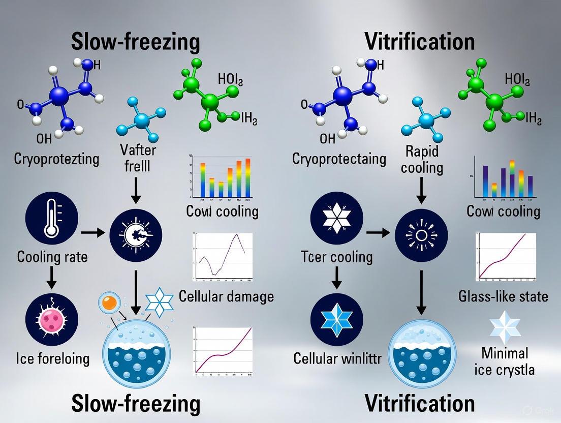
Comparative Analysis: Slow Freezing vs. Vitrification
Fundamental Principles
Table 2: Core Differences Between Slow Freezing and Vitrification
| Parameter | Slow Freezing | Vitrification |
|---|---|---|
| Primary Principle | Controlled extracellular ice formation with cellular dehydration | Ultra-rapid cooling to achieve glassy, ice-free state |
| CPA Concentration | Low (0.5-1.5 M) [3] | High (3-8 M) [3] |
| Cooling Rate | Slow (0.3-2°C/min) [4] | Ultra-rapid (>20,000°C/min) [3] |
| Ice Formation | Extracellular ice inevitable | No ice formation if successful |
| Primary Injury Mechanisms | Osmotic shock, solute effects, extracellular mechanical damage | CPA toxicity, devitrification during warming |
| Equipment Needs | Programmable freezer required [5] | Simple immersion in liquid nitrogen [6] |
Injury Mechanisms in Each Approach
Slow Freezing Injuries:
- Extracellular ice causes mechanical damage to plasma membranes and extracellular matrix
- Progressive solute concentration leads to toxic solution effects
- Incomplete dehydration results in intracellular ice formation in a subset of cells
- Recrystallization during warming can amplify damage [1] [5]
Vitrification Injuries:
- High CPA concentrations cause chemical toxicity and osmotic stress
- Devitrification (ice formation during warming) can cause severe damage
- Fracture damage from thermal stress can occur in vitrified samples
- Incomplete penetration of CPAs leads to heterogeneous protection [1] [5]
The physical state achieved in vitrification is a non-crystalline, glass-like solid that preserves the molecular organization of the liquid state while eliminating destructive ice crystals [3]. However, this state is metastable and susceptible to devitrification if warming rates are insufficient.
Quantitative Assessment of Cryoinjury
Physical Parameters and Cell Survival
Table 3: Quantitative Measures of Cryoinjury in Experimental Models
| Experimental System | Cryopreservation Method | Survival Metric | Result |
|---|---|---|---|
| Bovine Blastocysts [7] | Vitrification | Cryo-survival rate | 86% (138/161) |
| Bovine Blastocysts [7] | Slow freezing | Cryo-survival rate | 57% (81/142) |
| Mouse Tumor Tissues [2] | Zwitterion/DMSO solution | Cell recovery | Higher than commercial CPA |
| Human Ovarian Tissue [6] | Vitrification | DNA fragmentation | Significantly less than slow freezing |
| Human Ovarian Tissue [6] | Vitrification | Normal stromal cells | Significantly more than slow freezing |
| Cell Spheroids [2] | ZD-10/15 solution | Relative cell recovery | 1.51 vs. commercial CPA |
Advanced Measurement Techniques
Modern cryobiology employs multiple techniques to quantify cryoinjury:
- Cryo-electron microscopy: Direct visualization of ice crystal morphology and distribution [1]
- DNA fragmentation assays: Quantification of genetic damage (TUNEL assay) [7]
- Apoptosis markers: Detection of caspase activation and other cell death pathways [7]
- Membrane integrity tests: Assessment of plasma membrane damage post-thaw
- Functional assays: Measurement of cellular functions recovery after thawing [2]
These quantitative approaches enable researchers to move beyond simple morphological assessment to more sophisticated evaluations of cellular recovery.
Experimental Protocols for Studying Cryoinjury
Protocol 1: Assessment of Intracellular Ice Formation
Materials:
- Cryomicroscopy system with temperature control
- Appropriate culture medium
- Cryoprotective agents (DMSO, EG, etc.)
- Fluorescent dyes for membrane integrity (FDA, PI)
Methodology:
- Seed cells on cryomicroscopy stage and maintain at physiological temperature
- Add CPAs at appropriate concentrations
- Initiate cooling protocol with precise rate control (0.1-50°C/min)
- Monitor and record ice formation events visually
- Correlate ice formation events with temperature data
- Assess membrane integrity post-thaw using fluorescent markers
Key Parameters:
- Nucleation temperature (extracellular and intracellular)
- Crystal morphology and growth patterns
- Correlation between IIF and cell death
- Effect of cooling rate on IIF incidence [1]
Protocol 2: Comparative Analysis of Slow Freezing vs. Vitrification
Materials:
- Programmable freezer for slow freezing
- Vitrification carriers (open or closed systems)
- Liquid nitrogen storage system
- Warming equipment (37°C water bath)
Slow Freezing Protocol (based on ovarian tissue cryopreservation):
- Equilibrate tissue in CPA solution (1.5M ethylene glycol + 0.1M sucrose) [4]
- Transfer to programmable freezer
- Cool from 2°C to -6°C at 2°C/min
- Initiate ice seeding at -6°C
- Cool to -40°C at 0.3°C/min
- Rapid cool to -140°C at 10°C/min
- Transfer to liquid nitrogen for storage [4]
Vitrification Protocol (based on ovarian tissue cryopreservation):
- Equilibrate tissue in 3.8% EG + 0.5M sucrose for 3 min at RT
- Transfer to 19% EG + 0.5M sucrose for 1 min
- Final equilibration in 38% EG + 0.5M sucrose for 11 min
- Place on metallic grid and plunge into liquid nitrogen [4]
Assessment:
- Histological evaluation of morphological integrity
- DNA fragmentation assays (TUNEL)
- Apoptosis markers (caspase activation)
- Functional recovery after transplantation [6] [4]
Research Reagent Solutions for Cryoinjury Studies
Table 4: Essential Research Reagents for Cryoinjury Investigation
| Reagent/Category | Specific Examples | Function/Application |
|---|---|---|
| Permeating CPAs | DMSO, glycerol, ethylene glycol, 1,2-propanediol | Penetrate cell membranes, reduce intracellular ice formation |
| Non-Permeating CPAs | Sucrose, trehalose, hydroxyethyl starch, polymers | Create osmotic gradient, promote dehydration |
| Novel Cryoprotectants | Synthetic zwitterions (OE2imC3C), antifreeze proteins | Inhibit ice crystallization, stabilize membranes |
| Commercial Media | CELLBANKER series, CultureSure freezing medium | Standardized cryopreservation solutions |
| Viability Assays | Trypan blue, fluorescein diacetate, propidium iodide | Assess membrane integrity post-thaw |
| Apoptosis Detection | TUNEL assay, caspase-3 staining | Quantify programmed cell death |
| Ice Binding Agents | Antifreeze proteins (AFPs) from fish/insects | Modify ice crystal structure, inhibit recrystallization |
Signaling Pathways and Cellular Responses to Cryoinjury
The cellular response to cryopreservation involves multiple signaling pathways that determine survival versus death outcomes:
Diagram 1: Cellular Signaling Pathways Activated by Cryoinjury
The diagram illustrates how cryoinjury activates multiple signaling pathways that lead to either aberrant cellular activation (through Hippo and PI3K/AKT/mTOR pathways) or cell death (through mitochondrial damage and caspase activation) [5]. In ovarian tissue, for example, cryopreservation and transplantation procedures activate the mTOR pathway through phosphorylation of S6K, leading to abnormal primordial follicle activation and depletion of the follicular reserve [5].
The physics of cryoinjury centers on the destructive capacity of intracellular ice crystals, which damage cellular structures through mechanical disruption, osmotic stress, and signaling pathway activation. The comparison between slow freezing and vitrification reveals that both methods represent different approaches to managing the same fundamental challenge: the phase transition of water and its devastating consequences for cellular integrity.
Future directions in cryopreservation research include the development of novel cryoprotectants like synthetic zwitterions [2], advanced engineering strategies such as cell encapsulation and bioinspired structure design [1], and external physical field technologies for controlling ice crystals in both cooling and warming processes [1]. As our understanding of the physics of cryoinjury deepens, so too will our ability to preserve biological systems with increasing fidelity and success.
{article}
The Slow Freezing Paradigm: Controlled Cooling and Extracellular Ice Crystallization
Within the field of cryobiology, the slow freezing method represents a foundational technique for the long-term preservation of biological materials. This paradigm relies on precisely controlled cooling rates and the strategic management of extracellular ice crystallization to promote cell dehydration and minimize lethal intracellular ice formation. Despite the increasing prominence of vitrification techniques, slow freezing remains a critical procedure in both clinical and research settings. This technical guide delves into the core principles, methodologies, and quantitative outcomes of the slow freezing protocol, framing its role within the ongoing research discourse that compares it with vitrification. We provide detailed experimental protocols, synthesize comparative data in structured tables, and illustrate key conceptual and workflow frameworks to serve researchers, scientists, and drug development professionals.
Cryopreservation is an indispensable technique in biomedical research and clinical medicine, enabling the long-term storage of cells, tissues, and other biological constructs by halting biochemical activity at ultra-low temperatures [3]. The slow freezing paradigm has been a cornerstone of this field for decades. Its fundamental principle involves a carefully controlled, gradual reduction in temperature to facilitate the orderly efflux of water from the cell's interior before it can form destructive ice crystals internally [8] [9]. This process hinges on the physical phenomenon of extracellular ice crystallization. As ice forms in the extracellular solution, dissolved solutes are excluded, creating a hypertonic environment that osmotically draws water out of the cell [8] [3]. The success of this method is therefore a balance between the cooling rate and the cell's permeability to water; too slow a cooling rate subjects cells to prolonged "solution effects" from the hypertonic environment, while too rapid a cooling rate does not allow sufficient time for dehydration, leading to lethal intracellular ice formation [8]. This paper will explore the technical execution of this paradigm, its application in preserving human embryos and other cells, and its standing in direct comparison with the increasingly prevalent technique of vitrification.
Core Principles of Slow Freezing
The slow freezing process is governed by well-established biophysical phenomena. Understanding these core principles is essential for optimizing protocols and interpreting post-thaw outcomes.
The Two-Factor Hypothesis and Cooling Rate Optimization
The theoretical foundation of slow freezing is largely built upon Mazur's "Two-Factor Hypothesis" [8]. This hypothesis describes the interrelationship between cooling rates and cell survival, which is influenced by two primary mechanisms of cryoinjury. First, at inappropriately slow cooling rates, cells are exposed for a prolonged duration to a highly concentrated extracellular environment caused by freeze-concentration of solutes. This leads to toxic "solution effects" and excessive cellular dehydration, causing metabolic disruptions and membrane damage [8] [3]. Second, at excessively rapid cooling rates, there is insufficient time for water to exit the cell osmotically. Consequently, the supercooled intracellular water undergoes nucleation, resulting in the formation of lethal intracellular ice crystals that physically disrupt organelles and membrane structures [8] [9]. The optimal cooling rate, typically around -1°C/minute for many mammalian cells, is a compromise that minimizes both types of injury [10].
The Role of Cryoprotective Agents (CPAs)
Cryoprotective Agents (CPAs) are compounds added to the freezing medium to mitigate cryoinjury. They are broadly categorized as penetrating (membrane-permeating) or non-penetrating (membrane-impermeating) [3].
- Penetrating CPAs, such as Dimethyl sulfoxide (DMSO) and propanediol (PROH), permeate the cell and reduce the fraction of freezable water, thereby lowering the probability of intracellular ice formation. They also dilute the concentration of electrolytes as freezing progresses [8] [3].
- Non-penetrating CPAs, including polymers like sucrose and hydroxyethyl starch, remain outside the cell. They increase the osmolarity of the extracellular solution, promoting a more controlled and gentle cellular dehydration prior to freezing [11] [3].
The combination of both types of CPAs is common in slow freezing protocols. For instance, a commercial slow freezing kit for human cleavage-stage embryos uses 1.5 M PROH as a penetrating CPA and 0.1 M sucrose as a non-penetrating CPA [11].
Seeding and Extracellular Ice Crystallization
A critical technical step in controlled slow freezing is seeding. This involves the manual induction of ice nucleation in the extracellular solution at a temperature just below its freezing point (typically between -2°C and -7°C) [11] [8]. Seeding is performed to prevent massive supercooling of the sample, which could lead to uncontrolled ice growth. By triggering controlled extracellular ice crystallization at a defined moment, seeding ensures a reproducible and predictable osmotic gradient, facilitating a steady efflux of water from the cells during the subsequent cooling phases [9].
Diagram 1: The Slow Freezing Workflow and Cellular Response. This diagram illustrates the key stages of a programmable slow freezing protocol and the corresponding physiological responses of a cell, culminating in a vitrified intracellular state without lethal ice formation.
Experimental Protocols in Practice
The application of the slow freezing paradigm varies depending on the biological material. Below are detailed methodologies for two critical applications: freezing human cleavage-stage embryos and general mammalian cells.
Protocol 1: Slow Freezing of Human Cleavage-Stage Embryos
This protocol is adapted from a prospective randomized trial and a retrospective study comparing different cryopreservation methods [12] [11].
- Materials and Reagents: Commercial embryo freezing kit (e.g., Irvine Scientific) containing F1 medium (1.5 M PROH) and F2 medium (1.5 M PROH + 0.1 M sucrose); Programmable freezer (e.g., Planer Kryo 10); Cryogenic straws or vials; Liquid nitrogen storage system [11].
- Freezing Procedure:
- Equilibration: At room temperature (RT), expose good-quality cleavage-stage embryos (Day 2 or 3) first to F1 medium for 10 minutes, then transfer to F2 medium. Alternatively, a simplified one-step method uses F2 medium only for 10 minutes [11].
- Loading: Aspirate the embryos in a minimal volume of F2 medium into cryogenic straws.
- Programmable Freezing: Place straws in the programmable freezer, starting at 23°C.
- Cool from 23°C to -7°C at a rate of -2°C/min.
- Seeding: Perform manual seeding at -7°C using a forceps or cotton swab pre-cooled in LN2. Hold for 5-10 minutes to allow ice propagation.
- Cool from -7°C to -30°C at a slow rate of -0.3°C/min.
- Finally, cool rapidly from -30°C to -150°C at -50°C/min.
- Storage: Plunge the straws into liquid nitrogen for long-term storage at -196°C [11].
- Thawing and Rehydration:
- Rapidly thaw straws in a 30-37°C water bath or air.
- Rehydrate embryos by step-wise dilution in decreasing concentrations of PROH and sucrose (e.g., 1.0 M PROH/0.2 M sucrose, then 0.5 M PROH/0.2 M sucrose, then 0.2 M sucrose), spending 5-10 minutes in each solution at RT [11].
- Assess survival based on blastomere integrity. An embryo is typically considered to have survived if ≥50% of its original blastomeres remain intact [11].
Protocol 2: Standard Slow Freezing of Mammalian Cells
This is a generalized protocol for cryopreserving cell lines, common in research and drug development laboratories [10].
- Materials and Reagents: Log-phase cells; Complete growth medium; Cryoprotective agent (e.g., DMSO); Serum (e.g., Fetal Bovine Serum) or defined protein-free supplements; Sterile cryogenic vials; Controlled-rate freezer or isopropanol chamber (e.g., "Mr. Frosty"); Liquid nitrogen storage container [10].
- Freezing Procedure:
- Harvest and Count: Harvest cells via trypsinization (adherent cells) or centrifugation (suspension cells). Determine total cell count and viability (should be >90%). A typical target concentration is 1x10^6 to 1x10^7 cells/mL [10].
- Freezing Medium Preparation: Prepare freezing medium, commonly comprising 90% complete growth medium (e.g., basal medium + 10-20% serum) and 10% DMSO. Alternatively, use a commercial, serum-free, ready-to-use freezing medium like CryoStor CS10 or Gibco Synth-a-Freeze [13] [10].
- Resuspension and Aliquoting: Centrifuge the cell suspension, discard the supernatant, and gently resuspend the cell pellet in the cold freezing medium. Aliquot the cell suspension into cryovials.
- Controlled Cooling: Use one of two methods:
- Active Control: Place vials in a controlled-rate freezer and cool at approximately -1°C/min down to -80°C or lower before transferring to LN2.
- Passive Control: Place vials in an isopropanol freezing chamber and store at -80°C overnight. This apparatus achieves an approximate cooling rate of -1°C/min [10].
- Long-term Storage: Transfer the frozen cryovials to a liquid nitrogen tank for storage in the vapor phase (below -135°C) to prevent risks associated with liquid-phase storage [10].
Quantitative Data: Slow Freezing vs. Vitrification
A core component of the research paradigm is the quantitative comparison of slow freezing with vitrification. The following tables synthesize survival and clinical outcome data from multiple studies.
Table 1: Post-Warming Survival Rates of Human Embryos after Slow Freezing vs. Vitrification
| Embryo Stage | Cryopreservation Method | Survival Rate (%) | Study Details | Citation |
|---|---|---|---|---|
| Cleavage-Stage | Slow Freezing | 63.8% | 330 embryos warmed | [12] |
| Cleavage-Stage | Vitrification (Irvine) | 89.4% | 330 embryos warmed | [12] |
| Cleavage-Stage | Vitrification (Vitrolife) | 87.6% | 330 embryos warmed | [12] |
| Cleavage-Stage | Slow Freezing (Two-Step) | 83.1% | 891 embryos thawed | [11] |
| Cleavage-Stage | Slow Freezing (One-Step) | 86.9% | 693 embryos thawed | [11] |
| Blastocyst | Slow Freezing | Varies | Meta-analysis | [14] |
| Blastocyst | Vitrification | Significantly Higher | Meta-analysis (OR: 2.20) | [14] |
Table 2: Comparison of Clinical Outcomes and Technical Aspects
| Parameter | Slow Freezing | Vitrification | Citation |
|---|---|---|---|
| Implantation Rate (per embryo) | ~9.9% - 21.4% (NS) | ~12.1% - 17.0% (NS) | [12] |
| Cooling Rate | Slow (≈ -0.3°C/min) | Ultra-rapid (≈ -20,000°C/min) | [12] [9] |
| CPA Concentration | Low (e.g., 1.5 M PROH) | Very High (e.g., 6-8 M total) | [8] [3] |
| Physical Principle | Extracellular ice crystallization, cell dehydration | Glass-like solidification, no ice | [3] [9] |
| Primary Cryoinjury Risks | Solution effects, intracellular ice (if uncontrolled) | CPA toxicity, fracture damage | [8] [3] |
The Scientist's Toolkit: Essential Reagents and Equipment
A successful slow freezing protocol relies on a suite of specialized reagents and equipment. The following table details key components of a researcher's toolkit for this technique.
Table 3: Key Research Reagent Solutions and Equipment for Slow Freezing
| Item | Function/Description | Example Products/Formats |
|---|---|---|
| Permeating CPAs | Penetrate cell membrane, reduce intracellular ice formation. | DMSO, Ethylene Glycol (EG), Propanediol (PROH) |
| Non-Permeating CPAs | Create osmotic gradient for controlled dehydration. | Sucrose, Trehalose, Hydroxyethyl Starch |
| Base & Freezing Media | Provide nutrients, pH buffering, and protein support during freeze-thaw. | Commercial kits (e.g., Irvine Scientific), CryoStor CS10, Synth-a-Freeze |
| Programmable Freezer | Provides precise, active control over cooling rates for protocol standardization. | Planer Kryo 10 Series |
| Passive Cooling Devices | Insulated containers that provide a reproducible, passive cooling rate of ~-1°C/min. | Nalgene "Mr. Frosty", Corning CoolCell |
| Cryogenic Storage Vials | Sterile, leak-proof containers designed for ultra-low temperature storage. | Internal-threaded cryovials (e.g., Corning) |
| Liquid Nitrogen Storage | Long-term storage of frozen samples at -135°C to -196°C. | LN2 tanks (vapor phase storage recommended) |
The slow freezing paradigm, with its foundation in controlled cooling and the management of extracellular ice crystallization, remains a vital and well-understood technique in the cryobiologist's arsenal. While meta-analyses and numerous studies clearly demonstrate that vitrification is associated with significantly higher post-warming survival rates for sensitive materials like oocytes and embryos, slow freezing is far from obsolete [12] [14]. Its protocols are robust, standardized, and less susceptible to operator-induced variation and CPA toxicity concerns. Furthermore, modifications such as the one-step slow freezing method continue to refine its efficiency and outcomes [11]. The choice between slow freezing and vitrification is, therefore, not a simple matter of superiority but must be contextual, depending on the specific biological material, the required throughput, the available equipment, and the expertise of the personnel. Future research will continue to refine both paradigms, potentially leading to hybrid techniques that further enhance the survival and functionality of cryopreserved cells and tissues for research and clinical applications.
{/article}
Cryopreservation represents a fundamental technique in modern bioscience, enabling the long-term storage of biological systems ranging from individual cells to complex tissues. Within this field, two principal methodologies have emerged: conventional slow freezing and vitrification. While slow freezing involves controlled ice formation in extracellular spaces, vitrification offers an alternative pathway by achieving a complete ice-free solidification of aqueous solutions into a glass-like state using high concentrations of cryoprotective agents (CPAs) and ultra-rapid cooling [15] [16]. This technical guide examines the core principles and methodologies underlying vitrification solutions, with particular emphasis on the critical interplay between CPA concentration and cooling rates required to achieve stable vitreous states while minimizing cryoprotectant toxicity.
The fundamental advantage of vitrification lies in its ability to circumvent the mechanically and osmotically damaging effects of ice crystallization, which represents a significant limitation of slow-freezing protocols [17]. When properly executed, vitrification preserves the native molecular and structural organization of biological systems, maintaining viability and functionality after rewarming. This approach has demonstrated superior outcomes for sensitive biological materials including oocytes, embryos, stem cells, and complex tissues that are incompatible with conventional freezing methods [18] [6]. The following sections provide a comprehensive technical examination of vitrification methodology, from theoretical foundations to practical application and emerging technological innovations.
Theoretical Foundations: Principles of Glass Formation
Thermodynamic Basis of Vitrification
Vitrification represents a non-equilibrium process wherein a liquid solution transitions into an amorphous glassy solid without undergoing crystalline formation. This transition occurs when a solution is cooled at sufficient rates to bypass ice nucleation and crystal growth, resulting in a dramatic increase in viscosity that effectively immobilizes molecules in their liquid-state configurations [15]. The thermodynamic relationship between temperature and molecular mobility during this process is fundamental to understanding vitrification protocol design.
The vitrification process navigates a critical temperature pathway between several key transition points as illustrated in Figure 1. As an aqueous solution cools below its melting temperature (Tm), it enters a metastable supercooled state where ice formation is thermodynamically favorable but kinetically inhibited. With further cooling, the solution approaches the glass transition temperature (Tg), where molecular motion slows sufficiently to form an amorphous solid [15] [16]. The critical challenge in vitrification protocol design lies in traversing the temperature zone between Tm and Tg rapidly enough to avoid ice crystallization, either through enhanced cooling rates or manipulation of solution composition to depress the homogeneous nucleation temperature.
Critical Cooling and Warming Rates
The successful implementation of vitrification protocols depends critically on achieving cooling and warming rates that exceed material-specific thresholds. The critical cooling rate (CCR) defines the minimum rate required to prevent ice formation during temperature descent, while the critical warming rate (CWR) represents the minimum rate needed to prevent ice formation during temperature ascent (devitrification) [18] [16]. These critical rates are profoundly influenced by CPA concentration, with higher CPA concentrations depressing both CCR and CWR to more practically achievable levels.
For pure water, the theoretical CCR exceeds 10,000,000°C/min, making vitrification impossible for practically-sized biological samples without cryoprotective additives [15] [16]. The introduction of CPAs at sufficient concentrations dramatically reduces the CCR by increasing solution viscosity and interfering with water molecule organization into crystal lattices. However, this benefit is counterbalanced by increasing CPA toxicity at elevated concentrations, creating the fundamental optimization challenge in vitrification solution design: balancing CPA concentration against required cooling/warming rates to achieve vitrification while maintaining biological viability [17] [18].
Vitrification Solution Composition
Cryoprotectant Agents: Mechanisms and Classifications
Cryoprotectant agents function through multiple mechanisms to enable vitrification at practically achievable cooling rates. Their primary action involves * disrupting hydrogen bonding* between water molecules, increasing solution viscosity and depressing the freezing point of water. Additionally, CPAs stabilize cellular structures by interacting with membrane phospholipids and proteins, preventing denaturation during volume changes and temperature extremes [16].
CPAs are broadly categorized according to their membrane permeability characteristics:
- Permeating CPAs: Low molecular weight compounds that readily cross cell membranes (e.g., propylene glycol, ethylene glycol, glycerol). These agents provide intracellular protection but can induce osmotic stress during loading and removal.
- Non-permeating CPAs: Larger molecules that remain extracellular (e.g., trehalose, sucrose, polymers). These compounds primarily function as osmotic buffers and extracellular glass formers, and can stabilize cell membranes through specific molecular interactions [17] [19].
Formulation Strategies and Toxicity Mitigation
Effective vitrification solutions typically employ balanced combinations of permeating and non-permeating CPAs to achieve the necessary glass-forming tendency while mitigating individual component toxicity. The composition of VS55, a well-characterized vitrification solution, illustrates this balanced approach with 3.1 M glycerol, 3.1 M propylene glycol, and 0.5 M sucrose [18]. Empirical optimization has demonstrated that multi-component CPA cocktails often provide superior performance compared to single-CPA solutions at equivalent total concentrations, likely due to distributed toxicity profiles and synergistic effects on solution properties [19].
Toxicity management represents a critical aspect of vitrification solution design, particularly for sensitive cell types and complex tissues. Strategy implementations include:
- Multi-step loading protocols with progressively increasing CPA concentrations to minimize osmotic shock
- Temperature reduction during CPA exposure to decrease chemical reaction rates and metabolic activity
- Optimization of exposure durations to balance adequate permeation against cumulative toxicity
- Incorporation of specific membrane stabilizers and free radical scavengers [19] [16]
Table 1: Characteristic Vitrification Solution Formulations
| Solution Name | CPA Composition | Total Molarity | Application Examples | Cooling Rate Requirements |
|---|---|---|---|---|
| VS55 | 3.1 M Glycerol + 3.1 M Propylene Glycol + 0.5 M Sucrose | 6.7 M | Kidney slices, Ovarian tissue | >20°C/min [18] |
| QMC Solution | 2 M PROH + 0.5 M Trehalose | 2.5 M | Murine embryonic stem cells | >100,000°C/min [17] |
| EG-Sorbitol | 2.24 M EG + 1.57 M Sorbitol | 3.81 M | Drosophila embryos | >20,000°C/min [18] |
| VS70 | 4.25 M Glycerol + 4.25 M Propylene Glycol + 0.67 M Sucrose | 9.17 M | Bioengineered epithelial constructs | >45°C/min [19] |
Experimental Protocols and Methodologies
Quartz Microcapillary (QMC) Vitrification Protocol
The quartz microcapillary technique represents an advanced approach for achieving ultra-rapid cooling rates that enable vitrification at lower CPA concentrations. This method exploits the exceptional thermal properties of quartz and minimal dimensions to maximize heat transfer efficiency [17] [20].
Materials and Equipment:
- Quartz microcapillaries (0.2 mm outer diameter, 0.01 mm wall thickness)
- Controlled-rate cooling apparatus or liquid nitrogen immersion system
- CPA solutions prepared in appropriate base medium
- Murine embryonic stem cells (or other target cells) at 70-80% confluence
Methodology:
- Cell Preparation: Harvest and concentrate cells to approximately 2×10^7 cells/mL in maintenance medium.
- CPA Loading: Incubate cells sequentially in equilibration solution (1-2 M permeating CPA) for 5-10 minutes at 4°C, followed by vitrification solution (2 M PROH + 0.5 M trehalose) for 30-60 seconds.
- Loading into QMC: Aspirate approximately 0.5-1 µL of cell suspension in vitrification solution into the quartz microcapillary.
- Cooling: Immediately plunge the loaded QMC directly into liquid nitrogen, achieving cooling rates >100,000°C/min.
- Storage: Transfer QMC to cryogenic storage tanks maintaining temperatures below -135°C.
- Rewarming: Rapidly remove QMC from liquid nitrogen and immediately plunge into rewarming medium at 37°C.
- CPA Removal: Transfer cells through a series of decreasing CPA concentrations (0.75 M, 0.5 M, 0.25 M sucrose in base medium) at 4°C for osmotic balancing.
- Viability Assessment: Perform cell attachment assays, proliferation measurements, and lineage-specific differentiation potential evaluations [17] [20].
This protocol demonstrates that murine embryonic stem cells cryopreserved using the QMC method maintain viability (>70% attachment efficiency relative to controls), proliferation rates, and pluripotency markers (Oct-4 expression, SSEA-1 presentation, alkaline phosphatase activity) at levels comparable to non-frozen controls [17].
Joule Heating-Based Vitrification Platform
Recent advances in rewarming technology have addressed the critical challenge of achieving sufficiently rapid and uniform warming to prevent devitrification in larger biosystems. The joule heating platform utilizes electrical current passage through a conductor in contact with vitrified samples to generate extremely high warming rates through resistive heating [18].
Materials and Equipment:
- Stainless steel sheet (12 µm thickness) or mesh (30 µm wire diameter)
- RC discharge circuit pulse generator (e.g., ECM 830)
- Custom sample mounting fixtures
- Liquid nitrogen cooling system
- VS55 or other appropriate vitrification solution
Methodology:
- CPA Loading: Incubate biosystems (adherent cells, Drosophila embryos, or tissue slices) in VS55 using multi-step concentration increases at 4°C.
- Sample Preparation: Place biosystems in contact with stainless steel sheet or mesh, removing excess CPA solution to minimize thermal mass.
- Cooling: Plunge samples directly into liquid nitrogen, achieving vitrification.
- Joule Heating Rewarming: Connect steel conductor to pulse generator and administer controlled electrical pulses (10 µs to 100 ms duration) to achieve warming rates from 50,000 to 600,000,000°C/min.
- CPA Removal: Transfer rewarmed biosystems through decreasing CPA concentration solutions.
- Functional Assessment: Conduct viability staining, membrane integrity tests, and tissue-specific functional assays [18].
This platform technology has demonstrated successful cryopreservation of biosystems across multiple scales, including adherent cells (~4 µm thickness), Drosophila embryos (~50 µm), and rat kidney slices (~1.2 mm) using relatively low CPA concentrations (2-4 M) [18].
Comparative Analysis: Vitrification versus Slow Freezing
The fundamental distinction between vitrification and slow freezing protocols lies in their approach to managing the physical state of water during cryopreservation. While both methods aim to stabilize biological systems at cryogenic temperatures, their mechanisms of action and consequent applications differ significantly as detailed in Table 2.
Table 2: Vitrification versus Slow Freezing Method Comparison
| Parameter | Vitrification | Slow Freezing |
|---|---|---|
| Physical State | Amorphous glass | Crystalline ice + unfrozen fraction |
| CPA Concentration | High (4-8 M total) | Low (1-2 M) |
| Cooling Rate | Ultra-rapid (>100°C/min to >100,000°C/min) | Slow (0.3-2°C/min) |
| Ice Formation | Eliminated | Extracellular, controlled |
| Primary Damage Mechanisms | CPA toxicity, Devitrification | Solute effects, Intracellular ice, Mechanical damage |
| Technical Complexity | High (requires optimization) | Moderate (standardized) |
| Application Scope | Oocytes, embryos, complex tissues, organ fragments | Cell suspensions, robust tissues |
Meta-analytical comparisons of vitrification versus slow freezing for human ovarian tissue demonstrate vitrification's association with significantly less DNA fragmentation in primordial follicles (Relative Risk = 0.71; 95% CI, 0.62-0.80; P < 0.00001) and better preservation of normal stromal cells (RR = 1.69; 95% CI, 1.47-1.94; P < 0.00001) [6]. However, both methods showed equivalent performance in maintaining the proportion of morphologically intact primordial follicles (OR = 0.98; 95% CI, 0.74-1.28; P = 0.86), indicating context-dependent advantages [21] [6].
The Scientist's Toolkit: Essential Research Reagents and Materials
Successful implementation of vitrification protocols requires specialized materials and reagents optimized for specific biological applications. The following toolkit compiles essential components referenced across experimental methodologies.
Table 3: Essential Research Reagents for Vitrification Studies
| Reagent/Material | Function | Application Examples | Technical Notes |
|---|---|---|---|
| 1,2-Propanediol (PROH) | Permeating CPA | Murine ES cells, Oocytes | Lower toxicity alternative to DMSO [17] |
| Ethylene Glycol | Permeating CPA | Ovarian tissue, Drosophila embryos | Rapid permeation kinetics [21] [18] |
| Trehalose | Non-permeating CPA | Stem cells, Bioengineered constructs | Membrane stabilization, osmotic buffer [17] [19] |
| Quartz Microcapillaries | Ultra-rapid cooling device | ES cells, Sensitive cell types | 0.2 mm OD, 0.01 mm wall thickness [17] |
| Stainless Steel Mesh | Joule heating conductor | Tissue slices, Drosophila embryos | 30 µm wire diameter, 38 µm aperture [18] |
| Sucrose | Osmotic buffer | CPA removal steps, Vitrification solution | Stabilizes membranes during CPA dilution [21] [19] |
| Cinchonidine | API for vitrification studies | Pharmaceutical formulation | High crystallization tendency model compound [22] |
Vitrification technology represents a rapidly advancing frontier in cryopreservation science, offering solutions to fundamental limitations of conventional slow-freezing methodologies. The continued refinement of vitrification protocols centers on optimizing the critical balance between CPA toxicity minimization and achievable cooling/warming rates, enabled by advanced materials and engineering approaches.
Emerging trends in the field include the development of high-throughput vitrification platforms for drug discovery applications, equilibrium approaches using liquidus tracking to minimize supercooling, and nanoparticle-assisted warming technologies to improve thermal uniformity in larger systems [19] [18] [16]. The ongoing translation of vitrification methodologies from cellular to tissular and organ-level applications holds particular promise for transforming transplantation medicine, bio-banking, and regenerative therapeutics through the establishment of true biological storage systems.
As vitrification protocols continue to evolve, their integration within comparative cryopreservation research frameworks will further elucidate the fundamental biophysical principles governing successful recovery of complex biological systems from cryogenic storage. This advancement will ultimately enable the precise customization of preservation methodologies to the specific requirements of diverse biological materials from single cells to intact organs.
Cryopreservation is an indispensable technique in biomedical research and clinical practice, enabling long-term preservation of cells, tissues, and organs by suspending metabolic processes at ultra-low temperatures. The fundamental challenge in protocol development lies in navigating the critical trade-off between two competing injury mechanisms: the damaging effects of ice crystal formation and the cytotoxic effects of cryoprotective agents (CPAs) required to suppress ice formation [23] [24]. This technical guide examines the core principles and experimental approaches underlying this balance, framed within ongoing research into slow-freezing versus vitrification protocols.
The phase behavior of water during cooling and warming cycles dictates all cryopreservation outcomes. During freezing, water can either undergo a liquid-to-solid crystalline transition (freezing) or solidify into an amorphous, glass-like state (vitrification) without forming ice crystals [23]. The pathway taken determines the potential injury mechanisms—mechanical damage from ice in conventional freezing versus chemical toxicity from high CPA concentrations in vitrification. This guide provides researchers with a comprehensive framework for evaluating these trade-offs in their experimental systems.
Fundamental Injury Mechanisms
Ice-Mediated Damage
Ice formation presents multiple threats to cellular integrity throughout the cryopreservation cycle:
- Mechanical damage: Ice crystals can physically disrupt cellular membranes, organelles, and cytoskeletal structures [24] [3]. Intracellular ice formation (IIF) is almost universally lethal to cells [25].
- Solute effects: As ice forms, it excludes solutes, leading to hypertonic conditions in the remaining unfrozen fraction. This causes severe osmotic stress, cellular dehydration, and shrinkage that can damage membrane systems [23] [24].
- Recrystallization: During thawing, ice crystals can grow larger through recrystallization, particularly in the temperature range between -15°C and -60°C, exacerbating mechanical damage [24].
The extent and location of ice formation depend critically on cooling rates. At slow cooling rates (approximately 1°C/min), extracellular ice forms first, drawing water out of cells through osmotic effects and potentially leading to excessive dehydration [23] [13]. At rapid cooling rates, intracellular water cannot exit cells quickly enough, resulting in lethal intracellular ice formation [24].
CPA-Mediated Toxicity
Cryoprotective agents mitigate ice damage but introduce their own risks:
- Metabolic toxicity: High concentrations of CPAs can disrupt cellular metabolism, enzyme function, and signal transduction pathways [26] [24]. DMSO, for instance, has been shown to alter the epigenetic landscape and cellular differentiation pathways [3].
- Osmotic stress: During CPA addition and removal, cells experience significant volume changes that can cause membrane damage or rupture if not properly controlled [24] [27].
- Oxidative stress: The cryopreservation process, including CPA exposure, can generate reactive oxygen species (ROS) that damage lipids, proteins, and DNA [24].
The toxicity of CPAs generally increases with concentration, exposure time, and temperature [26] [27]. This creates a fundamental challenge for vitrification, which requires very high CPA concentrations (typically 6-9M) to completely suppress ice formation [23] [26].
Table 1: Key Characteristics of Primary Cryoprotective Agents
| CPA | Molecular Weight (Da) | Membrane Permeability | Relative Toxicity | Common Applications |
|---|---|---|---|---|
| DMSO | 78.1 | Moderate | Moderate-High | Cell lines, tissues, organs |
| Glycerol | 92.1 | Low | Low-Moderate | Microorganisms, spermatozoa |
| Ethylene Glycol | 62.1 | High | Low | Oocytes, embryos |
| Propylene Glycol | 76.1 | Moderate | Moderate | Oocytes, reproductive tissues |
| Formamide | 45.0 | High | High | Component in CPA mixtures |
Protocol Selection: Slow Freezing vs. Vitrification
Slow Freezing Principles
Slow freezing protocols employ controlled cooling rates (typically ~1°C/min) and relatively low CPA concentrations (1-2M). The gradual temperature decrease allows extracellular ice formation while permitting sufficient time for cellular dehydration, thereby minimizing intracellular ice formation [23] [13]. The process can be summarized as follows:
- CPA equilibration: Cells are exposed to permeating CPAs (e.g., DMSO, glycerol) at concentrations typically below 2M.
- Controlled cooling: Samples are cooled at precisely controlled rates using programmable freezers or passive cooling devices.
- Extracellular ice formation: Ice nucleation is initiated in the extracellular solution at temperatures slightly below the freezing point.
- Osmotic dehydration: As extracellular ice forms, solute concentration increases, drawing water out of cells.
- Storage: Samples are transferred to long-term storage at <-135°C (typically in liquid nitrogen).
- Thawing: Rapid warming is employed to minimize recrystallization [13].
The optimal cooling rate is cell-type dependent, reflecting differences in membrane permeability and surface area-to-volume ratio [23]. Slow freezing better accommodates larger sample volumes and facilitates more homogeneous CPA distribution [23]. However, it cannot completely eliminate ice-related damage and requires specialized, costly equipment [23] [13].
Vitrification Principles
Vitrification represents a fundamentally different approach—complete avoidance of ice crystallization through ultra-rapid cooling and high CPA concentrations (typically 6-9M). The process transforms the aqueous solution directly into a glassy state without ice formation [23] [26]. Key requirements include:
- High CPA concentrations: Sufficient CPAs must be present to depress the freezing point and increase solution viscosity.
- Rapid cooling: Extremely high cooling rates (>10,000°C/min) are typically required to outpace ice nucleation.
- Rapid warming: Similarly high warming rates are necessary to prevent devitrification (ice formation during warming) [23] [25].
The critical cooling rate (Vccr) and critical warming rate (Vcwr) are key parameters determining vitrification success [23] [26]. Higher CPA concentrations lower both critical rates but increase toxicity risks [23]. Recent research demonstrates that warming rate is often more critical than cooling rate for maintaining viability [25].
Comparative Analysis
Table 2: Protocol Comparison: Slow Freezing vs. Vitrification
| Parameter | Slow Freezing | Vitrification |
|---|---|---|
| CPA Concentration | Low (1-2M) | High (6-9M) |
| Cooling Rate | Slow (~1°C/min) | Ultra-rapid (>10,000°C/min) |
| Ice Formation | Extracellular ice permitted, intracellular ice minimized | Complete suppression in successful protocols |
| Primary Risks | Solution effects, excessive dehydration, intracellular ice (if too fast) | CPA toxicity, osmotic shock, devitrification during warming |
| Sample Volume | Wide range (small to large volumes) | Typically small volumes (<1µL for highest rates) |
| Equipment Needs | Controlled-rate freezer or passive cooling device | Vitrification devices (Cryotop, OPS, etc.), liquid nitrogen |
| Protocol Standardization | Highly reproducible and quantifiable | More variable, technique-dependent |
Quantitative Toxicity Assessment
Toxicity Measurement Models
Recent advances have established quantitative frameworks for CPA toxicity assessment. A toxicity cost function approach models cumulative damage during CPA exposure [26]:
$$ I{\text{tox}} = \int0^{tf} k \, dt = \int0^{t_f} \beta C^{\alpha} dt $$
Where:
- ( I_{\text{tox}} ) = toxicity cost function (lower values indicate less damage)
- ( t_f ) = duration of CPA exposure
- ( k ) = toxicity rate
- ( C ) = CPA concentration inside the tissue
- ( \beta ), ( \alpha ) = CPA-specific parameters
Cell viability following CPA exposure can be modeled as:
$$ \frac{N}{N0} = \exp(-I{\text{tox}}) $$
Where ( N_0 ) and ( N ) represent cell viability before and after exposure, respectively [26].
Experimental Toxicity Data
High-throughput screening approaches have enabled systematic toxicity comparisons across CPA formulations. Recent research evaluating scalable CPAs for organ vitrification reported significant differences in toxicity rates [26]:
- VM3 (8.46M): toxicity rate k = 0.007958 min⁻¹
- M22-PVP (9.34M): toxicity rate k = 0.01755 min⁻¹
- M22 (9.35M): toxicity rate k = 0.02339 min⁻¹
These findings demonstrate that formulation differences significantly impact toxicity, even at similar molar concentrations [26]. Research has identified that CPA toxicity increases with both concentration and exposure duration, creating time-dependent constraints on protocol development [27].
Advanced Experimental Approaches
High-Throughput Screening
Traditional CPA development has been limited to a narrow range of chemicals. Recent advances in automated liquid handling and screening technologies have dramatically increased throughput for CPA discovery [27] [28]. A fluorescence-based method enables simultaneous assessment of membrane permeability and toxicity in 96-well plates, allowing rapid screening of candidate molecules [28].
Key advantages of this approach include:
- Volume-dependent fluorescence: Intracellular calcein fluorescence serves as a marker for cell volume changes.
- Parallel assessment: Permeability and toxicity data are gathered from the same well plate.
- Temperature control: Measurements can be conducted at both 4°C and room temperature.
- Rapid screening: ~100x faster than previous permeability measurement methods [28].
This methodology has identified 23 candidate molecules with favorable permeability and toxicity profiles from an initial screen of 27 chemicals [28].
Mixture Toxicity Reduction
CPA mixtures can reduce overall toxicity through two primary mechanisms:
- Mutual dilution: Each CPA in a mixture lowers the concentration of the others, reducing toxicity while maintaining total solute concentration [27].
- Toxicity neutralization: Specific CPA combinations can partially neutralize each other's toxicity [27].
Research has confirmed previously observed neutralization of formamide toxicity by DMSO and identified new neutralization effects in formamide-glycerol mixtures [27]. These findings highlight the potential for optimized CPA cocktails that maintain vitrification capability while reducing toxicity.
Ice Visualization Techniques
Synchrotron-based X-ray diffraction has provided unprecedented insights into ice formation dynamics during cryopreservation [25]. This approach enables:
- Quantitative ice detection: Sensitive to ice volume fractions below 1%
- Time-resolved analysis: Observation of ice formation during rapid warming transients
- Crystal characterization: Identification of ice structure (cubic vs. hexagonal) and grain size
Critical findings from this research include:
- Oocytes cooled with current practice protocols typically show no ice after cooling but develop large ice fractions during warming
- Most ice-related damage likely occurs during the warming phase
- Increasing cooling rates allows oocytes to remain essentially ice-free during both cooling and warming [25]
These insights clarify that warming protocol optimization is equally important as cooling protocol development.
The Scientist's Toolkit
Table 3: Essential Research Reagents and Materials
| Category | Specific Examples | Function/Application |
|---|---|---|
| Permeating CPAs | DMSO, glycerol, ethylene glycol, propylene glycol | Penetrate cell membranes to protect against intracellular ice |
| Non-Penetrating CPAs | Sucrose, trehalose, hydroxyethyl starch, PVP | Create osmotic gradient, stabilize membranes, modify ice growth |
| Commercial Media | CryoStor, CELLBANKER series, mFreSR | Standardized, optimized formulations for specific cell types |
| Vitrification Devices | Cryotop, Open Pulled Straw (OPS) | Enable ultra-rapid cooling through minimal volume design |
| Cooling Equipment | Controlled-rate freezers, Mr. Frosty, CoolCell | Achieve precise cooling rates for slow freezing protocols |
| Assessment Tools | Synchrotron XRD, calcein-AM, PrestoBlue | Quantify ice formation, membrane integrity, and cell viability |
Decision Framework and Protocol Optimization
Protocol Selection Algorithm
Emerging Solutions and Future Directions
Several innovative approaches show promise for mitigating the fundamental toxicity-ice formation trade-off:
- Ice-binding materials: Antifreeze proteins and synthetic mimics can inhibit ice recrystallization, potentially allowing reduced CPA concentrations [24].
- Toxicity-neutralizing mixtures: Systematic screening of CPA combinations identifies mixtures with mutually reduced toxicity [27].
- Enhanced warming rates: Advanced warming technologies (e.g., laser heating, conductive materials) achieve faster warming, reducing devitrification risks [25].
- Biochemical modulation: Antioxidants and caspase inhibitors can mitigate secondary injury mechanisms, improving post-thaw recovery [24].
These approaches, combined with advanced screening methodologies, represent the future of cryopreservation protocol development—moving beyond simple trade-offs toward integrated solutions that address multiple injury mechanisms simultaneously.
The fundamental trade-off between CPA toxicity and ice crystal formation remains the central consideration in cryopreservation protocol selection. Slow freezing emphasizes control of ice formation through precise cooling kinetics and lower CPA exposure, while vitrification prioritizes complete ice avoidance through ultra-rapid temperature changes and high CPA concentrations. The optimal approach depends critically on the biological system, sample constraints, and application requirements.
Recent advances in high-throughput screening, ice visualization, and mixture optimization provide researchers with powerful tools to navigate this trade-off space more effectively. By applying quantitative toxicity assessment, exploring CPA combinations with neutralizing effects, and prioritizing both cooling and warming protocol optimization, researchers can develop cryopreservation protocols that maximize post-preservation recovery and functionality.
Protocol Deep Dive: Tissue-Specific Applications from Gametes to Complex Tissues
Ovarian tissue cryopreservation (OTC) has emerged as a vital fertility preservation strategy for women and girls facing gonadotoxic treatments, particularly those who cannot undergo ovarian stimulation or require immediate therapy initiation [29] [30]. This technique involves the surgical retrieval, freezing, and storage of ovarian cortical tissue containing primordial follicles, with the intention of future transplantation to restore fertility and endocrine function [29]. Since the first successful live birth reported in 2004, over 200 babies have been born worldwide through this technology [30] [31].
The cryopreservation of ovarian tissue presents unique challenges due to its complex composition of multiple cell types and structures [6]. The ultimate success of OTC depends on preserving the viability and function of not only primordial follicles but also the surrounding stromal cells, extracellular matrix, and vascular networks that support follicular development [31]. Two principal cryopreservation methods have been developed: conventional slow freezing and vitrification. While slow freezing has been the established standard in most clinical centers, vitrification is emerging as a promising alternative with potential advantages [30] [6].
This technical guide provides an in-depth comparison of these two cryopreservation methodologies, focusing on their biomechanical impacts, tissue integrity preservation, and functional outcomes post-transplantation. The analysis is framed within ongoing research debates regarding their relative effectiveness and the biological fundamentals underlying their protocols.
Fundamental Principles of Cryopreservation
Biophysical Basis of Cryopreservation
Cryopreservation conserves biological materials by reducing temperatures to levels that suspend metabolic activity. The primary challenge lies in navigating the intermediate temperature zone (approximately 0°C to -15°C) where ice crystal formation predominantly causes cryoinjury [32]. Both slow freezing and vitrification aim to minimize this damage through different physical approaches.
Slow freezing relies on controlled, gradual cooling that promotes extracellular ice formation, thereby increasing the solute concentration in the extracellular space. This creates an osmotic gradient that draws water out of cells, minimizing lethal intracellular ice formation [30]. The process requires precise cooling rates optimized for different cell types and uses relatively low concentrations of cryoprotectants (typically around 1.5 M) [30].
Vitrification employs ultra-rapid cooling rates and high concentrations of cryoprotectants (often exceeding 40%) to achieve a glass-like, amorphous solid state without ice crystal formation [30] [6]. This process avoids the mechanical damage associated with ice formation but introduces challenges related to cryoprotectant toxicity and sufficient permeation through dense tissue [32].
Cryoprotectant Agents (CPAs)
Cryoprotectants are essential components of both freezing protocols and can be categorized as:
- Permeating CPAs: Small molecules that diffuse through plasma membranes (e.g., dimethyl sulfoxide (DMSO), ethylene glycol (EG), propylene glycol). These form hydrogen bonds with intracellular water, reducing freezing points and inhibiting ice crystallization [30].
- Non-permeating CPAs: Larger molecules that remain extracellular (e.g., sucrose, trehalose, proteins). These create osmotic gradients that promote cellular dehydration and increase solution viscosity [30].
The choice and combination of CPAs significantly impact tissue survival. DMSO is commonly used in both methods, while EG is frequently employed in vitrification protocols due to its lower cytotoxicity [30] [33]. Recent research indicates that DMSO-containing protocols may better preserve cell viability (90.1% vs. 88.4% pre-vitrification, maintaining 82.9% vs. 72.4% post-vitrification) compared to DMSO-free alternatives [33].
Methodological Approaches: Protocol Specifications
Standardized Slow Freezing Protocol
The slow freezing protocol for ovarian tissue is largely based on the method described by Gosden et al. (1994) [29] [30]. The following represents the current standardized approach:
Table 1: Slow Freezing Protocol Specifications
| Parameter | Specification | Notes |
|---|---|---|
| Primary Cryoprotectant | 10% DMSO | In L-15 Leibovitz medium supplemented with 11% human serum albumin (HSA) [34] |
| Supplementation | 0.1 M sucrose | Optional non-permeating CPA [30] |
| Equilibration | 40 minutes at 0°C | In cryoprotective solution [34] |
| Cooling Rate 1 | -2°C/min from 0°C to -8°C | Programmable freezer required [29] |
| Seeding | Manual at -8°C for 5-10s | Using forceps prechilled in liquid nitrogen [29] |
| Soaking Time | 15 minutes at -8°C | After seeding [29] |
| Cooling Rate 2 | -0.3°C/min from -8°C to -40°C | Slow cooling phase [29] [34] |
| Cooling Rate 3 | -30°C/min from -40°C to -150°C | Rapid final cooling [29] |
| Storage | Liquid nitrogen at -196°C | Long-term preservation [29] |
The thawing process involves rapidly warming cryotubes in a 37°C water bath for 2 minutes, followed by stepwise removal of cryoprotectants through serial washes in L-15 medium or decreasing sucrose gradients (0.75 M, 0.375 M, 0.187 M) [29] [34].
Emerging Vitrification Protocols
Vitrification protocols show greater variability between centers, though two main approaches have emerged as prominent:
Table 2: Comparative Vitrification Protocols
| Parameter | Kagawa/Modified Protocol (VF2) | Amorim/Modified Protocol (VF1) |
|---|---|---|
| Base Medium | M199 with 20% SSS [4] | MEM-Glumax with 6% SSS [4] |
| Primary CPAs | 20% EG + 20% DMSO [4] | 38% EG [4] |
| Equilibration Step 1 | 10% EG + 10% DMSO for 25min at RT [4] | 3.8% EG + 0.5M sucrose for 3min at RT [4] |
| Equilibration Step 2 | Not applicable | 19% EG + 0.5M sucrose for 1min at RT [4] |
| Vitrification Solution | 20% EG + 20% DMSO + 0.5M sucrose + 20% SSS [4] | 38% EG + 0.5M sucrose + 6% SSS [4] |
| Vitrification Time | 15 minutes at RT [4] | 11 minutes at RT [4] |
| Carrier System | Metal meshes [34] | Metallic grid [4] |
| Storage | Liquid nitrogen at -196°C | Liquid nitrogen at -196°C |
| Warning Solution 1 | 1M sucrose + 20% SSS at 37°C for 1min [4] | 0.5M sucrose + 6% SSS for 5min at RT [4] |
| Warning Solution 2 | 0.5M sucrose + 20% SSS for 5min at RT [4] | 0.25M sucrose + 6% SSS for 5min at RT [4] |
| Additional Washes | 0M sucrose + 20% SSS for 5min (two steps) [4] | 0.125M sucrose + 6% SSS for 5min at RT, followed by 0M sucrose [4] |
Comparative Efficacy Analysis: Quantitative Outcomes
Follicular and Stromal Preservation
Multiple studies and meta-analyses have compared the effectiveness of slow freezing versus vitrification in preserving ovarian tissue integrity. The most recent comprehensive evidence suggests comparable outcomes for key parameters:
Table 3: Meta-Analysis of Cryopreservation Outcomes (2024-2025)
| Outcome Measure | Slow Freezing Results | Vitrification Results | Statistical Significance | Source |
|---|---|---|---|---|
| Follicular Viability | RR = 0.96 (95% CI: 0.84-1.09) | Reference | P = 0.520 | [31] |
| Intact Primordial Follicles | RR = 1.01 (95% CI: 0.94-1.09) | Reference | P = 0.778 | [31] |
| DNA Fragmented Follicles | RR = 1.20 (95% CI: 0.94-1.54) | Reference | P = 0.151 | [31] |
| Stromal Cell Integrity | RR = 0.58 (95% CI: 0.20-1.65) | Reference | P = 0.303 | [31] |
| Tissue Stiffness (Pa) | 1305.90 (IQR 503.51) | 2284.50 (IQR 3314.40) | P = 0.071 | [35] |
| Follicle Survival Post-Xenotransplantation | 90.9% | 82.6% | P < 0.001 | [36] |
| Ki-67 Positive Follicles | Median 2.5 (range 0-18) | Median 2 (range 0-11) | P = 0.04 | [36] |
| CD31-Positive Angiogenesis | 61% | 47% | P = 0.016 | [36] |
| AMH-Positive Follicles | Median 3 (range 0-23) | Median 2 (range 0-25) | P = 0.03 | [36] |
A 2025 meta-analysis of 18 studies found no statistically significant differences in follicular viability, intact primordial follicles, DNA fragmentation, or stromal cell preservation between the two methods [31]. This suggests that both techniques can effectively preserve the essential cellular components of ovarian tissue.
Biomechanical and Functional Post-Transplantation Outcomes
Recent research has expanded beyond morphological assessment to evaluate biomechanical properties and functional recovery after transplantation:
Biomechanical Properties: A 2025 study measuring ovarian cortex stiffness via Atomic Force Microscopy found median stiffness values of 3670.00 Pa in fresh tissue, decreasing to 1305.90 Pa after slow freezing and 2284.50 Pa after vitrification. Although vitrification showed better preservation of tissue stiffness, the difference was not statistically significant (F=2.750, p=0.071) [35].
Post-Transplantation Function: A 2025 Vietnamese study using xenotransplantation models demonstrated significantly higher follicle survival (90.9% vs. 82.6%), cell proliferation (Ki-67 positive follicles: 2.5 vs. 2), angiogenesis (CD31 positivity: 61% vs. 47%), and AMH-positive follicles (3 vs. 2) in slow frozen tissues compared to vitrified tissues [36]. This suggests potential functional advantages for slow freezing in transplantation contexts.
Angiogenic Potential: A 2023 study comparing angiogenic factor secretion found no significant differences between vitrified and slow frozen tissues in the expression of angiogenin, angiopoietin-2, EGF, bFGF, HB-EGF, HGF, Leptin, PDGF-BB, PLGF, and VEGF [34]. This indicates comparable angiogenic potential despite differences in neo-vascularization observed in transplantation models.
The Scientist's Toolkit: Essential Research Reagents
Table 4: Essential Research Reagents for Ovarian Tissue Cryopreservation
| Reagent Category | Specific Examples | Function & Application Notes |
|---|---|---|
| Base Media | Leibovitz L-15, M199, MEM-Glumax | Tissue transport and processing; formulation varies by protocol [29] [4] |
| Permeating Cryoprotectants | DMSO, Ethylene Glycol (EG), Propylene Glycol | Penetrate cell membranes; prevent intracellular ice formation; concentration-dependent toxicity [30] [33] |
| Non-Permeating Cryoprotectants | Sucrose, Trehalose | Osmotic regulation; promote cellular dehydration; reduce toxic CPA concentrations [30] |
| Protein Supplements | Human Serum Albumin (HSA), Serum Substitute Supplement (SSS) | Mitigate CPA toxicity; stabilize cell membranes; reduce ice crystal formation [34] [4] |
| Viability Assays | Hematoxylin & Eosin staining, TUNEL assay, Fluorescence Activated Cell Sorting (FACS) | Assess follicular morphology, DNA fragmentation, and cell viability post-preservation [31] [33] |
| Molecular Markers | Ki-67 (proliferation), CD31 (angiogenesis), AMH (follicular function) | Evaluate functional recovery and tissue quality after transplantation [36] [4] |
| Carrier Systems | Metal meshes, Cryotops, Closed vitrification systems | Facilitate ultra-rapid cooling; impact cooling rates and contamination risk [33] [34] |
Discussion and Future Research Directions
The comparative analysis of slow freezing versus vitrification reveals a complex landscape with nuanced trade-offs. While slow freezing remains the clinically validated method with the most live births reported worldwide, vitrification presents compelling advantages in terms of protocol simplicity, cost-effectiveness, and potential for better preservation of stromal integrity [6] [4].
The fundamental tension between these approaches reflects differing strategies for managing the biophysical challenges of cryopreservation. Slow freezing minimizes cryoprotectant toxicity but risks ice crystal formation, while vitrification eliminates ice formation but introduces potential cryoprotectant toxicity concerns [32]. Recent evidence suggesting comparable outcomes for key morphological parameters is encouraging for both techniques [31].
Future research should focus on standardizing vitrification protocols, particularly regarding optimal cryoprotectant combinations, exposure times, and carrier systems. The development of toxicity-mitigating strategies, such as the use of antioxidants and improved permeability enhancers, could further enhance vitrification outcomes [33]. Additionally, more long-term transplantation studies comparing endocrine function restoration and reproductive outcomes between the two methods are needed.
From a clinical implementation perspective, slow freezing currently offers the advantage of established protocols and proven success, while vitrification provides logistical benefits including reduced equipment costs and processing time [35] [6]. The choice between methods may ultimately depend on specific clinical scenarios, available resources, and intended applications, with both techniques likely to remain important tools in the fertility preservation arsenal.
For researchers entering this field, establishing robust quality assessment protocols encompassing morphological, molecular, and functional endpoints is essential for accurately evaluating cryopreservation outcomes and advancing the field toward improved patient care.
The cryopreservation of oocytes and embryos represents a cornerstone of modern assisted reproductive technology (ART), enabling fertility preservation, optimizing IVF cycles, and managing donor oocyte programs [37]. The fundamental techniques—slow freezing and vitrification—have historically occupied distinct positions in clinical practice. Vitrification, a flash-freezing process, has gained widespread adoption due to its rapid cooling rate which prevents ice crystal formation, a key advantage for the large, water-rich oocytes [38]. In contrast, conventional slow freezing, an older technique involving gradual cooling, has been hampered by lower survival rates, largely attributed to intracellular ice formation and cryodamage [39] [40].
However, recent research is reshaping this landscape by refining both established and emerging protocols. This guide examines two significant advancements within the broader thesis of cryopreservation protocol research: the development of a modified rehydration method for slow-frozen oocytes that challenges its perceived inefficiency, and the introduction of a shortened warming workflow for vitrified oocytes that enhances laboratory efficiency without compromising efficacy [39] [41]. These innovations not only improve clinical outcomes but also offer new strategic flexibility for ART clinics and the patients they serve.
Technical Breakdown of Core Methodologies
Fundamentals of Slow Freezing vs. Vitrification
A clear understanding of the underlying principles of each method is essential for evaluating the recent protocol modifications.
Traditional Slow-Freezing Protocol: This method is characterized by a slow, controlled cooling rate of -0.3°C/min to -50°C/min, typically using low concentrations of permeating cryoprotectants like 1.5 M 1,2-Propanediol (PrOH) and non-permeating agents such as 0.2-0.3 M sucrose [39] [42]. The process relies on an automated programmable freezer and involves manual seeding to induce ice crystallization in the extracellular solution. While this minimizes osmotic shock, the slow cooling process can permit the formation of detrimental intracellular ice crystals, leading to cellular damage and reduced post-thaw survival [38].
Traditional Vitrification Protocol: Vitrification is an ultra-rapid cooling process that solidifies cells into a glass-like state without ice crystal formation. It employs high concentrations of cryoprotectants (e.g., ethylene glycol, dimethyl sulfoxide) and extremely high cooling rates, achieved by direct plunging into liquid nitrogen [42] [38]. The conventional warming protocol (CWP) for vitrified oocytes is a multi-step process designed to gently remove these cryoprotectants, involving sequential incubation in Thawing Solution (TS), Dilution Solution (DS), and Wash Solution (WS) at specific temperatures to mitigate osmotic stress [41].
Table 1: Core Characteristics of Traditional Cryopreservation Methods
| Feature | Slow Freezing | Vitrification |
|---|---|---|
| Cooling Rate | Slow (-0.3°C/min) | Ultra-rapid (~20,000°C/min) |
| Cryoprotectant Concentration | Low | High |
| Primary Physical Risk | Intracellular ice crystal formation | Cryoprotectant toxicity & osmotic shock |
| Equipment | Programmable freezer | Minimal; open or closed carrier systems |
| Traditional Oocyte Survival Rate | ~61-65% [39] [40] | ~90-94% [39] [41] [40] |
Advanced Protocol 1: Modified Rehydration for Slow-Frozen Oocytes
A critical reassessment of the slow-freezing thaw process has identified the rehydration stage as a key area for improvement. The traditional PrOH-sucrose rehydration method, derived from embryo thawing protocols, has remained largely unchanged for years [39]. The modified approach abandons this in favor of a sucrose-only rehydration system that more closely resembles protocols used for vitrified specimens.
Detailed Experimental Methodology: The modified protocol was evaluated in a retrospective analysis of thawing cycles performed between 2007 and 2022 [39].
- Base Protocol: Slow-freezing was performed using 1.5 M PrOH with either 0.2 M or 0.3 M sucrose.
- Modified Intervention: After rapid thawing in a 30°C water bath, oocytes were rehydrated using a three-step sucrose gradient. This protocol starts with a high-concentration sucrose solution (e.g., 1.0 M) to control initial water influx and prevent swelling, followed by step-wise dilution (e.g., 0.5 M, 0.25 M) to gradually remove intracellular cryoprotectants [39].
- Control: The traditional rehydration method using PrOH and sucrose solutions.
- Outcome Measures: Oocyte survival rate, fertilization rate, clinical pregnancy rate (CPR), implantation rate (IR), and live birth rate. A parthenogenetic activation model was also used to assess developmental competence independent of sperm factors [39].
Advanced Protocol 2: Shortened Warming for Vitrified Oocytes
To address laboratory workflow inefficiencies, a Modified Warming Protocol (MWP) for vitrified oocytes has been developed, simplifying the rehydration process.
Detailed Experimental Methodology: A large-scale retrospective cohort study compared outcomes of donor oocyte cycles using fresh oocytes, vitrified oocytes warmed with a Conventional Warming Protocol (CWP), and vitrified oocytes warmed with an MWP [41].
- Base Protocol: Oocytes were vitrified using standard methods with a defined vitrification solution.
- Modified Intervention (MWP): This protocol eliminates the DS and WS steps. After warming, oocytes are placed directly in a Thawing Solution (TS) at 37°C for just one minute. This single-step, ultrafast process significantly reduces total warming time and procedural complexity [41].
- Control: The CWP, involving sequential incubation in TS, DS, and WS at room temperature.
- Outcome Measures: Oocyte survival rate, degeneration post-ICSI, normal fertilization rate, blastocyst formation rate, usable blastocyst formation rate, and ongoing pregnancy/live birth rate [41].
Comparative Data Analysis of Protocol Outcomes
The implementation of these refined protocols has yielded significant and quantifiable improvements in key performance metrics.
Table 2: Quantitative Outcomes of Modified vs. Traditional Protocols
| Outcome Measure | Traditional Slow-Freeze & Rehydration | Modified Slow-Freeze Rehydration | Vitrification (CWP) | Vitrification (MWP) |
|---|---|---|---|---|
| Oocyte Survival Rate | 65.1% [39] | 89.8% [39] | 93.7% [41] | 93.9% [41] |
| Normal Fertilization Rate | Not Specified | Comparable to Vitrification [39] | 79.6% [41] | 79.5% [41] |
| Blastocyst Formation Rate | N/A (via parthenogenesis) | 15.2% [39] | 57.5% [41] | 77.3% [41] |
| Usable Blastocyst Rate | N/A | N/A | 35.4% [41] | 51.4% [41] |
| Clinical Pregnancy Rate (CPR) | 23.5% [39] | 33.8% [39] | Not Specified | Not Specified |
| Implantation Rate (IR) | 13.8% [39] | 25.5% [39] | Not Specified | Not Specified |
| Ongoing Pregnancy/Live Birth | Not Specified | 23 births (25 babies) [39] | 50.4% [41] | 66.7% [41] |
The data demonstrate that the modified rehydration method for slow-frozen oocytes elevates their performance to a level comparable with vitrification, effectively closing the gap in survival and clinical outcomes [39]. Furthermore, the shortened MWP for vitrified oocytes not only maintains high survival but significantly enhances blastocyst development and live birth rates compared to the CWP, suggesting reduced cytoplasmic stress during the warming process [41].
Visualizing Experimental Workflows
The following diagrams illustrate the key procedural steps and logical relationships of the modified protocols, highlighting their streamlined nature.
Workflow for Modified Slow-Freeze Rehydration
Workflow for Shortened Vitrification Warming
The Scientist's Toolkit: Essential Research Reagents
Successful implementation of these cryopreservation protocols depends on a carefully selected suite of reagents and materials.
Table 3: Key Research Reagent Solutions for Cryopreservation
| Reagent / Material | Function & Role in Protocol | Example Application |
|---|---|---|
| 1,2-Propanediol (PrOH) | Permeating cryoprotectant; protects intracellular structures during slow-freezing by reducing ice crystal formation. | Primary cryoprotectant in slow-freezing solutions [39]. |
| Ethylene Glycol (EG) | Permeating cryoprotectant; commonly used in vitrification solutions for rapid penetration and stabilization. | Component of vitrification solutions (e.g., VF1: 3.8%-38%) [4]. |
| Dimethyl Sulfoxide (DMSO) | Permeating cryoprotectant; often used in combination with EG for effective vitrification. | Component of vitrification solutions (e.g., VF2: 10%-20%) [4]. |
| Sucrose | Non-permeating cryoprotectant; induces cellular dehydration via osmosis, reducing intracellular water content. | Critical in both freezing (0.2-0.3M) and rehydration/warming (0.125-1.0M) steps [39] [41]. |
| Serum Substitute Supplement (SSS) | Protein source; added to base media to stabilize cell membranes and reduce osmotic shock. | Standard supplement in vitrification and slow-freeze/thaw media [4] [41]. |
| HEPES-buffered M199 / MEM | Base media; provides a stable pH environment outside a CO₂ incubator during cell manipulation. | Used for handling and washing ovarian tissue and oocytes pre- and post-cryopreservation [4]. |
Discussion and Future Research Directions
The advancements in modified rehydration for slow freezing and shortened warming for vitrification represent a significant evolution in cryopreservation science. The modified slow-freeze protocol offers a vital retrospective solution for patients with existing slow-frozen oocyte inventories, potentially revitalizing their chances of achieving pregnancy [39]. Meanwhile, the shortened vitrification workflow points to a future of optimized laboratory efficiency and improved clinical outcomes, streamlining one of the most delicate procedures in ART [41].
From a research perspective, these developments reinforce that protocol refinement is as critical as the core freezing technology itself. The dramatic improvement in slow-freeze outcomes from a simple change in the thawing regimen challenges long-held assumptions about the technique's inherent limitations.
Future research should focus on:
- Standardizing these novel protocols across different laboratory settings.
- Conducting prospective randomized controlled trials to further validate the efficacy.
- Investigating the molecular and cellular mechanisms behind the improved outcomes, particularly regarding cytoskeletal integrity and metabolic function post-thaw.
- Exploring the application of these principles to the cryopreservation of other sensitive cell types and tissues, such as ovarian tissue, where the debate between slow freezing and vitrification remains active [4] [6].
In conclusion, the ongoing refinement of both slow-freezing and vitrification protocols ensures that cryopreservation will continue to be a dynamic and evolving field, directly contributing to enhanced reproductive autonomy and success for patients worldwide.
The cryopreservation of immature testicular tissue (ITT) represents a pivotal strategy for fertility preservation, particularly in prepubertal patients facing gonadotoxic medical treatments [43]. Unlike postpubertal males who can bank sperm, prepubertal individuals rely on preserving spermatogonial stem cells (SSCs) and gonocytes within testicular tissue to safeguard future fertility potential [44] [43]. The effectiveness of ITT cryopreservation fundamentally depends on the chosen protocol, with current approaches primarily divided between slow freezing methods and vitrification techniques.
Slow freezing methods encompass both controlled slow freezing (CSF), which requires programmable equipment to precisely regulate cooling rates, and uncontrolled slow freezing (USF), a simpler approach using passive cooling devices like "Mr. Frosty" containers [44] [43]. In contrast, vitrification—particularly solid surface vitrification (SSV)—employs ultra-rapid cooling to achieve a glass-like state without ice crystal formation [45] [46]. Each method presents distinct advantages and limitations regarding equipment requirements, technical complexity, and potential impacts on tissue integrity.
This technical analysis provides a comprehensive comparison of these cryopreservation methodologies, evaluating their efficacy in preserving ITT structure and function based on current scientific evidence. The assessment includes detailed experimental protocols, quantitative outcomes, and practical considerations to guide researchers and clinicians in selecting and optimizing cryopreservation strategies for male fertility preservation.
Cryopreservation Methodologies: Principles and Protocols
Slow Freezing Methods
Controlled Slow Freezing (CSF)
Controlled slow freezing utilizes programmable freezing equipment to precisely regulate cooling rates according to standardized protocols. For testicular tissue cryopreservation, the process typically begins with equilibration in a cryoprotectant medium. Research protocols commonly employ solutions containing 1.5 M DMSO supplemented with 0.1-0.15 M sucrose and 10 mg/mL human serum albumin (HSA) or bovine serum albumin (BSA) in a basal medium such as HBSS or DMEM/F12 [44] [43].
The freezing protocol follows a multi-stage cooling process: initial cooling at 1°C/min from 0°C to -8°C, followed by manual seeding to induce ice nucleation. After a holding period, cooling continues at 0.5°C/min to -40°C, then at 7-10°C/min to -70°C to -140°C, after which samples are transferred to liquid nitrogen for long-term storage at -196°C [43]. This controlled-rate approach aims to facilitate gradual cellular dehydration while minimizing intracellular ice crystal formation, which can cause mechanical damage to cellular structures.
Uncontrolled Slow Freezing (USF)
Uncontrolled slow freezing offers a simplified alternative that doesn't require specialized programmable equipment. In this method, tissue fragments are similarly equilibrated in cryoprotectant medium (typically containing 10% DMSO) but are then placed in insulated containers such as "Mr. Frosty" devices filled with isopropyl alcohol [44]. These containers are transferred directly to a -80°C freezer where they achieve an approximate cooling rate of -1°C/min, remaining there overnight before final transfer to liquid nitrogen [44] [43].
While this approach is more accessible and cost-effective for facilities without specialized equipment, it does not permit precise control or monitoring of cooling rates, potentially leading to increased variability in cryopreservation outcomes [44].
Vitrification Methods
Conventional Vitrification
Conventional vitrification employs high concentrations of cryoprotectants and ultra-rapid cooling to achieve a glass-like state without ice crystal formation. For testicular tissue, protocols typically involve a multi-step equilibration process. Tissue fragments are first incubated in an equilibration solution containing lower concentrations of cryoprotectants (e.g., 3.8% ethylene glycol + 0.5 M sucrose), followed by exposure to a vitrification solution with higher concentrations (e.g., 19-38% ethylene glycol + 0.5 M sucrose) [44] [4].
The tissue is then rapidly plunged into liquid nitrogen, either directly or using various carrier systems. Rapid warming is equally critical, typically performed in a 34-37°C water bath followed by stepwise dilution of cryoprotectants using sucrose solutions of decreasing concentrations (e.g., 1.0 M, 0.5 M, 0.25 M, 0.125 M) to prevent osmotic shock [44] [4].
Solid Surface Vitrification (SSV)
Solid surface vitrification represents a specialized approach that minimizes the risk of contamination by avoiding direct contact between samples and liquid nitrogen. In this method, tissue fragments or cell suspensions are placed in a minimal volume of vitrification solution and applied as microdroplets (~30 μL) onto a pre-cooled metal surface partially immersed in liquid nitrogen [45] [47]. This facilitates ultra-rapid heat transfer while maintaining sample integrity.
The SSV protocol for testicular cell suspensions has demonstrated promising results, with post-thaw viability of 74.8% compared to 80.6% in non-vitrified controls, effectively preserving the spermatogonial stem cell population as confirmed by PGP9.5 and DAZL marker expression [47].
Table 1: Key Cryopreservation Protocols for Immature Testicular Tissue
| Method | Cryoprotectant Composition | Cooling Rate/Temperature | Storage | Key Equipment |
|---|---|---|---|---|
| Controlled Slow Freezing | 1.5 M DMSO, 0.1 M sucrose, 10 mg/mL HSA [43] | 1°C/min to -8°C → 0.5°C/min to -40°C → 7°C/min to -70°C [43] | Liquid Nitrogen (-196°C) | Programmable freezer (e.g., Sy-lab IceCube, Planer Kryo 360) [44] [43] |
| Uncontrolled Slow Freezing | 10% DMSO in basic medium [44] | ~1°C/min in -80°C freezer overnight [44] | Liquid Nitrogen (-196°C) | Mr. Frosty freezing container [44] |
| Conventional Vitrification | Equilibration: 3.8% EG + 0.5 M sucrose; Vitrification: 19-38% EG + 0.5 M sucrose [44] [4] | Direct plunging into LN₂ [44] | Liquid Nitrogen (-196°C) | Open pulled straws, cryovials, metal grids [43] [46] |
| Solid Surface Vitrification | 20% EG, 20% DMSO, 0.5 M sucrose, 20% SSS [45] [4] | Metal surface cooled by LN₂ [45] [47] | Liquid Nitrogen (-196°C) | Metal cube/carrier, LN₂ thermos [45] |
Comparative Analysis of Cryopreservation Outcomes
Histological and Morphological Integrity
The structural preservation of seminiferous tubules following cryopreservation varies significantly between methods. Research using neonatal bovine testicular tissue demonstrates that vitrification results in a significantly lower proportion (19.15 ± 1.82%) of seminiferous tubules with >70% cellular attachment to the basement membrane compared to both controlled slow freezing (47.89 ± 10.98%) and uncontrolled slow freezing (39.05 ± 4.15%) [44]. This suggests that slow freezing methods may better maintain the structural integrity of the tubule architecture.
Ultrastructural analysis via scanning electron microscopy (SEM) reveals that vitrified ITT fragments exhibit mild crease shrinkage on the outer surface of seminiferous tubules, approximately two-thirds the size of fresh ITT, while vitrification without cryoprotectants causes significant shrinkage [45]. However, immunohistochemical staining confirms that solid surface vitrification preserves key cellular components, including undifferentiated spermatogonia (Oct4+), Sertoli cells (Sox-9+), Leydig cells (3β-HSD+), and germ cells (DDX4/VASA+) with intact ultrastructure comparable to fresh controls [45].
Cellular Viability and Apoptosis
Cell survival and death pathways following cryopreservation provide critical indicators of protocol efficacy. Controlled slow freezing and vitrification demonstrate comparable performance in maintaining cell membrane integrity and minimizing apoptosis, with no significant increases observed compared to fresh tissue [44]. In contrast, uncontrolled slow freezing shows significantly higher apoptosis levels (P < 0.05) relative to fresh tissue, suggesting inadequate cryoprotection in this method [44].
TUNEL assays analyzing DNA fragmentation reveal that vitrification generates minor apoptotic profiles that do not significantly differ from slow freezing methods [34]. Additionally, evaluation of spermatogonial stem cells in vitrified-warmed testicular cell suspensions shows preserved viability (74.8% vs 80.6% in controls) and maintenance of specific stem cell markers including PGP9.5 and DAZL [47].
Functional Capacity Post-Thaw
The functional competence of cryopreserved ITT represents the ultimate validation of any preservation protocol. Importantly, all three cryopreservation methods (controlled/uncontrolled slow freezing and vitrification) support the formation of germ cell colonies during in vitro culture of testicular cells, demonstrating retained proliferative capacity and differentiation potential [44].
In vivo transplantation studies using bioluminescence imaging (BLI) to track vitrified ITT grafts in mouse models show that solid surface vitrification preserves graft viability and function comparably to fresh tissue, with signals gradually increasing until adulthood and successful spermatogenesis observed [45]. This indicates that properly vitrified ITT maintains the biological activity necessary for fertility restoration.
Table 2: Quantitative Comparison of Cryopreservation Outcomes for ITT
| Evaluation Parameter | Controlled Slow Freezing | Uncontrolled Slow Freezing | Vitrification | Measurement Method |
|---|---|---|---|---|
| Tubule Integrity (>70% basement membrane attachment) | 47.89 ± 10.98% [44] | 39.05 ± 4.15% [44] | 19.15 ± 1.82% [44] | Histology/IHC |
| Germ Cell Density (per 10⁴ µm²) | 7.89 ± 1.83 [44] | 7.75 ± 1.75 [44] | 7.92 ± 1.23 [44] | PGP9.5 staining |
| Apoptosis Level | Not significant vs. fresh [44] | Significantly higher vs. fresh (P<0.05) [44] | Not significant vs. fresh [44] | TUNEL assay |
| SSC/Sertoli Cell Proportions | Similar across methods [44] [43] | Similar across methods [44] [43] | Similar across methods [44] [43] | Vimentin/Ki67 staining |
| Post-thaw Viability (cell suspensions) | ~70-80% [46] [47] | ~70-80% [46] [47] | 74.8% [47] | Live cell staining |
| Germ Cell Colony Formation | Positive [44] | Positive [44] | Positive [44] | In vitro culture |
The Scientist's Toolkit: Essential Research Reagents
Implementing effective ITT cryopreservation protocols requires specific reagents and equipment tailored to each methodology. The following table compiles key solutions and their applications based on current research protocols:
Table 3: Essential Research Reagents for ITT Cryopreservation
| Reagent Category | Specific Examples | Function & Application | Protocol Specificity |
|---|---|---|---|
| Base Media | HBSS, DMEM/F12, L-15 Leibovitz's medium, M199 [44] [43] [4] | Foundation for cryoprotectant solutions; maintains physiological pH and osmolarity | Universal across methods |
| Permeable Cryoprotectants | DMSO (1.5M-10%), Ethylene Glycol (10-38%) [44] [43] [4] | Penetrate cell membranes; suppress ice crystal formation | Concentration varies by method (higher for vitrification) |
| Non-permeable Cryoprotectants | Sucrose (0.1-0.5M), Trehalose [44] [43] [4] | Osmotic counteragents; promote dehydration | Critical for vitrification; used in slow freezing |
| Protein Supplements | Human Serum Albumin (HSA), Bovine Serum Albumin (BSA), Serum Substitute Supplement (SSS) [44] [43] [4] | Membrane stabilization; reduce cryoprotectant toxicity | Concentration varies (6-20%) |
| Viability Assays | DCFDA (ROS), TUNEL (apoptosis), Live/Dead staining [34] [47] | Assess post-thaw cell health and functionality | Quality control across methods |
| Cell Markers | PGP9.5, VASA/DDX4, Oct4, Sox-9, 3β-HSD, Vimentin, Ki67 [44] [45] | Identify specific cell types; evaluate preservation of target populations | Outcome assessment across methods |
Experimental Workflow and Decision Pathways
The following diagram illustrates the key decision points and experimental workflow for comparing cryopreservation methods in ITT research:
Experimental Workflow for ITT Cryopreservation Comparison
This workflow guides researchers through key methodological decisions and evaluation stages when comparing ITT cryopreservation techniques, from initial sample preparation through final protocol validation.
The cryopreservation of immature testicular tissue represents a critical biotechnology for fertility preservation, with both slow freezing and vitrification offering viable pathways. Controlled slow freezing demonstrates advantages in maintaining structural integrity of seminiferous tubules, while vitrification effectively preserves cellular viability and function with comparable outcomes for germ cell survival and potential for in vitro spermatogenesis.
The selection between these methods should consider available resources, technical expertise, and specific application requirements. Controlled slow freezing remains the established clinical standard in many centers, but vitrification—particularly solid surface vitrification—emerges as a promising alternative that may offer practical advantages in accessibility and efficiency without compromising functional outcomes.
Future protocol development should focus on standardizing procedures, optimizing cryoprotectant formulations, and validating long-term functional capacity through advanced in vitro culture systems and transplantation models. Such efforts will ultimately enhance the clinical application of ITT cryopreservation, ensuring effective fertility preservation options for prepubertal patients facing gonadotoxic therapies.
Cryopreservation has long been established for single cells and reproductive materials, yet the preservation of complex tissues and solid organs remains one of the most significant challenges in translational cryobiology. While cryopreservation of embryos, oocytes, and sperm has become routine in clinical practice, the cryopreservation of hearts, kidneys, and other multi-tissue architectures presents unique technical hurdles that existing protocols cannot adequately address. The fundamental obstacle lies in the structural complexity and functional heterogeneity of these systems, where different cell types with varying osmotic tolerance and cooling requirements coexist within an intricate extracellular matrix. This technical guide examines the current state of heart, kidney, and multi-tissue organ cryopreservation within the critical context of slow-freezing versus vitrification methodologies, providing researchers with a comprehensive analysis of both theoretical frameworks and practical experimental approaches.
The clinical imperative for solving these challenges is substantial. For patients with end-stage organ failure, transplantation remains the only curative option, yet the scarcity of viable organs constrains this life-saving treatment. The limited preservation time for organs using static cold storage—typically 3-12 hours for hearts and 24-36 hours for kidneys—creates tremendous logistical challenges and contributes to organ wastage [48] [49]. Effective cryopreservation could transform transplant medicine by creating organ banks analogous to contemporary cell banks, enabling better donor-recipient matching and elective transplantation procedures. Beyond transplantation, advances in multi-tissue cryopreservation would accelerate drug development by ensuring reliable access to physiologically relevant human tissue models for preclinical testing.
Fundamental Principles: Slow-Freezing Versus Vitrification
The cryopreservation of biological materials primarily employs two fundamental approaches: slow-freezing and vitrification. Understanding their distinct mechanisms, advantages, and limitations is essential for developing effective preservation strategies for complex tissues.
Slow-freezing, also known as equilibrium freezing, involves controlled cooling at modest rates (typically 0.3°C/min to 2.0°C/min) in the presence of cryoprotective agents (CPAs). This method works primarily through cellular dehydration—as extracellular ice forms, the unfrozen fraction becomes increasingly concentrated with solutes, creating an osmotic gradient that draws water out of cells, thereby minimizing lethal intracellular ice formation (IIF) [2]. The process requires precise control over cooling rates and often incorporates a "seeding" step to initiate controlled extracellular ice formation at a specific temperature. Slow-freezing typically utilizes lower concentrations of CPAs (generally 1-2 M) compared to vitrification, reducing potential chemical toxicity but introducing risks from solution effects injury and prolonged exposure to concentrated solutes during slow dehydration [50].
Vitrification, in contrast, is a non-equilibrium process that achieves an ice-free state through ultra-rapid cooling, transforming aqueous solutions into an amorphous glassy solid without ice crystal formation. This method requires much higher cooling rates (often exceeding 20,000°C/min) and higher CPA concentrations (typically 4-8 M) to suppress ice nucleation and growth [48] [49]. The primary advantage of vitrification is the complete avoidance of ice crystallization, both extracellular and intracellular, thereby preventing mechanical damage to delicate cellular structures. However, the requirement for high CPA concentrations introduces significant risks of CPA toxicity and osmotic damage during addition and removal steps [49].
For tissues and organs, both methods face substantial challenges. Slow-freezing struggles with ice crystal formation in complex extracellular matrices and varying dehydration responses among different cell types within the same tissue. Vitrification encounters difficulties in achieving uniform cooling and warming throughout larger tissue volumes and managing the toxic effects of high CPA concentrations on sensitive structures like vascular endothelium.
Table 1: Comparative Analysis of Slow-Freezing and Vitrification Methodologies
| Parameter | Slow-Freezing | Vitrification |
|---|---|---|
| Cooling Rate | 0.3°C/min to 2°C/min | >20,000°C/min |
| CPA Concentration | Low (1-2 M) | High (4-8 M) |
| Ice Formation | Extracellular only | None (amorphous glass) |
| Primary Mechanism | Cellular dehydration | Ultra-rapid cooling |
| Toxicity Concerns | Solution effects, prolonged exposure | CPA toxicity, osmotic shock |
| Technical Complexity | Moderate (requires controlled-rate freezer) | High (requires precision in timing) |
| Success with Tissues | Variable, structure-dependent | Promising for small tissues |
| Scaling Potential | More established for larger systems | Limited by heat transfer rates |
Current State of Heart Cryopreservation
Technical Challenges and Biological Barriers
The heart represents one of the most challenging organs for cryopreservation due to its structural complexity, high metabolic demand, and exquisite sensitivity to ischemia. Cardiomyocytes, vascular endothelial cells, and conduction system cells each have distinct physiological properties and cryobiological responses, creating a preservation conundrum. Current static cold storage preserves donor hearts for only 3-4 hours, severely limiting transplantation logistics [49]. Among the most significant challenges are the formation of ice crystals that damage contractile elements and specialized conduction tissues, cryoprotectant toxicity to cardiomyocyte mitochondria, and vascular damage that compromises coronary circulation upon reperfusion [48].
Perhaps the most formidable obstacle is the heart's functional requirement for coordinated electromechanical coupling. Even if individual cardiomyocytes survive cryopreservation, the preservation of their intricate intercellular connections through intercalated discs and gap junctions is essential for maintaining synchronous contraction. Research indicates that 7% of autopsies on patients with primary graft dysfunction following transplantation show evidence of cryoinjury, highlighting the vulnerability of cardiac tissue to freezing damage [48]. The heart's dense cellularity and limited interstitial space further complicate uniform CPA permeation and temperature distribution during both cooling and warming phases.
Research Advances and Experimental Approaches
Recent investigations have explored vitrification as a promising approach for heart cryopreservation, though successful long-term preservation with functional recovery remains elusive. Simulation studies suggest that human heart vitrification combined with nanowarming requires individualized thermal protocols to prevent thermal stress fractures and ensure uniform warming [48]. The development of customized thermal protocols based on the specific geometry and vascular architecture of each heart may be essential to overcome current limitations.
Advanced CPA cocktails have shown promise in preclinical models. For example, combinations of permeating CPAs (DMSO, ethylene glycol) with non-permeating agents (sucrose, trehalose) and ice-binding polymers have demonstrated reduced ice formation in cardiac tissues. The emerging technique of alginate hydrogel encapsulation has shown potential for preserving architectural integrity by providing external support that inhibits ice crystal formation and minimizes basement membrane contraction [49]. This approach has successfully preserved mitochondrial activity and cell morphology in testicular tissues and is now being adapted for cardiac applications.
Directional freezing techniques, which establish a controlled temperature gradient across the tissue, have yielded encouraging results in animal models. These methods promote organized extracellular ice formation in a specific direction, potentially minimizing random ice crystal damage to contractile elements. However, the translation of these techniques to human-scale hearts requires significant technological advancement in perfusion and cooling systems capable of handling larger organ sizes.
Table 2: Heart Cryopreservation: Current Limitations and Research Strategies
| Challenge | Current Status | Promising Research Directions |
|---|---|---|
| Ice Crystal Damage | Significant barrier to functional recovery | Nanowarming, ice-binding polymers, alginate encapsulation |
| CPA Toxicity | High susceptibility of cardiomyocytes | CPA cocktails with reduced toxicity, stepped perfusion protocols |
| Uniform Cooling/Warming | Limited by organ size and density | Customized thermal protocols, directional freezing |
| Vascular Integrity | Endothelial damage during freezing | Ischemic preconditioning, antioxidant supplements |
| Functional Recovery | Limited success in large mammals | Ex vivo functional assessment using Langendorff systems |
| Clinical Translation | No effective protocol for human hearts | Perfusion-based freezing, organ-specific CPA screening |
Current State of Kidney Cryopreservation
Technical Challenges and Biological Barriers
The kidney presents unique cryopreservation challenges due to its structural heterogeneity, comprising more than 20 different cell types organized into functionally distinct regions (cortex, medulla, pelvis) with varying susceptibilities to hypothermic and freezing injury. The kidney's complex vascular architecture and countercurrent multiplier system create regional variations in osmolarity that complicate CPA equilibration. Nephron components, particularly the energy-dependent tubular epithelial cells in the S3 segment, operate at near-physiological hypoxia and are exceptionally vulnerable to ischemic and cryo-injuries [51].
Current static cold storage limits kidney preservation to approximately 24-36 hours, after which transplantation outcomes significantly deteriorate [49]. The primary mechanisms of cryoinjury in renal tissue include ice crystal formation causing mechanical disruption of glomerular and tubular architecture, CPA toxicity specifically affecting proximal tubule transport systems, and vascular damage leading to thrombosis upon reperfusion. Research indicates that neither kidneys nor hearts have been consistently recovered after cooling to temperatures below -45°C [48]. The substantial volume of kidneys (approximately 150-200g in adults) creates formidable challenges for uniform cooling and warming, often resulting in catastrophic thermal gradients that cause fracturing in vitrified organs.
Research Advances and Experimental Approaches
Recent breakthroughs in kidney cryopreservation have centered on vitrification combined with advanced warming technologies. A landmark achievement demonstrated that rat kidneys could be vitrified and stored for up to 100 days with gradual restoration of function following nanowarmed thawing and allotransplantation [49]. This approach utilized a specialized perfusion system where organs were first rinsed with a diluted carrier solution, followed by perfusion with a full-strength carrier solution supplemented with specialized nanoparticles, before controlled-rate freezing for vitrification.
The emerging technique of subzero non-freezing preservation (supercooling) shows considerable promise for extending kidney preservation time. This approach maintains organs at subzero temperatures (-4°C to -6°C) without ice formation using preservation solutions enriched with cryoprotective osmolytes. Recent studies demonstrate that porcine kidneys can be safely preserved at -5°C for 120 hours using a peptoid-based preservation solution, showing superior effectiveness compared to the University of Wisconsin (UW) solution for urogenesis, blood flow, and oxygen consumption following hypothermic machine perfusion [49]. Similarly, the use of 3-O-methyl-glucose and polyethylene glycol as CPAs prevented intra- and extracellular freezing at -6°C and maintained rat liver viability for 96 hours, with 100% survival rates after 72 hours of preservation [49].
Machine perfusion technology represents a complementary approach that may bridge the gap toward successful cryopreservation. Normothermic machine perfusion (NMP) maintains kidneys in a near-physiological state ex vivo, potentially enabling metabolic resuscitation and pharmacological treatment before transplantation [51]. While currently used for short-term preservation, the principles of NMP are being adapted to improve CPA delivery and removal in cryopreservation protocols.
Emerging Technologies and Methodological Innovations
Advanced Cryoprotectant Strategies
The development of novel cryoprotectant formulations represents one of the most active areas of research in tissue cryopreservation. Traditional CPAs like DMSO and glycerol have limitations in tissue applications, including specific toxicities to sensitive cell types and inadequate penetration in dense tissues. Emerging alternatives include:
Zwitterionic Cryoprotectants: Synthetic zwitterions containing both positive and negative charges in a single molecule show exceptional ice crystallization inhibition properties. Research demonstrates that aqueous mixtures of imidazolium/carboxylate zwitterion with DMSO (e.g., 10 wt% zwitterion, 15 wt% DMSO, 75 wt% water) produce significantly better cryoprotective effects on cell spheroids and mouse tumor tissues compared to commercial cryoprotectants [2]. These zwitterions are cell-impermeable and function by increasing extracellular osmolarity, indirectly inhibiting intracellular ice formation through cellular dehydration while minimizing direct cytotoxic effects.
Biomimetic Approaches: Inspired by natural cryotolerance mechanisms in extremophiles, researchers are developing cryoprotective strategies based on antifreeze proteins and trehalose. The intracellular delivery of non-coding RNA, mimicking cryo-tolerance adaptations in North American wood frogs, has shown promise in activating intrinsic antioxidant systems and developing cold tolerance in mammalian cells [49]. Similarly, trehalose—a disaccharide found in naturally freeze-tolerant organisms—has been incorporated into preservation solutions like ET-Kyoto, demonstrating improved outcomes in lung preservation at -2°C [49].
Hydrogel Encapsulation: Alginate and gelatin-methacryloyl hydrogels provide physical protection against ice crystal formation while minimizing direct CPA toxicity. Studies show that 3% alginate or 5% gelatin-methacryloyl hydrogel encapsulation optimally preserved mouse testicular tissues by inhibiting ice crystal formation, minimizing basement membrane contraction, and improving cell morphology [49]. This approach is now being adapted for vascularized tissues like kidney cortical slices.
Thermal Management and Warming Technologies
Uniform and controlled warming remains a critical challenge, particularly for vitrified tissues where devitrification (ice formation during warming) can cause catastrophic damage. Several advanced approaches are under investigation:
Nanowarming: This technique utilizes magnetic nanoparticles dispersed throughout the tissue that generate heat when exposed to alternating magnetic fields. Nanowarming enables rapid and uniform warming throughout large tissue volumes, addressing the fundamental limitation of conventional warming methods that create damaging thermal gradients. Successful recovery of rat kidneys after 100 days of cryopreservation utilized this technology [49].
Radiofrequency Heating: Similar in principle to nanowarming but using different energy transfer mechanisms, radiofrequency heating shows promise for uniform warming of larger organs. Computational modeling indicates that individualized thermal protocols based on specific organ geometry may be necessary to prevent cracking and ensure uniform warming [48].
Isochoric Supercooling: A novel approach that enables subzero preservation without ice formation by maintaining constant volume conditions rather than constant pressure. This recently developed system facilitates real-time monitoring of the preservation process and prevents ice nucleation without requiring high CPA concentrations [49].
The Scientist's Toolkit: Essential Research Reagents
Table 3: Key Research Reagents for Tissue Cryopreservation Studies
| Reagent Category | Specific Examples | Function and Application |
|---|---|---|
| Permeating CPAs | DMSO, ethylene glycol, propylene glycol | Penetrate cell membranes, suppress intracellular ice formation |
| Non-Permeating CPAs | Sucrose, trehalose, 3-O-methyl-glucose | Create osmotic gradient, promote dehydration, stabilize membranes |
| Ice-Binding Polymers | Polyvinyl alcohol, polyglycerol, antifreeze proteins | Modify ice crystal structure, inhibit recrystallization |
| Zwitterionic Compounds | Imidazolium/carboxylate zwitterions (OE2imC3C) | Inhibit ice crystallization, low cytotoxicity alternatives |
| Hydrogel Matrix | Alginate, gelatin-methacryloyl (GelMA) | Provide physical protection, reduce CPA requirement |
| Nanoparticles | Iron oxide nanoparticles (for nanowarming) | Enable uniform warming through electromagnetic heating |
| Viability Indicators | ATP assays, mitochondrial membrane potential dyes, LDH release | Assess cellular function and recovery post-thaw |
Experimental Protocols for Tissue Cryopreservation
Zwitterion/DMSO Slow-Freezing Protocol for Tissues
Based on recent research demonstrating successful cryopreservation of tumor tissues and spheroids, the following protocol outlines a standardized approach for slow-freezing multicellular systems using zwitterion/DMSO solutions [2]:
Materials Preparation:
- Prepare zwitterion/DMSO/water solution at optimized composition (10 wt% zwitterion, 15 wt% DMSO, 75 wt% water)
- Have commercial controlled-rate freezer available
- Prepare culture medium for post-thaw assessment
- Have tumor tissues or spheroids (approximately 3-5 mm³) ready for processing
Procedure:
- Equilibration: Transfer tissue samples to zwitterion/DMSO solution and incubate at 4°C for 90 minutes with gentle agitation to ensure uniform CPA permeation.
- Loading: Transfer CPA-equilibrated tissues to cryovials containing fresh zwitterion/DMSO solution.
- Cooling Protocol:
- Initiate cooling at -1°C/min to -7°C
- Hold at -7°C for 12 minutes for thermal equilibration
- Perform manual seeding to initiate controlled ice formation
- Resume cooling at -0.3°C/min to -40°C
- Further cool at -10°C/min to -150°C
- Transfer to liquid nitrogen for long-term storage at -196°C
- Thawing: Rapidly warm in 37°C water bath with gentle agitation for 2-3 minutes until ice completely disappears.
- CPA Removal: Use a three-step dilution series with decreasing CPA concentrations (75%, 50%, 25% of original) containing constant non-permeating osmolyte (sucrose) to prevent osmotic shock.
- Viability Assessment: Evaluate cell recovery immediately post-thaw and after 24 hours culture to avoid "false positive" recovery results.
Validation Parameters:
- Relative cell recovery >1.3 compared to commercial CPA controls
- Maintenance of tissue-specific functions (e.g., metabolic activity, secretory functions)
- Preservation of tissue architecture and cell-cell contacts
Vitrification Protocol for Ovarian Tissue
Adapted from emerging standardized approaches for complex tissues, this protocol demonstrates the vitrification methodology with potential adaptation for other multi-tissue systems [52]:
Materials Preparation:
- Prepare vitrification solution (e.g., 2.75 M DMSO, 2.76 M ethylene glycol, 0.5 M sucrose in base medium)
- Prepare equilibration solution (half-strength vitrification solution)
- Have sterile tools and liquid nitrogen storage equipment ready
Procedure:
- Equilibration: Transfer tissue strips (max 8×5×1 mm) to equilibration solution for 15 minutes at room temperature.
- Vitrification Solution Exposure: Transfer to full-strength vitrification solution for 10 minutes at room temperature.
- Loading and Cooling: Place tissues on specialized vitrification devices and plunge directly into liquid nitrogen.
- Storage: Maintain in liquid nitrogen vapor phase below -150°C.
- Warming: Rapidly warm by transferring to 37°C warming solution (1.0 M sucrose in base medium) for 1 minute.
- CPA Removal: Sequentially transfer through decreasing sucrose concentrations (0.5 M, 0.25 M, 0 M) for 5 minutes each.
- Viability Assessment: Evaluate follicular count, apoptosis markers, and tissue integrity.
This protocol has yielded comparable or superior results to conventional slow-freezing approaches concerning follicular count and apoptosis rates, suggesting its potential as a universal warming approach for both slow-frozen and vitrified tissues [52].
Conceptual Framework and Signaling Pathways
The cellular response to cryopreservation involves complex signaling pathways that determine survival versus death decisions. Understanding these mechanisms is essential for developing targeted interventions to improve cryopreservation outcomes.
Diagram 1: Cellular Signaling Pathways in Cryopreservation Injury and Protection
The diagram illustrates key mechanistic pathways involved in cryopreservation injury. During cooling, hypothermia induces a metabolic shift toward glycolysis and mitochondrial dysfunction, while ice formation and osmotic stress trigger reactive oxygen species (ROS) production and calcium overload [51]. These insults converge on mitochondrial dysfunction, characterized by succinate accumulation at complex I of the electron transport chain. Upon reperfusion/rewarming, reverse electron flow through complex I causes a burst of ROS, promoting mitochondrial permeability transition pore (mPTP) formation [51]. This irreversible event leads to mitochondrial membrane potential collapse and release of mitochondrial DNA, activating innate immune responses and triggering apoptosis or necroptosis. Protective strategies target these pathways through antioxidant activation, induction of cold tolerance genes, and membrane stabilization.
Diagram 2: Decision Framework for Tissue Cryopreservation Method Selection
This decision framework guides researchers in selecting appropriate cryopreservation methods based on tissue characteristics. Slow-freezing is generally preferred for larger tissues with relatively homogeneous cell populations and simpler vascular networks, where structural integrity is the primary concern. The gradual dehydration process is less damaging to tissues where uniform CPA permeation is challenging. Vitrification is more suitable for smaller tissues with high cell type heterogeneity and complex extracellular matrices, where ice crystal formation would be particularly damaging, and functional precision is essential [48] [2]. The workflow diagrams illustrate the distinct procedural steps for each method, highlighting the different timeframes, CPA concentrations, and thermal profiles involved.
The cryopreservation of hearts, kidneys, and other complex tissues remains a formidable challenge, but recent advances in cryoprotectant design, thermal management, and understanding of cellular response pathways provide promising directions for future research. The ongoing debate between slow-freezing and vitrification methodologies is increasingly recognizing that each approach has distinct advantages for specific tissue types and applications, suggesting that a universal protocol may be neither feasible nor desirable.
The most promising near-term applications likely involve composite tissues rather than whole organs, with progressive scaling as technologies mature. Success will require interdisciplinary collaboration across cryobiology, materials science, systems biology, and clinical medicine. Particular attention should focus on the development of tissue-specific CPA cocktails, improved viability assessment protocols, and standardized warming methodologies that ensure reproducible outcomes across research laboratories.
For drug development professionals and researchers, the current state of tissue cryopreservation offers both limitations and opportunities. While whole organ cryopreservation remains elusive, the successful preservation of tissue slices, spheroids, and composite tissues continues to improve, enabling more physiologically relevant models for preclinical testing. As these technologies mature, they promise to transform not only transplantation medicine but also drug discovery, toxicology testing, and personalized medicine by creating stable, readily available human tissue resources for research and clinical application.
Solving Cryopreservation Challenges: Toxicity, Ischemia, and Protocol Standardization
Cryoprotectant agents (CPAs) are indispensable tools in cryopreservation, enabling the preservation of cells, tissues, and potentially entire organs by mitigating ice formation during cooling to cryogenic temperatures. However, the same compounds that confer protection against cryoinjury also introduce the significant challenge of CPA toxicity, which has been identified as the "single biggest hurdle" to advancing cryopreservation technologies, particularly for complex systems like tissues and organs [53] [54]. This technical guide examines CPA toxicity mitigation strategies within the broader context of slow-freezing versus vitrification protocol research, addressing the fundamental trade-off: vitrification requires higher CPA concentrations to achieve ice-free preservation but consequently increases toxicological risks, while slow-freezing utilizes lower CPA concentrations but introduces risks of ice crystal formation [53] [6].
The toxicity of CPAs manifests through multiple mechanisms, including disruption of protein function, alteration of membrane properties, induction of oxidative stress, and initiation of apoptotic pathways [53] [55]. The severity of toxic damage depends on multiple factors including CPA type, concentration, exposure time, temperature, and the specific biological system being preserved [53] [54]. This whitepaper provides researchers and drug development professionals with evidence-based strategies for selecting less toxic CPAs and optimizing exposure conditions to maximize post-preservation viability.
CPA Toxicity Profiles and Selection Strategies
Understanding the relative toxicity profiles of common penetrating CPAs provides the foundation for intelligent CPA selection. Research consistently demonstrates that toxicity is concentration-dependent and cell-type specific, necessitating empirical validation for each application.
Comparative Toxicity of Individual CPAs
Table 1: Comparative Toxicity Profiles of Common Penetrating Cryoprotectants
| Cryoprotectant | Relative Toxicity | Notable Characteristics & Optimal Use Cases | Key Evidence |
|---|---|---|---|
| Ethylene Glycol (EG) | Generally lowest toxicity | Lower molecular weight enables faster permeation; recommended for vitrification solutions [54] [55] | No significant toxicity to mouse oocytes at 1.5 M for 15 min at RT [55] |
| Dimethyl Sulfoxide (DMSO) | Low to moderate toxicity | Current gold standard for many applications; exhibits concentration-dependent membrane channel effects [53] [56] | Superior cell recovery in endothelial cells [56]; minimal oocyte toxicity at 1.5 M [55] |
| Propylene Glycol (PG)/Propanediol (PROH) | Highly variable, often highest | Significant temperature-dependent toxicity; particularly toxic to oocytes [55] | 54.2% oocyte degeneration at 1.5 M, 15 min, RT; increased to 85% at 37°C [55] |
| Glycerol (GLY) | Low to high (context-dependent) | Poor permeability in some cell types (e.g., oocytes); high toxicity in flounder embryos and E. coli [53] [55] | Excellent results in vitrified endothelial cells with K+TiP medium [56] |
| Formamide | Moderate to high | Strong hydrogen-bonding capability; can denature DNA; toxicity can be neutralized by glycerol [53] [54] | Toxicity neutralization observed when combined with glycerol in BPAEC studies [54] |
Strategic CPA Selection and Combination Approaches
No single CPA is universally superior, but strategic selection and combination can significantly reduce overall toxicity:
Prioritize EG for Vitrification Applications: Ethylene glycol consistently demonstrates favorable toxicity profiles across multiple cell types. Its lower molecular weight enables faster penetration, potentially reducing equilibration times and associated toxicity [54] [55].
Employ DMSO as a Balanced Option: DMSO remains a versatile choice, particularly for slow-freezing protocols, offering reasonable protection with manageable toxicity at moderate concentrations (typically 1.0-1.5 M) [56] [55].
Exercise Caution with PROH: Propanediol exhibits pronounced temperature-dependent toxicity, particularly damaging to oocytes even at moderate concentrations. Limit use in temperature-sensitive applications [55].
Leverage Toxicity Neutralization in Mixtures: Combining CPAs can mitigate the specific toxicity of individual components while maintaining sufficient total concentration for vitrification. For example, glycerol has demonstrated surprising ability to neutralize formamide toxicity in bovine pulmonary artery endothelial cells (BPAEC) [54].
Replace Toxic Components: When PROH toxicity is concerning, substitute with a less toxic alternative. Research demonstrates that combining 0.75 M DMSO with 0.75 M PROH (total 1.5 M CPA) completely avoided the toxicity observed with 1.5 M PROH alone in mouse oocytes [55].
Exposure Time and Temperature Optimization
Beyond CPA selection, exposure time and temperature constitute critical modifiable factors for toxicity reduction. The fundamental principle is to minimize the product of concentration and exposure time while ensuring sufficient CPA permeation for protection.
Quantitative Toxicity Kinetics Across CPA Types
Table 2: Toxicity Kinetics of Common CPAs on Bovine Pulmonary Artery Endothelial Cells (BPAEC) at Room Temperature
| CPA | Concentration (molal) | Exposure Time (min) | Relative Viability | Key Implications |
|---|---|---|---|---|
| Ethylene Glycol | 7 | 5 | High (>80%) | Maintains good viability even at high concentrations with brief exposure |
| 7 | 60 | Moderate (~60%) | Significant time-dependent toxicity at high concentrations | |
| DMSO | 7 | 5 | High (>80%) | Well-tolerated for short exposures |
| 7 | 60 | Low (~40%) | Marked time-dependent toxicity | |
| Propylene Glycol | 7 | 5 | Moderate (~60%) | Immediate toxicity even with brief exposure |
| 7 | 60 | Very Low (<20%) | Severe cumulative toxicity | |
| Glycerol | 7 | 5 | High (>80%) | Excellent short-term tolerance |
| 7 | 60 | Moderate (~50%) | Moderate time-dependent toxicity | |
| Formamide | 7 | 5 | Low (~40%) | Significant immediate toxicity |
| 7 | 60 | Very Low (<10%) | Severe concentration and time-dependent toxicity |
Protocol Optimization Strategies
Minimize Total Exposure Time: Data clearly demonstrate that viability decreases with increasing exposure time across all CPA types. Design protocols to achieve target intracellular concentrations in the shortest feasible timeframe [54].
Implement Temperature Reduction Strategically: While reducing temperature generally decreases toxicity kinetics, it also slows CPA permeation. Balance these competing factors based on the permeability characteristics of your biological system [53] [55].
Optimize Equilibration Endpoints: Consider dehydrating cells to their minimum volume tolerance limit rather than targeting isotonic volume at the end of CPA loading. This approach can reduce the required equilibration time and CPA exposure [57].
Employ Automated Liquid Handling: Automated systems improve timing precision and solution exchange accuracy in toxicity-sensitive protocols, enhancing reproducibility and potentially reducing toxic exposure [54].
Mathematical Optimization of CPA Equilibration
Mathematical modeling provides powerful tools for designing CPA equilibration protocols that minimize toxicity while preventing osmotic damage. The toxicity cost function approach represents a significant advancement beyond traditional step-wise equilibration methods.
Toxicity Cost Function Optimization
The core principle involves minimizing a toxicity cost function (J), which quantifies accumulated damage during CPA exposure:
J = ∫[CPA]^α dt
Where α is a constant describing concentration dependence (typically ~1.6 based on empirical data) [57]. This approach accounts for both concentration and time dependence of toxicity, enabling prediction of optimal equilibration paths.
Experimentally Facile Piecewise-Constant Protocols
For human oocyte cryopreservation with ethylene glycol, optimized piecewise-constant protocols predicted by mathematical optimization demonstrate significant toxicity reduction compared to conventional approaches [57]:
CPA Addition: Initial exposure to CPA-only solution causes controlled shrinkage followed by swelling to maximum volume limit, then high-CPA exposure to achieve target intracellular concentration while maintaining minimum volume constraint.
CPA Removal: Direct transition to CPA-free solution inducing swelling to maximum volume limit, followed by return to isotonic conditions.
These mathematically optimized procedures are predicted to reduce toxicity by approximately 60% compared to conventional step-wise protocols while maintaining compliance with osmotic tolerance limits [57].
Modified Thawing and Rehydration Methods
Post-thaw processing represents a critical phase for toxicity mitigation, as prolonged exposure to CPAs at elevated temperatures can compound pre-freeze damage.
Enhanced Slow-Freeze Thawing for Human Oocytes
A modified rehydration method for slow-frozen human oocytes demonstrates the profound impact of optimized post-thaw processing:
Traditional Approach: Single-step sucrose dilution resulting in 65.1% survival rate [39].
Modified Method: Three-step sucrose rehydration system (1.0M → 0.5M → 0M) mimicking vitrification warming protocols, achieving 89.8% survival - comparable to vitrification outcomes [39].
This modified approach yielded significantly improved clinical pregnancy rates (33.8% vs. 23.5%) and implantation rates (25.5% vs. 13.8%) compared to traditional rehydration, highlighting the importance of optimized CPA removal regardless of the freezing method employed [39].
Universal Warming Protocol Development
Emerging research explores universal warming protocols applicable to both slow-frozen and vitrified materials, potentially standardizing and simplifying the thawing process while maintaining viability. Preliminary data in ovarian tissue demonstrate comparable or superior follicular counts and apoptosis profiles compared to conventional thawing methods [52].
The Scientist's Toolkit: Essential Research Reagents
Table 3: Key Research Reagents for CPA Toxicity Investigation
| Reagent / Solution | Function / Application | Specific Examples |
|---|---|---|
| Penetrating CPAs | Primary cryoprotection through intracellular ice suppression | Ethylene Glycol, DMSO, Propylene Glycol, Glycerol, Formamide [54] [56] |
| Non-Penetrating CPAs | Osmotic buffering & extracellular ice mitigation | Sucrose, Trehalose, Dextrose [39] [56] |
| Cell Viability Assays | Quantification of post-exposure viability & function | PrestoBlue (metabolic activity), Trypan Blue (membrane integrity), TUNEL (apoptosis) [54] [58] |
| Membrane Integrity Stains | Assessment of structural damage post-CPA exposure | Hoechst 33258 (DNA quantification), Propidium Iodide [56] |
| Specialized Media | Enhanced cryoprotection during processing | K+TiP (high-potassium medium for endothelial cells) [56] |
| Equilibration Solutions | Gradual CPA introduction for osmotic balance | MEM-Glumax with EG/DMSO and sucrose for ovarian tissue [4] |
Mitigating cryoprotectant toxicity requires a multifaceted approach combining intelligent CPA selection, exposure time optimization, mathematical protocol design, and refined post-thaw processing. Key principles include prioritizing lower-toxicity CPAs like ethylene glycol, implementing minimal exposure times through precise protocol control, leveraging CPA mixtures for toxicity neutralization, and applying mathematical optimization to balance osmotic and toxic damage. As cryopreservation advances toward more complex systems like tissues and organs, these evidence-based strategies will become increasingly critical for achieving high viability and functionality post-preservation.
Within the ongoing research on the fundamentals of slow-freezing versus vitrification protocols, the post-storage phase—particularly the warming/thawing process—has emerged as a critical determinant of clinical and experimental success. While significant attention has been devoted to optimizing cooling rates and cryoprotectant equilibration, the thawing process represents a potential bottleneck where even optimally frozen samples can sustain irreversible damage. The development of universal protocols is complicated by fundamental differences in the physical processes of thawing slow-frozen versus vitrified samples. This technical guide examines the role of thawing rates and methodology within the broader context of cryopreservation research, providing researchers and drug development professionals with evidence-based strategies to optimize post-storage sample viability and function.
Quantitative Comparison of Thawing/Warming Outcomes Across Cryopreservation Methods
The following tables summarize key quantitative findings from recent studies comparing thawing outcomes after different cryopreservation methods and protocol variations.
Table 1: Clinical Outcomes Following Different Oocyte Thawing/Rehydration Protocols
| Thawing Protocol | Survival Rate | Clinical Pregnancy Rate | Implantation Rate | Study |
|---|---|---|---|---|
| Traditional Slow-Freeze Rehydration | 65.1% | 23.5% | 13.8% | [59] |
| Modified Slow-Freeze Rehydration | 89.8% | 33.8% | 25.5% | [59] |
| Vitrification Warming | 89.7% | 30.1% | 26.6% | [59] |
Table 2: Laboratory and Functional Outcomes in Ovarian Tissue Cryopreservation
| Evaluation Metric | Slow Freezing | Vitrification VF1 | Vitrification VF2 | Study |
|---|---|---|---|---|
| Normal Follicles (6 weeks post-transplant) | Lower | Intermediate | Significantly Higher | [4] |
| DNA Fragmentation in Primordial Follicles | Higher | Lower | Lower | [6] |
| Stromal Cell Apoptosis (4 weeks post-transplant) | Higher | Lower | Lower | [4] |
| Preservation of Normal Stromal Cells | Lower | Higher | Higher | [6] |
Fundamental Principles: The Science of Thawing and Warming
Physical and Biochemical Challenges During Thawing
The process of thawing/warming subjects biological samples to multiple stresses. During slow thawing, recrystallization occurs, where small ice crystals merge into larger, more damaging structures [60]. Simultaneously, samples experience osmotic shock as cryoprotectants re-equilibrate across cell membranes during the phase transition from solid to liquid. The temperature range of -30°C to -5°C is particularly critical, as it represents the zone where these processes are most active and potentially damaging.
For vitrified samples, the primary risk during warming is devitrification—the formation of ice crystals during the warming process if the rate is insufficiently rapid to bypass crystal formation. This necessitates ultra-rapid warming rates that can exceed 20,000°C/min for certain cell types. In contrast, slow-frozen samples require a thawing process that minimizes recrystallization while allowing for proper osmotic re-equilibration.
The "Last Point to Thaw" Concept in Process Optimization
Recent technical research has highlighted the importance of identifying and monitoring the Last Point to Thaw (LPT) within a container during process development. As described in pharmaceutical freeze-thaw studies, the LPT is "container and process specific" and is critical for determining the complete thawing time, which is a key parameter for process characterization [61]. Inefficient thawing that leaves portions of the sample in a partially frozen state for extended periods can lead to cryoconcentration, where solutes become highly concentrated in the remaining liquid phase, potentially damaging sensitive biologics or cellular structures.
Experimental Protocols for Thawing/Warming Optimization
Modified Rehydration Protocol for Slow-Frozen Oocytes
A landmark study demonstrated that modifying the rehydration protocol for slow-frozen oocytes could achieve outcomes comparable to vitrification [59]. The key modification involved adjusting sucrose concentrations during the rehydration process:
- Methodology: The study compared 22 thawing cycles using traditional rehydration methods against 73 cycles using a modified rehydration system and 105 warming cycles of vitrified oocytes.
- Procedure: The modified protocol implemented a graded sucrose dilution series during rehydration (exact concentrations not specified in the available excerpt) rather than the traditional single-step rehydration.
- Outcome Assessment: Oocyte survival was assessed morphologically by intact zona pellucida and cytoplasm without darkening or shrinkage. Functional competence was further evaluated through fertilization rates, embryo development, clinical pregnancy, and live birth rates. Additional validation was performed using chemical activation and parthenogenetic embryo development to isolate the thawing effect from fertilization variables.
Vitrification Warming Protocols for Ovarian Tissue
Two distinct vitrification warming protocols have shown efficacy for ovarian tissue preservation [4]:
VF1 Warming Protocol:
- Post-warming, tissues were incubated in solutions with decreasing concentrations of sucrose (0.5 M, 0.25 M, 0.125 M, and 0 M) supplemented with 6% Serum Substitute Supplement (SSS) in basic medium.
- Each incubation step lasted 5 minutes at room temperature.
VF2 Warming Protocol:
- Samples were submerged in a solution containing 1 M sucrose and 20% SSS in M199 at 37°C for 1 minute.
- Subsequent incubations were in solutions with different sucrose concentrations (0.5 M, 0 M, 0 M) with 20% SSS in M199 at room temperature for 5 minutes per step.
Technical Methodology for Thawing Process Characterization
For researchers characterizing thawing processes in pharmaceutical development, the following methodology has been optimized for manufacturing-scale Drug Substance (DS) bottles [61]:
- Temperature Mapping: Use Type-T thermocouples (1.5 mm diameter) passed through customized fixtures in container lids for precise placement at defined positions. Record temperature profiles with data loggers (measurement interval: 15 seconds).
- Camera-Assisted LPT Determination: Implement time-lapse camera monitoring to visually determine the Last Point to Thaw and account for ice detachment from temperature probes, which can cause measurement bias.
- Liquid Sampling After Thawing: Use optimized sampling procedures to precisely determine concentration gradients in the solution after thawing, which relates to process-related stresses.
The Researcher's Toolkit: Essential Materials for Thawing Optimization
Table 3: Key Research Reagent Solutions and Equipment
| Item | Function/Application | Examples/Specifications |
|---|---|---|
| Sucrose Solutions | Osmotic buffer during rehydration; controls water influx to prevent osmotic shock | Graded concentrations (e.g., 1M, 0.5M, 0.25M, 0.125M) [4] |
| Serum Substitute Supplement (SSS) | Provides macromolecular support; stabilizes cell membranes during osmotic stress | Typically used at 6-20% in base medium [4] |
| Ethylene Glycol (EG) & Dimethyl Sulfoxide (DMSO) | Permeating cryoprotectants that must be removed during warming; concentration steps critical | Used in vitrification solutions at varying concentrations [4] |
| Type-T Thermocouples | Temperature mapping during thawing to characterize profiles and identify LPT | 1.5mm diameter; with data loggers (15s interval) [61] |
| Time-Lapse Camera System | Visual determination of Last Point to Thaw (LPT) and ice detachment phenomena | Documents actual thawing time and completes temperature data [61] |
Decision Framework and Process Optimization
The following workflow illustrates the critical decision points in selecting and optimizing a thawing/warming protocol based on the cryopreservation method and sample type:
The evidence demonstrates that optimized thawing/warming protocols are critical for maximizing the success of both slow-freezing and vitrification approaches. While fundamental differences in the physical processes necessitate distinct approaches—rapid warming for vitrified samples to prevent devitrification versus controlled-rate rehydration for slow-frozen samples to prevent osmotic shock—common principles emerge. The systematic characterization of thawing parameters, particularly through temperature mapping and LPT determination, provides a scientific basis for protocol optimization across applications. The development of universal protocols remains challenging due to the diversity of biological systems and cryopreservation methods. However, standardized approaches to process characterization and validation, coupled with evidence-based modifications to traditional rehydration methods, offer a path toward improved reproducibility and efficacy in cryopreservation workflows. For researchers and drug development professionals, prioritizing thawing optimization as a critical component of cryopreservation protocol development represents a significant opportunity to enhance cell viability, tissue function, and clinical outcomes.
The process of transplanting cryopreserved tissues initiates a complex pathological cascade known as ischemia-reperfusion injury (IRI), a paradoxical phenomenon where the restoration of blood flow inadvertently exacerbates cellular damage beyond that caused by the initial ischemia [62]. This injury pattern represents a significant barrier to successful tissue transplantation in both clinical and research settings, particularly in the field of fertility preservation where ovarian tissue transplantation (OTT) has emerged as the sole option for prepubertal girls and patients requiring urgent oncologic treatment [63]. The underlying pathophysiology involves three interconnected mechanisms: increased formation of reactive oxygen species (ROS), microvascular vasoconstriction, and adhesion of neutrophils to endothelial lining with subsequent cytokine release [62]. Understanding and mitigating these mechanisms is essential for improving post-thaw tissue viability and revascularization success.
The transplantation of thawed tissues creates a critical period of hypoxia prior to revascularization establishment. In human ovarian grafts, this hypoxic interval lasts approximately 5 days before neovascularization can adequately restore oxygen and nutrient supply [63]. During this vulnerable window, ATP depletion triggers organelle and membrane degradation, generating ROS and inflammatory mediators that exacerbate cellular damage [63]. The subsequent reperfusion phase then generates substantial additional ROS, inducing vascular endothelial dysfunction, increasing microvascular permeability, promoting tissue edema, and amplifying inflammatory responses [63]. This comprehensive review examines the pathophysiology of IRI in thawed transplanted tissues and analyzes evidence-based strategies to enhance revascularization and tissue survival, with particular emphasis on the comparative efficacy of slow-freezing versus vitrification protocols.
Pathophysiological Mechanisms of Ischemia-Reperfusion Injury
Molecular Pathways of Cellular Damage
The ischemic phase initiates when blood flow cessation triggers a shift from aerobic to anaerobic metabolism, depleting cellular ATP reserves and crippling energy-dependent membrane ion exchangers [62] [64]. The failure of Na+/K+ ATPase pumps leads to intracellular sodium accumulation, which activates Na+/Ca2+ antiport systems and results in dangerous intracellular calcium overload [62]. This calcium excess activates proteolytic enzymes, including the conversion of xanthine dehydrogenase to xanthine oxidase, setting the stage for massive oxidative burst upon reperfusion [62] [65].
Upon reperfusion, the reintroduction of oxygen interacts with accumulated hypoxanthine via xanthine oxidase, generating superoxide anions and hydrogen peroxide [62]. These primary ROS then react with iron to form highly destructive hydroxyl radicals [64]. Mitochondrial damage further amplifies this process through opening of the mitochondrial permeability transition pore (mPTP), compromising energy production and releasing caspases and cytochromes that activate apoptotic pathways [62]. The rate of ROS formation during this critical phase typically exceeds endogenous detoxification capacity, allowing oxidative damage to proteins, lipids, and nucleic acids [62].
Vascular and Inflammatory Components
The vascular endothelium suffers particularly severe damage during IRI. ROS and tumor necrosis factor-alpha (TNF-α) impair endothelium-dependent vasodilation, while also stimulating the expression of adhesion molecules such as P-selectin and intercellular adhesion molecule-1 (ICAM-1) [64]. These surface proteins facilitate neutrophil adhesion to endothelial linings, followed by transmigration into interstitial spaces where they release additional proteases and ROS [62]. The combined effect of oxidative stress and neutrophilic infiltration creates a pro-inflammatory milieu characterized by cytokine release and complement activation, ultimately leading to microvascular thrombosis, increased permeability, tissue edema, and amplified cellular apoptosis [62] [64].
Table 1: Critical Ischemia Times for Various Tissue Types in Vascularized Composite Allotransplantation
| Tissue Type | Warm Ischemia Time (37°C) | Cold Ischemia Time (0-4°C) |
|---|---|---|
| Skin and Subcutaneous Tissue | ~6 hours | ~3.5 days |
| Skeletal Muscle | ~6 hours | ~16 hours |
| Nerve | Progressive damage with time | Tolerates extended periods |
| Bone | 3-7 hours | 25+ hours |
| Vessel Endothelium | Highly sensitive, minimal tolerance | Moderate tolerance |
Source: Adapted from [64]
Cryopreservation Methodologies: Slow Freezing Versus Vitrification
Technical Principles and Procedural Differences
The two predominant cryopreservation techniques—slow freezing and vitrification—employ fundamentally different approaches to stabilize tissues for long-term storage. Slow freezing, considered the conventional gold standard for ovarian tissue cryopreservation, involves specimen immersion in low-concentration cryoprotectants followed by controlled cooling in computerized programmable freezers [63]. Tissues are progressively frozen at defined rates (−2°C/min to -6°C, then -0.3°C/min to -40°C, and finally -10°C/min to -140°C) prior to long-term storage in liquid nitrogen [63]. This method's advantages include technical maturity and extensive clinical validation, with the majority of reported live births following ovarian tissue transplantation resulting from slow-frozen specimens [63]. However, it exhibits inherent limitations including heterogeneous cooling rates between cortical and medullary compartments, prolonged isothermal phases during phase transition, mechanical damage from extracellular ice crystallization, and potential cryoprotectant toxicity risks [63].
In contrast, vitrification employs ultra-rapid cooling of tissues in high-concentration cryoprotectants to achieve a glassy, ice crystal-free state through extreme viscosity [63] [34]. This technique offers operational simplicity, rapid processing times, and reduced equipment dependency but introduces risks of cryoprotectant-induced chemical toxicity, osmotic shock, and devitrification (ice nucleation upon temperature fluctuations) [63]. The specific protocols vary significantly between laboratories, with two representative approaches illustrated below:
- VF1 Protocol: Based on Amorim et al. with modifications, this method incubates ovarian tissues in equilibration solution (3.8% ethylene glycol [EG], 0.5M sucrose, 6% Serum Substitute Supplement [SSS]) for 3 minutes at room temperature, followed by transfer to 19% EG and 0.5M sucrose for 1 minute, and finally to vitrification solution (38% EG, 0.5M sucrose) for 11 minutes before plunging into liquid nitrogen [66].
- VF2 Protocol: Adapted from Kagawa et al., this approach uses a two-step protocol with 25 minutes in 10% EG, 10% dimethylsulphoxide (DMSO), 20% SSS in M199, followed by 15 minutes in 20% EG, 20% DMSO, 0.5M sucrose, and 20% SSS in M199 before cryopreservation [66].
Comparative Efficacy in Preserving Tissue Integrity
Recent comparative studies have yielded nuanced insights into the relative performance of these cryopreservation methodologies. Histopathological and molecular analyses of cryopreserved ovarian tissue demonstrate that slow freezing causes 42% depletion of primordial follicle reserves and substantial stromal cell viability loss (<65%) [63]. Vitrification appears to reduce apoptosis and DNA damage while improving follicular morphological normality, follicular density, and stromal cell integrity in some studies [63]. However, meta-analyses have found no significant differences in follicular morphological normality, estradiol secretion after in vitro culture, follicular proliferation, or apoptosis rates between the two methods [63].
Table 2: Comparative Outcomes of Slow Freezing Versus Vitrification in Ovarian Tissue Cryopreservation
| Parameter | Slow Freezing | Vitrification | Significance |
|---|---|---|---|
| Follicular Survival Rate | 42% primordial follicle depletion [63] | Improved in some studies [63] | Contested across studies |
| DNA Damage | Present | Decreased in some reports [63] | Varies by protocol |
| Stromal Integrity | <65% viability [63] | Better maintained [63] | Protocol-dependent |
| Angiogenic Factor Secretion | Comparable profile [34] | Comparable profile [34] | Not significant |
| Clinical Live Births | Majority of cases [63] | Limited but successful [34] | Both effective |
| Apoptotic Index | Moderate | Reduced in some studies [63] | Inconsistent findings |
A critical study comparing angiogenic potential found no significant differences in the secretion profiles of 10 key angiogenic factors—including angiogenin, angiopoietin-2, epidermal growth factor, basic fibroblast growth factor, heparin binding epidermal growth factor, hepatocyte growth factor, leptin, platelet-derived growth factor B, placental growth factor, and vascular endothelial growth factor A—between slow frozen/thawed and vitrified/rapid warmed ovarian cortical tissue after 48 hours of hypoxic culture [34]. This suggests that both methods similarly preserve the tissue's capacity to initiate revascularization post-transplantation.
Experimental Models for Assessing Post-Thaw Revascularization
Heterotopic Xenotransplantation Models
The heterotopic transplantation of human ovarian tissue into immunodeficient mice represents a validated experimental model for evaluating post-thaw revascularization efficacy. This model typically involves grafting thawed ovarian cortical strips beneath the skin or into muscle beds of SCID or nude mice, with subsequent analysis of neovascularization, follicular survival, and hormonal recovery [66] [67]. The procedural workflow generally follows these standardized steps:
- Tissue Preparation: Ovarian cortical strips are sized to approximately 10×5×1mm to optimize diffusion and revascularization [66].
- Transplantation: Tissue grafts are surgically implanted into recipient mice under sterile conditions.
- Monitoring Period: Grafts are recovered at predetermined intervals (e.g., 4 and 6 weeks post-transplantation) for analysis.
- Assessment Endpoints: Evaluation includes restoration of estrous cycle, serum estradiol levels, follicular morphology and density, stromal cell apoptosis, and angiogenesis markers [66].
In this model, initial angiogenesis begins by day 3 post-transplantation, characterized by sinusoidal sacculations and capillary sprouts [67]. By one week, these sprouts interconnect to form complete microvascular networks with glomerulus-like arrangements of newly formed capillaries [67]. Quantification of vessel density and maturity at various timepoints provides critical data on revascularization efficiency.
Angiogenic Profiling Methodologies
Comprehensive angiogenic profiling utilizes fluoroimmunoassays to quantify the secretion of multiple angiogenic factors from cultured tissue samples. The experimental workflow typically involves:
- Tissue Culture: Fresh, slow frozen/thawed, and vitrified/rapid warmed ovarian cortical pieces (2×2mm diameter) are cultured in bicarbonate-buffered medium under hypoxic conditions (5% O₂) for 48 hours [34].
- Supernatant Collection: Culture media is collected and analyzed for angiogenic factor concentrations.
- Multiplex Analysis: The Human Angiogenesis Array simultaneously measures 10 key angiogenic factors to generate comprehensive secretion profiles [34].
- Histological Correlation: TUNEL staining assesses apoptotic DNA fragmentation, while hematoxylin and eosin and proliferating cell nuclear antigen staining evaluate tissue architecture and cellular proliferation [34].
This methodology allows researchers to correlate specific cryopreservation techniques with functional angiogenic capacity, providing insights into how pre-transplantation processing influences post-transplantation revascularization potential.
Diagram 1: Pathophysiological Cascade of Ischemia-Reperfusion Injury in Transplanted Tissues. This diagram illustrates the sequence of events from cryopreservation through transplantation to tissue damage outcomes, highlighting key mechanistic pathways.
Strategic Interventions to Enhance Revascularization
Pharmacological Approaches
Multiple pharmacological strategies have been investigated to mitigate IRI and enhance revascularization in transplanted tissues. Antioxidant therapies target the oxidative burst that occurs during reperfusion. Combination therapy with vitamins C and E demonstrates particular promise due to their complementary mechanisms: vitamin E (α-tocopherol) protects membrane lipids from peroxidation, while vitamin C (ascorbate) clears aqueous reactive oxygen species and regenerates oxidized α-tocopherol back to its active form [65]. This combination has shown synergistic effects in reducing lipid peroxidation markers, attenuating neutrophil-mediated oxidative bursts, and suppressing NF-κB-driven pro-inflammatory signaling in preclinical models [65].
Other pharmacological interventions include:
- Adenosine: Demonstrates protective effects by replenishing ATP reserves, causing vasodilation, and attenuating the adverse effects of ROS through inhibition of neutrophilic and platelet aggregation [62].
- Allopurinol: Inhibits xanthine oxidase, reducing the generation of superoxide anions during reperfusion [68].
- Melatonin: Shown to promote cumulus-oocyte complexes quality of vitrified-thawed murine ovaries with increased mean number of follicles survival and ovary size following heterotopic transplantation [68].
- N-acetylcysteine: Improves function and follicular survival in mice ovarian grafts through inhibition of oxidative stress [68].
Bioengineering and Technical Strategies
Beyond pharmacological interventions, several bioengineering approaches have shown promise in enhancing revascularization outcomes:
- Ischemic Conditioning: Pre-conditioning involves exposing tissues to short, non-lethal ischemic periods before the actual ischemic insult, while post-conditioning utilizes partial or episodic, limited reperfusion after ischemia to slow the washout of protective factors like adenosine and allow nitric oxide formation [62].
- Therapeutic Hypothermia: Moderate hypothermia exhibits protective effects in ischemia models, potentially through upregulation of inhibitor of apoptosis stimulating protein of p53 (iASPP) and subsequent neuroprotective effects [62].
- Angiogenesis-Modulating Scaffolds: Biomaterial scaffolds incorporating controlled-release systems for vascular endothelial growth factor (VEGF), basic fibroblast growth factor (bFGF), and other angiogenic factors can guide and accelerate neovascularization [63].
- Hyperbaric Oxygen Therapy: Appears to reduce neutrophil-endothelial adhesion by preventing ICAM-1 binding to CD18 through nitric oxide synthase-mediated mechanisms [62].
Diagram 2: Strategic Interventions to Enhance Post-Transplantation Revascularization. This diagram categorizes the multifaceted approaches available to combat ischemia-reperfusion injury and improve tissue outcomes.
The Scientist's Toolkit: Essential Research Reagents and Methodologies
Table 3: Essential Research Reagents for Investigating Revascularization in Cryopreserved Tissues
| Reagent Category | Specific Examples | Research Application | Functional Significance |
|---|---|---|---|
| Cryoprotectants | Ethylene Glycol (EG), Dimethyl Sulfoxide (DMSO), Sucrose | Cryopreservation protocols | Prevent ice crystal formation; EG and DMSO penetrate cells while sucrose controls osmotic pressure [66] |
| Culture Media | M199, Leibovitz's L-15, GTL Vitrolife | Tissue transport and in vitro culture | Maintain tissue viability; HEPES-buffered M199 for transport, specialized media for culture [66] [34] |
| Serum Supplements | Serum Substitute Supplement (SSS), Human Serum Albumin (HSA) | Cryopreservation and culture | Provide essential proteins and growth factors; 10-20% concentration typical [66] [34] |
| Angiogenesis Assays | Human Angiogenesis Array (RayBiotech) | Angiogenic profiling | Multiplex detection of 10 key angiogenic factors including VEGF, angiogenin, FGF [34] |
| Viability Assays | TUNEL assay, H&E staining, PCNA immunohistochemistry | Tissue health assessment | Quantify apoptosis (TUNEL), tissue architecture (H&E), and proliferation (PCNA) [66] [34] |
| Animal Models | SCID mice, Balb/c nude mice | Xenotransplantation studies | Provide immunodeficient environment for human tissue grafting [66] [67] |
Protocol Optimization: Modified Rehydration for Slow-Freezing
A significant technical advancement in slow-freezing methodology comes from modified rehydration protocols applied during the thawing process. Traditional thawing/rehydration of slow-frozen oocytes yielded survival rates of only 65.1%, while a modified rehydration approach dramatically improved survival to 89.8%—comparable to vitrification outcomes at 89.7% [59]. This modified protocol enhanced clinical pregnancy rates (33.8% vs 23.5% with traditional method) and implantation rates (25.5% vs 13.8%), resulting in 25 healthy babies from 23 births in the modified protocol group [59]. This demonstrates that protocol refinement rather than complete methodology replacement can yield substantial improvements in outcomes.
The successful revascularization of transplanted cryopreserved tissues remains a formidable challenge centered on the paradoxical damage inflicted by ischemia-reperfusion injury. Current evidence suggests that both slow freezing and vitrification methods can effectively preserve tissue architecture and function when optimized, with neither approach demonstrating clear superiority across all parameters. The modified rehydration protocol for slow-frozen specimens represents a significant advancement, achieving survival rates comparable to vitrification while leveraging the technical maturity of established slow-freezing infrastructure.
Future research directions should prioritize several key areas: First, standardized protocols for both slow freezing and vitrification must be established to reduce inter-laboratory variability and enable meaningful comparative analyses. Second, combinatorial approaches integrating optimized cryopreservation with pharmacological adjuvants (particularly combination antioxidant therapies) and bioengineering solutions (such as angiogenic factor-releasing scaffolds) show particular promise. Third, advanced monitoring techniques enabling real-time assessment of revascularization progress in clinical settings would represent a significant advancement. Finally, large-scale prospective comparative trials are needed to definitively establish the relative efficacy of various intervention strategies in human clinical contexts.
As cryopreservation and transplantation technologies continue to evolve, the strategic integration of multiple protective approaches—targeting different phases of the IRI cascade—offers the most promising pathway toward significantly enhancing post-thaw revascularization and ultimately improving functional outcomes for transplanted tissues.
Cryopreservation represents a cornerstone technology in assisted reproductive technologies (ART), stem cell research, and biobanking, enabling long-term preservation of biological materials through controlled-temperature processes. The two predominant methodologies—slow freezing and vitrification—offer fundamentally distinct approaches to cryopreservation with significant implications for workflow integration, equipment infrastructure, and technical accessibility [69]. This technical guide provides a comprehensive analysis of both techniques framed within a broader research context, examining core operational parameters to inform strategic protocol selection by researchers, scientists, and drug development professionals. The escalating global vitrification market, projected to grow from USD 10.82 billion in 2025 to USD 31.15 billion by 2032 at a 16.3% CAGR, underscores the critical importance of understanding these technological distinctions [70]. This growth is largely driven by vitrification's demonstrated superiority in cellular survival rates across multiple applications, though technical and economic considerations continue to make slow freezing relevant in specific research contexts [71] [72].
Core Technological Principles and Workflow Comparison
Fundamental Cryobiological Mechanisms
The physiological impact of cryopreservation on oocytes and embryos varies significantly between techniques, primarily due to differing approaches to managing intracellular ice crystallization—the primary source of cellular damage during freezing [69].
Slow Freezing operates on the principle of controlled cellular dehydration, using low concentrations of cryoprotectants combined with precisely regulated cooling rates (typically 0.1-0.3°C/min) to gradually draw water out of cells before ice crystal formation can occur [69]. This method requires sophisticated programmable freezing equipment to maintain these exacting thermal gradients. The process typically requires several hours to complete, with the extended timeframe representing a trade-off for reduced immediate cryoprotectant toxicity [69].
Vitrification employs an alternative approach using high concentrations of cryoprotectants combined with ultra-rapid cooling rates (>20,000°C/min) to achieve a glass-like solidification without ice crystal formation [69]. This technique transforms the cellular solution directly into an amorphous glassy state through extreme cooling velocity, effectively vitrifying the sample within seconds [69]. While this method eliminates ice-related damage, it introduces challenges related to cryoprotectant toxicity and requires precise technical execution to maintain viability [73].
Comparative Workflow Analysis
Table 1: Direct Comparison of Slow Freezing versus Vitrification Workflow Characteristics
| Parameter | Slow Freezing | Vitrification |
|---|---|---|
| Cooling Rate | 0.1-0.3°C/min [69] | >20,000°C/min [69] |
| Process Duration | Several hours [69] | Seconds to minutes [69] |
| Cryoprotectant Concentration | Low [69] | High [69] |
| Primary Equipment | Programmable freezer [69] | Liquid nitrogen and carrier system [69] |
| Technical Skill Requirement | Moderate (equipment operation) | High (manual technique sensitive) |
| Ice Crystal Formation Risk | Moderate (managed through controlled cooling) | Minimal (glass-like solidification) |
| Automation Potential | High (equipment-driven process) | Low (technician-dependent) |
Equipment Infrastructure and Cost Analysis
Capital and Operational Expenditures
The equipment requirements for slow freezing versus vitrification represent fundamentally different cost structures and capital investment scenarios for research facilities and clinical laboratories.
Slow Freezing Infrastructure necessitates substantial initial investment in controlled-rate programmable freezers, which represent sophisticated instrumentation requiring regular calibration and maintenance [69]. These systems typically range from $15,000-$50,000 depending on capacity and feature sets, establishing a significant barrier to entry for smaller research operations. Additionally, these instruments consume laboratory space and require ongoing service contracts, contributing to a higher total cost of ownership despite lower per-use consumable expenses [70].
Vitrification Infrastructure dramatically reduces capital equipment requirements, instead relying on liquid nitrogen and relatively inexpensive carrier systems ($5-$50 per unit) [70]. This accessibility comes with trade-offs in technical dependency, as the process demands significant technical skill and manual dexterity rather than equipment automation. The global vitrification market's expansion at a 16.3% CAGR reflects growing adoption despite high procedural costs, with manufacturers increasingly focusing on developing automated vitrification systems to address technical consistency challenges [70].
Cost-Benefit Considerations for Research Applications
The economic analysis of cryopreservation methodology selection must extend beyond direct costs to encompass outcomes and downstream implications. While slow freezing offers lower consumable costs per cycle, vitrification demonstrates superior cost-efficacy in applications valuing maximum cellular viability, particularly in clinical ART settings where post-thaw survival rates directly impact success metrics [70] [74].
The high procedural costs of vitrification (a significant market challenge according to industry analysis) are increasingly justified by improved survival rates—89.7% for vitrified oocytes versus 65.1% for traditional slow freezing protocols [73]. Modified slow freezing approaches with improved rehydration protocols have demonstrated survival rates approaching 89.8%, potentially offering a middle ground for resource-constrained environments [73]. For research applications requiring high-throughput cryopreservation with moderate viability thresholds, slow freezing may remain preferable, while clinical and biobanking applications demanding maximal post-thaw functionality increasingly favor vitrification despite technical complexities [74].
Experimental Protocols and Methodologies
Vitrification Protocol for Oocytes/Embryos
The vitrification process employs rapid cooling to achieve a glass-like state without ice crystal formation, utilizing high cryoprotectant concentrations and minimal volume techniques to maximize cooling rates [73] [69].
Materials Required:
- Base medium (e.g., HTF medium with HSA) [75]
- Permeating cryoprotectants: Ethylene Glycol (EG) and Dimethyl Sulfoxide (DMSO) [75]
- Non-permeating cryoprotectant: Sucrose [73]
- Vitrification carrier system (Open: Cryotop; Closed: Rapid-i) [69]
- Liquid nitrogen storage system
Procedure:
- Equilibration: Transfer oocytes/embryos to equilibration solution (7.5% EG + 7.5% DMSO in base medium) for 10-15 minutes at room temperature [75].
- Vitrification Solution Exposure: Transfer samples to vitrification solution (15% EG + 15% DMSO + 0.5M sucrose) for 60 seconds at room temperature [75]. Precise timing is critical to minimize cryoprotectant toxicity.
- Loading and Cooling: Within 60 seconds, load minimal volume (~1μL) containing samples onto carrier system and immediately plunge into liquid nitrogen [69].
- Storage: Transfer to long-term liquid nitrogen storage tanks at -196°C.
- Warning: For warming, rapidly immerse carrier in warming solution (37°C) for 1 minute followed by serial dilution in sucrose solutions (0.28M → 0.17M → base medium) to remove cryoprotectants [75].
Slow Freezing Protocol with Modified Rehydration
Traditional slow freezing protocols have been enhanced with modified rehydration techniques demonstrating significantly improved outcomes [73].
Materials Required:
- Freezing medium (1.5M PrOH + 0.3M sucrose) [73]
- Programmable controlled-rate freezer
- Sealed straws or cryovials
- Thawing solutions with descending sucrose concentrations
Procedure:
- Cryoprotectant Equilibration: Incubate samples in freezing medium (1.5M PrOH + 0.3M sucrose) for 15 minutes at room temperature [73].
- Loading: Transfer samples into sealed straws (1-5 oocytes/embryos per straw) [73].
- Programmed Cooling:
- Thawing and Modified Rehydration:
Visualization of Technical Workflows
Cryopreservation Technical Workflows: This diagram illustrates the operational sequences for slow freezing and vitrification protocols, highlighting critical differences in equipment requirements and processing times that directly impact laboratory workflow planning and resource allocation.
The Scientist's Toolkit: Essential Research Reagents and Materials
Table 2: Key Research Reagent Solutions for Cryopreservation Protocols
| Reagent/Material | Function | Protocol Application | Technical Considerations |
|---|---|---|---|
| Ethylene Glycol (EG) | Permeating cryoprotectant | Vitrification [75] | Lower toxicity than DMSO; commonly used in combination |
| Dimethyl Sulfoxide (DMSO) | Permeating cryoprotectant | Vitrification [75] | Potential epigenetic effects; toxicity concerns at high concentrations |
| 1,2-Propanediol (PrOH) | Permeating cryoprotectant | Slow freezing [73] | Standard for oocyte/embryo slow freezing; lower toxicity profile |
| Sucrose | Non-permeating cryoprotectant | Both protocols [73] [75] | Controls osmotic pressure; critical for preventing cellular swelling during cryoprotectant removal |
| Programmable Freezer | Controlled-rate cooling | Slow freezing [69] | Capital intensive ($15,000-50,000); requires calibration and maintenance |
| Vitrification Carrier Systems | Minimal volume containment | Vitrification [69] | Open vs. closed systems present contamination trade-offs; low per-unit cost |
| Liquid Nitrogen | Cryogenic medium | Both protocols | Ongoing expense; storage capacity planning essential |
Comparative Performance and Research Applications
Cellular Viability and Functional Outcomes
Extensive meta-analyses of cryopreservation outcomes demonstrate vitrification's consistent superiority in post-thaw survival rates across multiple cell types. For oocytes, vitrification achieves survival rates of 89.7% compared to 65.1% with traditional slow freezing protocols [73]. Embryo cryosurvival rates show similar trends, with vitrification providing significantly improved outcomes (RR = 1.59, 95% CI: 1.30-1.93, P < 0.001) according to pooled data from seven randomized controlled trials [71].
Beyond simple survival metrics, vitrification better preserves cellular integrity and function. Vitrified oocytes demonstrate superior spindle apparatus maintenance and reduced protein leakage compared to slow-frozen counterparts [69]. Metabolic competence, as measured by pyruvate uptake in human day-3 embryos, remains significantly higher post-vitrification, indicating better preservation of mitochondrial function and embryonic developmental potential [69].
Application-Specific Protocol Selection
Protocol selection should align with specific research objectives and resource constraints:
Stem Cell Research and Biobanking: Vitrification is preferred for preserving valuable cell lines where maximum viability is paramount, despite technical demands [70]. The technique's superior survival rates justify additional procedural costs for irreplaceable samples.
High-Throughput Screening Applications: Slow freezing offers advantages for large-scale drug discovery applications requiring parallel processing of multiple samples, leveraging equipment automation over manual technique [70].
Clinical ART Implementation: Vitrification dominates in fertility clinics where pregnancy outcomes directly correlate with oocyte/embryo survival, achieving 89.7% survival versus 65.1% with traditional slow freezing [73]. Modified slow freezing with improved rehydration protocols (89.8% survival) offers a viable alternative where vitrification expertise is limited [73].
Agricultural and Veterinary Genetics: Both methods find application, with vitrification preferred for high-value genetics and slow freezing remaining relevant for larger-scale operations where cost considerations outweigh marginal viability differences [70].
Emerging Innovations and Future Research Directions
The cryopreservation field is experiencing rapid technological evolution, with several promising developments addressing current limitations of both slow freezing and vitrification protocols.
Automation and Standardization: The development of automated vitrification systems represents a significant innovation to reduce technical variability and training requirements [74]. These systems aim to maintain the superior outcomes of vitrification while minimizing its primary operational drawback—technical complexity and operator dependency.
Advanced Cryoprotectant Formulations: Research into less toxic cryoprotectant cocktails using machine learning approaches is yielding promising results [74]. These innovations aim to reduce the cellular stress associated with high cryoprotectant concentrations in vitrification while maintaining its ice-crystal-free solidification advantages.
Novel Carrier Systems: Continued refinement of closed vitrification systems addresses contamination concerns while maintaining cooling rates sufficient for effective vitrification [69]. The introduction of devices like the Rapid-i system demonstrates progress in standardizing the vitrification process without compromising outcomes.
Integrated Monitoring Technologies: Blockchain technology is being explored for cryostorage tracking, while AI-powered monitoring systems enhance real-time quality control throughout the cryopreservation workflow [74]. These digital innovations address important documentation and quality assurance requirements in regulated research environments.
The progressive maturation of these technologies promises to further redefine the accessibility and application-specific suitability of both cryopreservation methodologies, potentially narrowing the current technical simplicity gap between slow freezing and vitrification while maintaining their distinct performance characteristics.
Data-Driven Decisions: Validating Outcomes from Histology to Live Birth Rates
Ovarian tissue cryopreservation (OTC) has emerged as a vital fertility preservation strategy, particularly for prepubertal girls and cancer patients who cannot delay the start of gonadotoxic therapies [5]. The efficacy of OTC hinges on the successful preservation of follicular and tubular architecture during freeze-thaw cycles, as these structures are fundamental to reproductive function. The two predominant cryopreservation techniques—slow freezing and vitrification—employ distinct physical principles to mitigate ice crystal formation, which is a primary source of cellular damage [5] [4]. While slow freezing uses controlled, gradual cooling with lower concentrations of cryoprotectants, vitrification employs ultra-rapid cooling with high concentrations of cryoprotectants to achieve a glassy, ice-free state [5]. Understanding how these techniques impact the delicate morphological integrity of ovarian tissues is paramount for refining protocols and improving clinical outcomes. This review provides a comprehensive analysis of the morphological and functional preservation of follicular and tubular architecture following these two cryopreservation methods, contextualized within the broader research aims of optimizing fertility preservation protocols.
Fundamental Principles of Cryopreservation Damage
Cryopreservation inflicts multiscale damage on ovarian tissue through interconnected mechanisms. The formation of intracellular and extracellular ice crystals during freezing can cause direct mechanical damage to cell membranes and organelles [5]. Furthermore, cryoprotectant agents (CPAs) themselves can induce chemical toxicity and osmotic shock, leading to aberrant cell signaling and activation pathways [5] [76]. Post-thaw, particularly after transplantation, tissues undergo severe hypoxia and ischemia-reperfusion injury (IRI) prior to the re-establishment of vascular networks [5]. This hypoxic milieu disrupts mitochondrial function, depletes adenosine triphosphate (ATP), and generates reactive oxygen species (ROS), collectively triggering apoptosis and necrotic cell death in both follicles and stromal compartments [5]. A critical challenge is the aberrant activation of the primordial follicle pool. Cryopreservation and tissue sectioning can dysregulate key signaling pathways—including PI3K/AKT/mTOR and Hippo—pushing quiescent primordial follicles into the growth phase prematurely, which leads to accelerated depletion of the ovarian reserve and shortened graft lifespan [5].
Comparative Analysis of Slow Freezing and Vitrification
Principles and Technical Specifications
Slow Freezing, the conventional clinical method, involves specimen immersion in low-concentration CPAs followed by controlled cooling using programmable freezers. Tissues are cooled at defined rates (e.g., from -1.0°C/min to -0.1°C/min) to temperatures as low as -130°C before long-term storage in liquid nitrogen [5]. This "equilibrium" freezing allows for fluid exchange between extra- and intracellular spaces, reducing osmotic damage but risking intracellular ice crystallization due to the low CPA concentrations [42]. A typical protocol for ovarian tissue involves equilibration in solutions with increasing concentrations of dimethyl sulfoxide (DMSO) (e.g., 0.5 M, 1.0 M, 1.5 M), followed by a multi-step cooling process: cooling at 2°C/min to -7°C, holding for seeding, then cooling at 0.3°C/min to -40°C, and finally at 10°C/min to -140°C before storage [4] [77].
Vitrification, a "non-equilibrium" method, uses high CPA concentrations and ultra-rapid cooling rates to solidify tissue into a glassy state without ice crystal formation [42]. This method avoids expensive equipment and is less time-consuming but introduces risks of CPA toxicity and osmotic shock [5]. Protocols vary; for example, the "VF2" method for ovarian tissue uses a two-step protocol with ascending concentrations of ethylene glycol (EG) and DMSO (e.g., 10% EG + 10% DMSO for 25 min, then 20% EG + 20% DMSO + 0.5 M sucrose for 15 min) at room temperature before plunging into liquid nitrogen [4].
Quantitative Comparison of Morphological Outcomes
Table 1: Comparative Follicular Integrity Post-Thaw and Transplantation
| Parameter | Slow Freezing (SF) | Vitrification (VF) | Experimental Context |
|---|---|---|---|
| Follicular Viability | ~70-78% viability post-thaw [77] | Higher trend; Superior morphological integrity & stromal cell integrity reported [5] [4] | Human ovarian tissue post-thaw [77] |
| Normal Follicle Proportion (Post-Transplant) | Significantly lower at 6 weeks vs. VF2 (P < 0.05) [4] | VF2 group significantly higher than SF at 6 weeks (P < 0.05) [4] | Human tissue xenografted in nude mice [4] |
| Stromal Cell Apoptosis | Higher at 4 weeks post-transplant (P < 0.05) [4] | Lower at 4 weeks post-transplant [4] | Human tissue xenografted in nude mice (TUNEL assay) [4] |
| Follicular Density | No significant difference from fresh tissue after 30 & 180 days storage [77] | Comparable results maintained [5] | Human ovarian tissue post-thaw [77] |
| DNA Damage & Apoptosis | Elevated apoptosis indices post-thaw [5] | Reduces apoptosis and DNA damage [5] | Human ovarian tissue analysis [5] |
Table 2: Functional and Angiogenic Recovery Post-Transplantation
| Parameter | Slow Freezing (SF) | Vitrification (VF) | Experimental Context |
|---|---|---|---|
| Endocrine Function Recovery | Restoration of estrous cycle & increasing E2 levels [4] | Superior restoration; VF2 had significantly higher E2 at 6 weeks (P < 0.05) [4] | Human tissue xenografted in nude mice [4] |
| Angiogenesis (CD31 Expression) | Better CD31 expression at 4 & 6 weeks [4] | Established but inferior to SF [4] | Human tissue xenografted in nude mice (IHC) [4] |
| Follicular Proliferation (Ki-67) | Lower proportion of proliferating follicles [4] | Higher proportion (P > 0.05) [4] | Human tissue xenografted in nude mice (IHC) [4] |
| Clinical Pregnancy Rate (Embryos) | 21.4% [42] | 40.5% (Higher, OR: 2.427) [42] | Human cleavage-stage embryos [42] |
Mechanisms of Follicular Damage and Protective Signaling Pathways
The morphological integrity of follicles is compromised through specific molecular pathways activated during cryopreservation. The PI3K/AKT/mTOR pathway is a master regulator of primordial follicle activation. Cryopreservation upregulates phosphorylated ribosomal protein S6 kinase (p-s6K), activating this pathway and prompting abnormal transition of primordial follicles to primary stages, depleting the ovarian reserve [5]. The Hippo signaling pathway is disrupted when ovarian cortical tissue is sectioned for cryopreservation. This disruption reduces phosphorylation of Yes-associated protein (YAP), allowing non-phosphorylated YAP to translocate to the nucleus and drive follicular activation [5]. A synergistic crosstalk between the Hippo and PI3K/AKT pathways further amplifies this effect [5]. Additionally, cryopreservation-induced ROS overproduction activates other pathways like MAPK, JAK/STAT, and NF-κB, potentially exacerbating PI3K/PTEN/AKT-driven recruitment [5].
Diagram 1: Signaling Pathways in Aberrant Follicular Activation. This diagram illustrates the key molecular pathways—Hippo and PI3K/AKT/mTOR—activated by cryopreservation and tissue handling stresses, leading to premature follicle activation and depletion of the ovarian reserve.
Experimental Protocols for Assessment
Heterotopic Transplantation Model in Nude Mice
A critical protocol for evaluating the in vivo functional recovery of cryopreserved ovarian tissue involves heterotopic transplantation into immunodeficient mice [4]. The methodology can be summarized as follows:
- Tissue Source: Ovarian tissue is obtained from patients (e.g., aged 29-40) during surgery, dissected into fragments (~10x10x1-2 mm), and cryopreserved via slow freezing or vitrification [4].
- Animal Preparation: Female Balb/c nude mice (5-6 weeks old) are acclimatized and undergo castration surgery [4].
- Transplantation: Thawed ovarian tissues are transplanted into the mice. The specific site (e.g., subcutaneous, peritoneal) is determined by the experimental design.
- Post-Transplant Monitoring:
- Tissue Harvest and Analysis: Grafts are harvested at predetermined endpoints (e.g., 4 and 6 weeks) for:
Diagram 2: Experimental Workflow for Heterotopic Transplantation. This workflow outlines the key steps for assessing the functional recovery of cryopreserved ovarian tissue, from transplantation into nude mice to in-vivo monitoring and ex-vivo analysis.
Follicular Viability and Density Assessment
Direct assessment of follicular health post-thaw involves specific laboratory techniques:
- Viability Staining: Thawed ovarian tissue is digested with collagenase to isolate individual follicles. Follicular viability is assessed using a LIVE/DEAD viability/cytotoxicity kit, which relies on the cellular metabolism of calcein. Follicles are considered viable if they contain a viable oocyte and more than 90% viable granulosa cells [77].
- Follicular Density Calculation: Fixed, paraffin-embedded tissue is serially sectioned and stained with H&E. Follicles are counted under light microscopy. Follicular density (number per mm³) is calculated using the formula: Nt = (No × St × t), where Nt is the total follicles, No is the mean follicles observed in 1 mm², St is the total sections in 1 mm³, and t is the section thickness [77].
The Scientist's Toolkit: Essential Research Reagents and Materials
Table 3: Key Reagents and Materials for OTC Research
| Reagent/Material | Function | Example Use Case |
|---|---|---|
| Cryoprotectants (CPAs) | Penetrating (e.g., DMSO, EG, PROH) and non-penetrating (e.g., Sucrose) agents to protect cells from ice crystal damage. | Essential components of both slow freezing and vitrification solutions [5] [4] [77]. |
| Basal Culture Media | Provide nutritional support for tissue during processing and post-thaw culture. | M199, MEM-Glumax, α-MEM used for tissue transport, CPA equilibration, and in vitro culture [4] [78] [77]. |
| Serum Supplements | Provide macromolecules, growth factors, and hormones to support cell viability and function. | Serum Substitute Supplement (SSS), Human Serum Albumin added to media [4] [77]. |
| Viability Assay Kits | Quantify the proportion of live cells/follicles post-thaw. | LIVE/DEAD Viability/Cytotoxicity Kit used with enzymatically isolated follicles [77]. |
| Enzymes for Tissue Dissociation | Break down extracellular matrix to isolate individual follicles for analysis. | Collagenase Type IA for digesting ovarian tissue to assess follicular viability [77]. |
| Histology Reagents | Preserve tissue architecture and enable visualization of cellular morphology. | Paraformaldehyde for fixation; Hematoxylin & Eosin for staining tissue sections to evaluate follicular density and morphology [4] [77]. |
| Immunohistochemistry Kits | Detect and localize specific proteins (antigens) in tissue sections. | Antibodies against Ki-67 (proliferation) and CD31 (angiogenesis) to assess graft function post-transplantation [4]. |
| ELISA Kits | Measure hormone concentrations in culture media or serum. | Used to quantify Estradiol (E2) and Progesterone (P4) secreted by cultured tissue or in mouse serum to evaluate endocrine function [4] [77]. |
| Biomaterials | Provide a 3D scaffold that can mimic the extracellular matrix and offer cryoprotective properties. | Hyaluronic Acid (HA), Alginate, and other hydrogels used as cryoprotective matrices to improve cell viability and differentiation potential post-thaw [79]. |
The comparative analysis of slow freezing and vitrification reveals a nuanced landscape for preserving morphological integrity in ovarian tissue cryopreservation. While slow freezing remains the established clinical standard with proven success, vitrification demonstrates significant potential, often yielding superior outcomes in follicular viability, reduced apoptosis, and better restoration of endocrine function post-transplantation. The choice of technique involves a trade-off between the risk of ice crystal damage (more associated with slow freezing) and the risks of CPA toxicity and osmotic shock (more associated with vitrification). The critical mechanisms of follicular loss post-thaw—including ischemia-reperfusion injury and aberrant activation of primordial follicles via the PI3K/AKT/mTOR and Hippo pathways—highlight key targets for future protective strategies. Emerging technologies, such as DMSO-free cryoprotectant solutions, biomaterial-enhanced scaffolds, and automated microfluidic systems, promise to optimize these protocols further. The ongoing refinement of both slow freezing and vitrification, guided by robust experimental models and detailed morphological and functional assessment, is essential to advance the clinical efficacy of ovarian tissue cryopreservation and secure reproductive futures for patients.
The cryopreservation of cells and tissues represents a cornerstone technique in modern biomedical research, clinical assisted reproduction, and biobanking. The fundamental premise of these protocols is to maintain full cellular functionality post-preservation, with the central paradigm in the field being the comparison between conventional slow-freezing and vitrification methods. Within this context, the accurate assessment of key cellular health parameters—apoptosis rates, DNA integrity, and proliferative capacity—provides critical insights into the success of cryopreservation protocols and the long-term viability of preserved specimens [80] [4]. This technical guide provides an in-depth examination of the molecular mechanisms, assessment methodologies, and quantitative benchmarks essential for evaluating these parameters, with all data and protocols framed within the ongoing research discourse comparing slow-freezing versus vitrification.
The choice between slow-freezing and vitrification has significant consequences for cellular physiology. Slow-freezing, characterized by a gradual temperature decrease, risks intracellular ice crystal formation, which can mechanically damage cellular structures and trigger apoptotic pathways [12]. Vitrification, using high cooling rates and high concentrations of cryoprotectants to achieve a glass-like state, avoids ice formation but exposes cells to potential chemical toxicity and osmotic stress, which can similarly compromise cellular function [4]. A rigorous post-thaw assessment is therefore indispensable for validating and optimizing any cryopreservation protocol.
Molecular Mechanisms of Apoptosis in Cryopreservation
Apoptotic Signaling Pathways
Apoptosis, or programmed cell death, is a tightly regulated process essential for maintaining tissue homeostasis but represents a primary mode of post-thaw cell death when dysregulated following cryopreservation. The apoptotic cascade proceeds via two principal pathways that converge on a common execution phase [80] [81].
- Extrinsic Pathway (Death Receptor Pathway): Initiated by the binding of extracellular death ligands (e.g., TNF-α, Fas ligand) to their corresponding cell surface receptors. This leads to the formation of the Death-Inducing Signaling Complex (DISC) and activation of initiator caspase-8. In some cell types, this directly activates executioner caspases; in others, it cross-talks with the intrinsic pathway via Bid cleavage [80] [82].
- Intrinsic Pathway (Mitochondrial Pathway): Activated in response to intracellular stressors, including oxidative damage, DNA breakage, and loss of trophic support—stressors highly relevant to the cryopreservation microenvironment. This pathway is regulated by the Bcl-2 family of proteins, where an imbalance favoring pro-apoptotic members (e.g., Bax, Bak) over anti-apoptotic members (e.g., Bcl-2, Bcl-xL) triggers Mitochondrial Outer Membrane Permeabilization (MOMP) [80] [83]. MOMP leads to the release of apoptogenic factors such as cytochrome c into the cytosol. Cytochrome c then facilitates the formation of the apoptosome and activation of initiator caspase-9.
Both pathways culminate in the activation of executioner caspases (caspase-3, -6, and -7), which orchestrate the systematic dismantling of the cell through cleavage of key structural and functional proteins, resulting in the characteristic morphological changes of apoptosis: cell shrinkage, chromatin condensation, DNA fragmentation, and membrane blebbing [80] [81] [82].
The Role of DNA Damage and p53
The tumor suppressor protein p53 serves as a critical nexus linking DNA damage to apoptosis, a connection highly pertinent to cryopreservation where DNA integrity is often challenged [83]. In response to DNA double-strand breaks—severe lesions that can be induced by cryopreservation-associated oxidative stress—sensor kinases like ATM (Ataxia Telangiectasia Mutated) are activated. These kinases phosphorylate and stabilize p53, enabling it to function as a transcription factor. Active p53 then transactivates a suite of target genes, including pro-apoptotic members of the Bcl-2 family (e.g., Bax, Puma, Noxa), which promote MOMP and thus initiate the intrinsic apoptotic pathway [83]. The figure below illustrates the core signaling network connecting DNA damage to apoptosis.
Assessing Apoptosis: Techniques and Biomarkers
The accurate quantification of apoptosis requires a multi-faceted approach, leveraging assays that detect different stages of the apoptotic process, from early phosphatidylserine exposure to late-stage DNA fragmentation.
Key Biomarkers and Detection Assays
Table 1: Core Apoptosis Biomarkers and Corresponding Detection Methodologies
| Biomarker / Process | Detection Assay | Principle of Detection | Key Readout |
|---|---|---|---|
| Phosphatidylserine Externalization | Annexin V Staining | Binds to PS residues exposed on the outer leaflet of the plasma membrane. Often used with propidium iodide (PI) to differentiate early apoptosis (Annexin V+/PI-) from necrosis/late apoptosis (Annexin V+/PI+). | Flow Cytometry, Fluorescence Microscopy [82] |
| Caspase Activation | Cleaved Caspase-3 IHC/IF/WB | Antibody-based detection of the activated, cleaved form of executioner caspases (e.g., Caspase-3). | Immunohistochemistry (IHC), Immunofluorescence (IF), Western Blot (WB) [81] |
| Caspase Activity | Fluorogenic/Luminescent Substrates | Synthetic substrates containing caspase cleavage sites become fluorescent or luminescent upon cleavage. | Plate Reader Assay (ELISA-like) [81] |
| Caspase-Cleaved Substrates | M30 Apoptosense ELISA | Detects a caspase-cleaved neo-epitope of Cytokeratin 18 (CK18), a specific marker for epithelial cell apoptosis. | Serum/Plasma ELISA [81] |
| DNA Fragmentation | TUNEL Assay | Terminal deoxynucleotidyl transferase (TdT) labels 3'-OH ends of fragmented DNA with a fluorescent or colorimetric tag. | Flow Cytometry, Fluorescence Microscopy [84] [4] |
| Mitochondrial Membrane Potential (ΔΨm) JC-1 or TMRM Staining | Dye accumulation in mitochondria is dependent on ΔΨm. Loss of signal indicates mitochondrial membrane permeabilization, an early apoptotic event. | Flow Cytometry, Fluorescence Microscopy [82] |
Advanced and Serological Biomarkers
For a non-invasive or longitudinal assessment, particularly in clinical or pre-clinical trial settings, serological biomarkers are invaluable. The M30/M65 ELISA assays are prominent examples. The M30 assay specifically measures caspase-cleaved CK18, providing a selective readout of apoptosis in epithelial-derived cells (e.g., ovarian tissue, hepatocytes). In contrast, the M65 assay detects both full-length and cleaved CK18, serving as a measure of overall cell death (apoptosis and necrosis) [81]. The ratio of M30 to M65 can further elucidate the primary mode of cell death.
Similarly, circulating DNA nucleosomes (fragments of DNA wrapped around histones) in serum are a general marker of cell death from all nucleated cells, resulting from apoptotic endonuclease activity. Their levels can be quantified by ELISA and often correlate with the burden of cell death in conditions like cancer or following cytotoxic treatments [81].
Evaluating DNA Integrity
DNA integrity is a fundamental indicator of genetic stability and is particularly vulnerable to stresses encountered during cryopreservation, such as oxidative damage from reactive oxygen species (ROS) and physical shearing from ice crystals [84].
DNA Damage Detection Methods
Table 2: Methodologies for Assessing DNA Integrity Post-Cryopreservation
| Method | Principle | Application in Cryopreservation Research |
|---|---|---|
| TdT-Endo IV-Fluorescent Probe Biosensor | A highly sensitive method that uses TdT to extend 3'-OH ends at DNA breakpoints, forming a poly-A tail. A fluorescent probe binds and is cleaved by Endonuclease IV, generating an amplified fluorescence signal proportional to breakpoints (reported as Mean DNA Breakpoints, MDB). | Provides a highly sensitive and quantitative measure of DNA strand breaks. Validated for use in spermatogonial stem cells (SSCs) after heat stress and cryopreservation. Enables non-invasive assessment via culture supernatant analysis for extracellular DNA fragments [84]. |
| Comet Assay (Single Cell Gel Electrophoresis) | Cells embedded in agarose are lysed and subjected to electrophoresis. DNA with strand breaks migrates further, forming a "comet tail." The tail moment (length and intensity) is proportional to DNA damage. | A standard technique for detecting single and double-strand DNA breaks at the single-cell level. Its results can be subjective and require specialized fluorescence imaging [84]. |
| TUNEL Assay | As described in Table 1, this method labels DNA breakpoints. | A common histochemical or flow cytometric method for identifying cells with extensive DNA fragmentation, a late-stage apoptotic hallmark. Can be less sensitive for low levels of damage compared to the biosensor [84] [4]. |
| γH2AX Foci Staining | Immunofluorescence detection of phosphorylated histone H2AX (γH2AX), which rapidly forms foci at the sites of DNA double-strand breaks. | A specific and sensitive marker for the most severe form of DNA damage (DSBs). Useful for quantifying genotoxic stress induced by cryopreservation [83]. |
The experimental workflow for a comprehensive DNA integrity assessment, integrating both invasive and non-invasive techniques, is illustrated below.
Measuring Proliferative Capacity
The ultimate test of a successful cryopreservation protocol is the ability of recovered cells to resume normal function, with proliferative capacity being a primary metric. A loss of proliferation indicates significant sublethal damage, such as senescence or cell cycle arrest.
Proliferation and Viability Assays
Table 3: Assays for Determining Proliferative Capacity and Cell Viability
| Assay Category | Specific Assay | Principle | Readout |
|---|---|---|---|
| DNA Synthesis/Cell Division | BrdU/EdU Incorporation | Synthetic nucleosides (BrdU/EdU) are incorporated into newly synthesized DNA during S-phase. Detection is via anti-BrdU antibodies or click chemistry. | ELISA, Flow Cytometry, IF [82] |
| Cell Cycle Analysis | DNA is stained with a fluorescent intercalating dye (e.g., Propidium Iodide). DNA content per cell is quantified by flow cytometry to distinguish G0/G1, S, and G2/M phases. | Flow Cytometry [82] | |
| Metabolic Activity | MTT/XTT Assay | Living cells reduce yellow tetrazolium salts (MTT/XTT) to purple (MTT) or orange (XTT) formazan products via mitochondrial enzymes. The color intensity correlates with the number of viable, metabolically active cells. | Absorbance (450 nm) [82] |
| ATP Measurement | Quantification of ATP, the primary energy currency, using luciferase enzymes that generate light proportional to ATP concentration. A direct correlate of viable cell number. | Luminescence [82] | |
| Membrane Integrity | Trypan Blue Exclusion | A vital dye that is excluded by live cells with intact membranes but taken up by dead cells. | Bright-field Microscopy / Hemocytometer [82] |
| Lactate Dehydrogenase (LDH) Release | Measures the activity of the cytosolic enzyme LDH released into the culture medium upon plasma membrane damage. | Absorbance (490 nm) [82] | |
| Proliferation Marker Expression | Ki-67 / PH3 Staining | Antibody-based detection of proteins expressed only in active phases of the cell cycle (Ki-67 in all active phases; Phospho-Histone H3 in mitosis). | IHC, IF, Flow Cytometry [82] |
Comparative Analysis: Slow-Freezing vs. Vitrification
The application of the aforementioned assessment techniques has yielded critical comparative data on the performance of slow-freezing versus vitrification across different tissue and cell types.
Quantitative Outcomes in Embryo and Ovarian Tissue Cryopreservation
Table 4: Comparative Efficacy of Slow-Freezing vs. Vitrification on Cellular Function
| Cell/Tissue Type | Assessment Metric | Slow-Freezing Result | Vitrification Result | Citation |
|---|---|---|---|---|
| Human Cleavage-Stage Embryos | Post-Warming Survival Rate | 63.8% | 87.6% - 89.4%* | [12] |
| Implantation Rate (per embryo warmed) | 9.9% | 12.1% - 12.4%* | [12] | |
| Human Ovarian Tissue (post-transplantation in mice) | Stromal Cell Apoptosis (4 weeks post-op) | Higher | Significantly Lower | [4] |
| Estradiol Production (6 weeks post-op) | Lower | Significantly Higher (VF2 protocol) | [4] | |
| Proportion of Normal Follicles (6 weeks post-op) | Significantly Lower | Higher (VF2 protocol) | [4] | |
| Mouse Spermatogonial Stem Cells (SSCs) | DNA Integrity post-thaw (implicit from methodology) | Inferior (implicated by need for LBP antioxidants in freezing media to reduce damage) | N/A (Study focused on freezing) | [84] |
Note: Ranges represent two different commercial vitrification methods (Irvine and Vitrolife).
The data consistently demonstrates the superiority of vitrification in preserving immediate cell survival and tissue architecture. This is largely attributed to the avoidance of destructive intra- and extracellular ice crystals. However, the choice of protocol remains context-dependent, influenced by cell type, technical expertise, and available equipment.
The Scientist's Toolkit: Essential Reagents and Materials
Successful assessment requires a suite of reliable reagents and tools. The following table details key solutions used in the experiments cited within this guide.
Table 5: Key Research Reagent Solutions for Functionality Assessment
| Reagent / Kit Name | Function / Application | Specific Example from Literature |
|---|---|---|
| Cryoprotectant Solutions (e.g., DMSO, EG, Sucrose) | Penetrating (DMSO, EG) and non-penetrating (Sucrose) agents used to dehydrate cells and suppress ice formation during freezing/vitrification. | Vitrification solutions for ovarian tissue contained EG, DMSO, and sucrose [4]. Slow-freezing of embryos used DMSO and sucrose [12]. |
| TdT-Endo IV-Fluorescent Probe Biosensor | A novel, highly sensitive kit for quantifying DNA strand breaks (Mean DNA Breakpoints). | Used to assess DNA damage in mouse SSCs after cryopreservation and heat stress [84]. |
| M30 Apoptosense ELISA | A commercial kit for the specific serological detection of caspase-cleaved CK18, an apoptosis biomarker. | Cited as a validated method for measuring epithelial cell apoptosis in clinical trials [81]. |
| CCK-8 Kit (Cell Counting Kit-8) | A colorimetric assay using a tetrazolium salt (WST-8) to measure cellular metabolic activity, a proxy for viability. | Used to determine the proliferation of mouse SSCs after thawing [84]. |
| Antibodies: Anti-Ki-67, Anti-CD31, Anti-γH2AX | Key reagents for immunohistochemistry (IHC) and immunofluorescence (IF). Ki-67 (proliferation), CD31 (angiogenesis), γH2AX (DNA DSBs). | Ki-67 and CD31 IHC were used to evaluate follicular proliferation and angiogenesis in transplanted ovarian tissue [4]. |
| TUNEL Assay Kit | A standard kit for labeling DNA strand breaks in situ to identify apoptotic cells. | Used to quantify stromal cell apoptosis in thawed and transplanted ovarian tissues [4]. |
| Lycium barbarum polysaccharide (LBP) | An antioxidant used in cryopreservation media to mitigate oxidative stress-induced DNA damage and improve post-thaw survival. | Added to freezing media for SSCs at concentrations of 0.1-4 mg/mL to improve viability and reduce DNA damage [84]. |
The comprehensive assessment of apoptosis, DNA integrity, and proliferative capacity provides an indispensable, multi-parametric framework for evaluating cellular functionality following cryopreservation. The accumulated empirical evidence, derived from the sophisticated methodologies detailed herein, strongly indicates that vitrification protocols generally offer superior preservation of immediate cellular survival and function compared to slow-freezing across a range of sensitive cell types, including embryos and ovarian tissue. However, the optimal protocol must be determined through rigorous, standardized application of these assessment tools, as outcomes can be influenced by specific technical variations, cell-type-specific vulnerabilities, and the composition of cryoprotectant solutions. The ongoing integration of novel, non-invasive biomarkers and highly sensitive DNA integrity assays will continue to refine cryopreservation techniques, ultimately enhancing the efficacy of biobanking, assisted reproduction, and regenerative medicine.
The restoration of endocrine and reproductive function represents a critical frontier in reproductive medicine, particularly for cancer survivors facing infertility due to gonadotoxic treatments. Ovarian tissue cryopreservation and transplantation has emerged as a pivotal strategy that can not only preserve fertility but also restore hormonal function [85]. The fundamental techniques underpinning this field—slow freezing and vitrification—have been the subject of extensive research to optimize outcomes. Slow freezing, the conventional approach, involves gradual, controlled cooling with relatively low concentrations of cryoprotectants, while vitrification utilizes rapid cooling with high cryoprotectant concentrations to achieve a glass-like state without ice crystal formation [66]. This technical guide examines the efficacy of these protocols through the lens of endocrine recovery and transplantation success, providing researchers and drug development professionals with comprehensive experimental data and methodologies to advance the field.
Comparative Analysis of Cryopreservation Efficacy
Quantitative Outcomes of Transplantation
Table 1: Transplantation Outcomes Following Different Cryopreservation Methods
| Outcome Measure | Vitrification (VF2 Protocol) | Vitrification (VF1 Protocol) | Slow Freezing (SF) | Source |
|---|---|---|---|---|
| Pooled Live Birth Rate (Human) | 28% (Overall for cryopreservation) | 28% (Overall for cryopreservation) | 28% (Overall for cryopreservation) | [85] |
| Restoration of Estrous Cycle (Mouse Model) | 63% (VTo) | Not Reported | 68% (SF) | [86] |
| Estradiol Level (Post-Transplant, 6 weeks) | Significantly Higher (P < 0.05) | Intermediate | Lower | [66] |
| Normal Follicles (6 weeks post-transplant) | Significantly Higher vs. SF (P < 0.05) | Slightly Lower than VF2 | Significantly Lower than VF2 (P < 0.05) | [66] |
| Stromal Cell Apoptosis (4 weeks post-transplant) | Lower | Lower | Higher (P < 0.05) | [66] |
Hormone Level Recovery Profiles
A primary indicator of successful endocrine restoration is the normalization of reproductive hormone levels post-transplantation. Meta-analyses of human data reveal that ovarian tissue transplantation effectively restores hormonal function, with pre-transplant estrogen levels (pooled mean: 101.6 pmol/L, 95% CI: 47.9–155.3) rising significantly post-transplant to 522.4 pmol/L (95% CI: 315.4–729) [85]. Concurrently, elevated pre-transplant FSH levels (pooled mean: 66.4 IU/L, 95% CI: 52.8–84) decrease to 14.1 IU/L (95% CI: 10.9–17.3) following transplantation [85]. The median time for FSH to return to a level below 25 IU/L is 19 weeks [85].
Comparative studies indicate that the cryopreservation method influences the degree of hormonal recovery. In a heterotopic transplantation model using human tissue, hormone levels showed an increasing trend in vitrification groups, with the VF2 group achieving significantly higher estradiol levels at 6 weeks post-transplantation compared to both the VF1 and slow freezing groups (P < 0.05) [66]. This suggests that optimized vitrification protocols may better preserve the steroidogenic capacity of ovarian stromal cells.
Table 2: Hormonal Recovery Metrics After Ovarian Tissue Transplantation
| Hormone | Pre-Transplant Level | Post-Transplant Level | Time to Restoration | Clinical Significance |
|---|---|---|---|---|
| Estrogen | 101.6 pmol/L [85] | 522.4 pmol/L [85] | Median 19 weeks for FSH normalization [85] | Restoration of menstrual cyclicity, secondary sexual characteristics |
| FSH | 66.4 IU/L [85] | 14.1 IU/L [85] | Median 19 weeks (to <25 IU/L) [85] | Indicates recovery of negative feedback from ovarian hormones |
| LH | Not Reported | <15 IU/L (Definition of return) [85] | Not Specified | Supports ovulation and corpus luteum function |
Follicular Integrity and Apoptosis
The preservation of follicular architecture and minimization of stromal apoptosis are critical determinants of transplantation success. Histological evaluations demonstrate that at 6 weeks post-transplantation, the proportion of normal follicles was significantly higher in the VF2 vitrification group compared to the slow freezing group (P < 0.05) and slightly higher than the VF1 vitrification group [66]. Furthermore, immunohistochemistry analysis indicated a higher proportion of proliferating follicles in the vitrification groups compared to the slow freezing group, though this difference was not statistically significant (P > 0.05) [66].
TUNEL analysis of stromal cell apoptosis reveals notable differences between cryopreservation methods. At 4 weeks post-transplantation, stromal cell apoptosis was significantly higher in the slow freezing group compared to the vitrification groups (P < 0.05) [66]. This finding suggests that vitrification may better preserve stromal cell viability, potentially contributing to superior angiogenesis and hormonal support for follicular development. By 6 weeks post-transplantation, these differences in apoptosis were no longer statistically significant, indicating possible recovery or remodeling processes in the slow freezing grafts [66].
Experimental Protocols and Methodologies
Vitrification Protocols (VF1 and VF2)
VF1 Protocol (Based on Amorim et al. with modifications) [66]:
- Equilibration Solution: 3.8% ethylene glycol (EG), 0.5 M sucrose, 6% Serum Substitute Supplement (SSS) in MEM-Glumax medium.
- Equilibration Time: 3 minutes at room temperature (RT).
- Vitrification Solution: 38% EG, 0.5 M sucrose, 6% SSS in MEM-Glumax medium.
- Exposure Time: 11 minutes.
- Loading and Storage: Tissues placed on a metallic grid and plunged directly into liquid nitrogen before transfer to cryotubes for long-term storage.
- Warming Protocol: Sequential incubation in sucrose solutions of decreasing concentration (0.5 M, 0.25 M, 0.125 M, and 0 M) with 6% SSS in basic medium for 5 minutes each at RT.
VF2 Protocol (Based on Kagawa et al. with modifications) [66]:
- Equilibration Solution: 10% EG, 10% dimethylsulphoxide (DMSO), 20% SSS in M199 medium.
- Equilibration Time: 25 minutes at RT.
- Vitrification Solution: 20% EG, 20% DMSO, 0.5 M sucrose, 20% SSS in M199 medium.
- Exposure Time: 15 minutes at RT.
- Warming Protocol: Thawing in 1 M sucrose, 20% SSS in M199 at 37°C for 1 minute, followed by incubation in 0.5 M, 0 M, and 0 M sucrose solutions with 20% SSS in M199 for 5 minutes each at RT.
Slow Freezing Protocol
SF Protocol (Based on vonWolff et al. with modifications) [66]:
- Freezing Medium: L-15 medium supplemented with 10% SSS, 10% DMSO, and 0.1 M sucrose.
- Equilibration: 30 minutes at 4°C.
- Cooling Program: Utilizing a programmable freezer:
- From 2°C to -6°C at 2°C/min
- Manual seeding
- From -6°C to -40°C at 0.3°C/min
- From -40°C to -140°C at 10°C/min
- Final storage in liquid nitrogen
- Thawing Protocol: Rapid thawing in a 37°C water bath for 2 minutes, followed by stepwise dilution in L-15 medium with 10% SSS, 1% DMSO, and 0.05 M sucrose at 37°C for 5 minutes, and two 5-minute rinses in basic medium.
Transplantation and Assessment Models
Heterotopic Transplantation Model (Human-to-Mouse) [66]:
- Recipient: Balb/c nude mice (5-6 weeks old).
- Transplantation Site: Not specified in the available excerpt, but typically subcutaneous or under the kidney capsule.
- Monitoring: Estrous cycle observation and serum estradiol measurement via ELISA.
- Tissue Collection: At 4 and 6 weeks post-transplantation for histological and immunohistochemical analysis.
Auto-Transplantation Model (Mouse-to-Mouse) [86]:
- Design: C57Bl/6 mouse ovaries cryopreserved and auto-transplanted back into the same strain.
- Fertility Assessment: Natural mating to evaluate restoration of reproductive function.
- Outcome Measures: Pregnancy rates, number of litters, histological analysis of follicular density, apoptosis (TUNEL assay), and gene expression analysis (Bmp15, Amh).
Visualizing Experimental Workflows and Signaling Pathways
Experimental Workflow for Cryopreservation and Transplantation Studies
Hormonal Signaling Pathways in Endocrine Recovery
The Scientist's Toolkit: Essential Research Reagents and Materials
Table 3: Key Research Reagents for Ovarian Tissue Cryopreservation Studies
| Reagent/Category | Specific Examples | Function & Application | Protocol References |
|---|---|---|---|
| Cryoprotectants | Ethylene Glycol (EG), Dimethyl Sulfoxide (DMSO), 1,2-Propanediol, Sucrose | Protect cells from ice crystal damage; enable vitrification or slow freezing | [66] [42] [2] |
| Base Media | MEM-Glumax, M199, Leibovitz L-15, Ham's F-10 | Foundation for cryopreservation solutions; maintain pH and osmolarity | [66] [42] |
| Protein Supplements | Serum Substitute Supplement (SSS), Fetal Bovine Serum (FBS), Human Serum Albumin | Reduce osmotic shock; provide protective coating during freezing | [66] [86] |
| Viability & Apoptosis Assays | TUNEL Assay, H&E Staining, Ki67 Immunostaining | Assess follicular integrity, stromal apoptosis, and cell proliferation | [66] [86] [87] |
| Hormone Assays | ELISA for Estradiol, FSH, LH | Quantify endocrine function recovery post-transplantation | [66] [85] |
| Angiogenesis Markers | CD31 Immunohistochemistry | Evaluate revascularization of transplanted tissue | [66] |
| Molecular Biology Reagents | Primers for BMP15, AMH, PGP9.5, Vimentin | Analyze gene expression patterns specific to follicular health and cell types | [86] [87] |
Discussion and Future Directions
The cumulative evidence from both human clinical studies and animal models indicates that vitrification, particularly optimized protocols like VF2, demonstrates superior performance in preserving follicular integrity and minimizing stromal cell apoptosis compared to slow freezing methods [66]. This translates to enhanced recovery of endocrine function, as evidenced by significantly higher estradiol levels in vitrification groups post-transplantation [66]. The emerging success of vitrification is further supported by reports of successful deliveries after retransplantation of vitrified-warmed ovarian tissue in clinical settings [88].
However, the field continues to evolve with several critical considerations for future research. First, the combination of cell-permeable and cell-impermeable cryoprotectants, such as novel zwitterions combined with DMSO, shows promise for improving cryopreservation outcomes for complex tissues [2]. Second, the choice between open and closed vitrification systems presents a trade-off between achieving ultra-rapid cooling rates and minimizing potential contamination risks [86]. Finally, while mouse models provide valuable insights, the translation to human clinical outcomes requires further validation through standardized protocols and larger multicenter studies.
The restoration of endocrine function remains as crucial as fertility restoration for cancer survivors, as it mitigates menopausal symptoms and protects long-term health. As cryopreservation techniques continue to refine, the combination of optimized vitrification protocols with enhanced transplantation methodologies will likely improve both hormonal recovery and reproductive outcomes for patients worldwide.
The cryopreservation of embryos and oocytes represents a pivotal technology in assisted reproductive technology (ART), enabling fertility preservation, optimizing cycle management, and cumulative live birth rates. The ongoing scientific debate between two principal cryopreservation methodologies—slow freezing and vitrification—centers on their relative efficacy and impact on key clinical outcomes. While slow freezing represents an established, traditional approach, vitrification has emerged as a technically distinct alternative. This whitepaper provides a comprehensive, evidence-based comparison of these techniques, focusing on quantitative metrics of survival, fertilization, implantation, and live birth rates, framed within the broader research on cryopreservation fundamentals. The analysis is intended to inform researchers, scientists, and drug development professionals in their strategic planning and technological evaluations.
Vitrification consistently demonstrates superior performance across most clinical outcome measures compared to conventional slow-freezing protocols for human embryos. The most significant advantages are observed in post-warming survival rates and live birth rates for cleavage-stage embryos. However, recent research indicates that optimized slow-freezing protocols with modified rehydration can yield outcomes for oocytes that are comparable to vitrification. The detrimental impact of multiple vitrification cycles on embryo viability has been confirmed, and the duration of cryostorage may also influence success rates. The following data provide a high-level summary of these findings.
Table 1: Summary of Key Clinical Outcomes from Analyzed Studies
| Cryopreservation Comparison | Primary Outcome | Vitrification Result | Slow-Freezing Result | P-Value / Odds Ratio (OR) |
|---|---|---|---|---|
| Cleavage-Stage Embryos [42] | Survival Rate | 96.9% | 82.8% | OR 6.607 (4.184–10.434) |
| Clinical Pregnancy Rate | 40.5% | 21.4% | OR 2.427 (1.461–4.033) | |
| Implantation Rate | 16.6% | 6.8% | OR 2.726 (1.837–4.046) | |
| Cleavage-Stage Embryos (RCT) [89] | Live Birth Rate (per embryo) | 16.1% | 5.0% | P < 0.0022 |
| Survival Rate | 84.3% | 52.5% | P < 0.0001 | |
| Oocytes (Modified Rehydration) [39] | Survival Rate | 89.7% | 89.8% | P ≤ 0.0001 (vs. traditional thaw) |
| Clinical Pregnancy Rate | 30.1% | 33.8% | Not Significant |
Detailed Quantitative Outcomes Analysis
Survival and Morphology
Post-warming survival is the most fundamental metric for cryopreservation success. A large retrospective study on cleavage-stage embryos demonstrated a significantly higher survival rate with vitrification (96.9%) compared to slow freezing (82.8%), with an odds ratio of 6.607 [42]. Beyond simple survival, the morphological integrity of the embryos was also vastly superior after vitrification, with 91.8% of vitrified-warmed embryos exhibiting all blastomeres intact compared to only 56.2% in the slow-freezing group (OR 8.769) [42]. A subsequent randomized controlled trial (RCT) corroborated these findings, reporting survival rates of 84.3% for vitrification versus 52.5% for slow freezing [89]. Furthermore, the proportion of embryos that were fully intact after warming was markedly higher after vitrification (75.4%) than after slow freezing (28.6%) [89].
For oocytes, a critical distinction must be made regarding the slow-freezing protocol used. While traditional slow-freezing and thawing methods yielded a poor survival rate of 65.1%, a modified post-thawing rehydration method dramatically improved the survival of slow-frozen oocytes to 89.8%, making it statistically equivalent to the 89.7% survival rate achieved with vitrification [39].
Fertilization and Embryo Development
While the provided search results focus heavily on survival and clinical pregnancy endpoints, fertilization rates are more relevant to oocyte cryopreservation. The study on oocytes with modified rehydration showed that the improved survival translated into development potential, with fertilization and subsequent development rates comparable to those of vitrified oocytes [39]. This was further validated using a parthenogenetic activation model, where slow-frozen oocytes subjected to the superior rehydration method showed similar activation (76.0% vs. 64.6%) and blastocyst formation rates (15.2% vs. 9.4%) compared to vitrified oocytes [39].
Implantation, Pregnancy, and Live Birth
The ultimate measure of cryopreservation efficacy is the ability to result in a live birth. For cleavage-stage embryos, the implantation rate per embryo thawed/warmed was significantly higher after vitrification (20.7%) than after slow freezing (7.5%) [89]. This translates directly into higher clinical pregnancy rates and live birth rates (LBR). The LBR per embryo warmed was more than three times higher for vitrification (16.1%) than for slow freezing (5.0%) [89]. Another study confirmed a higher clinical pregnancy rate for vitrified cleavage-stage embryos (40.5%) versus slow-frozen ones (21.4%) [42].
For oocytes, when the modified slow-freeze protocol is used, the clinical pregnancy and implantation rates appear to be comparable to those achieved with vitrification [39].
Table 2: Summary of Live Birth and Implantation Outcomes
| Study Focus | Outcome Measure | Vitrification | Slow-Freezing | Statistical Significance |
|---|---|---|---|---|
| Cleavage-Stage Embryos [89] | Live Birth Rate (per embryo) | 35/217 (16.1%) | 10/200 (5.0%) | P = 0.0022; RR 3.23 |
| Implantation Rate | 45/217 (20.7%) | 15/200 (7.5%) | P = 0.0012; RR 2.76 | |
| Cleavage-Stage Embryos [42] | Clinical Pregnancy Rate | 40.5% | 21.4% | OR 2.427 (1.461–4.033) |
| Oocytes (Modified) [39] | Clinical Pregnancy Rate | 30.1% | 33.8% | Not Significant |
| Implantation Rate | 26.6% | 25.5% | Not Significant |
Experimental Protocols and Methodologies
Vitrification Protocol for Cleavage-Stage Embryos
The vitrification process described involves the use of high concentrations of cryoprotectants and ultra-rapid cooling to achieve a glass-like state [42]. While the specific commercial media kits may vary, the underlying principle is consistent.
Workflow Overview:
Detailed Steps:
- Equilibration: Embryos are initially exposed to a lower concentration equilibration solution, typically containing 7.5% ethylene glycol (EG) and 7.5% dimethyl sulfoxide (DMSO), for approximately 8-10 minutes at room temperature [90]. This allows for partial dehydration and permeation of cryoprotectants.
- Vitrification: Embryos are then transferred to a highly concentrated vitrification solution, often containing 15% EG, 15% DMSO, and 0.5M sucrose, for less than 60 seconds [90]. The sucrose acts as a non-permeating osmotic agent to draw out additional water.
- Loading and Cooling: The embryos are loaded onto a specialized device (e.g., Cryotop, Cryoloop) in a minimal volume (<1µL) and immediately plunged into liquid nitrogen, achieving cooling rates of >20,000°C/min to prevent ice crystal formation [42].
- Warming: For warming, the process is rapidly reversed. The embryo is quickly transferred from LN₂ to a warming solution containing 1.0M sucrose at 37°C, followed by serial dilution through solutions with decreasing sucrose concentrations (e.g., 0.5M, 0.25M, 0.125M) to gradually remove cryoprotectants and rehydrate the cells without causing osmotic shock [42] [39].
Slow Freezing Protocol for Cleavage-Stage Embryos
Slow freezing uses lower concentrations of cryoprotectants and controlled, slow cooling rates to achieve dehydration before ice crystal formation can occur.
Workflow Overview:
Detailed Steps:
- Equilibration: Embryos are incubated in an equilibrium solution, typically containing 1.5M 1,2-Propanediol (PrOH), for about 10 minutes at room temperature [42] [89].
- Loading: Embryos are transferred to a freezing solution, often containing 1.5M PrOH and 0.1M to 0.3M sucrose, loaded into straws, and placed into a programmable freezer [42] [89].
- Controlled Cooling: The freezer lowers the temperature from 20°C to -6.5°C to -7°C at a rate of -2°C/min. Manual seeding is performed at this stage to induce extracellular ice crystallization [42] [39].
- Further Cooling: After a hold for seeding, slow cooling continues at a rate of -0.3°C/min to -30°C or -33°C before the straws are plunged into liquid nitrogen [42].
- Thawing: Thawing is performed rapidly by exposing the straw to air for 30 seconds followed by a 30°C water bath. Cryoprotectant removal is achieved by stepping embryos through decreasing concentrations of PrOH (e.g., 1.0M, 0.5M) in solutions containing 0.5M to 1.0M sucrose [42] [39].
The Scientist's Toolkit: Research Reagent Solutions
Table 3: Key Reagents and Materials for Cryopreservation Research
| Reagent / Material | Function | Example Use Case |
|---|---|---|
| Permeating Cryoprotectants (EG, DMSO, PrOH) | Penetrate the cell, lowering the freezing point and preventing intracellular ice formation. | EG/DMSO for vitrification [90]; PrOH for slow freezing [42]. |
| Non-Permeating Cryoprotectants (Sucrose) | Create an osmotic gradient, drawing water out of the cell to promote dehydration. | Used in both vitrification and slow freezing solutions at varying concentrations (0.1M - 1.0M) [42] [39] [90]. |
| Programmable Freezer | Precisely controls the cooling rate for slow freezing protocols. | Essential for slow freezing; not required for vitrification [42] [39]. |
| Vitrification Device (Cryotop, Cryoloop) | Holds embryos in a minimal volume for ultra-rapid cooling. | Critical for achieving the high cooling rates necessary for successful vitrification [42]. |
| Base Medium (e.g., M199, Ham's F-10) | Provides the foundational buffered solution for cryoprotectant mixtures. | Serves as the vehicle for preparing equilibration, vitrification, and freezing solutions [42] [4]. |
| Serum Substitute Supplement (SSS) | Provides macromolecular support, stabilizing the cell membrane during the cryopreservation process. | Added to cryopreservation media to increase survival rates [4]. |
Emerging Considerations and Adverse Outcome Pathways
Impact of Multiple Vitrification Cycles
While single vitrification is highly effective, repeated cycles are detrimental. A study on blastocysts found that double vitrification significantly reduced live birth rates (35.7% vs. 53.6%) and ongoing pregnancy rates (35.7% vs. 54.1%) compared to a single vitrification cycle [91]. A separate study confirmed these findings, reporting a significantly lower LBR for double-vitrified blastocysts (30.2%) compared to those vitrified once (45.6%) [92]. The number of vitrification-warming cycles was identified as the only factor significantly associated with reduced live birth rates (OR 1.95) [91].
Long-Term Cryostorage and Re-expansion Potential
The duration of cryostorage may also influence success. A large retrospective study suggested that vitrification of embryos for more than six months is associated with progressively reduced live birth rates, independent of maternal age and embryo quality, though no adverse neonatal outcomes were noted [93]. Furthermore, the post-warming assessment of blastocysts is critical. Blastocysts that fail to re-expand within 2-4 hours after warming have significantly lower clinical pregnancy (28.8% vs. 61.5%) and live birth rates (20.2% vs. 50.0%) compared to re-expanded blastocysts. However, they still retain some implantation potential, especially if they are day 5 blastocysts [90].
The body of evidence firmly establishes vitrification as the superior cryopreservation method for human embryos, with significantly enhanced survival, implantation, and live birth outcomes compared to conventional slow-freezing techniques. The fundamental principles of ultra-rapid cooling and high cryoprotectant concentration effectively mitigate the primary pathway of cryo-injury—intracellular ice formation. However, research into optimized slow-freezing protocols, particularly for oocytes, demonstrates that methodological refinements can narrow this performance gap. Critical considerations for clinical and research applications include the avoidance of multiple vitrification cycles and attention to the potential effects of long-term storage. Future research should continue to refine cryoprotectant formulations, optimize warming protocols, and further elucidate the molecular and cellular pathways affected by cryopreservation to push the boundaries of ART efficacy and safety.
Conclusion
The choice between slow freezing and vitrification is not a matter of universal superiority but is dictated by the specific biological material, available resources, and desired clinical outcome. While vitrification demonstrates significant advantages in terms of follicular morphology, proliferative capacity, and restoration of endocrine function in ovarian tissue, along with superior oocyte survival and workflow efficiency, slow freezing remains a robust and validated method, particularly when enhanced with modern rehydration techniques. The critical factor for both methods is continuous protocol optimization to mitigate their inherent limitations—ice crystal formation for slow freezing and CPA toxicity for vitrification. Future directions must focus on developing less toxic CPAs, establishing universal warming protocols, standardizing methods for complex tissues and organs, and validating long-term safety and efficacy through robust clinical outcomes and large-scale biobanking initiatives. This will ultimately expand the frontiers of regenerative medicine, fertility preservation, and drug development.
References
- 1. Ice Inhibition for Cryopreservation: Materials, Strategies, ... [https://pmc.ncbi.nlm.nih.gov/articles/PMC7967093/]
- 2. Cryopreservation of tissues by slow-freezing using an ... [https://www.nature.com/articles/s41598-022-23913-3]
- 3. Cryopreservation: A Review Article - PMC [https://pmc.ncbi.nlm.nih.gov/articles/PMC9756256/]
- 4. Comparative effectiveness of vitrification and slow freezing ... [https://bmcwomenshealth.biomedcentral.com/articles/10.1186/s12905-024-03505-1]
- 5. The mechanism and protective strategies of follicle injury ... [https://pmc.ncbi.nlm.nih.gov/articles/PMC12487330/]
- 6. Vitrification versus slow freezing for human ovarian tissue ... [https://www.nature.com/articles/s41598-017-09005-7]
- 7. Vitrification yields higher cryo-survival rate than slow ... [https://www.sciencedirect.com/science/article/pii/S0093691X21001680]
- 8. Cryopreservation: An emerging paradigm change - PMC [https://pmc.ncbi.nlm.nih.gov/articles/PMC2781087/]
- 9. The Evolution of the Cryopreservation Techniques in ... [https://www.mdpi.com/2673-3897/1/2/11]
- 10. Cell Freezing Protocols | Thermo Fisher Scientific - US [https://www.thermofisher.com/us/en/home/references/gibco-cell-culture-basics/cell-culture-protocols/freezing-cells.html]
- 11. Human embryo cryopreservation: one-step slow freezing ... [https://pmc.ncbi.nlm.nih.gov/articles/PMC4096892/]
- 12. A randomized controlled trial comparing two vitrification ... [https://pmc.ncbi.nlm.nih.gov/articles/PMC3933602/]
- 13. Protocols and Best Practices for Freezing Cells [https://www.stemcell.com/cryopreservation-basics-protocols-and-best-practices-for-freezing-cells]
- 14. Cryopreservation of human embryos by vitrification or slow ... [https://www.sciencedirect.com/science/article/pii/S0015028207012617]
- 15. Thermodynamic aspects of vitrification [https://www.sciencedirect.com/science/article/abs/pii/S0011224009000674]
- 16. Technologies for Vitrification Based Cryopreservation - PMC [https://pmc.ncbi.nlm.nih.gov/articles/PMC10215456/]
- 17. Vitrification by Ultra-fast Cooling at a Low Concentration of ... [https://pmc.ncbi.nlm.nih.gov/articles/PMC2728604/]
- 18. Rapid joule heating improves vitrification based ... [https://www.nature.com/articles/s41467-022-33546-9]
- 19. Development of a Vitrification Preservation Process for ... [https://www.mdpi.com/2073-4409/11/7/1115]
- 20. Vitrification by ultra-fast cooling at a low concentration of ... [https://www.sciencedirect.com/science/article/abs/pii/S0011224008000485]
- 21. Comparison between Slow Freezing and Vitrification in ... [https://pmc.ncbi.nlm.nih.gov/articles/PMC10374242/]
- 22. Vitrification of two active pharmaceutical ingredients by fast ... [https://www.sciencedirect.com/science/article/abs/pii/S0378517317311407]
- 23. Freezing vs. vitrification [https://bio-protocol.org/exchange/minidetail?id=9216502&type=30]
- 24. Overcoming ice: cutting-edge materials and advanced ... [https://pmc.ncbi.nlm.nih.gov/articles/PMC11887326/]
- 25. Ice formation and its elimination in cryopreservation of ... [https://www.nature.com/articles/s41598-024-69528-8]
- 26. Toxicity Measurements for Human-Scale Organ Vitrification ... [https://www.jyi.org/2025-june/2025/6/30/toxicity-measurements-for-human-scale-organ-vitrification-cpas]
- 27. High-throughput evaluation of cryoprotective agents for ... [https://www.sciencedirect.com/science/article/pii/S0011224025001221]
- 28. High throughput method for simultaneous screening of ... [https://www.nature.com/articles/s41598-025-85509-x]
- 29. Ovarian tissue cryopreservation [https://www.isfp-fertility.org/scientific-topic/ovarian-tissue-cryopreservation/]
- 30. Ovarian tissue cryopreservation: a narrative review on ... [https://pmc.ncbi.nlm.nih.gov/articles/PMC12130658/]
- 31. Comparison of the quality of ovarian tissue cryopreservation ... [https://ovarianresearch.biomedcentral.com/articles/10.1186/s13048-024-01561-7]
- 32. Vitrification as an alternative means of cryopreserving ... [https://www.sciencedirect.com/science/article/abs/pii/S1472648311002318]
- 33. The effect of different vitrification protocols on cell survival in ... [https://ovarianresearch.biomedcentral.com/articles/10.1186/s13048-021-00924-8]
- 34. Comparison of angiogenic potential in vitrified vs. slow ... [https://www.nature.com/articles/s41598-023-39920-x]
- 35. Comparison of the biomechanical characteristics of human ... [https://pubmed.ncbi.nlm.nih.gov/41060888/]
- 36. The first comparative study on the effectiveness of slow ... [https://pubmed.ncbi.nlm.nih.gov/40029565/]
- 37. Planned oocyte cryopreservation to preserve future ... [https://www.asrm.org/practice-guidance/ethics-opinions/planned-oocyte-cryopreservation/]
- 38. What's the difference between slow freezing and vitrification? [https://extendfertility.com/slow-freezing-vs-vitrification/]
- 39. Equivalent outcomes of human oocytes after vitrification or ... [https://pmc.ncbi.nlm.nih.gov/articles/PMC11998440/]
- 40. Comparison of survival and embryonic development in ... [https://www.sciencedirect.com/science/article/pii/S0015028208035565]
- 41. Improved clinical outcomes with a modified warming protocol ... [https://ovarianresearch.biomedcentral.com/articles/10.1186/s13048-025-01776-2]
- 42. Vitrification versus slow freezing gives excellent survival ... [https://pmc.ncbi.nlm.nih.gov/articles/PMC2729856/]
- 43. Establishment of a controlled slow freezing-based ... [https://pmc.ncbi.nlm.nih.gov/articles/PMC8978083/]
- 44. A comparative analysis of vitrification and two slow freezing ... [https://pmc.ncbi.nlm.nih.gov/articles/PMC11950551/]
- 45. Tracking Immature Testicular Tissue after Vitrification In ... [https://pmc.ncbi.nlm.nih.gov/articles/PMC9368802/]
- 46. Cryopreservation and culture strategies for testicular tissue ... [https://www.frontiersin.org/journals/veterinary-science/articles/10.3389/fvets.2025.1638248/full]
- 47. Solid surface vitrification of goat testicular cell suspension ... [https://www.sciencedirect.com/science/article/abs/pii/S0011224021003527]
- 48. Current State and Challenges of Tissue and Organ ... [https://www.mdpi.com/1422-0067/25/20/11124]
- 49. Organ preservation: current limitations and optimization ... [https://pmc.ncbi.nlm.nih.gov/articles/PMC11980443/]
- 50. Cryopreservation of biological materials: applications and ... [https://link.springer.com/article/10.1007/s11626-025-01027-0]
- 51. Opportunities and challenges with the implementation of ... [https://www.nature.com/articles/s41467-025-60410-3]
- 52. Towards a universal rapid warming protocol for ... [https://www.sciencedirect.com/science/article/pii/S147264832500358X]
- 53. Cryoprotectant Toxicity: Facts, Issues, and Questions - PMC [https://pmc.ncbi.nlm.nih.gov/articles/PMC4620521/]
- 54. RAPID QUANTIFICATION OF MULTI-CRYOPROTECTANT ... [https://pmc.ncbi.nlm.nih.gov/articles/PMC7897298/]
- 55. Comparison and Avoidance of Toxicity of Penetrating ... [https://journals.plos.org/plosone/article?id=10.1371/journal.pone.0027604]
- 56. Vitrification versus Slow Freezing Methods | PLOS One [https://journals.plos.org/plosone/article?id=10.1371/journal.pone.0149660]
- 57. Mathematically optimized cryoprotectant equilibration ... [https://tbiomed.biomedcentral.com/articles/10.1186/1742-4682-11-13]
- 58. A comparative analysis of vitrification and two slow freezing ... [https://link.springer.com/article/10.1007/s44164-025-00085-8]
- 59. Equivalent outcomes of human oocytes after vitrification or ... [https://pubmed.ncbi.nlm.nih.gov/40234976/]
- 60. Theoretical and experimental basis of slow freezing [https://www.sciencedirect.com/science/article/abs/pii/S1472648310007078]
- 61. Optimization of Methodologies to Study Freeze/Thaw ... [https://www.mdpi.com/2409-9279/7/5/68]
- 62. Vascular Reperfusion Injury - StatPearls - NCBI Bookshelf - NIH [https://www.ncbi.nlm.nih.gov/books/NBK562210/]
- 63. The mechanism and protective strategies of follicle injury after ... [https://ovarianresearch.biomedcentral.com/articles/10.1186/s13048-025-01793-1]
- 64. Improving the ischemia-reperfusion injury in vascularized ... [https://www.frontiersin.org/journals/immunology/articles/10.3389/fimmu.2022.998952/full]
- 65. Possible role of vitamins C and E co-administration in ... - PMC [https://pmc.ncbi.nlm.nih.gov/articles/PMC12621144/]
- 66. Comparative effectiveness of vitrification and slow freezing ... [https://pmc.ncbi.nlm.nih.gov/articles/PMC11662470/]
- 67. Re-vascularisation in human ovarian tissue after ... [https://www.sciencedirect.com/science/article/abs/pii/S0301211509006903]
- 68. Ovarian tissue damage after grafting: systematic review of ... [https://www.sciencedirect.com/science/article/abs/pii/S1472648321003035]
- 69. PPT - Cryopreservation Techniques: Slow or Freezing ? Vitrification [https://www.slideserve.com/watta/cryopreservation-slow-freezing-or-vitrification-powerpoint-ppt-presentation]
- 70. Vitrification Market Size to Hit USD 31.15 Billion by 2032, ... [https://finance.yahoo.com/news/vitrification-market-size-hit-usd-134500778.html]
- 71. Oocyte, embryo and blastocyst cryopreservation in ART: systematic... [https://pubmed.ncbi.nlm.nih.gov/27827818/]
- 72. Cryopreservation of human embryos by vitrification or slow ... [https://www.ncbi.nlm.nih.gov/books/NBK75344/]
- 73. Equivalent outcomes of human oocytes after vitrification or slow ... [https://rbej.biomedcentral.com/articles/10.1186/s12958-025-01383-2]
- 74. Vitrification Market Size & Outlook, 2025-2033 [https://straitsresearch.com/report/vitrification-market]
- 75. of the effects of Comparison and vitrification on the... slow freezing [https://www.imrpress.com/journal/CEOG/47/5/10.31083/j.ceog.2020.05.2079/htm]
- 76. An all-in-one microfluidic cryopreservation system and ... [https://pubs.rsc.org/en/content/articlelanding/2025/lc/d4lc00888j]
- 77. Cryopreservation time does not decrease follicular viability ... [https://pmc.ncbi.nlm.nih.gov/articles/PMC3226605/]
- 78. Morphological and ultrastructural evaluation of cultured ... [https://www.sciencedirect.com/science/article/pii/S0015028205041816]
- 79. Advances in Cryopreservation Strategies for 3D Biofabricated ... [https://pmc.ncbi.nlm.nih.gov/articles/PMC12295925/]
- 80. Apoptosis: A Comprehensive Overview of Signaling Pathways ... [https://pmc.ncbi.nlm.nih.gov/articles/PMC11592877/]
- 81. Biomarkers of apoptosis | British Journal of Cancer [https://www.nature.com/articles/6604519]
- 82. Overview of Cell Viability and Survival [https://www.cellsignal.com/science-resources/cell-viability-and-survival?srsltid=AfmBOoop0b7zGRHHBkCirwECYKfYYNPu9bsL4sKOqS8lwpFaPzAfUXOM]
- 83. New Insights into the Link Between DNA Damage and Apoptosis [https://pmc.ncbi.nlm.nih.gov/articles/PMC3717195/]
- 84. Non-invasive assessment of stem cell DNA integrity in ... [https://www.nature.com/articles/s41598-025-00701-3]
- 85. Fresh and cryopreserved ovarian tissue transplantation for ... [https://pmc.ncbi.nlm.nih.gov/articles/PMC9733829/]
- 86. Slow Freezing Versus Vitrification of Mouse Ovaries [https://www.nature.com/articles/s41598-019-56182-8]
- 87. A comparative analysis of vitrification and two slow freezing methods for gonocyte-containing neonatal calf testicular tissue and subsequent in vitro culture - PubMed [https://pubmed.ncbi.nlm.nih.gov/40160213/]
- 88. First report on successful delivery after retransplantation of ... [https://www.sciencedirect.com/science/article/pii/S1472648324001299]
- 89. Vitrification of cleavage stage day 3 embryos results in ... [https://pubmed.ncbi.nlm.nih.gov/26089301/]
- 90. The impact of completely shrunken blastocysts on clinical ... [https://www.nature.com/articles/s41598-025-17526-9]
- 91. Double Vitrification-Warming Cycles Reduce Live Birth ... [https://bmrat.org/index.php/BMRAT/article/view/978]
- 92. Double vitrification of embryos adversely affects clinical ... [https://pubmed.ncbi.nlm.nih.gov/38546119/]
- 93. Long-term embryo vitrification is associated with reduced ... [https://www.sciencedirect.com/science/article/abs/pii/S0301211524001167]