Spheroids vs. Organoids: A Comprehensive Guide to Advanced 3D Cell Culture Models
This article provides researchers, scientists, and drug development professionals with a definitive guide to three-dimensional (3D) cell culture models.
Spheroids vs. Organoids: A Comprehensive Guide to Advanced 3D Cell Culture Models
Abstract
This article provides researchers, scientists, and drug development professionals with a definitive guide to three-dimensional (3D) cell culture models. We explore the foundational science behind spheroids and organoids, detail established and emerging methodologies for their culture, and address common troubleshooting challenges. A direct, application-focused comparison is provided to guide model selection for specific research goals, from high-throughput drug screening to personalized disease modeling. The content synthesizes the latest advancements and market trends, positioning these technologies as pivotal tools for revolutionizing preclinical research and precision medicine.
Beyond the Monolayer: Defining Spheroids, Organoids, and Their Core Biology
From Flat Biology to Physiological Reality
For decades, two-dimensional (2D) cell culture has been the standard workhorse in biological research, utilizing flat, treated plastic surfaces to grow cells as monolayers [1]. This approach powered breakthroughs in antibiotics, vaccines, and basic cancer biology due to its simplicity, low cost, and well-established protocols [2]. However, a fundamental limitation plagues this model: in the human body, cells do not exist as flat sheets on plastic but within complex three-dimensional microenvironments [3].
The critical shortcoming of 2D models became starkly apparent in drug development, where promising compounds that successfully killed cancer cells in 2D culture and animal trials subsequently failed in human clinical testing [2]. This high failure rate, driven by poor predictive power, prompted a fundamental re-evaluation of in vitro models. Researchers realized that when a model system does not mimic the body's natural architecture, the results do not translate to patients [2].
This realization has driven a paradigm shift toward three-dimensional (3D) cell culture, a transformative approach that allows cells to grow and interact in all three dimensions, thereby creating more physiologically relevant models of human tissues [3]. These advanced models, including spheroids and organoids, are now indispensable tools for advancing spheroid and organoid research, offering unprecedented insights into cancer, drug development, and regenerative medicine [4].
Fundamental Differences Between 2D and 3D Cultures
The distinction between 2D and 3D culture is not merely geometrical but foundational, affecting every aspect of cell behavior and experimental outcomes.
The 2D Microenvironment
In 2D culture, cells are forced to adhere and spread on a rigid, flat plastic surface. This artificial environment drastically alters cell morphology, polarity, and differentiation [1]. Cells exhibit limited cell-cell and cell-matrix interactions, and they are uniformly exposed to nutrients, oxygen, and signaling molecules in the culture media. This lack of spatial organization and physiological signaling leads to aberrant gene expression profiles and drug responses that often overestimate compound efficacy [2] [1].
The 3D Microenvironment
In contrast, 3D cultures enable cells to self-assemble into complex structures such as spheroids (aggregates of cells) and organoids (miniaturized, simplified organs that mimic key aspects of in vivo tissue architecture) [5] [4]. Within these 3D structures, cells dynamically engage with a surrounding extracellular matrix (ECM) and establish natural gradients of oxygen, pH, and nutrients [2]. This results in:
- Physiologically Relevant Architecture: The formation of distinct, functional regions, such as a proliferating outer layer and a hypoxic, necrotic core in large spheroids, closely mimicking the pathophysiological conditions of a tumor [2] [6].
- Accurate Gene Expression and Signaling: Cells in 3D culture exhibit gene expression profiles, signaling pathway activities, and drug resistance behaviors that are more representative of in vivo tissues [2].
- Biomimetic Tissue Barriers: 3D systems, especially those incorporating microfluidics, greatly enhance the representation of barrier tissues like epithelia, which is crucial for studying absorption and disease [1].
Table 1: Core Characteristics of 2D vs. 3D Cell Culture Models
| Feature | 2D Cell Culture | 3D Cell Culture |
|---|---|---|
| Growth Pattern | Monolayer on a flat surface [3] | Three-dimensional structure (spheroids, organoids) [3] |
| Cell Morphology | Altered, flattened spreading [3] | In vivo-like morphology and polarity [2] |
| Cell-ECM Interactions | Limited and artificial [2] | Complex, dynamic interactions with a 3D matrix [2] |
| Spatial Organization | None | Distinct regions (e.g., proliferative, hypoxic, necrotic zones) [2] |
| Predictive Power for Drug Response | Often poor, can overestimate efficacy [2] [1] | Higher, more accurately models drug penetration and resistance [2] [3] |
Establishing Robust 3D Culture Models: A Methodological Framework
Transitioning to 3D culture requires careful consideration of cell sourcing, support materials, and culture conditions. The following workflow outlines the critical steps for creating and analyzing reliable 3D models.
A Five-Step Workflow for 3D Culture
A standardized workflow is essential for generating consistent and reproducible 3D cultures [5].
- Source Cells: Select cell lines or primary cells that best represent the biology under investigation. Primary cells often provide high physiological relevance, while stem cells can be used to derive complex "mini-organs" or organoids containing multiple cell types [5].
- Choose 3D Support Material: Decide between scaffold-based or scaffold-free systems.
- Scaffold-based methods use hydrogels like Corning Matrigel or Geltrex matrix to provide an extracellular matrix (ECM) mimic that supports 3D growth and signaling [5] [1].
- Scaffold-free methods utilize low-attachment plates or the hanging drop technique to promote cell self-aggregation into spheroids [2] [3].
- Select Culture Media and Supplements: Use specialized media optimized for 3D growth. Fine-tuning with recombinant growth factors and cytokines is often necessary for proper differentiation and long-term maintenance of organoids [5].
- Monitor and Visualize Growth: Confirm the development of appropriate 3D morphology using brightfield and fluorescence microscopy. High-content analysis systems and specialized clearing reagents (e.g., CytoVista) are often required to image deep into thick 3D structures [5] [7].
- Characterize and Assay: Validate models by analyzing gene expression profiles, phenotypic markers, and functional responses to ensure they accurately resemble the target tissue [5].
Diagram 1: 3D culture establishment workflow.
Optimizing Culture Conditions: Insights from High-Throughput Screening
Recent research analyzing over 32,000 spheroids has quantified the impact of key variables, providing guidelines for robust model development [6].
Table 2: Impact of Experimental Variables on 3D Spheroid Attributes [6]
| Variable | Impact on Spheroid Attributes | Recommended Practice |
|---|---|---|
| Media Composition | Significant differences in growth kinetics and viability. Varying glucose/calcium levels in DMEM, DMEM/F12, and RPMI 1640 alter size, shape, and cell death. | Standardize media formulations across experiments. Select media based on physiological relevance to the tissue being modeled. |
| Serum Concentration | Directly tied to structural integrity. Low or serum-free conditions cause spheroid shrinkage and detachment. 10–20% FBS produces compact, viable spheroids with distinct zones. | Use 10-20% FBS to balance cell growth and physiological architecture for most applications. |
| Oxygen Levels | Hypoxic conditions (3% O₂) decrease dimensions, viability, and ATP content. Mimics tumor microenvironment and influences immune cell interactions. | Culture under physiologically relevant oxygen levels (e.g., hypoxia for tumor models) to improve accuracy. |
| Seeding Density | Affects growth kinetics and stability. High densities (6,000-7,000 cells) can cause large but unstable spheroids. Lower densities yield smaller, more stable structures. | Select density based on study goals; optimize for a balance between size and structural integrity. |
The Scientist's Toolkit: Essential Reagents and Technologies
Success in 3D culture relies on a suite of specialized tools and reagents designed to support and analyze complex tissue models.
Table 3: Essential Reagents and Tools for 3D Cell Culture Research
| Item | Function/Description | Examples/Notes |
|---|---|---|
| Extracellular Matrices (ECM) | Scaffold materials that mimic the in vivo basement membrane, providing structural support and biochemical cues for 3D growth. | Corning Matrigel matrix, Geltrex matrix [8] [5]. |
| Low-Attachment Plates | Cultureware with specially treated surfaces to minimize cell adhesion, forcing cells to self-assemble into spheroids. | Corning spheroid microplates, Thermo Scientific Nunclon Sphera plates [8] [5]. |
| Specialized Media | Cell culture media formulations optimized for the long-term growth, differentiation, and maintenance of 3D models like organoids. | Gibco organoid culture media; often require growth factor supplements [5]. |
| Viability Assays | Luminescent or fluorescent assays optimized to penetrate and measure metabolic activity (ATP) or cell death within dense 3D structures. | CellTiter-Glo 3D Cell Viability Assay; Propidium Iodide for dead cells [6]. |
| Advanced Imaging Systems | Microscopes capable of optical sectioning to visualize the interior of thick 3D samples. | Confocal microscopy, multiphoton microscopy, high-content screening systems [5] [7]. |
| Clearing Reagents | Chemical agents that render dense 3D cultures optically transparent, enabling deep-layer fluorescence imaging. | CytoVista 3D Cell Culture Clearing Reagent [5]. |
Signaling in 3D: Modeling the Tumor Microenvironment
The 3D architecture of spheroids and organoids directly enables the recapitulation of complex signaling pathways found in vivo, which are absent in 2D monolayers. A key example is the modeling of the tumor microenvironment (TME), crucial for oncology research and therapy development.
In a mature spheroid, the internal structure creates distinct signaling niches. The hypoxic core, a result of impaired oxygen diffusion, stabilizes Hypoxia-Inducible Factors (HIFs), which activate pro-survival pathways and upregulate genes like VEGF to promote angiogenesis and Glycolysis to adapt metabolism [2]. This metabolic reprogramming creates an acidic extracellular pH that further influences drug efficacy and immune cell function. Meanwhile, interactions between cancer cells and the surrounding ECM, mediated by integrins, activate key survival and proliferation pathways such as PI3K/Akt and MAPK/ERK, contributing to drug resistance [2]. These pathways are aberrantly regulated in 2D but emerge naturally in 3D models, providing a powerful platform for studying drug penetration, resistance mechanisms, and immune cell infiltration.
Diagram 2: Key signaling pathways in a 3D tumor spheroid.
The paradigm shift from 2D to 3D cell culture represents a fundamental evolution in how we model human biology. Moving from the simplistic "sketch" of a 2D monolayer to the detailed "blueprint" provided by 3D models like spheroids and organoids has dramatically improved the predictive power of in vitro research [2]. This transition is not about the complete obsolescence of 2D culture, which remains valuable for high-throughput screening and basic research, but rather about strategically matching the model to the research question [3].
The future of the field lies in integrated, multi-model workflows that combine the speed of 2D with the physiological realism of 3D and the personalization potential of patient-derived organoids, further enhanced by AI-driven analytics [2]. As 3D technologies continue to mature and standardize, they are poised to bridge the long-standing gap between traditional cell culture and clinical outcomes, accelerating the development of safer and more effective therapies. For researchers, embracing this third dimension is no longer an option but a necessity for exploring the complex realities of human physiology and disease.
What are Spheroids? Simple, Self-Assembling 3D Aggregates
Cell culture has been a fundamental tool for researchers across diverse scientific fields. For decades, traditional two-dimensional (2D) cell culture—where cells grow as a monolayer on flat plastic surfaces—has been the standard approach in laboratories worldwide [9]. However, the scientific community increasingly recognizes that these 2D models cannot accurately replicate the complex three-dimensional architecture and microenvironment of living tissues [10]. This limitation is particularly problematic in cancer research and drug development, where physiological relevance directly impacts the predictive accuracy of preclinical studies [11].
The evolution from 2D to three-dimensional (3D) cellular systems represents a paradigm shift in experimental biology [9]. Among various 3D models, spheroids have emerged as a powerful yet accessible platform that bridges the gap between simple 2D cultures and complex animal models [12]. These simple, self-assembling 3D aggregates provide a crucial link between in vitro systems and in vivo physiology, offering valuable tools for investigating cell biology within a 3D environment and for testing drug candidates with reduced reliance on animal models [13]. By more closely mimicking the in vivo cellular environment, spheroids enable researchers to study disease mechanisms, screen drug compounds, and investigate basic biological processes with greater physiological relevance [10] [11].
Defining Spheroids and Distinguishing Them from Organoids
What are Spheroids?
Spheroids are defined as three-dimensional spherical cell aggregates that self-assemble through cell-cell adhesion [14] [15]. First introduced in the 1970s, spheroids form when cells—typically from primary cells or established cell lines—are cultured under conditions that prevent adhesion to a flat surface, prompting them to aggregate into sphere-like formations [14] [15]. These multicellular clusters replicate aspects of native tissue architecture that 2D cultures cannot, including differential nutrient availability, oxygen gradients, and complex cell-cell signaling [9].
The process of spheroid formation occurs in three distinct phases: aggregation, compaction, and growth [9]. Initially, dispersed cells form loose aggregates through interactions between transmembrane receptors (integrins) and extracellular matrix components. These aggregates then compact into denser, spherical structures before entering a growth phase where they develop internal organization and potentially form necrotic cores due to diffusion limitations [9].
Spheroids vs. Organoids: Key Differences
While often mentioned together, spheroids and organoids represent distinct model systems with different characteristics and applications [14]. The table below summarizes the key distinctions between these two 3D culture platforms:
Table 1: Comparison Between Spheroid and Organoid 3D Culture Models
| Characteristic | Spheroids | Organoids |
|---|---|---|
| Cell Source | Primary cells, cell lines, multicellular mixes, or tumor cells [14] | Adult and embryonic stem cells, induced pluripotent stem cells (iPSCs), tumor cells, progenitor cells [14] |
| Architecture & Morphology | Typically uniform, spherical structures that self-assemble via cell-cell adhesion [14] | Self-organization into complex morphologies that recapitulate organ structure or tissue of origin [14] |
| Complexity | Simple cell aggregates | Complex structures with multiple cell types and organ-specific functions [16] |
| Culture Conditions | Can be cultured with or without extracellular matrix (ECM) support [14] | Often requires addition of ECM and supplementary growth factors [14] |
| Culture Timeline | ~2-3 days [14] | 21-28 days and longer [14] |
| Maintenance | Difficult to maintain long-term [14] | Long-term viability [14] |
| Applications | Study of tumor microenvironment, drug screening, biomarker discovery [14] | Disease and cancer modeling, organ development, drug screening, personalized medicine [14] |
The fundamental distinction lies in their developmental capacity: spheroids are simple aggregates of cells, while organoids are self-organizing structures that recapitulate organ-specific features and functions [9]. Organoids are generated from tissue-specific progenitor or stem cells and require specific ECM and growth factors to direct their differentiation into structures resembling the organ or tissue of origin [14] [16].
Formation and Key Characteristics of Spheroids
Methods of Spheroid Formation
Several techniques have been developed to generate spheroids, each with unique advantages and applications. These methods can be broadly classified as scaffold-based or scaffold-free approaches [10]:
- Hanging Drop Method: Cells are seeded into small droplets of medium suspended from a horizontal surface. This technique produces uniform spheroids but can be challenging for handling and downstream applications [11].
- Ultra-Low Attachment Plates: Specially treated plates prevent cell adhesion to the plastic surface, forcing cells to aggregate in each well. This widely used approach is simple and compatible with high-throughput screening [10] [16].
- Liquid Overlay Technique: Utilizes culture plates with ultra-low adhesive properties to promote cell aggregation through minimized substrate attachment [10].
- Spinner Cultures & Bioreactors: Continuous agitation prevents adhesion to vessel walls, promoting cell aggregation in suspension. These systems can support larger-scale spheroid production [14] [9].
- Magnetic Levitation: Uses magnetic forces to concentrate cells into aggregates, providing spatial control over spheroid formation [10].
- 3D Bioprinting: Enables precise deposition of cells and biomaterials to create spheroids with defined architecture and composition, offering improved standardization [15].
The following diagram illustrates the experimental workflow for a representative spheroid formation method using low-attachment plates:
Diagram: Experimental workflow for spheroid formation in low-attachment plates
Architectural Hierarchy and Zonal Organization
As spheroids grow and mature, they develop a distinct spatial organization that closely mimics aspects of real tumors [10]. This architectural hierarchy typically consists of three concentric cellular zones, each with unique characteristics:
- Proliferative Outer Zone: Composed of highly proliferative cells with ready access to oxygen and nutrients from the culture medium. This region contains actively cycling cells and represents the growing edge of the spheroid [10].
- Quiescent Intermediate Zone: Contains senescent or quiescent cells with limited metabolic activity. These cells are in a state of dormancy but remain viable [10] [9].
- Necrotic Core: Characterized by hypoxic (low oxygen) and acidic conditions that lead to cell death and necrosis. This region forms due to limited diffusion of oxygen and nutrients into the spheroid center, creating a gradient environment that mimics diffusion limitations in solid tumors [10] [9].
This cellular heterogeneity creates critical gradients of nutrients, oxygen, pH, and signaling molecules that significantly influence drug penetration and efficacy—properties that make spheroids invaluable for studying tumor progression and therapeutic resistance [10].
Applications in Research and Drug Development
Cancer Biology and Drug Screening
The use of tumor spheroids in cancer research has become increasingly common [15]. Multicellular tumor spheroid (MCTS) models are designed to mimic key features of the in vivo tumor microenvironment, making them valuable tools for various applications [9]:
- Drug Screening & Evaluation: Spheroids provide a more physiologically relevant platform for assessing drug efficacy compared to 2D models. They better predict in vivo drug responses by replicating the diffusion barriers and heterogeneous cell populations found in actual tumors [10] [11].
- Therapeutic Resistance Studies: The zonal organization within spheroids—particularly the presence of quiescent and hypoxic cell populations—enables researchers to study mechanisms of drug resistance that are difficult to investigate in conventional 2D cultures [10].
- Nanocarrier Delivery Systems: Spheroids serve as excellent models for evaluating the penetration and efficacy of nanocarrier-based drug delivery systems, which must navigate through multiple cell layers to reach their targets [11].
- High-Throughput Screening: The relatively simple production of spheroids enables medium-to-high throughput screening of compound libraries, bridging the gap between simple 2D screens and complex in vivo models [16].
Tissue Engineering and Regenerative Medicine
Beyond oncology, spheroid technology shows significant promise in tissue engineering and regenerative medicine [17]. In cartilage regeneration, for example, 3D cell spheroids enhance cell-cell communication and signaling, which facilitates extracellular matrix secretion while suppressing fibrosis and inflammatory responses [17]. Spheroids formed with mesenchymal stem cells (MSCs) have shown excellent tissue regeneration and repair properties, with the potential to increase survival periods after tissue implantation [15].
Toxicological Testing
Spheroids derived from primary human hepatocytes have become valuable tools for predictive toxicology testing [12]. These liver spheroid models better replicate human metabolic function and drug-induced liver injury responses compared to 2D hepatocyte cultures, providing more clinically relevant toxicity data during drug development [12].
Experimental Protocols: Generating and Utilizing Spheroids
Representative Protocol for Pancreatic Cancer Spheroid Formation
The following methodology, adapted from recent literature, details the generation of pancreatic ductal adenocarcinoma (PDAC) spheroids for drug evaluation studies [11]:
- Cell Lines and Culture: Utilize appropriate PDAC cell lines such as PANC-1 (KRASG12D mutant) or BxPC-3 (wild-type KRAS). Maintain cells in standard culture conditions using recommended media supplemented with 10% FBS and 1% penicillin-streptomycin [11].
- Stromal Cell Co-culture: To better mimic the tumor microenvironment, include human pancreatic stellate cells (hPSCs) at a ratio of 1:1 with PDAC cells. hPSCs represent a major source of cancer-associated fibroblasts in PDAC [11].
- Spheroid Formation:
- Prepare cell suspension mixture containing both PDAC cells and hPSCs.
- Seed cell suspension into low-attachment 96-well plates (e.g., Corning Ultra-Low Attachment spheroid microplates).
- Centrifuge plates at 1000 × g for 10 minutes to force cells into close proximity and promote cell-cell contact.
- Incubate under standard tissue culture conditions (37°C, 5% CO2).
- Matrix Optimization: For certain cell lines (e.g., PANC-1), supplement culture medium with 2.5% Matrigel to improve spheroid compaction and density. Other matrix options include collagen I at concentrations of 15-60 μg/mL, though this may induce invasiveness in some models [11].
- Growth Monitoring: Monitor spheroid formation and growth using live-cell analysis systems (e.g., Incucyte) to track size and morphology over time. Most spheroids will be fully formed within 2-5 days [11].
Drug Treatment and Analysis Workflow
Once spheroids are established, they can be used for therapeutic evaluation through the following workflow:
Table 2: Research Reagent Solutions for Spheroid Experiments
| Reagent/Equipment | Function/Application | Examples/Alternatives |
|---|---|---|
| Ultra-Low Attachment Plates | Prevents cell adhesion, forcing 3D aggregation | Corning Ultra-Low Attachment, Elplasia plates [18] |
| Extracellular Matrix (ECM) | Provides structural support, mimics tumor microenvironment | Matrigel, collagen I, Cultrex Basement Membrane Extract [10] [11] |
| Live-Cell Analysis System | Monitors spheroid growth and morphology in real-time | Incucyte Live-Cell Analysis System [11] |
| Viability Assays | Assesses cell viability and drug response | ATP-based assays, calcein AM/ethidium homodimer staining [11] |
| Imaging Systems | Visualizes spheroid structure and drug penetration | Confocal microscopy, light sheet microscopy [11] |
Diagram: Spheroid drug treatment and analysis workflow
Current Challenges and Future Perspectives
Technical Limitations and Solutions
Despite their significant advantages, spheroid models face several technical challenges that researchers continue to address:
- Reproducibility and Standardization: The great variety of spheroid generation techniques makes it difficult to directly compare results across studies [11]. Standardized protocols and specialized equipment like 3D bioprinters are helping address this issue by improving consistency [15].
- Necrotic Core Formation: As spheroids grow beyond 400-500 μm in diameter, their core regions often become necrotic due to diffusion limitations [9]. Perfusion systems, rotating bioreactors, and specialized culture techniques can help mitigate this problem by improving nutrient and oxygen exchange [9].
- Scalability for High-Throughput Applications: While simpler than organoids, spheroid production can still be challenging to scale for large-scale screening campaigns. Automated platforms and standardized culture vessels are increasingly addressing this limitation [18].
Emerging Trends and Market Growth
The organoids and spheroids market is experiencing rapid growth, expected to increase from USD 1.8 billion in 2025 to USD 9.6 billion in 2034, representing a compound annual growth rate (CAGR) of 20.3% [18]. This expansion is driven by:
- Rising Demand for Human-Relevant Models: Increasing recognition of the limitations of animal models in predicting human responses is accelerating adoption of 3D culture systems [18].
- Personalized Medicine Applications: Patient-derived spheroids are increasingly used to simulate individual disease profiles and predict therapeutic responses, supporting personalized treatment strategies [18].
- Technological Integration: The combination of spheroid models with advanced platforms such as organ-on-chip systems, AI-powered image analysis, and high-content screening is enhancing their capabilities and applications [18].
- Toxicology and Safety Testing: Regulatory agencies are showing increased interest in human-relevant models for safety assessment, driving adoption in pharmaceutical toxicology [12].
Spheroids represent a significant advancement in cell culture technology, offering a versatile platform that bridges the gap between traditional 2D cultures and complex in vivo models. Their ability to self-assemble into three-dimensional aggregates that mimic key features of native tissues—particularly the spatial organization, gradient environments, and cell-cell interactions found in tumors—makes them invaluable tools for cancer research, drug discovery, and regenerative medicine [10] [11].
While not as complex as organoids in their architectural and functional sophistication, spheroids provide an accessible entry point into 3D cell culture with advantages in simplicity, cost-effectiveness, and compatibility with medium-to-high throughput screening [14] [16]. As the field continues to evolve, ongoing technical improvements in standardization, scalability, and analytical methods will further enhance the utility and application of spheroid models across biomedical research [11] [18].
For researchers embarking on 3D cell culture, spheroids offer a balanced approach—providing greater physiological relevance than 2D models while being more accessible and scalable than organoid systems. Their continued adoption and refinement will undoubtedly contribute to more predictive preclinical models, ultimately accelerating drug development and improving clinical translation.
What are Organoids? Complex, Stem Cell-Derived Mini-Organs
Organoids are three-dimensional (3D), in vitro miniature structures that are derived from stem cells and self-organize to recapitulate the cellular heterogeneity, structure, and functions of human organs [19]. They represent a paradigm shift in biomedical research, bridging the critical gap between traditional two-dimensional (2D) cell cultures and animal models [19] [9]. Unlike 2D cultures, which lack spatial architecture and complex cell-cell interactions, organoids mimic the intricate organization of native tissues, providing a more physiologically relevant platform for studying human biology and disease [10] [20].
The development of organoid technology is intrinsically linked to a broader exploration of three-dimensional cellular models, which includes the more foundational spheroid systems [9]. While both are 3D aggregates, spheroids are generally simpler, scaffold-free clusters of cells used to study basic processes like tumor biology [10] [9]. Organoids, however, possess a higher level of biological complexity. They are defined by their self-organization and self-renewal capabilities, often containing multiple cell types found in the original organ and exhibiting specific organ functionality [19] [21]. This distinction makes organoids an invaluable tool for a wide range of applications, from modeling human development and disease to drug screening and personalized medicine [19] [18] [21].
Historical Development and Technical Foundations
A Brief History of Organoid Technology
The conceptual foundation for organoids was laid by early 20th-century work on cellular self-organization [20]. However, the field was truly catalyzed by breakthroughs in stem cell biology. The derivation of human embryonic stem cells (hESCs) in 1998 and the subsequent creation of induced pluripotent stem cells (iPSCs) by Shinya Yamanaka in 2006 provided the essential cellular raw materials [19]. The modern era of organoids began in 2009 with a landmark study by Hans Clevers and his team, who successfully cultivated long-term, expanding 3D structures from Lgr5+ intestinal stem cells, mimicking the crypt-villus architecture of the gut [19] [20]. This work established a stable culture system using a basement membrane extract (Matrigel) and a defined cocktail of niche factors, including EGF, Noggin, and R-spondin [19]. This paradigm was rapidly adapted, leading to the generation of organoids from a multitude of organs, including the brain, liver, kidney, lung, and pancreas [19] [20].
Core Biological Principles: The Niche and Self-Organization
The successful generation of organoids relies on recapitulating the stem cell niche—the in vivo microenvironment that governs stem cell fate decisions through a combination of biochemical signals (growth factors, morphogens) and physical cues (extracellular matrix, stiffness) [19] [22]. By providing this niche in vitro, stem cells can be guided through the processes of proliferation, differentiation, and spatial self-organization, ultimately forming a miniature organ-like structure [21].
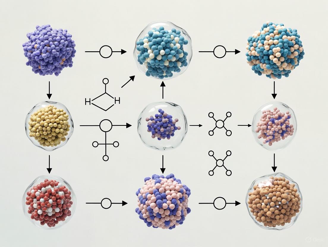
The following diagram illustrates the core decision process and signaling inputs involved in establishing different types of organoids from the two primary stem cell sources.
Organoid Generation: From Stem Cells to Functional Mini-Organs
The choice of stem cell source is a primary determinant of the organoid's characteristics and potential applications. The two main sources are Pluripotent Stem Cells (PSCs) and Adult Stem Cells (AdSCs), each with distinct advantages and limitations [19].
PSC-derived organoids are generated from either embryonic stem cells (ESCs) or induced pluripotent stem cells (iPSCs). iPSCs, created by reprogramming somatic cells (e.g., skin fibroblasts) using defined factors (Oct4, Sox2, Klf4, c-Myc), are particularly powerful as they enable the creation of patient-specific disease models [19] [21]. PSC-derived organoids are ideal for studying early human organogenesis because their differentiation process mirrors embryonic development [19]. Protocols often involve stepwise addition of specific morphogens to guide PSCs through intermediate stages, a process that can take several months [19] [23]. A key advantage is their complex cellular composition, which can include mesenchymal and epithelial components, more fully representing an embryonic organ [19].
AdSC-derived organoids are obtained directly from adult tissues (e.g., intestinal crypts, liver biopsies) [19]. These organoids are typically simpler and faster to generate, often taking only weeks, and their maturity is closer to adult tissue [19] [22]. They are predominantly composed of epithelial cell types and are exceptionally well-suited for studying adult tissue homeostasis, repair, and diseases like cancer and viral infections [19] [22].
Table 1: Comparison of Pluripotent Stem Cell (PSC) and Adult Stem Cell (AdSC) Derived Organoids
| Feature | PSC-Derived Organoids | AdSC-Derived Organoids |
|---|---|---|
| Cell Source | Embryonic Stem Cells (ESCs), Induced Pluripotent Stem Cells (iPSCs) | Tissue-specific adult stem cells (e.g., Lgr5+ intestinal stem cells) |
| Differentiation Process | Directed, multi-step differentiation mimicking embryogenesis | Expansion and maturation of committed lineage |
| Culture Duration | Several months | Several weeks |
| Cellular Complexity | High; can include multiple germ layer-derived cells | Lower; primarily epithelial cell types |
| Maturity | Fetal-like | Adult-like |
| Primary Applications | Developmental biology, disease modeling, toxicology | Adult tissue function, disease modeling (e.g., cancer), personalized medicine |
| Key Advantage | Models organ development; can generate tissues inaccessible in adults | Faster, simpler, and more genetically stable for adult disease research |
General Protocol for Organoid Culture
A standard workflow for establishing and maintaining organoid cultures involves several critical steps, each requiring optimization for the specific organoid type [20]. The process heavily relies on a carefully optimized combination of an extracellular matrix scaffold and a defined culture medium [22] [20].
- Tissue or Cell Collection: Obtain starting material, which can be somatic cells (for reprogramming to iPSCs), established PSCs, or tissue fragments containing AdSCs [20].
- Stem Cell Culture and Differentiation (for PSCs): For PSC-derived organoids, cells are first aggregated into embryoid bodies and then guided through a series of differentiation stages using specific cocktails of growth factors and small molecules added to the culture medium at precise timepoints [19] [23].
- 3D Culture in Matrix Scaffold: The stem cells or progenitor cells are embedded in a 3D extracellular matrix, most commonly Basement Membrane Extracts (BME) like Matrigel or Geltrex. This matrix provides crucial biochemical and mechanical cues for 3D growth and self-organization [19] [22].
- Maintenance and Passaging: Organoids are fed with specialized medium and maintained long-term. For passaging, organoids are typically enzymatically and/or mechanically dissociated into smaller fragments and re-embedded in fresh matrix to expand the culture [20].
- Characterization and Analysis: The successful culture of organoids is confirmed through morphological observation (e.g., microscopy) and analytical techniques such as immunofluorescence, gene expression analysis (qPCR, RNA-seq), and functional assays relevant to the target organ [20].
Advanced Applications in Research and Medicine
Organoid technology has moved beyond basic science to become a cornerstone in advanced biomedical applications, offering unprecedented opportunities for personalized medicine and drug development.
Disease Modeling and Personalized Medicine
Patient-derived organoids (PDOs), particularly from cancer biopsies, have emerged as a transformative tool for personalized medicine [18] [21]. These PDOs retain the genetic and phenotypic heterogeneity of the patient's tumor, creating an avatars for ex vivo drug testing [21] [10]. In oncology, patient-derived tumor organoids (PDTOs) can be used for medium-throughput drug screens to identify the most effective therapeutic strategies for an individual patient, predict responses to chemotherapy, and study mechanisms of drug resistance [21]. This approach is being piloted in clinical settings for colorectal, pancreatic, and lung cancers to inform treatment decisions [21]. Beyond cancer, organoids from patients with genetic disorders like cystic fibrosis have been used to study disease mechanisms and test potential therapies [24].
Drug Discovery and Development
The pharmaceutical industry is increasingly adopting organoids to improve the predictive power of preclinical models, thereby reducing high attrition rates in clinical trials [21] [9]. Organoids provide human-specific pathophysiological data that is more relevant than data from animal models, which are often compromised by species differences [21]. Key applications include:
- Drug Efficacy Screening: Organoids enable high-throughput screening of compound libraries in a human, physiologically relevant context [21] [20].
- Toxicity Testing: Liver and heart organoids are used to assess drug-induced hepatotoxicity and cardiotoxicity, two major causes of drug failure [21]. hPSC-derived cardiomyocytes, for instance, can detect the cardiotoxic effects of drugs like doxorubicin [21].
Addressing Technical Challenges: Long-Term Culture and Analysis
A significant challenge in organoid research is maintaining viability during long-term culture. As organoids grow in size, they develop diffusion-limited necrotic cores due to hypoxia and nutrient deprivation [24]. To address this, researchers have developed an efficient cutting method using 3D-printed jigs. This technique involves periodically slicing organoids into smaller pieces under sterile conditions, which improves nutrient diffusion, increases cell proliferation, and enables cultures to be maintained for over five months [24]. Furthermore, for high-throughput analysis, novel mold-based approaches have been created to generate densely packed organoid arrays for consistent cryosectioning, facilitating techniques like single-cell spatial transcriptomics [24].
The Organoid and Spheroid Market Landscape
The broad utility of 3D models has fueled a rapidly growing market, reflecting their adoption in research and industry. The global organoids and spheroids market, valued at USD 1.5 billion in 2024, is projected to grow at a compound annual growth rate (CAGR) of 20.3% to reach USD 9.6 billion by 2034 [18]. This growth is primarily driven by the demand for physiologically relevant models that are more predictive and ethically viable than animal testing, especially in oncology, neurology, and regenerative medicine [18]. The organoids segment dominates this market, accounting for 76.2% share in 2024, due to its superior ability to replicate organ-specific architecture and function [18].
Table 2: Comparative Analysis of Preclinical Research Models
| Characteristic | 3D Organoids | 2D Cell Cultures | Animal Models |
|---|---|---|---|
| Physiological Representation | Semiphysiologic | Limited | Physiologic |
| Success Rate | High | High | Low |
| Time Required | Moderate | Short | Long |
| Cost | Moderate | Low | High |
| Genomic Stability | High | Low | High |
| Cellular Heterogeneity | High | Low | High |
| Clinical Relevance | High | Low | High |
| Gene Editing | Easy | Easy | Hard |
| High-Throughput Screening | Applicable | Applicable | Not Applicable |
| Biobanking | Feasible | Feasible | Not Feasible |
The Scientist's Toolkit: Essential Reagents and Materials
Successful organoid culture depends on a suite of specialized reagents and materials designed to mimic the in vivo stem cell niche.
Table 3: Key Research Reagent Solutions for Organoid Culture
| Reagent/Material | Function | Examples & Notes |
|---|---|---|
| Extracellular Matrix (ECM) | Provides a 3D scaffold that delivers biochemical and mechanical cues for cell growth and polarization. | Basement Membrane Extracts (BME) like Matrigel, Geltrex, and Cultrex are most common. They are versatile but have undefined composition and batch-to-batch variability [22]. |
| Growth Factors & Cytokines | Key signaling molecules that direct stem cell self-renewal, differentiation, and patterning. | Combinations of EGF, Noggin, R-spondin-1 (for intestinal organoids); FGF, Wnt agonists, BMP inhibitors. Must be tailored to the specific organoid type [19] [20]. |
| Culture Media | A defined basal medium supplemented with specific growth factors, hormones, and nutrients. | Formulations are organ-specific. Companies offer specialized medium kits for brain, liver, intestinal, and other organoids [18] [20]. |
| Dissociation Enzymes | Used to break down the ECM and dissociate organoids into single cells or small fragments for passaging. | Enzymes like Accutase, Trypsin, or Collagenase are used, often in combination with mechanical disruption [24] [20]. |
| Characterization Tools | Reagents and kits for validating organoid identity, structure, and function. | Includes antibodies for immunofluorescence (detecting cell-specific markers), qRT-PCR kits for gene expression, and functional assay kits (e.g., insulin release for pancreatic organoids) [20]. |
Organoids, as complex, stem cell-derived mini-organs, have firmly established themselves as a transformative technology in biomedical research. By faithfully mimicking the structure and function of human organs in a controlled in vitro setting, they provide a powerful and versatile platform that bridges the gap between traditional 2D cell lines and animal models. The ability to generate organoids from patients is ushering in a new era of personalized medicine, enabling tailored drug screening and disease modeling. While challenges related to standardization, scalability, and full recapitulation of organ complexity remain, ongoing interdisciplinary innovations in bioengineering, automation, and data analysis are rapidly addressing these limitations. As the field continues to mature, organoids are poised to dramatically accelerate our understanding of human biology, improve the efficiency of drug development, and ultimately, reshape future therapeutic strategies.
The advancement from conventional two-dimensional (2D) cell cultures to three-dimensional (3D) models represents a paradigm shift in biomedical research. Spheroids and organoids have emerged as powerful tools that bridge the gap between oversimplified monolayer cultures and complex in vivo environments [25] [9]. These 3D structures recapitulate crucial aspects of tissue architecture, including cell-cell and cell-extracellular matrix (ECM) interactions, metabolic gradients, and heterotypic cellular crosstalk that drive tissue function and dysfunction [26]. The formation and integrity of these sophisticated models depend fundamentally on precise biological mechanisms, with E-cadherin-mediated cell-cell adhesion and integrin-mediated cell-ECM interactions serving as the primary architectural pillars. Understanding these mechanisms is essential for researchers leveraging 3D models to study development, disease progression, and therapeutic interventions with greater physiological relevance.
Core Biological Mechanisms
E-Cadherin in 3D Formation
E-cadherin is a calcium-dependent transmembrane adhesion molecule encoded by the CDH1 gene that serves as the principal mediator of epithelial cohesion [27]. In the context of 3D model formation, it functions as a master regulator of tissue architecture through several key mechanisms:
- Initiation of Cell-Cell Contact: The process of spheroid formation begins with the initial aggregation of cells, during which E-cadherin molecules on adjacent cells engage in trans-homophilic binding, forming stable intercellular junctions [25] [9].
- Spheroid Compaction and Integrity: Following initial contact, the accumulation of E-cadherin at cell surfaces and its linkage to the actin cytoskeleton via catenins (α-catenin, β-catenin) facilitates dramatic tissue reorganization, transforming loose cell aggregates into compact, spherical structures [28] [25]. This compaction is essential for establishing the dense cellular organization characteristic of spheroids.
- Dimensional Context Determinant: The functional outcome of E-cadherin expression displays a fascinating dimensional dichotomy. In 3D in vitro settings, E-cadherin potently promotes spheroid formation, as demonstrated by experiments where ectopic overexpression enhanced spheroid generation while knockdown retarded it [27] [29]. Paradoxically, in in vivo contexts, the same molecule acts as a metastasis suppressor, with its downregulation promoting anoikis resistance and invasiveness [27] [29].
- Regulation of Stemness: In lung cancer models, downregulation of E-cadherin was shown to increase cancer stem cell (CSC) properties, though it simultaneously adversely affected the survival of non-CSCs, highlighting its complex role in cellular phenotypes within 3D architectures [27].
Integrins in 3D Formation
Integrins are heterodimeric transmembrane receptors composed of α and β subunits that connect the intracellular cytoskeleton to the extracellular matrix (ECM) [30] [31]. Their role in 3D model establishment is multifaceted:
- Initial Aggregate Formation: During the early aggregation phase of spheroid formation, integrins, particularly those containing the β1 subunit, bind to RGD (Arg-Gly-Asp) motifs in long-chain ECM fibers, facilitating the initial cell-cell and cell-ECM contacts that nucleate 3D structures [9] [30].
- Dimensionality-Dependent Signaling: Integrin function exhibits profound contextual dependence on the culture environment. In 2D cultures, integrins cluster into focal adhesions that serve as anchors, while in 3D contexts, they form distinctive 3D-matrix adhesions with different morphology and molecular composition [32]. These 3D adhesions show more rapid formation and lower levels of tyrosine phosphorylation of focal adhesion kinase (FAK) compared to their 2D counterparts [32].
- Mechanotransduction and Survival Signaling: In prostate cancer models, β1 integrin has been identified as a critical regulator of anchorage-independent growth in 3D cultures, where its depletion abolished the ability of tumor cells to form colonies in soft agarose and Matrigel [31]. This integrin-ECM interaction prevents detachment-mediated death (anoikis) by activating survival pathways, highlighting its essential role in 3D cellular viability.
- Modulation of Therapeutic Response: Integrin-mediated adhesion complexes modulate cellular responses to anti-cancer agents, with these influences being largely masked in 2D culture but becoming readily apparent under 3D culture conditions and in tumor growth in vivo [31].
Table 1: Comparative Roles of E-Cadherin and Integrins in 3D Model Formation
| Feature | E-Cadherin | Integrins |
|---|---|---|
| Primary Function | Mediates homophilic cell-cell adhesion | Mediates heterophilic cell-ECM adhesion |
| Structural Role | Forms adherens junctions; enables spheroid compaction | Forms focal adhesions (2D) or 3D-matrix adhesions (3D) |
| Signaling Role | Regulates stemness; contextual survival signaling | Mechanotransduction; survival signaling via FAK/ILK |
| Dimensional Context | Promotes in vitro spheroid formation but suppresses in vivo metastasis | Exhibits distinct signaling in 2D vs. 3D environments |
| Key Binding Partners | Catenins, actin cytoskeleton | ECM proteins (fibronectin, collagen), cytoskeletal linkers |
Quantitative Experimental Data
Research into the molecular mechanisms of 3D formation has yielded substantial quantitative insights, particularly regarding the functional consequences of manipulating adhesion molecules.
Table 2: Experimental Findings on Adhesion Molecule Manipulation in 3D Models
| Experimental Manipulation | Experimental Model | Key Quantitative Findings | Biological Impact |
|---|---|---|---|
| E-cadherin knockdown [27] | Lung cancer cells (H460) | Retarded formation of tumor spheroids in vitro | Disrupted compact spheroid morphology |
| E-cadherin overexpression [27] | Lung cancer cells (H460) | Promoted formation of tumor spheroids in vitro | Enhanced compact spheroid formation |
| E-cadherin overexpression [27] [29] | Lung cancer xenograft mouse model | Inhibited tumor formation and metastasis in vivo | Suppressed metastatic progression |
| β1 integrin depletion [31] | Prostate cancer cells (PC3) | Abolished colony formation in soft agarose and Matrigel | Disrupted anchorage-independent growth |
| Enrichment of NSF cells [28] | Colon cancer cells (SW620) | Projected cell-covered area increased twofold in NSF vs SF | Markedly reduced cell-cell adhesion |
| Anti-fibronectin neutralizing antibodies [31] | Prostate cancer cells (PC3) | Inhibition of anchorage-independent growth similar to β1 depletion | Disrupted integrin-ECM survival signaling |
Methodologies for Investigating Adhesion Mechanisms
Spheroid Formation Assays
The fundamental protocol for assessing 3D formation capability involves several standardized approaches:
- Ultra-Low Attachment (ULA) Plates: Cells are detached into a single-cell suspension using EDTA in PBS and seeded at densities ranging from 5×10³ to 1×10⁴ cells/ml onto ULA plates [27] [28]. These plates prevent cell attachment to the substrate, forcing cells to self-assemble into spheroids. Cells are then allowed to form primary spheroids for 7-14 days with periodic medium changes [27].
- Hanging Drop Method: This technique utilizes gravity to aggregate cells in suspended droplets, typically of 20-40 µl volume, forcing cell-cell contact and spheroid formation without artificial substrate interactions [9].
- Spheroid Compaction Analysis: The degree of spheroid formation can be quantified by measuring the projected cell-covered area using image analysis software. Non-spheroid forming (NSF) cells exhibit significantly larger projected areas compared to their spheroid-forming (SF) counterparts [28].
Functional Characterization Assays
- Anoikis Assay: Cells are detached and dissociated into a single-cell suspension in medium containing 1% FBS and 1 mM EDTA. The suspended cells are seeded onto ULA plates at a density of 1×10⁴ cells/ml and analyzed for cell viability by MTT assay over time [27]. This assay specifically assesses detachment-induced cell death resistance.
- Invasion Assay: Cell invasion capacity is evaluated using Boyden chambers with 8-µm pore filter inserts in 24-well plates. Cells are added to the upper chamber at a density of 3×10⁴ cells/ml, with the lower chamber filled with medium containing 10% FBS as a chemoattractant. After 48 hours, non-invading cells are removed, and invading cells are fixed, stained, and quantified [27].
- Flow Cytometry for Stem Cell Markers: Spheroids are dissociated into single cells and analyzed for CSC markers. For ALDH activity assessment, cells are stained with Aldefluor reagent with or without the ALDH inhibitor DEAB as a negative control [27]. Cells can also be stained for surface markers like CD133 using specific antibodies and analyzed by fluorescence-activated cell sorting (FACS) [27].
Signaling Pathways and Molecular Interactions
The formation and maintenance of 3D structures involve complex signaling networks that integrate adhesion signals with cellular fate decisions. E-cadherin and integrins function as key signaling hubs in these processes.
Diagram 1: E-cadherin and integrin signaling in 3D formation. The pathways demonstrate how both adhesion systems converge on critical cellular processes essential for 3D structure establishment and maintenance.
The diagram illustrates two parallel but interconnected signaling modules. The integrin-mediated pathway begins with ECM binding, which triggers FAK activation and subsequent signaling through ERK/AKT pathways to promote cell survival and proliferation [30] [31]. Simultaneously, the E-cadherin-mediated pathway drives junction assembly through catenins, links to the actin cytoskeleton to enable mechanical cohesion, and activates ERK/AKT signaling to regulate secretory processes like VEGF production [33]. These pathways converge to establish the structural integrity and functional capacity of 3D models.
Experimental Workflow
The systematic investigation of adhesion mechanisms in 3D models follows a logical progression from model establishment to functional analysis.
Diagram 2: Experimental workflow for investigating adhesion mechanisms. The process begins with model establishment and progresses through targeted manipulation, phenotypic characterization, and functional analysis to achieve comprehensive mechanistic understanding.
This workflow emphasizes the importance of contextual interpretation, particularly given the dimensional dichotomy observed with adhesion molecules like E-cadherin, which can display opposite functions in 3D in vitro versus in vivo settings [27] [29]. Researchers must carefully consider this contextual dependency when extrapolating findings from 3D models to physiological or pathological scenarios.
The Scientist's Toolkit: Essential Research Reagents
Table 3: Key Research Reagents for Investigating Adhesion Mechanisms in 3D Models
| Reagent/Category | Specific Examples | Research Application | Experimental Context |
|---|---|---|---|
| Genetic Manipulation Tools | shRNA E-cadherin plasmids; E-cadherin-GFP overexpression vectors [27] | Functional studies of E-cadherin in spheroid formation | Gain- and loss-of-function experiments in cancer cells |
| Blocking Antibodies | E-cadherin neutralizing antibodies [33]; fibronectin neutralizing antibodies [31] | Acute inhibition of specific adhesion pathways | Mechanistic studies of compaction and survival signaling |
| Specialized Cultureware | Ultra-low attachment (ULA) plates [27] [28]; Boyden chambers [27] | Spheroid formation and invasion assays | Standardized 3D culture and migration/invasion quantification |
| Detection Reagents | E-cadherin antibodies for immunofluorescence [27]; Aldefluor assay kit [27] | Molecular characterization and stem cell analysis | Spatial protein localization and cancer stem cell identification |
| Matrix Components | Matrigel; collagen I; fibronectin [31] | Scaffold-based 3D culture and mechanistic studies | Providing physiological context for integrin-mediated signaling |
E-cadherin and integrins constitute the fundamental architectural framework governing the formation and functionality of 3D spheroid and organoid models. Their roles extend far beyond simple mechanical adhesion to encompass complex signaling functions that regulate cell survival, proliferation, stemness, and therapeutic responses. The context-dependent nature of these adhesion mechanisms—particularly the divergent functions observed in 3D in vitro versus in vivo settings—underscores the critical importance of careful experimental design and interpretation in 3D model research. As the field advances, a deeper understanding of these core biological mechanisms will enable the development of increasingly sophisticated 3D models that more accurately recapitulate tissue physiology and pathology, thereby enhancing their predictive value in drug development and disease modeling. The integration of the experimental frameworks, reagents, and methodologies outlined in this technical guide provides a solid foundation for researchers to systematically investigate and manipulate these key biological mechanisms in their 3D model systems.
The evolution of three-dimensional (3D) cell culture models, primarily spheroids and organoids, represents a paradigm shift in biomedical research, offering a bridge between traditional two-dimensional (2D) cultures and in vivo models. The physiological relevance of these 3D structures is profoundly influenced by the choice of the originating cell source. The decision to use stem cells, primary tissues, or immortalized cell lines dictates the model's complexity, scalability, genetic stability, and translational potential. This guide provides an in-depth technical analysis of these cell sources, framing them within the context of modern spheroid and organoid research to inform strategic experimental design for scientists and drug development professionals.
Defining Spheroids and Organoids in Modern Research
While the terms are sometimes used interchangeably, spheroids and organoids represent distinct classes of 3D models with fundamental differences in origin and complexity.
Spheroids are simple, spherical aggregates of cells that form through self-assembly, typically from primary cells or immortalized cell lines [34]. They can be generated with or without extracellular matrix (ECM) support and develop over a relatively short timeline of 2-3 days [34]. Their architecture often includes gradients that mimic the tumor microenvironment, with proliferating cells on the exterior, quiescent cells in an intermediate layer, and a necrotic core at the center due to oxygen and nutrient diffusion limitations [9]. Their primary value lies in modeling cell-cell interactions and studying tumor biology and drug screening [34].
Organoids are more complex structures that are generated from tissue-specific progenitor cells or stem cells—including adult stem cells, embryonic stem cells, or induced pluripotent stem cells (iPSCs) [35] [34]. They require an ECM scaffold and specific growth factors to enable progenitor cell expansion, differentiation, and self-organization into structures that recapitulate the organ or tissue of origin [35] [34]. This process can take several weeks to months to achieve full complexity [34]. Organoids model organ-specific architecture and function, making them ideal for studying development, disease mechanisms, and personalized medicine [18] [34].
Table 1: Fundamental Characteristics of Spheroids and Organoids
| Characteristic | Spheroids | Organoids |
|---|---|---|
| Cell Source | Primary cells, cell lines, multicellular mixes [34] | Adult stem cells, embryonic stem cells, iPSCs, progenitor cells [34] |
| Architecture | Simple, spherical aggregates [34] | Complex, self-organizing, organ-specific structures [34] |
| Culture Time | ~2-3 days [34] | 21-60+ days [34] |
| ECM Requirement | Optional; can be scaffold-free [34] | Typically required (e.g., Matrigel) [35] [34] |
| Key Applications | Tumor microenvironment studies, initial drug screening [9] [34] | Disease modeling, developmental biology, personalized medicine [18] [34] |
The selection of a cell source involves a critical trade-off between physiological relevance and practical considerations like scalability and cost.
Immortalized Cell Lines
Definition and Origin: Immortalized cell lines are cells that have been genetically modified—either intentionally via viruses or spontaneously via carcinogenic transformation—to proliferate indefinitely in culture [36]. Common examples include HeLa (cervical cancer), SH-SY5Y (neuroblastoma), and MCF-7 (breast cancer) cells [37].
Advantages and Limitations: The primary advantage of immortalized lines is their practicality. They offer unlimited lifespan, are simple to culture, and are easily scalable, making them ideal for high-throughput screening and foundational research [37] [36]. However, this comes at the cost of physiological relevance. Being mostly cancer-derived, they are optimized for proliferation rather than native function and are subject to genetic drift and phenotypic changes with continuous passage [38] [37]. Furthermore, studies have shown that findings in immortalized lines frequently fail to translate to human tissue or in vivo models, contributing to high attrition rates in drug development [37].
Primary Cells
Definition and Origin: Primary cells are isolated directly from human donor or animal tissue and undergo minimal manipulation to preserve their original characteristics and functions [36]. They are not passaged indefinitely and have a finite lifespan in culture.
Advantages and Limitations: Primary cells are considered the gold standard for physiological relevance as they retain genomic and phenotypic stability, native cell morphology, and key functions of the tissue of origin [38]. This makes them highly valuable for immunology, inflammation, and vaccination experiments [36]. Their main drawbacks include limited scalability due to a finite lifespan, technically complex isolation and culture protocols, and significant donor-to-donor variability, which can introduce noise and reduce reproducibility [38] [37]. The commercial supply, however, has made sourcing more efficient, with donor screening and regulatory documentation provided [38].
Stem Cells
Definition and Origin: This category includes induced Pluripotent Stem Cells (iPSCs), which are adult cells reprogrammed to an embryonic-like state, and adult stem cells. iPSCs can be renewed indefinitely and differentiated into various somatic cell types [37].
Advantages and Limitations: Stem cells, particularly iPSCs, offer a powerful combination of human relevance and scalability. They provide a renewable source of human-specific cells that can be differentiated to model different tissues, overcoming the ethical concerns of embryonic stem cells and the species-mismatch of animal primary cells [37]. However, traditional directed differentiation protocols can be time-consuming and variable, leading to batch-to-batch inconsistency and heterogeneous cell populations [37]. Newer technologies like deterministic cell programming (e.g., opti-ox technology) are emerging to address these challenges by enabling the production of highly consistent, functionally validated iPSC-derived cells at scale [37].
Table 2: Strategic Comparison of Cell Sources for 3D Models
| Attribute | Immortalized Cell Lines | Primary Cells | Stem Cells (iPSCs) |
|---|---|---|---|
| Physiological Relevance | Low; often non-physiological, cancer-derived [37] | High; retain native morphology & function [38] [36] | High; human-specific, can model native biology [37] |
| Scalability | High; unlimited lifespan [36] | Low; finite lifespan [36] | High; renewable [37] |
| Reproducibility | High initially, but prone to genetic drift [38] | Low; high donor-to-donor variability [37] | Variable; improved with new programming tech [37] |
| Ease of Use | Simple to culture [37] | Technically complex, time-intensive [37] | Varies; ready-to-use cryopreserved cells available [37] |
| Typical Cost | Low | High | High, but decreasing |
| Ideal Use Case | High-throughput screening, preliminary functional studies [37] | Studies requiring high physiological fidelity (e.g., immunology) [36] | Disease modeling, personalized medicine, regenerative medicine [18] [37] |
Technical Protocols and Methodologies
The following diagram summarizes the general workflow for generating spheroids and organoids from the different cell sources discussed.
Protocol for Automated Spheroid Production
For high-content screening, consistency in spheroid size and shape is critical. The following is a detailed protocol adapted from a 2025 method for producing uniform spheroids from HeLa Kyoto cells, suitable for automated pipelines [39].
Objective: To establish a robust, automated method for generating consistent populations of spheroids for investigating organelle biology and membrane trafficking pathways [39].
Key Features:
- Production of uniform spheroids in a short time frame.
- Elimination of ECM removal steps, simplifying downstream applications.
- Compatibility with both manual and automated high-throughput/high-content screens [39].
Materials and Reagents:
- Cell Line: HeLa Kyoto cells (or other cell line of interest).
- Micropatterned Plates: Commercially available plates designed for uniform spheroid formation (e.g., Elplasia plates from Corning) [18] [39].
- Cell Culture Medium: Appropriate for the cell line used (e.g., DMEM with 10% FBS).
- Trypsin-EDTA: For cell detachment.
- PBS (Phosphate Buffered Saline): For washing.
- Fixative: e.g., 4% Paraformaldehyde (PFA).
- Permeabilization Buffer: e.g., 0.1% Triton X-100 in PBS.
- Staining Solutions: Primary and fluorescently-labeled secondary antibodies, phalloidin, DAPI.
Procedure:
- Cell Seeding:
- Harvest cells using trypsin-EDTA and create a single-cell suspension.
- Count cells and adjust the concentration to the optimal seeding density (e.g., 50-100 cells per microwell, requires optimization).
- Seed the cell suspension into the micropatterned plate.
- Spheroid Culture:
- Incubate the plate under standard culture conditions (37°C, 5% CO2) for the desired period (typically 2-5 days). The micropatterned wells guide the cells to aggregate into a single, uniform spheroid per microcavity.
- Treatment (Optional):
- After spheroid formation, introduce nanoparticles, drugs, or other compounds to the culture medium for the intended experimental treatment [35].
- Fixation and Staining:
- Carefully remove the culture medium.
- Wash spheroids gently with PBS.
- Fix with 4% PFA for 15-30 minutes at room temperature.
- Permeabilize and block with an appropriate buffer (e.g., 1% BSA, 0.1% Triton X-100 in PBS) for 1 hour.
- Incubate with primary antibodies diluted in blocking buffer overnight at 4°C.
- Wash and incubate with fluorescent secondary antibodies and nuclear stain (e.g., DAPI) for 2-4 hours at room temperature.
- Imaging and Analysis:
- Image the entire spheroid volumetrically using confocal fluorescence microscopy or high-content imaging systems.
- Use automated image analysis software to quantify changes in fluorescence intensity, spatial distribution, and morphology [39].
Essential Research Reagents and Tools
Successful culture and analysis of 3D models rely on a suite of specialized reagents and tools.
Table 3: Key Research Reagent Solutions for 3D Cell Culture
| Reagent/Tool | Function | Examples & Notes |
|---|---|---|
| Extracellular Matrix (ECM) | Provides a 3D scaffold that mimics the in vivo basement membrane, supporting cell adhesion, differentiation, and self-organization. | Matrigel is the most widely used natural ECM polymer [35]. Cultrex UltiMatrix is a commercial alternative [34]. Batch-to-batch variability is a known challenge [35]. |
| Specialized Media & Kits | Provides optimized growth factors, cytokines, and nutrients to support the expansion and differentiation of stem cells and primary cells in 3D culture. | Companies like STEMCELL Technologies offer organoid-specific kits and media (e.g., STEMdiff products) for neural, intestinal, and other organoid types [18]. |
| Micropatterned & ULA Plates | Physical tools that promote scaffold-free spheroid formation by preventing cell attachment, forcing aggregation. | Ultra-Low Attachment (ULA) plates and micropatterned plates (e.g., Corning Elplasia) enable high-throughput production of uniform spheroids [18] [39]. |
| Bioreactors | Dynamic culture systems that improve mass transfer of oxygen and nutrients, allowing for larger and more viable 3D structures. | Stirred-tank bioreactors help scale up organoid production and mitigate necrotic core formation [40]. |
| High-Content Imaging Systems | Automated microscopes capable of capturing 3D volumetric data from spheroids and organoids for quantitative analysis. | Confocal fluorescence microscopes are essential for high-resolution z-stacking and 3D reconstruction of models [35] [39]. |
The selection of cell sources—immortalized lines, primary tissues, or stem cells—is a foundational decision that dictates the biological fidelity, applicability, and scalability of spheroid and organoid research. Immortalized lines offer practical advantages for high-throughput screening but lack physiological relevance. Primary cells provide high translational relevance but are hampered by scalability and reproducibility issues. Stem cells, particularly with advances in iPSC programming, present a compelling path forward, balancing human specificity with the potential for scale. The ongoing integration of automation, AI, and advanced imaging with these cell sources is poised to further enhance the precision and predictive power of 3D models, solidifying their role in accelerating drug discovery and personalized medicine.
From Theory to Bench: Culture Techniques and Transformative Applications
The advancement from traditional two-dimensional (2D) cell cultures to three-dimensional (3D) cellular systems represents a paradigm shift in biological research, offering an experimental tool that more closely mimics the in vivo environment [9]. Within this realm, multicellular tumor spheroids (MCTS) have emerged as one of the most popular and versatile methods for culturing cells in 3D [9]. These spherical cell aggregates allow for critical cell-cell and cell-matrix interactions that are absent in 2D monolayers, where cells are stretched and undergo cytoskeletal rearrangements, acquiring artificial polarity and aberrant gene expression [41].
Spheroids are particularly valuable for modeling avascular tumor microenvironments, where they develop physiochemical gradients of oxygen, nutrients, and metabolic wastes [42] [41]. This leads to the formation of concentric zones of heterogeneous cell populations: an outermost layer of proliferating cells, intermediate layers of quiescent viable cells, and an innermost core that can become necrotic due to oxygen and nutrient limitations [41] [9]. This architectural complexity enables more physiologically relevant studies of drug penetration, therapeutic efficacy, and resistance mechanisms that better reflect in vivo conditions than traditional 2D cultures [41] [43].
Table 1: Fundamental Characteristics of Spheroid Models Compared to Other Culture Systems
| Feature | 2D Monolayer | Spheroids | In Vivo Tumors |
|---|---|---|---|
| Spatial organization | Flat monolayer | 3D spherical structure | 3D architecture with tissue context |
| Cell-cell interactions | Limited, primarily at edges | Extensive throughout structure | Extensive and complex |
| Nutrient/O₂ gradients | Uniform exposure | Established gradients from periphery to core | Present, influenced by vasculature |
| Cellular heterogeneity | Limited | Zonal heterogeneity (proliferating, quiescent, necrotic) | High, with multiple cell types |
| Drug response | Typically more sensitive | More resistant, better predicts in vivo efficacy | Clinical response |
| Cost & throughput | Low cost, high throughput | Moderate cost and throughput | High cost, low throughput |
| Experimental control | High | Moderate | Limited |
Core Spheroid Culture Techniques
Hanging Drop Method
The hanging drop method is a scaffold-free technique that facilitates spontaneous spheroid formation through gravitational force. In this approach, cell suspensions are dispensed as discrete droplets on the inner surface of a Petri dish lid, which is then inverted over a reservoir containing phosphate-buffered saline (PBS) to prevent evaporation [44] [45]. The hanging surface tension maintains the droplet integrity while gravity encourages cells to sediment and aggregate at the liquid-air interface, promoting cell-cell contact and spheroid formation.
Protocol for Hanging Drop Spheroid Formation:
- Prepare a single-cell suspension at the desired density, typically ranging from 1,500 to 15,000 cells per droplet depending on the target spheroid size [45].
- Using a pipette, dispense 10-60 µL droplets onto the inner surface of a Petri dish lid [44] [45].
- Carefully invert the lid and place it over a Petri dish bottom filled with PBS or culture medium to maintain humidity [44] [45].
- Incubate the assembly at 37°C with 5% CO₂ for 24-72 hours to allow spheroid formation [45].
- For extended culture, partial medium exchange can be performed by carefully removing a limited volume from the droplet and adding fresh medium [45].
The hanging drop method produces spheroids with high uniformity and reproducibility due to consistent droplet volumes. However, challenges include potential evaporation, limited scale-up capacity, and difficulties in feeding and handling during long-term culture [45]. To enhance droplet stability, additives like methylcellulose can be incorporated, though this may affect cellular interactions [45].
Ultra-Low Attachment (ULA) Plates
Ultra-low attachment (ULA) plates feature specially treated surfaces with hydrophilic and neutrally charged coatings that resist protein adsorption and cell attachment, effectively promoting cell aggregation into spheroids [44] [43]. These plates are available in various formats, including flat-bottom and round-bottom designs, with the latter further enhancing spheroid uniformity by guiding cells toward the well center through gravitational forces.
Protocol for ULA Plate Spheroid Formation:
- Prepare a single-cell suspension at an appropriate density, typically ranging from 1,000 to 10,000 cells per well in 96- or 384-well plates, optimizing for specific cell lines [42] [43].
- Seed the cell suspension into ULA plate wells, ensuring even distribution [44].
- Centrifuge the plates briefly at low speed (100-400 × g for 1-3 minutes) to collect cells at the well bottom and initiate contact [42].
- Incubate plates at 37°C with 5% CO₂ for several days, with medium changes every 2-4 days if needed for extended culture [42].
- Monitor spheroid formation and growth using microscopy until they reach the desired size for experimentation.
ULA plates enable medium-to-high throughput screening and are compatible with automated imaging and analysis systems [43]. The main advantages include technical simplicity, reproducibility, and compatibility with standard laboratory equipment. However, specialized plates represent a recurring cost, and optimization of seeding density is required for different cell types to achieve uniform spheroid size and structure [9].
Bioreactors
Bioreactor systems for spheroid culture utilize dynamic conditions through continuous agitation or perfusion to promote cell aggregation and maintain viability. Rotating wall vessels and spinner flasks are common configurations that create low-shear stress environments, preventing cell attachment while encouraging the formation of numerous spheroids simultaneously [46] [9].
Protocol for Spheroid Formation in Bioreactors:
- Prepare a single-cell suspension at the recommended density for the specific bioreactor system, typically within the range of 1×10⁵ to 1×10⁶ cells/mL [46].
- Inoculate the cell suspension into the sterile bioreactor vessel containing appropriate culture medium.
- Initiate agitation at an optimized speed (typically 50-100 rpm for spinner flasks) to maintain cells in suspension without generating destructive shear forces [46].
- Maintain cultures at 37°C with proper gas exchange (O₂ and CO₂), with continuous perfusion or periodic medium changes to replenish nutrients and remove wastes [9].
- Sample at intervals to monitor spheroid growth and morphology, typically over 1-4 weeks depending on application.
Bioreactors support the generation of large quantities of spheroids and enable long-term culture by ensuring efficient nutrient and oxygen exchange throughout the aggregates [9]. This method is particularly valuable for producing spheroids at scales needed for tissue engineering or extensive drug screening campaigns. The limitations include higher equipment costs, more complex operation, and potential for shear stress if agitation is not properly controlled [9].
Table 2: Quantitative Comparison of Spheroid Culture Techniques
| Parameter | Hanging Drop | ULA Plates | Bioreactors |
|---|---|---|---|
| Throughput | Low to medium (dozens to hundreds) | High (96- to 384-well format) | Very high (thousands) |
| Spheroid uniformity | High (controlled by droplet volume) | Medium to high (depends on well geometry) | Variable (requires optimization) |
| Spheroid size range | 100-500 µm (limited by droplet volume) | 200-1000 µm (adjustable by cell density) | 100-1000 µm (wide distribution possible) |
| Culture duration | Short to medium (up to 1 week) | Medium (1-2 weeks) | Long-term (weeks to months) |
| Hands-on time | High (manual setup and feeding) | Low (automation compatible) | Medium (system maintenance) |
| Cost per spheroid | Low (minimal special equipment) | Medium (specialized plates) | Low at scale (high initial investment) |
| Ease of manipulation | Challenging (accessibility issues) | Easy (direct well access) | Moderate (sampling required) |
| Suitability for co-culture | Good | Excellent | Excellent |
| Scalability | Limited | Medium | High |
| Compatibility with imaging | Moderate | High | Low to moderate |
Technical Workflows and Spheroid Analysis
Standardized Workflow for Spheroid Culture and Analysis
The following diagram illustrates a generalized experimental workflow for spheroid culture, treatment, and analysis, integrating common elements across different formation techniques:
Spheroid Culture and Analysis Workflow
Structural Development in Spheroids
As spheroids mature, they develop characteristic structural features that mimic those observed in avascular tumors. The relationship between spheroid size, culture duration, and internal architecture follows predictable patterns:
Spheroid Structure vs Size Relationship
The development of these structural zones directly influences experimental outcomes. Research has demonstrated that transient spheroid structure is independent of initial spheroid size, and the limiting structure can be independent of seeding density [42]. This understanding has led to the recommendation that comparing spheroid structure as a function of overall size produces results that are relatively insensitive to variability in spheroid size, providing more robust experimental comparisons [42].
Advanced Imaging and Analysis Techniques
Cutting-edge 3D imaging and analysis methods are crucial to characterize, reproduce, and monitor spheroids [43]. High-content imaging systems with confocal capabilities and water immersion objectives enable the acquisition of z-stacks that capture the full three-dimensional structure of spheroids [43]. For analysis, specialized software can quantify multiple parameters, including:
- Size and volume measurements to track growth dynamics
- Circularity/sphericity to assess structural development
- Viability and cytotoxicity through fluorescent staining patterns
- Cell proliferation and death markers to characterize zonal heterogeneity
- Protein expression and localization via immunohistochemistry
The emergence of large-scale image atlases like SLiMIA (Spheroid Light Microscopy Image Atlas), which contains approximately 8,000 images of spheroids from 47 cell lines, provides valuable resources for method development and comparison [47]. Such resources are particularly useful for training segmentation models and establishing standardized analysis pipelines for 3D cell cultures.
Essential Reagents and Materials
Successful spheroid culture requires specific reagents and materials tailored to the selected methodology. The following table summarizes key components and their functions in spheroid research:
Table 3: Essential Research Reagents and Materials for Spheroid Culture
| Reagent/Material | Function/Application | Examples/Notes |
|---|---|---|
| Ultra-low attachment plates | Prevents cell adhesion, promotes spheroid formation | Round-bottom plates enhance uniformity; available in 96-384 well formats [44] [43] |
| Basement membrane extracts | Scaffold for embedded culture; provides ECM components | Matrigel (>1,800 proteins); defined hydrogels offer more consistency [44] |
| Collagen solutions | Defined scaffold material for 3D culture | Type I collagen (3 mg/mL concentration); adjustable pore size [44] |
| Viability stains | Distinguish live/dead cells in 3D structures | Calcein-AM (esterase activity for live cells); propidium iodide (dead cell nuclei) [45] [43] |
| Cell cycle indicators | Monitor proliferation and quiescence | FUCCI (fluorescent ubiquitination-based cell cycle indicator) [42] |
| Methylcellulose | Increases medium viscosity; stabilizes hanging drops | Prevents droplet dispersion; enhances spheroid compactness [45] |
| Oxygen-sensitive probes | Detect hypoxia development in spheroid core | Useful for monitoring gradient formation [41] |
| Fixation reagents | Preserve spheroid morphology for analysis | 4% paraformaldehyde; requires penetration optimization [42] |
The selection of an appropriate spheroid culture technique—whether hanging drop, ULA plates, or bioreactors—depends on the specific research objectives, required throughput, and available resources. Each method offers distinct advantages that can be leveraged for different applications in cancer research, drug development, and basic biological studies. As the field advances, standardization of protocols and analytical approaches will be crucial for improving reproducibility and comparability across studies. The integration of these 3D models with advanced imaging technologies and computational analysis promises to further enhance their predictive value in translational research.
The transition from traditional two-dimensional (2D) cell culture to three-dimensional (3D) models represents a paradigm shift in biomedical research, enabling unprecedented study of human physiology and disease. While 2D cultures have been foundational, they fail to replicate the spatial and microenvironmental complexity of living tissues [48]. Among 3D models, spheroids and organoids have emerged as powerful tools, though they possess distinct characteristics. Spheroids are simple, spherical clusters of broad-ranging cells that form via cell-cell adhesion, typically used for studying tumor microenvironments and drug screening [49] [50]. In contrast, organoids are complex, self-organizing 3D structures derived from stem or progenitor cells that recapitulate the functional and structural aspects of their organ of origin, making them invaluable for disease modeling, regenerative medicine, and developmental biology [49] [51] [50]. The critical distinction lies in organoids' ability to self-assemble and differentiate into organ-specific cell types, a process fundamentally dependent on precisely engineered extracellular matrix (ECM) scaffolds and tailored growth factor combinations [52] [51].
The Extracellular Matrix: Foundation of Organoid Development
The ECM provides the essential structural and biochemical niche for organoid formation, influencing cell behavior through mechanical, topographical, and biochemical cues [52]. In native tissues, the ECM is a fibrous network of macromolecules—including fibrous proteins, glycoproteins, proteoglycans, and glycosaminoglycans—with tissue-specific architecture that regulates cell adhesion, migration, proliferation, and differentiation [52].
Matrigel: The Traditional Standard and Its Limitations
Matrigel, a basement membrane extract purified from Engelbreth-Holm-Swarm mouse sarcoma, remains the most widely used ECM scaffold for organoid culture [52] [51]. Composed primarily of laminin and collagen IV, it provides a complex mixture of ECM proteins that support the self-organization of various organoids [52]. However, Matrigel presents significant limitations for advanced applications. Its tumor-derived origin means it contains residual growth factors and enzymes, introducing undefined variables [52] [51]. Additionally, Matrigel exhibits substantial lot-to-lot variability in composition, lacks tailorability for specific organoid niches, and has potential immunogenicity that hampers clinical translation [52] [51]. These limitations have driven the development of defined, reproducible alternatives.
Advanced ECM Scaffolds for Defined Organoid Culture
Table 1: Comparison of ECM Scaffolds for Organoid Culture
| Material Type | Examples | Key Characteristics | Organoid Applications | Advantages | Limitations |
|---|---|---|---|---|---|
| Natural Polymers | Collagen I, Alginate, Fibrin-Laminin | Biocompatible, bioactive, tunable mechanical properties | Intestinal, stomach, colon, kidney, liver, pancreatic organoids [52] | FDA-approved components (some), mimic natural ECM [52] | Limited mechanical strength, batch variability (natural sources) |
| Decellularized ECM | Liver, intestine, pancreas-derived ECM | Tissue-specific biochemical composition, preserved vascular architecture [51] | Liver, intestinal, pancreatic organoids [51] | Provides native tissue-specific signals [51] | Complex preparation, potential immunogenicity, variability |
| Synthetic Hydrogels | PEG, PLGA, PCL, PVA | Highly defined, tunable mechanical properties, reproducible | Intestinal, lung, kidney, cardiac organoids [52] | Full control over biochemical and mechanical properties [52] | Often lacks natural bioactive motifs without modification |
| Hybrid Systems | PEG-fibrin, PEG-gelatin | Combines synthetic control with natural bioactivity | Liver organoids [52] | Tunable mechanics with enhanced cellular recognition | More complex fabrication |
Natural polymer-based scaffolds include protein-derived materials like collagen I, which has been used for intestinal, stomach, and colonic organoids [52]. Interestingly, intestinal organoids cultured in collagen I display different morphology—appearing smooth without buds—compared to their budding structure in Matrigel [52]. Alginate, a seaweed-derived polysaccharide, supports intestinal and islet organoid culture with viability and structure comparable to Matrigel, though with lower long-term yields [52]. Fibrin-laminin hydrogels have successfully supported various epithelial organoids [52]. Multi-component systems combining collagen with RGD peptide-functionalized cellulose nanofibers or creating composites of collagen, hyaluronan, laminin, and fibronectin have demonstrated enhanced support for organoid formation [52].
Decellularized ECM from tissues like liver, intestine, and pancreas offers tissue-specific biochemical compositions that can enhance organoid differentiation and function [51]. For instance, liver organoids grown in decellularized liver ECM form complex, branching structures similar to biliary ducts, while those in Matrigel form simple cysts [51]. Similarly, pre-differentiated human intestinal organoids successfully seed onto decellularized porcine intestinal ECM and form correctly spatially oriented structures [51].
Synthetic hydrogels like polyethylene glycol (PEG) provide fully defined systems with tunable mechanical properties and have supported intestinal, lung, and cardiac organoids [52]. These materials allow precise control over biochemical and biophysical cues but often require modification with bioactive peptides to support cell adhesion and function.
Essential Growth Factors and Signaling Pathways
Organoid formation requires precise temporal and spatial presentation of growth factors that mimic the signaling environment of the native stem cell niche. The specific combination varies by organoid type but typically includes factors regulating stemness, proliferation, and differentiation.
Core Signaling Pathways in Organoid Development
Core Signaling Pathways in Organoid Development
The minimal essential growth factor combination for many epithelial organoids includes WNT, R-spondin, Noggin, and EGF, often called the "WREN" system [51]. WNT agonists (e.g., WNT-3A) and R-spondin maintain stemness and promote proliferation, particularly in intestinal organoids [51]. Noggin, a bone morphogenetic protein (BMP) inhibitor, prevents differentiation and supports undifferentiated stem cell expansion [51]. Epidermal growth factor (EGF) stimulates epithelial proliferation across multiple organoid types [48].
Additional factors are incorporated based on tissue-specific requirements. Fibroblast growth factors (FGFs) like FGF-10 are crucial for lung and liver organoid development [53]. For colorectal cancer organoids, supplements including N-acetylcysteine, nicotinamide, A-83-01 (a TGF-β inhibitor), SB202190 (a p38 inhibitor), and prostaglandin E2 are essential [48]. Neural organoids require N2 and B-27 supplements for neural differentiation and survival [48].
Experimental Protocols for Organoid Culture
Establishing Patient-Derived Tumor Organoids (PDTOs)
Table 2: Key Research Reagents for Organoid Culture
| Reagent Category | Specific Examples | Function | Application Examples |
|---|---|---|---|
| ECM Scaffolds | Matrigel, BME, Collagen I, Fibrin-Laminin | Provide 3D structural support, biochemical and mechanical cues | Intestinal, hepatic, neural organoids [52] [48] |
| Stem Cell Niche Factors | R-spondin-1, WNT-3A, Noggin | Maintain stemness, promote self-renewal | Intestinal, gastric organoids [53] [51] |
| Mitogens | EGF, FGF-10, HGF | Stimulate epithelial proliferation and expansion | Lung, liver, colon organoids [48] [53] |
| Differentiation Supplements | N2, B-27, N-acetylcysteine | Support lineage specification and cell survival | Neural, intestinal organoids [48] |
| Signaling Modulators | A-83-01 (TGF-β inhibitor), SB202190 (p38 inhibitor), Y-27632 (ROCK inhibitor) | Enhance stem cell survival, prevent anoikis | Colorectal cancer PDTOs, epithelial organoids [48] |
A established protocol for generating colorectal cancer PDTOs involves obtaining tumor tissue from surgical resections with proper ethical approval [48]. The tissue is washed with PBS, minced, and digested in a solution containing collagenase (1.5 mg/mL), hyaluronidase (20 µg/mL), and Y-27632 (10 µM, a ROCK inhibitor that prevents anoikis) at 37°C for 30 minutes [48]. The digested tissue is filtered through a 100 µm strainer, centrifuged, and washed before resuspension in Basement Membrane Extract (BME) such as Cultrex Reduced Growth Factor BME [48]. The cell-BME mixture is plated as domes in 24-well plates and solidified at 37°C before adding organoid growth medium [48].
The growth medium for colorectal cancer PDTOs typically consists of Advanced DMEM/F12 supplemented with 10% FBS, antibiotics, and essential factors including 1× N2, 1× B-27, 1 mM N-acetylcysteine, 50 ng/mL EGF, 100 ng/mL Noggin, 10 mM nicotinamide, 500 nM A-83-01, 10 µM SB202190, and 0.01 µM PGE2 [48]. For passaging, organoids are recovered from BME using gentle cell dissociation reagent, dissociated, and re-plated in fresh BME [48].
Organoid-Immune Cell Co-Culture Systems
To study immune-epithelial interactions, organoids can be co-cultured with immune cells such as T cells [53]. This requires establishing the organoid culture first, then adding activated immune cells in appropriate ratios. For extrahepatic cholangiocyte organoids (ECOs) co-cultured with polarized human effector T cells, the organoids are embedded in Matrigel and cultured with specific growth factors including human R-spondin-1, EGF, FGF-10, and HGF [53]. Immune cells are typically cultured in specialized media like TexMACS Medium with T cell activators such as T Cell TransAct and IL-2 [53]. These co-culture systems enable investigation of immune-mediated effects on organoid growth and development, particularly valuable for cancer immunotherapy research and inflammatory disease modeling.
Analysis and Characterization Techniques
The complex 3D nature of organoids presents unique challenges for analysis and quantification. Traditional 2D analysis methods are often inadequate, necessitating specialized approaches.
Imaging and Quantitative Analysis
Advanced imaging techniques are essential for organoid characterization. High-content imaging systems with 4D capabilities (dynamic, multiple z-level scanning) enable volumetric reconstruction and temporal tracking of organoid development [48] [53]. Common quantitative parameters include organoid number, size (volume, diameter), shape (sphericity, ellipticity), and live/dead cell ratios [48] [53].
To facilitate analysis, organoids can be labeled with fluorescent reporters like H2B-GFP using lentiviral transduction, enabling nuclear tracking [48]. Vital dyes like DRAQ7 can identify dead cells within organoids [48]. These tools allow correlation of cellular events (birth, death) with organoid-level morphological changes [48].
Organoid Analysis Workflow
Computational and Machine Learning Approaches
The heterogeneity and complexity of organoid cultures have driven the development of specialized computational tools. Traditional image analysis pipelines struggle with organoid co-cultures where immune cells and organoids have similar morphologies [53]. To address this, novel applications like the Organoid App have been developed for high-throughput identification and quantification of organoids in co-culture systems [53].
Machine learning approaches are increasingly valuable for organoid classification and analysis. The CLORG framework, based on supervised contrastive learning, incorporates Fourier transform to enhance frequency-domain information representation, achieving 91.68% accuracy in colon organoid classification [54]. Other available tools include OrganoSeg, OrgaQuant, OrganoidTracker, and D-CryptO, each with specific capabilities for different organoid types and analysis needs [53].
Future Directions and Concluding Remarks
Organoid technology continues to evolve rapidly, with several emerging trends shaping its future. The development of defined, tunable hydrogel systems will gradually replace poorly defined matrices like Matrigel, enhancing reproducibility and clinical translation [52] [51]. Organoid-on-a-chip platforms that combine organoids with microfluidic systems create more physiologically relevant conditions for studying organ development, disease modeling, and drug screening [50]. The integration of machine learning and automated image analysis will enable more robust, high-throughput screening applications [53] [54].
The essential roles of ECM scaffolds and growth factors in organoid culture systems cannot be overstated. These components collectively recreate the stem cell niche, providing the necessary biochemical, biophysical, and mechanical cues that guide self-organization and differentiation. As these systems become more refined and physiologically relevant, they will increasingly bridge the gap between traditional 2D cultures and in vivo models, accelerating drug discovery, personalized medicine, and our fundamental understanding of human development and disease.
The transition from traditional two-dimensional (2D) cell culture to three-dimensional (3D) models represents a paradigm shift in cancer research. While 2D cultures have contributed significantly to our understanding of cancer biology, they lack the cellular complexity and architecture of human tumors, often leading to poor translatability of preclinical findings [55]. Three-dimensional models, primarily spheroids and organoids, have emerged as powerful tools that better mimic the structural and functional complexity of in vivo tumors, filling a critical gap between simple 2D cultures and complex, costly animal models [56] [57]. These advanced models recapitulate key tumor features such as cellular heterogeneity, nutrient and oxygen gradients, drug penetration barriers, and cell-matrix interactions—all essential factors influencing tumor behavior and therapeutic response [56] [55]. Their adoption is particularly timely given the FDA's recent plans to phase out animal testing requirements for drug development in favor of New Approach Methodologies (NAMs) [57]. This technical guide provides an in-depth examination of spheroid and organoid applications in cancer research, with specific focus on their roles in tumor modeling and therapeutic response assessment.
Defining Spheroids and Organoids: Key Characteristics and Distinctions
Spheroids: The Original 3D Tumor Model
First introduced in the 1970s, spheroids are simple, spherical cell aggregates that form through self-assembly of primary cells or cancer cell lines [58] [56]. They can be cultured with or without extracellular matrix (ECM) support and generated using various methods including hanging-drop, ultra-low attachment plates, hydrogels, rotary cell cultures, microfluidics, and bioprinting [58] [56]. Spheroids replicate important tumor characteristics such as the secretion of soluble mediators, drug resistance mechanisms, gene expression patterns, and physiological responses better than 2D cultures [56]. As spheroids grow beyond approximately 500μm in diameter, they develop distinct microenvironments: an outer proliferating zone, an internal quiescent zone, and a necrotic core caused by limited oxygen and nutrient diffusion [55]. This cellular heterogeneity closely resembles that of solid in vivo tumors, making spheroids particularly valuable for studying tumor pathophysiology and treatment resistance [55].
Organoids: Complex, Self-Organizing Micro-Organs
Organoids represent a more advanced 3D culture system characterized by greater complexity and tissue fidelity. These miniature, self-organizing 3D structures are derived from stem cells (adult tissue-specific stem cells, induced pluripotent stem cells [iPSCs], or embryonic stem cells) or primary tissues and can simulate the structure and function of real organs [59] [57]. Unlike spheroids, organoids require addition of ECM (such as Matrigel or Basement Membrane Extract) and specific growth factors to allow for progenitor cell expansion, differentiation, and self-organization into cultures that recapitulate the organ or tissue of origin [58]. The landmark development of organoid technology is credited to the Hans Clevers team, which in 2009 used a single LGR5+ intestinal stem cell to establish intestinal organoids with crypt-villus structures [59]. Organoids can be classified as either tissue-derived or pluripotent stem cell-derived, with patient-derived tumor organoids (PDTOs) becoming particularly valuable for cancer modeling and personalized medicine applications [59].
Table 1: Fundamental Differences Between Spheroids and Organoids
| Characteristic | Spheroids | Organoids |
|---|---|---|
| Cell Source | Primary cells, cell lines, multicellular mix, or tumor cells | Adult stem cells, embryonic stem cells, iPSCs, tumor cells, progenitor cells |
| Architecture & Morphology | Uniform, spherical structure via cell-cell adhesion | Self-organization into complex morphology recapitulating organ structure |
| Cellular Complexity | Single cell type or limited cell types | Multiple cell types representing tissue of origin |
| Culture Requirements | With or without ECM; relatively simple | Requires ECM and specific growth factors |
| Culture Timeline | ~2-3 days | 21-28 days or longer |
| Maintenance | Difficult to maintain long-term | Long-term viability with self-renewal capacity |
| Key Applications | Tumor microenvironment studies, drug screening, biomarker discovery | Disease and cancer modeling, organ development, personalized medicine |
Applications in Tumor Modeling
Recapitulating the Tumor Microenvironment
Spheroids excel at modeling the tumor microenvironment (TME), particularly for solid tumors. Their 3D architecture allows for the development of physiological gradients that mirror in vivo conditions. As spheroids increase in size, they develop distinct regional characteristics: proliferating cells in the oxygen-rich outer layer, quiescent cells in the intermediate zone, and necrotic cells in the hypoxic core [55]. This spatial heterogeneity enables researchers to study critical aspects of tumor biology, including hypoxia-induced signaling, metabolic adaptation, and cell death mechanisms [56] [55]. The incorporation of multiple cell types within spheroids—such as cancer cells, cancer-associated fibroblasts (CAFs), and immune cells—further enhances their physiological relevance for modeling stromal interactions and immunosuppressive environments [11].
Advanced imaging techniques have revealed that spheroid morphology significantly influences their biological properties. Studies using open-source software like AnaSP have demonstrated that both spheroid volume and shape (quantified by sphericity index) affect cellular viability and treatment response [55]. Pre-selection of spheroids with homogeneous volume and shape is recommended to reduce experimental variability, with spherical spheroids (sphericity index ≥0.90) showing greater stability over time compared to irregularly shaped aggregates [55].
Modeling Tumor Heterogeneity and Progression
Organoids provide unprecedented opportunities for modeling tumor heterogeneity and cancer progression. Their ability to recapitulate the genetic, phenotypic, and behavioral characteristics of source tumors makes them particularly valuable for studying tumor biology [59]. Patient-derived tumor organoids (PDTOs) maintain the heterogeneity of the original tumors and patients, providing a robust platform for investigating tumor pathogenesis [59]. The preservation of native tissue architecture in organoids enables researchers to study dynamic processes such as tumor initiation, progression, and metastasis in a controlled in vitro setting.
The "bottom-up" approach to cancer modeling using organoids has emerged as a powerful strategy for deciphering oncogenic processes. This method involves introducing specific genetic alterations into wild-type organoids to model human tumor initiation and progression in a tissue-specific fashion [60]. For example, combinatorial genetic engineering of normal human colonic organoids with common colon cancer alterations (APC−/−, KRASG12V/D, SMAD4−/−, PIK3CAE545K, and TP53−/−) has successfully recapitulated the multistep process of colorectal carcinogenesis [60]. Similarly, oncogenic conversion of normal gastric and pancreatic organoids to adenocarcinoma has been achieved through targeted genetic manipulations, providing insights into tissue-specific transformation mechanisms [60].
Applications in Drug Response and Therapeutic Screening
Predictive Drug Screening and Assessment
Spheroids serve as valuable platforms for drug screening due to their ability to mimic critical therapeutic resistance mechanisms observed in solid tumors. The compact 3D structure of spheroids presents physical barriers to drug penetration that more accurately reflect in vivo conditions compared to 2D cultures [55] [11]. Studies have consistently demonstrated that cancer cells cultured as spheroids show significantly reduced susceptibility to chemotherapeutic agents compared to their 2D counterparts, mirroring the chemoresistance observed in clinical settings [11]. This enhanced predictive capability makes spheroids particularly useful for filtering ineffective drug candidates early in the development pipeline, potentially saving substantial resources in subsequent animal and clinical studies.
Advanced screening platforms have been developed to enhance the throughput and reproducibility of spheroid-based drug testing. For instance, agarose micro-dish systems capable of generating 81 tumor spheroids in a single platform enable robust quantitative analysis of drug responses [61]. Similarly, the SpheroidSync method has been developed to create uniform, size-adjustable spheroids at low cost without requiring special growth factors or supplements, addressing key reproducibility challenges in high-throughput screening applications [62]. These technological advances are making spheroid models increasingly accessible and standardized for drug discovery programs.
Table 2: Comparison of Model Systems for Drug Screening Applications
| Parameter | 2D Models | Spheroids | Organoids | Animal Models |
|---|---|---|---|---|
| Physiological Relevance | Low | Intermediate | High | High |
| Reproducibility | High | Intermediate | Intermediate | Low |
| Throughput Capacity | High | Intermediate | Intermediate | Low |
| Cost Efficiency | High | Intermediate | Intermediate | Low |
| Stromal Components | Limited | Can be incorporated | Can be incorporated | Present |
| Zonal Heterogeneity | Absent | Present | Present | Present |
| Drug Penetration Effects | Not modeled | Modeled | Modeled | Modeled |
| Personalization Potential | Low | Intermediate | High | Low (except PDX) |
Nanomedicine and Targeted Therapy Evaluation
Three-dimensional models are particularly valuable for evaluating novel therapeutic approaches, including nanocarrier-based drug delivery systems. Spheroids provide a more physiologically relevant environment for assessing nanocarrier penetration, distribution, and efficacy compared to conventional 2D cultures [11]. The dense structure of spheroids mimics the physical barriers that nanotherapeutics encounter in solid tumors, enabling more accurate prediction of their performance in vivo. Light sheet fluorescence microscopy (LSFM) has emerged as particularly suitable for visualizing nanocarrier distribution within spheroids, overcoming limitations associated with conventional confocal microscopy which may not adequately penetrate thicker 3D structures [11].
Organoids have proven instrumental in personalized medicine approaches, enabling patient-specific therapy selection. Patient-derived tumor organoids (PDTOs) can be established from individual patient biopsies and used to screen drug sensitivity profiles before treatment initiation [59]. Several clinical studies have demonstrated the successful use of biopsy-derived organoids for selecting optimal patient-specific treatments, highlighting their translational potential [11]. The ability to maintain patient-specific genetic and phenotypic characteristics in organoid cultures makes them particularly valuable for guiding precision oncology decisions, especially for cancers with significant interpatient heterogeneity.
Technical Methodologies and Protocols
Spheroid Generation Techniques
Multiple methods exist for generating tumor spheroids, each with distinct advantages and limitations. The hanging drop technique represents a simple, economical approach that creates spheroids in distinct compartments without requiring special equipment [56] [62]. However, this method can be labor-intensive for large-scale production and presents challenges for medium exchange and downstream processing [11]. Ultra-low attachment plates provide a more accessible high-throughput alternative by preventing cell adhesion and promoting spontaneous aggregation into spheroids [58] [57]. The liquid overlay technique enhances this approach by coating plates with non-adhesive materials such as agarose to further inhibit attachment [56].
Advanced methods have been developed to address specific research needs. The SpheroidSync method combines aspects of hanging drop and agarose gel cultures to produce highly uniform spheroids without requiring viscosity-increasing agents [62]. This approach involves cutting sampler tips to maintain spheroid integrity during transfer to agarose gel medium, resulting in structures with enhanced viability and consistent intracellular esterase activity over extended culture periods [62]. For more specialized applications, microfluidic systems enable precise control over the spheroid microenvironment but require specialized equipment and expertise [56] [11]. Similarly, magnetic levitation and bioprinting techniques offer additional options for creating complex 3D structures with spatial control [56].
Organoid Establishment and Culture
Organoid culture requires more specialized conditions than spheroid generation. The submerged culture method represents the most common approach, involving growth within solid ECM gels (typically Matrigel or similar basement membrane extracts) submerged beneath tissue culture media [60]. This method requires precise formulation of growth factor cocktails tailored to the specific tissue type being modeled. For intestinal organoids, for example, essential components typically include Wnt pathway ligands (Wnt3a and/or R-spondin), epidermal growth factor (EGF), and the bone morphogenetic protein (BMP) inhibitor Noggin to recapitulate the stem cell niche [60]. The inclusion of Rho kinase (ROCK) inhibitors has proven critical for enhancing organoid survival by preventing anoikis during the initial culture establishment phase [60].
Air-liquid interface (ALI) culture provides an alternative method that preserves native tissue architecture including stromal components. In this approach, mechanically dissociated tissues are grown in a type I collagen matrix on transwell inserts with culture medium provided through a permeable membrane [60]. The direct air exposure facilitates oxygen diffusion and supports the growth of larger multicellular organoids that retain endogenous stromal cells without requiring exogenous growth factor supplementation [60]. For tissues that cannot be cultured using submerged or ALI methods, such as brain, induced pluripotent stem cell (iPSC)-derived organoids offer a third alternative. This approach involves directed differentiation of iPSCs to the target tissue type using specialized differentiation protocols that typically require weeks to months to complete [60].
The Scientist's Toolkit: Essential Research Reagents and Materials
Table 3: Essential Reagents and Materials for 3D Cancer Models
| Reagent/Material | Function | Application Examples |
|---|---|---|
| Matrigel/Basement Membrane Extract | Provides extracellular matrix support for 3D structure formation | Organoid culture, some spheroid models [58] [60] |
| Ultra-Low Attachment Plates | Prevents cell adhesion, promoting spontaneous aggregation | Spheroid formation via liquid overlay [58] [57] |
| Rho Kinase (ROCK) Inhibitor | Prevents anoikis during cell dissociation | Enhancing organoid survival after passage [60] |
| Tissue-Specific Growth Factor Cocktails | Supports stem cell maintenance and differentiation | Organoid culture (e.g., EGF, Noggin, R-spondin for intestinal organoids) [60] |
| Collagen I | Provides stromal-like matrix structure | Modeling invasive behavior in spheroids [11] |
| Live/Dead Viability Stains | Assesses cell viability in 3D structures | Evaluating treatment efficacy in spheroids and organoids [62] [55] |
| Oxygen-Sensitive Probes | Measures hypoxia gradients | Characterizing microenvironment in spheroids [55] |
| Dispase/Accutase | Gentle enzymatic dissociation | Passaging organoids while maintaining viability [60] |
Signaling Pathways in 3D Model Biology and Applications
The biological fidelity of spheroids and organoids depends on recapitulating key signaling pathways that govern tissue development and cancer progression. In organoid systems, pathway modulation is particularly critical for maintaining stemness and directing differentiation. The Wnt/β-catenin pathway, activated through supplementation with Wnt agonists like R-spondin, is essential for maintaining intestinal stem cells in organoid cultures [60]. Similarly, BMP signaling inhibition via Noggin supports epithelial stem cell maintenance, while epidermal growth factor (EGF) signaling promotes proliferation across multiple organoid types [60]. These precisely tuned signaling environments enable the self-organization and multilineage differentiation that distinguish organoids from simpler 3D models.
In cancer modeling, pathway manipulation enables "bottom-up" engineering of tumorigenesis. Sequential introduction of oncogenic mutations in wild-type organoids—such as APC loss combined with KRAS activation and TP53 deletion—recapitulates the multistep process of colorectal carcinogenesis [60]. The response to pathway inhibition also provides valuable insights for therapeutic development. For instance, matrix-induced regional patterning in brain organoids has been linked to WNT and Hippo (YAP1) signaling pathways, with spatially restricted induction of WNT ligand secretion mediator (WLS) marking the earliest emergence of non-telencephalic brain regions [63]. Understanding these pathway dynamics in 3D models provides crucial insights for developing targeted therapies that account for the complex signaling networks operating in native tissue contexts.
Current Challenges and Future Perspectives
Despite their significant advantages, spheroid and organoid models present distinct challenges that require methodological consideration. Spheroid models often face issues with reproducibility, particularly regarding size uniformity and structural consistency [55] [11]. The development of automated image analysis tools like AnaSP has helped address these concerns by enabling quantitative assessment of morphological parameters and pre-selection of homogeneous spheroids for experimental use [55]. Standardization of culture protocols remains an ongoing effort, with the diversity of existing methods complicating direct comparison between studies [11]. For organoids, the extended culture timeline (typically 21-28 days or longer) presents practical limitations for high-throughput applications [58]. Additionally, the absence of vascularization and immune components in most current organoid systems limits their ability to fully recapitulate tissue-level physiology and immune responses [59] [60].
Future developments in 3D cancer modeling are likely to focus on enhancing physiological complexity and scalability. Organoid-on-a-chip technology represents an emerging frontier that combines organoid culture with microfluidics to create more physiologically relevant conditions for studying organ development, disease, and drug screening [58]. The integration of immune cells into organoid systems will enable more comprehensive modeling of tumor-immune interactions and immunotherapy response [60]. Similarly, vascularization strategies—including the incorporation of endothelial cells and the application of mechanical flow—will address current limitations in nutrient diffusion and enable modeling of larger, more complex tissue structures [63]. From a methodological perspective, efforts to standardize protocols and analytical approaches will enhance reproducibility and data comparability across research platforms [11]. As these technologies mature, spheroids and organoids are poised to become increasingly central to cancer research, drug discovery, and personalized medicine, ultimately contributing to more predictive preclinical models and improved clinical translation.
The development of pharmaceuticals is a time-consuming and expensive process, with an average cost of over $1.2 billion and a timeline of approximately 12 years to bring a new drug to market [9]. More than half of all failures in late-stage clinical trials are due to lack of efficacy, while safety concerns account for about one-third of failures [9]. Traditional two-dimensional (2D) cell cultures, while simple and economical, represent oversimplified versions of tumors that lack essential cellular organization and interactions occurring in vivo [25]. These models often lose the cellular heterogeneity observed in original tumors and cannot accurately replicate the conditions of cell development and differentiation in vivo due to differences in cell shape, biochemical properties, and morphology compared to original tissue [9] [64].
The limitations of conventional models have accelerated the adoption of three-dimensional (3D) culture systems that better mimic in vivo conditions. Among these, patient-derived organoids (PDOs) have emerged as powerful avatars that faithfully recapitulate patient-specific tumor biology [65] [66]. These 3D structures are grown from stem cells and consist of organ-specific cell types that self-organize through cell sorting and spatially restricted lineage commitment [65]. Unlike simpler spheroid models, PDOs maintain the histological architecture, genomic landscapes, and gene expression profiles of their parental tumors, even after long-term culture [67]. This preservation of critical tumor characteristics positions PDOs as transformative tools for advancing personalized medicine by serving as patient avatars for drug sensitivity testing and treatment prediction [66] [64].
Distinguishing 3D Model Systems: Spheroids vs. Organoids
Fundamental Definitions and Characteristics
While the terms "spheroid" and "organoid" are sometimes used interchangeably in the literature, they represent distinct model systems with different cellular sources, formation processes, and applications [25] [49]. Understanding these differences is essential for selecting the appropriate model for specific research questions.
Spheroids are simple clusters of broad-ranging cells that form through cell-cell aggregation and adhesion [49]. They can be generated from tumor tissue, embryoid bodies, hepatocytes, nervous tissue, or mammary glands without requiring scaffolding to form 3D cultures [49]. The formation of spheroids typically involves three phases: aggregation, compaction, and growth [9]. During aggregation, transmembrane receptors (integrins) facilitate cell-cell and cell-extracellular matrix adhesion, initially forming loose cell aggregates [9]. These interactions lead to compaction, where spheroids become densely packed and assume a round shape [9]. As spheroids continue to grow, they develop metabolic and proliferation gradients similar to in vivo tumors, including an outer layer of proliferating cells, intermediate senescent and quiescent cells, and an inner apoptotic and necrotic core caused by limited oxygen and nutrient diffusion [9] [43].
Organoids are complex clusters of organ-specific cells made of stem cells or progenitor cells that self-assemble when provided with a scaffolding extracellular matrix environment [49]. Unlike spheroids, organoids demonstrate self-organization and self-assembly involving differentiation of cells in response to physical and chemical cues, forming complex structures that partially resemble the parent organ in both structure and function [25]. Organoids require extracellular matrix and a cocktail of growth factors specific to their tissue of origin, allowing them to develop different cell lineages that reflect the organization and function of the original organ [25].
Table 1: Comparison between Spheroid and Organoid Model Systems
| Feature | Spheroids | Organoids |
|---|---|---|
| Cellular Source | Cell lines, multicellular mixtures, primary cells, tumor cells and tissues | Embryonic stem cells, adult stem cells or induced pluripotent cells, tumor cells and tissues |
| 3D Organization | Self-assembly involving cell-cell aggregation and adhesion | Self-organization and self-assembly involving differentiation in response to physical and chemical cues |
| Organ Physiology | Layers of heterogeneous cells (proliferating, quiescent, necrotic); transiently resembles 3D cellular organization | Different cell lineages that reflect the structure and function of the organ, at least in part |
| Culture Conditions | With or without extracellular matrix and growth factors | Requires extracellular matrix and a cocktail of growth factors |
| Complexity | Lower complexity structurally | High complexity; mimics organ microstructure and function |
| Self-Renewal Capacity | Limited self-renewal and differentiation | Can self-renew and differentiate into multiple cell types |
Formation Techniques and Methodologies
Different techniques have been developed for generating spheroids and organoids, each with distinct advantages and applications. Spheroid formation methods include:
- Scaffold-based methods: Utilizing artificial hydrogels, extracellular matrix, or solid scaffolds to shape cell growth and encourage spheroid formation [49]
- Anti-adherence methods: Growing cells in bioreactors with constantly flowing media or using low-attachment coatings in plates to prevent cell adherence to surfaces, forcing cells to clump together [49] [43]
- Hanging-drop method: Seeding cells in media to form hanging droplets below a surface where cells clump together by gravity, forming one spheroid per drop [49]
- Pellet culture: Centrifugation of cell suspension to concentrate cells and promote cell-cell adhesion through centrifugal force [25]
Organoid generation typically involves culturing cells dissociated from tissue in 3D semisolid extracellular matrix scaffolds (such as Corning Matrigel matrix or collagen) in a defined medium containing appropriate growth factors [67]. The growth factor cocktails vary according to the source tumor but often contain combinations of Wnt, R-Spondin-1 (a Wnt amplifier), epidermal growth factor (EGF), prostaglandin E2, fibroblast growth factor 10 (FGF10), noggin (inhibitor of bone morphogenetic protein signaling), A83-01 (inhibitor of TGF-signaling), SB202190 (p38 inhibitor), and Y-27632 (Rho/ROCK kinase inhibitor) [67].
Patient-Derived Organoids: Generation and Validation
Establishment of PDO Cultures
The generation of PDOs begins with obtaining patient tumor tissue through surgical resection or biopsy. The process involves mechanical or enzymatic dissociation of tumor tissue into single cells or small clusters, which are then embedded in an extracellular matrix (typically Matrigel) and cultured in a specialized medium containing growth factors and supplements specific to the tumor type [67]. This methodology enables the expansion of tumor cells while preserving the genetic heterogeneity and pathological features of the original tumor [65] [68].
Critical to this process is the formulation of tumor-specific culture media. Unlike traditional serum-containing media, PDO media are precisely defined with growth factors and small molecules that support the growth of epithelial tumor cells while inhibiting the expansion of normal stromal components [66]. For example, breast cancer organoids may require neuregulin, while ovarian cancer organoids need β-estradiol in their culture media [66]. This selective pressure helps maintain the representation of tumor cells in the resulting organoid cultures.
The success rates for establishing PDOs vary by cancer type but are generally higher than those for patient-derived xenograft (PDX) models. While PDX engraftment rates can be as low as 10-20% for some cancer types like breast and prostate cancer, PDO establishment success rates are typically higher, making them more feasible for clinical applications [65].
Diagram 1: PDO Generation and Utilization Workflow
Quality Control and Validation
Ensuring that PDOs faithfully represent the original patient tumor is critical for their use as predictive avatars. Multiple quality control assays are employed to verify that PDOs maintain key characteristics of the source tumor [66]:
- Histopathological assessment: Comparing morphological features between PDOs and original tumor tissue
- Genomic analysis: DNA and/or RNA sequencing to confirm preservation of mutational profiles and gene expression patterns
- Niche-dependency assays: Testing growth factor requirements to ensure maintained dependencies
- Engraftment studies: Transplanting organoids into immunodeficient mice to assess tumor formation capacity
Studies have demonstrated that PDOs preserve the genetic mutations of the original tumor, and initial drug sensitivity tests have shown their ability to faithfully recapitulate patient-specific responses to chemotherapies and targeted therapies [65]. The maintenance of tumor heterogeneity and key pathological features through multiple passages makes PDOs reliable models for both basic research and clinical applications [67].
PDOs in Drug Screening and Personalized Therapy
Predictive Biomarker for Treatment Response
The application of PDOs as predictive biomarkers for treatment response represents one of the most promising clinical applications. Since 2018, multiple studies have examined PDOs as potential predictive biomarkers in cancer treatment [66]. A pooled analysis of 17 studies demonstrated that PDOs can effectively predict patient responses to various anticancer agents, including chemotherapy, targeted therapy, and immunotherapy [66].
In colorectal cancer, studies have shown significant correlations between PDO drug screen results and clinical responses to irinotecan-based regimens [66]. The TUMOROID study found that ex vivo drug screen parameters were predictive for the best RECIST response to irinotecan-based treatment in metastatic colorectal cancer patients [66]. Similarly, the Phase 3 CinClare trial examined PDO drug response in 80 locally advanced rectal cancer patients receiving neoadjuvant chemoradiation, demonstrating the potential of PDOs to predict treatment efficacy in larger patient cohorts [66].
The predictive value of PDOs extends beyond conventional chemotherapy to targeted therapies. Sensitivity to epidermal growth factor receptor (EGFR) tyrosine kinase inhibitors like Erlotinib correlates with EGFR mutations in lung cancer-derived organoids [67]. Similarly, PDOs from breast and lung cancer patients with BRCA mutations show increased sensitivity to PARP inhibitors, mirroring clinical observations [67]. This capacity to match drug sensitivity with specific genomic alterations enhances the utility of PDOs for precision medicine approaches.
Table 2: PDO Predictive Performance Across Cancer Types
| Cancer Type | Treatment Modality | Prediction Accuracy | Key Findings |
|---|---|---|---|
| Colorectal Cancer | Irinotecan-based chemotherapy | Significant correlation | PDO drug screen parameters predictive of RECIST response [66] |
| Locally Advanced Rectal Cancer | Capecitabine ± Irinotecan with chemoradiation | Significant correlation | Phase 3 CinClare trial (n=80) demonstrated predictive value [66] |
| Ovarian Cancer | Platinum drugs, paclitaxel, PARP inhibitors | High correlation | BRCA1 mutant organoids sensitive to PARP inhibitors [67] |
| Lung Cancer | EGFR inhibitors (Erlotinib) | High correlation | Sensitivity correlated with EGFR mutations [67] |
| Various Cancers | Radiotherapy | Correlation demonstrated | Response to radiation in PDOs correlated with patient response [67] |
High-Throughput Screening Platforms
PDOs are amenable to high-throughput drug screening, making them suitable for evaluating large compound libraries and combination therapies. The experimental setup for PDO-based drug screening typically involves:
- Matrix-embedded or suspension cultures: PDOs can be screened in matrix or adapted to suspension formats for higher throughput
- Duration of drug exposure: Varying from 2 to 24 days depending on the treatment and cancer type
- Endpoint read-outs: Including cell viability measurements (luminescence assays), immunofluorescence with dead/alive staining, or quantification of immune cell cytokines in co-culture models [66]
Advanced analytical approaches enhance the predictive power of PDO drug screens. Some studies employ growth rate inhibition metrics (GR) that account for proliferation rate differences, a known source of variance in drug screens [66]. Optical metabolic imaging (OMI) can measure the metabolic state of single cells within PDOs, capturing metabolic heterogeneity during treatment in addition to treatment effect size [66].
The area under the drug response curve (AUC) is frequently used as an index test for in vitro response rather than other drug response curve parameters like IC50 (50% inhibitory concentration) [66]. The AUC combines the potency and efficacy of a drug and represents a robust parameter when comparing agents across multiple tissue lines exposed to the same concentration range [66].
For combination treatments, two approaches are used: analyzing each agent separately for a combined response classification or analyzing the response to combination treatment directly in vitro [66]. Evidence suggests that analyzing combination drug screen results, rather than each drug separately, may more accurately discriminate clinical response in patients [66].
The Scientist's Toolkit: Essential Reagents and Materials
Successful establishment and maintenance of PDO cultures require specific reagents and materials optimized for 3D cell culture. The following table details key research reagent solutions essential for working with PDOs:
Table 3: Essential Research Reagents for PDO Culture and Analysis
| Reagent Category | Specific Examples | Function and Application |
|---|---|---|
| Extracellular Matrices | Corning Matrigel matrix, collagen | Provides 3D scaffolding for organoid growth and self-organization; mimics basement membrane environment [49] [67] |
| Growth Factors and Supplements | Wnt-3A, R-spondin-1, EGF, noggin, FGF10, prostaglandin E2 | Supports stem cell maintenance and proliferation; formulation varies by tumor type [67] |
| Small Molecule Inhibitors | A83-01 (TGF-β inhibitor), SB202190 (p38 inhibitor), Y-27632 (ROCK inhibitor) | Prevents differentiation and apoptosis; enhances organoid establishment and growth [67] |
| Culture Media Formulations | Advanced DMEM/F12, defined media components | Base medium supplemented with tumor-specific factors; often serum-free to maintain selective pressure [66] |
| Dissociation Reagents | Trypsin-EDTA, collagenase, dispase, Accutase | Enzymatic dissociation of tumor tissue and passaging of established organoids [67] |
| Viability Assays | CellTiter-Glo, Calcein AM/propidium iodide staining | Measures cell viability and drug response in 3D cultures; adapted for spheroid formats [66] [43] |
| Cryopreservation Solutions | DMSO-containing freezing media, specialized cryopreservation formulations | Enables long-term storage and biobanking of PDOs; refined solutions improve recovery rates [67] |
Technological Advances and Future Directions
Emerging Technologies in PDO Research
Several technological advances are enhancing the utility and applicability of PDOs in personalized medicine:
Microfluidic and Organ-on-Chip Platforms: These systems combine organoid methods with sophisticated micro-chip technology to emulate the dynamic microenvironment of tumor pathophysiology as well as tissue-tissue interactions [25]. Organoid-chip models can incorporate fluid flow and mechanical forces that better mimic in vivo conditions [69].
Co-culture Systems: Traditional PDO models selectively enrich for tumor cells, but newer co-culture systems incorporate various components of the tumor microenvironment (TME), including immune cells, cancer-associated fibroblasts (CAFs), and endothelial cells [65] [68]. These advanced models can more accurately predict responses to immunotherapies and account for stromal influences on drug efficacy [65].
Circulating Tumor Cell (CTC)-Derived Organoids: The development of organoids from circulating tumor cells offers a less invasive approach for serial monitoring of treatment response and tumor evolution [67]. This technology is particularly valuable for cases where tumor tissue is difficult to access or for tracking clonal evolution during treatment.
High-Content Imaging and Analysis: Advanced imaging platforms incorporating confocal microscopy, automated image acquisition, and artificial intelligence-based analysis enable detailed characterization of PDO morphology, cell viability, and drug penetration in 3D space [43]. These technologies facilitate high-throughput screening while providing rich multidimensional data.
Clinical Implementation Challenges
Despite the promising potential of PDOs as patient avatars, several challenges remain for widespread clinical implementation:
Establishment Rates and Timelines: While PDO establishment success rates are generally higher than PDX models, they still vary across cancer types. The time required to establish expandable PDO cultures (typically 2-8 weeks) may limit applicability for patients requiring immediate treatment decisions [66] [67].
Standardization and Reproducibility: The lack of standardized protocols for tissue processing, culture conditions, and drug testing across different laboratories presents challenges for comparing results and establishing universal thresholds for clinical decision-making [67]. Development of commercial media formulations and standardized protocols is addressing this limitation.
Cost and Infrastructure: PDO culture requires specialized expertise, materials, and equipment, making it more expensive than traditional 2D cultures [67]. Widespread clinical implementation will require demonstrating cost-effectiveness through improved patient outcomes and reduced ineffective treatments.
Tumor Microenvironment Representation: While co-culture systems are being developed, current PDO models often lack the complete tumor microenvironment, including vascular networks and diverse immune cell populations [25]. Ongoing research aims to address this limitation through more complex engineered systems.
Diagram 2: Evolution of PDO Model Systems
Patient-derived organoids represent a transformative technology in the progression from simple 2D cultures to sophisticated 3D patient avatars. By faithfully maintaining the histological and genetic characteristics of original tumors, PDOs bridge the critical gap between conventional preclinical models and clinical response. Their demonstrated ability to predict treatment outcomes across diverse cancer types positions PDOs as powerful tools for advancing personalized medicine.
While challenges remain in standardization, timeline reduction, and complete tumor microenvironment recapitulation, ongoing technological advances in co-culture systems, microfluidic platforms, and high-throughput screening are rapidly addressing these limitations. As the field moves toward more automated, reproducible, and clinically feasible platforms, PDOs are poised to become integral components of cancer drug development and treatment selection, truly fulfilling their potential as patient avatars in personalized oncology.
High-Throughput Drug Screening and Toxicity Testing with 3D Models
The high failure rate of drug candidates in clinical trials, often due to insufficient efficacy or unmanageable toxicity discovered too late, underscores a critical need for more predictive preclinical models [9]. Traditional two-dimensional (2D) cell cultures, while simple and inexpensive, fall short in mimicking the complex three-dimensional architecture and tumor microenvironment (TME) of in vivo tissues [10] [11]. This limitation is a significant factor in the high attrition rates, with over 90% of anti-cancer clinical trials failing, largely due to drugs lacking clinical efficacy or showing unmanageable toxicity [11].
Three-dimensional (3D) cell culture models, primarily spheroids and organoids, have emerged as powerful tools that bridge the gap between conventional 2D cultures and animal models [10]. These models provide a platform for mimicking the function and structure of tissues and organs in vitro, enabling more accurate study of complex biological processes, disease modeling, and drug response [70]. The global market for these technologies is experiencing rapid growth, projected to rise from USD 1.8 billion in 2025 to USD 9.6 billion in 2034, reflecting their increasing adoption and value in biomedical research [18].
This guide provides an in-depth technical overview of the application of spheroid and organoid models in high-throughput drug screening and toxicity testing, framed within the broader context of 3D biology research.
While both are 3D models, spheroids and organoids possess distinct characteristics, advantages, and limitations, making them suitable for different research applications. The table below summarizes their core differences.
Table 1: Fundamental Characteristics of Spheroid and Organoid Models
| Feature | Spheroids | Organoids |
|---|---|---|
| Cell Source | Primary cells, immortalized cancer cell lines, multicellular mixes [70] [16] | Adult stem cells (ASCs), induced pluripotent stem cells (iPSCs), embryonic stem cells (ESCs), patient-derived tumor cells [70] [40] |
| Architecture & Complexity | Simple, spherical aggregates; lack tissue-specific organization [70] [16] | Complex structures that recapitulate organ-specific architecture and function [70] [9] |
| Self-Organization & Differentiation | Limited self-organization; typically do not self-differentiate [9] | High capacity for self-organization and differentiation into multiple, organ-specific cell types [9] [40] |
| Culture Timeline | ~2-3 days to form [70] | 21-28 days or longer to achieve full complexity [70] |
| Culture Requirements | Can be scaffold-free (e.g., ULA plates, hanging drop) or use simple ECM support [70] [10] | Requires complex ECM (e.g., Matrigel, BME) and specific growth factor cocktails [70] [16] |
| Genetic & Phenotypic Fidelity | Lower; spheroids from cell lines harbor hypermutated profiles not representative of patient tumors [16] | High; patient-derived organoids (PDOs) closely retain the mutational profile and heterogeneity of the original tissue [16] |
| Primary Applications in Screening | High-throughput drug screening, tumor biology studies, biomarker discovery [70] [18] | Disease modeling, personalized medicine, drug toxicity & efficacy testing [70] [18] |
Model Selection: Spheroids for Throughput, Organoids for Specificity
Choosing Spheroids: Spheroids are ideal for high-throughput screening campaigns due to their simplicity, reproducibility, and lower cost [16] [9]. They excel in modeling basic tumor properties like nutrient gradients, drug penetration, and the initial study of the TME [70] [10]. Their formation is relatively straightforward, using methods like the hanging drop technique, ultra-low attachment (ULA) plates, or rotary cell cultures [70] [10].
Choosing Organoids: Organoids are the model of choice when physiological relevance and genetic fidelity are paramount [16]. They are particularly valuable for personalized medicine, where patient-derived organoids (PDOs) can be used to test therapeutic responses on a patient-specific basis [16] [40]. They are also indispensable for studying organ development, complex disease mechanisms, and for applications where species-specific human biology is critical [70] [40].
Diagram 1: Decision workflow for model selection
High-Throughput Applications in Drug Screening & Toxicity Testing
Spheroids in HTS: Mimicking the Tumor Microenvironment
Spheroids replicate key in vivo tumor features, making them superior to 2D models for drug assessment. Their 3D architecture creates physiochemical gradients that mimic those in solid tumors [10]. Spheroids develop three distinct zones: an outer layer of proliferating cells, an intermediate layer of quiescent cells, and an inner core characterized by hypoxia and necrosis due to limited diffusion of oxygen and nutrients [10] [9]. This spatial heterogeneity allows for the study of drug resistance mechanisms, as drugs may kill outer cells but fail to penetrate and eradicate the inner, often more resistant, cell populations [10] [11].
Spheroids are widely used to evaluate nanocarrier-based drug delivery systems. For instance, a 2025 study on pancreatic ductal adenocarcinoma (PDAC) spheroids demonstrated the utility of a co-culture spheroid model (cancer cells with pancreatic stellate cells) to study the penetration and efficacy of polymeric nanocarriers loaded with the chemotherapeutic SN-38, providing critical pre-clinical data before advancing to in vivo trials [11].
Table 2: Quantitative Advantages of 3D Spheroid Models over 2D Cultures in Drug Screening
| Parameter | 2D Monolayer Culture | 3D Spheroid Model | Implication for Drug Discovery |
|---|---|---|---|
| Gene Expression Profile | Does not match in vivo tumors [10] | More closely matches in vivo gene expression [10] [11] | More predictive data for clinical response |
| Chemosensitivity | Highly sensitive; does not mimic clinical resistance [11] | Significantly less susceptible to chemotherapy (e.g., in PDAC models) [11] | Better identifies ineffective candidates early |
| Cellular Heterogeneity | Uniform exposure to nutrients and drugs [9] | Gradients of proliferation, quiescence, and hypoxia [10] [9] | Models resistance from tumor microenvironments |
| Drug Penetration | No barrier to diffusion | Limited diffusion creates a penetration barrier [10] | Critical for evaluating drug and nanocarrier efficacy |
Organoids in Predictive Toxicology and Personalized Medicine
Organoids bring a new level of biological relevance to toxicity testing and efficacy screening. The integration of AI-powered image analysis is now automating and enhancing the throughput and accuracy of organoid-based assays. For example, one demonstrated workflow uses the CellXpress.ai Automated Cell Culture System to maintain and treat intestinal organoids, with the ImageXpress HCS.ai system and IN Carta Software employing deep learning for phenotypic classification to evaluate drug-induced toxicity [71].
A major trend is the use of Patient-Derived Organoids (PDOs) to power personalized medicine. PDOs, established from a patient's tumor tissue, retain the genetic and phenotypic heterogeneity of the original tumor [16]. This allows researchers to create "living biobanks" from patients with diverse genetic backgrounds, enabling the screening of multiple drug candidates to identify the most effective, personalized therapy before it is administered to the patient [18] [40]. This approach helps incorporate human diversity into the earliest stages of drug development, moving beyond the limitations of standardized cell lines and animal models [40].
Experimental Protocols for High-Throughput Screening
Protocol: Generating Spheroids for Drug Screening in 96-Well ULA Plates
This protocol is adapted from recent research on pancreatic and lung adenocarcinoma spheroid models and is suitable for high-throughput applications [11].
Methodology:
- Cell Preparation: Harvest and count cells. For co-culture spheroids (e.g., cancer cells and stromal cells), prepare a single-cell suspension with the desired cell ratio (e.g., 1:1 PANC-1 pancreatic cancer cells to human pancreatic stellate cells - hPSCs) [11].
- Seeding: Seed the cell suspension into a 96-well ultra-low attachment (ULA) round-bottom plate. A common seeding density is 5,000 - 10,000 cells per well, but this should be optimized for each cell line [11].
- Centrifugation: Centrifuge the plate at a low speed (e.g., 500 x g for 5 minutes) to force cells to gather at the bottom of the well and initiate contact.
- Culture and Maturation: Incubate the plate under standard tissue culture conditions (37°C, 5% CO2) for 2-3 days to allow for spheroid formation and compaction. For some cell lines (e.g., PANC-1), adding a low concentration of Matrigel (e.g., 2.5%) to the culture medium can improve spheroid density and uniformity [11].
- Quality Control: Monitor spheroid formation and growth using a live-cell analysis system (e.g., Incucyte) or brightfield microscopy. Use only spheroids with consistent, spherical morphology for screening.
Protocol: Automated Toxicity Screening Using Intestinal Organoids
This protocol summarizes an AI-enabled workflow for high-content toxicity screening, as demonstrated in a recent scientific poster [71].
Methodology:
- Organoid Culture & Maturation: Culture healthy human intestinal organoids in an appropriate extracellular matrix (e.g., Matrigel). Maintain and expand the organoids for 20-60 days to achieve full maturity and complexity [70] [71].
- Automated Compound Treatment: Using an automated cell culture system (e.g., CellXpress.ai), seed mature organoids into assay plates and treat them with a library of test compounds across a range of concentrations. Include controls (vehicle and a known toxicant).
- High-Content Imaging: After a defined exposure period (e.g., 24-72 hours), fix and stain the organoids. Use a high-content screening system (e.g., ImageXpress HCS.ai) to automatically acquire high-resolution 3D images of the entire organoid population.
- AI-Powered Phenotypic Classification: Analyze the images using AI-driven software (e.g., IN Carta). Train a deep learning algorithm to classify organoids into different phenotypic categories based on morphology, viability markers, and other relevant readouts (e.g., "healthy," "apoptotic," "necrotic").
- Data Analysis and Hit Identification: Quantify the percentage of organoids in each phenotypic category per treatment condition. Calculate IC50 values and determine the toxicity profile of each compound based on the phenotypic shifts.
Diagram 2: High-throughput spheroid screening workflow
The Scientist's Toolkit: Essential Research Reagents and Solutions
Successful high-throughput screening with 3D models relies on specialized reagents and tools. The following table details key solutions used in the field.
Table 3: Key Research Reagent Solutions for 3D Cell Culture and Screening
| Reagent / Tool | Function | Example Use-Case |
|---|---|---|
| Ultra-Low Attachment (ULA) Plates | Prevents cell attachment to the plastic surface, forcing cells to aggregate and form spheroids. | Generating scaffold-free spheroids in a 96- or 384-well format for high-throughput drug screening [10] [11]. |
| Basement Membrane Extract (BME)/Matrigel | A complex, reconstituted extracellular matrix (ECM) providing structural support and biochemical cues. | Essential for the growth and differentiation of most organoid cultures; can be used to enhance spheroid compaction [70] [11]. |
| Gibco OncoPro Tumoroid Culture Medium | A specialized, standardized culture medium formulated to support the growth of patient-derived tumoroids (cancer organoids). | Enables suspension culture of tumoroids from multiple epithelial cancers, increasing scalability and standardizing protocols [16]. |
| Specialized Culture Media & Kits | Chemically defined media and kits (e.g., from STEMCELL Technologies) containing the necessary growth factors and supplements. | Directed differentiation of iPSCs into specific organoid types (neural, intestinal, etc.) [72] [18]. |
| CellXpress.ai / ImageXpress HCS.ai | Automated systems for cell culture maintenance, high-content imaging, and AI-powered image analysis. | Automated compound screening and phenotypic classification of 3D organoids for toxicity assessment [71]. |
Current Challenges and Future Perspectives
Despite their promise, 3D models face hurdles for universal adoption in high-throughput screening. Key challenges include:
- Standardization and Reproducibility: Variability in organoid size, shape, and cellular composition between batches can complicate data interpretation [40]. There is a significant need for standardized protocols and quality control metrics.
- Scalability and Cost: Organoid culture, in particular, is complex, time-consuming, and expensive compared to 2D cultures, with high costs for specialized ECM and media [70] [16].
- Model Complexity Limitations: Many organoids lack key in vivo features such as a vascular system, immune cells, and functional innervation, limiting their physiological relevance [40]. The development of a necrotic core in larger spheroids and organoids due to poor nutrient diffusion is another persistent issue [9] [40].
Future innovation is focused on addressing these limitations. Key trends include:
- Integration with Organ-on-a-Chip and Microfluidics: Combining organoids with microfluidic "organ-chips" introduces dynamic fluid flow, mechanical cues, and allows for the interconnection of different organoid types to model systemic human physiology [70] [40].
- Vascularization: Co-culturing organoids with endothelial cells to create a rudimentary vascular network is a major research focus. This would improve nutrient delivery, limit necrosis, and enable the study of drug delivery via the bloodstream [40].
- Automation and AI: The increased use of fully automated culture systems and AI-driven design and analysis, as previously described, will be critical for improving reproducibility, scalability, and data robustness [71] [40].
- Biobanking and Diverse PDO Libraries: Expansion of biobanks containing patient-derived organoids from diverse genetic backgrounds and rare diseases will enhance drug discovery and the development of personalized therapies [18] [40].
The transition from traditional two-dimensional (2D) cell cultures to three-dimensional (3D) models represents a paradigm shift in biological research, particularly for organogenesis studies and regenerative medicine. While 2D cultures have served as fundamental tools, they present significant limitations in replicating the intricate architecture and microenvironment of in vivo tissues [10]. Spheroids and organoids, as two prominent classes of 3D models, bridge this critical gap by offering more physiologically relevant systems that better mimic human biology [73] [9]. The global market value for these technologies, estimated at USD 1.5 billion in 2024 and projected to reach USD 9.6 billion by 2034, underscores their rapidly expanding influence in biomedical science [18].
Spheroids are defined as simple, scaffold-free clusters of broad-ranging cells that form through cell-to-cell adhesion, unable to self-assemble or regenerate [73]. In contrast, organoids are complex, self-organizing 3D structures derived from stem cells or progenitor cells that, when provided with appropriate extracellular scaffolding, can differentiate into microscopic versions of parent organs, capturing key aspects of organ structure and function [73] [74]. This technical guide explores the advanced applications of these sophisticated models in deconstructing organogenesis principles and accelerating regenerative medicine therapies.
Fundamental Differences Between Spheroids and Organoids
Understanding the distinct characteristics of spheroids versus organoids is prerequisite to selecting the appropriate model for specific research applications in organogenesis and regenerative medicine. The following table summarizes their core differences:
Table 1: Comparative Analysis of Spheroids and Organoids
| Characteristic | Spheroids | Organoids |
|---|---|---|
| Definition | Simple cell clusters [73] | Complex, self-organizing 3D structures [73] |
| Cellular Composition | Often monoculture; limited diversity [74] | Multiple cell types; organ-specific diversity [73] [74] |
| Origin | Tumor tissue, embryoid bodies, primary cells, or cell lines [73] [74] | Stem cells (adult, embryonic, induced pluripotent) or progenitor cells [73] [75] |
| Formation Mechanism | Cell-cell adhesion; forced aggregation [73] [74] | Spontaneous self-organization and differentiation [74] |
| Self-Renewal Capacity | Limited or absent [73] | Present [73] |
| Physiological Relevance | Moderate; mimics cell interactions and gradients [10] | High; recapitulates organ microstructure and function [74] |
| Key Applications | Drug penetration studies, tumor biology, high-throughput screening [73] [10] | Disease modeling, personalized medicine, regenerative medicine, developmental biology [73] [18] |
| Culture Duration | Typically short-term [74] | Long-term culture possible [74] |
The diagram below illustrates the fundamental formation mechanisms and structural outcomes for spheroids versus organoids:
Organoids in Modeling Organogenesis
Organoids excel as models for studying organogenesis—the process by which organs develop—as they recapitulate the spatial organization and cell lineage differentiation observed during embryonic development. Recent advances have enabled unprecedented control over these processes.
Controlled Self-Renewal and Differentiation in Intestinal Organoids
A 2025 study demonstrated a tunable human intestinal organoid system that achieves a controlled balance between stem cell self-renewal and differentiation, a crucial aspect of organogenesis [76]. Researchers leveraged a combination of small molecule pathway modulators—Trichostatin A (TSA, an HDAC inhibitor), 2-phospho-L-ascorbic acid (pVc, Vitamin C), and CP673451 (CP, a PDGFR inhibitor)—collectively termed TpC, to enhance stemness of organoid stem cells, thereby amplifying their differentiation potential [76].
This protocol significantly increased the proportion of LGR5+ stem cells and their relative expression, improved colony-forming efficiency of dissociated single cells, and increased total cell count in culture [76]. Under the TpC condition, organoids efficiently generated multiple intestinal lineage cells, evidenced by positive staining for mature enterocytes (ALPI), goblet cells (MUC2), enteroendocrine cells (CHGA), and Paneth cells (DEFA5, LYZ) [76]. The system enabled real-time observation of dynamic differentiation and dedifferentiation processes, where a single LGR5+ stem cell could give rise to organoids containing various secretory cell types [76].
Table 2: Key Signaling Pathways in Intestinal Organoid Development
| Signaling Pathway | Manipulation Method | Effect on Organogenesis |
|---|---|---|
| Wnt/β-catenin | CHIR99021 (agonist) [76] | Promotes self-renewal of intestinal stem cells [76] |
| Notch | Small molecule inhibitors [76] | Regulates secretory vs. absorptive cell fate decision [76] |
| BMP | DMH1 or Noggin (inhibitors) [76] | Creates permissive environment for stem cell maintenance [76] |
| HDAC | Trichostatin A (inhibitor) [76] | Enhances stemness and differentiation potential [76] |
| PDGFR | CP673451 (inhibitor) [76] | Increases proportion of LGR5+ stem cells [76] |
The experimental workflow and key signaling interactions in this intestinal organoid system are summarized below:
Protocol: Midbrain Organoid Differentiation from Human Pluripotent Stem Cells
The generation of region-specific organoids enables the study of human brain development, which is otherwise inaccessible. The following protocol details the differentiation of human pluripotent stem cells (hPSCs) into midbrain organoids, with particular utility for modeling neurodegenerative diseases and neural development [77]:
- Maintenance of hPSCs: Culture human induced pluripotent stem cells (iPSCs) in essential 8 medium on vitronectin-coated plates until 70-80% confluent.
- Neural Induction: Transition cells to neural induction medium containing dual SMAD inhibitors (dorsomorphin and SB431542) to pattern cells toward neural lineage.
- Midbrain Patterning: Add activation factors for SHH (sonic hedgehog) and Wnt pathways to specify midbrain identity, along with FGF8b for regional patterning.
- 3D Aggregation: Dissociate patterned neural progenitor cells and transfer to low-attachment U-bottom plates to allow self-organization into 3D structures.
- Maturation: Maintain organoids in differentiation medium containing BDNF, GDNF, ascorbic acid, and db-cAMP to promote neuronal maturation and dopaminergic neuron specification over 60-90 days.
- Viral Infection (Optional): For disease modeling or gene manipulation, use a microinjection apparatus for precise viral delivery into the organoid core to minimize viral load and preserve structural integrity [77].
This protocol generates midbrain organoids containing dopaminergic neurons and other relevant neural cell types, exhibiting features of the developing human midbrain.
Spheroid Models in Regenerative Medicine Research
While organoids excel in complexity, spheroids offer valuable simplicity and reproducibility for specific regenerative medicine applications. Their ability to mimic nutrient and oxygen gradients makes them particularly useful for tissue engineering and tumor modeling.
Spheroid Formation Techniques for Regenerative Applications
Multiple techniques exist for generating spheroids, each with advantages for specific regenerative medicine applications:
Table 3: Spheroid Formation Techniques and Applications
| Technique | Mechanism | Advantages | Regenerative Applications |
|---|---|---|---|
| Hanging Drop | Gravity forces cells to aggregate at the bottom of a droplet [73] [9] | Simple, low cost; uniform size [9] | Embryoid body formation, initial tissue patterning [73] |
| Ultra-Low Attachment Plates | Prevents cell adhesion, forcing 3D aggregation [73] [10] | High-throughput capability; reproducible [10] | Large-scale drug screening, toxicity testing [10] [78] |
| Liquid Overlay | Cells aggregate on non-adhesive surfaces [10] | Technical simplicity; minimal equipment [10] | Basic cancer biology, co-culture systems [10] |
| Spinner Culture | Constant stirring prevents adhesion [10] | Scalable for large spheroid production [10] | Biofabrication, tissue building blocks [73] |
| Magnetic Levitation | Magnetic nanoparticles pull cells into 3D structures [10] | Rapid formation; controllable environment [10] | Dynamic tissue modeling, bioprinting [10] |
Spheroid Zonal Organization and Its Physiological Relevance
Spheroids spontaneously develop distinct concentric zones that mirror the microenvironments found in regenerating tissues or tumors:
This zonal organization creates gradients of nutrients, oxygen, and metabolic waste that influence cellular behavior and drug responses—critical considerations in regenerative medicine [10] [9]. Spheroids develop an outer layer of proliferating cells, an intermediate layer of quiescent cells, and an inner core characterized by hypoxic and acidic conditions that can lead to necrosis, particularly valuable for modeling avascular tumor regions and predicting drug efficacy [10].
The Scientist's Toolkit: Essential Reagents and Materials
Successful organoid and spheroid culture requires specialized reagents and materials. The following table details essential components for establishing these 3D culture systems:
Table 4: Essential Research Reagents for Organoid and Spheroid Culture
| Reagent Category | Specific Examples | Function | Application Notes |
|---|---|---|---|
| Extracellular Matrices | Corning Matrigel matrix [73] [76], Collagen [10], Cultrex BME [78] | Provides scaffolding for 3D growth; mimics basement membrane [73] | Matrigel concentration and lot consistency critical for organoid formation [76] |
| Stem Cell Media | IntestiCult Organoid Growth Medium [78], STEMdiff products [18] | Supports stem cell maintenance and differentiation [78] | Specific formulations required for different organ types [18] [78] |
| Growth Factors & Cytokines | EGF [76], R-Spondin1 [76], Noggin [76], IGF-1 [76], FGF-2 [76] | Regulates signaling pathways for self-renewal and patterning [76] | Concentrations and combinations determine cell fate [76] |
| Small Molecule Inhibitors/Activators | CHIR99021 (Wnt activator) [76] [78], Y-27632 (ROCK inhibitor) [78], A83-01 (TGF-β inhibitor) [76] | Precisely controls signaling pathways [76] | Enables fine-tuning of differentiation vs. proliferation [76] |
| Specialized Cultureware | Ultra-low attachment plates [10] [9], Transwell inserts [73], Microfluidic chips [79] [75] | Provides physical environment for 3D formation [10] | Different formats suit various applications and throughput needs [10] |
Quantitative Analysis of Model Systems in Drug Development
The predictive value of 3D models represents one of their most significant advantages for drug development and regenerative medicine applications. The following table summarizes performance comparisons between different model systems:
Table 5: Predictive Performance of Model Systems in Drug Development
| Model System | Predictive Accuracy for Human Toxicity | Advantages | Limitations |
|---|---|---|---|
| Rodent Models | 45% for liver toxicity [79] | Whole-system physiology; established historical data | Species-specific differences; ethical concerns [79] |
| 2D Cell Cultures | Limited for gastrointestinal toxicity [78] | Simple, inexpensive, high-throughput | Lacks cellular complexity; poor clinical correlation [10] [78] |
| Liver Organoids | 85% for drug-induced liver injury [79] | Human-relevant; captures cellular diversity | Limited scalability in some systems; cost [79] |
| Intestinal Organoids | Correlates with clinical diarrhea incidence [78] | Recapitulates major intestinal cell lineages [78] | Differentiation state impacts toxicity readouts [78] |
| Tumor Spheroids | Valuable for drug penetration studies [73] [10] | Mimics tumor microenvironment gradients [10] | Limited cellular diversity; necrotic core issues [73] [9] |
Future Directions and Concluding Remarks
The integration of organoid and spheroid technologies with advancing methodologies represents the next frontier in organogenesis research and regenerative medicine. Several promising directions are emerging:
Vascularization remains a critical challenge limiting the size and complexity of 3D models. Recent breakthroughs include the demonstration of complete vascularization of organoids on microfluidic chips in 2024 [79]. The development of multi-organoid systems and organ-on-chip platforms enhances the simulation of complex tissue interactions, improving outcomes in drug screening and disease progression studies [79] [18]. The integration of AI-powered image analysis and digital platforms accelerates organoid-based diagnostics, enabling real-time monitoring and personalized treatment strategies [79] [18].
In conclusion, spheroids and organoids have transitioned from novel research tools to essential platforms for studying organogenesis and advancing regenerative medicine. Spheroids offer simplicity and reproducibility for modeling basic tissue interactions and drug penetration, while organoids provide unprecedented physiological relevance for studying human development and disease. As these technologies continue to evolve through improvements in vascularization, standardization, and analytical methods, they promise to further bridge the gap between laboratory discoveries and clinical applications, ultimately enabling more effective regenerative therapies and personalized medicine approaches.
Navigating Challenges: Standardization, Scalability, and Analysis in 3D Culture
The transition from traditional two-dimensional (2D) cell cultures to three-dimensional (3D) models like spheroids and organoids represents a paradigm shift in biomedical research. These 3D structures better replicate the complex architecture, cell-cell interactions, and physiological environments of human tissues, making them invaluable for studying development, disease mechanisms, and drug responses [9]. However, their three-dimensional nature introduces fundamental biological challenges that researchers must overcome to ensure experimental validity and reproducibility.
As spheroids and organoids grow in size, they inevitably develop diffusion-limited gradients of oxygen, nutrients, and metabolic waste [80] [81]. This leads to the formation of hypoxic regions and, subsequently, necrotic cores—areas of cell death at the center of these structures [82] [9]. These phenomena not only compromise cell viability but also alter cellular behavior, gene expression profiles, and ultimately, the reliability of data generated from these models [82]. Understanding and controlling these pitfalls is therefore critical for researchers across academic, pharmaceutical, and clinical settings who depend on these advanced tissue models for predictive outcomes.
The Underlying Biology of Hypoxia and Necrosis
Mass Transport Limitations and Microenvironmental Gradients
The development of hypoxia and necrosis in 3D models is a direct consequence of mass transport limitations. Unlike in vivo tissues, which are supported by extensive vascular networks, spheroids and organoids rely primarily on passive diffusion for substance exchange.
Oxygen and Nutrient Gradients: Cells consume oxygen and nutrients from the culture medium, creating concentration gradients from the periphery to the core. In spheroids with radii exceeding 150-200 μm, these gradients become significant, with oxygen tension decreasing toward the center [80]. Similar limitations affect organoids, where inadequate vascularization restricts nutrient supply and waste removal, ultimately limiting their survival time and functional maintenance [83].
Metabolic Waste Accumulation: Simultaneously, metabolic waste products such as lactate and carbon dioxide accumulate in the core, creating an acidic environment that further stresses central cells and contributes to necrosis [80].
Proliferation and Viability Gradients: These physicochemical gradients establish concentric zones of cellular activity. The outer layer consists of proliferating cells with access to ample oxygen and nutrients. Deeper layers contain quiescent, senescent, and eventually necrotic cells as conditions become increasingly harsh toward the center [9] [81].
Table 1: Characteristic Zones in Large Spheroids and Organoids
| Zone | Location | Cellular Characteristics | Microenvironment |
|---|---|---|---|
| Proliferative Zone | Outer periphery | Actively dividing cells, high proliferation markers | Normoxic, nutrient-replete |
| Senescent/Quiescent Zone | Intermediate layer | Viable but non-dividing cells, cell cycle arrest | Moderate hypoxia, nutrient-limited |
| Necrotic Core | Center | Apoptotic and necrotic cells, debris | Severe hypoxia, acidic, waste accumulation |
Impact on Model Fidelity and Experimental Outcomes
The presence of hypoxic and necrotic regions fundamentally alters the biology of 3D models, with several critical implications for research:
Compromised Cellular Function and Stress: Organoids consistently show increased expression of cellular stress marker genes, indicating metabolic stress, endoplasmic reticulum stress, and electron transport dysfunction [82]. This chronic stress is not a feature of normal neural development and may interfere with authentic developmental programs, fate specification, and maturation.
Altered Drug Response and Resistance: The hypoxic microenvironment within these models can induce drug resistance, mirroring a key challenge in treating solid tumors [80]. Cells in hypoxic regions often exhibit reduced proliferation rates and altered metabolic states, making them less susceptible to chemotherapeutic agents that target rapidly dividing cells.
Impaired Maturation and Differentiation: For organoids intended to model later developmental stages or adult tissues, the presence of a necrotic core can severely limit their utility. For instance, brain organoids often simulate only the fetal brain phenotype, and their maturation toward adult brain characteristics is hampered by core necrosis [83].
The diagram below illustrates the spatial relationship between the microenvironmental gradients and the resulting cellular zones in a mature spheroid or organoid.
Quantitative Analysis of Diffusion Limits and Size Control
Effective management of 3D models requires a rigorous, quantitative understanding of the relationship between size, diffusion, and viability. The data below provides critical parameters for experimental design.
Table 2: Quantitative Parameters for Size and Diffusion in 3D Models
| Parameter | Spheroids | Organoids | Impact and Implications |
|---|---|---|---|
| Critical Diffusion Limit | ~150-200 μm radius [80] | Up to 5 mm diameter, but with central necrosis [82] | Defines the maximum distance for effective O₂/nutrient diffusion before hypoxia occurs. |
| Onset of Necrosis | Diameters > 400-500 μm [80] | Develops in core as size increases [24] | Necrotic cores compromise cellular viability and introduce confounding variables. |
| Typical Experimental Size Range | 200-500 μm diameter | Varies by type; cutting enables long-term culture (>5 months) [24] | Must balance physiological relevance (size/complexity) against viability (diffusion limits). |
| Oxygen Tension in Core | Can reach hypoxic levels (<5 mmHg) [80] | Hypoxia present in interior of growing organoids [82] | Drives formation of quiescent cell layers and necrotic cores, altering drug responses. |
The data underscores a critical trade-off: while larger models can develop more complex architectures and better mimic in vivo tissues, they inevitably face diffusion limitations. Therefore, maintaining dimensions below the critical diffusion threshold is essential for sustaining uniform viability in non-vascularized models. For studies requiring larger tissues, implementing the engineering solutions and advanced culture protocols is necessary to overcome these inherent limitations.
Methodological Solutions for Long-Term Culture
Protocol: Mechanical Cutting for Enhanced Viability
Regular cutting is an effective method to mitigate necrotic core formation and maintain organoids in culture for extended periods. The following protocol, adapted from a 2025 study, details an efficient and sterile method using 3D-printed jigs [24].
Step 1: Preparation of Tools and Organoids
- Materials: Sterile 3D-printed cutting jig (e.g., flat-bottom design), blade guide, double-edge safety razor blades, fine-point tweezers, cut pipette tips (1000 μL and 200 μL), polydimethylsiloxane (PDMS) sheet (if using a racetrack jig).
- Pre-sterilize all tools and jigs prior to use. Collect organoids from the bioreactor (e.g., mini-spin bioreactor) into a 50 mL conical tube containing an appropriate buffer such as DMEM/F12 with HEPES.
Step 2: Loading and Alignment
- Aspirate approximately 30 organoids in a small volume of medium using a cut 1000 μL pipette tip.
- Deposit the organoids into the channel of the cutting jig base, which is placed in a 100 mm cell culture dish or on a PDMS sheet.
- Carefully remove excess medium from the channel using a 200 μL pipette tip.
- Use sterile fine-point tweezers to gently align the organoids at the bottom of the channel, ensuring they do not contact each other.
Step 3: Sectioning and Recovery
- Position the blade guide onto the jig base. Push the sterile razor blade down through the blade guide slots until it contacts the bottom of the channel, cleanly slicing all organoids.
- Remove the blade and blade guide. Flush the cut organoid halves with culture medium into a clean dish. Check the underside of the blade guide for any stuck halves and collect them with tweezers.
- Transfer all sliced organoids to a new 50 mL conical tube and return them to the culture system.
- Cutting Schedule: Initiate cutting on day 34-35 of culture, and repeat every 3 weeks (± 3 days). Allow a 6-day recovery period after cutting before analysis [24].
This method significantly improves nutrient diffusion, increases cell proliferation, and enhances long-term growth by systematically reducing organoid size before hypoxia and necrosis can establish [24].
Advanced Culture Technologies
Beyond manual intervention, several advanced technologies can help maintain homogeneity and viability:
Spinner Cultures and Rotating Bioreactors: These systems use convectional force or constant circular rotation to provide continuous agitation and nutrient exchange, promoting spheroid growth and viability by ensuring adequate oxygen and nutrient supply throughout the culture [81] [84].
Microfluidic and Organ-on-Chip Platforms: These devices allow for precise control over the culture microenvironment, including chemical gradients and mechanical forces. They can be perfused with media to mimic blood flow, enhancing nutrient delivery and waste removal, thereby supporting larger and more complex tissue structures [83] [84].
Perfusion Bioreactors: These systems allow for the continuous perfusion of fresh media through the 3D culture, further enhancing the delivery of oxygen and nutrients to the inner layers of spheroids and organoids [9].
The workflow below outlines the key decision points for selecting and applying these solutions.
The Scientist's Toolkit: Essential Research Reagents and Materials
Successfully navigating the challenges of 3D culture requires a specific set of tools and reagents. The following table catalogs key solutions for maintaining healthy, viable spheroids and organoids.
Table 3: Essential Research Reagents and Materials for Managing Hypoxia and Necrosis
| Tool/Reagent | Function | Application Example |
|---|---|---|
| 3D-Printed Cutting Jigs | Enables rapid, uniform, and sterile sectioning of organoids to reduce size and prevent necrotic core formation. | Fabricated from BioMed Clear resin; used for regular (e.g., every 3 weeks) slicing of organoids in long-term cultures [24]. |
| Ultra-Low Attachment Plates | Provides a scaffold-free environment that promotes the spontaneous aggregation of cells into spheroids. | U-bottom plates with cell-repellent surfaces are used in forced-floating and liquid overlay methods for spheroid formation [16] [81]. |
| Basement Membrane Extracts (BME)/Matrigel | Acts as a natural hydrogel scaffold, providing crucial extracellular matrix (ECM) cues for organoid growth and polarization. | Used in most tumoroid (cancer organoid) culture protocols to drive growth and maintain donor-specific morphology [16]. |
| Specialized Culture Media | Formulated to support the growth and maintenance of specific 3D models, often replacing serum to better control differentiation. | Gibco OncoPro Tumoroid Culture Medium supports the culture of epithelial cancer tumoroids in suspension, enabling scale-up [16]. |
| Rotating Bioreactors | Generates a dynamic culture environment via constant agitation, improving nutrient/waste exchange and spheroid uniformity. | Spinner flasks with magnetic stir bars are used for large-scale production of spheroids [9] [81]. |
The challenges of hypoxia, necrotic cores, and size control are intrinsic to the three-dimensional nature of spheroid and organoid models. Rather than being insurmountable obstacles, they represent critical parameters that must be rigorously managed. Through a combination of vigilant size monitoring, methodological interventions like mechanical cutting, and the adoption of advanced culture technologies, researchers can successfully mitigate these pitfalls. Mastering these aspects is fundamental to harnessing the full potential of 3D models, thereby accelerating the development of more effective therapeutics and advancing our understanding of human biology and disease.
Reproducibility is a critical challenge in three-dimensional (3D) cell culture. Spheroid and organoid models, which bridge the gap between conventional 2D cultures and in vivo studies, can exhibit significant variability without strict protocol standardization and robust quality control (QC). This guide details the essential practices and parameters for achieving reliable and consistent results in 3D research.
Defining the Models: Spheroids and Organoids
While both are 3D cellular structures, spheroids and organoids have fundamental differences that influence their applications and the approaches needed for standardization.
Table 1: Core Differences Between Spheroids and Organoids
| Feature | Spheroids | Organoids |
|---|---|---|
| Definition | Simple, spherical clusters of broad-ranging cells [49] [9]. | Complex, miniaturized organ models with self-organizing structures [49] [85]. |
| Cellular Origin | Cell lines (cancerous or primary), typically a single cell type [49] [74]. | Stem cells (adult, embryonic, or induced pluripotent) [49] [85]. |
| Cellular Complexity | Often monocellular, though co-culture spheroids exist [74]. | Multicellular, containing multiple organ-specific cell types [86] [74]. |
| Formation Driver | Cell-cell adhesion; aggregation forced by physical/chemical culture conditions [49] [74]. | Inherent self-organization and differentiation of stem cells [85] [74]. |
| Key Applications | Drug screening, toxicology studies, tumor biology, basic cellular process studies [49] [86] [9]. | Disease modeling, personalized medicine, drug discovery, organ development studies [49] [85]. |
Standardized Protocols for Generation and Culture
Consistency begins with standardized methods for generating and maintaining 3D cultures. The choice of method depends on the model, required throughput, and application.
Spheroid Formation Techniques
Table 2: Standardized Methods for Spheroid Formation
| Method | Principle | Key Protocol Steps | Advantages | Limitations |
|---|---|---|---|---|
| Liquid Overlay (ULA Plates) [49] [86] | Prevents cell attachment using Ultra-Low Attachment (ULA) surfaces, forcing aggregation. | 1. Coat plates with non-adherent hydrogel if required.2. Seed a uniform cell suspension at optimized density.3. Incubate; media changes may be needed for large spheroids [86]. | High-throughput, simple protocol, compatible with standard plates [86]. | Potential well-to-well size variability; requires optimization of seeding density [86]. |
| Hanging Drop [49] [87] | Uses gravity to aggregate cells suspended in a droplet. | 1. Prepare a uniform cell suspension.2. Dispense droplets (e.g., 20-30 µL) on a plate lid.3. Invert lid; cells aggregate into one spheroid per drop [49] [87]. | Simple, low-cost, uniform spheroid size. | Low-throughput, difficult media changes, not suitable for long-term culture [87]. |
| Agitated Rotation [88] | Uses constant slow rotation to prevent cell attachment to vessel walls. | 1. Prepare a uniform cell suspension in standard culture tubes.2. Place tubes on a clinostat for slow rotation around a horizontal axis.3. Culture to form spheroids with reduced gravitational clumping [88]. | Scalable, consistent size and shape, uses standard labware. | Requires specialized rotation equipment. |
| Scaffold-Based (Hydrogels) [49] [86] | Cells are embedded in an extracellular matrix (ECM) to support 3D structure. | 1. Mix cells with hydrogel (e.g., Corning Matrigel matrix or collagen).2. Dispense mixture into a plate and allow to solidify.3. Overlay with culture media [86]. | Provides in vivo-like ECM environment; good for invasive cell types. | Can be costly; may complicate imaging and analysis [86]. |
Organoid Culture Techniques
Organoid protocols are generally more complex and require ECM scaffolds and specific growth factors.
- Embedded Culture: The most common method where stem or progenitor cells are mixed with an ECM hydrogel (like Matrigel) and dispensed into a plate to form a dome, providing a scaffold for self-organization [86].
- Suspension Culture: For specific applications, organoids can be cultured in suspension using ULA surfaces with ECM supplements in the media to guide correct polarity, which can reduce costs and simplify imaging [86].
- "Sandwich" Culture: A plate is first coated with a thick layer of ECM, followed by seeding cells in a dilute ECM mixture on top. This technique positions organoids in a single focal plane, significantly simplifying imaging and analysis [86].
A general workflow for establishing 3D cultures is outlined below.
Critical Quality Control Parameters
Rigorous QC is non-negotiable for reproducible 3D models. Key parameters must be monitored throughout the culture period.
Table 3: Essential Quality Control Parameters for 3D Cultures
| QC Parameter | Target & Importance | Standardized Assessment Techniques |
|---|---|---|
| Size & Uniformity | Spheroids: Ideal diameter ~50-250 µm [87]. Larger spheroids develop necrotic cores [49] [9].Importance: Directly impacts nutrient diffusion, viability, and drug penetration [49]. | Bright-field microscopy with image analysis software to measure diameter and circularity across a large sample set (e.g., n≥30 per condition) [88]. |
| Viability | A high percentage of viable cells is crucial for all functional assays. | - Live/Dead Staining: Use of Calcein-AM (live, green) and Propidium Iodide (dead, red) followed by confocal microscopy to visualize viability throughout the structure [88].- Metabolic Assays: ATP-based assays (e.g., CellTiter-Glo); may require spheroid disaggregation for accurate quantification [49]. |
| Morphology & Structure | Spheroids: Consistent spherical shape.Organoids: Evidence of expected tissue-specific architecture and polarity (e.g., lumen formation, budding) [86]. | - Bright-field/Phase-Contrast Microscopy: For basic morphology.- Histology: H&E staining of formalin-fixed, paraffin-embedded sections to assess internal structure [49].- Immunofluorescence: Staining for tissue-specific markers and polarity proteins (e.g., ZO-1 for tight junctions) [89]. |
| Phenotypic Stability | Organoids: Must retain genetic and phenotypic characteristics of the parent tissue over multiple passages [74]. | - Flow Cytometry: For quantifying expression of stem cell and differentiation markers.- Genomic Analysis: PCR or sequencing to confirm retention of key mutations (for tumor organoids) [74]. |
The following diagram illustrates the core components of a robust QC framework and their relationships.
The Scientist's Toolkit: Essential Reagents and Materials
Successful standardization relies on using defined and high-quality reagents.
Table 4: Essential Research Reagent Solutions for 3D Culture
| Reagent / Material | Function in Protocol | Key Considerations for Standardization |
|---|---|---|
| Ultra-Low Attachment (ULA) Plates | Prevents cell attachment, forcing aggregation into spheroids [49] [86]. | Use plates with covalently bound hydrogel surfaces for maximum consistency. Avoid reusable non-coated plates. |
| Basement Membrane Matrix (e.g., Corning Matrigel) | Provides a scaffold of extracellular matrix proteins for embedded organoid and spheroid culture, supporting growth and polarity [49] [86]. | Lot-to-lot variability is a major concern. For critical studies, pre-test and reserve a large, single lot. Keep on ice during handling. |
| Defined Culture Media | Provides nutrients and specific signaling cues. Organoid media often requires specific growth factor cocktails [86] [85]. | Use commercially available, pre-formulated media or prepare large, single-batch aliquots of custom media to minimize variability. |
| Enzymatic Dissociation Reagents (e.g., Trypsin/EDTA) | Used for passaging organoids and dissociating spheroids for analysis [86] [88]. | Standardize enzyme concentration, volume, and incubation time to prevent over-digestion and loss of cell viability. |
| Wide-Bore Pipette Tips | For handling 3D structures without causing mechanical stress or disruption [89]. | Essential for transferring intact spheroids and organoids. Never use standard-bore tips. |
| Validated Antibodies | For immunofluorescence analysis of protein expression and spatial organization within 3D structures [89]. | Antibodies must be validated for use on 3D, fixed samples. Titration is often required [89]. |
Achieving reproducibility in spheroid and organoid research is a multifaceted endeavor that demands meticulous attention to detail. It requires a clear understanding of model selection, strict adherence to standardized protocols, and the implementation of a rigorous quality control framework. By systematically addressing variables from cell source and formation method to ongoing monitoring of critical quality attributes, researchers can significantly enhance the reliability and translational value of their 3D culture data, solidifying the role of these powerful models in advancing biomedical science and drug development.
Three-dimensional (3D) cell cultures, including spheroids and organoids, have emerged as indispensable tools in biomedical research for their ability to closely mimic human physiology and disease states. However, their structural complexity presents significant analytical challenges that surpass those of traditional two-dimensional (2D) cultures. This technical guide explores cutting-edge methodologies that enable researchers to extract rich, quantitative data from these intricate models. By integrating advanced imaging, sophisticated viability assays, and artificial intelligence (AI), scientists can now perform high-content screening at single-cell resolution within 3D structures, revolutionizing drug discovery, personalized medicine, and basic biological research [35] [90].
The transition from 2D to 3D models represents more than just a technical shift—it necessitates a fundamental rethinking of analytical approaches. While 3D models provide superior physiological relevance through enhanced cell-cell interactions, nutrient gradients, and tissue-like architecture, their analysis requires overcoming obstacles related to light penetration, data complexity, and structural heterogeneity [35]. This guide provides a comprehensive framework for implementing the advanced techniques that are setting new standards in 3D model characterization and screening.
High-Resolution 3D Imaging Modalities
Core Imaging Technologies
Advanced imaging forms the foundation of modern 3D analysis, enabling researchers to visualize and quantify biological processes within intact structures. The following table summarizes the key imaging modalities and their applications in spheroid and organoid research:
Table 1: Advanced Imaging Modalities for 3D Cell Culture Analysis
| Imaging Technology | Key Principles | Resolution & Penetration | Optimal Applications | Notable Advantages |
|---|---|---|---|---|
| Confocal Microscopy | Laser-based point scanning with pinhole elimination of out-of-focus light | High resolution up to 200μm depth | Immunofluorescence, multiparametric analysis, live-cell imaging | Excellent optical sectioning, reduced background fluorescence |
| Light-Sheet Fluorescence Microscopy (LSFM) | Selective illumination of a single plane with orthogonal detection | Cellular resolution with minimal phototoxicity, suitable for large samples (>500μm) | Long-term live imaging, high-content screening, delicate samples | Extreme speed, minimal photobleaching, high penetration depth [90] |
| High-Content Screening (HCS) Imaging Systems | Automated multi-well plate imaging with automated focus | Variable based on objectives (2.5x to 60x) | Drug screening, toxicity assessment, phenotypic characterization | High-throughput capability, integrated analysis software |
| Multimode Reader Platforms | Hybrid detection systems combining absorbance, fluorescence, and luminescence | Macroscopic to mesoscopic | Viability assays, metabolic readouts, kinetic studies | Compatibility with standard plates, quantitative robustness |
Implementation Considerations for 3D Imaging
Successful 3D imaging requires careful optimization of multiple parameters. For light-sheet microscopy, which is particularly valuable for large-scale 3D analysis, specialized sample mounting in fluorinated ethylene propylene (FEP) foil multiwell plates significantly improves image quality by reducing optical aberrations [90]. Objective selection must balance resolution with practical constraints; while 20x objectives provide superior cellular detail, 5x and 10x objectives offer a 45% and 20% increase in imaging speed respectively while maintaining acceptable feature extraction accuracy [90].
Image quality optimization must address the unique challenges of 3D samples, including light scattering in dense structures, limited antibody penetration for immunostaining, and spherical aberrations. Clearing techniques using refractive index-matched solutions can dramatically improve penetration depth and signal-to-noise ratio. For high-throughput applications, automated focus systems combined with computational refocusing algorithms ensure consistent image quality across large sample sets [91].
Viability and Functional Assays in 3D Systems
Adaptation of Classical Viability Assays
Translating conventional viability assays to 3D models requires addressing diffusion limitations and ensuring homogeneous reagent distribution throughout the structure. Resazurin-based assays (such as CellTiter-Blue) have proven particularly adaptable to 3D formats, where the non-fluorescent blue dye penetrates spheroids and is reduced by metabolically active cells to fluorescent red resorufin, providing a quantitative measure of viability [92].
The table below compares commonly adapted viability assays for spheroid and organoid research:
Table 2: Viability and Functional Assays for 3D Models
| Assay Type | Readout Method | Key Metrics | Compatibility with 3D | Technical Considerations |
|---|---|---|---|---|
| Resazurin Reduction (CellTiter-Blue) | Fluorescence (560Ex/590Em) | Metabolic activity, cell viability | Excellent with optimized incubation | Requires extended incubation (4+ hours) for penetration [92] |
| ATP Content (CellTiter-Glo) | Luminescence | ATP-dependent luminescence, viability | Moderate (limited by reagent penetration) | May require spheroid disruption for accurate quantification |
| Live/Dead Staining | Fluorescence microscopy | Membrane integrity, spatial viability patterns | Excellent with confocal/LSFM | Enables visualization of necrotic cores and viability gradients |
| High-Resolution IC50 Profiling | Fluorescence or luminescence | Dose-response curves, drug efficacy | Excellent in microfluidic systems | Droplet-based systems enable continuous gradient testing [92] |
Innovative Platforms for High-Resolution Screening
Droplet-based microfluidic systems represent a breakthrough in 3D assay technology, addressing key limitations of well-plate-based approaches. The pipe-based bioreactors (pbb) technology enables the generation of hundreds of droplets per minute with volumes significantly smaller than conventional systems, creating a continuous drug gradient with up to 290 concentration levels within a single droplet sequence [92]. This high-resolution approach far surpasses traditional methods that test only discrete concentrations.
A standardized protocol for droplet-based viability screening involves several critical steps: (1) droplet generation with precise cell counts and drug concentration gradients using a gradient module; (2) cultivation and spheroid formation during 20 hours of incubation; (3) injection of viability assay reagent and incubation for 4 hours for signal development; and (4) analysis of fluorescence intensities correlated with viable cell count [92]. This integrated approach demonstrates excellent reproducibility while minimizing reagent consumption and evaporation issues common in well-plate systems.
Diagram 1: Droplet-based Viability Assay Workflow. This microfluidic process enables high-resolution drug screening with continuous concentration gradients in 3D models [92].
AI-Powered Analytics for 3D Models
Multilevel Segmentation and Digital Reconstruction
The complexity and volume of data generated from 3D samples necessitate advanced computational approaches. AI-driven segmentation enables quantitative analysis at multiple biological scales—from subcellular compartments to entire organoids. A robust pipeline typically incorporates three sequential segmentation levels: (1) nuclei identification using convolutional neural networks (CNNs) like StarDist; (2) cytoplasmic segmentation using watershed algorithms seeded from nuclei; and (3) whole-organoid contour detection through thresholding and morphological filtering [91].
The DeepStar3D CNN represents a significant advancement in nuclei segmentation, having been trained on diverse simulated datasets to ensure robust performance across various image qualities, staining methods, and cell types. Benchmarking against alternative models (AnyStar, Cellos, OpSeF) demonstrates its consistent ranking, maintaining satisfactory F1IoU50 scores (>0.5) across different resolutions and imaging modalities [91]. This resilience to variations in signal-to-noise ratio and nuclei density makes it particularly valuable for real-world laboratory conditions.
Integrated AI Platforms for High-Content Screening
Comprehensive AI platforms such as HCS-3DX combine multiple technologies to address the entire 3D screening workflow. This next-generation system integrates three core components: an AI-driven micromanipulator (SpheroidPicker) for selecting morphologically homogeneous 3D-oids; an optimized HCS foil multiwell plate for superior imaging; and AI software for single-cell data analysis [90]. This integrated approach overcomes critical bottlenecks in 3D screening, including sample variability, imaging limitations, and analytical complexity.
The 3DCellScope software package provides a user-friendly interface for implementing these advanced analytics without requiring programming expertise. Its versatility accommodates various image qualities, anisotropic voxels, and multiple microscopy techniques while supporting both fluorescent reporters and chemical stains [91]. This accessibility democratizes advanced 3D analysis, making it practicable for standard laboratory settings.
Diagram 2: AI-Powered Multi-scale Segmentation Pipeline. This workflow digitalizes organoids from subcellular to tissue-level organization for comprehensive quantification [91].
Integrated Workflows and Research Applications
Standardized Protocols for Reproducible 3D Analysis
Implementing robust standardized protocols is essential for generating reproducible, high-quality data from 3D models. The following integrated workflow represents best practices for AI-enhanced 3D analysis:
Protocol: Comprehensive 3D-oid Analysis Pipeline
- Sample Preparation: Generate spheroids in 384-well U-bottom cell-repellent plates using standardized cell seeding numbers (e.g., 100 cells/well for monoculture, 40+160 cells/well for co-culture) [90].
- AI-Assisted Selection: Implement SpheroidPicker or similar AI-driven tools to select morphologically homogeneous 3D-oids based on diameter, circularity, and sphericity parameters [90].
- Experimental Treatment: Apply compounds using microfluidic gradient systems for high-resolution dose-response studies or traditional well-plate formats for discrete concentration testing [92].
- Viability Assessment: Incubate with resazurin-based reagents for 4 hours followed by fluorescence measurement, or alternative assays appropriate for the research question.
- High-Content Imaging: Acquire 3D image stacks using light-sheet or confocal microscopy with optimized settings for penetration and resolution.
- AI-Based Analysis: Process images through multi-level segmentation pipelines to extract quantitative data at nuclear, cellular, and organoid levels [91].
- Data Integration and Mining: Utilize specialized software (3DCellScope, BIAS) or open-source platforms (ImageJ, KNIME) for multidimensional data analysis and visualization.
Application Case Studies
Cancer Research and Drug Screening: Patient-derived organoids (PDOs) have demonstrated remarkable success in predicting clinical responses. In a 2024 study, researchers established 108 PDOs from 135 metastatic colorectal cancer samples, achieving an 80% success rate in guiding third-line treatment decisions with results delivered within 7 days [93]. High-throughput screening platforms can now test thousands of individual spheroids daily, dramatically reducing drug development costs and timelines [93].
Toxicology and Disease Modeling: Liver organoids have shown 85% predictive accuracy for human hepatotoxicity compared to 45% for rodent models, driving their adoption in safety assessment [93]. The integration of these models with AI analytics has enabled pharmaceutical companies to reduce animal testing by 30-50% while improving predictive accuracy [93] [94].
Essential Research Reagents and Tools
Successful implementation of advanced 3D analysis requires specific reagents and specialized materials. The following table catalogues key solutions referenced in the cited research:
Table 3: Essential Research Reagent Solutions for Advanced 3D Analysis
| Product Category | Specific Examples | Primary Function | Application Notes |
|---|---|---|---|
| Extracellular Matrices | Corning Matrigel matrix | Scaffold for organoid growth, mimics basement membrane | Batch-to-batch variability requires optimization; temperature-sensitive gelling [35] [8] |
| Specialized Microplates | Corning Elplasia plates, U-bottom cell-repellent plates | Microwell design for standardized spheroid formation | Enables parallel formation of thousands of uniform spheroids for HTS [18] [90] |
| Viability Assay Kits | CellTiter-Blue Reagent | Fluorometric measurement of metabolic activity | Resazurin-based; requires 4h incubation for penetration in 3D models [92] |
| Microfluidic Systems | Pipe-based bioreactors (pbb), Droplet generators | High-resolution dose-response testing, minimal reagent use | Enables continuous concentration gradients; reduces volumes and evaporation [92] |
| Cell Lines & Cultures | HEK-293, Patient-derived organoids (PDOs) | Biologically relevant models for screening | PDOs maintain patient-specific drug responses for personalized medicine [92] [93] |
| AI Analysis Software | 3DCellScope, BIAS, HCS-3DX | Automated image analysis and segmentation | User-friendly interfaces requiring no programming expertise [91] [90] |
The integration of advanced imaging, sophisticated viability assays, and AI-powered analytics has transformed the landscape of 3D spheroid and organoid research. These technologies enable researchers to extract rich, quantitative data from complex biological systems with unprecedented resolution and scale. As these methodologies continue to evolve, they promise to further bridge the gap between in vitro models and human physiology, accelerating drug discovery, enhancing personalized medicine, and deepening our understanding of fundamental biological processes. The standardized protocols and analytical frameworks presented in this guide provide a roadmap for implementing these cutting-edge technologies in diverse research settings.
The transition from two-dimensional (2D) cell cultures to three-dimensional (3D) models, particularly spheroids and organoids, represents a paradigm shift in biomedical research. Spheroids are three-dimensional cellular aggregates that mimic basic tissue organization, while organoids are more complex, self-organizing structures that recapitulate key aspects of native organ architecture and function [9] [95]. These models provide a more physiologically relevant environment for studying cellular behavior, disease mechanisms, and drug responses compared to traditional monolayer cultures [96]. However, a significant challenge limiting their broader application is the difficulty in producing and handling these models at scales necessary for drug screening, biobanking, and clinical applications.
The inherent variability in self-assembling 3D systems creates a critical bottleneck for high-throughput applications. Conventional methods for generating spheroids and organoids often yield populations with heterogeneous sizes, shapes, and cellular compositions, compromising experimental reproducibility and reliability [97]. Furthermore, traditional imaging and analysis techniques are often inadequate for dense 3D structures, as light scattering limits penetration and resolution [98]. Addressing these challenges requires integrated strategies combining advanced engineering technologies with robust biological protocols to enable scalable production, standardized quality control, and efficient preservation of 3D cellular models.
Scalable Production Technologies
High-Throughput Bioprinting Systems
Bioprinting technology has emerged as a powerful tool for precise spatial patterning of 3D cellular constructs. Recent advancements have specifically addressed the throughput limitations previously associated with spheroid-based bioprinting. The High-throughput Integrated Tissue Fabrication System for Bioprinting (HITS-Bio) represents a significant leap forward by employing a digitally-controlled nozzle array (DCNA) to position multiple spheroids simultaneously rather than sequentially [99].
This parallel processing approach achieves speeds approximately ten times faster than conventional single-nozzle systems while maintaining cell viability exceeding 90% [99]. The system operates through a coordinated process: first, the DCNA aspirates multiple spheroids from a suspension culture; second, a bioink substrate is extruded onto the printing surface; third, the DCNA positions the spheroids onto the substrate with high spatial precision; finally, the structure is stabilized through photo-crosslinking [99]. This technology has demonstrated practical utility in multiple applications, including the fabrication of scalable cartilage constructs containing approximately 600 chondrogenic spheroids in under 40 minutes and calvarial bone regeneration in rodent models [99].
Micropatterned Substrates for Controlled Morphogenesis
Micropatterned surfaces provide geometrically defined niches that guide cellular self-organization into uniform 3D structures. The micropatterned Gut Spheroid Generator (μGSG) system enhances the biogenesis of region-specific gut spheroids from human pluripotent stem cells (hPSCs) through mechanically enforced tissue morphogenesis [100]. This system utilizes circular adhesive micropatterns (typically 400 μm diameter) to control the size and shape of colonies of definitive endoderm cells, which subsequently undergo programmed differentiation into various gut lineages.
Notably, the μGSG system enhances spheroid generation efficiency independent of micropattern shape and size, instead relying on mechanically enforced cell multilayering and crowding as a general mechanism for promoting spheroid formation [100]. The efficiency of this system is remarkable, producing approximately 850 spheroids per 300,000 hPSCs input compared to only about 60 spheroids using conventional monolayer-based induction methods [100]. This represents more than a 14-fold improvement in yield, demonstrating the profound impact of mechanical guidance on production efficiency. Furthermore, μGSG-derived spheroids maintain proper developmental potential, successfully generating fundic and antral gastric organoids, as well as esophageal, lung, and intestinal organoids with appropriate region-specific markers [100].
Automated Sorting and Quality Control Platforms
The inherent variability in self-assembled 3D cultures necessitates robust quality control measures for high-throughput applications. Automated sorting platforms address this challenge by enabling label-free selection of spheroids based on morphological criteria compatible with downstream bioprinting and screening applications [97]. These systems typically integrate high-resolution imaging, computational analysis, and precise fluid handling to identify and isolate high-quality spheroids from heterogeneous populations.
Advanced platforms now incorporate deep learning algorithms to classify spheroids based on brightfield images without requiring invasive fluorescent labels [97]. This approach allows for complex morphological analysis, including assessment of spheroid viability and structural integrity, while preserving sample viability. The integration of transfer learning techniques enables effective model training even with limited datasets, addressing a common challenge in biological applications [97]. For context, the production of a single 0.5 cm³ liver construct requires approximately 12,500 tri-cellular liver spheroids, underscoring the critical importance of efficient sorting systems for scalable tissue fabrication [97].
Table 1: Comparison of High-Throughput Production Technologies
| Technology | Throughput | Key Advantages | Applications | Reference |
|---|---|---|---|---|
| HITS-Bio Bioprinting | ~600 spheroids/40 minutes | Parallel processing, high cell viability (>90%), precise spatial control | Scalable tissue fabrication, bone regeneration, cartilage constructs | [99] |
| μGSG System | ~850 spheroids/300,000 hPSCs | 14-fold improvement over monolayers, maintains developmental potential | Region-specific gut spheroids, gastrointestinal and pulmonary organoids | [100] |
| Automated Sorting Platform | High-throughput (specific numbers not provided) | Label-free analysis, deep learning classification, preserves viability | Quality control for bioprinting, population homogenization | [97] |
Quality Assessment and Standardization
Advanced Imaging and Clearing Techniques
The quantitative analysis of 3D cellular models presents unique challenges due to their dense architecture and associated light scattering properties. Light-sheet fluorescence microscopy (LSFM) has emerged as a preferred method for volumetric imaging of spheroids and organoids, offering high imaging speed with minimal photobleaching and phototoxicity [98] [95]. However, even with LSFM, imaging depth remains limited without additional processing to reduce light scattering.
Optical clearing techniques address this limitation by equilibrating refractive indices throughout the sample to reduce inhomogeneities in light scattering. Various approaches have been developed, including dehydration-based, solvent-based, and water-based methods [98]. A comparative study evaluating five water-based clearing protocols (ClearT, ClearT2, CUBIC, ScaleA2, and Sucrose) on spheroids derived from three human carcinoma cell lines found that CUBIC and ScaleA2 protocols generally produced the best results for most cell lines, while Sucrose was particularly effective for improving image quality in the challenging Huh-7D12 spheroid line [98].
Expansion microscopy provides an alternative approach by physically expanding the specimen within a swellable hydrogel, simultaneously enabling super-resolution imaging on conventional microscopes and rendering samples optically transparent [95]. This technique significantly improves antibody penetration throughout the sample volume and enhances the signal-to-background ratio, facilitating more accurate image segmentation and analysis compared to chemically cleared samples [95].
Quantitative Metrics for Quality Assessment
Standardized quality assessment is crucial for ensuring reproducibility in high-throughput applications. Research has identified intensity variance as a particularly reliable metric for quantitatively evaluating the efficacy of optical clearing protocols on 3D multicellular spheroids [98]. This metric demonstrated strong correlation with evaluations by microscopy experts, who scored images based on sharpness and clarity using a 1-5 scale.
The development of user-friendly computational tools, such as the Spheroid Quality Measurement (SQM) ImageJ/Fiji plugin, implements this and six other no-reference sharpness metrics (Laplacian variance, gradient magnitude variance, histogram threshold, histogram entropy, kurtosis, and frequency threshold) to facilitate standardized quality assessment [98]. These quantitative approaches provide objective criteria for comparing experimental conditions and optimizing protocols, moving beyond subjective visual assessments that have traditionally limited reproducibility in 3D culture research.
Table 2: Quality Assessment Techniques for 3D Models
| Technique | Principle | Advantages | Limitations | Applications | |
|---|---|---|---|---|---|
| Light-Sheet Fluorescence Microscopy | Optical sectioning with thin laser light sheet | High speed, low photobleaching, good penetration | Limited depth in uncleared samples | Live imaging, long-term development studies | [98] [95] |
| Optical Clearing | Chemical equilibration of refractive indices | Reduces light scattering, improves penetration | May cause sample shrinkage/expansion | Enhanced imaging of fixed samples | [98] |
| Expansion Microscopy | Physical sample expansion in hydrogel | Enables super-resolution, improves labeling | Specialized protocol required | High-resolution imaging of dense structures | [95] |
| Intensity Variance Metric | Quantifies pixel intensity variations | Objective assessment, correlates with expert evaluation | Requires implementation in analysis pipeline | Standardized quality control | [98] |
Biobanking Methodologies
Standardized Culture Protocols
Robust biobanking of spheroids and organoids requires standardized protocols that ensure consistency across batches and laboratories. The process typically begins with controlled spheroid formation using methods such as non-adherent round-bottom plates, hanging drops, or microfluidic devices [89]. Critical parameters include initial cell seeding density, medium composition, and incubation time, all of which must be optimized for specific cell types. For example, HCT116 colorectal carcinoma cells typically form spheroids at 2,000 cells per well over 96 hours in 96-well round-bottom plates [89].
For organoid cultures, the cellular source significantly influences protocol requirements. Pluripotent stem cell (PSC)-derived organoids, including those from embryonic stem cells (ESCs) and induced pluripotent stem cells (iPSCs), recapitulate organogenesis but often exhibit fetal-like characteristics and may lack important interactions with co-developing cell types [96]. Adult stem cell (ASC)-derived organoids, typically from epithelial tissues, more closely resemble adult tissue but often contain a more limited repertoire of cell types [96]. Tumor-derived organoids (tumoroids) preserve the histological structure and molecular characteristics of the original tumor, making them valuable for personalized medicine applications [96].
Cryopreservation Considerations
Effective cryopreservation represents a critical step in biobanking workflows, though specific protocols for spheroids and organoids are less well-established compared to single-cell suspensions. The fundamental challenges include differential penetration of cryoprotectants and variable cooling rates throughout the 3D structure, which can lead to ice crystal formation and cellular damage. Successful cryopreservation typically requires optimization of key parameters including pre-culture conditions, selection of cryoprotective agents (e.g., DMSO, glycerol), cooling rates (typically -1°C/min using controlled-rate freezers), and thawing procedures (generally rapid thawing at 37°C) [96].
Following thawing, recovery cultures often require specific medium formulations, including Rho-associated coiled-coil kinase (ROCK) inhibitors to ameliorate apoptosis associated with the freeze-thaw process [96]. The implementation of quality control checks post-thaw, including viability assays (e.g., calcein-AM/ethidium homodimer staining), molecular characterization, and functional assessments, ensures the preservation of critical biological characteristics throughout the biobanking process.
Experimental Protocols
High-Throughput Spheroid Production Using μGSG
Materials:
- Human pluripotent stem cells (iPSCs or ESCs)
- Circular adhesive micropatterns (400 μm diameter recommended)
- Definitive endoderm induction medium
- Posterior foregut differentiation medium
- Rho kinase inhibitor (Y27632)
Procedure:
- Differentiate hPSCs to definitive endoderm (DE) cells using standard protocols (approximately 4 days).
- Dissociate DE cells to single cells using appropriate enzymes (e.g., Accutase).
- Plate DE cells at 350,000 cells cm⁻² onto circular adhesive micropatterns.
- Culture cells in posterior foregut differentiation medium supplemented with Y27632 for 24 hours.
- Continue differentiation in basal medium without Y27632 for 2-3 days.
- Monitor spheroid formation daily; finger-like tissue columns should emerge by day 6, followed by spheroid fission on days 6-7.
- Harvest spheroids by gentle pipetting or enzymatic release if necessary.
Quality Control:
- Confirm spheroid morphology under brightfield microscopy
- Verify expression of region-specific markers (e.g., GATA4, SOX2 for posterior foregut spheroids) via immunostaining
- Determine size distribution using automated image analysis [100]
Automated Sorting of Spheroids for Bioprinting
Materials:
- SpheroidSorter platform or equivalent automated system
- Capillary tubes (250 μm ID, 360 μm OD)
- Low-pressure gear pump system
- Culture plates with mature spheroids
- Deep learning model for classification
Procedure:
- Acquire brightfield images of spheroids directly in culture plates using integrated microscopy.
- Process images through trained deep learning model for detection and classification.
- Set classification thresholds based on morphological criteria compatible with bioprinting.
- Activate picking system for low-quality spheroids using capillary tube and low-pressure aspiration.
- Transfer selected high-quality spheroids to bioprinting cartridge or assembly platform.
- Verify sorting efficiency by comparing pre- and post-sorting populations.
Quality Control:
- Validate deep learning classification against manual assessments
- Confirm post-sorting viability using fluorescence markers (e.g., calcein-AM for live cells)
- Verify functionality of sorted spheroids in downstream applications [97]
Immunofluorescence and Imaging of 3D Models
Materials:
- Wide-bore pipette tips (to prevent mechanical damage)
- 4% paraformaldehyde in PBS
- Permeabilization buffer (PBS with 0.5-2% Triton X-100)
- Blocking buffer (PBS with 0.1% Tween, 1% BSA, 10% goat serum)
- Primary and secondary antibodies
- Mounting medium or storage buffer (PBS with 0.1% sodium azide)
Procedure:
- Fix spheroids with 4% paraformaldehyde for 10 minutes at room temperature.
- Permeabilize with 0.5% Triton X-100 for 1 hour at room temperature with gentle shaking.
- Block nonspecific binding with blocking buffer overnight at room temperature.
- Incubate with primary antibodies diluted in wash buffer according to manufacturer recommendations.
- Wash four times with PBS containing 0.1% Tween (1 hour per wash).
- Incubate with secondary antibodies and nuclear stain (e.g., DAPI) overnight.
- Wash four times with PBS containing 0.1% Tween (1 hour per wash).
- Clear using appropriate protocol (e.g., CUBIC, ScaleA2, or Sucrose) or proceed to imaging.
- Image using light-sheet or confocal microscopy with appropriate settings for 3D samples.
Troubleshooting:
- For poor antibody penetration, consider increasing permeabilization time or including an antigen retrieval step
- For high background, optimize blocking conditions and antibody concentrations
- For imaging deep structures, implement optical clearing protocols [89] [98]
The Scientist's Toolkit
Table 3: Essential Research Reagent Solutions for High-Throughput Workflows
| Reagent/Category | Specific Examples | Function | Application Notes |
|---|---|---|---|
| Stem Cell Sources | iPSCs, ESCs, ASCs | Provide cellular starting material | iPSCs avoid ethical concerns; ASCs yield more mature organoids [96] |
| 3D Culture Systems | Non-adherent plates, micropatterns, microfluidic devices | Support 3D structure formation | Micropatterns enhance efficiency; choice depends on application [100] [9] |
| Extracellular Matrices | Matrigel, synthetic hydrogels, collagen | Provide structural support and biochemical cues | Matrix stiffness influences morphogenesis; composition affects differentiation [89] |
| Differentiation Factors | Growth factors, cytokines, small molecules | Direct lineage specification | Combinations generate region-specific spheroids (foregut, mid/hindgut) [100] |
| Clearing Reagents | CUBIC, ScaleA2, Sucrose solutions | Reduce light scattering for imaging | CUBIC and ScaleA2 generally provide best results [98] |
| Analysis Tools | Intensity variance metric, deep learning models | Quantitative quality assessment | Enable standardized, objective evaluation of 3D models [98] [97] |
Visualizing Workflows
High-Throughput Spheroid Production Workflow
Automated Spheroid Sorting Workflow
The field of 3D cellular models is transitioning from proof-of-concept studies to robust, scalable platforms capable of supporting drug discovery, disease modeling, and regenerative medicine. The technologies and methodologies outlined in this review—including high-throughput bioprinting, micropatterned morphogenesis, automated sorting, and standardized quality assessment—provide a roadmap for overcoming the critical bottlenecks in spheroid and organoid research. As these approaches continue to mature and integrate, they will undoubtedly accelerate the adoption of 3D models in both academic and industrial settings, ultimately enhancing the predictive power of preclinical research and advancing the development of novel therapeutics.
The continued refinement of biobanking protocols will further support the creation of comprehensive repositories of patient-derived organoids, enabling large-scale personalized medicine initiatives and facilitating the sharing of standardized research materials across institutions. Through the concerted application of engineering principles to biological challenges, the vision of routinely employing physiologically relevant 3D models in high-throughput applications is rapidly becoming a reality.
Addressing Cost and Accessibility in Complex 3D Model Systems
The transition from traditional two-dimensional (2D) cell cultures to three-dimensional (3D) models represents a paradigm shift in biomedical research, enabling scientists to study complex biological processes in environments that closely mimic in vivo conditions. Among these advanced models, spheroids—simple, spherical aggregates of cells—and organoids—complex, self-organizing, miniaturized organs derived from stem cells—have become indispensable tools [9] [101]. However, the very complexity that makes these systems biologically compelling also introduces significant challenges regarding cost and accessibility, particularly for organoids [16] [101]. The global organoids and spheroids market, valued at USD 1.5 billion in 2024 and projected to reach USD 9.6 billion by 2034, reflects both the growing adoption and substantial economic footprint of these technologies [18].
The high failure rates in drug development—where more than half of Phase II and III clinical trials fail due to lack of efficacy—have intensified the search for more predictive models [9]. While 3D systems offer a promising solution, their implementation must be economically viable. This technical guide examines the specific cost components and accessibility barriers associated with spheroid and organoid technologies, providing researchers with actionable strategies for implementing these systems in a resource-conscious manner without compromising scientific rigor.
Cost Structures and Economic Analysis of 3D Culture Systems
Comparative Cost Analysis: Spheroids vs. Organoids
The economic considerations for implementing 3D model systems span multiple dimensions, from initial setup to long-term maintenance. The table below summarizes the key cost differentiators between spheroid and organoid cultures:
Table 1: Cost and Resource Comparison Between Spheroids and Organoids
| Parameter | Spheroids | Organoids |
|---|---|---|
| Cell Source | Primary cells, cell lines, or multicellular mixes [101] | Adult stem cells, embryonic stem cells, or induced pluripotent stem cells (iPSCs) [101] [102] |
| Culture Timeline | ~2-3 days [101] | 21-60 days or longer [101] |
| ECM Requirements | Can be cultured with or without extracellular matrix support [101] | Requires specialized ECM (e.g., Matrigel, BME) and growth factors [16] [101] |
| Protocol Standardization | Relatively standardized protocols [101] | Significant line-to-line heterogeneity; protocol standardization challenging [101] |
| Specialized Equipment | Minimal (e.g., ultra-low attachment plates) [16] | Often requires advanced equipment (e.g., bioprinters, microfluidic systems) [103] [102] |
| Maintenance Requirements | Difficult to maintain long-term [101] | Long-term viability possible but requires continuous resource investment [101] |
| Personnel Costs | Lower (simpler techniques, less training required) | Higher (specialized technical expertise needed) |
The cost differential between these systems is substantial. Organoids command approximately 76.2% of the total organoids and spheroids market share, reflecting both their higher complexity and greater resource requirements [18]. The primary cost drivers for organoid culture include specialized extracellular matrices (e.g., Corning Matrigel), growth factors and cytokines, stem cell maintenance, and the extended culture timeline requiring ongoing resource commitment [101] [18].
Framework for Cost Analysis in 3D Culture Systems
A comprehensive understanding of cost structures enables better resource allocation. The following framework, adapted from pharmaceutical 3D printing costing studies, provides a systematic approach to analyzing expenses in 3D model systems [104]:
Table 2: Cost Component Framework for 3D Model Implementation
| Phase | Cost Categories | Specific Applications in 3D Cultures |
|---|---|---|
| Pre-Culture | Personnel, equipment, facility costs | Protocol development, ethical approvals, specialized equipment acquisition (e.g., bioprinters, microfluidic systems) [104] [102] |
| Culture Establishment | Materials, personnel, quality assurance | Cell acquisition, ECM components, growth factors, differentiation media [104] [101] |
| Maintenance & Expansion | Materials, personnel, facility costs | Medium changes, expansion reagents, routine monitoring [104] |
| Analysis & Application | Equipment, personnel, quality assurance | Specialized imaging systems, analytical reagents, data analysis tools [104] |
A case study examining microwell arrays for spheroid production revealed that the main cost parameters were associated with the device itself and qualified staff, with per-spheroid costs decreasing significantly at larger production scales (100 vs. 5000 spheroids) [105]. This highlights the importance of scale in achieving cost-efficiency in 3D model production.
Experimental Protocols for Cost-Effective 3D Model Implementation
Low-Cost Spheroid Production Using Hanging Drop and Microwell Methods
For researchers seeking to implement 3D models with limited resources, spheroid systems offer an accessible entry point. The hanging drop method represents one of the most cost-effective approaches:
Protocol: Hanging Drop Method for Spheroid Formation
- Prepare a single-cell suspension at optimized density (typically 1×10⁴ to 2.5×10⁴ cells/mL) in complete medium [102].
- Piper 20-30 μL droplets of cell suspension onto the underside of a culture dish lid.
- Carefully invert the lid and place it over a chamber containing PBS to maintain humidity.
- Incubate for 48-72 hours at 37°C with 5% CO₂ to allow for aggregate formation [102].
- Transfer resulting spheroids to ultra-low attachment plates for long-term maintenance.
This method requires minimal specialized equipment beyond standard laboratory supplies, though it has limitations for large-scale production and can be cumbersome for drug handling and morphological observation [102]. For higher throughput needs, microwell arrays provide a balance of cost and efficiency, with research indicating that in-house developed systems can offer significant cost savings over commercial alternatives [105].
Strategic Implementation of Organoid Systems
While organoids inherently involve greater complexity and cost, strategic approaches can enhance their accessibility:
Protocol: Scaffold-Based Organoid Culture with Cost-Saving Modifications
- Cell Source Selection: Utilize commercially available organoid lines (e.g., Gibco OncoPro Tumoroid Cell Lines) rather than deriving new lines from patient tissue when possible [16].
- ECM Optimization: Use Corning Matrigel at minimal effective concentrations; test lot-to-lot variability to maximize efficiency [18].
- Medium Formulation: Consider standardized media kits (e.g., STEMCELL Technologies' STEMdiff products) to reduce optimization time and improve reproducibility [18].
- Scale Management: Begin with small-scale pilot cultures (96-well format) before scaling to larger formats to minimize reagent waste during optimization.
The development of standardized culture media, such as Gibco OncoPro Tumoroid Culture Medium, which supports suspension culture methods, has decreased complexity and enabled scaling in standard culture flasks [16]. This approach increases throughput and reduces the technical barriers associated with traditional organoid culture.
The Scientist's Toolkit: Essential Research Reagents and Materials
Successful implementation of 3D culture systems requires careful selection of core reagents and materials. The following table details essential components and their functions:
Table 3: Essential Research Reagent Solutions for 3D Cell Culture
| Reagent/Material | Function | Application Notes |
|---|---|---|
| Ultra-Low Attachment Plates | Prevents cell adhesion, promoting 3D self-assembly [16] [101] | Essential for scaffold-free spheroid formation; reusable options available |
| Basement Membrane Extract (BME)/Matrigel | Provides extracellular matrix support for organoid growth and differentiation [16] [101] | High cost driver; optimize concentration for specific applications |
| Specialized Culture Media | Provides tissue-specific signals for growth and differentiation [16] [18] | Standardized kits (e.g., STEMCELL Technologies) reduce optimization time |
| Growth Factor Cocktails | Directs stem cell differentiation toward target tissue types [101] | Significant cost component; consider alternative sourcing |
| Dissociation Reagents | Enables organoid passaging and expansion [16] | Essential for long-term culture maintenance |
| Microfluidic Chips | Creates controlled microenvironments for advanced model systems [103] | Higher initial investment but enables complex modeling |
| Cryopreservation Media | Enables long-term storage of established lines [16] | Critical for preserving valuable models and reducing repeat work |
The selection of appropriate reagents should balance cost with performance requirements. For example, while Matrigel remains the gold standard for many organoid cultures, investigating alternative natural and synthetic hydrogels may offer cost savings for certain applications [102].
Strategic Pathways for Enhancing Accessibility
Technical Strategies for Cost Reduction
Several technical approaches can significantly reduce the economic barriers to implementing 3D model systems:
Protocol Standardization and Validation: The lack of standardized protocols contributes to reagent waste and failed experiments. Leveraging commercially available, validated protocols can improve success rates and reduce costs associated with optimization [101] [18].
Scale Optimization: Implement systematic scale testing to identify the minimal effective scale for specific applications. Microscale approaches (96- and 384-well formats) can reduce reagent consumption by 50-80% compared to standard formats [105].
Alternative Matrix Materials: Investigate defined, synthetic hydrogel systems as alternatives to biological matrices like Matrigel. While development requires initial investment, these systems offer better lot-to-lot consistency and potential long-term savings [102].
In-House Reagent Production: For high-volume applications, consider in-house preparation of key reagents. Studies have shown that in-house developed microwell arrays can offer significant cost savings over commercial systems [105].
Organizational and Infrastructure Approaches
Beyond technical considerations, strategic organizational approaches can enhance accessibility:
Core Facility Establishment: Institutions can establish shared core facilities to distribute equipment costs across multiple research groups and provide expert technical support [18].
Biobanking Resources: Utilize emerging organoid biobanks to access diverse models without the need for de novo derivation. The expansion of biobanks with genetic diversity is bridging the gap between population-level research and individualized therapy development [18].
Strategic Reagent Procurement: Implement consolidated purchasing agreements for high-use reagents and materials to leverage volume discounts across research groups or institutions.
The following diagram illustrates the strategic framework for enhancing accessibility in complex 3D model systems:
Strategic Framework for Enhancing 3D Model Accessibility
The adoption of spheroid and organoid technologies represents a significant advancement in biomedical research, offering unprecedented physiological relevance compared to traditional 2D systems. While cost and accessibility challenges remain substantial—particularly for complex organoid models—strategic implementation approaches can mitigate these barriers. The key lies in matching model complexity to research questions, utilizing spheroids for studies where their simplicity is sufficient and reserving organoids for investigations requiring higher physiological fidelity [16] [101].
Future directions in the field point toward continued improvement in accessibility through technologies such as organ-on-chip systems, AI-integrated diagnostics, and patient-derived organoid biobanks [18]. As standardization improves and costs decrease with broader adoption, these sophisticated 3D model systems will likely transition from specialized tools to mainstream resources, ultimately enhancing the predictive power of preclinical research and accelerating the development of novel therapeutics.
For researchers implementing these systems, a phased approach—beginning with simpler spheroid models before progressing to more complex organoid systems—represents a prudent strategy for balancing scientific ambition with practical resource constraints. Through careful planning, strategic reagent selection, and thoughtful implementation, the considerable benefits of 3D model systems can be realized across a broad spectrum of research environments and budgetary constraints.
Choosing Your Model: A Direct Comparison of Spheroids vs. Organoids
The field of preclinical research is undergoing a significant transformation, moving away from traditional two-dimensional (2D) cell cultures and animal models toward more physiologically relevant three-dimensional (3D) systems. Spheroids and organoids stand at the forefront of this shift, offering unprecedented ability to mimic human biology in vitro [57]. While the terms are sometimes used interchangeably, they represent distinct models with different capabilities, applications, and technical requirements. The global market growth for these technologies, projected to rise from USD 1.8 billion in 2025 to USD 9.6 billion by 2034 at a compound annual growth rate (CAGR) of 20.3%, underscores their rapidly expanding role in life sciences [18].
Understanding the fundamental differences between spheroids and organoids is critical for researchers aiming to bridge the gap between conventional cell culture and in vivo conditions. This guide provides an in-depth technical comparison to help scientists, researchers, and drug development professionals select the appropriate 3D model for their specific research objectives, whether in basic biology, disease modeling, drug discovery, or personalized medicine [25] [9].
Fundamental Definitions and Key Distinctions
What are Spheroids?
Spheroids are simple, spherical cellular aggregates that form through the self-assembly of cells, typically via cell-cell adhesion [49] [9]. First introduced in the early 1970s, they represent one of the earliest and most established 3D culture systems [25] [106]. Spheroids can be generated from a variety of sources, including primary cells, established cell lines, or multicellular mixtures, and they can be cultured with or without the support of an extracellular matrix (ECM) [25] [107].
A key characteristic of spheroids is their ability to mimic certain aspects of the in vivo microenvironment, such as the development of nutrient, oxygen, and proliferation gradients. As spheroids grow in size, their internal structure evolves to contain:
- An outer layer of proliferating cells
- An intermediate layer of quiescent or senescent cells
- A central core that can become necrotic or apoptotic due to limited diffusion [25] [9]
This makes them particularly valuable for studying tumor biology and drug penetration [9].
What are Organoids?
Organoids are complex, self-organizing 3D structures derived from stem cells (adult, embryonic, or induced pluripotent) or progenitor cells [49] [108]. Unlike spheroids, organoids require specific culture conditions including an extracellular matrix scaffold (such as Matrigel or synthetic hydrogels) and a cocktail of growth factors that guide their development [25] [106].
The defining feature of organoids is their ability to recapitulate the structural and functional characteristics of specific organs [108]. Through processes of self-organization and differentiation, organoids develop multiple cell lineages that reflect the organ of origin, at least in part, making them miniature versions of organs that can be studied in vitro [25]. They have been successfully generated for a wide range of tissues including brain, liver, kidney, stomach, intestine, and lung [106] [107].
Comparative Analysis: Spheroids vs. Organoids
Table 1: Fundamental characteristics of spheroids and organoids
| Feature | Spheroids | Organoids |
|---|---|---|
| Cellular Source | Primary cells, cell lines, multicellular mixtures, tumor cells and tissues [25] [106] | Adult stem cells, embryonic stem cells, induced pluripotent stem cells (iPSCs), tumor cells and tissues, progenitor cells [25] [106] [107] |
| Complexity | Simple spherical aggregates [49] | Complex structures with multiple cell types and tissue-specific organization [49] [108] |
| 3D Organization | Self-assembly via cell-cell adhesion and aggregation [25] | Self-organization and differentiation into complex structures resembling organs [25] |
| ECM Requirement | Can be cultured with or without ECM [25] [106] | Requires ECM support and growth factors [25] [106] |
| Culture Timeline | ~2-3 days [106] [107] | 21-28 days or longer [106] [107] |
| Genetic Stability | May not retain original genetic profile long-term [25] | Retains genetic and histological features of original tissue [25] [109] |
| Maintenance | Difficult to maintain long-term [106] [107] | Long-term viability and self-renewal capability [106] |
Technical Considerations and Experimental Design
Decision Framework for Model Selection
Selecting between spheroid and organoid models requires careful consideration of research goals, resources, and technical constraints. The following decision pathway provides a systematic approach to model selection:
Spheroid Culture Methodologies
Several well-established techniques exist for generating spheroids, each with distinct advantages and limitations:
Table 2: Comparison of spheroid formation methods
| Method | Principle | Advantages | Disadvantages | Best Applications |
|---|---|---|---|---|
| Hanging Drop [49] [9] | Gravity forces cells to aggregate at the bottom of a suspended media droplet | Simple, low-cost, uniform size | Limited scale, difficult to manipulate, media exchange challenges | Small-scale studies, preliminary optimization |
| Ultra-Low Attachment (ULA) Plates [49] [106] | Specialized polymer coatings prevent cell attachment, promoting 3D aggregation | Easy to use, amenable to high-throughput screening, scalable | Potential well-to-well variability, cost of specialized plates | Drug screening, toxicity testing, large-scale production |
| Liquid Overlay [9] [57] | Agarose or other non-adherent coatings applied to culture vessels prevent attachment | Low-cost, customizable for various vessel formats | Potential for irregular shapes, manual coating required | Academic labs with budget constraints |
| Rotary Cell Culture [106] [9] | Continuous gentle mixing prevents attachment while maintaining cells in suspension | Enhanced nutrient/waste exchange, uniform environment | Specialized equipment required, higher cost | Co-culture systems, simulating mechanical stimulation |
| Microfluidics [106] [9] | Precise control of fluid flow in microchambers enables controlled aggregation | Precise size control, gradient generation, high-content imaging | Complex fabrication, technical expertise required | Drug penetration studies, tumor microenvironment modeling |
| Magnetic Levitation [57] | Nanoparticles internalized by cells allow magnetic manipulation into 3D structures | Rapid formation, controllable density | Introduction of foreign particles, potential cellular effects | Tissue engineering, bioprinting applications |
The process of spheroid formation typically occurs in three distinct phases:
- Aggregation: Initial cell-cell contact mediated by integrins and other surface receptors [9]
- Compaction: Spheroids become more densely packed through strong E-cadherin interactions [25] [9]
- Growth: Cellular proliferation, differentiation, and development of internal gradients [9]
Organoid Culture Techniques
Organoid establishment requires more specialized protocols tailored to the tissue of origin. The general workflow involves:
Critical factors for successful organoid culture include:
- Matrix Composition and Stiffness: Optimal stiffness varies by tumor type (e.g., ~4 kPa for pancreatic carcinoma vs. 20-30 kPa for lung tumors) [109]
- Oxygen Tension: Controlled conditions to match the low-oxygen environment tumors experience in vivo [109]
- Growth Factor Cocktails: Tissue-specific combinations (e.g., Wnt/β-catenin and BMP signaling pathways) [109]
- Mechanical Cues: Matrix stiffness influences cell behavior including proliferation, migration, and therapeutic responses [109]
Essential Research Reagents and Materials
Table 3: Key reagents and materials for 3D cell culture
| Category | Specific Examples | Function | Application Notes |
|---|---|---|---|
| Extracellular Matrices | Matrigel, Corning Elplasia plates, Cultrex UltiMatrix RGF BME, Collagen hydrogels, PeptiGels [49] [18] [108] | Provides structural support and biochemical cues for 3D growth | Selection depends on tissue type; stiffness optimization critical [109] |
| Specialized Media | STEMdiff organoid kits, Serum-free media with growth factors (hydrocortisone, insulin, progesterone) [25] [18] | Supports stem cell maintenance and directed differentiation | Formulations are often tissue-specific; components may include Wnt agonists, R-spondin, Noggin [25] |
| Culture Platforms | Ultra-low attachment plates, Hanging drop plates, Microfluidic chips, Bioreactors [49] [106] | Enables 3D aggregation by preventing adhesion or providing scaffolding | Choice impacts throughput, uniformity, and experimental capabilities [9] |
| Analysis Tools | Odyssey imaging systems, Plate readers, Confocal microscopy, Flow cytometry [49] [57] | Enables visualization and quantification of 3D structures | Specialized imaging often required for interior analysis [49] |
Research Applications and Case Studies
Applications of Spheroid Models
Spheroids serve as valuable tools across multiple research domains:
Tumor Microenvironment Modeling: Multicellular tumor spheroids (MCTS) incorporating cancer cells, fibroblasts, and immune cells mimic the cellular complexity and metabolic gradients of in vivo tumors [25] [9]. These models enable the study of drug penetration and resistance mechanisms that cannot be replicated in 2D cultures [106].
High-Throughput Drug Screening: The simplicity and scalability of spheroid formation make them ideal for pharmaceutical screening. One research team successfully screened a natural product library against non-small cell lung cancer spheroids in 1536-well plates, identifying several active compounds [49].
Nanoparticle Drug Delivery Evaluation: Spheroids provide a intermediate complexity model for testing drug-carrying nanoparticles. Researchers have used techniques such as two-photon microscopy and flow cytometry to analyze nanoparticle penetration depth and distribution within the spheroid architecture [49].
Cancer Stem Cell Enrichment: Tumor-derived spheroids cultured in serum-free media with specific growth factors enrich for cancer stem cells, providing models for studying tumor initiation, metastasis, and therapeutic resistance [25].
Applications of Organoid Models
Organoids enable more sophisticated research applications that require tissue-like complexity:
Personalized Medicine and Drug Testing: Patient-derived organoids (PDOs) retain the genetic and phenotypic characteristics of the original tumor, allowing for ex vivo drug testing to guide clinical treatment decisions [25] [109]. Studies have demonstrated that PDOs preserve >90% of the original tumor's genetic alterations and accurately predict patient responses to therapies [109].
Disease Modeling: Organoids generated from patients with specific genetic disorders recapitulate disease pathophysiology, enabling mechanistic studies and drug discovery. Examples include cystic fibrosis modeling in lung organoids and colorectal cancer progression in intestinal organoids [57].
Organ Development and Regeneration: The self-organization capacity of organoids makes them powerful models for studying embryonic development, tissue morphogenesis, and regenerative processes [18] [106].
Host-Pathogen Interactions: Organoids provide physiologically relevant models for studying infectious diseases, including viral infections (e.g., SARS-CoV-2) and bacterial pathogenesis in tissues such as lung, gut, and brain [18].
Organ-on-Chip and Advanced Systems: Integration of organoids with microfluidic platforms creates organ-on-chip models that incorporate fluid flow, mechanical forces, and multi-tissue interactions for enhanced physiological relevance [25] [109].
Future Perspectives and Emerging Trends
The field of 3D cell culture continues to evolve rapidly, with several emerging trends shaping future research directions:
Integration of AI and Digital Platforms: AI-powered image analysis and digital diagnostics are enhancing the precision, scalability, and predictive capabilities of both spheroid and organoid models [18].
Multi-Organoid Systems and Organ-on-Chip Platforms: These advanced models simulate complex tissue interactions and systemic responses, improving outcomes in drug screening, toxicity testing, and disease progression studies [25] [18].
Standardization and Biobanking Expansion: Development of scalable manufacturing platforms, standardized protocols, and genetically diverse organoid biobanks is facilitating broader adoption in both academic and industrial environments [18].
Immune-Organoid Co-culture Systems: Growing popularity of co-culture models that incorporate immune cells is transforming immunotherapy research by providing insights into tumor-immune interactions and enabling evaluation of checkpoint inhibitors [18].
Organoid-Driven Nanomedicine Evaluation: Tumor organoids are emerging as transformative tools for evaluating nanoparticle drug delivery efficiency, therapeutic effects, and safety profiles in contexts that more accurately mimic human tumors [109].
The choice between spheroid and organoid models ultimately depends on the specific research question, required biological complexity, available resources, and technical expertise. Spheroids offer a relatively simple, cost-effective approach for studying basic cellular interactions, drug penetration, and high-throughput screening applications. Organoids provide superior physiological relevance for modeling organ-specific functions, disease mechanisms, and personalized therapeutic responses.
As 3D culture technologies continue to advance, they promise to further bridge the gap between traditional in vitro models and human physiology, ultimately enhancing the predictive power of preclinical research and accelerating the development of novel therapeutics. By carefully considering the comparative advantages outlined in this guide, researchers can select the most appropriate 3D model system to address their specific experimental goals.
In the evolving landscape of biomedical research, three-dimensional (3D) cell cultures have emerged as a transformative technology, bridging the gap between traditional two-dimensional (2D) monolayers and complex in vivo environments [9]. Among these 3D models, spheroids and organoids represent two of the most prominent and widely adopted systems. This document provides an in-depth technical guide for researchers, scientists, and drug development professionals, framing the core characteristics of these models within the broader thesis of their application in modern research.
The fundamental distinction lies in their biological complexity: spheroids are simple, often self-assembling, 3D aggregates of cells, while organoids are more sophisticated structures that exhibit self-organization and can recapitulate key aspects of organ functionality and microarchitecture [16] [9]. This comparative analysis will dissect the complexity, cost, and culture timelines of these systems to inform experimental design and resource allocation in research and development.
Core Comparative Analysis
The following section provides a detailed, data-driven comparison of spheroids and organoids across multiple technical and operational dimensions.
Key Characteristics and Comparative Metrics
Table 1: Comprehensive comparison of spheroids and organoids.
| Feature | Spheroids | Organoids |
|---|---|---|
| Definition & Complexity | Simple 3D cellular aggregates; lack native tissue architecture [16]. | Complex 3D structures that mimic organ-specific architecture and function; exhibit self-organization and differentiation [18] [9]. |
| Cellular Origin | Typically derived from immortalized cell lines [16]. | Patient-derived tissues or pluripotent stem cells [16]. |
| Genetic & Morphological Heterogeneity | Low heterogeneity; uniform structure and gene expression [16]. | High heterogeneity; complex, donor-dependent morphologies and diverse gene expression profiles [16]. |
| Tumor Mutation Burden | Hypermutated compared to patient cancers [16]. | Closely reflects mutation profiles seen in clinical patient samples [16]. |
| Key Applications | Basic cellular processes, early-stage drug screening, tumour biology [9]. | Disease modelling (especially cancer), personalized therapy selection, regenerative medicine, developmental biology [18] [9]. |
| Relative Cost | Cost-effective; lower media and reagent costs [16]. | High cost; requires specialized techniques, matrices, and growth factors [110] [16]. |
| Culture Duration (Typical) | Days to 1 week for formation (e.g., 5 days for stable spheroids) [111]. | Weeks to months for maturation [9]. |
| Throughput | High; amenable to high-throughput screening [16]. | Low to medium; less amenable to high-throughput due to complexity [16]. |
| Standardization & Reproducibility | Moderate; reproducibility can be challenged by variability in formation protocols [9]. | Lower; can be difficult to grow and maintain with high consistency across batches [110] [9]. |
Table 2: Quantitative market and application data.
| Parameter | Spheroids | Organoids | Market Context |
|---|---|---|---|
| Market Size (2024) | Part of the broader 3D cell culture market [112]. | USD 1.5 Billion (as part of the combined Organoids and Spheroids market) [18]. | The global organoids and spheroids market was valued at USD 1.5 billion in 2024 [18]. |
| Market Growth (CAGR) | Steady growth, supported by cost-effectiveness [18]. | 20.2% (forecast 2025-2034) [18]. | Combined market expected to grow at a CAGR of 20.3% to USD 9.6 billion by 2034 [18]. |
| Application Dominance | Used in ~40% of cancer studies for drug resistance modeling [113]. | Dominated the type segment with 76.2% market share in 2024 [18]. | The organoids segment is expected to exceed USD 7.2 billion by 2034 [18]. |
Decision Workflow for Model Selection
The following diagram illustrates the key decision-making process for researchers when choosing between spheroid and organoid models.
Model Selection Workflow: A logical pathway for choosing between spheroid and organoid models based on research goals and resources.
Experimental Protocols
This section outlines foundational methodologies for generating and assaying 3D models, providing a technical starting point for researchers.
Protocol 1: Spheroid Formation via Hanging Drop and Microplates
The hanging drop method is a scaffold-free technique for generating uniform spheroids, while using ultra-low attachment (ULA) plates is a common scalable alternative [9].
Detailed Methodology:
- Cell Suspension Preparation: Harvest and count cells from a 2D culture. Prepare a single-cell suspension in a standard growth medium supplemented with serum or other necessary factors at a pre-optimized density (e.g., 62,500 cells per well for some cell types [111]). Densities typically range from (1 \times 10^4) to (1 \times 10^6) cells/mL, depending on the desired spheroid size and cell type.
- Hanging Drop Technique:
- Pipette 20-50 µL droplets of the cell suspension onto the inner surface of a petri dish lid.
- Carefully invert the lid and place it over a dish filled with phosphate-buffered saline (PBS) to maintain humidity and prevent evaporation.
- Culture the cells under standard conditions (37°C, 5% CO₂) for 3-7 days. Gravity causes the cells to aggregate at the bottom of the droplet, forming a single spheroid.
- ULA Plate Method:
- Seed the prepared cell suspension into U-bottom or round-bottom ULA plates.
- Centrifuge the plates at low speed (e.g., 300-500 x g for 3-5 minutes) to facilitate cell aggregation at the well bottom.
- Culture under standard conditions for 3-7 days, with medium changes as needed.
- Monitoring and Harvesting: Monitor spheroid formation and morphology daily using bright-field or phase-contrast microscopy. For the hanging drop method, carefully pipette to collect spheroids from the droplets for downstream assays.
Protocol 2: Establishing Patient-Derived Tumor Organoids
This protocol describes the establishment of organoids from patient tumor tissues, a key tool for personalized medicine [16].
Detailed Methodology:
- Tissue Processing and Digestion:
- Obtain patient tumor tissue with ethical approval. Mince the tissue into ~1 mm³ fragments using sterile scalpels.
- Wash fragments with a buffer like Dulbecco's PBS (DPBS) containing antibiotics.
- Digest the tissue fragments using a collagenase solution (e.g., 1-2 mg/mL) or other suitable enzyme cocktails for 30-60 minutes at 37°C with gentle agitation. This dissociates the tissue into small cell clusters and single cells.
- Cell Extraction and Embedding:
- Neutralize the digestion enzyme with a complete medium containing serum. Pass the cell suspension through a strainer (e.g., 70-100 µm) to remove undigested fragments.
- Centrifuge the filtrate and resuspend the cell pellet in a cold, growth factor-reduced extracellular matrix (ECM) surrogate, such as Basement Membrane Extract (BME) or Matrigel.
- Plating and Culture:
- Pipette small droplets (e.g., 20-50 µL) of the cell-BME suspension into the center of a pre-warmed cell culture plate.
- Polymerize the BME droplet by incubating the plate at 37°C for 20-30 minutes.
- Once solidified, carefully overlay the droplets with a specialized organoid culture medium, such as Gibco OncoPro Tumoroid Culture Medium [16], which contains a defined cocktail of growth factors (e.g., Wnt, R-spondin, Noggin) essential for stem cell maintenance and organoid growth.
- Maintenance and Passaging:
- Culture the organoids at 37°C, 5% CO₂, changing the medium every 2-3 days.
- Organoids typically become visible within 1-2 weeks and can be passaged every 2-4 weeks. For passaging, dissolve the BME droplet, mechanically or enzymatically break the organoids into smaller fragments, and re-embed them in fresh BME as in Step 2.
The Scientist's Toolkit: Essential Research Reagents and Materials
Successful culture of spheroids and organoids relies on specialized materials and reagents. The following table details key solutions for establishing these 3D models.
Table 3: Key research reagent solutions for 3D cell culture.
| Item | Function | Application Examples |
|---|---|---|
| Ultra-Low Attachment (ULA) Plates | Prevents cell attachment to the plastic surface, forcing cells to aggregate and form spheroids in a scaffold-free environment [16]. | Generation of spheroids from cancer cell lines (e.g., HCT-116) for high-throughput drug screening [16]. |
| Basement Membrane Extract (BME)/Matrigel | A solubilized basement membrane preparation rich in ECM proteins like laminin and collagen. Provides a physiologically relevant 3D scaffold for organoid growth and polarization [16]. | Essential for embedding and culturing patient-derived tumor organoids to support complex structure formation [16]. Corning dominates this segment with products like Matrigel [18]. |
| Gibco OncoPro Tumoroid Culture Medium | A specialized, standardized, serum-free medium formulated to support the growth of tumoroids from multiple epithelial-origin cancers. Simplifies culture and improves reproducibility [16]. | Suspension culture of tumoroids to scale up cell production and increase experimental throughput [16]. |
| Organoid Culture Media Kits | Defined media kits (e.g., STEMCELL Technologies' STEMdiff products) containing specific growth factors and supplements tailored for different organoid types (neural, intestinal, etc.) [18]. | Supporting stem cell-derived organoid models for specific tissue types in developmental biology and disease modeling [18]. |
| Specialized Growth Factors | Key signaling molecules (e.g., Wnt, R-spondin, Noggin, EGF) that are critical for maintaining stemness and guiding the self-organization and lineage specification within organoids. | A core component of any defined organoid culture medium to recapitulate the niche signaling environment [9]. |
Technological Advances and Future Outlook
The field of 3D cell culture is rapidly evolving, driven by technological innovations that address current limitations and expand applications.
Enhancing Model Complexity and Throughput
A major trend involves the development of multi-organoid systems and organ-on-chip platforms that connect different organoid types via microfluidic channels. These systems enhance the simulation of complex tissue interactions and systemic drug responses, improving outcomes in drug screening and toxicity testing [18]. To address the low-throughput nature of traditional organoid cultures, companies are developing suspension culture methods that enable scaling to tens of millions of cells in standard culture flasks, significantly increasing throughput for drug discovery applications [16].
Integration of AI and Automation
The integration of AI-powered image analysis and digital platforms is accelerating the use of organoid-based diagnostics. These technologies enable real-time monitoring, high-content analysis of complex organoid morphology, and the prediction of therapeutic outcomes, facilitating personalized treatment strategies [18]. Furthermore, automation through robotic systems and advanced bioreactors is reducing manual handling errors and improving the reproducibility of both spheroid and organoid cultures [110].
Standardization and Accessibility
Efforts are underway to overcome the challenges of standardization and high cost. The expansion of organoid biobanks with genetic diversity provides researchers with standardized, clinically annotated models, bridging the gap between population-level research and individualized therapy development [18]. The market is also responding with the development of validated, well-characterized tumoroid cell lines and kit-based protocols, which lower the barrier to entry for labs new to organoid technology and promote wider adoption [110] [16].
The pursuit of physiologically relevant in vitro models has driven the adoption of three-dimensional (3D) cell culture systems in cancer research, bridging the gap between traditional two-dimensional (2D) monolayers and in vivo animal models [25]. Among these, tumor spheroids and patient-derived tumoroids (often referred to as cancer organoids) have emerged as pivotal tools for studying tumor biology and drug responses [16] [114]. While both models aim to mimic the 3D architecture of tumors, they differ fundamentally in their origin, complexity, and application. Tumor spheroids are 3D aggregates of immortalized cancer cell lines, offering a simplified and reproducible system for high-throughput studies [16] [10]. In contrast, patient-derived tumoroids are cultivated directly from patient tumor tissue, retaining much of the original tumor's heterogeneity, genetic profile, and microenvironmental cues, making them powerful tools for personalized medicine [16] [115] [116]. This review delineates the strengths and limitations of each model within the broader context of spheroid and organoid research, providing a technical guide for researchers and drug development professionals.
Definitions and Fundamental Characteristics
Tumor Spheroids
Tumor spheroids are defined as three-dimensional aggregates of immortalized cancer cell lines [16]. They typically form through self-assembly involving cell-cell aggregation and adhesion, often facilitated by culture techniques that prevent adhesion, such as ultra-low attachment plates or the hanging drop method [25] [10]. Their formation is driven by the upregulation of E-cadherin, leading to compact structures [25]. Structurally, spheroids often develop gradients that mimic in vivo tumors: an outer layer of proliferating cells, an intermediate layer of quiescent cells, and a necrotic core under hypoxic conditions [10]. A key characteristic is their genetic uniformity, as they are derived from established, often hypermutated, cell lines, which can limit their representation of patient-specific cancer genetics [16].
Patient-Derived Tumoroids
Patient-derived tumoroids (PDOs) are 3D, self-organized multicellular structures derived directly from patient tumor samples [115] [116] [117]. Their establishment requires a supportive extracellular matrix (ECM), such as Basement Membrane Extract (BME), and a cocktail of specific growth factors to maintain the stem-like and proliferative capacity of the tumor cells [16] [115] [25]. A defining feature is their high genetic fidelity to the original tumor; they retain patient-specific mutations, gene expression patterns, and intratumoral heterogeneity, making them more physiologically relevant [115] [116] [117]. Furthermore, they exhibit complex, donor-dependent morphologies that more accurately reflect the architecture of the primary tissue [16] [115].
Table 1: Core Definitions and Characteristics
| Feature | Tumor Spheroids | Patient-Derived Tumoroids |
|---|---|---|
| Cellular Source | Immortalized cancer cell lines [16] | Patient tumor tissue (primary or metastatic) [16] [115] |
| 3D Organization | Self-assembly via cell-cell aggregation; spherical, compact structures [25] | Self-organization and self-assembly; complex, donor-specific morphologies [16] [25] |
| Key Culture Requirements | Often scaffold-free (e.g., U-bottom plates); may use simple basal media with serum [16] [10] | Requires ECM (e.g., BME/Matrigel) and a defined cocktail of growth factors [16] [115] [25] |
| Genetic Profile | Hypermutated profile of parental cell line; lacks patient-specific genetics [16] | Retains mutations and gene expression of the parent tumor; high genetic fidelity [115] [116] |
Diagram 1: Fundamental workflow and characteristics of spheroid vs. tumoroid models.
Comprehensive Comparison: Strengths and Limitations
Strengths and Limitations of Tumor Spheroids
Strengths:
- Cost-Effectiveness and Accessibility: Spheroids are generally more cost-effective to produce than tumoroids. They utilize widely available, immortalized cell lines and simpler culture media, making them accessible to many labs [16].
- High Throughput and Reproducibility: The relative simplicity and uniformity of spheroids make them highly amenable to high-throughput screening, which is invaluable for large-scale drug discovery and toxicity testing [16] [10]. Their clonal nature supports high experimental reproducibility [25].
- Modeling of Core Tumor Features: Despite their simplicity, spheroids recapitulate critical in vivo tumor features such as nutrient, oxygen, and drug penetration gradients, as well as zones of proliferating, quiescent, and necrotic cells [16] [10].
Limitations:
- Lack of Tumor Heterogeneity: Spheroids lack the structural and genetic heterogeneity found in patient tumors. This uniformity limits their ability to accurately mimic the complex nature of human cancers and their varied response to treatments [16].
- Non-Representative Genetics: The immortalized cell lines used often have hypermutated genomes that are not representative of mutations found in patient cancers, potentially compromising the translational relevance of findings [16] [115].
- Simplified Microenvironment: Traditional spheroid models often lack key components of the tumor microenvironment (TME), such as a realistic extracellular matrix and diverse stromal and immune cell populations [25] [10].
Strengths and Limitations of Patient-Derived Tumoroids
Strengths:
- High Biological and Clinical Relevance: Tumoroids closely mirror the genotype, gene expression, and histopathology of the original patient tumor, making them highly clinically relevant [115] [116] [117]. They maintain patient-specific mutational profiles and intratumoral heterogeneity.
- Personalized Medicine Applications: Tumoroids can be used as avatars for functional precision medicine (FPM), allowing for ex vivo drug sensitivity testing to guide patient-specific treatment selection [118] [116] [117]. Studies have shown correlation between tumoroid drug response and patient clinical outcomes [117].
- Retention of Tumor Microenvironment: While still evolving, tumoroid cultures can better retain and mimic aspects of the native TME, including specific cell populations and cell-ECM interactions, especially when co-culture systems are employed [25] [119].
Limitations:
- Complexity, Cost, and Low Throughput: The process of establishing and maintaining tumoroids is complex, costly, and requires specialized techniques and reagents [16] [118]. Their patient-specific nature and culture complexity often make them less suitable for high-throughput screening.
- Variable Establishment Success and Time: The success rate of deriving tumoroids is not 100% and can be variable, often correlated with tumor grade and aggressiveness [118] [115]. The timeline for establishment and drug testing may be protracted, which can be a challenge for clinical decision-making in urgent cases [118].
- Selective Pressure and Drift: There is a risk of selective pressure during culture, where certain subclones may be favored over others, potentially leading to genetic drift away from the original tumor composition over time [118].
Table 2: Comparative Analysis of Strengths and Limitations
| Aspect | Tumor Spheroids | Patient-Derived Tumoroids |
|---|---|---|
| Biological Relevance | Moderate; recapitulates gradients but lacks heterogeneity [16] [10] | High; retains genetic fidelity, heterogeneity, and morphology [16] [115] |
| Cost & Accessibility | High; low cost, uses existing cell lines [16] | Low; high cost, requires specialized media/ECM [16] |
| Throughput | High; suitable for HTS drug discovery [16] [10] | Low; complex for HTS, better for focused studies [16] |
| Personalized Medicine | Limited; lacks patient-specific genetics [16] | High; functional avatars for treatment prediction [118] [116] [117] |
| Key Strength | Simplified, reproducible model for initial screening | Biomimetic avatar for predictive therapeutic testing |
| Key Limitation | Limited translational relevance due to genetic divergence | Time-consuming, costly establishment and culture |
Establishment and Experimental Protocols
Protocol for Generating Tumor Spheroids
A standard scaffold-free protocol for generating spheroids using the liquid overlay technique is as follows [25] [10]:
- Cell Preparation: Harvest immortalized cancer cells from a 2D monolayer culture. Create a single-cell suspension and count the cells to determine viability and concentration.
- Seeding: Adjust the cell concentration and seed the suspension into ultra-low attachment (ULA) multi-well plates (e.g., U-bottom 96-well plates). The forced aggregation in these plates, which prevent cell adhesion, promotes spheroid formation.
- Culture and Monitoring: Culture the plates under standard conditions (37°C, 5% CO2). Spheroids typically form compact, spherical aggregates within 24-72 hours.
- Maintenance and Assay: Monitor spheroid growth and morphology. The spheroids are typically ready for drug treatment or other assays (e.g., viability, imaging) within 3-7 days, depending on the cell line and desired size.
Protocol for Establishing Patient-Derived Tumoroids
The derivation and culture of tumoroids is a more involved process [115] [117]:
- Tissue Processing: Obtain patient tumor tissue from surgical resection or biopsy. The tissue should be processed as quickly as possible (often within hours). Mechanically mince and enzymatically digest the tissue to obtain a suspension of single cells and small clusters.
- Initial Seeding in ECM: Mix the dissociated cell pellet with a cold, liquid ECM like Basement Membrane Extract (BME) or Matrigel. Plate small droplets (domes) of this cell-ECM mixture into a culture dish and allow it to polymerize at 37°C.
- Tumoroid Culture: Overlay the polymerized domes with a specialized, serum-free culture medium. This medium is crucial and is typically a defined cocktail containing nutrients (e.g., Advanced DMEM/F-12), supplements (e.g., B-27), and specific growth factors (e.g., Noggin, R-spondin, niche-specific factors) to support the growth of epithelial cancer cells [115].
- Passaging and Expansion: Culture the tumoroids, refreshing the medium every 2-3 days. When tumoroids reach a sufficient size (typically 100-300 μm in diameter), they can be enzymatically and/or mechanically dissociated into single cells or small clusters and re-seeded into new ECM domes for expansion.
- Cryopreservation: Established tumoroid lines can be cryopreserved for long-term storage and future use, enabling the creation of living biobanks [115] [25].
Diagram 2: Key workflow for establishing and utilizing patient-derived tumoroid models.
Applications in Cancer Research and Drug Development
Applications of Tumor Spheroids
Spheroids are extensively used for initial drug screening and cytotoxicity assays due to their compatibility with high-throughput formats [16] [10]. They serve as excellent models for studying drug penetration and gradient formation, as the compact structure creates a barrier that mimics the limited diffusion of therapeutics into solid tumors [10]. Furthermore, they are valuable for investigating tumor metabolism and the effects of hypoxia on treatment efficacy, given the distinct zones that develop within larger spheroids [10]. Their simplicity also makes them a good starting point for engineering more complex models, such as co-culture spheroids that incorporate endothelial or fibroblast cells to study specific interactions [119].
Applications of Patient-Derived Tumoroids
The primary application of tumoroids lies in functional precision medicine (FPM). Prospective clinical trials have demonstrated the feasibility of using tumoroid drug sensitivity testing to guide treatment for cancer patients, leading to improved progression-free survival in some cases [118] [116] [117]. For example, studies in colorectal cancer (CRC) have shown a significant correlation between tumoroid sensitivity to chemotherapies (e.g., 5-fluorouracil, irinotecan) and the patient's clinical response [117]. Tumoroids are also powerful platforms for studying drug resistance mechanisms and for screening novel targeted therapies and immunotherapies, especially when co-cultured with immune cells like T cells or NK cells [117] [119]. The ability to create living biobanks from diverse patients enables research into tumor heterogeneity and the identification of sub-populations that respond to specific drugs [25] [117].
The Scientist's Toolkit: Essential Reagents and Materials
Successful establishment and experimentation with 3D models rely on specific reagents and materials. The following table details key components for both systems.
Table 3: Essential Research Reagents and Materials for 3D Cancer Models
| Reagent/Material | Function | Application |
|---|---|---|
| Ultra-Low Attachment (ULA) Plates | Prevents cell adhesion, forcing cells to aggregate into spheroids. | Essential for scaffold-free spheroid formation [16] [10]. |
| Basement Membrane Extract (BME)/Matrigel | Provides a complex ECM scaffold for cell attachment, polarization, and signaling. | Critical for embedding and supporting tumoroid growth and structure [16] [115]. |
| Defined Serum-Free Media | Provides nutrients and maintains stemness; prevents differentiation. | Base medium for tumoroid culture (e.g., Advanced DMEM/F-12) [115]. |
| Growth Factor Supplements | Mimics niche signals to support proliferation and survival. | Added to base media (e.g., B-27, EGF, Noggin, R-spondin) for tumoroids [115]. |
| Gibco OncoPro Tumoroid Culture Medium | A standardized, commercial medium for culturing tumoroids from multiple cancer types. | Simplifies and standardizes tumoroid culture, improving accessibility and reproducibility [16] [115]. |
| ROCK Inhibitor (Y-27632) | Inhibits Rho-associated kinase to reduce anoikis (cell death after detachment). | Often used during passaging and thawing of tumoroids to improve cell survival [115]. |
Tumor spheroids and patient-derived tumoroids represent complementary yet distinct tools in the cancer researcher's arsenal. Tumor spheroids offer a streamlined, cost-effective, and high-throughput platform for initial drug screening and studying fundamental tumor biology principles like gradient formation. Conversely, patient-derived tumoroids provide a high-fidelity, patient-specific model that is revolutionizing personalized medicine by enabling functional therapeutic testing and the study of complex tumor heterogeneity. The choice between models hinges on the specific research question, balancing the need for throughput and simplicity against the requirement for biological relevance and clinical predictability. Future advancements will focus on standardizing tumoroid protocols, reducing establishment time and cost, and incorporating more complex TME components (e.g., vasculature, immune cells) into both models to further enhance their predictive power in the drug development pipeline.
The pharmaceutical industry faces a critical challenge in translating preclinical findings into clinical success, with high attrition rates in late-stage clinical trials representing a significant bottleneck. For decades, traditional two-dimensional (2D) cell cultures and animal models have served as the foundation of preclinical research, yet they often fail to faithfully recapitulate human-specific physiology and disease responses [21]. This translational gap has driven the emergence of more sophisticated three-dimensional (3D) cell culture systems—specifically spheroids and organoids—that better mimic human tissue architecture and function.
The validation of these 3D models against existing animal and clinical data is paramount for their integration into regulatory and drug development pipelines. Recent regulatory shifts, including the FDA's 2025 roadmap to reduce animal testing and the NIH's establishment of the Standardized Organoid Modeling (SOM) Center, underscore the growing importance of establishing robust, human-relevant New Approach Methodologies (NAMs) [120] [121] [122]. This technical guide examines the current evidence validating spheroid and organoid models against traditional preclinical data and clinical outcomes, providing researchers with methodologies and frameworks for assessing the predictive power of these advanced in vitro systems.
Defining Spheroids and Organoids in Biomedical Research
While both spheroids and organoids represent 3D cell culture models, they differ significantly in their biological complexity and applications. Understanding these distinctions is crucial for selecting the appropriate model for validation studies.
Spheroids are simple, spherical aggregates of cells that can form through self-assembly under conditions that prevent adhesion to cell culture surfaces. They are typically generated from immortalized cell lines and are characterized by their cell-to-cell interactions and gradient distributions of nutrients, oxygen, and metabolic waste [16] [9]. This architecture creates physiologically relevant microenvironments with proliferating cells at the periphery and quiescent or necrotic cells in the core, making them particularly valuable for studying tumor biology and drug penetration [9].
Organoids are more complex, self-organizing structures that recapitulate key aspects of native organ architecture and function. Derived from tissue-specific adult stem cells or pluripotent stem cells (both embryonic and induced), organoids exhibit remarkable cellular differentiation and organization, mimicking organ-specific features such as the crypt-villus structure in intestinal organoids or bile canaliculi in hepatic organoids [121] [21]. Patient-derived organoids (PDOs) maintain the genetic and phenotypic characteristics of the donor tissue, making them particularly valuable for disease modeling and personalized medicine applications [123] [21].
Table 1: Fundamental Characteristics of Spheroids and Organoids
| Feature | Spheroids | Organoids |
|---|---|---|
| Cellular Origin | Immortalized cell lines | Adult stem cells, pluripotent stem cells, or tissue fragments |
| Complexity | Simple 3D aggregates | Complex, self-organizing structures |
| Differentiation Capacity | Limited | Multiple cell types, organ-specific differentiation |
| Genetic Stability | Often hypermutated (cell lines) | Retains donor genetic profile |
| Primary Applications | Drug screening, tumor biology, basic cellular processes | Disease modeling, personalized medicine, regenerative medicine |
| Throughput | High | Medium to low |
| Cost | Lower | Higher |
Quantitative Validation: Correlating 3D Model Data with Clinical Outcomes
Substantial evidence has accumulated demonstrating the predictive value of 3D models, particularly in oncology and specific disease applications. The tables below summarize key validation studies comparing model outputs with clinical response data.
Table 2: Validation of Cancer Models Against Clinical Response
| Model Type | Cancer Type | Clinical Correlation Metric | Performance | Study Details |
|---|---|---|---|---|
| Tumor Organoids (PDOs) | Colorectal | Drug response prediction | High correlation (significant in multiple studies) | Herpers et al., 2022: 5-year timeline from discovery to clinical trials [121] |
| Tumor Organoids (PDOs) | Various (e.g., pancreatic, lung) | Treatment response | Retention of original tumor's genetic features and drug resistance patterns [21] | Enables personalized therapy selection in clinical pilot studies [21] |
| Spheroids | Various | Drug efficacy and toxicity screening | Moderate correlation (limited by genetic differences from original tumors) | Useful for early-stage drug evaluation despite hypermutated status [16] |
Table 3: Validation Metrics Across Model Systems
| Validation Parameter | Traditional 2D Models | Spheroids | Organoids | Animal Models |
|---|---|---|---|---|
| Genetic Concordance with Human Tissue | Low | Moderate | High | Variable (species-dependent) |
| Prediction of Clinical Drug Efficacy | ~5% success rate in oncology [121] | Improved over 2D, but limited | Significantly improved (evidence in colorectal cancer) [121] | Variable (often poor human translation) |
| Tumor Microenvironment Recapitulation | None | Basic (oxygen/nutrient gradients) | High (cellular heterogeneity, structure) | High (but mouse-specific) |
| Personalized Response Prediction | Not possible | Limited | High (using PDOs) [21] | Limited (PDX models possible but resource-intensive) |
Beyond oncology, organoid models have demonstrated predictive value for rare diseases. For instance, cystic fibrosis patient-derived organoids have been used to predict individual responses to CFTR modulator therapies, in some cases informing treatment decisions for patients with ultra-rare mutations not included in clinical trials [121].
Methodologies for Model Validation
Standardized Validation Frameworks
The NIH Standardized Organoid Modeling (SOM) Center has established a comprehensive framework for organoid validation to ensure reproducibility and translational relevance [120]. This framework integrates:
- Artificial Intelligence and Machine Learning: Mining scientific literature and experimental data to optimize and validate protocols in real-time.
- Advanced Robotics and Imaging: Enabling high-throughput production and analysis of over 100,000 samples daily to generate statistically robust validation datasets.
- Heterogeneous Human Cell Sources: Ensuring organoids reflect real-world biological differences, including age, sex, and genetic ancestry.
- Open-Access Digital and Physical Repositories: Providing standardized protocols, data, and living organoids to enable cross-laboratory validation.
Experimental Protocol: Validating Drug Response in Patient-Derived Organoids
The following detailed protocol outlines the methodology for establishing and validating patient-derived tumor organoids (PDTOs) for drug response prediction, based on established workflows from HUB Organoids and other leading institutions [121] [21]:
Step 1: Tissue Acquisition and Processing
- Obtain patient tumor tissue via biopsy or surgical resection (minimum 1-2 mm³).
- Mechanically dissociate tissue using scalpels or forceps, followed by enzymatic digestion with collagenase (1-2 mg/mL) or dispase (1-2 U/mL) for 30-60 minutes at 37°C with gentle agitation.
- Filter cell suspension through 70-100 μm cell strainers to obtain single cells and small organoid fragments.
- Centrifuge at 300-500 × g for 5 minutes and resuspend in appropriate organoid culture medium.
Step 2: Organoid Establishment and Culture
- Embed cell suspension in extracellular matrix (ECM) substitute (e.g., Matrigel or BME) at a density of 10,000-20,000 cells per 50 μL dome.
- Plate ECM domes in cell culture plates and polymerize for 20-30 minutes at 37°C.
- Overlay with organoid-specific culture medium supplemented with niche factors (e.g., Wnt agonists, R-spondin, Noggin for intestinal organoids).
- Maintain cultures at 37°C with 5% CO₂, with medium changes every 2-3 days.
- Passage organoids every 7-21 days by mechanical fragmentation or enzymatic dissociation.
Step 3: Quality Control and Characterization
- Validate organoids through histology (H&E staining) to confirm tissue architecture resemblance to original tumor.
- Perform immunohistochemistry for tissue-specific markers.
- Conduct genomic analysis (whole exome or targeted sequencing) to verify retention of key driver mutations from the original tumor.
- Exclude samples with contamination or poor growth characteristics.
Step 4: Drug Screening Protocol
- Harvest organoids by dissolving ECM in cell recovery solution or PBS on ice.
- Mechanically dissociate into single cells or small fragments (approximately 4-10 cells).
- Seed 5,000-10,000 cells per well in 384-well plates in ECM.
- After 24-48 hours, treat with drug compounds across a concentration range (typically 8-point serial dilutions).
- Include positive (standard chemotherapy) and negative (DMSO vehicle) controls.
- Incubate for 5-7 days with drug exposure.
Step 5: Response Assessment and Data Analysis
- Measure cell viability using ATP-based assays (e.g., CellTiter-Glo 3D).
- Acquire imaging data for morphological assessment.
- Calculate IC₅₀ values and dose-response curves.
- Classify responses as sensitive, intermediate, or resistant based on established thresholds.
- Compare organoid response with patient clinical response when available.
Step 6: Validation Against Clinical Data
- Compare organoid drug sensitivity data with patient treatment outcomes.
- Calculate predictive values (sensitivity, specificity, positive predictive value, negative predictive value).
- Establish concordance rates between organoid response and clinical response.
This protocol has been validated in multiple studies, including one demonstrating that organoid models could accelerate the progression of a colorectal cancer drug from discovery to clinical trials in just five years, significantly faster than traditional oncology development timelines [121].
The Scientist's Toolkit: Essential Reagents and Platforms
Table 4: Key Research Reagent Solutions for 3D Model Validation
| Reagent/Platform | Function | Application in Validation |
|---|---|---|
| Matrigel/BME | Extracellular matrix substitute providing structural support and biochemical cues | Essential for organoid growth and differentiation; enables proper 3D architecture formation [18] |
| Organoid Culture Media | Specialized media formulations containing tissue-specific growth factors | Supports long-term expansion and maintenance of organoids while preserving tissue identity [16] |
| OncoPro Tumoroid Culture Medium | Standardized medium for cancer organoid culture | Enables suspension culture method, decreasing complexity and improving scalability [16] |
| CellTiter-Glo 3D | ATP-based viability assay optimized for 3D cultures | Quantifies cell viability in drug screening applications; accounts for 3D-specific penetration limitations [16] |
| Single-Cell RNA Sequencing | High-resolution transcriptomic profiling | Validates cellular heterogeneity and differentiation states against original tissues [21] |
| Microfluidic Organ-on-Chip Platforms | Systems for housing organoids under physiological flow conditions | Enables assessment of drug absorption, distribution, and metabolism under more physiologically relevant conditions [123] [21] |
Visualizing the Validation Workflow
The following diagram illustrates the comprehensive workflow for validating 3D models against animal and clinical data:
Model Validation Workflow
Technological Integration Enhancing Predictive Power
The convergence of organoid technology with advanced engineering and computational approaches is addressing key limitations and enhancing the predictive capabilities of 3D models:
Microfluidic Systems and Organ-on-Chip Platforms Integration of organoids with microfluidic technologies creates dynamic microenvironments with controlled fluid flow, mechanical forces, and multi-tissue interactions. For example, lung-on-a-chip models have demonstrated that breathing-induced shear stress exacerbates nanoparticle toxicity, with rod-shaped silica nanoparticles causing greater damage under high strain—a finding not observable in static cultures [123]. These systems enable more accurate simulation of human pharmacokinetics and pharmacodynamics.
Artificial Intelligence and Machine Learning AI-powered image analysis enables high-content screening of complex 3D structures, extracting subtle morphological features that predict drug response [18] [120]. Machine learning algorithms can integrate multi-omics data from organoids to identify biomarkers of drug sensitivity and resistance. The NIH SOM Center employs AI to mine scientific literature and experimental data to optimize organoid protocols in real-time [120].
Genetic Engineering and Biosensors CRISPR-Cas9 genome editing allows introduction of disease-specific mutations or reporter systems into organoids [21]. Fluorescent biosensors can monitor real-time drug responses, cellular signaling, and metabolic activities within 3D structures, providing dynamic readouts of drug effects.
3D Bioprinting Bioprinting technologies enable precise spatial arrangement of multiple cell types within organoid structures, enhancing physiological relevance and reproducibility [18]. This approach allows creation of more complex tissue models with controlled architectures.
The validation of spheroids and organoids against animal and clinical data represents a paradigm shift in preclinical research. Substantial evidence now demonstrates that these 3D models, particularly patient-derived organoids, can predict clinical drug responses with greater accuracy than traditional 2D systems—significantly improving upon the 5% success rate typical of oncology drug candidates that pass conventional preclinical testing [121].
The ongoing standardization efforts led by the NIH SOM Center, coupled with technological innovations in AI, microfluidics, and high-throughput screening, are addressing key challenges of reproducibility and scalability [120]. As these human-relevant systems continue to be refined and validated, they are poised to reduce the pharmaceutical industry's reliance on animal models, align with evolving regulatory frameworks, and ultimately improve the efficiency of drug development.
Future directions will focus on creating increasingly complex multi-tissue systems, enhancing cellular maturity within organoids, and developing computational frameworks for extrapolating in vitro data to clinical predictions. The continued validation of these models against clinical outcomes will be essential for their widespread adoption as trusted tools in the drug development pipeline, potentially ushering in a new era of more predictive, human-relevant preclinical research.
The field of biomedical research is undergoing a transformative shift, moving away from traditional two-dimensional (2D) cell cultures and animal models toward more physiologically relevant human-based systems. Organ-on-a-chip (OOC) and multi-organoid technologies represent the vanguard of this revolution, offering unprecedented opportunities to model human disease, accelerate drug development, and advance personalized medicine. These microphysiological systems bridge the critical translational gap between animal studies and human clinical trials, where an estimated 40% of drugs fail despite promising preclinical results, largely due to species-specific differences [124] [125].
The convergence of organoid biology with microfluidic engineering has created a powerful platform that recapitulates the complex microenvironment of human tissues and organs. According to recent market analysis, the global organ-on-a-chip market is projected to grow from $153.2 million in 2024 to $651.9 million by 2029, reflecting a remarkable compound annual growth rate (CAGR) of 33.6% [126]. Similarly, the organoid market is expected to reach $15.01 billion by 2031, driven by a CAGR of 22.1% from 2023's $3.03 billion [40]. This explosive growth underscores the significant confidence and investment flowing into these technologies from both the pharmaceutical industry and academic research institutions.
Foundational Concepts: Spheroids, Organoids, and Organ-on-a-Chip
Defining the Terminology and Technological Evolution
Understanding the distinctions between various 3D culture models is essential for appreciating their respective applications and limitations. While often used interchangeably, spheroids, organoids, and organ-on-a-chip systems represent distinct technologies with unique characteristics.
Table 1: Comparison of 3D Cell Culture Models [106] [49] [25]
| Feature | Spheroids | Organoids | Organ-on-a-Chip |
|---|---|---|---|
| Cell Source | Primary cells, cell lines, multicellular mixtures | Adult stem cells, embryonic stem cells, induced pluripotent stem cells | Primary cells, cell lines, stem cells, organoid-derived cells |
| Architecture | Simple spherical aggregates through cell-cell adhesion | Complex self-organization recapitulating organ structure | Engineered microfluidic chambers with tissue-tissue interfaces |
| Key Components | Cell aggregates, possibly self-produced ECM | Stem cells, ECM, growth factor cocktails | Microfluidic device, living cells, ECM, mechanical cues |
| Culture Timeline | 2-3 days | 21-60 days | Days to weeks (depending on complexity) |
| Physiological Relevance | Moderate (models cell aggregates and gradients) | High (mimics developmental processes) | Very high (incorporates fluid flow, mechanical forces) |
| Primary Applications | Tumor microenvironment studies, initial drug screening | Disease modeling, developmental biology, personalized medicine | Drug ADME-tox profiling, disease modeling, host-microbiome interactions |
The Scientific Basis for 3D Model Superiority
The transition from 2D to 3D culture systems represents more than a technical refinement; it fundamentally changes cellular behavior and tissue organization. Unlike conventional flat cultures, spheroids exhibit metabolic and proliferation gradients that mimic the in vivo tumor microenvironment, including hypoxic cores and differential drug penetration [25]. Organoids advance this further by recapitulating organ-specific cellular heterogeneity and self-organization through stem cell differentiation, effectively creating "mini-organs" that mirror the structure and function of their in vivo counterparts [40] [127]. The organ-on-a-chip platform integrates these complex 3D structures with dynamic microenvironments, introducing fluid shear stress, mechanical stretching, and tissue-tissue interfaces that are critical for maintaining physiological functionality [124] [125].
Current Technological Landscape and Applications
Single-Organ Systems: From Brain to Liver
Significant progress has been made in developing individual organ models that faithfully replicate key aspects of human physiology:
- Lung-on-a-Chip: This pioneering model, developed by the Ingber group, co-cultures alveolar and capillary cells on opposite sides of a porous membrane, allowing for the study of breathing mechanisms and pulmonary diseases, including COVID-19 infection and immune responses [124] [125].
- Liver-on-a-Chip: These systems support the long-term culture of primary hepatocytes and non-parenchymal cells, maintaining metabolic function for weeks—a significant improvement over conventional cultures. They are particularly valuable for predicting drug-induced liver injury, a major cause of drug failure [124].
- Brain Organoids: Derived from induced pluripotent stem cells (iPSCs), cerebral organoids model neurodevelopmental disorders and have been used to study conditions like microcephaly and the effects of the Zika virus, providing unprecedented access to human-specific brain development processes [127].
- Intestinal Systems: Gut chips and intestinal organoids successfully model the complex epithelial structure, host-microbiome interactions, and even the presence of functional immune cells, enabling studies of inflammatory bowel disease and nutrient absorption [125].
Multi-Organ and Body-on-a-Chip Systems
The integration of multiple organ systems represents the next frontier in microphysiological research. Multi-organ chips sequentially connect different tissue compartments—such as gut (absorption), liver (metabolism), and kidney (excretion)—to simulate whole-body responses to pharmaceutical compounds [124]. For instance:
- A heart-liver-skin three-organ system has been developed to analyze the effects of acute and chronic drug exposure on multiple tissues simultaneously [124].
- A four-organ chip integrating intestine, liver, skin, and kidney compartments demonstrated stable homeostasis for testing systemic drug toxicity [124].
- Advanced "Human-on-a-Chip" platforms aim to mirror the physiology of the entire human body, with one proof-of-concept system incorporating 13 different organ equivalents to investigate inter-organ communication and system-level drug responses [124].
Table 2: Quantitative Market Data and Growth Projections [40] [126]
| Parameter | Organoid Technology | Organ-on-a-Chip Technology |
|---|---|---|
| Market Size (2023/2024) | $3.03 billion (2023) | $153.2 million (2024) |
| Projected Market Size | $15.01 billion (2031) | $651.9 million (2029) |
| Compound Annual Growth Rate (CAGR) | 22.1% (2023-2031) | 33.6% (2024-2029) |
| Market Drivers | Personalized medicine, drug screening alternatives, FDA Modernization Act 2.0 | Alternatives to animal testing, personalized medicine, pharmaceutical partnerships |
| Key Regional Markets | Global | North America dominating (projected $275.7M by 2029) |
The Scientist's Toolkit: Essential Research Reagents and Materials
Successful implementation of organ-on-a-chip and organoid technologies requires specialized materials and reagents that support complex 3D cell culture and microfluidic manipulation.
Table 3: Essential Research Reagents and Materials for OOC and Organoid Research
| Reagent/Material | Function | Examples/Specifications |
|---|---|---|
| Extracellular Matrix (ECM) | Provides scaffolding for 3D cell growth and differentiation | Corning Matrigel, Cultrex UltiMatrix RGF BME, collagen hydrogels [106] [49] |
| Microfluidic Chips | Platform for housing tissues and controlling fluid flow | PDMS-based chips, often fabricated via soft lithography [124] |
| Growth Factors & Cytokines | Direct stem cell differentiation and tissue development | EGF, Noggin, R-spondin for intestinal organoids; tissue-specific combinations [40] [127] |
| Cell Sources | Foundation for building physiological models | iPSCs, adult stem cells (e.g., Lgr5+ intestinal stem cells), primary tissue-derived cells [127] [25] |
| Specialized Culture Media | Supports long-term viability and functionality | Tissue-specific formulations with precise growth factor concentrations [40] |
Technical Protocols and Methodological Approaches
Standardized Workflow for Organoid Generation
The establishment of reproducible organoid cultures requires meticulous attention to protocol details and quality control measures. The following workflow outlines the key steps for generating patient-derived organoids:
Critical Protocol Steps:
Tissue Acquisition and Processing: Obtain patient tissue via biopsy or surgical resection. Mechanically dissociate followed by enzymatic digestion (collagenase/dispase) to create single-cell suspensions or small tissue fragments [25].
ECM Embedding: Resuspend cells in ECM hydrogel (e.g., Matrigel or BME) at optimized density. Plate as small droplets in pre-warmed culture dishes and polymerize at 37°C for 20-30 minutes [49] [127].
Specialized Media Formulation: Overlay with organ-specific growth media containing essential niche factors. For intestinal organoids, this includes EGF, Noggin, R-spondin, and Wnt agonists to support stem cell maintenance and differentiation [127] [25].
Long-term Culture and Passaging: Culture for 21-60 days with regular media changes (every 2-4 days). For passaging, mechanically disrupt organoids and enzymatically digest to single cells or small fragments before re-embedding in fresh ECM [106].
Quality Control and Characterization: Validate organoids through morphological assessment, immunostaining for tissue-specific markers, and genomic analysis to ensure they retain key characteristics of the original tissue [40] [25].
Organ-on-a-Chip Fabrication and Experimental Integration
The development of functional organ-on-a-chip systems requires interdisciplinary expertise in microfabrication, cell biology, and engineering principles.
Key Technical Considerations:
Microfabrication: Organ-on-a-chip devices are predominantly fabricated from polydimethylsiloxane using soft lithography techniques, which create microfluidic channels with precise architectural features. PDMS is favored for its optical clarity, gas permeability, and biocompatibility [124].
Dynamic Cell Culture: Incorporate fluid flow using precision pumps to generate physiological shear stresses. For lung chips, apply cyclic vacuum to side chambers to simulate breathing motions. These mechanical cues are essential for maintaining differentiated cellular phenotypes [124] [125].
Integration with Organoids: Combine organoids with OOC technology by seeding pre-formed organoids into microfluidic chambers or differentiating stem cells directly within the chip. This "organoid-on-a-chip" approach enhances reproducibility and enables direct access to apical and basal surfaces for functional assays [40] [128].
Future Outlook and Emerging Trends
Regulatory Shifts and Industry Adoption
The regulatory landscape is rapidly evolving to embrace these human-relevant models. Recent developments include:
- The FDA Modernization Act 2.0 empowers researchers to use innovative non-animal methods, including organoids, for drug safety and efficacy testing [40].
- In 2025, the FDA issued guidance to phase out animal trials in favor of organoids and organ-on-a-chip systems, swiftly followed by the NIH inauguration of the Office of Research Innovation, Validation, and Application to spearhead human-centric technology development [128].
- Pharmaceutical companies are increasingly forming strategic partnerships with OOC and organoid technology developers to integrate these platforms into their drug discovery pipelines [126].
Technological Convergence and Addressing Current Challenges
The future development of OOC and multi-organoid systems will focus on overcoming existing limitations through technological integration:
Key Focus Areas for Advancement:
Standardization and Scalability: Current challenges include lack of protocol standardization, batch-to-batch variability, and limited scalability. Solutions combining automation and artificial intelligence are being deployed to produce more reliable human-relevant models in a reproducible and efficient manner [40]. Automated systems eliminate human bias and ensure consistent culture conditions, while AI algorithms optimize culture parameters and analyze complex outcomes.
Enhanced Physiological Relevance: Future systems will focus on incorporating functional vascular networks to overcome diffusion limitations that lead to necrotic cores [40] [49]. Integration of immune components will enable study of complex inflammatory diseases and immunotherapy responses. Additionally, achieving adult-like maturity in organoid models remains a critical challenge, particularly for studying late-onset diseases [40].
Advanced Sensing and Analytics: Next-generation platforms will incorporate integrated biosensors for real-time monitoring of metabolic parameters, barrier integrity, and electrophysiological responses. Advances in spatial biology technologies will enable detailed molecular characterization of these complex 3D structures without the need for destructive sampling [40] [49].
Personalized Medicine Applications: Patient-derived organoids are increasingly used as "patient avatars" for personalized treatment selection, particularly in oncology where they can predict individual responses to chemotherapy, radiation, and targeted therapies [125] [25]. The creation of living organoid biobanks from diverse genetic backgrounds will help incorporate human population heterogeneity into early drug development stages [40].
The convergence of organoid biology with organ-on-a-chip engineering represents a paradigm shift in biomedical research, drug development, and personalized medicine. These human-centric technologies offer unprecedented opportunities to model human physiology and disease with high fidelity, potentially transforming the pharmaceutical development pipeline and reducing reliance on animal models. While challenges remain in standardization, scalability, and achieving full physiological complexity, rapid advances in automation, AI integration, and multi-organ system development are steadily addressing these limitations. With strong regulatory support and growing industry adoption, organ-on-a-chip and multi-organoid systems are poised to become indispensable tools for understanding human biology, developing safer therapeutics, and delivering personalized medical solutions in the coming decade.
Conclusion
Spheroids and organoids represent a fundamental advancement in biomedical research, offering unprecedented physiological relevance for disease modeling, drug discovery, and personalized medicine. While spheroids provide a cost-effective, high-throughput model for initial screening, organoids deliver unparalleled complexity for studying organ-specific functions and patient-specific responses. The ongoing challenges of standardization and scalability are being met with innovations in bioprinting, AI integration, and microfluidics. Supported by a rapidly growing market and a regulatory shift away from animal testing, these 3D models are poised to dramatically increase the efficiency and success rate of clinical drug development, ultimately paving the way for more effective, personalized therapies.
References
- 1. 2D Versus 3D Cell Cultures: Advantages and Disadvantages [https://www.mimetas.com/en/blogs/315/2d-versus-3d-cell-cultures-advantages-and-disadvantages.html]
- 2. 3D vs 2D Cell Culture: Which Model Suits Your Lab in 2025? [https://www.pharmanow.live/knowledge-hub/research/3d-vs-2d-cell-culture-comparison]
- 3. 2D vs 3D Cell Culture: Advantages, Limitations, and Lab ... [https://www.astorscientific.us/blogs/news/2d-vs-3d-cell-culture?srsltid=AfmBOooAo5K27mlLATqT_6BpHsOADhlv7HwhKW3Dn9t4R-quZqK28eQ8]
- 4. Organoids, Volume 4, Issue 3 (September 2025) – 7 articles [https://www.mdpi.com/2674-1172/4/3]
- 5. 5 steps to creating 3D cell culture models [https://www.thermofisher.com/us/en/home/life-science/cell-culture/organoids-spheroids-3d-cell-culture/5-steps-creating-3d-cell-models.html]
- 6. What 32000 3D Spheroids Revealed About Culture ... [https://www.promegaconnections.com/what-32000-3d-spheroids-revealed-about-culture-conditions/]
- 7. Imaging and Analysis of Three-Dimensional Cell Culture ... [https://pmc.ncbi.nlm.nih.gov/articles/PMC3699323/]
- 8. 3D Cell Culture Summit Roadshow 2025 [https://www.corning.com/worldwide/en/products/life-sciences/news-events/events/2025/09/3d-summit-2025.html]
- 9. An Overview on Spheroid and Organoid Models in Applied ... [https://www.mdpi.com/2413-4155/7/1/27]
- 10. Mimicking the Complexity of Solid Tumors: How Spheroids ... [https://pmc.ncbi.nlm.nih.gov/articles/PMC11987746/]
- 11. A 3D spheroid model for assessing nanocarrier-based ... [https://www.nature.com/articles/s44385-025-00041-x]
- 12. All About Spheroids (Podcast) | Eureka blog [https://www.criver.com/eureka/all-about-spheroids]
- 13. Enzymatic Self-Assembly of Short Peptides for Cell ... [https://pmc.ncbi.nlm.nih.gov/articles/PMC11540748/]
- 14. Spheroids vs Organoids: 3D Cell Culture Models [https://www.novusbio.com/antibody-news/spheroids-vs-organoids-which-3d-cell-culture-model-is-best-for-you?srsltid=AfmBOoos7UHQZwWGCHs515qdDcvi5712Ov5F71wPew9BOJJ5rXwlNMp8]
- 15. What is a spheroid? And why they are important [https://www.cellink.com/blog/what-are-spheroids-and-why-are-they-important/]
- 16. Cancer organoids vs cancer spheroids - Behind the Bench [https://www.thermofisher.com/blog/behindthebench/cancer-organoids-vs-cancer-spheroids/]
- 17. Three-dimensional cell spheroid technology: Recent ... [https://www.sciencedirect.com/science/article/abs/pii/S174270612500724X]
- 18. Organoids and Spheroids Market Trends 2025-2034 [https://www.gminsights.com/industry-analysis/organoids-and-spheroids-market]
- 19. Human organoids in basic research and clinical applications [https://www.nature.com/articles/s41392-022-01024-9]
- 20. Organoids Review: Definition, History, Culture, and ... [https://www.sinobiological.com/resource/organoid-review]
- 21. Stem Cells and Organoids: A Paradigm Shift in Preclinical ... [https://pmc.ncbi.nlm.nih.gov/articles/PMC12299584/]
- 22. Developments in gastrointestinal organoid cultures to ... [https://www.frontiersin.org/journals/bioengineering-and-biotechnology/articles/10.3389/fbioe.2025.1521044/full]
- 23. 2025 Project: Stem Cell-derived Pancreatic Organoids [https://kcl-mrcdtp.com/project/2025-project-stem-cell-derived-pancreatic-organoids-elucidating-molecular-regulators-of-beta-cell-differentiation-for-regenerative-diabetes-therapies/]
- 24. An Efficient Organoid Cutting Method for Long-Term ... [https://pmc.ncbi.nlm.nih.gov/articles/PMC12209159/]
- 25. Organoid and Spheroid Tumor Models - PubMed Central - NIH [https://pmc.ncbi.nlm.nih.gov/articles/PMC7922036/]
- 26. Modelling the complex nature of the tumor microenvironment [https://jbiomedsci.biomedcentral.com/articles/10.1186/s12929-024-00997-9]
- 27. Detachment-induced E-cadherin expression promotes 3D ... [https://pmc.ncbi.nlm.nih.gov/articles/PMC5792170/]
- 28. Exclusion from spheroid formation identifies loss of ... [https://www.nature.com/articles/s41598-018-19384-0]
- 29. Detachment-induced E-cadherin Expression Promotes 3D ... [https://pubmed.ncbi.nlm.nih.gov/28931539/]
- 30. Targeting integrin pathways: mechanisms and advances in ... [https://www.nature.com/articles/s41392-022-01259-6]
- 31. β1 integrin: An emerging player in the modulation ... [https://pmc.ncbi.nlm.nih.gov/articles/PMC3499315/]
- 32. Dimensions and dynamics in integrin function - PubMed - NIH [https://pubmed.ncbi.nlm.nih.gov/12886449/]
- 33. Original Article Spherical Bullet Formation via E-cadherin ... [https://www.sciencedirect.com/science/article/pii/S1525001616325734]
- 34. Spheroids vs Organoids: 3D Cell Culture Models [https://www.novusbio.com/antibody-news/spheroids-vs-organoids-which-3d-cell-culture-model-is-best-for-you?srsltid=AfmBOopd0OsR4nJF5auz9V8R_9CsUQW0R_mrXkgmkBBAy9ER2mI7eJhg]
- 35. Harnessing 3D cell models and high-resolution imaging to ... [https://pmc.ncbi.nlm.nih.gov/articles/PMC12277333/]
- 36. Primary vs Immortal Cell Lines [https://www.criver.com/eureka/ask-scientist-whats-difference-between-primary-and-immortalized-cells]
- 37. Primary cells vs. cell lines: Is it not time for something better? [https://www.bit.bio/blog/primary-cells-vs-cell-lines]
- 38. Cell Lines vs. Primary Cells [https://www.stemcell.com/physiological-relevance-of-human-primary-cells-vs-cell-lines]
- 39. An Automated Imaging Method for Quantification of ... [https://pubmed.ncbi.nlm.nih.gov/40511411/]
- 40. 2025 Trends: Organoids [https://www.genengnews.com/topics/genome-editing/2025-trends-organoids/]
- 41. Three-Dimensional Culture Systems in Cancer Research - PMC [https://pmc.ncbi.nlm.nih.gov/articles/PMC4961208/]
- 42. Quantitative analysis of tumour spheroid structure - PMC [https://pmc.ncbi.nlm.nih.gov/articles/PMC8741212/]
- 43. Spheroids - 3D Cell Models [https://www.moleculardevices.com/applications/3d-cell-models/spheroids]
- 44. Comparison of three-dimensional cell culture techniques ... [https://www.frontiersin.org/journals/cell-and-developmental-biology/articles/10.3389/fcell.2024.1362696/full]
- 45. SpheroidSync as edge cutting transfer strategy for uniform ... [https://www.nature.com/articles/s41598-025-25202-1]
- 46. 3D spheroid culture synchronizes heterogeneous MSCs into ... [https://pmc.ncbi.nlm.nih.gov/articles/PMC11404176/]
- 47. The Spheroid Light Microscopy Image Atlas for ... [https://pmc.ncbi.nlm.nih.gov/articles/PMC11833042/]
- 48. Comparison of Cell and Organoid-Level Analysis of Patient ... [https://pmc.ncbi.nlm.nih.gov/articles/PMC7372585/]
- 49. Spheroid vs. Organoid: What's the Difference and Definition? [https://www.corning.com/worldwide/en/products/life-sciences/resources/stories/at-the-bench/organoid-vs-spheroid-what-is-the-difference.html]
- 50. Spheroids vs Organoids: 3D Cell Culture Models [https://www.novusbio.com/antibody-news/spheroids-vs-organoids-which-3d-cell-culture-model-is-best-for-you?srsltid=AfmBOopIgJduQ6Q2LBoM8tV4h_s3GwNXfB8uzac59EkUSLIl1ejiMzus]
- 51. Towards organoid culture without Matrigel [https://www.nature.com/articles/s42003-021-02910-8]
- 52. Engineering the Extracellular Matrix for Organoid Culture - PMC [https://pmc.ncbi.nlm.nih.gov/articles/PMC8889330/]
- 53. Analysis of organoid and immune cell co-cultures by machine ... [https://pmc.ncbi.nlm.nih.gov/articles/PMC10827864/]
- 54. CLORG: A contrastive learning-based framework for ... [https://www.sciencedirect.com/science/article/pii/S2590005625000736]
- 55. 3D tumor spheroid models for in vitro therapeutic screening [https://www.nature.com/articles/srep19103]
- 56. Three-Dimensional In Vitro Tumor Spheroid Models for ... [https://pmc.ncbi.nlm.nih.gov/articles/PMC10571930/]
- 57. What Are Organoids and Spheroids? [https://www.licorbio.com/blog/what-are-organoids-and-spheroids]
- 58. Spheroids vs Organoids: 3D Cell Culture Models [https://www.novusbio.com/antibody-news/spheroids-vs-organoids-which-3d-cell-culture-model-is-best-for-you?srsltid=AfmBOoqwcb3kFdCo-y0A9ueTwRFT1cKpgcqyGtVPScYwzq-1HNXgu-zb]
- 59. The role of organoids in cancer research [https://ehoonline.biomedcentral.com/articles/10.1186/s40164-023-00433-y]
- 60. Applications of Organoids for Cancer Biology and Precision ... [https://pmc.ncbi.nlm.nih.gov/articles/PMC8208643/]
- 61. Design of 3D spheroid models for drug-response studies ... [https://www.sciencedirect.com/science/article/pii/S2772950825004480]
- 62. SpheroidSync as edge cutting transfer strategy for uniform and ... [https://pmc.ncbi.nlm.nih.gov/articles/PMC12638756/]
- 63. Morphodynamics of human early brain organoid ... [https://www.nature.com/articles/s41586-025-09151-3]
- 64. Patient-derived organoid (PDO) platforms to facilitate ... [https://translational-medicine.biomedcentral.com/articles/10.1186/s12967-020-02677-2]
- 65. Patient-derived organoids (PDOs) and PDO ... - PubMed Central [https://pmc.ncbi.nlm.nih.gov/articles/PMC11256726/]
- 66. Patient-derived organoids as a predictive biomarker for ... [https://www.nature.com/articles/s41698-021-00168-1]
- 67. Patient-Derived Organoids as a Model for Cancer Drug ... [https://pmc.ncbi.nlm.nih.gov/articles/PMC8038043/]
- 68. Patient-derived organoids in translational oncology and ... [https://www.sciencedirect.com/science/article/pii/S0304383523001313]
- 69. From spheroids to organoids: next-generation models for CAR ... [https://pmc.ncbi.nlm.nih.gov/articles/PMC12289599/]
- 70. Spheroids vs Organoids: 3D Cell Culture Models [https://www.novusbio.com/antibody-news/spheroids-vs-organoids-which-3d-cell-culture-model-is-best-for-you?srsltid=AfmBOopCLOz8eyOjhPOl6Jf4QJhrS9-0zJAT6iYeFeUFreAT-Or94Ona]
- 71. AI-Driven Toxicity Screening in 3D Intestinal Organoids [https://www.moleculardevices.com/en/assets/scientific-posters/dd/3d-biology/ai-compound-screening-for-toxicity-using-intestinal-3d-organoids]
- 72. Applications of Human iPSCs and Organoids 2025 [https://mdibl.org/course/ipsc-organoid-2025/]
- 73. . Spheroid : What's the vs ? Organoid Difference [https://www.corning.com/emea/en/products/life-sciences/resources/stories/at-the-bench/organoid-vs-spheroid-what-is-the-difference.html]
- 74. : The 3D Organoids Matters! Versus Spheroids Difference [https://blog.crownbio.com/organoids-versus-spheroids-the-3d-difference-matters]
- 75. Applications of Organoids and Spheroids in Anaplastic and ... [https://www.mdpi.com/2674-1172/4/3/18]
- 76. A tunable human intestinal organoid system achieves ... [https://www.nature.com/articles/s41467-024-55567-2]
- 77. Protocol for differentiating human pluripotent stem cells into ... [https://www.sciencedirect.com/science/article/pii/S2666166725003892]
- 78. The differentiation state of small intestinal organoid models ... [https://www.frontiersin.org/journals/cell-and-developmental-biology/articles/10.3389/fcell.2025.1508820/full]
- 79. Organoids and Spheroids Market to Reach US$ ... [https://finance.yahoo.com/news/organoids-spheroids-market-reach-us-133000316.html]
- 80. Opportunities and Challenges for use of Tumor Spheroids as ... [https://pmc.ncbi.nlm.nih.gov/articles/PMC3436947/]
- 81. Spheroid Culture System Methods and Applications for ... [https://pmc.ncbi.nlm.nih.gov/articles/PMC6953111/]
- 82. Challenges of Organoid Research - PMC - PubMed Central [https://pmc.ncbi.nlm.nih.gov/articles/PMC10559943/]
- 83. Strategies to overcome the limitations of current organoid ... [https://pmc.ncbi.nlm.nih.gov/articles/PMC12033597/]
- 84. 3D Cell Culture Methods and Applications [https://elveflow.com/microfluidic-reviews/3d-cell-culture-methods-and-applications-a-short-review/]
- 85. Therapeutic strategies of three-dimensional stem cell ... [https://pmc.ncbi.nlm.nih.gov/articles/PMC8987319/]
- 86. Cell Culture Protocol: Best Practice in Spheroid and ... [https://www.corning.com/worldwide/en/products/life-sciences/resources/stories/at-the-bench/cell-culture-protocol-best-practices-in-spheroid-and-organoid-cultures.html]
- 87. Spheroids and organoids: Their implications for oral and ... [https://pmc.ncbi.nlm.nih.gov/articles/PMC11292544/]
- 88. Protocol for generation of multicellular spheroids through ... [https://pmc.ncbi.nlm.nih.gov/articles/PMC10323117/]
- 89. Spheroid preparation and immunofluorescence protocol. [https://www.abcam.com/en-us/technical-resources/protocols/spheroid-preparation-and-immunofluorescence-protocol]
- 90. HCS-3DX, a next-generation AI-driven automated 3D-oid ... [https://www.nature.com/articles/s41467-025-63955-5]
- 91. Digitalized organoids: integrated pipeline for high-speed ... [https://www.nature.com/articles/s41592-025-02685-4]
- 92. Droplet-based cell viability assay for analysis of spheroid ... [https://pubs.rsc.org/en/content/articlehtml/2025/lc/d5lc00495k]
- 93. Organoids and Spheroids Market to Reach US$ 6613.97 [https://www.globenewswire.com/news-release/2025/10/21/3170323/0/en/Organoids-and-Spheroids-Market-to-Reach-US-6-613-97-Million-by-2033-Astute-Analytica.html]
- 94. Organoids and Spheroids Market Size, Share & Growth ... [https://www.metatechinsights.com/industry-insights/organoids-and-spheroids-market-3068]
- 95. High-Resolution Imaging of Tumor Spheroids and Organoids ... [https://pmc.ncbi.nlm.nih.gov/articles/PMC7543521/]
- 96. Organoids: The current status and biomedical applications [https://pmc.ncbi.nlm.nih.gov/articles/PMC10192887/]
- 97. High-throughput platform for label-free sorting of 3D ... [https://www.frontiersin.org/journals/bioengineering-and-biotechnology/articles/10.3389/fbioe.2024.1432737/full]
- 98. A quantitative metric for the comparative evaluation of ... [https://pmc.ncbi.nlm.nih.gov/articles/PMC7907228/]
- 99. High-throughput bioprinting of spheroids for scalable tissue ... [https://www.nature.com/articles/s41467-024-54504-7]
- 100. Mechanically enhanced biogenesis of gut spheroids with ... [https://pmc.ncbi.nlm.nih.gov/articles/PMC10533890/]
- 101. Spheroids vs Organoids: 3D Cell Culture Models [https://www.novusbio.com/antibody-news/spheroids-vs-organoids-which-3d-cell-culture-model-is-best-for-you?srsltid=AfmBOoroyiy53kW_wMawAojz1_oJjEv-thpKrnpXtNJExWOtsZXtIEeI]
- 102. 3D tumor cultures for drug resistance and screening ... [https://molecular-cancer.biomedcentral.com/articles/10.1186/s12943-025-02281-2]
- 103. From spheroids to organoids: next-generation models for ... [https://www.frontiersin.org/journals/immunology/articles/10.3389/fimmu.2025.1626369/full]
- 104. What Does Pharmaceutical 3D Printing Cost? A Framework ... [https://pmc.ncbi.nlm.nih.gov/articles/PMC11865368/]
- 105. Static systems to obtain 3D spheroid cell models: a cost ... [https://www.sciencedirect.com/science/article/pii/S1369703X22000833]
- 106. Spheroids vs. Organoids: Which 3D Cell Culture Model ... [https://www.bio-techne.com/resources/blogs/spheroids-vs-organoids-which-3d-cell-culture-model-is-best]
- 107. Spheroids vs Organoids: 3D Cell Culture Models [https://www.novusbio.com/antibody-news/spheroids-vs-organoids-which-3d-cell-culture-model-is-best-for-you?srsltid=AfmBOorgQMYU2Cpm2WQiSsJ2ViiCgfogDjMIS4mysC-elHstwyx-B3LV]
- 108. What's the difference: Organoids vs. Spheroids? [https://www.cellgs.com/blog/whats-the-difference-organoids-vs-spheroids.html]
- 109. Harnessing Organoid Platforms for Nanoparticle Drug ... [https://pmc.ncbi.nlm.nih.gov/articles/PMC12282546/]
- 110. Organoids And Spheroids Market Size & Share Analysis [https://www.mordorintelligence.com/industry-reports/organoids-and-spheroids-market]
- 111. Imaging cell spheroid clusters: An MRI protocol for non ... [https://pmc.ncbi.nlm.nih.gov/articles/PMC11787676/]
- 112. 3d Cell Culture Market: Comprehensive Analysis and ... [https://www.pharmiweb.com/press-release/2025-02-12/3d-cell-culture-market-comprehensive-analysis-and-forecast-2025-2035]
- 113. Scaffold Free 3D Cell Culture Market Size - Growth Report ... [https://www.researchnester.com/reports/scaffold-free-3d-cell-culture-market/3934]
- 114. What Are the Differences between Them? [https://pubmed.ncbi.nlm.nih.gov/40254399/]
- 115. Long-term maintenance of patient-specific characteristics ... [https://www.nature.com/articles/s41598-025-86979-9]
- 116. Review The future of tumor organoids in precision therapy [https://www.sciencedirect.com/science/article/pii/S2405803325000731]
- 117. Patient-derived organoids (PDOs) as a promising platform ... [https://pmc.ncbi.nlm.nih.gov/articles/PMC12530486/]
- 118. Opportunities and challenges for patient-derived models of ... [https://www.nature.com/articles/s41698-025-00832-w]
- 119. Tumoroids, a valid preclinical screening platform for ... [https://stemcellres.biomedcentral.com/articles/10.1186/s13287-024-03880-4]
- 120. NIH Standardized Organoid Modeling (SOM) Center [https://www.nih.gov/som]
- 121. Beyond animal models: how organoids could transform ... [https://www.nature.com/articles/d42473-025-00219-2]
- 122. FDA and NIH Are Moving Beyond Animal Models. Is ... [https://inventia.life/resources/blog/fda-and-nih-are-moving-beyond-animal-models]
- 123. Organoids as predictive platforms: advancing disease ... [https://www.sciencedirect.com/science/article/abs/pii/S016836592500834X]
- 124. Organ-on-a-Chip: A new paradigm for drug development - PMC [https://pmc.ncbi.nlm.nih.gov/articles/PMC7990030/]
- 125. Human organs-on-chips for disease modelling, drug ... [https://www.nature.com/articles/s41576-022-00466-9]
- 126. Organ-on-a-Chip Market [https://www.bccresearch.com/pressroom/bio/organ-on-a-chip-market?srsltid=AfmBOopJBzSb1MLxM6tmBNhm0bahvOBqzfLM_NaIcpOcD4fxbVtD7ilG]
- 127. Advances in the Development and Application of Human ... [https://pmc.ncbi.nlm.nih.gov/articles/PMC11775963/]
- 128. Synergistic innovation in organ-on-a-chip and organoid ... [https://pubmed.ncbi.nlm.nih.gov/40653861/]