Strategies for Antibiotic Treatment of Mycoplasma Contamination: From Foundational Mechanisms to Clinical and Research Applications
This article provides a comprehensive analysis of contemporary strategies for treating Mycoplasma pneumoniae, with a specific focus on overcoming antibiotic resistance, a challenge highly relevant to researchers and drug development...
Strategies for Antibiotic Treatment of Mycoplasma Contamination: From Foundational Mechanisms to Clinical and Research Applications
Abstract
This article provides a comprehensive analysis of contemporary strategies for treating Mycoplasma pneumoniae, with a specific focus on overcoming antibiotic resistance, a challenge highly relevant to researchers and drug development professionals. It synthesizes the latest evidence on the global resurgence of M. pneumoniae post-COVID-19 and the escalating prevalence of macrolide-resistant strains. The scope spans from foundational knowledge of resistance mechanisms and biofilm formation to methodological approaches for diagnosis and tailored treatment. It further explores troubleshooting for complex cases, including refractory pneumonia and extrapulmonary manifestations, and validates therapeutic efficacy through comparative analyses of antibiotic classes. This resource is designed to inform both clinical management and the development of novel anti-mycoplasmal agents.
Understanding Mycoplasma pneumoniae: Biology, Resistance, and Post-Pandemic Resurgence
Mycoplasma pneumoniae is a significant human pathogen responsible for community-acquired pneumonia and other respiratory tract infections. Belonging to the class Mollicutes, it is characterized by the absence of a cell wall, a defining feature that fundamentally shapes its biology, pathogenic mechanisms, and clinical management [1]. This unique cellular structure results from reductive evolution from Firmicutes ancestors, leading to a genome with reduced complexity and the loss of major anabolic pathways, including those for peptidoglycan synthesis [2] [1]. The lack of a cell wall not only renders M. pneumoniae inherently resistant to beta-lactam antibiotics but also necessitates a parasitic lifestyle dependent on host resources [3] [1]. This application note details the core biological features of M. pneumoniae, with a specific focus on the implications of its cell wall-deficient nature for pathogenesis and antibiotic treatment, providing essential context for researchers engaged in antibiotic development and mycoplasma contamination studies.
Unique Biological Characteristics ofM. pneumoniae
Cellular and Genomic Features
M. pneumoniae is among the smallest self-replicating organisms, with cell dimensions of approximately 0.1–0.2 μm in width and 1–2 μm in length [1]. Its genome is dramatically reduced to 816,394 base pairs, encoding merely 687 proteins, which reflects a profound dependency on the host for essential nutrients [1]. The metabolic network is linear and inefficient, with a significant portion of energy (71-88%) dedicated to non-growth associated maintenance tasks, such as upholding proton gradients across its extensive membrane surface area [1] [4]. Due to the absence of biosynthetic pathways, it must import essential building blocks including cholesterol, fatty acids, and amino acids from the host or culture medium [1] [4].
The Attachment Organelle and Gliding Motility
A critical virulence determinant is the attachment organelle (AO), a polar membrane protrusion also known as the terminal organelle [2] [5]. This complex structure functions in adherence to host respiratory epithelium, gliding motility, and cell division [2]. The interior of the AO contains a proteinaceous core that is insoluble in Triton X-100, a characteristic reminiscent of the eukaryotic cytoskeleton [2]. This core acts as a scaffold, providing structural integrity and facilitating the assembly of adhesion complexes. The gliding motility of M. pneumoniae is a unique form of movement on solid surfaces, powered by ATP and directed by the AO, which serves as the leading edge [5]. This motility is crucial for host colonization and dissemination, and unlike many other motile bacteria, M. pneumoniae lacks a general chemotactic signaling system to control movement direction [5].
Table 1: Key Characteristics of the Mycoplasma pneumoniae Attachment Organelle
| Feature | Description | Functional Role |
|---|---|---|
| Structure | Polar extension of the cell with an internal, detergent-insoluble proteinaceous core [2]. | Provides a structural scaffold for adhesion and motility machinery. |
| Core Proteins | Enriched in alpha-helical coiled-coil motifs and acidic, proline-rich (APR) domains [2]. | Facilitates protein-protein interactions and structural stability. |
| Gliding Motility | ATP-dependent, continuous unidirectional movement on solid surfaces [5]. | Facilitates colonization of the host respiratory tract and dissemination. |
| Role in Division | New AOs are synthesized in coordination with the cell cycle, often coupled to the preexisting organelle [2]. | Ensures daughter cells inherit the machinery for adherence and motility. |
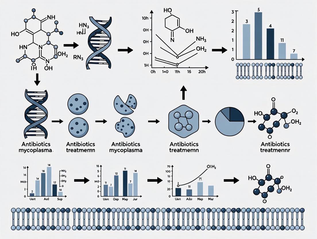
Figure 1: Functional Architecture of the Attachment Organelle. The diagram illustrates the central role of the attachment organelle in adhesion (via membrane adhesins), structural integrity (via the internal core), gliding motility, and cell division.
Experimental Protocols for Key Analyses
Protocol 1: Differentiating Mycoplasma Contamination in Cell Culture
The absence of a cell wall makes Mycoplasma contamination resistant to standard antibiotics like penicillin and streptomycin, a critical concern for cell culture integrity.
- Principle: Exploit the bacterium's unique biology—specifically its lack of a cell wall and essential cholesterol requirement—for detection and elimination.
- Materials: See Table 3 for specific reagent solutions.
- Procedure:
- Detection by DNA Staining: Use a DNA fluorochrome (e.g., DAPI or Hoechst 33258) to stain a fixed sample of test cells. Visualize under a fluorescence microscope. The presence of Mycoplasma will appear as particulate or filamentous extranuclear fluorescence on the surface of the infected cells.
- PCR-Based Detection: Isolate DNA from the culture supernatant. Perform PCR using primers specific for the M. pneumoniae 16S rRNA gene or other conserved genomic regions. Analyze the amplified products by gel electrophoresis.
- Culture-Based Detection (for confirmation): Inoculate the suspect sample into a specialized broth medium (e.g., Hayflick medium). Incubate at 37°C and monitor for a color change of the phenol red indicator to yellow, signifying acid production from glucose fermentation [6].
- Decontamination: Treat contaminated cultures with non-beta-lactam antibiotics effective against Mycoplasma. TET or fluoroquinolones like MOX are common choices, as macrolide resistance is prevalent [6]. Always confirm eradication post-treatment.
Protocol 2: Assessing Macrolide Resistance via 23S rRNA Gene Mutation Analysis
Mutations in the 23S rRNA gene are the primary mechanism of macrolide resistance in M. pneumoniae. This protocol outlines the steps for genotypic resistance testing.
- Principle: Identify point mutations in domain V of the 23S rRNA gene that reduce macrolide binding affinity, using PCR followed by sequencing.
- Materials: See Table 3 for specific reagent solutions.
- Procedure:
- DNA Extraction: Obtain a clinical specimen (e.g., throat swab or bronchoalveolar lavage). Extract total DNA using a commercial kit (e.g., Qiagen DNA Mini-kit) per the manufacturer's protocol [6].
- PCR Amplification: Set up a PCR reaction mixture containing the extracted DNA, primers flanking the critical region of the 23S rRNA gene (encompassing positions 2063 and 2064), and a DNA polymerase. Run the PCR with appropriate cycling conditions [6].
- Sequencing and Analysis: Purify the PCR product and perform Sanger sequencing. Analyze the sequencing results by comparing them to a wild-type reference sequence (e.g., GenBank accession M129). Identify the presence of resistance-associated mutations (A2063G, A2064G, etc.) [7] [6].
Figure 2: 23S rRNA Mutation Analysis Workflow. The flowchart outlines the process for detecting macrolide-resistant mutations in Mycoplasma pneumoniae, from sample collection to final genotypic report.
Clinical Implications for Antibiotic Therapy
Innate and Acquired Antibiotic Resistance
The defining biological feature of M. pneumoniae—the lack of a cell wall—confers innate resistance to all beta-lactam antibiotics (e.g., penicillin, cephalosporins), which target peptidoglycan synthesis [3]. Consequently, first-line treatment relies on antibiotics that inhibit protein synthesis, such as macrolides (e.g., azithromycin), TET, and fluoroquinolones [3].
However, acquired resistance to macrolides has become a major global health challenge. This resistance is primarily driven by point mutations in the 23S rRNA gene, with A2063G and A2064G being the most common [7] [6]. These mutations alter the macrolide binding site on the bacterial ribosome, reducing drug affinity and leading to treatment failure [6]. The resistance rates show significant geographical variation, exceeding 90% in some parts of Asia, while remaining below 10% in the United States, though it is a growing concern worldwide [7] [3] [6].
Table 2: Impact of 23S rRNA Point Mutations on Clinical Outcomes (Meta-Analysis Data) [7]
| Genotype | Fever Duration (Hazard Ratio vs. Wild-type) | Risk of Severe Illness (Hazard Ratio vs. Wild-type) | Notes |
|---|---|---|---|
| Wild Type | Reference (HR=1.0) | Reference (HR=1.0) | - |
| Single Mutation (A2063G) | HR = 3.66 (95% CI: 1.89–7.09) | HR = 5.89 (95% CI: 2.03–17.08) | Moderate resistance, longer illness. |
| Double Mutation (A2063G + A2064G) | HR = 5.32 (95% CI: 4.27–6.61) | HR = 7.80 (95% CI: 2.51–24.18) | Higher MIC, more severe outcomes. |
Treatment Strategies and Decontamination Protocols
Treatment decisions must account for local resistance patterns and patient factors like age. The following workflow, based on current clinical guidelines and research, provides a logical framework for managing M. pneumoniae infections and contamination.
Figure 3: Antibiotic Treatment and Decontamination Decision Workflow. This chart guides the selection of appropriate antibiotics based on clinical response and resistance testing results.
For researchers, decontaminating cell cultures requires a different antibiotic approach than for typical bacterial contaminants. The protocol in Section 3.1 should be followed, with antibiotic selection informed by the resistance patterns below.
- Macrolides: The historical first-line choice, especially for children. However, efficacy is now compromised in regions with high resistance [3] [8].
- Tetracyclines: An effective second-line option (e.g., doxycycline, minocycline), but typically avoided in young children due to the risk of tooth discoloration [3] [6].
- Fluoroquinolones: Another effective second-line class for adults (e.g., levofloxacin, moxifloxacin). Use in children is restricted due to potential effects on cartilage development [3] [6]. Current surveillance shows no signs of resistance to TET or fluoroquinolones in M. pneumoniae, though MIC values on the edge of resistance for quinolones have been observed, signaling a potential future threat [6].
The Scientist's Toolkit: Essential Research Reagents
Table 3: Key Reagent Solutions for Mycoplasma pneumoniae Research
| Reagent / Material | Function / Application | Specific Examples / Notes |
|---|---|---|
| Hayflick Medium | Culture of M. pneumoniae; requires serum as a source of cholesterol and fatty acids [1] [4]. | Contains horse serum, yeast extract, glucose. Phenol red indicates acid production from glucose fermentation [6]. |
| Serum-Free Defined Medium | Reproducible, large-scale cultivation for applications like vaccine development; eliminates serum variability [4]. | Formulations are model-driven, supplemented with essential lipids (cholesterol), nucleotides, and nutrients [4]. |
| PCR Reagents for 23S rRNA | Genotypic detection of macrolide resistance mutations [7] [6]. | Primers for domain V, DNA polymerase, dNTPs. Followed by sequencing or RFLP analysis. |
| DNA Fluorochromes | Rapid detection of mycoplasma contamination in cell culture via fluorescence microscopy. | DAPI, Hoechst 33258. Stains extranuclear Mycoplasma DNA on infected cell surfaces. |
| Non-Beta-Lactam Antibiotics | Treatment of M. pneumoniae infections and decontamination of cell cultures. | Macrolides (Azithromycin), Tetracyclines (Doxycycline), Fluoroquinolones (Moxifloxacin) [3] [6]. |
The COVID-19 pandemic, caused by the novel coronavirus SARS-CoV-2, disrupted global circulation patterns of numerous respiratory pathogens through widespread non-pharmaceutical interventions (NPIs) and immunity debt [9]. Among the most significantly affected pathogens was Mycoplasma pneumoniae (MP), a common cause of community-acquired pneumonia. This application note analyzes the delayed post-COVID-19 resurgence and changing epidemiological patterns of MP within the broader context of antibiotic treatment research, particularly focusing on macrolide-resistant Mycoplasma pneumoniae (MRMP). We present comprehensive surveillance data from multiple global regions and provide detailed experimental protocols for monitoring MP resurgence and resistance patterns, essential for researchers, scientists, and drug development professionals working in respiratory pathogen epidemiology and antimicrobial resistance.
Epidemiological Shifts inMycoplasma pneumoniaeIncidence
Global Resurgence Patterns
The unprecedented decline in MP incidence during the peak COVID-19 pandemic period (2020-2022) has been followed by a significant global resurgence beginning in late 2023. Surveillance data from the U.S. Centers for Disease Control and Prevention (CDC) indicates that MP infections began increasing in late spring/early summer of 2024 from a lower baseline observed since the start of the COVID-19 pandemic [10]. This resurgence pattern has been observed across multiple continents, with studies from Southern Italy confirming a sharp increase in MP positivity rates from negligible levels in early 2023 to a peak of 15.8% by May 2025 [11]. The re-emergence occurred after a prolonged period of low incidence since the start of the COVID-19 pandemic, characteristic of the 3-7 year cyclical epidemic pattern typical of MP but with altered dynamics in the post-pandemic era [10].
Changing Demographic Patterns
A significant shift in the demographic distribution of MP cases has been observed in the post-pandemic period. Historically affecting primarily school-age children and adolescents, recent surveillance has identified an increased incidence in young children [10]. Data from Southern Italy demonstrates this altered age distribution, with the highest burden of disease observed in children aged 5-14 years [11]. The table below summarizes key epidemiological features of the post-pandemic MP resurgence across different geographic regions.
Table 1: Comparative Global Epidemiology of Post-COVID-19 Mycoplasma pneumoniae Resurgence
| Geographic Region | Pre-Pandemic Pattern | Peak Positivity Rate (Post-COVID-19) | Affected Age Groups | Key Epidemiological Shifts |
|---|---|---|---|---|
| United States [10] | Cyclical peaks every 3-7 years | Increasing through summer 2024 | All ages, with notable increase in 0-4 year olds | Departure from historical seasonality; increased cases in younger children |
| Southern Italy [11] | Not specified | 15.8% (May 2025) | Median age: 10 years (IQR: 6-12) | Sharp increase from near-zero prevalence during pandemic; highest burden in 5-9 and 10-14 year age groups |
| Global Pattern [10] | Regular 3-7 year cycles | Varied by region, beginning late 2023 | Traditionally school-aged children; now including younger children | Resurgence after prolonged suppression during COVID-19; potential immunity debt effect |
Experimental Protocols for MP Surveillance and Resistance Monitoring
Multiplex PCR Detection Protocol for Respiratory Pathogens
Principle: Simultaneous detection of MP alongside other respiratory pathogens, including SARS-CoV-2, enables comprehensive surveillance of co-circulation patterns and identification of co-infections in the post-COVID-19 era.
Materials:
- Nasopharyngeal swabs in Universal Transport Medium (UTM)
- Nucleic acid extraction kit (STARMag Universal Cartridge, Seegene)
- Automated extraction platform (Nimbus IV, Seegene)
- Multiplex PCR assays (Allplex Respiratory Panel Assays, Seegene; FilmArray RP2.1 Plus, bioMérieux; QIAstat-Dx Respiratory SARS-CoV-2 Panel, QIAGEN)
- Real-time PCR instrument
Procedure:
- Sample Collection: Collect nasopharyngeal swabs using standardized procedures and transport in UTM at 4°C.
- Nucleic Acid Extraction: Extract nucleic acids using automated platforms according to manufacturer protocols.
- Multiplex PCR Setup: Prepare reaction mixtures according to panel specifications. Commonly targeted pathogens include: Mycoplasma pneumoniae, SARS-CoV-2, influenza A/B, RSV, rhinovirus/enterovirus, adenovirus, parainfluenza, human metapneumovirus, seasonal coronaviruses, and various bacterial pathogens including Streptococcus pneumoniae.
- Amplification and Detection: Run PCR protocols according to manufacturer specifications.
- Data Analysis: Interpret results using manufacturer-provided software, determining positivity for each target.
Application Notes: This protocol enables researchers to monitor MP within the broader context of respiratory pathogen co-circulation, essential for understanding the changing epidemiology in the post-COVID-19 landscape [11] [9].
Macrolide Resistance Detection Protocol
Principle: Identification of point mutations in domain V of the 23S rRNA gene associated with macrolide resistance in MP, particularly A2063G and A2064G substitutions.
Materials:
- MP-positive clinical samples or isolates
- PCR primers targeting 23S rRNA domain V
- PCR amplification reagents
- Gel electrophoresis equipment
- Sanger sequencing reagents
- Sequence analysis software (BioEdit, MEGA)
Procedure:
- DNA Extraction: Extract DNA from MP-positive samples as described in protocol 3.1.
- PCR Amplification: Amplify domain V of the 23S rRNA gene using previously described primers and conditions [11].
- Amplification Verification: Confirm successful amplification via gel electrophoresis.
- Sequencing: Purify PCR products and perform Sanger sequencing.
- Sequence Analysis: Align sequences with reference strains using analysis software. Identify resistance-associated mutations (A2063G, A2064G).
- Resistance Reporting: Categorize samples as macrolide-resistant or susceptible based on identified mutations.
Application Notes: This protocol is essential for monitoring the emergence and spread of MRMP, particularly important in the context of altered antibiotic usage patterns during and after the COVID-19 pandemic [11].
Research Reagent Solutions for MP Studies
Table 2: Essential Research Reagents for Mycoplasma pneumoniae Studies
| Reagent/Category | Specific Examples | Function/Application | Implementation in Post-COVID-19 Research |
|---|---|---|---|
| Multiplex PCR Panels | Allplex Respiratory Panel Assays (Seegene); FilmArray RP2.1 Plus (bioMérieux); QIAstat-Dx Respiratory SARS-CoV-2 Panel (QIAGEN) | Simultaneous detection of multiple respiratory pathogens | Critical for monitoring MP resurgence within complex pathogen co-circulation patterns post-pandemic [11] [9] |
| Antimicrobial Susceptibility Testing Systems | Mycoplasma IST2/IST3 (BioMérieux) | Culture-based identification and antibiotic susceptibility profiling | Determines resistance patterns to macrolides, tetracyclines, and fluoroquinolones; IST3 provides improved differentiation of ureaplasma species [12] |
| Molecular Resistance Detection | 23S rRNA domain V PCR primers; Sanger sequencing reagents | Detection of macrolide resistance-associated mutations (A2063G, A2064G) | Essential for tracking MRMP epidemiology in response to altered antibiotic usage patterns during/after pandemic [11] |
| Culture Systems | Standardized culture media for mollicutes | Pathogen isolation and propagation | Enables further characterization of circulating strains and validation of molecular findings |
Visualization of Research Workflow
The following diagram illustrates the integrated experimental workflow for monitoring the post-COVID-19 resurgence of Mycoplasma pneumoniae and associated antimicrobial resistance patterns:
MP Resurgence Research Workflow
Discussion: Implications for Antibiotic Treatment and Future Research
The delayed resurgence of Mycoplasma pneumoniae following the COVID-19 pandemic presents significant challenges for clinical management and antibiotic stewardship. The emergence of macrolide-resistant strains (MRMP) complicates empirical treatment decisions, particularly in pediatric populations where tetracyclines and fluoroquinolones have safety concerns [13]. Recent data from Southern Italy indicates an overall MRMP prevalence of 7.5%, with rates peaking at 12.6% in preadolescents (aged 10-14 years) and predominantly associated with the A2063G mutation (96% of resistant cases) [11]. Global surveillance shows significant geographical variation in resistance patterns, with higher rates observed in Asia (>50% in Japan, ~80% in China) compared to Europe (averaging ~5%) and the United States (<10% overall, though with regional hotspots >20%) [10].
The changing demographic patterns, with increased incidence in younger children, necessitates enhanced vigilance and tailored treatment approaches. The high rate of coinfections (23.3% in Southern Italy data, particularly among children <5 years) further complicates clinical management and highlights the importance of comprehensive diagnostic approaches that can detect multiple respiratory pathogens simultaneously [11]. Research indicates that tetracyclines demonstrate superior efficacy for MRMP pneumonia, with meta-analyses showing significantly reduced febrile duration (weighted mean difference 1.64 days) and hospital stays (WMD 1.22 days) compared to macrolides [13]. However, balance of efficacy against potential side effects remains crucial, particularly in younger pediatric patients.
Future research directions should focus on: (1) ongoing molecular surveillance of resistance patterns across different geographic regions; (2) development of rapid point-of-care tests for resistance detection to guide targeted therapy; (3) clinical trials evaluating alternative antimicrobial agents for MRMP; and (4) investigation of the long-term impact of pandemic-related NPIs on MP evolution and population immunity. These efforts will be essential for optimizing therapeutic strategies and mitigating the public health impact of MP resurgence in the post-COVID-19 era.
Macrolide antibiotics are a cornerstone of treatment for infections caused by Mycoplasma pneumoniae, a major cause of community-acquired pneumonia, particularly in children [14]. Their widespread and often indiscriminate use has led to the emergence and rapid global spread of macrolide-resistant M. pneumoniae (MRMP) [14] [15]. The primary molecular mechanism of this resistance involves point mutations in the 23S ribosomal RNA (rRNA) gene, which disrupt the drug's binding site on the bacterial ribosome [14] [16]. This application note details the specific mutations, their clinical consequences, and provides standardized protocols for their detection, framed within the broader context of antimicrobial resistance research and therapeutic development.
The Molecular Basis of 23S rRNA-Mediated Resistance
Mechanism of Action and Resistance
Macrolides exert their antibacterial effect by binding to the 50S ribosomal subunit and inhibiting bacterial protein synthesis [16]. Specifically, they target the peptidyl transferase loop in domain V of the 23S rRNA [14]. Nucleotides in this central loop are critical for macrolide binding. When mutations alter these key nucleotides, the affinity of the ribosome for the drug is reduced, leading to clinical resistance [14] [16]. M. pneumoniae is intrinsically resistant to beta-lactam antibiotics due to its lack of a cell wall, making macrolides a first-line therapy and underscoring the clinical impact of this resistance mechanism [14] [15].
Primary Resistance-Associated Mutations
The following table summarizes the most clinically significant mutations in domain V of the 23S rRNA gene that confer macrolide resistance in M. pneumoniae.
Table 1: Key 23S rRNA Mutations and Their Phenotypic Effects in M. pneumoniae
| Nucleotide Position (E. coli numbering) | Nucleotide Change | Prevalence and Notes | Resistance Level and Profile |
|---|---|---|---|
| 2063 | A→G (Transition) | The most common mutation, accounts for up to 93.7% of all resistance mutations [14] [17]. | Confers high-level resistance to 14- and 15-membered macrolides (e.g., erythromycin, azithromycin, clarithromycin) [14] [17]. |
| 2063 | A→T, A→C (Transversions) | Rare mutations [17]. | Associated with high-level macrolide resistance [17]. |
| 2064 | A→G (Transition) | The second most common mutation [14]. | Confers high-level resistance to 14- and 15-membered macrolides [14] [17]. |
| 2064 | A→C (Transversion) | Very rare; reported but not commonly found [15] [17]. | Associated with high-level macrolide resistance [17]. |
| 2067 | A→G, A→C | Less common; can be selected in vitro by 16-membered macrolides [15]. | Confers high-level resistance to 16-membered macrolides (e.g., josamycin) [14] [15]. |
| 2617 | C→A, C→G, C→T | Rare mutation [15] [17]. | Associated with a low level of macrolide resistance [17]. |
| 2353 | C→T | A novel variant identified in Vietnam in 2023; clinical significance under investigation [18]. | Hypothesized to confer macrolide resistance [18]. |
Other mechanisms, such as mutations in ribosomal proteins L4 and L22, have been observed in laboratory-derived mutants and in other bacterial species like Streptococcus pneumoniae [15] [19]. However, in clinical isolates of M. pneumoniae, point mutations in 23S rRNA remain the dominant and most clinically relevant mechanism [14].
Detection Protocols and Methodologies
Accurate and rapid detection of MRMP is crucial for clinical decision-making and antimicrobial stewardship. The following section outlines key experimental protocols.
Standard PCR and Sequencing for Mutation Detection
This protocol is the reference standard for identifying mutations in the 23S rRNA gene.
Table 2: Key Reagents for PCR and Sequencing
| Research Reagent | Function/Explanation |
|---|---|
| Specific Primers (e.g., MRMP-F1/R1) | Designed to amplify a ~748 bp region of domain V of the 23S rRNA gene (nt 1963-2710), encompassing all known major resistance loci [18]. |
| DNA Polymerase for Long-Range PCR | Essential for robust amplification of the target region from genomic DNA [20]. |
| BigDye Terminator Cycle Sequencing Kit | Used for Sanger sequencing of the PCR amplicons to determine the nucleotide sequence at critical positions [18]. |
Workflow:
- DNA Extraction: Extract genomic DNA from clinical samples (e.g., nasopharyngeal swabs, bronchoalveolar lavage fluid) using a commercial kit (e.g., QIAamp DNA Mini Kit) [17] [21].
- PCR Amplification:
- Primers: Use primers MRMP-F1 (5′-CGTCCCGCTTGAATGGTGTA-3′) and MRMP-R1 (5′-GGCGCTACAACTGGAGCATA-3′) [18].
- Reaction Setup: Prepare a 25 µL reaction mixture containing template DNA, primers, dNTPs, and a suitable DNA polymerase.
- Cycling Conditions: Initial denaturation at 95°C for 5 min; 35 cycles of denaturation at 94°C for 35 s, annealing at 56°C for 40 s, and elongation at 72°C for 45 s; final elongation at 72°C for 5 min [22].
- Sequencing and Analysis: Purify the PCR product and perform Sanger sequencing. Compare the resulting sequence to a reference strain (e.g., GenBank NR_077056.1) to identify mutations at positions 2063, 2064, etc. [18].
Diagram Title: Standard PCR and Sequencing Workflow
Novel Real-time PCR with Two Non-Overlapping Probes
This advanced method allows for simultaneous detection of M. pneumoniae and identification of macrolide resistance mutations in a single, rapid assay [17].
Principle: Two probes bind to the same DNA strand in the target region. Probe A covers the mutable loci 2063/2064, while Probe B binds downstream on the same strand, serving as an internal control. The difference in cycle threshold (ΔCT) values between the two probes determines the resistance status.
Workflow:
- Assay Design:
- Primer F: 5′-AATCCAGGTACGGGTGAAGACA-3′
- Primer R: 5′-TGCTCCTACCTATTCTCTACATGATAATG-3′
- Probe A (VIC-labeled): Covers 2063/2064 loci (5′-VIC-ACGGGACGGAAAGA-MGB-3′)
- Probe B (FAM-labeled): Internal control probe (5′-FAM-ACTGTAGCTTAATATTGATCAG-MGB-3′) [17].
- Reaction Setup: Prepare a 25 µL reaction mix with 2x PCR mix, primers, probes, and template DNA.
- Real-time PCR Cycling: 95°C for 10 min; 45 cycles of 95°C for 15 s and 65°C for 15 s [17].
- Result Interpretation:
- ΔCT (|CTVIC - CTFAM|) < 0.5: Macrolide-sensitive M. pneumoniae (MSMP)
- ΔCT > 2.0: Macrolide-resistant M. pneumoniae (MRMP) [17]
Diagram Title: Dual-Probe Real-time PCR Principle
The Researcher's Toolkit
Table 3: Essential Research Reagents and Materials
| Category | Item | Specific Function in Research |
|---|---|---|
| Bacterial Strains | M. pneumoniae reference strain M129 (ATCC 29342) | Susceptible control parent strain for in vitro selection of resistant mutants and assay validation [15]. |
| Culture Media | PPLO Broth / Mycoplasma Broth Base | Specialized culture medium essential for cultivating fastidious Mycoplasma species from clinical samples or for in vitro studies [15] [22]. |
| Antibiotics for MIC Testing | Clarithromycin, Azithromycin, Erythromycin, Doxycycline, Levofloxacin | Used in broth microdilution assays to determine Minimum Inhibitory Concentrations (MICs) and validate resistance phenotypes [14] [22]. |
| Molecular Detection | Primer sets for 23S rRNA, P1 gene | Specific primers for PCR identification of M. pneumoniae (P1 gene) and amplification of the resistance-conferring region (23S rRNA domain V) [18] [22]. |
| Next-Generation Sequencing | tNGS Panels / Whole-Genome Sequencing | For comprehensive analysis of resistance mutations and discovery of novel genetic changes in ribosomal proteins or other genes associated with resistance [15] [21]. |
Application in Drug Development and Contamination Control
Understanding these molecular mechanisms is vital beyond clinical therapy, directly impacting drug development and cell culture contamination control.
- Guiding Antibiotic Stewardship in Research: The high prevalence of MRMP in Asia (up to 90%) compared to North America and Europe (∼10%) highlights the need for regional resistance monitoring in pre-clinical and clinical trial planning [14]. Research facilities, especially those with global collaborations, must validate the efficacy of macrolides used in cell culture media against local Mycoplasma strains.
- Developing Next-Generation Antimicrobials: The knowledge of precise resistance mutations (e.g., A2063G) enables the rational design of novel macrolides and ketolides that can bind effectively to mutant ribosomes [16] [19]. In vitro selection studies can identify which drugs are less prone to inducing resistance, informing future drug candidates [15].
- Ensuring Robust Contamination Testing: Effective eradication of Mycoplasma contamination from cell lines requires using antibiotics to which the contaminant is susceptible. Molecular detection kits based on the protocols described herein (e.g., the two-probe qPCR assay) can provide rapid resistance profiling, ensuring the selection of effective decontamination agents and maintaining the integrity of biological models in drug development [17].
Biofilms are structured communities of microbial cells embedded in a self-produced matrix of extracellular polymeric substances (EPS) and represent a dominant mode of bacterial growth in nature [23]. For Mycoplasma species, which lack a cell wall and possess highly reduced genomes, biofilm formation represents a critical survival strategy that contributes significantly to persistent infections and treatment failures in clinical settings [24]. The biofilm lifestyle provides intrinsic tolerance to antimicrobials through multiple mechanisms, including physical barrier function, metabolic dormancy, and enhanced horizontal gene transfer [25]. Understanding the characteristics and control of mycoplasma biofilms is therefore essential for developing effective therapeutic strategies against these problematic contaminants in research and clinical contexts.
Biofilm Architecture and Development in Mycoplasma Species
Structural Characteristics
Mycoplasma biofilms exhibit complex three-dimensional architectures that vary between species and growth conditions. M. pneumoniae typically forms volcano-like structures when grown axenically, while M. synoviae biofilms display mushroom- and tower-like formations [24]. These structures are not static but rather dynamic communities connected by channels that facilitate nutrient transport and waste removal [24]. The extracellular matrix of mycoplasma biofilms comprises various biopolymers, including polysaccharides, proteins, and extracellular DNA (eDNA), which provide structural integrity and protection [24].
Developmental Lifecycle
The formation of mycoplasma biofilms follows a staged process similar to other bacterial species, though with unique adaptations due to their minimal genome:
Figure 1: The developmental lifecycle of mycoplasma biofilms, illustrating the transition from free-living planktonic cells to structured communities and subsequent dispersion.
- Initial Reversible Attachment: Planktonic mycoplasma cells adhere to biotic or abiotic surfaces via variable surface antigen (Vsa) proteins. Short Vsa proteins facilitate this attachment, while longer variants may inhibit adhesion through steric hindrance [24].
- Irreversible Attachment and Microcolony Formation: Following initial attachment, cells begin secreting extracellular polymers that form an EPS matrix, enabling firm surface adhesion and the development of microcolonies [24].
- Biofilm Maturation: Microcolonies develop into mature biofilms with species-specific architectural features. During this phase, mycoplasma within biofilms demonstrate attenuated virulence factor production (including reduced H₂O₂ and CARDS toxin production), consistent with establishing long-term residence while limiting excessive host damage and immune responses [26].
- Dispersion: Upon nutrient depletion or environmental stress, biofilms activate dispersal mechanisms, often through enzymatic degradation of EPS components, allowing cells to colonize new niches [24].
Biofilm-Mediated Antimicrobial Resistance Mechanisms
Mycoplasma biofilms confer dramatically enhanced resistance to antimicrobial agents compared to their planktonic counterparts. M. pneumoniae biofilm towers exhibit extreme resistance to erythromycin, tolerating concentrations up to 512 µg/mL, which represents 8,500-128,000 times the minimal inhibitory concentration (MIC) for planktonic cells [26]. The mechanisms underlying this enhanced tolerance are multifaceted:
Physical and Physiological Barriers
- Matrix-Mediated Protection: The EPS matrix acts as a structural barrier that hinders antibiotic penetration through binding or enzymatic degradation of antimicrobial compounds [25]. The matrix composition (including polysaccharides, proteins, and eDNA) can bind antibiotics such as aminoglycosides, reducing their effective concentration within the biofilm interior [25].
- Metabolic Heterogeneity: Biofilms contain subpopulations of metabolically dormant persister cells that exhibit reduced susceptibility to antimicrobials targeting active cellular processes [25]. This physiological heterogeneity creates gradients of metabolic activity that further complicate treatment efficacy.
Genetic Adaptations
Biofilm environments facilitate enhanced horizontal gene transfer between bacterial cells, promoting the dissemination of resistance determinants [25]. In mycoplasma species, this is complemented by chromosomal mutations in target genes and the action of efflux pumps that further enhance the resistant phenotype [24].
Table 1: Key Mechanisms of Antimicrobial Resistance in Mycoplasma Biofilms
| Resistance Mechanism | Functional Basis | Impact on Treatment |
|---|---|---|
| Physical Barrier | EPS matrix limits antibiotic penetration | Reduced drug concentration at target sites |
| Metabolic Dormancy | Heterogeneous metabolic activity including persister cells | Tolerance to growth-dependent antibiotics |
| Genetic Exchange | Enhanced horizontal gene transfer in structured communities | Dissemination of resistance genes |
| Efflux Systems | Upregulation of efflux pumps | Active removal of antimicrobial compounds |
| Target Modification | Mutations in antibiotic target sites | Reduced drug binding affinity |
Quantitative Analysis of Biofilm-Associated Resistance
Recent investigations have quantified the dramatic increase in antimicrobial resistance associated with mycoplasma biofilm formation. The data reveal not only intrinsic tolerance but also promising synergistic approaches for biofilm eradication.
Table 2: Antibiotic Efficacy Against Planktonic vs. Biofilm Forms of Mycoplasma pneumoniae
| Antibiotic | MIC for Planktonic Cells (µg/mL) | MIC for Biofilm Cells (µg/mL) | Resistance Fold-Increase | Synergistic Combinations (FICI) |
|---|---|---|---|---|
| Erythromycin | 0.004-0.06 | 512 | 8,500-128,000 | Erythromycin + Moxifloxacin (FICI<0.5) |
| Moxifloxacin | 0.25-1.0 | 8-16 | 16-32 | Doxycycline + Moxifloxacin (FICI<0.5) |
| Doxycycline | 0.125-1.0 | 16-32 | 16-128 | Erythromycin + Doxycycline (FICI<0.5) |
Data compiled from synergy testing against M. pneumoniae strains M129 and 19294 [26]. FICI (Fractional Inhibitory Concentration Index) values <0.5 indicate synergistic interactions.
The quantitative data demonstrate that combination therapies utilizing antibiotic pairs show particular promise against mycoplasma biofilms, with synergistic interactions (FICI<0.5) observed between erythromycin, moxifloxacin, and doxycycline [26]. These combinations achieve substantial efficacy against pre-formed biofilm towers at clinically relevant concentrations, with scanning electron microscopy confirming more complete eradication than indicated by crystal violet assays alone [26].
Experimental Protocols for Biofilm Analysis
Mycoplasma Biofilm Culture and Synergy Testing
Purpose: To establish in vitro mycoplasma biofilms and evaluate antimicrobial synergy against mature structures.
Materials:
- Mycoplasma strains (e.g., M. pneumoniae M129 and 19294)
- SP-4 broth medium
- 24- or 96-well tissue culture plates
- Antimicrobial stock solutions (erythromycin, moxifloxacin, doxycycline)
- Crystal violet staining solution
- Scanning electron microscopy (SEM) equipment
Methodology:
- Inoculum Preparation: Syringe mycoplasma stocks through a 26-g needle multiple times and dilute in SP-4 broth to achieve approximately 1.0 × 10⁴ CFU/mL [26].
- Biofilm Formation: Inoculate diluted mycoplasma into 24- or 96-well plates and incubate at 37°C for 7-10 days to allow mature biofilm tower development [26].
- Checkerboard Assay Preparation:
- Prepare antibiotic stocks at twice the MIC of each drug.
- Create decreasing concentrations of both antibiotics in 96-well plates.
- Include controls: growth control (no antibiotics), sterility control (medium only), and vehicle controls.
- Synergy Assessment:
- Inoculate plates with prepared mycoplasma suspension.
- Incubate until growth control shows color change from red to yellow.
- Calculate FICI = (MIC of drug A in combination/MIC of drug A alone) + (MIC of drug B in combination/MIC of drug B alone).
- Interpret results: FICI ≤0.5 indicates synergy; >0.5-4.0 indicates additive or indifferent effects; >4.0 indicates antagonism [26].
- Biofilm Quantification:
- Use crystal violet staining to assess total biofilm biomass.
- Validate results with SEM for structural analysis of biofilm eradication.
Figure 2: Experimental workflow for assessing synergistic antibiotic efficacy against mycoplasma biofilms.
Biofilm Disruption Using Engineered Biologicals
Purpose: To evaluate enzymatic disruption of mycoplasma biofilms using engineered bacterial delivery systems.
Rationale: Engineered attenuated Mycoplasma pneumoniae strains (e.g., CV8_HAD) can be designed to secrete multiple biofilm-degrading enzymes simultaneously, including PelAh, PslGh, A1-II', and Dispersin B, which target different EPS components [27].
Materials:
- Engineered M. pneumoniae CV8_HAD strain
- Wild-type mycoplasma biofilm cultures
- Supernatant collection equipment
- Crystal violet staining solution
- CFU enumeration materials
Methodology:
- Supernatant Preparation: Culture engineered M. pneumoniae CV8_HAD strain and collect sterile supernatant containing secreted enzymes [27].
- Biofilm Treatment: Apply supernatant to 24-hour pre-formed mycoplasma biofilms and incubate for 6 hours.
- Efficacy Assessment:
- Quantify biofilm biomass using crystal violet staining.
- Enumerate viable cells through CFU counting.
- Compare treatment groups to controls (untreated or wild-type supernatant).
- In Vivo Validation: Utilize Galleria mellonella larvae models to assess efficacy in complex infection environments [27].
Emerging Control Strategies and Research Reagents
Novel approaches to mycoplasma biofilm control extend beyond conventional antibiotics to include enzymatic disruption, engineered biologicals, and combination therapies. The limited genetic capacity of mycoplasma species presents both challenges and opportunities for targeted interventions.
The Scientist's Toolkit: Essential Research Reagents
Table 3: Key Research Reagents for Mycoplasma Biofilm Studies
| Reagent/Category | Specific Examples | Research Application | Functional Role |
|---|---|---|---|
| Culture Media | SP-4 Broth | Mycoplasma cultivation | Supports axenic growth with essential cholesterol |
| Biofilm Detection | Crystal Violet | Biofilm quantification | Stains biomass for spectrophotometric analysis |
| Engineered Strains | M. pneumoniae CV8_HAD | Biofilm disruption research | Secretes multiple EPS-degrading enzymes |
| Synergy Assessment | Checkerboard Assay | Antibiotic combination screening | Determines FICI for drug interactions |
| In Vivo Models | Galleria mellonella | Infection/therapeutic validation | Cost-effective alternative to mammalian systems |
| Analytical Tools | Scanning Electron Microscopy | Structural analysis | Visualizes 3D biofilm architecture |
Innovative Therapeutic Approaches
- Enzyme-Based Disruption: Glycoside hydrolases such as Dispersin B (targeting PNAG polysaccharides) and alginate lyases (targeting alginate polymers) can effectively degrade key EPS matrix components, sensitizing biofilms to conventional antibiotics [27].
- Engineered Biologicals: Attenuated bacterial vectors designed to secrete multiple biofilm-disrupting enzymes simultaneously offer a promising approach for targeted delivery and enhanced efficacy against complex biofilm communities [27].
- Synergistic Antibiotic Combinations: The demonstrated synergy between macrolides, fluoroquinolones, and tetracyclines provides viable treatment options even against extensively resistant mycoplasma biofilms [26].
Mycoplasma biofilm formation represents a significant challenge in both research and clinical contexts, contributing substantially to persistent infections and treatment failures. The structured nature of biofilms confers dramatically enhanced antimicrobial resistance through combined physical, physiological, and genetic mechanisms. However, recent advances in understanding biofilm biology have revealed promising intervention strategies, particularly synergistic antibiotic combinations and enzymatic disruption approaches. Future research directions should focus on optimizing delivery methods for biofilm-disrupting agents, identifying additional synergistic drug pairs, and validating efficacy in complex infection models that better recapitulate the in vivo environment.
The accurate differentiation between asymptomatic carriage and active infection represents a critical challenge in clinical microbiology and therapeutic development. This distinction is particularly acute in infections caused by Mycoplasma pneumoniae, where the presence of the organism does not necessarily correlate with disease state. Within antibiotic treatment research for mycoplasma contamination, this diagnostic dilemma directly impacts study outcomes, treatment efficacy assessments, and resistance monitoring. Asymptomatic carriage refers to the presence and multiplication of a pathogen without manifest symptoms in the host, while active infection involves both pathogen presence and symptomatic disease. This distinction is crucial for appropriate antimicrobial stewardship, as treating carriage may contribute unnecessarily to antibiotic resistance without clinical benefit.
Diagnostic Methods and Their Clinical Utility
Multiple laboratory methods are available for detecting Mycoplasma pneumoniae, each with distinct advantages and limitations for differentiating carriage from active infection. The following table summarizes the key characteristics of these diagnostic approaches:
Table 1: Comparison of Diagnostic Methods for Mycoplasma pneumoniae
| Method | Target | Time Required | Advantages | Limitations | Indication for Active Infection |
|---|---|---|---|---|---|
| Culture | Viable organism | Weeks (slow-growing) | Gold standard, provides isolate for resistance testing | Low sensitivity, technically demanding, not clinically practical [28] | Positive result confirms current infection |
| Serology (MP-IgM) | Host antibody response | Hours to days | Indicates immune response, useful for later diagnosis | Cannot distinguish current vs. past infection; false negatives in early infection [29] | ≥4-fold rise in titre between acute and convalescent phases [30] |
| Real-time PCR (DNA detection) | Microbial DNA | Hours | High sensitivity and specificity, rapid results | Detects DNA but not necessarily viable organisms; may remain positive after infection resolution [29] | Positive result suggests presence but cannot distinguish carriage |
| Nucleic Acid Amplification (RNA detection) | Microbial RNA | Hours | Detects viable organisms (RNA degrades quickly); higher specificity for active infection | Technically demanding; not universally available [29] | Strong indicator of active infection due to association with viable organisms |
The combination of serological and molecular testing provides complementary insights into both past exposure and ongoing infections [31]. For research purposes, integrating multiple methods significantly enhances diagnostic accuracy, particularly when differentiating complicated infection states.
Experimental Protocols for Diagnostic Differentiation
Protocol for Combined Serological and Molecular Testing
This integrated protocol is designed specifically for research settings requiring precise differentiation between carriage and active infection.
Sample Collection: Collect paired samples (acute and convalescent) for comprehensive analysis:
Nucleic Acid Extraction and Detection:
- Extract DNA and RNA using commercial kits (QIAamp DNA Mini Kit or equivalent)
- Perform real-time PCR targeting MP-specific DNA sequences (e.g., 23S rRNA region)
- Conduct simultaneous amplification and testing (SAT) for MP-RNA detection using isothermal amplification [29]
- Include controls: negative controls, positive controls, and internal extraction controls
Serological Testing:
Data Interpretation:
- Active Infection: MP-RNA positive OR MP-DNA positive with seroconversion
- Recent Infection: Seroconversion alone with negative nucleic acid tests
- Asymptomatic Carriage: MP-DNA positive with stable/low antibody titres and no symptoms
- Past Infection: High stable antibody titres with negative nucleic acid tests
Protocol for Macrolide Resistance Detection
Given the high prevalence of macrolide-resistant M. pneumoniae strains, particularly in Asian countries where resistance rates exceed 80%, resistance testing should be incorporated into research protocols [29] [30].
- Sample Processing: Use respiratory specimens positive for MP-DNA
- PCR Amplification: Amplify domain V of the 23S rRNA gene using primers:
- Forward: 5′-AACTATAACGGTCCTAAGGTAGCG-3′
- Reverse: 5′-GCTCCTACCTATTCTCTACATGAT-3′ [29]
- Sequence Analysis: Sequence PCR products and analyze for point mutations (A2063G, A2064G) associated with macrolide resistance
- Interpretation: Identify mutations conferring resistance compared to reference strain M129
Diagnostic Decision Pathway
The following diagnostic workflow integrates multiple testing modalities to optimally distinguish between asymptomatic carriage and active infection in research settings:
Diagram 1: Diagnostic pathway for differentiating Mycoplasma pneumoniae infection states. This integrated approach combines molecular and serological testing to distinguish between active infection, asymptomatic carriage, and past exposure.
Research Reagent Solutions
Table 2: Essential Research Reagents for Mycoplasma pneumoniae Detection and Characterization
| Reagent/Category | Specific Examples | Research Application | Considerations |
|---|---|---|---|
| Nucleic Acid Extraction Kits | QIAamp DNA Mini Kit, Maxwell RSC PureFood GMO and Authentication Kit | DNA extraction from respiratory specimens for PCR-based detection | Ensure compatibility with sample type; include contamination controls |
| PCR Master Mixes | TaqMan Universal PCR Master Mix, SYBR Green-based kits | Amplification of MP-specific DNA targets | Select based on detection method; optimize primer concentrations |
| Real-time PCR Systems | AriaMx Real-Time PCR System, Applied Biosystems platforms | Quantitative detection of MP-DNA with cycle threshold (Ct) values | Standardize Ct cutoffs for positivity (typically Ct ≤35) [32] |
| Serological Assays | SERODIA MYCO-II (Particle Agglutination), immuno-colloidal gold kits | Detection of host antibody response to MP infection | Pair acute and convalescent samples; establish institution-specific titre thresholds |
| RNA Detection Kits | Simultaneous amplification and testing (SAT) kits | Detection of viable organisms through RNA amplification | Requires RNA stabilization during collection; more technically challenging |
| Culture Media | SP4 broth and agar | Gold standard cultivation of viable organisms | Limited clinical utility due to slow growth (up to 6 months) [28] |
| Antimicrobial Testing | Macrolide resistance detection primers | Identification of resistance mutations (A2063G) in 23S rRNA | Essential in regions with high resistance prevalence; requires sequencing |
The distinction between asymptomatic carriage and active M. pneumoniae infection has profound implications for antibiotic treatment research. The high rate of macrolide resistance, particularly in Asian countries where rates approach 90%, underscores the importance of accurate diagnostic classification in therapeutic studies [29] [30]. Without proper differentiation, research outcomes may be confounded by inclusion of carriers who clear infection without intervention or who harbor resistant strains that respond differently to investigated therapies.
For antibiotic development research, the combined approach of MP-RNA detection plus MP-IgM serology provides the most reliable differentiation, with studies demonstrating this combination yields sensitivity of 84.2%, specificity of 78.7%, and a Youden index of 62.9% [29]. This diagnostic precision is essential for enrolling appropriate subject populations, evaluating treatment efficacy, and monitoring resistance patterns in interventional studies.
Future research should prioritize the development of rapid, point-of-care tests that can distinguish carriage from active infection, particularly tests that detect viable organisms through RNA or other viability markers. Additionally, greater understanding of host-pathogen interactions in asymptomatic carriage may reveal new therapeutic targets for preventing progression to active disease. Until such advances emerge, the integrated application of currently available diagnostic modalities within well-designed research protocols represents the most effective approach to addressing this persistent diagnostic dilemma.
Diagnostic and Therapeutic Protocols: From First-Line to Alternative Regimens
The effective management of Mycoplasma infections, particularly Mycoplasma pneumoniae in respiratory illnesses and Mycoplasma genitalium in sexually transmitted infections, presents a significant challenge in clinical practice due to the rising prevalence of antimicrobial resistance. The integration of advanced molecular diagnostics—including PCR, resistance gene detection, and metagenomic next-generation sequencing (mNGS)—is revolutionizing the approach to these pathogens. These tools enable precise pathogen identification and resistance profiling, facilitating targeted antibiotic therapy and advancing stewardship efforts. This protocol outlines the application of these integrated diagnostic technologies within the broader context of antibiotic treatment research for Mycoplasma contamination, providing a structured framework for researchers and drug development professionals.
Comparative Analysis of Diagnostic Methods
Table 1: Performance Comparison of Mycoplasma Diagnostic Methods
| Method | Target | Time | Sensitivity | Specificity | Key Advantage | Key Limitation |
|---|---|---|---|---|---|---|
| Culture | Viable organism | Weeks to months [28] [29] | Low [33] [29] | High [33] | Traditional gold standard | Extremely slow, insensitive [28] [29] |
| Serology (IgM PA) | Host antibodies | Hours | 74.0% [29] | 79.7% [29] | Indicates current/ recent infection | Cannot differentiate active infection; lower specificity [29] |
| Real-Time PCR (RT-PCR) | Pathogen DNA (e.g., 23S rRNA) | 6-12 hours [33] | 36.6% Positivity Rate [29] | N/A | Rapid, quantitative; detects macrolide resistance mutations (e.g., A2063G) [29] | Limited to targeted pathogens; false positives possible [33] |
| RNA SAT | Pathogen RNA | Hours | 84.2% (in combo with IgM PA) [29] | 97.5% [29] | High specificity; indicates viable organism [29] | Requires specialized isothermal amplification |
| mNGS | All nucleic acids in sample | 24-48 hours [33] | 86.7%-95.9% [33] [34] | 90.9%-95.2% [33] | Hypothesis-free; detects polymicrobial/rare pathogens [33] [34] | High cost; host DNA interference; complex bioinformatics [33] |
The diagnostic landscape for Mycoplasma is diverse, with method selection dependent on the required balance of speed, sensitivity, and scope. Traditional culture, while specific, is impractical for clinical decision-making due to its prolonged turnaround time and low sensitivity [33] [28] [29]. Serological methods like IgM particle agglutination (PA) offer a rapid indication of infection but lack the specificity of molecular methods and cannot be used for resistance testing [29].
Molecular techniques form the cornerstone of modern diagnosis. Real-time PCR (RT-PCR) provides a rapid, sensitive, and specific tool for detecting Mycoplasma DNA and, critically, for identifying key point mutations (e.g., A2063G and A2064G in domain V of 23S rRNA) conferring macrolide resistance [29]. However, its targeted nature means it will not detect unexpected or novel pathogens. In contrast, mNGS sequences all nucleic acids in a sample, providing an unbiased "hypothesis-free" detection capable of identifying polymicrobial infections, rare pathogens, and predicting antimicrobial resistance (AMR) genes, albeit with higher costs and bioinformatic complexity [33] [34]. Combining methods, such as MP-RNA (SAT) with MP-IgM (PA), has been shown to improve sensitivity (84.2%) and specificity (78.7%), offering a reliable early diagnostic approach [29].
Experimental Protocols
Protocol 1: Detection ofMycoplasma pneumoniaeand Macrolide Resistance via RT-PCR
This protocol details the procedure for identifying Mycoplasma pneumoniae and the A2063G macrolide resistance mutation in sputum or respiratory samples using real-time PCR [29].
3.1.1 Research Reagent Solutions
| Item | Function | Specification/Example |
|---|---|---|
| Sputum Sample | Source of pathogen DNA | Collected from patients with CAP symptoms [29]. |
| DNA Extraction Kit | Isolation of nucleic acids | QIAamp DNA Mini Kit (Qiagen) [29]. |
| Specific Primers | Amplification of target gene | Forward: 5′-AACTATAACGGTCCTAAGGTAGCG-3′; Reverse: 5′-GCTCCTACCTATTCTCTACATGAT-3′ (targeting 23S rRNA) [29]. |
| PCR Master Mix | Enzymes and buffers for amplification | Contains DNA polymerase, dNTPs, and buffer. |
| Gel Electrophoresis System | Verification of PCR product | Agarose gel, running buffer, DNA stain. |
| Sanger Sequencing | Confirmatory analysis | Validates the presence of the A2063G mutation [29]. |
3.1.2 Step-by-Step Procedure
- Sample Processing: Sputum samples are liquefied and homogenized using a digestion buffer to release microbial DNA.
- Nucleic Acid Extraction: Extract DNA from 200 µL of processed sample using the QIAamp DNA Mini Kit, following the manufacturer's instructions. Elute the DNA in a final volume of 50-100 µL of elution buffer.
- Primer Design: Design primers to flank the specific point mutations in domain V of the M. pneumoniae 23S rRNA gene, particularly positions 2063 and 2064 [29].
- PCR Amplification: Prepare the reaction mix containing the extracted DNA, specific primers, and PCR master mix. Amplify using a standard thermocycling protocol.
- Product Verification: Verify the PCR product using gel electrophoresis.
- Sequencing and Analysis: Purify the PCR product and perform Sanger sequencing. Analyze the resulting sequence by comparing it to a reference strain (e.g., M129) using sequence analysis software (e.g., SnapGene). The A2063G transition is confirmed by a sequence mismatch at the corresponding position [29].
Protocol 2: Metagenomic Next-Generation Sequencing (mNGS) for Pathogen Detection
This protocol describes the mNGS workflow for unbiased pathogen identification from clinical samples like bronchoalveolar lavage fluid (BALF), which is critical for detecting Mycoplasma and co-infecting pathogens in culture-negative cases [33] [34].
3.2.1 Research Reagent Solutions
| Item | Function | Specification/Example |
|---|---|---|
| BALF Sample | Source of microbial nucleic acids | Collected using sterile technique via bronchoscopy [34]. |
| Nuclease-Free Water | Negative control | Monitors for laboratory contamination during sequencing runs [34]. |
| Nucleic Acid Extraction Kit | Simultaneous isolation of DNA and RNA | Designed to efficiently extract low-biomass microbial nucleic acids. |
| Library Prep Kit | Preparation for sequencing | Fragments nucleic acids and adds sequencing adapters. |
| High-Throughput Sequencer | Massive parallel sequencing | Platforms like Illumina or Ion Torrent. |
| Bioinformatics Pipeline | Data analysis | For host sequence depletion, microbial classification, and AMR gene prediction [33]. |
3.2.2 Step-by-Step Procedure
- Sample Collection & Processing: Collect BALF aseptically. Process samples within 4 hours of collection using sterile techniques to minimize contamination. Include a nuclease-free water negative control.
- Nucleic Acid Extraction: Extract total nucleic acids (DNA and RNA) from the sample. The extracted nucleic acids are then converted to a DNA sequencing library.
- Library Preparation: The extracted nucleic acids are randomly fragmented. Sequencing adapters are ligated to the fragments, and the library is amplified via PCR.
- High-Throughput Sequencing: Load the library onto a sequencer for massive parallel sequencing.
- Bioinformatic Analysis:
- Quality Control: Filter raw sequencing reads for low-quality and short sequences.
- Host Depletion: Map reads to the human reference genome (e.g., hg38) and remove them to enrich for microbial data.
- Microbial Identification: Align non-host reads to comprehensive microbial databases to identify bacteria, viruses, fungi, and parasites. The number of pathogen-specific reads and genome coverage are key metrics [33].
- AMR Gene Prediction: Align reads to curated antimicrobial resistance gene databases to predict potential resistance profiles [33].
The following workflow diagram illustrates the core mNGS process:
Protocol 3: Resistance-Guided Therapy forMycoplasma genitalium
This protocol outlines a two-stage, resistance-guided treatment strategy for M. genitalium infections, which is recommended by the CDC to combat high rates of macrolide resistance [28].
3.3.1 Research Reagent Solutions
| Item | Function | Specification/Example |
|---|---|---|
| FDA-cleared NAAT | Initial detection of M. genitalium | Aptima Mycoplasma genitalium Assay (Hologic) for urine or swab samples [28]. |
| Macrolide Resistance Test | Detection of resistance mutations | Molecular assay for mutations in 23S rRNA (not yet commercially available in the U.S. but under evaluation) [28]. |
| Doxycycline | First-stage empiric therapy | Reduces bacterial load [28]. |
| Azithromycin | Second-stage therapy | For macrolide-sensitive infections (high-dose, extended regimen) [28]. |
| Moxifloxacin | Second-stage therapy | For macrolide-resistant infections [28]. |
3.3.2 Step-by-Step Procedure
- Initial Detection: Test symptomatic patients (e.g., recurrent NGU or cervicitis) using an FDA-cleared NAAT on a urogenital sample to confirm M. genitalium infection [28].
- Resistance Testing (if available): Perform a molecular test for macrolide resistance mutations on the positive sample.
- Two-Stage Treatment:
- Stage 1 (All Patients): Initiate with doxycycline 100 mg orally twice daily for 7 days. This pre-treatment reduces the organism load and can improve clearance rates [28].
- Stage 2 (Guided by Resistance):
- If macrolide-sensitive: Prescribe azithromycin (1g orally on day 1, followed by 500mg once daily for 3 days).
- If macrolide-resistant OR if resistance testing is unavailable: Prescribe moxifloxacin 400 mg orally once daily for 7 days [28].
- Test of Cure: Consider a test of cure (via NAAT) at least 21 days after treatment completion, especially if symptoms persist or if the alternative regimen (doxycycline + azithromycin) was used without resistance testing [28].
The logical pathway for clinical decision-making is as follows:
Discussion and Clinical Integration
The integration of PCR, resistance gene detection, and mNGS provides a powerful, multi-tiered diagnostic strategy for Mycoplasma infections. RT-PCR remains the workhorse for rapid, sensitive detection and first-line resistance screening for M. pneumoniae. In contrast, the two-step approach with NAAT and resistance testing is critical for managing M. genitalium in the face of widespread macrolide resistance, which can exceed 90% in some Asian regions for M. pneumoniae and ranges from 44% to 90% for M. genitalium in the United States [28] [29].
mNGS plays a pivotal role in complex, severe, or culture-negative cases, such as suspected periprosthetic joint infections (PJI) or lower respiratory tract infections (LRTI) where conventional methods fail. It offers a significant advantage in detecting polymicrobial infections and rare or fastidious pathogens, with studies showing a positive rate of 86.7% for mNGS versus 41.8% for traditional methods in LRTI [33] [34]. This can directly impact patient care, leading to changes in antibiotic therapy in a majority of cases (72.13%), including de-escalation in over 30% of patients [34].
However, challenges remain. The high cost and lack of standardization for mNGS limit its widespread adoption, and the clinical relevance of all detected nucleic acids (from viable vs. non-viable organisms) must be carefully interpreted [33]. Furthermore, while mNGS can predict AMR genes, the concordance between genotypic prediction and phenotypic resistance requires further validation [33]. The future of Mycoplasma diagnostics and treatment research lies in the continued refinement of these technologies, including the development of more accessible resistance tests and the validation of standardized mNGS protocols to guide targeted antibiotic therapy and uphold antimicrobial stewardship principles.
Mycoplasma pneumoniae is a significant cause of community-acquired pneumonia (CAP) in both children and adults, accounting for an estimated 10% to 40% of all cases [35] [36]. As a cell wall-less bacterium, it is intrinsically resistant to beta-lactam antibiotics, making macrolides the cornerstone of empirical treatment for decades [37] [36]. These antibiotics inhibit protein synthesis by binding to the 50S ribosomal subunit and are favored for their safety profile, particularly in pediatric populations [37] [38].
However, the global rise of macrolide-resistant M. pneumoniae (MRMP), driven predominantly by point mutations in the 23S rRNA gene, now threatens this first-line status [37] [38]. This application note details the established role of macrolides, defines the scope and impact of resistance, and provides researchers with standardized protocols for surveillance and investigation into overcoming this critical limitation in antimicrobial therapy.
The Established Role of Macrolides in Therapy
Macrolides, including azithromycin, clarithromycin, and erythromycin, have been the first-line treatment for M. pneumoniae infections due to their clinical efficacy and favorable safety profile [36] [38]. Their mechanism of action is bacteriostatic, involving binding to the 50S ribosomal subunit, which prevents bacterial protein synthesis and halts replication [37] [36].
- Clinical Usage and Efficacy: Azithromycin is often the preferred macrolide due to its convenient once-daily dosing and long half-life [36]. In susceptible infections, macrolide treatment is highly effective, with most cases resolving within 7 to 10 days [36].
- Anti-inflammatory Effects: Beyond their antibacterial activity, macrolides are noted for their immunomodulatory effects, which may contribute to clinical improvement by reducing cytokine production and inflammation in the airways [37].
The Growing Challenge of Macrolide Resistance
Epidemiology and Geographic Variation
The prevalence of MRMP exhibits significant geographic variation, with the most alarming rates reported in Asia.
Table 1: Global Prevalence of Macrolide-Resistant M. pneumoniae
| Region | Reported Resistance Prevalence | Key Mutations Identified | Population Context |
|---|---|---|---|
| China (Beijing) | 41.7% (in adults) [35] | A2063G [35] | Adult patients (2011-2017) |
| China (Pediatric) | 73.2% - 78.6% [39] [40] | A2063G [39] [40] | Hospitalized children (2022-2023) |
| Southern Italy | 7.5% [11] | A2063G (96% of resistant cases) [11] | Post-pandemic period (2023-2025) |
| South Korea, Japan | >80% in recent years [37] | A2063G [37] | Pediatric populations |
The COVID-19 pandemic temporarily disrupted the circulation of M. pneumoniae, but a pronounced global resurgence has been observed since late 2023, underscoring the need for continued vigilance [11] [37].
Primary Molecular Mechanisms of Resistance
Resistance in M. pneumoniae is primarily mediated by point mutations in domain V of the 23S rRNA gene, which alter the macrolide binding site [35] [37].
- A2063G Mutation: This is the most prevalent and significant mutation, conferring high-level resistance to macrolides [37] [38]. It is found in the vast majority of resistant strains globally [11] [37].
- Other Mutations: Mutations at position 2064 (A2064G) and in ribosomal proteins L4 and L22 have also been implicated, though they are less common [35] [37].
The following diagram illustrates the primary mechanism of macrolide resistance.
Beyond target site mutation, other mechanisms contribute to resistance. Some clinical isolates have been found to harbor efflux pump genes (msrA/B, mefA), and the addition of the efflux pump inhibitor reserpine reduced the MIC of azithromycin in these strains, confirming a partial role for this mechanism [35] [41]. In contrast, other common bacterial resistance mechanisms, such as target methylation genes (ermA/B/C) or drug-inactivating enzymes (mphC), were not detected in these adult patient isolates [35] [41].
Clinical Impact of Resistance
Macrolide resistance has direct clinical consequences, complicating patient management and worsening outcomes.
Table 2: Clinical Comparisons of Macrolide-Sensitive vs. Resistant M. pneumoniae Pneumonia
| Clinical Parameter | Macrolide-Sensitive M. pneumoniae (MSMP) | Macrolide-Resistant M. pneumoniae (MRMP) | Significance |
|---|---|---|---|
| Fever Duration | ~4.0 days [39] | ~6.0 days [39] | Prolonged in MRMP [39] [38] |
| Hospital Stay | ~5.0 days [39] | ~7.0 days [39] | Prolonged in MRMP [39] [38] |
| Antibiotic Course | Shorter duration | ~2.93 days longer [38] | Increased drug exposure |
| Treatment Failure | Low | High (OR 21.24) [38] | Likely requires therapy change |
| Switch to 2nd-line | Less common | More common (OR 4.42) [38] | Necessitates less preferred agents |
Despite these treatment challenges, studies indicate that the intrinsic severity of pneumonia and the risk of extrapulmonary manifestations are not necessarily greater in MRMP infections; the core issue is the reduced efficacy of first-line macrolide therapy [38].
Experimental Protocols for Resistance Monitoring and Investigation
Protocol 1: Broth Microdilution for Antimicrobial Susceptibility Testing
This protocol determines the Minimum Inhibitory Concentration (MIC) of macrolides against M. pneumoniae clinical isolates [35] [41].
- Strain Isolation and Culture: Isolate M. pneumoniae from oropharyngeal swabs or other respiratory specimens in broth medium (e.g., OXOID CM0403). Incubate at 37°C until a color change from red to yellow is observed, indicating growth. Confirm isolation by observing characteristic "fried egg" colonies on agar plates (e.g., OXOID CM0401) [35] [41].
- Preparation of Inoculum: Harvest fresh cultures and adjust the turbidity to match a 0.5 McFarland standard. Further dilute the suspension in broth medium to achieve a final inoculum density of 10⁴–10⁵ CFU/mL [35].
- Antibiotic Dilution Series: Prepare two-fold serial dilutions of macrolide antibiotics (e.g., erythromycin, azithromycin, midecamycin) in a 96-well microdilution plate using broth medium. The tested concentration range should typically span from 0.001 µg/mL to ≥128 µg/mL [35] [41].
- Inoculation and Incubation: Transfer the adjusted inoculum into each well of the plate. Include growth control (inoculum without antibiotic) and sterility control (broth only) wells. Seal the plate and incubate at 37°C under appropriate atmospheric conditions [35].
- MIC Determination: The MIC is defined as the lowest concentration of antibiotic that completely prevents a color change in the broth medium. Read the plates at the time when the growth control first shows a visible color change. Perform all tests in triplicate for reliability. A reference strain like M. pneumoniae FH (ATCC 15531) should be used as a control [35] [41].
Protocol 2: Molecular Detection of 23S rRNA Resistance Mutations
This protocol identifies the key point mutations associated with macrolide resistance via PCR amplification and sequencing of domain V of the 23S rRNA gene [11] [39].
- DNA Extraction: Extract genomic DNA from clinical isolates or directly from respiratory specimens using a commercial kit (e.g., QIAamp DNA Mini Kit, QIAGEN) according to the manufacturer's instructions [35] [41].
- PCR Amplification: Set up a PCR reaction mix targeting domain V of the 23S rRNA gene.
- Primers: Use previously published or designed primers specific for this region [35] [11].
- Reaction Mix: Typically includes buffer, dNTPs, primers, DNA polymerase, and template DNA.
- Cycling Conditions: Initial denaturation (95°C for 5 min); followed by 35-40 cycles of denaturation (95°C for 30 s), annealing (55-60°C for 30 s), and extension (72°C for 1 min); with a final extension (72°C for 7 min) [11].
- Sequencing and Analysis: Purify the PCR products and perform Sanger sequencing. Analyze the resulting sequences using bioinformatics software (e.g., BioEdit, MEGA). Align sequences with a wild-type reference to identify mutations at positions 2063 and 2064 [11].
The workflow for detecting and analyzing macrolide resistance is summarized below.
The Scientist's Toolkit: Essential Research Reagents
Table 3: Key Reagents for M. pneumoniae Macrolide Resistance Research
| Reagent / Kit | Specific Example | Research Application |
|---|---|---|
| Culture Media | OXOID CM0401 (Agar), CM0403 (Broth) [35] [41] | Cultivation and propagation of clinical M. pneumoniae isolates. |
| Antibiotic Standards | Erythromycin, Azithromycin, Midecamycin [35] [41] | For in vitro susceptibility testing (MIC determination via broth microdilution). |
| DNA Extraction Kit | QIAamp DNA Mini Kit (QIAGEN) [35] [41] | High-quality genomic DNA extraction from cultures or clinical samples. |
| PCR Master Mix | MP nucleic acid and resistance mutation detection kit (e.g., Jiangsu Mole Bioscience) [40] | Amplification of 23S rRNA gene targets for subsequent mutation analysis. |
| Efflux Pump Inhibitor | Reserpine [35] [41] | Investigate the contribution of efflux mechanisms to macrolide resistance. |
| Reference Strain | M. pneumoniae FH (ATCC 15531) [35] [41] | Quality control for culture, DNA extraction, and antimicrobial susceptibility testing. |
Macrolides remain a foundational first-line treatment for M. pneumoniae, but their utility is being critically eroded by globally rising resistance, primarily mediated by the A2063G mutation in the 23S rRNA gene. This resistance does not inherently cause more severe disease but manifests as prolonged illness and treatment failure, necessitating a switch to second-line antibiotics like tetracyclines or fluoroquinolones [39] [38].
Future research must focus on several critical areas:
- Novel Antimicrobials: Developing new antibiotics that are effective against MRMP while maintaining a good safety profile for all patient groups.
- Rapid Diagnostics: Advancing point-of-care molecular tests that can quickly identify resistance mutations to guide targeted therapy from the onset of illness [39].
- Epidemiological Surveillance: Implementing continuous global surveillance to monitor the evolution and spread of resistant clones and novel resistance mechanisms [11] [37]. The experimental frameworks and tools provided here are essential for supporting these research endeavors, ultimately aimed at preserving the efficacy of existing treatments and pioneering new ones against this significant respiratory pathogen.
Mycoplasma pneumoniae is a significant cause of community-acquired pneumonia, particularly in school-age children and adolescents, with cyclic epidemics occurring approximately every 3 to 6 years [42]. Unlike other bacteria, Mycoplasma species lack a cell wall, rendering them inherently resistant to beta-lactam antibiotics such as penicillin and amoxicillin [42] [3]. This fundamental characteristic necessitates the use of antibiotics that target bacterial protein synthesis or DNA replication.
The emergence and global spread of macrolide-resistant M. pneumoniae (MRMP) has complicated treatment paradigms. Resistance was first reported in Japan in 2000 and has since increased worldwide, with prevalence rates exceeding 50% in some Asian countries [42] [10]. This resistance development has created an urgent need for alternative antibiotic classes, primarily tetracyclines and fluoroquinolones, particularly in research settings investigating treatment efficacy and resistance mechanisms.
Quantitative Comparison of Alternative Antibiotic Classes
The efficacy of tetracyclines and fluoroquinolones for MRMP has been demonstrated in multiple clinical studies. The data below summarize key comparative metrics for these antibiotic classes based on current clinical evidence.
Table 1: Efficacy Outcomes for Alternative Antibiotics in Macrolide-Refractory M. pneumoniae Infection
| Antibiotic Class | Fever Duration Reduction (Days) | Hospital Stay Reduction (Days) | Defervescence Rate within 48 hours (OR) | Therapeutic Efficacy (OR) |
|---|---|---|---|---|
| Tetracyclines | -1.45 (WMD: -2.55 to -0.36) [43] | -3.33 (WMD: -4.32 to -2.35) [43] | 18.37 (95% CI: 8.87-38.03) [43] | 8.80 (95% CI: 3.12-24.82) [43] |
| Fluoroquinolones | Data not pooled in meta-analysis | Data not pooled in meta-analysis | 2.78 (95% CI: 1.41-5.51) [43] | Data not pooled in meta-analysis |
Table 2: Global Macrolide Resistance Patterns in M. pneumoniae (2023-2025 Data)
| Region | Macrolide Resistance Prevalence | Tetracycline Efficacy | Fluoroquinolone Efficacy |
|---|---|---|---|
| China | ~80% [42] [10] | High [42] [43] | Moderate [42] |
| Japan | >50% [42] [10] | High [42] [43] | Moderate [42] |
| United States | <10% overall (>20% in some regions) [10] | Presumed effective | Presumed effective |
| Europe | ~5% average (up to 20% in Italy) [10] | Presumed effective | Presumed effective |
| Canada | ~12% [10] | Presumed effective | Presumed effective |
Experimental Protocols for Antibiotic Efficacy Testing
Protocol 1: In Vitro Assessment of Antibiotic Efficacy Against MRMP
Purpose: To determine the minimum inhibitory concentration (MIC) of tetracyclines and fluoroquinolones against clinical isolates of macrolide-resistant M. pneumoniae.
Materials:
- Mycoplasma pneumoniae clinical isolates (including macrolide-resistant strains)
- SP4 broth and agar media
- Antibiotic stock solutions: doxycycline, minocycline, levofloxacin, moxifloxacin
- 96-well microtiter plates
- Automated broth dilution system or E-test strips
- Incubator (37°C with 5% CO₂)
Procedure:
- Prepare two-fold serial dilutions of antibiotics in SP4 broth across 96-well plates
- Standardize bacterial inoculum to 10⁵-10⁶ CCU/mL and add to each well
- Include growth control (no antibiotic) and sterility control (no inoculum)
- Incubate plates at 37°C with 5% CO₂ for 7-14 days
- Assess bacterial growth by color change in phenol red indicator
- Determine MIC as the lowest antibiotic concentration preventing color change
- Confirm results by subculturing from clear wells onto SP4 agar plates
Interpretation: Compare MIC values to Clinical and Laboratory Standards Institute (CLSI) breakpoints where available. For research purposes, categorize isolates as susceptible based on pharmacokinetic/pharmacodynamic targets and clinical correlation data.
Protocol 2: Time-Kill Kinetics Assay for Tetracyclines and Fluoroquinolones
Purpose: To evaluate the bactericidal activity and rate of kill of alternative antibiotics against MRMP.
Materials:
- Log-phase cultures of MRMP strains
- Antibiotic solutions at concentrations of 0.5×, 1×, 2×, and 4× MIC
- Sterile phosphate-buffered saline (PBS)
- 24-well cell culture plates
- Incubator (37°C with 5% CO₂)
Procedure:
- Dilute MRMP cultures to approximately 10⁶ CCU/mL in SP4 broth
- Add antibiotics at test concentrations to bacterial suspensions
- Incubate at 37°C with 5% CO₂
- Remove aliquots at 0, 6, 12, 24, 48, and 72 hours
- Perform 10-fold serial dilutions in SP4 broth
- Spot 10μL of each dilution onto SP4 agar plates
- Incubate plates for 7-14 days until colonies appear
- Count colonies and calculate CCU/mL for each time point
Interpretation: Plot log₁₀ CCU/mL versus time to determine bactericidal activity (≥3-log reduction in CFU/mL) and rate of kill. Compare concentration-dependent versus time-dependent killing patterns between antibiotic classes.
Mechanisms of Action and Resistance Pathways
The diagram below illustrates the molecular mechanisms of tetracyclines and fluoroquinolones against Mycoplasma pneumoniae, alongside documented resistance pathways.
Figure 1: Molecular Mechanisms of Tetracyclines, Fluoroquinolones, and Resistance Pathways in Mycoplasma pneumoniae
The Scientist's Toolkit: Essential Research Reagents
Table 3: Key Research Reagents for Mycoplasma Antibiotic Resistance Studies
| Reagent/Material | Function/Application | Research Considerations |
|---|---|---|
| SP4 Broth & Agar Media | Culture and propagation of M. pneumoniae isolates | Essential for maintaining fastidious mycoplasma; supports growth for antibiotic susceptibility testing |
| Doxycycline Hydrochloride | Tetracycline-class antibiotic for MRMP research | Research formulation; consider solubility in DMSO/water; working concentrations based on clinical MIC data |
| Levofloxacin | Fluoroquinolone antibiotic for comparative studies | Use research-grade powder; prepare fresh solutions to avoid degradation; test multiple concentrations |
| PCR Reagents for Resistance Mutations | Detection of macrolide resistance (23S rRNA mutations) and tetracycline/fluoroquinolone resistance determinants | Design primers specific for M. pneumoniae resistance markers (e.g., 23S rRNA, gyrA/gyrB) |
| Antibiotic Susceptibility Test Strips (E-test) | Determination of minimum inhibitory concentrations (MICs) | Commercial availability for mycoplasma; validate against broth microdilution as reference method |
| Cell Culture Systems | Host-pathogen interaction studies for intracellular antibiotic efficacy | Use human respiratory epithelial cell lines to model natural infection environment |
Research Considerations and Safety Profiles
When investigating tetracyclines and fluoroquinolones for mycoplasma contamination research, several critical considerations emerge from clinical experience:
Tetracycline Safety in Pediatric Research Models: While historical concerns about tetracycline-induced tooth staining in children under 8 years existed, recent evidence suggests doxycycline's lower calcium affinity minimizes this risk [42]. Contemporary guidelines from pediatric societies support its use for courses under 21 days when indicated [42].
Fluoroquinolone Adverse Effect Monitoring: Research protocols should incorporate surveillance for potential adverse effects observed in clinical settings, particularly musculoskeletal events. A meta-analysis of pediatric studies found no statistically significant difference in bone and muscle damage between children who received fluoroquinolones versus those who did not (risk ratio=1.145; 95% CI 0.974 to 1.345) [42].
Refractory Infection Models: Some cases of persistent M. pneumoniae infection may involve excessive host immune responses rather than antibiotic resistance alone [42]. Research models should account for this immunopathological component when evaluating antibiotic efficacy, potentially incorporating immunomodulatory approaches in combination with antimicrobial therapy.
The escalating global prevalence of macrolide-resistant M. pneumoniae necessitates continued research into alternative antibiotic classes. Tetracyclines, particularly doxycycline, demonstrate superior efficacy in resolving MRMP infections compared to macrolides, with fluoroquinolones serving as important alternatives when tetracyclines are contraindicated. Future research directions should include:
- Developing standardized antimicrobial susceptibility testing methods specifically for mycoplasma species
- Investigating combination therapies for severe or refractory cases
- Monitoring emerging resistance patterns to tetracyclines and fluoroquinolones
- Exploring novel antibiotic classes and non-antibiotic approaches for mycoplasma eradication
As M. pneumoniae resistance patterns continue to evolve, judicious use of antibiotics in both clinical and research settings remains paramount to prevent further resistance development.
Glucocorticoids (GCs) are a class of steroid hormones with potent anti-inflammatory and immunomodulatory properties that have been used therapeutically for over 70 years [44] [45]. Their immunomodulatory functions are mediated through an intracellular glucocorticoid receptor (GR), which is expressed on nearly all immune cells [44] [46]. Upon GC binding, the activated GR complex translocates to the nucleus and modulates gene expression through several genomic mechanisms: transactivation by binding to glucocorticoid response elements (GREs), transrepression via tethering to other transcription factors like NF-κB and AP-1, and binding to negative GREs (nGREs) [47] [45] [46]. These mechanisms ultimately lead to suppression of pro-inflammatory cytokines and induction of anti-inflammatory mediators.
Therapeutically, GCs exert their effects through different interconnected mechanisms: they regulate the transcription of numerous genes (genomic mechanisms), interfere with cell activation factors (repression of cell activation factors), and inhibit cell activation via direct interaction with cell membrane components (non-genomic mechanisms) [47]. Most anti-inflammatory effects are mediated through repression of transcriptor factors, while metabolic effects appear predominantly mediated by genomic mechanisms [47]. This understanding has prompted the development of new steroid compounds with more selective anti-inflammatory properties than currently available options [47].
Quantitative Analysis of Glucocorticoid Mechanisms
Table 1: Key Anti-inflammatory Proteins Induced by Glucocorticoid Signaling
| Protein | Molecular Function | Effect on Signaling Pathways | Cellular Outcome |
|---|---|---|---|
| IκBα | Inhibitor of NF-κB | Binds to NF-κB, preventing nuclear translocation | Suppression of NF-κB target genes |
| A20 (TNFAIP3) | Deubiquitinase | Disrupts NF-κB activation cascade | Attenuation of inflammatory signaling |
| DUSP1 | Phosphatase | Dephosphorylates MAPKs (p38, JNK, ERK) | Reduced MAPK pathway activation |
| GILZ | Multifunctional adapter | Suppresses NF-κB and MAPK signaling; inhibits Ras and Raf-1 | Broad anti-inflammatory and immunomodulatory effects |
| Annexin A1 | Phospholipid-binding protein | Regulates leukocyte migration and phagocytosis | Resolution of inflammation |
Table 2: Glucocorticoid Effects on Specific Immune Cell Populations
| Cell Type | GC-Induced Changes | Functional Consequences |
|---|---|---|
| Monocytes/Macrophages | Increased phagocytic potential; suppressed pro-inflammatory mediator production | Enhanced clearance of pathogens and debris; promoted anti-inflammatory phenotype |
| Dendritic Cells | Inhibition of co-stimulatory molecule upregulation (MHCII, CD86, CD40); conversion to tolerogenic state | Reduced T cell stimulation; promotion of regulatory T cells |
| Neutrophils | Increased bone marrow egress; reduced tissue transmigration | Blood neutrophilia; containment at vascular compartment |
| T Lymphocytes | Enhanced migration to bone marrow and lymphoid organs; induction of apoptosis | Reduced circulating T cell levels; immunosuppression |
Signaling Pathway Visualization
Figure 1: GC-GR Signaling Mechanisms. This diagram illustrates the two primary genomic mechanisms of glucocorticoid action: transrepression (red) through tethering to pro-inflammatory transcription factors NF-κB and AP-1, and transactivation (green) through binding to GREs to induce anti-inflammatory gene expression.
Synergistic Approaches with Adjunctive Therapies
Table 3: Synergistic Adjunctive Therapies with Glucocorticoids
| Adjunctive Therapy | Mechanism of Synergy | Experimental Evidence | Research Applications |
|---|---|---|---|
| Probiotics | Restore microbial diversity; increase SCFA production; modulate immune responses [48] | Enhance GC therapy by restoring gut barrier integrity; reduce systemic inflammation [48] | Gut inflammation models; critical illness with intestinal barrier disruption |
| Vitamin D | Balances T-cell subsets; promotes antimicrobial peptides [48] | Stabilizes tight junctions; mitigates oxidative stress [48] | Autoimmune disease models; respiratory infection studies |
| Vitamin C | Supports collagen synthesis; antioxidant defenses; immune function [48] | Strengthens mucosal immunity; epithelial regeneration [48] | Critical illness; wound healing models; sepsis |
| Antihistamines | Potentiate GC-induced suppression of pro-inflammatory genes via H1 receptor [45] | Counteract GC effects on bone metabolism markers; may reduce osteoporosis risk [45] | Allergy and inflammation models; potential for dose reduction |
| Antioxidants | Combat oxidative stress; enhance mitochondrial resilience; improve GR signaling [49] | Address GC resistance mechanisms; restore GRα function [49] | Models of steroid resistance; chronic inflammation |
| Melatonin | Regulate circadian rhythm of HPA axis; antioxidant properties [49] | Enhance mitochondrial function; support redox stability [49] | Circadian rhythm studies; sleep-related inflammation |
Experimental Protocol: Evaluating Combination Therapies for Mycoplasma Contamination
Materials and Reagents
Table 4: Essential Research Reagent Solutions
| Reagent/Category | Specific Examples | Research Function | Application Notes |
|---|---|---|---|
| Glucocorticoids | Dexamethasone, Prednisolone, Corticosterone | Immunosuppressive control; inflammation modulation | Dose-response critical; consider circadian timing [46] |
| Immunomodulators | Probiotics (Lactobacillus spp.), Vitamin D3, Ascorbic Acid | Gut barrier protection; antioxidant support; epithelial integrity [48] | Synergistic effects depend on timing and dosage |
| Cell Culture Supplements | Glutamine, Pyruvate, Serum Alternatives | Support metabolic needs during antibiotic stress | Critical for primary cell viability |
| Antibiotics | Macrolides (Azithromycin), Tetracyclines, Quinolones | Direct mycoplasma eradication; experimental variable [50] | Account for direct immunomodulatory effects |
| GR Signaling Modulators | Mifepristone (GR antagonist), Selective GR Agonists (SEGRAMs) | Mechanism investigation; receptor specificity studies | Essential for pathway validation |
| Detection Assays | ELISA (Cytokines), qPCR (Gene Expression), Immunoblotting | Outcome measurement; pathway activation assessment | Multiplex approaches recommended |
Methodology
Phase 1: System Establishment and Mycoplasma Infection
- Cell Culture Preparation: Utilize relevant cell lines (e.g., RAW 264.7 macrophages, primary respiratory epithelial cells) known to be susceptible to mycoplasma contamination.
- Mycoplasma Infection Model: Infect cells with a standardized inoculum of Mycoplasma pneumoniae or other relevant species at multiplicity of infection (MOI) 10:1 for 4 hours.
- Antibiotic Baseline: Apply primary antibiotic (e.g., azithromycin at 1-10 μg/mL based on previous susceptibility testing) to establish eradication baseline.
Phase 2: Adjunctive Therapy Application
Experimental Groups:
- Group 1: Antibiotic only (control)
- Group 2: Antibiotic + low-dose glucocorticoid (e.g., 10 nM dexamethasone)
- Group 3: Antibiotic + immunomodulator (e.g., probiotic supernatant, 100 μM vitamin C)
- Group 4: Antibiotic + GC + immunomodulator (combination therapy)
- Group 5: Untreated infected control
- Group 6: Uninfected control
Treatment Protocol:
- Apply treatments 2 hours post-antibiotic initiation
- Maintain treatments for 24-72 hours based on experimental endpoints
- Include GR antagonist controls (e.g., 10 μM mifepristone) to verify receptor specificity
Phase 3: Assessment and Analysis
Mycoplasma Clearance Assessment:
- qPCR for mycoplasma DNA at 0, 24, 48, and 72 hours
- Culture-based viability assays on selective media
- Immunofluorescence for mycoplasma attachment and invasion
Host Response Parameters:
- Inflammatory mediators: ELISA for IL-6, TNF-α, IL-1β at 6, 12, and 24 hours
- Barrier integrity: TEER measurements for epithelial cells; FITC-dextran flux
- Cell viability: MTT assay; apoptosis markers (Annexin V/PI)
- Gene expression: qPCR for GR-responsive genes (GILZ, FKBP5, DUSP1)
GR Signaling Analysis:
- Immunoblotting for GR phosphorylation and nuclear translocation
- Immunofluorescence for GR subcellular localization
- RNA-seq for global transcriptome changes with combination therapies
Advanced Research Applications and Protocol Variations
Circadian Rhythm Integration
The HPA axis exhibits continuous oscillatory activity characterized by circadian and ultradian variations, with GCs secreted in a highly pulsatile fashion [44]. This circadian regulation significantly impacts immune responses, as endogenous GCs induced by the diurnal cycle can enhance immune responses against some infections [46]. Experimental designs should account for these temporal factors:
- Timed Interventions: Schedule treatments to coincide with physiological GC peaks (early morning in humans, evening in rodents)
- Circadian Disruption Models: Investigate combination therapies in shift work or jet lag simulations
- Chronotherapy Assessment: Compare efficacy of same interventions administered at different circadian timepoints
Nanocarrier Delivery Systems
Advanced delivery approaches can enhance therapeutic efficacy while reducing side effects:
- Liposome Formulations: Encapsulate GCs in PEGylated liposomes for prolonged circulation [45]
- Cell-Targeted Nanoparticles: Develop formulations that selectively target macrophages or epithelial cells [45]
- Controlled Release Systems: Implement sustained-release platforms for combination therapies
Microbial Resistance Considerations
In the context of mycoplasma contamination, account for resistance patterns:
- Macrolide Resistance: Incorporate resistant mycoplasma strains when testing combination approaches [51]
- Adjuvant Sensitization: Test whether immunomodulators can resensitize resistant strains to antibiotics
- Biofilm Penetration: Evaluate combination therapies against biofilm-associated mycoplasma
Data Interpretation Guidelines
When analyzing results from these experimental protocols:
- Synergy Assessment: Use Chou-Talalay combination index method to quantify synergistic effects
- Time-Course Considerations: Account for temporal patterns in GC response and antibiotic action
- Receptor Specificity Verification: Always include GR antagonist controls to confirm mechanism
- Physiological Relevance: Compare drug concentrations to physiological GC levels and clinical dosing
- Resistance Monitoring: Include long-term passages to assess resistance development with combination therapies
This comprehensive protocol framework enables systematic investigation of glucocorticoid and immunomodulator synergies in the context of mycoplasma contamination research, providing standardized methodologies while allowing customization for specific research questions and model systems.
Antibiotic combination therapy represents a cornerstone strategy for enhancing treatment efficacy and overcoming resistance in infectious diseases. Within the specific context of mycoplasma contamination research, particularly concerning Mycoplasma pneumoniae, combining antimicrobials with adjunctive agents has shown significant promise. This protocol focuses on the evidence-based pairing of azithromycin (AZM), a first-line macrolide antibiotic, with budesonide (BUD), an inhaled corticosteroid, for managing Mycoplasma pneumoniae pneumonia (MPP). Furthermore, it explores other synergistic pairs with relevance to mycoplasma research models. The rationale hinges on simultaneously targeting the pathogenic organism and modulating the host's inflammatory response, which is often disproportionate and contributes to tissue damage in MPP [52] [53]. The following sections provide a detailed summary of clinical evidence, structured protocols for evaluating these combinations in preclinical and clinical settings, and a toolkit for researchers.
Recent meta-analyses and clinical studies robustly demonstrate the superior efficacy of combining azithromycin with budesonide compared to azithromycin monotherapy in pediatric MPP, without a significant increase in adverse events.
Table 1: Summary of Clinical Efficacy Outcomes for AZM+BUD vs. AZM Monotherapy
| Outcome Measure | Findings (AZM+BUD vs. AZM) | Source |
|---|---|---|
| Overall Therapeutic Efficacy | Significantly superior clinical response rate (OR: 3.517, 95% CI: 1.200-10.304) [52]. Superior total treatment effectiveness (96.67% vs 80.00%) [54]. | Multiple Studies |
| Symptom Resolution | Accelerated resolution of fever, cough, and pulmonary rales [52] [54] [55]. | Meta-Analysis & RCTs |
| Inflammatory Markers | Significant reduction in serum CRP, PCT, IL-6, and TNF-α levels [54] [55]. | Multiple Studies |
| Pulmonary Function | Marked improvement in FEV1, FVC, and PEF [55]. | Meta-Analysis |
| Immunomodulation | Improved serum immunoglobulin levels [54]. | RCT |
| Safety Profile | No significant difference in the incidence of adverse events [52] [54] [55]. | Multiple Studies |
Table 2: Efficacy of Different Nebulized Drug Combinations with AZM (Network Meta-Analysis)
| Intervention | Key Efficacy Findings | Ranking (SUCRA) |
|---|---|---|
| Budesonide + Terbutaline + AZM | Superior overall efficacy and safety for non-severe MPP [53] [56]. | Highest |
| Ambroxol + AZM | Particularly effective in shortening the duration of fever and lung rales [53] [56]. | High |
| Budesonide + AZM | Significant improvement in pulmonary function and reduction of inflammation [53] [56]. | High |
| Terbutaline + AZM | Significantly improved pulmonary function [53] [56]. | High |
Experimental Protocols
Protocol 1: Checkerboard Synergy Assay for Antibiotic Combinations
This methodology is used to quantitatively assess the interaction between two antimicrobial agents against a specific bacterial strain, such as Mycoplasma pneumoniae.
1. Reagents and Materials
- Mueller-Hinton broth (or SP-4 broth for Mycoplasmas)
- Sterile 96-well flat-bottom microtiter plates
- Stock solutions of antibiotics (e.g., Azithromycin, Doxycycline, Moxifloxacin)
- Standardized bacterial inoculum (~1 x 10^4 CFU/mL)
- Multichannel pipettes and sterile tips
- Plate reader (or visual inspection for color change)
2. Procedure
- Step 1: Plate Preparation. Dilute antibiotic A in broth along the rows of the plate, creating a serial two-fold dilution series. Dilute antibiotic B along the columns. The final volume in each well before inoculation should be 100 µL. The last row and column should contain only one antibiotic each, and the last well should be a growth control (broth + inoculum, no antibiotic).
- Step 2: Inoculation. Prepare a standardized bacterial suspension and add 100 µL to each well of the plate, resulting in a final volume of 200 µL per well and the desired final inoculum.
- Step 3: Incubation. Incub the plate under appropriate conditions (e.g., 37°C for M. pneumoniae) until visible growth is observed in the growth control well.
- Step 4: Determination of Minimum Inhibitory Concentration (MIC). Record the lowest concentration of each antibiotic alone and in combination that completely inhibits visible growth.
- Step 5: Data Analysis - FICI Calculation. Calculate the Fractional Inhibitory Concentration Index (FICI) for each combination well using the formula: FICI = (MIC of drug A in combination / MIC of drug A alone) + (MIC of drug B in combination / MIC of drug B alone). Interpret the results as follows: Synergy (FICI ≤ 0.5); Additive (0.5 < FICI ≤ 1); Indifferent (1 < FICI ≤ 4); Antagonism (FICI > 4) [26].
Protocol 2: In Vivo Assessment of Combination Therapy in a Murine Model
This protocol outlines the evaluation of AZM and BUD efficacy in a mouse model of Mycoplasma pneumoniae pneumonia.
1. Reagents and Materials
- Pathogen: Mycoplasma pneumoniae strain (e.g., M129)
- Animals: Specific pathogen-free (SPF) mice (e.g., C57BL/6)
- Drugs: Azithromycin for injection, Nebulized Budesonide
- Equipment: Nebulizer chamber, Aerosol containment setup, Microdissection tools
- Assay Kits: ELISA kits for cytokines (IL-6, TNF-α), PCR reagents for bacterial load
2. Procedure
- Step 1: Infection. Anesthetize mice and intranasally inoculate with a predetermined dose of M. pneumoniae in a small volume (e.g., 50 µL) to establish pulmonary infection. Use a control group inoculated with sterile vehicle.
- Step 2: Group Randomization. 24 hours post-infection, randomly assign mice to treatment groups (e.g., Placebo, AZM alone, BUD alone, AZM+BUD). Ensure each group has an adequate sample size (n≥5).
- Step 3: Drug Administration.
- Azithromycin: Administer via intraperitoneal injection or oral gavage daily at a clinically relevant dose (e.g., 50 mg/kg).
- Budesonide: Administer via nebulization. Place mice in a nebulization chamber and expose them to an aerosolized solution of budesonide for a defined period (e.g., 20 minutes) daily.
- Step 4: Monitoring. Monitor and record clinical signs (weight, posture, respiration) daily.
- Step 5: Sample Collection. At a predetermined endpoint (e.g., 7 days post-infection), euthanize mice. Collect bronchoalveolar lavage fluid (BALF) and lung tissue.
- Step 6: Analysis.
- Bacterial Load: Homogenize lung tissue and perform quantitative PCR or plate counts on serial dilutions to determine M. pneumoniae load.
- Cytokine Analysis: Use ELISA to measure pro-inflammatory cytokine levels (e.g., IL-6, TNF-α) in BALF and lung homogenates.
- Histopathology: Fix lung sections in formalin, embed in paraffin, section, and stain with H&E for blinded scoring of inflammation and tissue damage [52] [55].
The Scientist's Toolkit: Research Reagent Solutions
Table 3: Essential Reagents for Mycoplasma Combination Therapy Research
| Reagent / Material | Function / Application | Example / Note |
|---|---|---|
| Azithromycin | Macrolide antibiotic; inhibits bacterial protein synthesis. First-line treatment for M. pneumoniae. | Used in both in vitro checkerboard assays and in vivo models. |
| Budesonide | Inhaled corticosteroid; potent anti-inflammatory agent to mitigate immune-mediated lung damage. | Key for nebulization studies in animal models. |
| SP-4 Broth | Complex culture medium optimized for the fastidious growth of Mycoplasma pneumoniae. | Essential for in vitro cultivation and MIC assays. |
| Doxycycline | Tetracycline antibiotic; protein synthesis inhibitor. Alternative for macrolide-resistant strains. | Used in synergy testing with other antibiotics like MOX [26]. |
| Moxifloxacin | Fluoroquinolone antibiotic; inhibits DNA gyrase and topoisomerase IV. | Part of synergistic pairs against biofilms [26]. |
| Checkerboard Plates | High-throughput platform for systematic testing of two-drug interactions across concentration gradients. | 96-well microtiter plates are standard. |
| Cytokine ELISA Kits | Quantification of specific inflammatory markers (e.g., IL-6, TNF-α, CRP) to assess host immune response. | Critical for evaluating the immunomodulatory effect of BUD. |
| Nebulization Chamber | Controlled delivery of aerosolized drugs to live animals for modeling inhaled therapies. | Ensures consistent and uniform drug delivery to the lungs. |
Visualizing Workflows and Pathways
Combination Therapy Evaluation Workflow
AZM+BUD Mechanism of Action in MPP
Managing Complex Cases: Resistance, Severe Disease, and Extrapulmonary Manifestations
Defining and Managing Refractory and Severe Mycoplasma pneumoniae Pneumonia (RMPP/SMPP)
Refractory Mycoplasma pneumoniae pneumonia (RMPP) and severe M. pneumoniae pneumonia (SMPP) represent significant clinical challenges within the broader context of antibiotic treatment for mycoplasma infections. These conditions highlight the critical interplay between pathogen characteristics and host immune responses, presenting a complex landscape for therapeutic intervention [57].
RMPP is specifically defined as a difficult-to-treat state of Mycoplasma pneumoniae infection characterized by prolonged fever and clinical or radiographic deterioration despite administration of appropriate antimicrobial therapy (typically macrolides) for 7 days or more [57] [58]. This definition emphasizes the treatment-resistant nature of the condition rather than purely severity metrics.
In contrast, SMPP describes a severe disease state meeting criteria for severe community-acquired pneumonia, often requiring intensive care support due to respiratory failure or life-threatening extrapulmonary complications [57]. The distinction is clinically important as RMPP focuses on treatment response challenges, while SMPP emphasizes disease severity and acuity.
Pathogenesis and Key Mechanisms
The development of RMPP/SMPP involves a complex interaction between bacterial virulence factors, antibiotic resistance mechanisms, and excessive host immune responses [57].
Bacterial Virulence Factors
Mycoplasma pneumoniae employs several key virulence mechanisms that contribute to severe and refractory disease:
- Adhesion proteins: Facilitate attachment to respiratory epithelium, enabling close contact and material exchange between bacterium and host cell [57]
- Community-Acquired Respiratory Distress Syndrome (CARDS) toxin: Induces vacuolation and ciliostasis in mucosal cells, causing the characteristic spasmodic cough and promoting proinflammatory cytokine induction [57]
- Reactive oxygen species production: Hydrogen peroxide (H₂O₂) and hydrogen sulfide (H₂S) cause direct epithelial cell damage, oxidative stress, and erythrocyte lysis [57]
Macrolide Resistance Mechanisms
Macrolide resistance represents a critical factor in RMPP pathogenesis, particularly concerning antibiotic treatment strategies:
Table 1: Macrolide Resistance Mechanisms in M. pneumoniae
| Mechanism | Molecular Basis | Geographic Prevalence | Clinical Impact |
|---|---|---|---|
| Point mutations in 23S rRNA | A2063G, A2064G, A2063T, A2063C transitions in domain V | >90% in East Asia during epidemic years; ~41.7% in Beijing adults [59] | High-level resistance to all macrolides; associated with prolonged fever and treatment failure |
| Efflux pump mechanisms | Presence of msrA/B and mefA efflux pump genes [59] | Less common; identified in some clinical isolates | Partial contribution to resistance; MIC reduction with efflux pump inhibitors |
| Ribosomal protein alterations | Insertions/deletions in L4 and L22 ribosomal proteins [60] | Rare | Variable resistance patterns |
The A2063G mutation represents the most common resistance mechanism, exhibiting very high minimal inhibitory concentrations (MICs) against all macrolides [60]. Temporal studies suggest that emergence of significant macrolide resistance often precedes peaks of M. pneumoniae epidemics, indicating that activation of resistant strains may drive outbreak dynamics [57].
Host Immune Response Dysregulation
Excessive host immune responses play a pivotal role in RMPP pathogenesis, with three proposed hypotheses:
- Repeated MP infections priming immune hyperreactivity
- Persistent MP infection due to impaired clearance mechanisms
- Overactive innate immune response through Toll-like receptor heterodimerization [57]
The resulting cytokine storm and inflammatory cascade leads to significant tissue damage and clinical deterioration, often despite appropriate antibiotic therapy.
Diagnostic Approaches and Predictive Modeling
Clinical and Laboratory Predictors
Recent advances in machine learning have improved early identification of RMPP. An XGBoost model demonstrated high predictive capability (AUC: 0.93) using key clinical features [58]:
Table 2: Key Predictive Features for RMPP Identification
| Predictor Category | Specific Features | Clinical Significance |
|---|---|---|
| Clinical course | Fever duration, peak fever temperature, macrolide treatment before hospitalization | Direct indicators of treatment response failure |
| Laboratory markers | Lactate dehydrogenase (LDH), neutrophil-to-lymphocyte ratio (NLR), alanine aminotransferase (ALT) | Measures of systemic inflammation and tissue damage |
| Radiographic findings | Extensive lung consolidation, severe MP pneumonia (SMP) criteria | Indicators of disease severity and parenchymal involvement |
| Disease severity | Meeting SMPP criteria [58] | Strong predictor of progression to refractory disease |
For SMPP prediction, a nomogram model incorporating eight key predictors demonstrated excellent discriminative ability (AUC: 0.972-0.975), including fever duration, peak body temperature, wheezing, extrapulmonary complications, hemoglobin levels, pulmonary consolidation, mosaic sign, and bronchial occlusion [61].
Diagnostic Protocols
Protocol 1: Microbiological Diagnosis and Resistance Detection
Sample Collection: Obtain pharyngeal swabs, nasopharyngeal aspirates, or bronchoalveolar lavage fluid using standardized collection kits [61].
Nucleic Acid Amplification Testing:
- Extract DNA using commercial extraction kits
- Perform real-time PCR targeting MP-specific DNA sequences (e.g., P1 adhesin gene, 16S rRNA)
- For resistance detection: Amplify domain V of 23S rRNA gene and sequence for A2063G, A2064G mutations [59] [61]
- Interpretation: Positive MP-DNA with mutation indicates macrolide-resistant MP
Culture Methods (Note: Not routine due to slow growth):
- Inoculate samples in SP4 or Hayflick medium
- Incubate at 37°C with 5% CO₂ for 7-21 days
- Confirm growth by color change or microscopic examination
- Perform antimicrobial susceptibility testing using broth microdilution method [59]
Protocol 2: Host Immune Response Profiling
Sample Processing:
- Collect venous blood within 24 hours of admission
- Process serum for inflammatory marker analysis
- Preserve aliquots at -80°C for batch testing
Laboratory Analysis:
- Quantitative CRP, LDH, ferritin using automated clinical analyzers
- Cytokine profiling (IL-6, IL-10, TNF-α) via ELISA or multiplex immunoassay
- Complete blood count with differential for NLR calculation
- Cold agglutinin titers (supportive, not diagnostic) [58] [61]
Therapeutic Management Protocols
Antimicrobial Therapy Strategies
Table 3: Antibiotic Options for RMPP/SMPP Management
| Antibiotic Class | Specific Agents | Dosing Regimens | Efficacy Evidence | Safety Considerations |
|---|---|---|---|---|
| Macrolides | Azithromycin | Children: 10 mg/kg day 1, then 5 mg/kg days 2-5; Adults: 500 mg day 1, then 250 mg days 2-5 [36] | First-line for macrolide-sensitive MP; limited efficacy in MRMP | Gastrointestinal effects, QT prolongation, infantile hypertrophic pyloric stenosis |
| Tetracyclines | Doxycycline, Minocycline | Children: 2-4 mg/kg/day divided twice daily; Adults: 100 mg twice daily [62] | Observational studies report efficacy in shortening fever duration in MRMP [60] | Tooth discoloration in children <8 years; generally tolerable in short courses |
| Fluoroquinolones | Levofloxacin, Moxifloxacin | Levofloxacin: 500 mg/day adults; Moxifloxacin: 400 mg/day adults [36] | Alternative for macrolide-resistant cases; higher MICs than macrolides [36] | Black box warnings for tendonitis, neuropathy; cartilage toxicity in children |
Protocol 3: Stepwise Antimicrobial Approach for RMPP
Initial Assessment:
- Confirm diagnosis of MP pneumonia with PCR or serology
- Assess for macrolide resistance risk factors (regional prevalence, prior macrolide exposure)
- Evaluate disease severity using SMPP criteria [61]
First-line Therapy:
- Initiate macrolide therapy unless high local resistance prevalence (>30%)
- Monitor clinical response (fever curve, respiratory symptoms) for 48-72 hours
Escalation Protocol:
- If no improvement after 72 hours or clinical deterioration: a. Switch to second-line agent based on local resistance patterns b. For children >8 years: Doxycycline 2-4 mg/kg/day divided BID c. For severe cases in adults: Levofloxacin 500 mg/day or Moxifloxacin 400 mg/day d. Consider combination therapy in life-threatening cases [60] [62]
Duration:
- Continue antibiotics for 7-14 days based on clinical response
- Extended courses may be necessary for complicated disease
Immunomodulatory Therapy
Protocol 4: Corticosteroid Administration for Immune Modulation
Indications: RMPP with significant hyperimmune response evidenced by:
- Persisting fever despite appropriate antibiotics
- Worsening radiographic findings
- Elevated inflammatory markers (CRP >40-50 mg/L, LDH >400 IU/L) [62]
Dosing Regimens:
- Methylprednisolone: 1-2 mg/kg/day IV for 3-5 days, then taper over 1-2 weeks
- Prednisolone: 1-2 mg/kg/day orally for 5-7 days, then taper [57]
Monitoring Parameters:
- Daily assessment of clinical symptoms and fever pattern
- Serial inflammatory markers (CRP, LDH) every 2-3 days
- Monitoring for corticosteroid side effects (hyperglycemia, hypertension, infection)
Research Reagents and Methodologies
Table 4: Essential Research Reagents for M. pneumoniae Studies
| Reagent Category | Specific Examples | Research Application | Key Features |
|---|---|---|---|
| Culture Media | SP4 medium, Hayflick medium | Isolation and propagation of clinical isolates | Serum-supplemented; supports fastidious growth; 7-21 day incubation |
| Molecular Detection | MP-specific PCR primers (P1 adhesin, 16S rRNA), 23S rRNA domain V sequencing | Detection and resistance profiling | Targets: A2063G, A2064G mutations; enables macrolide resistance determination |
| Antibiotic Testing | Broth microdilution panels, efflux pump inhibitor (reserpine) | Antimicrobial susceptibility assessment | Determines MIC values; identifies resistance mechanisms |
| Immunoassays | CARDS toxin ELISA, cytokine profiling arrays (IL-6, TNF-α, IL-10) | Host response characterization | Quantifies bacterial virulence factors and inflammatory mediators |
| Animal Models | Syrian hamster, mouse pneumonia models | Pathogenesis and therapeutic studies | Models immune response and antibiotic efficacy; requires specialized facilities |
Conceptual Framework and Pathways
The following diagram illustrates the integrated pathogenesis of RMPP/SMPP, connecting bacterial factors, host responses, and clinical outcomes:
The management of RMPP and SMPP requires a multifaceted approach addressing both antimicrobial resistance and host immune dysregulation. Future research should focus on:
- Rapid diagnostic systems for point-of-care macrolide resistance detection
- Novel antimicrobial agents effective against resistant strains
- Immunomodulatory protocols with precise biomarkers for patient selection
- Vaccine development targeting virulence factors to prevent infection
The high global prevalence of macrolide-resistant M. pneumoniae, particularly in East Asia, underscores the urgent need for continued research into alternative treatment strategies and improved diagnostic modalities within the broader context of antibiotic stewardship and resistance management [60] [59] [62].
Within antibiotic treatment research for Mycoplasma pneumoniae, a critical challenge is the timely identification of patients progressing to severe or refractory disease. Severe Mycoplasma pneumoniae pneumonia (SMPP) and refractory MPP (RMPP) are associated with prolonged illness, significant complications, and poor treatment responses, often driven by macrolide-resistant strains and host immune-pathological reactions [63] [64]. This application note details a suite of predictive biomarkers and validated protocols for early risk stratification, enabling researchers to identify high-risk patients for targeted therapeutic interventions in clinical studies.
Predictive Biomarker Profiles
The following biomarkers, derivable from routine clinical samples, demonstrate significant predictive value for severe MPP.
Key Serum and Cellular Biomarkers
Table 1: Predictive Biomarkers for Severe Mycoplasma pneumoniae Pneumonia
| Biomarker Category | Specific Biomarker | Predictive Value for Severity | Reported Cut-off Value | AUC (Area Under Curve) | Primary Association |
|---|---|---|---|---|---|
| Inflammation Proteins | C-Reactive Protein (CRP) | Independent risk factor for RMPP [65] | >39.34 mg/L [65] | 0.870 [65] | Systemic inflammation |
| Lactate Dehydrogenase (LDH) | Independent risk factor for RMPP [65] | >379 IU/L [65] | 0.893 [65] | Tissue/cellular damage | |
| Serum Amyloid A (SAA) | Elevated in SMPP [66] | - | - | Acute phase response | |
| Coagulation Factors | D-dimer | Independent risk factor for RMPP; Predictor for necrotizing pneumonia [65] [67] | >1.47 mg/L [65]; >3.705 mg/L for NP [67] | 0.841 [65] | Hypercoagulable state |
| Fibrinogen Degradation Products (FDP) | Elevated in SMPP and mucus plug formation [67] | - | - | Fibrinolysis activation | |
| Cellular Ratios | Systemic Immune-Inflammation Index (SII) | (Neutrophils × Platelets)/Lymphocytes; Independent risk factor for SMPP [66] | - | 0.883 [66] | Immune-inflammatory status |
| Neutrophil-Lymphocyte Ratio (NLR) | Elevated in SMPP/RMPP; >3.92 predictive of RMPP [63] | 3.92 [63] | - | Inflammation imbalance | |
| Lymphocyte Subsets | CD4+ T-cell count | Decreased in severe disease; predictive for RMPP [63] | <599.89 cells/µL [63] | 0.900 [63] | Host immune response |
Integrated Predictive Models
Multivariable models combining biomarkers enhance predictive accuracy.
- Model for Severe MPP (SMPP): A machine learning model identified Erythrocyte Sedimentation Rate (ESR), Procalcitonin (PCT), Interleukin-6 (IL-6), and abnormal lung auscultation as key predictors, achieving an AUC of 0.964 [64].
- Model for Lobar Pneumonia: A nomogram incorporating Albumin (ALB), LDH, the presence of rales, and co-bacterial infection showed strong predictive accuracy for lobar pneumonia (AUC: 0.846) [68].
Experimental Protocols for Biomarker Analysis
Protocol 1: Serum Biomarker Quantification for Risk Stratification
Objective: To quantify levels of LDH, CRP, and D-dimer from blood samples for early prediction of severe MPP.
Materials:
- Research Reagent Solutions: See Section 5.1.
- Serum separator tubes, centrifuge, -80°C freezer, clinical chemistry analyzer, immunoassay platform.
Procedure:
- Specimen Collection & Processing: Draw 3-5 mL of venous blood into a serum separator tube from patients within 24 hours of diagnosis [65]. Invert tube gently 5-8 times. Allow blood to clot for 30 minutes at room temperature. Centrifuge at 1,300-2,000 × g for 10 minutes. Aliquot the supernatant serum into cryovials and store at -80°C if not analyzed immediately.
- LDH Measurement: Use an automated chemistry analyzer following the manufacturer's instructions for a kinetic LDH assay. Report results in IU/L.
- CRP Measurement: Quantify CRP level using a particle-enhanced immunoturbidimetric assay on a clinical chemistry analyzer or a validated ELISA. Report results in mg/L.
- D-dimer Measurement: Use an automated quantitative immunoanalyzer with a latex-enhanced immunoturbidimetric assay specific for D-dimer. Report results in mg/L.
Data Analysis: Compare patient values against established cut-offs (LDH >379 IU/L, CRP >39.34 mg/L, D-dimer >1.47 mg/L). Perform logistic regression to evaluate their value as independent risk factors [65].
Protocol 2: Flow Cytometric Analysis of Lymphocyte Subsets
Objective: To analyze peripheral blood lymphocyte subsets (CD3+, CD4+, CD8+) in children with MPP to assess immune status and predict progression to RMPP.
Materials:
- Research Reagent Solutions: See Section 5.2.
- EDTA blood collection tubes, flow cytometer, centrifuge.
Procedure:
- Sample Preparation: Collect 2 mL of venous blood into an EDTA tube [63]. Process within 24 hours.
- Staining: Label three flow cytometry tubes for each sample. Add 100 µL of whole blood to each tube. Add the pre-titrated antibody cocktails:
- Tube 1: Anti-CD3 FITC, Anti-CD4 PE, Anti-CD8 PerCP.
- Tube 2: Anti-CD3 FITC, Anti-CD19 PE, Anti-CD56 PerCP.
- Tube 3: Isotype control antibodies. Incubate for 15-20 minutes in the dark at room temperature.
- Red Blood Cell Lysis: Add 2 mL of lysing solution to each tube. Vortex gently and incubate for 10-15 minutes in the dark. Centrifuge at 500 × g for 5 minutes. Decant the supernatant.
- Wash and Resuspend: Wash the cell pellet with 2 mL of phosphate-buffered saline (PBS). Centrifuge and decant. Resuspend the cells in 0.5 mL of 1% paraformaldehyde in PBS.
- Flow Cytometric Acquisition & Analysis: Acquire a minimum of 10,000 events per tube on a flow cytometer. Gate on lymphocytes based on forward and side scatter properties. Analyze the percentage and absolute counts of CD3+, CD4+, and CD8+ T cells, and calculate the CD4+/CD8+ ratio.
Data Analysis: A CD4+ count below 599.89 cells/µL is highly predictive of RMPP [63].
Biomarker Discovery and Application Workflow
The following diagram illustrates the pathway from biomarker measurement to clinical risk stratification and its application in a research context focused on antibiotic efficacy.
Biomarker Risk Stratification Workflow
The Scientist's Toolkit: Research Reagent Solutions
Reagents for Serum Biomarker Analysis
Table 2: Essential Reagents for Serum Biomarker Quantification
| Item Name | Function/Application | Brief Explanation |
|---|---|---|
| LDH Assay Kit | Quantifies lactate dehydrogenase activity. | Measures cellular/tissue damage; a key independent risk factor for RMPP [65]. |
| CRP Immunoassay | Quantifies C-reactive protein concentration. | Standardized measure of systemic inflammatory response [65] [66]. |
| D-dimer Immunoassay | Quantifies D-dimer levels in plasma/serum. | Critical marker of coagulation activation and predictor for necrotizing pneumonia [65] [67]. |
| Serum Separator Tubes | Collection and processing of blood samples. | Ensures quality serum sample for downstream analytical applications [65]. |
Reagents for Cellular Immune Phenotyping
Table 3: Essential Reagents for Immune Cell Analysis
| Item Name | Function/Application | Brief Explanation |
|---|---|---|
| Anti-human CD3/CD4/CD8 Antibodies | Surface staining of T lymphocyte subsets. | Fluorescently-conjugated antibodies for flow cytometric analysis of cell-mediated immunity [63]. |
| Anti-human CD19/CD56 Antibodies | Surface staining of B and NK cells. | For comprehensive immunophenotyping of major lymphocyte populations [63]. |
| Flow Cytometry Lysing Solution | Lyses red blood cells in whole blood samples. | Prepares samples for flow cytometry by removing erythrocytes [63]. |
| Flow Cytometer | Analyzes light scatter and fluorescence of cells. | Essential instrument for acquiring and quantifying lymphocyte subset data [63]. |
The integration of validated serum, cellular, and clinical biomarkers into a structured risk stratification protocol provides a powerful tool for clinical researchers. The application of these predictive models and experimental protocols allows for the early identification of patients at high risk for severe MPP, facilitating their targeted enrollment in studies investigating novel antibiotic regimens or adjunctive therapies, ultimately contributing to improved outcomes in mycoplasma contamination research.
Antimicrobial resistance in Mycoplasma genitalium presents a formidable challenge in both clinical and research settings, particularly in the context of cell culture contamination. The rising incidence of macrolide resistance, exceeding 50% in many regions, has rendered traditional first-line treatments like azithromycin increasingly ineffective [69] [70]. This protocol outlines evidence-based strategies for transitioning to doxycycline and fluoroquinolones, leveraging resistance-guided therapy and sequential treatment approaches to overcome these challenges. The emergence of dual macrolide and fluoroquinolone resistance mutations in up to 8.6% of clinical specimens further underscores the necessity for precise and structured protocols [70]. The principles outlined are critical for researchers and drug development professionals managing mycoplasma contamination in experimental systems, where compromised cell lines can lead to invalidated data and significant resource loss.
The escalating global challenge of macrolide-resistant Mycoplasma genitalium necessitates a clear understanding of resistance patterns and corresponding treatment efficacies. The data demonstrate that strategic antibiotic switching can maintain cure rates above 90%, even in populations with high baseline resistance.
Table 1: Macrolide and Fluoroquinolone Resistance Profiles in M. genitalium
| Resistance Type | Key Mutations | Reported Prevalence | Geographic Notes | Primary Citations |
|---|---|---|---|---|
| Macrolide Resistance | 23S rRNA (A2058G, A2059G) | 68.4% (Melbourne, Australia) >50% (multiple countries) 73% (Trondheim, Norway) | High in Asia-Pacific region | [69] [70] [71] |
| Fluoroquinolone Resistance | parC (S83, D87) | 13.6% (pre-treatment, Melbourne) | Increasing in Asia-Pacific | [70] |
| Dual Resistance | 23S rRNA + parC | 8.6% (Melbourne) | Recommended therapies ineffective | [70] |
Table 2: Comparative Efficacy of Alternative Treatment Regimens for M. genitalium
| Treatment Regimen | Population/Condition | Microbiological Cure Rate | Key Findings | Primary Citations |
|---|---|---|---|---|
| Sequential (Doxy → RGT) | Macrolide-susceptible infection | 94.8% (73/77) | ≥92% overall cure in high-resistance setting | [69] |
| Sequential (Doxy → RGT) | Macrolide-resistant infection | 92.2% (154/167) | ≥92% overall cure in high-resistance setting | [69] |
| Doxycycline (14-day) | Macrolide-resistant infection | 58.9% (155/263) | Higher than previously reported; supports role prior to moxifloxacin | [71] |
| Moxifloxacin | Pre-2010 cohorts | ~100% | Efficacy declining over time | [70] [71] |
| Moxifloxacin | 2010-2016 cohorts | ~89% | Efficacy declining over time | [70] [71] |
Experimental Protocols
Resistance-Guided Sequential Therapy Protocol
The resistance-guided sequential therapy protocol is a high-efficacy strategy for eradicating M. genitalium, particularly valuable in scenarios with known or suspected high macrolide resistance. This approach involves an initial bacterial load reduction phase followed by a targeted second-line antibiotic [69].
Workflow Overview:
Materials:
- Doxycycline hydate: (e.g., Sigma-Aldrich D9891). Prepare a 10 mg/mL stock solution in sterile water or DMSO. Store at -20°C protected from light.
- Nucleic Acid Extraction Kit: (e.g., QIAamp DNA Mini Kit, MagMAX Core Nucleic Acid Purification Kit).
- Resistance PCR Assay: Commercial kit (e.g., ResistancePlus MG, SpeeDx) or in-house validated assay targeting 23S rRNA mutations (A2058G, A2059G).
- Azithromycin: (e.g., Sigma-Aldrich PZ0007). Prepare a 50 mg/mL stock in ethanol or DMSO.
- Sitafloxacin or Moxifloxacin: (e.g., Sitafloxacin from MedChemExpress, HY-B0266; Moxifloxacin hydrochloride from Sigma-Aldrich, SML1273). Prepare stock solutions in sterile water or DMSO according to manufacturer instructions.
- Quantitative PCR (qPCR) System: and reagents for M. genitalium load quantification and test of cure.
Procedure:
- Initial Doxycycline Treatment:
- Apply doxycycline to the contaminated culture system at a final concentration correlating to the clinical dose of 100 mg twice daily for 7 days. The exact in vitro concentration must be determined empirically via MIC testing but often falls between 1-10 µg/mL.
- Incubate the culture under standard growth conditions for the 7-day treatment period.
- Post-Doxycycline Sample Collection & Resistance Testing:
- Collect a sample (culture supernatant or cell lysate) for DNA extraction immediately following the initial doxycycline course.
- Extract genomic DNA using a commercial kit, following the manufacturer's protocol.
- Perform the resistance PCR assay according to the kit's instructions to detect the presence of macrolide resistance mutations (MRMs).
- Second-Line Antibiotic Administration:
- If MRM-Negative (Macrolide-Susceptible): Administer azithromycin. A common in vitro regimen involves an initial high dose (e.g., 1-2 µg/mL for 24-48 hours) followed by a lower maintenance dose (e.g., 0.5 µg/mL for 3-5 days).
- If MRM-Positive (Macrolide-Resistant): Administer a fluoroquinolone.
- Sitafloxacin: Apply at a concentration correlating to 100 mg twice daily for 7 days.
- Moxifloxacin (Alternative): Apply at a concentration correlating to 400 mg daily for 7-10 days.
- Test of Cure (TOC):
- Perform a qPCR assay specific for M. genitalium 21-28 days after completing the second antibiotic.
- A negative PCR result indicates successful eradication. A positive result indicates treatment failure, necessitating a switch to a third-line agent like pristinamycin.
Protocol for Synergistic Antibiotic Combination Testing
For highly persistent infections or biofilm-associated mycoplasma, combination therapy can provide a synergistic effect, potentially eradicating organisms that survive single-agent regimens [72].
Workflow Overview:
Materials:
- Antibiotic Stocks: Erythromycin (ERY, 25.6 mg/mL in ethanol), Doxycycline (DOX, 20 mg/mL in water), Moxifloxacin (MOX, 2.048 mg/mL in water).
- 96-Well Microtiter Plates: Sterile, tissue culture-treated.
- SP-4 Broth Medium.
- Crystal Violet Solution: 0.1% (w/v) in water.
- Acetic Acid: 33% (v/v).
- Scanning Electron Microscope (SEM).
Procedure:
- Checkerboard Assay for MIC & Synergy:
- Prepare a 96-well plate with serial two-fold dilutions of antibiotic A along the rows and antibiotic B along the columns, creating a matrix of combinations.
- Inoculate each well with a standardized inoculum of Mycoplasma (e.g., ~1x10^4 CFU/mL in SP-4 broth).
- Incubate the plate at 37°C until the growth control (no antibiotic) shows a color change.
- Determine the Minimum Inhibitory Concentration (MIC) for each antibiotic alone and in combination.
- Calculate the Fractional Inhibitory Concentration (FIC) Index: ΣFIC = (MIC of drug A in combination / MIC of drug A alone) + (MIC of drug B in combination / MIC of drug B alone).
- Interpretation: ΣFIC ≤ 0.5 indicates synergy; >0.5 to 4 indicates indifference; >4 indicates antagonism [72].
- Biofilm Eradication Assay:
- Grow M. pneumoniae biofilm towers in 24-well plates for several days.
- Treat pre-formed biofilms with synergistic combinations of antibiotics (e.g., ERY+DOX, MOX+DOX) at clinically relevant concentrations.
- After incubation, quantify remaining biofilm biomass using crystal violet staining: stain with 0.1% crystal violet, solubilize in 33% acetic acid, and measure absorbance at 570-600 nm.
- Validate eradication qualitatively using Scanning Electron Microscopy (SEM) to visualize the integrity of the biofilm structure.
The Scientist's Toolkit: Research Reagent Solutions
Table 3: Essential Research Reagents for Mycoplasma Eradication Studies
| Reagent / Material | Function / Application | Example Product / Specification |
|---|---|---|
| Doxycycline Hyclate | First-line bacteriostatic agent; reduces bacterial load in sequential therapy. | Sigma-Aldrich, D9891; prepare 10 mg/mL stock in H₂O. |
| Azithromycin | Macrolide for MRM-negative infections in sequential therapy. | Sigma-Aldrich, PZ0007; prepare 50 mg/mL stock in EtOH. |
| Sitafloxacin | Fluoroquinolone for MRM-positive infections; retains activity against some moxifloxacin-resistant strains. | MedChemExpress, HY-B0266. |
| Moxifloxacin HCl | Alternative fluoroquinolone for macrolide-resistant infections. | Sigma-Aldrich, SML1273. |
| ResistancePlus MG Kit | Commercial PCR assay for simultaneous detection of M. genitalium and macrolide resistance mutations. | SpeeDx Pty Ltd. |
| Nucleic Acid Purification Kit | DNA extraction from culture samples for PCR-based resistance testing and load quantification. | QIAamp DNA Mini Kit (Qiagen), MagMAX Core Kit (Thermo Fisher). |
| SP-4 Broth Medium | Specialized medium for the axenic culture and propagation of Mycoplasma pneumoniae. | Prepared in-house per standard protocols or commercial equivalents. |
Discussion
The strategic implementation of these protocols provides a robust framework for addressing macrolide-resistant mycoplasma in research. The sequential therapy approach is highly effective, with documented cure rates >92% in clinical settings, and its reliance on molecular resistance guidance makes it a precise tool [69]. The synergistic combination protocol is particularly relevant for tackling biofilms, a state associated with chronic persistence and heightened antibiotic resistance where macrolides alone become ineffective [72].
Critical considerations for researchers include the potential for fluoroquinolone resistance, often mediated by mutations in the parC gene (particularly at position S83), which is a significant predictor of moxifloxacin failure [70]. Furthermore, while doxycycline monotherapy for 14 days shows a modest cure rate of ~59% for resistant strains, its primary value lies in its role as an effective bacterial load-reducing agent in sequential protocols and its lack of association with selecting for further tetracycline resistance [71].
Adherence to these structured protocols, utilizing the provided toolkit, will enhance the reliability of mycoplasma eradication in vital research systems and drug development pipelines, safeguarding biological models from the compounding variable of antimicrobial resistance.
Biofilm-associated infections represent a significant challenge in clinical microbiology due to their inherent resistance to antimicrobial treatments. Biofilms are structured communities of microbial cells enclosed in a self-produced matrix of extracellular polymeric substance (EPS) that adhere to biological or inert surfaces [73]. This EPS matrix, composed of polysaccharides, proteins, lipids, and extracellular DNA (eDNA), creates a protective barrier that restricts antibiotic penetration and contributes to treatment failure [73] [74]. The minimum inhibitory concentration (MIC) for antibiotics against biofilm-embedded bacteria can be 100-800 times greater than for their planktonic counterparts, necessitating innovative therapeutic approaches [74].
The clinical relevance of biofilms is substantial, with an estimated 65% of all bacterial infections and nearly 80% of chronic wounds involving biofilm formation [74]. The global impact of biofilm-associated infections reaches approximately $280 billion annually in healthcare costs and productivity losses, highlighting the urgent need for effective eradication strategies [74]. This application note focuses specifically on addressing Mycoplasma pneumoniae biofilm infections through the combined use of hydrogen peroxide and synergistic antibiotic combinations, providing detailed protocols for researchers investigating antibiotic treatment for mycoplasma contamination.
Experimental Data and Efficacy Assessment
Quantitative Assessment of Hydrogen Peroxide and Antibiotic Efficacy Against M. pneumoniae Biofilms
Table 1: Efficacy of Single Agents Against M. pneumoniae Biofilm Towers
| Therapeutic Agent | Concentration Range Tested | Efficacy Against Biofilm | Key Findings |
|---|---|---|---|
| Hydrogen Peroxide (H₂O₂) | Up to 2% | High efficacy | Biofilm towers provide no defense; complete eradication observed [72] |
| Erythromycin (Macrolide) | Up to 512 µg/mL | Low efficacy | 8,500-128,000× MIC required for planktonic cells; highly resistant [72] |
| Moxifloxacin (Fluoroquinolone) | Clinically relevant concentrations | Moderate efficacy | Shows improved activity when used in combinations [72] |
| Doxycycline (Tetracycline) | Clinically relevant concentrations | Moderate efficacy | Enhanced activity observed in synergistic pairs [72] |
Table 2: Synergistic Antibiotic Combinations Against M. pneumoniae Biofilms
| Antibiotic Combination | Synergistic Effect | Eradication Completeness | Assessment Method |
|---|---|---|---|
| Erythromycin + Moxifloxacin | Strong synergy | Virtually complete | Crystal violet assay & SEM [72] |
| Erythromycin + Doxycycline | Strong synergy | Virtually complete | Crystal violet assay & SEM [72] |
| Moxifloxacin + Doxycycline | Strong synergy | Virtually complete | Crystal violet assay & SEM [72] |
| g-D50 SNAP + Silver Sulfadiazine | Synergy (FIC <0.5) | Significant reduction | Checkerboard assay & resazurin assay [75] |
| a-T50 SNAP + Colistin | Strong synergy (FIC <0.5) | Significant reduction | Checkerboard assay in SCFM [75] |
Recent investigations have demonstrated that M. pneumoniae biofilm towers, which exhibit features consistent with chronic infection, show intriguing sensitivity profiles. While these structures demonstrate profound resistance to erythromycin (requiring concentrations up to 512 µg/mL for efficacy), they offer no defense against hydrogen peroxide, even though H₂O₂ is itself a virulence factor produced by M. pneumoniae [72]. This paradoxical vulnerability presents a promising therapeutic avenue.
Checkerboard assays assessing dual antibiotic combinations against two strains of M. pneumoniae (M129 and 19294) revealed that pairs of erythromycin, moxifloxacin, and doxycycline acted synergistically against both strains [72]. Crystal violet assays initially suggested substantial efficacy at clinically relevant concentrations, but scanning electron microscopy (SEM) provided visual confirmation that eradication was even more complete than indicated by colorimetric methods [72].
Broader Applications of Combination Strategies Against Biofilms
Beyond mycoplasma-specific applications, combination strategies show promise against various biofilm-forming pathogens. Synthetic nano-engineered antimicrobial polymers (SNAPs) combined with conventional antibiotics demonstrate particularly potent synergistic effects [75]. The guanidinium copolymer g-D50 shows synergy with silver sulfadiazine against Staphylococcus aureus USA300 biofilms, while the ammonium copolymer a-T50 combined with colistin exhibits strong synergy against Pseudomonas aeruginosa PA14 biofilms [75].
The fractional inhibitory concentration (FIC) index serves as a key metric for quantifying these synergistic interactions, with values <0.5 indicating synergy, 0.5-2.0 indicating additive or indifferent effects, and >2.0 suggesting antagonism [75]. This framework provides researchers with a standardized approach for evaluating potential combination therapies.
Diagram 1: Therapeutic Strategy Decision Pathway for Biofilm Eradication. This workflow illustrates the mechanistic approaches to addressing biofilm infections through hydrogen peroxide, synergistic antibiotics, or SNAP-antibiotic combinations, all leading to biofilm eradication.
Experimental Protocols
Protocol 1: Hydrogen Peroxide Susceptibility Testing for M. pneumoniae Biofilms
Principle: This protocol assesses the susceptibility of pre-formed M. pneumoniae biofilm towers to hydrogen peroxide treatment, exploiting the unique vulnerability of biofilms to oxidative damage despite their resistance to conventional antibiotics [72].
Materials:
- M. pneumoniae strains (M129 and 19294 recommended)
- SP-4 broth medium
- 24-well or 96-well tissue culture plates
- Hydrogen peroxide (30% stock solution)
- Phosphate-buffered saline (PBS), sterile
- Crystal violet staining solution (0.1% w/v)
- Acetic acid (30% v/v)
- Scanning electron microscopy (SEM) equipment
Procedure:
- Biofilm Formation: Inoculate M. pneumoniae in SP-4 broth in 24-well or 96-well plates. Incubate at 37°C for 7-10 days to allow mature biofilm tower development [72].
- Hydrogen Peroxide Preparation: Prepare fresh dilutions of hydrogen peroxide in SP-4 broth to concentrations ranging from 0.1% to 2%.
- Treatment: Carefully remove spent medium from wells and add hydrogen peroxide solutions to pre-formed biofilms. Include SP-4 broth without H₂O₂ as a negative control.
- Incubation: Incubate plates at 37°C for 24 hours.
- Assessment Methods:
- Crystal Violet Staining: Remove treatment solutions, gently wash wells with PBS, and stain with 0.1% crystal violet for 15 minutes. Rinse excess stain, elute with 30% acetic acid, and measure absorbance at 590 nm [76].
- SEM Visualization: Fix biofilms with glutaraldehyde, dehydrate through ethanol series, critical point dry, sputter-coat with gold, and visualize using SEM for structural assessment [72].
Interpretation: Compared to untreated controls, H₂O₂-treated biofilms should show significant structural disintegration. Crystal violet quantification typically reveals >80% reduction in biofilm biomass, while SEM provides visual confirmation of complete tower disruption [72].
Protocol 2: Checkerboard Assay for Synergistic Antibiotic Combinations
Principle: This protocol evaluates synergistic interactions between antibiotic pairs against M. pneumoniae biofilms using checkerboard assays in microtiter plates, enabling systematic assessment of combination efficacy [72] [75].
Materials:
- Antibiotic stock solutions: erythromycin (25.6 mg/mL in ethanol), moxifloxacin (2.048 mg/mL in water), doxycycline (20 mg/mL in water)
- SP-4 broth medium
- 96-well U-bottom microtiter plates
- Multichannel pipettes
- M. pneumoniae biofilm suspension
Procedure:
- Antibiotic Preparation: Prepare serial two-fold dilutions of each antibiotic in SP-4 broth, covering concentrations from well below to above clinical relevance.
- Checkerboard Setup: Arrange one antibiotic in decreasing concentrations along the rows and the second antibiotic in decreasing concentrations along the columns.
- Inoculation: Add M. pneumoniae biofilm suspension to each well, ensuring final inoculum consistency.
- Controls: Include growth control (media + inoculum), sterility control (media only), and drug controls (antibiotics without inoculum).
- Incubation and Reading: Incubate plates at 37°C until growth control shows color change from red to yellow. Record the MIC as the lowest concentration preventing color change.
- FIC Calculation: Calculate the fractional inhibitory concentration index using the formula: FIC index = (MIC of drug A in combination/MIC of drug A alone) + (MIC of drug B in combination/MIC of drug B alone) [75].
Interpretation:
- Synergy: FIC index ≤0.5
- Additive/Indifferent: FIC index >0.5 to 2.0
- Antagonism: FIC index >2.0 [75]
For M. pneumoniae, synergistic pairs (erythromycin+moxifloxacin, erythromycin+doxycycline, moxifloxacin+doxycycline) typically demonstrate FIC indices <0.5, indicating strong synergy [72].
Protocol 3: Assessment of Combination Efficacy on Pre-formed Biofilms
Principle: This protocol evaluates the efficacy of synergistic antibiotic combinations against pre-formed M. pneumoniae biofilm towers, simulating clinical treatment scenarios for established infections.
Materials:
- Pre-formed M. pneumoniae biofilms (7-10 days old)
- Synergistic antibiotic combinations at predetermined ratios
- SP-4 broth
- 24-well tissue culture plates
- Crystal violet staining materials
- SEM preparation equipment
Procedure:
- Biofilm Preparation: Grow M. pneumoniae biofilms for 7-10 days in 24-well plates until mature towers form [72].
- Combination Treatment: Prepare antibiotic combinations at clinically relevant concentrations based on checkerboard results.
- Treatment Application: Replace spent medium with antibiotic combinations and incubate at 37°C for 24-48 hours.
- Viability Assessment: Use crystal violet staining for biomass quantification and resazurin assay for metabolic activity [75].
- Structural Analysis: Process samples for SEM to visualize structural integrity of treated versus untreated biofilms.
Interpretation: Effective combinations typically show >70% reduction in biofilm biomass by crystal violet and significant diminution of tower structures by SEM, with near-complete eradication observed for synergistic pairs [72].
The Scientist's Toolkit: Essential Research Reagents and Materials
Table 3: Essential Research Reagents for Biofilm Eradication Studies
| Reagent/Material | Function/Application | Examples/Specifications |
|---|---|---|
| SP-4 Broth Medium | Culture medium for M. pneumoniae growth and biofilm formation | Contains peptones, supplements; supports mycoplasma growth [72] |
| Hydrogen Peroxide | Oxidative biofilm eradication agent | 30% stock solution; working concentrations 0.1-2% [72] |
| Erythromycin | Macrolide antibiotic for combination studies | 25.6 mg/mL stock in ethanol; testing range up to 512 µg/mL [72] |
| Moxifloxacin | Fluoroquinolone antibiotic for synergy testing | 2.048 mg/mL stock in water; clinically relevant concentrations [72] |
| Doxycycline | Tetracycline antibiotic for combination therapy | 20 mg/mL stock in water; clinically relevant concentrations [72] |
| Crystal Violet | Biofilm biomass staining and quantification | 0.1% w/v solution; absorbance measurement at 590 nm [76] |
| Synthetic Nano-engineered Antimicrobial Polymers (SNAPs) | Antimicrobial polymer for enhanced combination therapy | g-D50 (against S. aureus), a-T50 (against P. aeruginosa) [75] |
| Checkerboard Assay Plates | Systematic combination therapy screening | 96-well U-bottom microtiter plates [75] |
Mechanism of Action and Technical Considerations
Diagram 2: Mechanism of Action for Biofilm Eradication Strategies. This diagram illustrates how hydrogen peroxide and synergistic antibiotic combinations overcome major biofilm resistance mechanisms including the EPS barrier, persister cells, and antibiotic resistance genes.
The efficacy of hydrogen peroxide against M. pneumoniae biofilms presents a paradoxical phenomenon since H₂O₂ is itself a virulence factor produced by this pathogen [72]. During biofilm tower growth, M. pneumoniae significantly attenuates its production of H₂O₂, potentially rendering the community more susceptible to exogenous oxidative stress [72]. The small molecular size and oxidative mechanism of H₂O₂ enable it to bypass the EPS diffusion barriers that restrict conventional antibiotics, directly damaging cellular components.
Synergistic antibiotic combinations overcome biofilm resistance through simultaneous multiple target engagement. For example, the combination of protein synthesis inhibitors (macrolides, tetracyclines) with DNA replication inhibitors (fluoroquinolones) creates concurrent stress on essential cellular processes that the metabolically heterogeneous biofilm population cannot collectively withstand [72]. This multi-target approach circumvents the limitations of single-agent therapies, which often fail due to phenotypic heterogeneity within biofilms.
The strategies outlined in this application note provide researchers with validated approaches for addressing challenging M. pneumoniae biofilm infections. The demonstrated efficacy of hydrogen peroxide against mycoplasma biofilms, despite their resistance to conventional antibiotics, offers a promising therapeutic avenue worthy of further investigation. Similarly, the consistent synergy observed between specific antibiotic classes provides multiple options for combination therapies that could overcome resistance limitations in clinical settings.
These protocols and findings significantly contribute to the broader thesis on antibiotic treatment for mycoplasma contamination by establishing standardized methodologies for biofilm eradication testing and demonstrating the superior efficacy of mechanism-based combination approaches over monotherapies. Future research directions should focus on optimizing concentration ratios for clinical translation, exploring in vivo efficacy in animal models, and investigating the potential of nanoparticle-based delivery systems to enhance targeted delivery of these anti-biofilm agents to infection sites.
Mycoplasma pneumoniae is a significant human pathogen responsible for community-acquired pneumonia. Beyond its pulmonary effects, it is increasingly recognized for causing substantial extrapulmonary manifestations, particularly mucocutaneous and neurological complications. These manifestations occur through direct microbial invasion or immune-mediated mechanisms, including molecular mimicry and autoantibody production. The management of these conditions is complicated by the rising global challenge of antibiotic resistance, which threatens the efficacy of standard therapeutic regimens [77] [78]. This document provides detailed application notes and experimental protocols to support research efforts aimed at improving the diagnosis, understanding, and treatment of Mycoplasma-associated mucocutaneous and neurological syndromes within the broader context of antibiotic treatment investigations.
The clinical burden of these manifestations is considerable. Mucocutaneous diseases such as erythema multiforme and reactive infectious mucocutaneous eruption represent severe skin reactions that can significantly impact patient quality of life and require specialized management approaches [77]. Simultaneously, the expanding challenge of antimicrobial resistance (AMR) underscores the urgency of this research. World Health Organization reports indicate that in 2023, approximately one in six bacterial infections globally demonstrated resistance to standard antibiotic treatments, with rates exceeding 40% for some pathogen-antibiotic combinations monitored between 2018 and 2023 [78]. This evolving landscape necessitates refined research protocols and innovative therapeutic strategies, which form the focus of this document.
Quantitative Data Analysis
Prevalence and Resistance Patterns of Mycoplasma Infections
Table 1: Epidemiological and Resistance Profile of Mycoplasma and Other Bacterial Pathogens
| Pathogen / Condition | Key Manifestations / Characteristics | Resistance Patterns / Notes |
|---|---|---|
| Mycoplasma pneumoniae | Extrapulmonary manifestations: Mucocutaneous disease (e.g., erythema multiforme), neurological involvement [77]. | Lacks cell wall; intrinsically resistant to beta-lactams. High rates of macrolide resistance reported [28]. |
| Mycoplasma genitalium | Urethritis, cervicitis, PID, associated with infertility [28]. | Macrolide resistance: 44%-90% in some regions. Quinolone resistance is lower but concerning [28]. |
| Global AMR Burden (WHO 2023) | 1 in 6 laboratory-confirmed bacterial infections show antibiotic resistance [78]. | Resistance to 3rd-gen cephalosporins in >40% of E. coli and >55% of K. pneumoniae isolates [78]. |
| Drug-Resistant Gonorrhea | Sexually transmitted infection caused by Neisseria gonorrhoeae [79]. | Ciprofloxacin resistance: ~95%; rising resistance to cefixime (11%) and ceftriaxone (5%) [79]. |
Analysis of Emerging Therapeutic Candidates
Table 2: Promising Anti-Mycoplasma and Antimicrobial Research Compounds
| Compound / Agent | Target Pathogen(s) | Proposed Mechanism of Action | Research Stage |
|---|---|---|---|
| NG1 | Neisseria gonorrhoeae (Gonorrhea) [80] [81]. | Unique, non-specified mode of action distinct from existing antibiotics [80]. | Mouse model; significantly reduced bacterial load [80]. |
| DN1 | Staphylococcus aureus (including MRSA) [80] [81]. | Unique, non-specified mode of action distinct from existing antibiotics [80]. | Mouse model; bactericidal, faster than vancomycin [80]. |
| High-Dose Azithromycin | Mycoplasma genitalium (macrolide-sensitive strains) [28]. | Binds to the 50S ribosomal subunit, inhibiting protein synthesis [28]. | Clinical use (Resistance-Guided Therapy); >90% cure rate in sensitive infections [28]. |
| Moxifloxacin | Mycoplasma genitalium (macrolide-resistant strains) [28]. | Inhibits DNA gyrase and topoisomerase IV [28]. | Clinical use; 7-day regimen recommended after doxycycline pre-treatment [28]. |
Diagnostic and Therapeutic Protocols
Protocol for Suspected Neurological and Mucocutaneous Manifestations
Principle: Patients with suspected M. pneumoniae-associated neurological (e.g., encephalitis, transverse myelitis) or severe mucocutaneous (e.g., Stevens-Johnson syndrome) manifestations require a combined diagnostic and therapeutic approach that acknowledges the potential role of immune-mediated damage alongside active infection.
Procedure:
Sample Collection and Diagnostic Testing:
- Collect respiratory specimens (nasopharyngeal swab, sputum) and acute-phase serum upon presentation.
- Test respiratory specimens using FDA-cleared NAAT (Nucleic Acid Amplification Test) for M. pneumoniae [28].
- Test acute serum for M. pneumoniae IgM and IgG antibodies. A convalescent serum sample should be collected 2-4 weeks later for paired serology.
- For neurological symptoms, cerebrospinal fluid (CSF) analysis is mandatory. Test CSF for M. pneumoniae via NAAT and perform serological testing on CSF/serum pairs to calculate an antibody index for intrathecal synthesis.
Resistance Testing (if available):
- If the NAAT assay used incorporates macrolide resistance detection, utilize this information to guide therapy [28].
- If resistance testing is not commercially available, consider empirical therapy based on local resistance prevalence.
Therapeutic Intervention:
- Initial Empiric Antibiotic Therapy: Initiate doxycycline (100 mg orally twice daily for 7 days) regardless of resistance markers. This reduces bacterial load and has good CNS penetration [28].
- Resistance-Guided Second-Line Therapy:
- If the strain is macrolide-sensitive: Follow doxycycline with azithromycin (1g orally on day 1, then 500mg daily for 3 days) [28].
- If the strain is macrolide-resistant or unknown: Follow doxycycline with moxifloxacin (400 mg orally once daily for 7 days) [28]. For severe neurological involvement, consider intravenous formulation.
- Immunomodulatory Therapy: For severe neurological or mucocutaneous manifestations, consider adjunctive immunomodulation (e.g., corticosteroids, intravenous immunoglobulin) after initiating antibiotic therapy, as the pathophysiology is often immune-mediated.
Follow-up and Test of Cure:
- A test of cure is not recommended for asymptomatic patients after a recommended regimen [28].
- For patients with persistent or worsening symptoms, repeat testing and expert consultation are recommended.
Diagram 1: Diagnostic and therapeutic workflow for extrapulmonary manifestations.
Protocol for AI-Guided Design and Testing of Novel Anti-Mycoplasma Compounds
Principle: Generative Artificial Intelligence (AI) can explore vast chemical spaces beyond existing compound libraries to design novel antibiotic candidates with potential efficacy against resistant Mycoplasma strains and other pathogens [80] [81]. This protocol outlines a workflow for the AI-guided design and experimental validation of new molecules.
Procedure:
Model Training and Compound Generation:
- Training Data Curation: Compile a dataset of known chemical compounds, including their structures and associated bioactivity data (e.g., minimum inhibitory concentrations against Mycoplasma species, cytotoxicity against human cell lines) [80] [82] [81].
- Model Selection and Training: Train a generative deep learning model, such as a Variational Autoencoder (VAE) or a model using chemical rational mutation (CReM), on the curated dataset. The model learns the structural features correlated with antibacterial activity and low toxicity [80].
- De Novo Generation: Use the trained model to generate millions of novel molecular structures. Apply filters to exclude compounds that are highly similar to existing antibiotics, predicted to be toxic, or lack drug-like properties [80] [81].
In Silico Screening and Prioritization:
Experimental Validation:
- Compound Synthesis: Chemically synthesize the prioritized AI-designed compounds.
- In Vitro Susceptibility Testing:
- Determine the Minimum Inhibitory Concentration (MIC) against a panel of relevant pathogens, including M. pneumoniae, M. genitalium, and control strains.
- Assess cytotoxicity on mammalian cell lines (e.g., from liver, lung) to calculate a selectivity index.
- Mechanism of Action Studies: Investigate the compound's mechanism using techniques like whole-genome sequencing of potential resistant mutants, transcriptomics, or metabolic profiling.
- In Vivo Efficacy Testing:
- Utilize a relevant animal infection model (e.g., mouse pulmonary infection model for M. pneumoniae).
- Treat infected animals with the lead compound(s) and compare the reduction in bacterial load (e.g., in lungs, target organs) against placebo and standard-of-care antibiotics [80].
Diagram 2: AI-guided antibiotic discovery pipeline.
The Scientist's Toolkit: Research Reagent Solutions
Table 3: Essential Research Reagents and Models for Investigating Mycoplasma and AMR
| Reagent / Model | Specification / Type | Research Application |
|---|---|---|
| NAAT Assay | FDA-cleared for urine, urethral, endocervical, vaginal swab samples [28]. | Detection of Mycoplasma genitalium in clinical specimens. Gold standard due to inability to culture routinely. |
| 96-Channel Potentiometer | High-throughput metabolic measurement device [83]. | Measuring metabolic power output of bacteria (e.g., P. aeruginosa) in low-energy, "hibernating" states relevant to antibiotic tolerance. |
| C. elegans Model | Nematode (soil roundworm) [84]. | Anti-infective drug screening in a whole-organism context; pathogen infection reduces worm lifespan, which can be rescued by test compounds. |
| Phenazines | Small redox-active molecules (e.g., pyocyanin) [83]. | Study the role of bacterial metabolites in maintaining metabolic activity in biofilms and under hypoxia, mechanisms linked to antibiotic persistence. |
| Phage Paride | Bacteriophage specific for P. aeruginosa [83]. | Investigate alternative therapeutic approaches to target and kill dormant/hibernating bacteria that are tolerant to antibiotics. |
Evaluating Therapeutic Efficacy: Safety, Outcomes, and Novel Agents
The escalating global prevalence of antimicrobial resistance, particularly in Mycoplasma pneumoniae infections, necessitates a critical re-evaluation of therapeutic strategies. Macrolide-resistant Mycoplasma pneumoniae (MRMP) represents a significant public health challenge, especially in pediatric populations, compelling clinicians and researchers to investigate alternative antibiotic classes. This application note synthesizes evidence from recent meta-analyses to compare the clinical efficacy of macrolides versus tetracyclines for resistant infections. Framed within a broader thesis on antibiotic treatment for mycoplasma contamination research, this document provides structured quantitative data, experimental protocols, and essential research tools to support drug development professionals and scientists in making evidence-based decisions and advancing therapeutic interventions.
Key Findings from Meta-Analyses
Quantitative Efficacy Outcomes
Recent comprehensive meta-analyses have demonstrated consistent superiority of tetracyclines over macrolides in managing MRMP infections across multiple clinical outcome measures. The synthesized data below present pooled estimates from comparative studies.
Table 1: Comparative Efficacy Outcomes for Macrolide-Resistant Mycoplasma pneumoniae Pneumonia
| Outcome Measure | Therapeutic Class | Pooled Effect Estimate | 95% Confidence Interval | Statistical Significance |
|---|---|---|---|---|
| Duration of Fever | Macrolides vs. Tetracyclines | WMD = 1.64 days | 0.68 to 2.59 | Significant [85] |
| Hospital Stay Duration | Macrolides vs. Tetracyclines | WMD = 1.22 days | 0.82 to 1.62 | Significant [85] |
| Therapeutic Efficacy | Macrolides vs. Tetracyclines | OR: 0.33 | 0.20 to 0.57 | Significant [85] |
| Fever Duration (Adults) | Macrolides vs. Tetracyclines | Median difference: 1.0 day | - | Significant [86] |
Table 2: Ranking of Antibiotics for Mycoplasma pneumoniae Infections Based on Network Meta-Analysis
| Antibiotic | Clinical Response Ranking | Cough Relief Ranking | Fever Reduction Ranking | Safety Profile in Pediatrics |
|---|---|---|---|---|
| Minocycline | 1st | 1st | 1st | Favorable for children >8 years [87] |
| Moxifloxacin | 2nd | 2nd | 3rd | Potential option for children <8 years [87] |
| Levofloxacin | 3rd | - | 1st (24-hour fever reduction) | Higher rate of adverse reactions [87] |
| Doxycycline | 4th | - | - | Favorable for children >8 years [85] |
Contextual Considerations for Clinical Application
The compelling efficacy data favoring tetracyclines must be contextualized within practice settings. Current Infectious Diseases Society of America (IDSA) guidelines maintain macrolides as first-line treatment for pediatric atypical pneumonia despite emerging resistance patterns [13]. This conservative approach stems from several considerations: tetracycline class antibiotics have historically been limited in pediatric populations due to potential tooth discoloration concerns, though recent evidence suggests this risk may be minimal with short-course doxycycline therapy. Furthermore, geographical resistance patterns vary significantly, with MRMP prevalence highest in the Western Pacific region (up to 90% in some areas) compared to more modest rates in North America and Europe (8.4% and 5.1%, respectively) [13]. This regional variation necessitates location-specific treatment algorithms and underscores the importance of local antimicrobial resistance surveillance.
Experimental Protocols
Meta-Analysis Methodology for Comparative Antibiotic Efficacy
Objective: To systematically identify, evaluate, and synthesize evidence comparing the clinical efficacy of macrolides versus tetracyclines for resistant Mycoplasma pneumoniae infections.
Search Strategy:
- Data Sources: Execute comprehensive searches across multidisciplinary databases including PubMed, Embase, Web of Science, Scopus, and regional databases (e.g., CNKI, WanFang Data for Chinese literature) [85] [87].
- Time Frame: No date restrictions should be applied initially, with final analysis typically focusing on studies from inception to current date [88] [89].
- Search Terms: Utilize controlled vocabulary (MeSH terms) and keywords: ("Mycoplasma pneumoniae" OR "MRMP") AND ("macrolides" OR "azithromycin" OR "clarithromycin") AND ("tetracyclines" OR "doxycycline" OR "minocycline") AND ("resistance" OR "resistant") AND ("comparative study" OR "clinical trial") [85] [87].
Study Selection Criteria:
- Inclusion: Comparative studies (randomized controlled trials, cohort studies, case-control studies) reporting clinical outcomes of macrolides versus tetracyclines in patients with confirmed MRMP infections; all age groups; studies published in English and major Asian languages with English abstracts [85].
- Exclusion: Case reports, reviews, studies without comparative data, studies without confirmed macrolide resistance, studies using historical controls only, and studies with insufficient outcome data [85] [87].
Data Extraction Protocol:
- Utilize standardized electronic data extraction forms implemented through REDCap or similar systems.
- Extract patient demographics, sample size, confirmation method for MRMP (e.g., nucleic acid amplification with resistance detection), specific antibiotics and dosages used, treatment duration, and outcome measures [85] [86].
- Primary outcomes: duration of fever, hospital length of stay, time to defervescence, and overall therapeutic efficacy [85].
- Secondary outcomes: fever disappearance at 24 and 48 hours, cough resolution time, radiographic improvement, and adverse events [87].
Quality Assessment and Statistical Analysis:
- Risk of Bias Assessment: Employ Joanna Briggs Institute (JBI) critical appraisal tools or Newcastle-Ottawa Scale (NOS) for non-randomized studies [87] [89]. Studies should be rated by two independent reviewers with disagreements resolved through consensus or third-party adjudication.
- Meta-Analytical Methods: Calculate weighted mean differences (WMD) for continuous variables and odds ratios (OR) for dichotomous variables, both with 95% confidence intervals using random-effects models to account for between-study heterogeneity [85].
- Assessment of Heterogeneity: Quantify using I² statistic, with values >50% indicating substantial heterogeneity. Conduct pre-specified subgroup analyses by age, disease severity, specific antibiotics, and geographic region to explore sources of heterogeneity [85] [87].
- Network Meta-Analysis: When comparing multiple interventions, employ Bayesian network meta-analysis using Markov chain Monte Carlo methods to rank treatments and calculate surface under the cumulative ranking curve (SUCRA) values [87].
Laboratory Protocol for Mycoplasma pneumoniae Resistance Detection
Sample Processing and Nucleic Acid Extraction:
- Collect respiratory specimens (throat swabs, nasopharyngeal aspirates, or bronchoalveolar lavage) in appropriate transport media.
- Extract DNA using commercial kits (e.g., QIAamp DNA Mini Kit) with inclusion of internal extraction controls to monitor efficiency.
- Elute DNA in nuclease-free water and store at -20°C until amplification [86].
Molecular Detection of Mycoplasma pneumoniae and Resistance Mutations:
- Real-time PCR: Perform qualitative detection of M. pneumoniae using assays targeting specific genes (e.g., P1 adhesin gene or Community Acquired Respiratory Distress Syndrome toxin gene) with appropriate positive and negative controls [86].
- Resistance Genotyping: Implement conventional PCR followed by sequencing of the 23S rRNA gene domain V to identify point mutations associated with macrolide resistance (A2063G, A2064G, A2067G) [85] [86].
- Alternative Methods: For high-throughput settings, consider developing multiplex PCR assays or employing MALDI-TOF mass spectrometry for rapid resistance detection.
Antimicrobial Susceptibility Testing:
- Broth Microdilution: Perform reference method using modified Hayflick medium with serial dilutions of antibiotics (macrolides, tetracyclines, fluoroquinolones) [90].
- Resistance Breakpoints: Define resistance according to current Clinical and Laboratory Standards Institute (CLSI) guidelines if available, or use epidemiological cut-offs when breakpoints are not established.
- Quality Control: Include reference strains with known susceptibility profiles in each assay run.
The Scientist's Toolkit
Essential Research Reagents and Platforms
Table 3: Key Research Reagents and Platforms for Antimicrobial Resistance Studies
| Reagent/Platform | Application | Specific Function | Examples/Specifications |
|---|---|---|---|
| Nucleic Acid Extraction Kits | Sample Processing | Isolation of high-quality DNA from clinical specimens | QIAamp DNA Mini Kit (Qiagen), MagNA Pure System (Roche) [86] |
| Real-time PCR Systems | Pathogen Detection | Qualitative and quantitative detection of M. pneumoniae | Applied Biosystems 7500, Roche LightCycler 480 [86] |
| PCR Master Mixes | Amplification | Efficient amplification of target genes | TaqMan Fast Advanced Master Mix, LightCycler 480 Probes Master [86] |
| Sanger Sequencing | Resistance Genotyping | Identification of 23S rRNA resistance mutations | BigDye Terminator v3.1 Cycle Sequencing Kit [85] |
| Broth Microdilution Plates | Susceptibility Testing | Determination of minimum inhibitory concentrations | Custom plates with macrolides, tetracyclines, fluoroquinolones [90] |
| Quality Control Strains | Assay Validation | Ensuring accuracy of susceptibility testing | M. pneumoniae FH (macrolide-susceptible), M. pneumoniae 309 (macrolide-resistant) [90] |
| Statistical Software | Data Analysis | Meta-analysis and network meta-analysis | R packages ("meta", "metafor", "netmeta"), STATA [85] [87] |
Mechanisms of Resistance and Therapeutic Implications
The efficacy differential between macrolides and tetracyclines in MRMP infections stems from distinct resistance mechanisms. Macrolide resistance primarily occurs through point mutations in the 23S rRNA gene, particularly at positions A2063, A2064, and A2067 in domain V, which reduce drug binding affinity without significantly impairing bacterial viability [85]. In contrast, tetracyclines maintain activity against these resistant strains because their mechanism of action—inhibition of protein synthesis by binding to the 30S ribosomal subunit—remains unaffected by 23S rRNA mutations. This fundamental difference explains the consistent clinical superiority of tetracyclines in MRMP pneumonia documented across multiple meta-analyses [85] [87].
The resistance landscape extends beyond M. pneumoniae, with concerning patterns emerging in other pathogens. Morganella morganii, for instance, shows increasing fluoroquinolone resistance (pooled global prevalence of 21%) mediated by chromosomal mutations and plasmid-mediated resistance mechanisms (Qnr proteins, AAC(6')-Ib-cr, efflux pumps) [90]. Similarly, foodborne pathogens in Zambia demonstrate remarkable resistance escalation, with Salmonella spp., E. coli, and L. monocytogenes exhibiting heightened resistance patterns linked to ineffective surveillance and antimicrobial stewardship [91] [92]. These trends underscore the interconnected nature of antimicrobial resistance across human, animal, and environmental domains and highlight the critical need for novel therapeutic approaches.
The consolidated evidence from multiple meta-analyses definitively establishes the superior efficacy of tetracyclines over macrolides for macrolide-resistant Mycoplasma pneumoniae pneumonia, demonstrating statistically significant reductions in fever duration (1.64 days), hospital stay (1.22 days), and improved therapeutic response (OR: 0.33). These findings compellingly support the reconsideration of tetracyclines as first-line therapy for MRMP infections in appropriate patient populations, particularly children over eight years of age where the risk-benefit ratio favors efficacy over potential adverse effects.
This application note provides researchers and drug development professionals with validated experimental protocols, essential research tools, and comprehensive efficacy data to advance the study of antimicrobial resistance and therapeutic interventions. The documented methodologies enable standardized investigation of resistance patterns and comparative drug efficacy, facilitating the development of evidence-based treatment guidelines. As resistance dynamics continue to evolve, ongoing surveillance, rigorous comparative effectiveness research, and innovative therapeutic development remain imperative to address the escalating global threat of antimicrobial resistance across human, animal, and environmental domains.
Doxycycline, a broad-spectrum tetracycline-class antibiotic, is a critical agent in the treatment of various bacterial infections, including those caused by Mycoplasma pneumoniae [93]. While historically used with caution in the pediatric population due to concerns about tooth discoloration and effects on bone growth, recent evidence has prompted a reevaluation of its safety profile, particularly for severe or resistant infections [94] [95]. This application note provides a comprehensive safety profile of doxycycline in children, synthesizing quantitative adverse event data and presenting standardized protocols for monitoring and studying these events in the context of pediatric drug development and clinical research. The focus is on generating reliable, actionable data for researchers and clinicians managing pediatric infectious diseases, particularly within the scope of antimicrobial treatment for mycoplasma and other atypical pathogens.
Quantitative Safety Profile of Doxycycline in Pediatrics
Analysis of large-scale pharmacovigilance data and clinical studies provides a detailed overview of the adverse event (AE) landscape associated with pediatric doxycycline use. The following tables summarize key quantitative findings.
Table 1: Overview of Adverse Event Reports for Tetracyclines in Pediatrics (FAERS Database 2005-2023) [96]
| Antibiotic | Total AE Reports (n) | Most Frequently Associated System Organ Classes (SOCs) |
|---|---|---|
| Doxycycline | 782 | General disorders and administration site conditions, Gastrointestinal disorders |
| Minocycline | 981 | Skin and subcutaneous tissue disorders, Gastrointestinal disorders |
| Tigecycline | 140 | General disorders and administration site conditions, Gastrointestinal disorders |
Table 2: Specific Adverse Event Signals and Incidence in Pediatric Doxycycline Use [96] [94] [95]
| Adverse Event Category | Specific Adverse Event | Reported Incidence / Signal Strength | Notes |
|---|---|---|---|
| Dental Effects | Tooth discoloration | 7 cases (ROR=20.11, 95% CI: 9.48–42.67) in children <8 yrs [96]; No staining observed in multiple controlled studies [95] | Risk associated with younger children; recent evidence suggests short-term use (median 8.5 days) has minimal risk [95]. |
| Gastrointestinal Disorders | Nausea, vomiting, discomfort | Common; precise incidence varies [96] [94] | Most frequently reported category alongside general disorders. |
| Psychiatric Disorders | Depression, suicidal ideation, suicide attempt | Identified as a potential risk signal [96] | Not currently mentioned in FDA prescribing information for doxycycline. |
| Endocrine Disorders | Thyroid dysfunction | Identified as a potential risk signal [96] | Shared risk with minocycline. |
| General Disorders | Unspecified administration site conditions | Common [96] | - |
| Skin & Subcutaneous Tissue | Mild rash | Reported in clinical trials [94] | Less frequent and severe than minocycline-induced DRESS syndrome. |
Experimental Protocols for Safety Assessment
Protocol: Pharmacovigilance Signal Detection using Spontaneous Reporting System Data
This protocol outlines the methodology for mining and analyzing large-scale pharmacovigilance databases, such as the FDA Adverse Event Reporting System (FAERS), to identify and quantify adverse event signals associated with doxycycline in pediatric populations [96].
I. Data Acquisition and Preparation
- Data Source: Obtain publicly available AE report data from the FAERS database for a defined period (e.g., January 2005 to September 2023) [96].
- Data Cleaning: Remove duplicate reports based on FDA-recommended deduplication rules: retain the report with the latest FDA receipt date for identical CASEIDs; if CASEID and FDA_DT are identical, retain the report with the higher PRIMARYID [96].
- Patient Subgroup: Filter reports to include only those where the patient age is documented as under 18 years.
- Drug Identification: Create a comprehensive list of all generic, brand, and research code names for doxycycline (e.g., from PharnexCloud or FDA Orange Book). Use this list to extract all relevant reports where doxycycline is listed as a "suspect" drug [96].
II. Data Analysis and Signal Detection
- Coding: Map all AE reports using the Medical Dictionary for Regulatory Activities (MedDRA), focusing on Preferred Terms (PTs) and their primary System Organ Classes (SOCs) [96].
- Statistical Analysis:
- Disproportionality Analysis: Employ the Reporting Odds Ratio (ROR) to identify potential signals.
- Calculation: Calculate ROR and 95% Confidence Intervals (CI) using the following formulas [96]:
- ( \text{ROR} = \frac{a/c}{b/d} )
- ( 95\%\ CI = e^{\ln(\text{ROR}) \pm 1.96 \sqrt{\frac{1}{a} + \frac{1}{b} + \frac{1}{c} + \frac{1}{d}}} )
- Where:
- ( a ): Number of specific AE reports for doxycycline.
- ( b ): Number of other AE reports for doxycycline.
- ( c ): Number of specific AE reports for all other drugs.
- ( d ): Number of other AE reports for all other drugs.
- Signal Criteria: A potential signal is defined as an AE with ≥3 reports and a lower limit of the 95% CI >1 [96].
The following workflow visualizes the multi-step process of this pharmacovigilance analysis:
Protocol: Clinical Assessment of Dental Safety in Children Under 8 Years
This protocol details the methodology for evaluating the risk of tooth discoloration and enamel hypoplasia in young children following doxycycline exposure, a primary historical concern [95].
I. Study Population and Design
- Design: Retrospective cohort or case-control study.
- Participants: Children who received doxycycline under the age of 8. A control group of age-matched children without doxycycline exposure is ideal.
- Data Collection: Extract data from medical records on doxycycline dose (mg/kg/day), route of administration, treatment duration, and age at exposure [95].
II. Dental Examination and Outcome Measurement
- Timing: Conduct dental examinations at least one year after doxycycline exposure, ideally when children are older than 8 years to allow for permanent tooth eruption [95].
- Method: Perform a standardized dental examination.
- Visual Inspection: Use a dental unit light, explorer, and mirror.
- Photography: Photograph teeth using a standardized setup (e.g., 35-mm intraoral camera or DSLR) from multiple angles [95].
- Color Assessment: Objectively assess tooth color using a standardized shade guide (e.g., Lumin Vacuum Shade Guide, VITA Easy shade spectrophotometer) [95].
- Assessment: All examinations should be performed or reviewed by trained pediatric dentists. In case of multiple reviewers, inter-rater reliability should be assessed. The primary outcome is the presence or absence of tetracycline-like staining (yellow, brown, or grey discoloration) or enamel hypoplasia [95].
The Scientist's Toolkit: Research Reagent Solutions
Table 3: Essential Reagents and Resources for Doxycycline Safety Research
| Item / Resource | Function / Application in Research | Example / Specification |
|---|---|---|
| FAERS Database | Primary data source for large-scale post-marketing surveillance and pharmacovigilance signal detection. | U.S. FDA Adverse Event Reporting System (FAERS) quarterly data files [96]. |
| MedDRA Terminology | Standardized medical terminology for coding and analyzing adverse event reports. | Medical Dictionary for Regulatory Activities (MedDRA), current version [96]. |
| Dental Shade Guide | Objective, quantitative measurement of tooth color for assessing drug-induced discoloration. | VITA Easy shade Compact spectrophotometer; Lumin Vacuum Shade Guide [95]. |
| Statistical Software | Performing disproportionality analysis (e.g., ROR) and managing datasets. | R software (e.g., version 4.3.1), Microsoft Excel [96]. |
| Pediatric Pharmacovigilance Guidelines | Framework for designing and interpreting pediatric drug safety studies. | FDA Guidance, ICH E11A guidelines on pediatric extrapolation. |
Mechanistic Pathways and Clinical Decision Logic
Understanding the safety profile of doxycycline involves considering its mechanism of action and the logical flow of clinical decision-making. The following diagram illustrates the risk-benefit assessment and management strategy for using doxycycline in a pediatric patient with a severe or resistant infection.
The safety profile of doxycycline in the pediatric population is more favorable than historically perceived, particularly for short-course therapy. Quantitative analysis confirms that while gastrointestinal events are common, the risk of serious adverse events like permanent tooth discoloration is low with brief treatment durations, even in children under eight years old [94] [95]. However, pharmacovigilance signals for potential psychiatric and endocrine effects warrant further investigation [96]. The structured protocols and tools provided herein offer a framework for researchers to systematically evaluate doxycycline's safety, ensuring that its application in treating serious pediatric infections like refractory mycoplasma pneumonia is both effective and grounded in robust evidence.
Within the scope of antibiotic treatment for Mycoplasma pneumoniae research, validating key clinical outcomes is fundamental for assessing therapeutic efficacy. This document provides detailed application notes and standardized protocols for measuring three critical endpoints: fever duration, hospital length of stay (LOS), and radiologic resolution. These metrics are vital for determining the success of antimicrobial interventions in clinical trials and patient management. Standardized measurement is crucial, as non-standardized approaches can lead to inconsistent data, hindering the validation of new antibacterial agents and treatment strategies [97] [98].
The following table summarizes the key clinical outcomes and their quantitative associations based on current literature, providing a benchmark for research evaluation.
Table 1: Summary of Key Clinical Outcomes in Respiratory Infection Research
| Outcome Measure | Key Findings/Association | Population/Context | Reference/Source |
|---|---|---|---|
| Fever Duration | No significant difference in time to fever resolution between antibiotic (Abx1) and non-antibiotic (Abx0) groups (HR 0.84, CI: 0.57–1.2). | Pediatric viral respiratory tract infections (VRTIs) [97] | |
| Persistent fever at 48 hours is a significant predictor of ESBL-producing bacteria (OR 1.17, CI: 1.05–1.30). | Non-critically ill patients with urinary tract infection [99] | ||
| Definition for Severe M. pneumoniae Pneumonia (SMPP): Continuous high fever (>39°C) for ≥5 days or fever for ≥7 days. | Pediatric community-acquired pneumonia [100] | ||
| Hospital Length of Stay (LOS) | Antibiotic administration increased LOS by an average of 2.19 days (p-value: 0.00). | Pediatric viral respiratory tract infections (VRTIs) [97] | |
| Presence of diarrhea reduced LOS by 2.26 days; higher albumin levels reduced LOS by 0.40 days. | Pediatric viral respiratory tract infections (VRTIs) [97] | ||
| Radiologic Resolution | Imaging criteria for SMPP include uniform high-density consolidation of ≥2/3 of a single lobe, or high-density consolidation of two or more lobes. | Pediatric community-acquired pneumonia [100] | |
| Follow-up chest imaging is not routinely recommended if symptoms resolve within 5-7 days. | Adult community-acquired pneumonia [101] |
Experimental Protocols for Outcome Measurement
Protocol for Measuring Fever Duration and Hospital Stay
This protocol outlines the standardized methodology for tracking core clinical metrics, derived from retrospective cohort analyses [97].
Objective: To objectively measure the time to fever resolution and the total duration of hospitalization in patients with respiratory infections.
Materials:
- Electronic Medical Record (EMR) system or patient charts.
- Calibrated thermometers.
- Data collection form (electronic or paper).
Procedure:
- Patient Enrollment: Define inclusion/exclusion criteria. For M. pneumoniae studies, confirmation via PCR or serological testing (four-fold increase in antibody titer) is required [98] [100].
- Fever Monitoring:
- Record body temperature at least every 6-8 hours.
- Define fever resolution operationally. One established definition is the point at which fever abates within 72 hours of its first documentation. Any protracted or non-diminishing febrile episode beyond this should be treated as a censored event for analysis [97].
- For severe M. pneumoniae pneumonia (SMPP), define persistent fever as >39°C for ≥5 days [100].
- Hospital Stay Tracking:
- Record the admission date and time, and the discharge date and time.
- Calculate the total length of stay (LOS) in days.
- Document reasons for prolonged stays (e.g., complications, slow clinical recovery).
- Data Analysis:
- Use survival analysis (e.g., Kaplan-Meier curves, Cox proportional hazards models) to analyze time to fever resolution, accounting for censored data [97].
- For LOS, use linear regression models to identify factors that significantly impact duration, adjusting for confounders like comorbidities and clinical severity [97].
Protocol for Assessing Radiologic Resolution
This protocol provides a framework for standardized imaging evaluation to assess lung pathology resolution.
Objective: To evaluate the resolution of pulmonary infiltrates and other radiologic findings on chest imaging following antibiotic treatment.
Materials:
- Chest X-ray (CXR) or Computed Tomography (CT) scanner.
- Picture Archiving and Communication System (PACS).
- Standardized radiologic reporting form.
Procedure:
- Baseline Imaging: Obtain a chest X-ray or CT scan at the time of diagnosis to establish the baseline extent of pneumonia [101] [102].
- Follow-up Imaging:
- The timing of follow-up imaging should be guided by clinical response. It is not routinely recommended if symptoms resolve within 5-7 days [101].
- For severe or complicated cases (e.g., SMPP), repeat imaging may be necessary to monitor progression or complications, such as pleural effusion or necrotizing pneumonia [100].
- Image Analysis:
- Use a standardized scoring system. For research purposes, this may involve:
- Lobar Involvement: Scoring the number of lobes affected (e.g., 0-5).
- Infiltrate Density: Grading the opacity (e.g., on a scale of 0-3).
- Specific Findings: Documenting the presence/absence of complications (pleural effusion, abscess, bronchial obstruction).
- A key criterion for SMPP includes "uniform high-density consolidation of ≥2/3 of a single lobe" or extension of the lesion range by >50% in 24-48 hours [100].
- Use a standardized scoring system. For research purposes, this may involve:
- Data Interpretation:
- Compare follow-up images to baseline scans.
- Report the degree of radiologic resolution as "complete," "partial," or "none/worsened."
- Correlate radiologic findings with clinical outcomes like fever duration and LOS.
Visualization of Workflows and Pathways
Clinical Outcome Validation Workflow
The following diagram illustrates the sequential process for validating key clinical outcomes in a research setting.
Treatment Response Monitoring Logic
This diagram outlines the decision-making logic for monitoring patient response to initial antibiotic therapy, incorporating fever as a key clinical indicator.
The Scientist's Toolkit: Research Reagent Solutions
Table 2: Essential Research Materials for Mycoplasma pneumoniae Studies
| Item/Category | Function/Application | Specific Examples / Notes |
|---|---|---|
| Diagnostic & Detection | ||
| PCR Reagents | Confirm active M. pneumoniae infection via nucleic acid amplification. | Multiplex PCR panels for respiratory pathogens; MP-DNA/RNA detection assays [97] [100]. |
| Serological Assays | Detect host immune response; confirm recent infection. | SERODIA-MYCO II agglutination test kit; ELISA for IgG/IgM antibodies [103] [98]. |
| Laboratory Biomarkers | ||
| Inflammatory Markers | Quantify systemic inflammation and correlate with disease severity. | C-Reactive Protein (CRP), Procalcitonin (PCT), Erythrocyte Sedimentation Rate (ESR), Interleukin-6 (IL-6) [103] [100]. |
| Microbiology & AST | ||
| Culture Media | Isolate and culture M. pneumoniae, though it is methodologically challenging. | SP4 broth and agar; used less frequently due to slow growth [98] [102]. |
| Antimicrobial Susceptibility Testing (AST) Guidelines | Standardize interpretation of antibiotic efficacy testing. | Follow EUCAST or CLSI guidelines for breakpoints; note emerging macrolide resistance [104] [101]. |
Antimicrobial resistance (AMR) represents one of the most significant public health challenges of the 21st century, fundamentally threatening the efficacy of current antimicrobial therapies [105]. The World Health Organization (WHO) has classified AMR as a top global health threat, with estimates suggesting it could lead to 10 million deaths annually by 2050 if no urgent action is taken [105]. Within this crisis, Mycoplasma pneumoniae poses a particular challenge as a common cause of community-acquired pneumonia (CAP), accounting for approximately 40% of cases in children in specific regions [106]. This pathogen is especially problematic in research settings where it can contaminate cell cultures, requiring effective eradication protocols that address both planktonic and biofilm forms [72].
This application note provides a comprehensive evaluation of two emerging therapeutic agents with distinct mechanisms of action: nafithromycin, a novel synthetic ketolide antibiotic, and toosendanin (TSN), a natural tetracyclic triterpene with demonstrated anti-inflammatory properties. We present structured preclinical and clinical data, experimental protocols, and analytical frameworks to support researchers and drug development professionals in evaluating these compounds for both clinical applications and laboratory contamination control.
Comprehensive Agent Profiles
Nafithromycin: A Novel Ketolide Antibiotic
Nafithromycin (WCK 4873, marketed as MIQNAF) is an innovative lactone-ketolide antibiotic developed by Wockhardt Ltd. to combat multidrug-resistant pathogens responsible for community-acquired bacterial pneumonia (CABP) [105]. Its development timeline spans over 14 years, addressing the urgent demand for new antibiotics in the face of rising AMR [105]. Structurally, nafithromycin belongs to the ketolide class, featuring modifications at the C-11 and C-12 positions of the macrolide ring and a unique amidoxime core with a 2-pyridine-1,3,4-thiadiazole biaryl tether separated by a non-flexible four-atom spacer [105]. These structural characteristics enable it to overcome common bacterial resistance mechanisms that compromise traditional macrolides.
The compound received Qualified Infectious Disease Product (QIDP) designation from the U.S. Food and Drug Administration (USFDA) and has recently obtained regulatory approval from the Central Drugs Standard Control Organization (CDSCO) in India, where it was formally launched on November 20, 2024 [105]. Its development represents a significant milestone for India's pharmaceutical innovation capabilities in addressing global AMR challenges.
Table 1: Key Characteristics of Nafithromycin
| Parameter | Specification |
|---|---|
| Class | Lactone-ketolide antibiotic |
| Developer | Wockhardt Ltd. |
| Mechanism of Action | Binds to 50S ribosomal subunit, inhibiting protein synthesis |
| Structural Features | Ketone functional group at C-3, amidoxime core, biaryl tether |
| Administration Route | Oral |
| Treatment Duration | 3 days (800 mg once daily) |
| Resistance Overcome | ermB-mediated methylation, mef (A/E)-mediated efflux pumps |
| Regulatory Status | Approved in India; QIDP designation from USFDA |
Toosendanin: A Natural Compound with Therapeutic Potential
Toosendanin (TSN) is a tetracyclic triterpene derived from the bark and fruits of the Melia toosendan plant [106]. Previous research has highlighted its diverse therapeutic potentials, including anti-inflammatory, antioxidant, anti-parasitic, and anticancer properties [106]. Recent investigations have explored its efficacy against Mycoplasma pneumoniae pneumonia (MPP) in murine models, where it significantly attenuated disease severity through modulation of key inflammatory pathways [106].
Unlike conventional antibiotics that directly target bacterial viability, TSN primarily addresses the host inflammatory response to infection, making it a promising candidate for adjunctive therapy or situations where antimicrobial resistance limits conventional treatment options. This mechanism is particularly valuable for research applications where mycoplasma contamination may trigger inflammatory responses in cell cultures without necessarily causing immediate cell death.
Quantitative Efficacy Data Analysis
Clinical and Preclinical Efficacy Metrics
Table 2: Comparative Efficacy Data for Nafithromycin and Toosendanin
| Agent | Experimental Model | Efficacy Endpoint | Result | Reference |
|---|---|---|---|---|
| Nafithromycin | Phase III clinical trial (CABP patients) | Early Clinical Response (Day 4) | 91.3% (220/241 patients) | [107] |
| Nafithromycin | Phase III clinical trial (CABP patients) | Non-inferiority to moxifloxacin | 2.3% difference [95% CI: -3.1, 7.8] | [107] |
| Toosendanin | MP-induced pneumonia (mice) | Lung weight reduction | ~25% decrease (P < 0.05) | [106] |
| Toosendanin | MP-induced pneumonia (mice) | CRP reduction | 40% decrease | [106] |
| Toosendanin | MP-induced pneumonia (mice) | MDA reduction | 35% decrease | [106] |
| Toosendanin | MP-induced pneumonia (mice) | Pro-inflammatory cytokines (IL-1β, IL-6) | 40-50% decrease | [106] |
| Toosendanin | MP-induced pneumonia (mice) | Antioxidant enzymes (SOD, CAT) | 20-25% increase | [106] |
Antibiotic Resistance Profiles
Table 3: Antibiotic Resistance Capabilities Against Mycoplasma pneumoniae
| Antibiotic Class | Example Agents | Resistance Mechanisms | Efficacy Against Resistant Strains |
|---|---|---|---|
| Ketolides | Nafithromycin | Overcomes erm-mediated methylation, efflux pumps | Effective against macrolide-resistant M. pneumoniae [105] |
| Macrolides | Azithromycin, Erythromycin | erm methylation, mef efflux pumps | Increasing resistance, especially in Asia (>75%) [72] |
| Tetracyclines | Doxycycline | Limited resistance reported | Generally effective, side effects in children [86] |
| Fluoroquinolones | Moxifloxacin | Target site mutations | Generally effective, side effects in children [86] |
| Synergistic Combinations | Erythromycin + Doxycycline + Moxifloxacin | Biofilm disruption | Enhanced eradication of biofilm towers [72] |
Mechanism of Action and Signaling Pathways
Nafithromycin: Ribosomal Targeting and Resistance Evasion
Nafithromycin exerts its antibacterial effect through inhibition of bacterial protein synthesis via interaction with the 50S ribosomal subunit, specifically targeting the peptidyl transferase center [105]. Its structural features, including the ketone functional group and amidoxime core, strengthen binding affinity and facilitate circumvention of typical resistance mechanisms such as ribosomal methylation and efflux pumps [105]. The unique 2-pyridine-1,3,4-thiadiazole biaryl tether enables dual-target contact within the ribosomal structure, enhancing activity against resistant strains [105].
Toosendanin: Anti-inflammatory Pathway Modulation
Toosendanin primarily functions through modulation of host inflammatory responses rather than direct bactericidal activity. Molecular docking analyses have confirmed strong binding interactions between TSN and key inflammatory cytokines, including interleukin-1 beta (IL-1β), interleukin-6 (IL-6), transforming growth factor-beta 1 (TGF-β1), and nuclear factor kappa B (NF-κB) [106] [108]. By inhibiting NF-κB-mediated inflammatory responses, TSN reduces the production of pro-inflammatory cytokines and oxidative stress markers, thereby attenuating tissue damage associated with mycoplasma infections [108].
Experimental Protocols
Protocol 1: Evaluation of Anti-Mycoplasma Efficacy Using Biofilm Models
Background: Mycoplasma pneumoniae forms biofilm towers that demonstrate increased resistance to antibiotics, representing a challenge for both clinical treatment and laboratory contamination control [72]. This protocol describes methods for evaluating the efficacy of antimicrobial agents against both planktonic and biofilm forms of M. pneumoniae.
Materials and Methods:
Bacterial Strains and Growth Conditions:
- Utilize M. pneumoniae wild-type strains M129 and 19294 to represent both subtypes of the species [72].
- Culture in SP-4 broth at 37°C until mid-to-late log phase (color change from red to yellow) [72].
- For biofilm formation, incubate in 24- or 96-well plates for extended periods (several days) until dome-shaped biofilm towers develop [72].
Antimicrobial Agents Preparation:
- Prepare stock solutions: Erythromycin (25.6 mg/ml in ethanol), Moxifloxacin (2.048 mg/ml in water), Doxycycline (20 mg/ml in water) [72].
- Filter-sterilize and store at -20°C.
- Prepare fresh dilutions in SP-4 broth for each experiment.
Minimum Inhibitory Concentration (MIC) Testing:
- Syringe bacterial stocks through a 26-g needle multiple times to disperse aggregates [72].
- Dilute in SP-4 broth to achieve final inoculum of 1.0 × 10^4 CFU/ml for antibiotic testing [72].
- Incubate bacteria with antimicrobial agents in 96-well plates with twofold dilution series.
- Incubate at 37°C until growth control changes color from red to yellow.
- Record MIC as the lowest concentration with no color change [72].
Biofilm Eradication Assay:
- Grow biofilm towers for 7-10 days until mature structures form [72].
- Treat pre-formed biofilm towers with antimicrobial agents at clinically relevant concentrations.
- For combination therapy, use checkerboard assays to identify synergistic interactions [72].
- Assess biofilm viability using crystal violet staining or scanning electron microscopy for structural analysis [72].
Hydrogen Peroxide Sensitivity Testing:
Expected Results: Mature biofilm towers show significant resistance to single antibiotics (e.g., erythromycin MIC >512 µg/ml) but increased sensitivity to antibiotic combinations acting synergistically [72]. Biofilm towers demonstrate no enhanced protection against H2O2 compared to planktonic cells [72].
Protocol 2: Evaluation of Anti-Inflammatory Effects in Murine Pneumonia Model
Background: This protocol outlines the procedures for assessing the therapeutic effects of compounds like toosendanin in a mouse model of Mycoplasma pneumoniae-induced pneumonia, with applicability for evaluating both anti-inflammatory and antimicrobial agents.
Materials and Methods:
Animal Model Preparation:
- Use Swiss albino mice (average weight 24 ± 6 g) housed under standard conditions (23 ± 2°C, 50-60% humidity, 12h light/dark cycle) [106].
- Allow one week for acclimatization before experiments.
- Randomly divide mice into experimental groups (n=6 per group).
Pneumonia Induction and Compound Administration:
- Anesthetize mice lightly using appropriate anesthetic protocol.
- Administer 100 µL of Mycoplasma pneumoniae culture intranasally daily for 2 consecutive days to induce pneumonia [106].
- For treatment group, administer toosendanin intraperitoneally at 20 mg/kg once daily for 3 consecutive days post-infection [106].
- Include appropriate control groups (healthy control, infected untreated).
Sample Collection and Analysis:
- Euthanize mice via cervical dislocation under light anesthesia at end of treatment period [106].
- Collect blood and lung tissue samples immediately.
- Measure lung wet and dry weights to evaluate pulmonary edema.
Biomarker Assessment:
- Serum Analysis: Measure C-reactive protein (CRP) and IgM levels using commercial test kits according to manufacturer protocols [106].
- Oxidative Stress Markers: Homogenize lung tissues in ice-cold buffer, centrifuge at 15,000 rpm for 20 minutes at 4°C, and analyze supernatant for MDA, SOD, and GSH using colorimetric assay kits [106].
- Inflammatory Cytokines: Collect bronchoalveolar lavage fluid (BALF) by injecting 30 mL saline into right middle lobe. Centrifuge at 6000 rpm for 5 minutes and analyze supernatant for IL-1β, IL-6, NF-κB, and TGF-β1 using ELISA kits [106].
- Total Protein and Cell Count: Measure total protein concentration in BALF using assay kit and enumerate total cell counts using hemocytometer [106].
Histopathological Analysis:
- Fix lung tissues in 10% neutral formalin followed by paraffin embedding [106].
- Section at 5 µm thickness and stain with hematoxylin and eosin.
- Examine under microscope for inflammatory cell infiltration and alveolar damage, using semi-quantitative scoring system.
Molecular Docking Analysis (in silico):
- Retrieve protein structures of inflammatory targets (IL-1β, IL-6, TGF-β1, NF-κB) from Protein Data Bank or AlphaFold Database [106].
- Validate structures using Ramachandran Plot via MolProbity database.
- Prepare protein structures and ligand (TSN) for docking simulations.
- Perform molecular docking to evaluate binding interactions and affinity.
Expected Results: TSN treatment should significantly reduce lung weight, inflammatory markers (CRP, pro-inflammatory cytokines), oxidative stress (MDA), and histological damage scores while increasing antioxidant enzyme levels (SOD, CAT) [106]. Molecular docking should confirm strong binding interactions between TSN and inflammatory targets [106].
The Scientist's Toolkit: Essential Research Reagents
Table 4: Essential Research Reagents for Mycoplasma Therapeutic Studies
| Reagent/Category | Specific Examples | Function/Application | Protocol Reference |
|---|---|---|---|
| Bacterial Strains | M. pneumoniae M129, 19294 | Representative strains for efficacy studies | Section 5.1 [72] |
| Culture Media | SP-4 Broth | Supports mycoplasma growth and biofilm formation | Section 5.1 [72] |
| Antibiotic Standards | Erythromycin, Doxycycline, Moxifloxacin | Comparator agents for resistance studies | Section 5.1 [72] |
| Animal Models | Swiss albino mice | In vivo efficacy and toxicity evaluation | Section 5.2 [106] |
| Biomarker Assay Kits | CRP, IgM, MDA, SOD, GSH ELISA kits | Quantitative assessment of therapeutic effects | Section 5.2 [106] |
| Molecular Docking Tools | Protein Data Bank, AlphaFold, MolProbity | In silico analysis of compound-target interactions | Section 5.2 [106] |
| Histopathology Supplies | Neutral formalin, paraffin, H&E stains | Tissue morphology and inflammation assessment | Section 5.2 [106] |
Comparative Clinical Applications
Treatment Regimen Considerations
Nafithromycin offers a significant advantage in treatment duration with a 3-day once-daily regimen (800 mg) demonstrating non-inferiority to a 7-day regimen of moxifloxacin (400 mg once daily) in phase III CABP trials [107]. This shortened course improves patient adherence and may reduce overall antibiotic selection pressure, important considerations for both clinical practice and research applications where extended treatment might affect experimental outcomes.
In comparative studies of M. pneumoniae pneumonia, tetracyclines have been associated with shorter durations of fever (median difference 0.3 days, P=0.02) and hospitalization (median difference 1.0 day, P<0.001) compared to macrolides and fluoroquinolones [86]. These findings suggest that tetracyclines may represent effective first-line options despite traditional concerns about side effects in pediatric populations.
Biofilm Eradication Strategies
For addressing mycoplasma contamination in research settings where biofilm formation may complicate eradication, combination antibiotic approaches show particular promise. Checkerboard assays have revealed that dual combinations of erythromycin, moxifloxacin, and doxycycline act synergistically against both reference and clinical strains of M. pneumoniae [72]. When used at clinically relevant concentrations, these combinations demonstrate substantial efficacy against pre-formed biofilm towers, with scanning electron microscopy confirming more complete eradication than indicated by crystal violet assays alone [72].
Hydrogen peroxide also exhibits potent activity against M. pneumoniae biofilm towers, which display no enhanced protection against this virulence factor despite their resistance to antibiotics [72]. This suggests potential utility of oxidative stress-inducing agents as adjuncts to conventional antibiotic treatments for stubborn contaminations.
The emerging therapeutic agents nafithromycin and toosendanin represent complementary approaches to addressing mycoplasma infections in both clinical and research contexts. Nafithromycin offers potent antibacterial activity with the practical advantage of a short-course regimen and efficacy against resistant strains, while toosendanin provides anti-inflammatory protection that may ameliorate infection-associated tissue damage. The experimental protocols and analytical frameworks presented herein provide researchers with robust methodologies for evaluating these and similar compounds, contributing to the ongoing battle against antimicrobial resistance and improving outcomes in both therapeutic and laboratory settings.
The respiratory microbiome plays a critical role in modulating host immunity and pathogen susceptibility. Within the context of antibiotic treatment for Mycoplasma pneumoniae infection, dysbiosis of commensal bacteria influences disease severity and therapeutic outcomes, particularly in macrolide-resistant M. pneumoniae (MRMP) cases. This document outlines experimental protocols and analytical workflows to evaluate microbiome-metabolome interactions driving MRMP progression, enabling the development of microbiome-targeted adjuvant therapies.
Key Experimental Findings
A 2017–2020 cohort study of 92 pediatric MRMP pneumonia patients revealed significant associations between respiratory microbiome composition, metabolomic profiles, and clinical outcomes [109] [110]. Key results are summarized below:
Table 1: Microbial Taxa and Metabolites Associated with MRMP Severity
| Category | Findings | Association with Severity |
|---|---|---|
| Alpha-Diversity | Higher Shannon/Chao1 indices in WDT* vs. DT* groups | Inverse correlation |
| Protective Genera | Fusobacterium, Haemophilus, Gemella, Oribacterium | Inverse correlation |
| Pathogenic Species | Fusobacterium periodonticum, Gemella sanguinis, Solobacterium moorei | Inverse correlation |
| Discriminative Metabolites | 15 amino/fatty acid-related metabolites (e.g., LysoPC(18:1(9Z))) | Direct/inverse correlation |
| Inflammatory Metabolites | Platelet-activating factor (PAF) linked to F. periodonticum abundance | Direct correlation |
*WDT: Without doxycycline treatment; DT: Doxycycline-treated.
Experimental Protocols
Sample Collection and Storage
- Patient Enrollment: Recruit pediatric patients (<18 years) with community-acquired pneumonia (CAP). Exclude those with underlying diseases or pre-admission macrolide/doxycycline use [109] [110].
- Oropharyngeal Swabbing: Collect samples within 48 hours of admission using FLOQSwabs. Store at –80°C until DNA extraction [111].
- MRMP Diagnosis: Confirm via real-time PCR and 23S rRNA gene sequencing for macrolide resistance mutations (e.g., A2063G) [110].
16S rRNA Microbiome Profiling
- DNA Extraction:
- Use mechanical (bead beating) and chemical lysis to isolate DNA from low-biomass URT samples [111].
- Quantify DNA purity (A260/A280 ratio ≥1.8).
- Library Preparation:
- Bioinformatics:
- Process sequences with DADA2 pipeline (trim positions: Fwd 290, Rev 220).
- Assign taxonomy via SILVA database (v138.1). Filter ASVs with <10 reads [110].
Metabolomic Analysis
- Untargeted Metabolomics:
- Extract metabolites from swab supernatants using methanol/water solvents.
- Analyze via LC-MS (positive/negative ionization modes).
- Annotate metabolites with HMDB/KEGG databases [109].
Statistical Integration
- Alpha/Beta Diversity: Calculate Shannon/Chao1 indices (alpha) and PCoA with UniFrac distances (beta).
- LEfSe Analysis: Identify differentially abundant taxa (LDA score >3, p < 0.05) [110].
- Network Analysis: Construct co-occurrence networks to link taxa with metabolites (e.g., Fusobacterium LysoPC(18:1(9Z))).
Visualization of Workflows and Pathways
Diagram 1: Experimental Workflow for Microbiome-Metabolome Integration
Diagram 2: Microbiome-Metabolite Interactions in MRMP
Research Reagent Solutions
Table 2: Essential Reagents for Respiratory Microbiome/Metabolome Studies
| Reagent | Function | Example Product |
|---|---|---|
| FLOQSwabs | Standardized oropharyngeal sampling | Copan FLOQSwabs |
| DNA Extraction Kit | Mechanical/chemical lysis for low-biomass samples | PowerSoil DNA Isolation Kit |
| 16S rRNA Primers | Amplification of V4 hypervariable region | 515F/806R |
| Illumina MiSeq Reagents | High-throughput 16S sequencing | MiSeq v3 Reagent Kit |
| SILVA Database | Taxonomic assignment of 16S sequences | Release 138.1 |
| LC-MS Solvents | Metabolite extraction and separation | Methanol, Water (HPLC-grade) |
| HMDB/KEGG | Metabolite annotation and pathway mapping | Public Databases |
Integrating 16S rRNA sequencing with untargeted metabolomics reveals how respiratory microbiome dysbiosis and associated metabolite shifts (e.g., PAF and lysophospholipids) exacerbate MRMP severity. The protocols outlined here provide a framework for identifying microbiome-based biomarkers and therapeutic targets, advancing precision medicine in antibiotic research.
Conclusion
The management of Mycoplasma pneumoniae contamination and infection is at a critical juncture, defined by the global rise of macrolide resistance and the need for personalized treatment strategies. Key takeaways include the necessity of rapid resistance gene detection to guide therapy, the proven efficacy of tetracyclines like doxycycline as safe and effective alternatives, and the importance of adjunctive anti-inflammatory treatments in severe cases. The establishment of predictive models for complications like lobar pneumonia represents a significant advance in preemptive management. Future directions for biomedical research must focus on the development of novel small-molecule therapeutics and next-generation macrolides active against resistant strains, a deeper investigation into the role of the respiratory microbiome, and the refinement of diagnostic tools to accurately distinguish infection from mere colonization. These efforts are essential for improving clinical outcomes and controlling the spread of this adaptable pathogen.
References
- 1. Mycoplasma pneumoniae [https://en.wikipedia.org/wiki/Mycoplasma_pneumoniae]
- 2. Mycoplasma pneumoniae, an Underutilized Model for ... [https://pmc.ncbi.nlm.nih.gov/articles/PMC4248795/]
- 3. Clinical Care of Mycoplasma pneumoniae Infection [https://www.cdc.gov/mycoplasma/hcp/clinical-care/index.html]
- 4. Model-driven design allows growth of Mycoplasma ... [https://www.nature.com/articles/s41540-020-00153-7]
- 5. Unraveling the role of distinct cytoskeletal motility ... [https://bmcmicrobiol.biomedcentral.com/articles/10.1186/s12866-025-04320-w]
- 6. Macrolide-resistant Mycoplasma pneumoniae prevalence ... [https://pmc.ncbi.nlm.nih.gov/articles/PMC5723809/]
- 7. Frontiers | Association between point mutations of macrolide-resistant... [https://www.frontiersin.org/journals/pharmacology/articles/10.3389/fphar.2025.1682167/full]
- 8. Clinical features and treatment of macrolide-resistant ... [https://pmc.ncbi.nlm.nih.gov/articles/PMC7389752/]
- 9. Respiratory Virus Prevalence Across Pre-, During-, and ... [https://pmc.ncbi.nlm.nih.gov/articles/PMC12390457/]
- 10. Mycoplasma pneumoniae Infection Surveillance and Trends [https://www.cdc.gov/mycoplasma/php/surveillance/index.html]
- 11. Prevalence of Macrolide-Resistant Mycoplasma ... [https://pmc.ncbi.nlm.nih.gov/articles/PMC12602751/]
- 12. Evaluating the Antibiotic Resistance Patterns in ... [https://www.mdpi.com/2674-0710/4/3/12]
- 13. How Should We Treat Resistant Mycoplasma pneumoniae? [https://journalfeed.org/article-a-day/2024/how-should-we-treat-resistant-mycoplasma-pneumoniae/]
- 14. Mechanism of resistance acquisition and treatment of ... [https://pmc.ncbi.nlm.nih.gov/articles/PMC5500384/]
- 15. Novel mechanisms of macrolide resistance revealed by in ... [https://www.frontiersin.org/journals/cellular-and-infection-microbiology/articles/10.3389/fcimb.2023.1186017/full]
- 16. Macrolide Resistance Conferred by Base Substitutions in 23S ... [https://pmc.ncbi.nlm.nih.gov/articles/PMC90232/]
- 17. A Novel Detection Procedure for Mutations in the 23S ... [https://www.mdpi.com/2076-2607/12/1/62]
- 18. Novel Variant and Known Mutation in 23S rRNA Gene of < ... [https://wwwnc.cdc.gov/eid/article/30/5/23-1632_article]
- 19. Mutations in 23S rRNA and Ribosomal Protein L4 Account for ... [https://pmc.ncbi.nlm.nih.gov/articles/PMC90022/]
- 20. Mutation at the position 2058 of the 23S rRNA as a cause of ... [https://pmc.ncbi.nlm.nih.gov/articles/PMC420483/]
- 21. Clinical efficacy of macrolide antibiotics in mycoplasma ... [https://bmcinfectdis.biomedcentral.com/articles/10.1186/s12879-024-09612-6]
- 22. The Prevalence of 23S rRNA Mutations in ML-Resistant M. ... [https://pmc.ncbi.nlm.nih.gov/articles/PMC7603257/]
- 23. Biofilms: structure, resistance mechanism, emerging ... [https://pubs.rsc.org/en/content/articlehtml/2025/pm/d5pm00094g]
- 24. Mycoplasma Biofilms: Characteristics and Control Strategies [https://www.mdpi.com/2076-2607/13/8/1850]
- 25. Mechanisms of antimicrobial resistance in biofilms [https://www.nature.com/articles/s44259-024-00046-3]
- 26. Eradication of Mycoplasma pneumoniae biofilm towers by ... [https://pmc.ncbi.nlm.nih.gov/articles/PMC12393767/]
- 27. Engineered Mycoplasma pneumoniae targeting dual ... [https://www.nature.com/articles/s41522-025-00835-2]
- 28. Mycoplasma genitalium - STI Treatment Guidelines [https://www.cdc.gov/std/treatment-guidelines/mycoplasmagenitalium.htm]
- 29. Comparison of different detection methods for Mycoplasma ... [https://bmcpediatr.biomedcentral.com/articles/10.1186/s12887-021-02523-4]
- 30. Evidence‐based guideline for the diagnosis and treatment of ... [https://pmc.ncbi.nlm.nih.gov/articles/PMC11998179/]
- 31. Seroprevalence and molecular detection of SARS-CoV-2 ... [https://pmc.ncbi.nlm.nih.gov/articles/PMC12619265/]
- 32. Asymptomatic Pneumococcal Carriage, Antimicrobial ... [https://www.mdpi.com/2414-6366/10/9/263]
- 33. The application value and challenges of metagenomic next ... [https://pmc.ncbi.nlm.nih.gov/articles/PMC12528123/]
- 34. Diagnostic value of metagenomic next-generation ... [https://www.nature.com/articles/s41598-025-23695-4]
- 35. Macrolide resistance in Mycoplasma pneumoniae in adult ... [https://www.frontiersin.org/journals/cellular-and-infection-microbiology/articles/10.3389/fcimb.2025.1496521/full]
- 36. Mycoplasma Pneumonia | Diagnosis & Disease Information [https://www.infectiousdiseaseadvisor.com/ddi/mycoplasma-pneumonia/]
- 37. Growing Threat of Macrolide-Resistant Mycoplasma ... [https://pmc.ncbi.nlm.nih.gov/articles/PMC12602143/]
- 38. Macrolide-Resistant Mycoplasma pneumoniae Infections in ... [https://wwwnc.cdc.gov/eid/article/26/7/20-0017_article]
- 39. Effect of Macrolide Resistance and Mycoplasma ... [https://link.springer.com/article/10.1007/s13312-025-00190-7]
- 40. Clinical features associated with macrolides resistance of ... [https://bmcinfectdis.biomedcentral.com/articles/10.1186/s12879-025-11234-5]
- 41. Macrolide resistance in Mycoplasma pneumoniae in adult ... [https://pmc.ncbi.nlm.nih.gov/articles/PMC11955812/]
- 42. Resistant Mycoplasma pneumoniae in children - PMC [https://pmc.ncbi.nlm.nih.gov/articles/PMC12312865/]
- 43. Efficacy of tetracyclines and fluoroquinolones for the treatment ... [https://bmcinfectdis.biomedcentral.com/articles/10.1186/s12879-021-06508-7]
- 44. Frontiers | The HPA – Immune Axis and the Immunomodulatory Actions... [https://www.frontiersin.org/journals/immunology/articles/10.3389/fimmu.2014.00136/full]
- 45. Immune Modulations by Glucocorticoids: From Molecular ... [https://www.mdpi.com/2073-4409/11/24/4032]
- 46. Pleiotropic Effects of Glucocorticoids on the Immune ... [https://www.frontiersin.org/journals/immunology/articles/10.3389/fimmu.2021.706951/full]
- 47. [ Glucocorticoids at the threshold of the new millennium: recent...] [https://pubmed.ncbi.nlm.nih.gov/11692903/]
- 48. , vitamins, and microbiome strategies for... Synergistic glucocorticoids [https://www.explorationpub.com/Journals/eemd/Article/101432]
- 49. Factors Influencing Glucocorticoid Treatment Response [https://pubmed.ncbi.nlm.nih.gov/40876825/]
- 50. Advances in clinical pharmacotherapy for neonatal ... [https://pmc.ncbi.nlm.nih.gov/articles/PMC12548651/]
- 51. 【汇总】肺炎支原体:指南&专家共识合集 [https://virion-serion.net/2025/02/26/e18b429827/]
- 52. Clinical efficacy of azithromycin-budesonide combination ... [https://pubmed.ncbi.nlm.nih.gov/40821059/]
- 53. Efficacy and safety of nebulized drugs in the treatment of non ... [https://pmc.ncbi.nlm.nih.gov/articles/PMC12436391/]
- 54. Analysis of the effects of azithromycin combined with ... [https://pubmed.ncbi.nlm.nih.gov/40550081/]
- 55. Meta-analysis of combined azithromycin and inhaled ... [https://pubmed.ncbi.nlm.nih.gov/38875395/]
- 56. Efficacy and safety of nebulized drugs in the treatment ... [https://www.frontiersin.org/journals/pharmacology/articles/10.3389/fphar.2025.1587152/full]
- 57. Refractory Mycoplasma pneumoniae Pneumonia in Children [https://pmc.ncbi.nlm.nih.gov/articles/PMC9144103/]
- 58. Predicting and interpreting key features of refractory ... [https://www.nature.com/articles/s41598-025-02962-4]
- 59. Macrolide resistance in Mycoplasma pneumoniae in adult ... [https://pubmed.ncbi.nlm.nih.gov/40166372/]
- 60. Consideration in treatment decisions for refractory ... [https://pmc.ncbi.nlm.nih.gov/articles/PMC8426095/]
- 61. Prediction model for severe mycoplasma pneumoniae ... [https://ijponline.biomedcentral.com/articles/10.1186/s13052-025-02039-y]
- 62. Rational stepwise approach for Mycoplasma pneumoniae ... [https://www.sciencedirect.com/science/article/pii/S1684118220302474]
- 63. Biomarkers associated with the diagnosis and prognosis of ... [https://pmc.ncbi.nlm.nih.gov/articles/PMC11958718/]
- 64. Building a diagnostic prediction model for severe ... [https://pmc.ncbi.nlm.nih.gov/articles/PMC12488690/]
- 65. Clinical Features and Biomarkers for Early Prediction of ... [https://pmc.ncbi.nlm.nih.gov/articles/PMC10787049/]
- 66. Application value of systemic immune-inflammation index in ... [https://journals.plos.org/plosone/article?id=10.1371/journal.pone.0333267]
- 67. Changes in coagulation markers in children with Mycoplasma ... [https://ijponline.biomedcentral.com/articles/10.1186/s13052-023-01545-1]
- 68. Establishment and validation of a predictive model for ... [https://www.nature.com/articles/s41598-025-05548-2]
- 69. Outcomes of Resistance-guided Sequential Treatment of ... [https://pmc.ncbi.nlm.nih.gov/articles/PMC6355821/]
- 70. Increasing Macrolide and Fluoroquinolone Resistance in ... [https://pmc.ncbi.nlm.nih.gov/articles/PMC5403035/]
- 71. Evaluation of treatment with two weeks of doxycycline on ... [https://bmcinfectdis.biomedcentral.com/articles/10.1186/s12879-021-06910-1]
- 72. Eradication of Mycoplasma pneumoniae biofilm towers by ... [https://journals.plos.org/plosone/article?id=10.1371/journal.pone.0329571]
- 73. Therapeutic Strategies against Biofilm Infections - PMC [https://pmc.ncbi.nlm.nih.gov/articles/PMC9866932/]
- 74. Strategies for combating antibiotic resistance in bacterial ... [https://www.frontiersin.org/journals/cellular-and-infection-microbiology/articles/10.3389/fcimb.2024.1352273/full]
- 75. Combining SNAPs with antibiotics shows enhanced ... [https://www.nature.com/articles/s41522-023-00401-8]
- 76. Quantitative and Qualitative Assessment Methods for Biofilm ... [https://pmc.ncbi.nlm.nih.gov/articles/PMC6133255/]
- 77. Extrapulmonary Manifestations in Children With Focus on ... [https://pubmed.ncbi.nlm.nih.gov/40839886/]
- 78. 世卫组织警告称全球普遍存在常用抗生素耐药性 [https://news.un.org/zh/story/2025/10/1140905]
- 79. 世卫组织警告:更多国家报告耐药性淋病出现上升趋势 [https://www.who.int/zh/news/item/19-11-2025-more-countries-report-rising-levels-of-drug-resistant-gonorrhoea--warns-who]
- 80. 从头设计全新抗生素,精准杀灭耐药菌- 组学专区 [https://news.bioon.com/article/526d891956cf.html]
- 81. AI設計用於抗生素研究,治療淋病和金黃色葡萄球菌超級細菌 [https://www.bbc.com/zhongwen/articles/c30zv4ndpr8o/trad]
- 82. AI筛选发现全新抗生素 [https://www.cas.cn/kj/202312/t20231225_4992604.shtml]
- 83. 细菌冬眠状态与抗生素耐药性的研究进展:2025年新技术与 ... [https://www.forwardpathway.com/133409]
- 84. 抗感染药物筛选服务 [https://china.sunybiotech.com/index.php?c=page&id=46]
- 85. A Meta-analysis of Macrolides Versus Tetracyclines [https://pubmed.ncbi.nlm.nih.gov/39383346/]
- 86. Tetracyclines May Be Effective First-Line Treatment for M ... [https://www.vaccineadvisor.com/news/tetracyclines-first-line-treatment-mycoplasma-pneumoniae-pneumonia/]
- 87. Effectiveness and safety of tetracyclines and quinolones in ... [https://www.sciencedirect.com/science/article/pii/S2589537024001688]
- 88. The Economics of Antibiotic Resistance: A Systematic ... [https://link.springer.com/article/10.1007/s40258-025-01001-7]
- 89. A systematic review and meta-analysis on antibiotic resistance ... [https://bmcmedgenomics.biomedcentral.com/articles/10.1186/s12920-024-02050-y]
- 90. A systematic review and meta-analysis of the global ... [https://www.nature.com/articles/s41598-025-18294-2]
- 91. A systematic review and meta-analysis of antibiotic ... [https://pubmed.ncbi.nlm.nih.gov/40234749/]
- 92. A systematic review and meta-analysis of antibiotic ... [https://www.amr-insights.eu/a-systematic-review-and-meta-analysis-of-antibiotic-resistance-of-foodborne-pathogenic-bacteria/]
- 93. Mycoplasma Pneumonia - StatPearls - NCBI Bookshelf - NIH [https://www.ncbi.nlm.nih.gov/books/NBK430780/]
- 94. Efficacy and safety of doxycycline for severe Mycoplasma ... [https://pmc.ncbi.nlm.nih.gov/articles/PMC12283874/]
- 95. Dental safety of short-term doxycycline use in children under 8 ... [https://pmc.ncbi.nlm.nih.gov/articles/PMC12504218/]
- 96. Safety profiles of doxycycline, minocycline, and tigecycline ... [https://pmc.ncbi.nlm.nih.gov/articles/PMC11317777/]
- 97. Exploring the Impact of Antibiotics on Fever Recovery Time ... [https://pmc.ncbi.nlm.nih.gov/articles/PMC11200729/]
- 98. Evidence-based guideline for the diagnosis and treatment ... [https://pubmed.ncbi.nlm.nih.gov/40241891/]
- 99. Fever lasting 48 hours as a predictive factor of ESBL ... [https://www.nature.com/articles/s41598-024-61824-7]
- 100. Building a diagnostic prediction model for severe ... [https://www.frontiersin.org/journals/public-health/articles/10.3389/fpubh.2025.1585042/full]
- 101. Clinical Practice Guidelines [https://emedicine.medscape.com/article/223609-guidelines]
- 102. About Mycoplasma pneumoniae Infection [https://www.cdc.gov/mycoplasma/about/index.html]
- 103. Clinical analysis of hospital acquired mycoplasma ... [https://pmc.ncbi.nlm.nih.gov/articles/PMC9840008/]
- 104. Validation of Antibiotic Susceptibility Testing Guidelines in ... [https://pmc.ncbi.nlm.nih.gov/articles/PMC4068562/]
- 105. Nafithromycin: a potential ketolide antibiotic for combating ... [https://www.sciencedirect.com/science/article/abs/pii/S3050475925004610]
- 106. Toosendanin Attenuates Mycoplasma pneumonia Induced ... [https://pmc.ncbi.nlm.nih.gov/articles/PMC12446804/]
- 107. Efficacy and safety of a 3-day once-daily regimen of oral ... [https://pmc.ncbi.nlm.nih.gov/articles/PMC12494923/]
- 108. Toosendanin Attenuates Mycoplasma pneumonia Induced ... [https://pubmed.ncbi.nlm.nih.gov/40979309/]
- 109. Respiratory microbiome and metabolome features ... [https://www.frontiersin.org/journals/cellular-and-infection-microbiology/articles/10.3389/fcimb.2025.1537182/full]
- 110. Respiratory microbiome and metabolome features associate ... [https://pmc.ncbi.nlm.nih.gov/articles/PMC12336209/]
- 111. Protocol for microbial profiling of low-biomass upper ... [https://www.sciencedirect.com/science/article/pii/S2666166725001467]
- 112. Molecular Accounting and Profiling of Human Respiratory ... [https://pmc.ncbi.nlm.nih.gov/articles/PMC9966333/]