Transformer Architecture in Single-Cell Biology: A Comprehensive Guide to Foundation Models, Applications, and Benchmarking
The integration of transformer architectures into single-cell biology is revolutionizing how we interpret complex cellular systems.
Transformer Architecture in Single-Cell Biology: A Comprehensive Guide to Foundation Models, Applications, and Benchmarking
Abstract
The integration of transformer architectures into single-cell biology is revolutionizing how we interpret complex cellular systems. This article provides a comprehensive overview for researchers and drug development professionals, covering the foundational concepts of single-cell foundation models (scFMs), their diverse methodological applications across omics data, current limitations and optimization strategies, and rigorous validation through benchmarking studies. We synthesize key insights from recent literature to offer a clear roadmap for leveraging these powerful AI tools to unlock deeper biological insights, enhance drug discovery pipelines, and advance clinical translation.
Demystifying Single-Cell Foundation Models: From NLP Concepts to Biological Insights
The analysis of single-cell genomics data represents one of the most computationally challenging problems in modern biology. The field has witnessed a paradigm shift with the introduction of transformer-based architectures, originally developed for natural language processing (NLP). This shift is underpinned by a powerful central analogy: cells as sentences and genes as tokens [1] [2]. In this framework, the gene expression profile of an individual cell is treated as a meaningful sentence, with each expressed gene representing a discrete word or token within that sentence [3]. The collective corpus of single-cell data across tissues, conditions, and species thus forms a complex "language of biology" that foundation models can learn to decipher. This analogy provides the conceptual foundation for single-cell foundation models (scFMs), which are revolutionizing how researchers interpret cellular heterogeneity, regulatory networks, and disease mechanisms [1] [4].
Architectural Foundations: From Natural Language to Biological Grammar
Core Transformer Components in Biology
Transformer architectures adapted for single-cell analysis retain the fundamental components of their NLP counterparts but apply them to biological data [3]:
Self-Attention Mechanism: Enables the model to learn contextual relationships between all genes in a cell simultaneously. Instead of focusing on word relationships in a sentence, it identifies which genes co-vary, potentially revealing functional pathways or regulatory relationships [1] [3]. The attention mechanism is mathematically defined as
Attention(Q, K, V) = softmax(QK^T/√d_k)V, where Q (Query), K (Key), and V (Value) are matrices derived from the input gene embeddings [3].Multi-Head Attention: Allows the model to jointly attend to information from different representation subspaces, potentially capturing diverse biological relationships (e.g., metabolic pathways, signaling cascades, stress responses) in parallel [3].
Positional Encoding: Since gene expression data lacks natural sequence order, scFMs implement various strategies to impose structure, most commonly by ranking genes by expression level or binning them into expression value ranges [1].
Feed-Forward Networks: Transform the representations produced by the attention layers, enabling complex, non-linear combinations of biological features [3].
Model Architecture Variants for Single-Cell Data
Different transformer architectures have been adapted for single-cell analysis, each with distinct advantages:
Table 1: Transformer Architecture Variants in Single-Cell Biology
| Architecture Type | Key Characteristics | Biological Applications | Example Models |
|---|---|---|---|
| Encoder-Only | Uses bidirectional attention; views all genes simultaneously | Cell type annotation, embedding generation | scBERT, scReformer-BERT [1] [5] |
| Decoder-Only | Uses masked self-attention; predicts genes based on context | Generative modeling, perturbation prediction | scGPT [1] |
| Hybrid Architectures | Combines local and global attention mechanisms | Long-range genomic interaction modeling | OmniReg-GPT [6] |
| Efficient Transformers | Employs techniques to handle high-dimensional gene space | Processing full transcriptomes without gene filtering | Reformer-based models [5] |
Tokenization Strategies: From Continuous Expression to Discrete Tokens
Defining Biological Tokens
Tokenization converts raw gene expression data into discrete units processable by transformer models. Several approaches have emerged:
Gene Identity Tokens: Each gene is treated as a unique token, analogous to words in a vocabulary. Expression values are incorporated through additional encoding strategies [1].
Expression-Bin Tokens: Genes are categorized into bins based on expression levels (e.g., low, medium, high), with each bin representing a different token [1] [7].
Rank-Based Ordering: Genes are sorted by expression magnitude within each cell, creating a deterministic sequence for transformer processing [1].
Multimodal Tokens: Incorporate multiple data types by adding special tokens indicating modality (e.g., scATAC-seq, spatial transcriptomics, proteomics) [1] [4].
Handling the Non-Sequential Nature of Genomic Data
A fundamental challenge in applying transformers to single-cell data is that gene expression lacks inherent sequence, unlike natural language. The field has developed several innovative solutions:
Deterministic Ordering: Most models impose sequence by ranking genes based on expression values, creating a consistent input structure [1].
Positional Encoding Adaptations: Standard sinusoidal positional encodings are often replaced with learned embeddings that can better accommodate the arbitrary nature of gene ordering [1].
Metadata Enrichment: Some models prepend special tokens representing cell-level metadata (e.g., tissue type, disease state) to provide biological context [1].
Experimental Protocols and Benchmarking
Standardized Evaluation Frameworks
Rigorous benchmarking is essential for comparing scFMs. The community has developed standardized evaluation protocols:
Data Sourcing and Curation: Models are typically pretrained on large, integrated atlases such as CZ CELLxGENE (containing over 100 million cells), Human Cell Atlas, Tabula Sapiens, and other publicly available resources [1] [5]. Careful filtering and quality control are critical steps.
Train-Test Splits: To prevent data leakage, datasets are split at the study or batch level rather than at the cell level, ensuring that models are evaluated on truly novel biological contexts [8].
Task-Specific Fine-Tuning: After pretraining, models are adapted to specific downstream tasks (e.g., cell type annotation, perturbation response prediction, gene regulatory network inference) with limited task-specific labeled data [1] [8].
Performance Benchmarks
Quantitative evaluation across multiple benchmarks demonstrates the effectiveness of transformer-based approaches:
Table 2: Performance Comparison of Single-Cell Foundation Models
| Model | Pretraining Scale | Key Applications | Reported Performance |
|---|---|---|---|
| scGPT | 33+ million cells [4] | Cell type annotation, multi-omic integration, perturbation response | Superior cross-task generalization, zero-shot annotation [4] |
| scGREAT | Not specified | Gene regulatory network inference | 91.30% average AUROC on 7 benchmark datasets [8] |
| scBERT | Millions of cells [5] | Cell type classification | Effective classification of major cell categories [5] |
| scReformer-BERT | ~15 million cells [5] | Automated cell type classification | Superior classification accuracy on heart cell datasets [5] |
| OmniReg-GPT | Human reference genome (20kb windows) [6] | Cis-regulatory elements identification, gene expression prediction | State-of-the-art on 9/13 genome understanding tasks [6] |
| scPlantFormer | 1 million Arabidopsis thaliana cells [4] | Cross-species annotation | 92% cross-species annotation accuracy [4] |
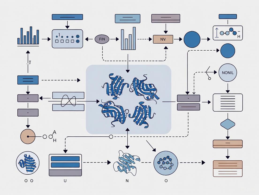
Visualization of Model Architectures and Workflows
Single-Cell Transformer Architecture
Single-Cell Data Processing Workflow
Table 3: Essential Resources for Single-Cell Foundation Model Research
| Resource Category | Specific Examples | Function and Utility |
|---|---|---|
| Data Repositories | CZ CELLxGENE [1] [4], Human Cell Atlas [1], DISCO [4], Tabula Sapiens [5] | Provide standardized, annotated single-cell datasets for model pretraining and benchmarking |
| Computational Frameworks | BioLLM [4], scGPT [1] [4], scGREAT [8] | Offer standardized interfaces and implementations of foundation models for single-cell analysis |
| Benchmarking Platforms | BEELINE [8], Nucleotide Transformer Benchmark [6] | Provide standardized evaluation pipelines and datasets for comparing model performance |
| Pretrained Models | scGPT [4], OmniReg-GPT [6], scBERT [5] | Ready-to-use models that can be fine-tuned for specific applications without costly pretraining |
| Specialized Architectures | Reformer encoders [5], Hybrid attention mechanisms [6], Sparse transformers | Enable efficient processing of long genomic sequences and high-dimensional gene expression data |
Future Directions and Challenges
While transformer-based models have demonstrated remarkable success in single-cell biology, several challenges remain. Model interpretability continues to be a significant hurdle, as understanding the biological relevance of latent embeddings and attention weights remains nontrivial [1]. Computational intensity for training and fine-tuning presents practical barriers to widespread adoption [1]. Additionally, inconsistencies in data quality and batch effects across studies can impact model robustness [1] [4]. Future developments will likely focus on enhancing model efficiency through improved architectures, developing better interpretation tools, and creating more standardized benchmarking frameworks [1] [4]. The integration of multimodal data at scale and the development of generative capabilities for in silico experimentation represent particularly promising directions for advancing both computational methodology and biological discovery [4] [6].
The transformer architecture, first introduced in the seminal paper "Attention Is All You Need," has revolutionized natural language processing (NLP) and is now fundamentally reshaping computational biology [9] [10]. This neural network architecture, which relies solely on attention mechanisms rather than recurrence or convolution, provides a powerful framework for capturing complex relationships in sequential data. In single-cell biology, researchers have creatively adapted this architecture to decipher the "language of cells," where individual cells are treated as sentences and genes or genomic features as words [11]. This paradigm shift enables the development of sophisticated single-cell foundation models (scFMs) that learn from millions of cells across diverse tissues and conditions, then adapt to various downstream analytical tasks through fine-tuning [11].
The integration of transformers with self-supervised learning (SSL) has been particularly transformative for single-cell genomics (SCG). SSL allows models to learn meaningful representations from vast, unlabeled datasets by solving pretext tasks, capturing universal patterns that transfer well to specific biological questions with limited labeled examples [12] [11]. As single-cell technologies rapidly generate data at an unprecedented scale, transformer-based scFMs offer a unified framework to integrate and analyze this complex biological information, providing insights into cellular heterogeneity, gene regulatory networks, and disease mechanisms that were previously challenging to uncover [11].
Architectural Foundations: The Core Transformer Building Blocks
Self-Attention Mechanism
The self-attention mechanism forms the fundamental operating principle of transformer models, enabling them to dynamically weigh the importance of different elements in a sequence when processing each element. Unlike recurrent neural networks that process sequences sequentially, self-attention computes relationships between all elements in parallel, making it highly efficient for modern hardware accelerators [9] [10].
The mechanism operates through three learned vectors for each input element: the Query (Q), Key (K), and Value (V) vectors. For a given element, the Query vector represents what the element is looking for, the Key vector represents what the element contains, and the Value vector represents the actual information the element contributes [9]. The attention output for a position is computed as a weighted sum of Value vectors, where the weights are determined by the compatibility between the Query vector of that position and the Key vectors of all positions in the sequence.
The mathematical formulation of self-attention is expressed as: [ \text{Attention}(Q, K, V) = \text{softmax}\left(\frac{QK^T}{\sqrt{dk}}\right)V ] where (dk) is the dimension of the Key vectors, and the scaling factor (\frac{1}{\sqrt{d_k}}) prevents the softmax function from entering regions with extremely small gradients [9].
Multi-Head Attention and Positional Encoding
Transformers enhance the basic self-attention through multi-head attention, which allows the model to jointly attend to information from different representation subspaces at different positions [9] [10]. Instead of performing a single attention function, the model linearly projects the Q, K, and V vectors multiple times with different learned projections and performs the attention function in parallel across these projected versions. The outputs are concatenated and projected again to produce the final result [9]. This architecture enables the model to capture different types of relationships—for instance, some attention heads might focus on syntactic patterns while others capture semantic relationships.
Since transformers process all tokens in parallel without inherent sequential processing, they require positional encoding to incorporate information about the position of each token in the sequence [9]. In single-cell applications, this presents a unique challenge because gene expression data lacks natural ordering. Common strategies include ranking genes by expression levels within each cell or partitioning genes into expression bins to create a deterministic sequence [11]. Positional encodings are then added to the token embeddings to provide positional context to the model.
Encoder-Decoder Architecture
The original transformer architecture follows an encoder-decoder structure [9]. The encoder processes the input sequence and generates contextualized representations. It consists of multiple identical layers, each containing a multi-head self-attention mechanism and a position-wise feed-forward network, with residual connections and layer normalization after each sub-layer [9].
The decoder generates the output sequence autoregressively. It shares similar components with the encoder but includes an additional multi-head cross-attention layer that attends to the encoder's output. To prevent the decoder from "peeking" at future tokens during training, it employs masked self-attention, which ensures that predictions for position i can only depend on known outputs at positions less than i [9].
Table: Core Components of Transformer Architecture
| Component | Function | Single-Cell Adaptation |
|---|---|---|
| Self-Attention | Computes contextual relationships between all sequence elements | Models gene-gene interactions and co-expression patterns |
| Multi-Head Attention | Attends to different representation subspaces simultaneously | Captures distinct biological relationships (e.g., regulatory, functional) |
| Positional Encoding | Provides sequence order information | Ranks genes by expression level or uses biological gene groupings |
| Feed-Forward Network | Applies non-linear transformation to each position independently | Enriches representations through biological pathway information |
| Layer Normalization | Stabilizes training by normalizing activations | Standardizes gene expression scales across different cell types |
| Residual Connections | Preserves gradient flow through deep networks | Enables training of deep biological models without degradation |
Self-Supervised Learning Paradigms for Single-Cell Data
Pretext Tasks for Biological Representation Learning
Self-supervised learning in single-cell genomics employs various pretext tasks that enable models to learn meaningful biological representations without explicit labeling. The most common approach adapts the masked language modeling objective from NLP, where randomly selected portions of the input data are masked, and the model is trained to reconstruct them [12] [11]. In single-cell applications, this translates to masking certain genes in a cell's expression profile and training the model to predict their values based on the remaining genes.
More sophisticated masking strategies have been developed for biological data. Gene program masking involves masking biologically coherent sets of genes that function together in pathways or complexes, forcing the model to learn higher-order functional relationships [12]. Contrastive learning methods represent another important SSL approach, where the model learns to identify similar and dissimilar pairs of cells or gene expression patterns [12]. Negative-pair-free methods like Bootstrap Your Own Latent (BYOL) and Barlow Twins have shown particular promise in single-cell applications [12].
Empirical Performance of SSL in Single-Cell Genomics
Recent large-scale benchmarking studies have illuminated the nuanced effectiveness of SSL in single-cell applications. Research evaluating SSL methods on over 20 million cells from the CELLxGENE census data has demonstrated that SSL particularly excels in transfer learning scenarios where models are pre-trained on large auxiliary datasets then fine-tuned on smaller target datasets [12].
Table: Performance of Self-Supervised Learning on Single-Cell Tasks
| Task | Dataset | Baseline Performance | SSL Performance | Key Improvement |
|---|---|---|---|---|
| Cell-type prediction | PBMC (422k cells, 30 types) | 0.7013 ± 0.0077 (Macro F1) | 0.7466 ± 0.0057 (Macro F1) | Better identification of underrepresented cell types |
| Cell-type prediction | Tabula Sapiens (483k cells, 161 types) | 0.2722 ± 0.0123 (Macro F1) | 0.3085 ± 0.0040 (Macro F1) | Correct classification of 6,881 type II pneumocytes vs. 2,441 baseline |
| Gene expression reconstruction | Multiple datasets | Varies by dataset | Significant improvement (weighted explained variance) | Better capture of technical and biological variations |
| Zero-shot cell typing | Multiple datasets | N/A | Competitive performance with kNN classification | Enables annotation without labeled training data |
Masked autoencoders have demonstrated particular effectiveness in single-cell genomics, outperforming contrastive methods—a finding that diverges from trends in computer vision [12]. The performance gains from SSL are most pronounced when the pre-training dataset is substantially larger and more diverse than the fine-tuning dataset, highlighting the importance of rich biological context for effective representation learning [12].
Experimental Framework: Implementing Transformers in Single-Cell Research
Tokenization Strategies for Single-Cell Data
A critical implementation challenge for transformers in single-cell biology is tokenization—the process of converting raw gene expression data into discrete input tokens [11]. Unlike natural language, where words have natural token boundaries, gene expression data is continuous and lacks inherent sequential structure. The most common approach represents each gene as a separate token, with the expression value incorporated through the token embedding [11].
Several strategies have emerged for ordering genes into sequences for transformer input:
- Expression-based ranking: Genes are sorted by expression level within each cell [11]
- Binning approaches: Genes are partitioned into expression bins that determine their position [11]
- Biological grouping: Genes are ordered based on biological knowledge (chromosomal location, pathway membership) [11]
- Fixed vocabulary: Some models use a standardized gene ordering across all cells [11]
Special tokens are often prepended to the gene token sequence, including a [CELL] token that aggregates cell-level information and modality indicators for multi-omics applications [11]. Positional encodings are then added to inform the model of each gene's position in the sequence.
Model Pre-training and Fine-tuning Protocols
The development of single-cell foundation models follows a two-stage process: self-supervised pre-training on large-scale diverse datasets followed by task-specific fine-tuning [12] [11].
Pre-training Protocol:
- Data Collection: Curate large-scale single-cell datasets from public repositories (CELLxGENE, Human Cell Atlas, GEO, SRA)
- Quality Control: Filter cells and genes based on quality metrics (mitochondrial content, detected genes)
- Normalization: Standardize expression values (CP10k normalization, log transformation)
- Pretext Task Training: Implement masked autoencoding or contrastive learning objectives
- Validation: Evaluate representation quality through zero-shot performance on benchmark tasks
Fine-tuning Protocol:
- Task Formulation: Define specific downstream task (cell type annotation, perturbation response, disease classification)
- Data Preparation: Split target dataset into training/validation/test sets
- Model Adaptation: Add task-specific layers to pre-trained backbone
- Transfer Learning: Fine-tune entire model or only task-specific layers on labeled data
- Evaluation: Assess performance on held-out test set using task-relevant metrics
Transformer Pre-training and Fine-tuning Workflow in Single-Cell Biology
Research Reagent Solutions: Essential Tools for scFM Development
Table: Key Research Resources for Single-Cell Foundation Models
| Resource Category | Specific Examples | Function/Purpose |
|---|---|---|
| Data Resources | CELLxGENE Census [12] [11], Human Cell Atlas [11], GEO/SRA [11] | Provide standardized, annotated single-cell datasets for model training |
| Preprocessing Tools | Scanpy [13], Seurat | Perform quality control, normalization, and feature selection |
| Model Architectures | scBERT [11], GeneFormer [11], scGPT [11] | Offer pre-designed transformer architectures for single-cell data |
| Tokenization Methods | Expression ranking [11], Gene binning [11], Biological grouping [11] | Convert continuous expression values to discrete token sequences |
| SSL Methods | Masked Autoencoders [12], Contrastive Learning (BYOL, Barlow Twins) [12] | Enable self-supervised pre-training on unlabeled data |
| Benchmarking Suites | Custom evaluation pipelines [12] | Standardized assessment of model performance across multiple tasks |
Advanced Applications and Future Directions
Emerging Applications in Single-Cell Biology
Transformer models are enabling new analytical capabilities across diverse single-cell applications. In cell type annotation, scBERT and similar models achieve high accuracy by framing annotation as a token prediction task [11]. For gene expression reconstruction, transformers can impute missing values or predict expression under different conditions [12]. In cross-modality prediction, models can translate between different molecular measurements (e.g., RNA to protein expression) [12]. Data integration represents another powerful application, where transformers remove batch effects and align cells across different experiments or technologies [12].
The DiffFormer model exemplifies architectural innovation, combining diffusion models with transformers for bulk RNA-seq deconvolution [13]. This approach reframes deconvolution as a conditional generation task, where the transformer's attention mechanism models complex, non-linear dependencies between bulk expression profiles and cell-type proportions [13]. Similarly, the White-Box Diffusion Transformer integrates mathematical interpretability with generative modeling for scRNA-seq data generation [14].
Technical Challenges and Research Frontiers
Despite rapid progress, several challenges remain in applying transformer architectures to single-cell biology. The non-sequential nature of genomic data continues to motivate research into optimal tokenization and positional encoding strategies [11]. Computational intensity presents practical constraints, especially as model sizes and dataset volumes continue to grow [11]. Interpretability remains challenging, as researchers seek to extract biologically meaningful insights from model attention patterns and latent representations [11].
Future research directions include developing more efficient attention mechanisms tailored to biological data, creating multi-modal foundation models that integrate transcriptomic, epigenomic, proteomic, and spatial information, and improving zero-shot capabilities for predicting cellular responses to unseen conditions or perturbations [11]. As these technical advances mature, transformer-based models are poised to become increasingly central tools for extracting biological knowledge from single-cell data.
Technical Challenges in Single-Cell Foundation Model Development
The transformer architecture, with its core attention mechanism and compatibility with self-supervised learning paradigms, has emerged as a powerful backbone for single-cell genomic analysis. By enabling the development of foundation models trained on millions of cells, this technology provides researchers with versatile tools that can be adapted to diverse downstream tasks through fine-tuning. The capacity of transformers to capture long-range dependencies and complex gene-gene interactions has proven particularly valuable for modeling the intricate regulatory networks underlying cellular identity and function.
As single-cell technologies continue to evolve, generating increasingly large and complex datasets, transformer-based approaches will likely play an expanding role in extracting biological insights from this data deluge. Future advances in model architecture, training efficiency, and interpretability will further enhance the utility of these methods, potentially transforming how researchers analyze cellular heterogeneity, decipher disease mechanisms, and develop targeted therapeutic interventions.
The analysis of single-cell RNA sequencing (scRNA-seq) data presents unique computational challenges due to its high-dimensionality, sparsity, and complex biological noise [5] [15]. Transformer-based foundation models, pre-trained on massive-scale single-cell atlases, have emerged as powerful tools to address these challenges. These models adapt the core architectural paradigms of natural language processing—specifically encoder-based (BERT-like) and decoder-based (GPT-like) models—to interpret the "language of cells," where genes are treated as words and individual cells as sentences [11] [3]. This technical guide examines these two architectural frameworks within the context of single-cell biology research, providing researchers and drug development professionals with a comprehensive comparison of their underlying mechanisms, applications, and experimental implementations.
Core Architectural Principles
The Transformer Foundation
The transformer architecture serves as the fundamental building block for both BERT-like and GPT-like models. Its core innovation is the self-attention mechanism, which allows the model to weigh the importance of different elements in a sequence when processing each element [3] [16]. For single-cell data, this enables the model to capture complex gene-gene interactions and regulatory relationships.
The multi-head self-attention mechanism is mathematically defined as:
Attention(Q,K,V) = softmax(QKᵀ/√dₖ)V [3]
Where:
- Q (Query) represents the current focus of attention
- K (Key) represents what the model can attend to
- V (Value) represents the actual information to extract
- dₖ is the dimensionality of the key vectors
In biological terms, this allows the model to learn which genes are most informative about a cell's identity or state, and how they co-vary across different cellular contexts [11].
Encoder-Based (BERT-like) Architectures
Encoder-based models utilize the transformer encoder stack to process all tokens in the input sequence simultaneously. This bidirectional attention enables the model to understand context from both directions, making it particularly effective for comprehension-oriented tasks [17] [18].
Key Characteristics:
- Architecture: Encoder-only transformer
- Attention Type: Multi-head attention (unmasked)
- Context Handling: Considers both left and right context simultaneously
- Training Objective: Masked Language Modeling (MLM)
- Primary Purpose: Understanding and extracting meaning from data [17]
In single-cell applications, BERT-like models such as scBERT [5] and Geneformer [15] excel at tasks requiring deep biological understanding, including cell type annotation, gene function prediction, and identifying disease-specific cellular signatures.
Decoder-Based (GPT-like) Architectures
Decoder-based models employ a causal attention mechanism that processes sequences autoregressively—each token can only attend to previous tokens in the sequence. This unidirectional approach is inherently suited for generative tasks [17] [18].
Key Characteristics:
- Architecture: Decoder-only transformer
- Attention Type: Masked multi-head attention
- Context Handling: Considers only left context
- Training Objective: Causal Language Modeling
- Primary Purpose: Generating coherent and contextually relevant sequences [17]
In single-cell biology, GPT-like models such as scGPT [15] demonstrate exceptional capability in generating synthetic cell profiles, predicting cellular responses to perturbations, and simulating developmental trajectories.
Architectural Comparison
Table 1: Fundamental Differences Between BERT-like and GPT-like Architectures
| Feature | BERT-like (Encoder) | GPT-like (Decoder) |
|---|---|---|
| Architecture Type | Encoder-only Transformer | Decoder-only Transformer |
| Attention Mechanism | Bidirectional, unmasked | Causal, masked |
| Context Processing | Full sequence simultaneously | Left-to-right sequentially |
| Training Objective | Masked Language Modeling (MLM) | Causal Language Modeling |
| Primary Strength | Understanding & classification | Generation & prediction |
| Computational Complexity | O(n²) for sequence length n | O(n²) for sequence length n |
| Typical Output | Classifications, embeddings | Generated sequences, completions |
Implementation in Single-Cell Biology
Data Tokenization Strategies
Applying transformer architectures to single-cell data requires innovative tokenization approaches since gene expression data lacks the inherent sequential order of natural language [11] [15]. The tokenization process converts raw gene expression values into discrete tokens that can be processed by transformer models.
Common Tokenization Methods:
- Expression-based ranking: Genes are ordered by expression level within each cell
- Value binning: Expression values are discretized into bins
- Genomic position ordering: Genes are ordered by genomic coordinates
- HVG selection: Using only highly variable genes as tokens [15]
Table 2: Tokenization Approaches in Single-Cell Foundation Models
| Model | Tokenization Strategy | Input Genes | Value Representation |
|---|---|---|---|
| Geneformer | Ranking by expression level | 2,048 ranked genes | Ordering |
| scGPT | HVG selection + value binning | 1,200 HVGs | Value binning |
| scFoundation | Full gene set | ~19,000 genes | Value projection |
| UCE | Sampling by expression + genomic position | 1,024 non-unique genes | Binary expression |
Model Architectures and Pre-training
Single-cell foundation models adapt the core transformer architecture with specific modifications for biological data. The pre-training phase typically uses self-supervised learning on large-scale single-cell atlases containing millions of cells [11] [15].
Encoder-based Pre-training (BERT-like):
- Masked Gene Modeling (MGM): Randomly mask a percentage of gene tokens and train the model to predict them using contextual information
- Binary Expression Prediction: Predict whether a gene is expressed or not
- Dataset: Models pre-trained on 30-50 million cells from diverse tissues and conditions [15]
Decoder-based Pre-training (GPT-like):
- Next-Gene Prediction: Autoregressively predict the next gene in the sequenced expression profile
- Generative Pre-training: Learn to generate plausible gene expression profiles
- Multi-task Learning: Combine multiple self-supervised objectives [15]
Experimental Protocols and Applications
Cell Type Annotation Protocol
Objective: Automatically identify and label cell types in scRNA-seq data Input: Raw count matrix (cells × genes) Protocol:
Data Preprocessing:
- Quality control and normalization of raw counts
- Gene filtering based on expression thresholds
- Library size normalization and log transformation
Tokenization:
- Select top 2,000 highly variable genes
- Rank genes by expression level within each cell
- Convert to token sequences with special [CLS] token
Model Inference:
- Process token sequences through transformer encoder (e.g., scBERT)
- Extract [CLS] token embedding as cell representation
- Pass through classification layer for cell type prediction
Validation:
Perturbation Response Prediction
Objective: Predict how cells respond to genetic or chemical perturbations Input: Baseline gene expression profile + perturbation information Protocol:
Data Preparation:
- Collect single-cell data from perturbation experiments
- Create paired samples (control vs. treated)
- Tokenize expression profiles with perturbation indicators
Model Architecture:
- Use decoder-based model (e.g., scGPT) with causal attention
- Incorporate perturbation tokens as special inputs
- Implement teacher forcing during training
Training:
- Pre-train on large-scale single-cell atlases
- Fine-tune on perturbation datasets
- Use mean squared error loss between predicted and actual expression
Evaluation:
Visualization of Model Architectures and Workflows
Performance Benchmarking and Evaluation
Quantitative Performance Comparison
Table 3: Performance Comparison on Common Single-Cell Tasks
| Task Type | Best Performing Architecture | Key Metrics | Representative Performance |
|---|---|---|---|
| Cell Type Annotation | Encoder-based (BERT-like) | Accuracy: 85-95% [5] | scBERT: >90% on major cell types [5] [19] |
| Batch Integration | Encoder-based (BERT-like) | ASW: 0.7-0.9 [15] | Geneformer: Superior batch correction [15] |
| Perturbation Prediction | Decoder-based (GPT-like) | MSE: 0.1-0.3 [15] | scGPT: Accurate response simulation [15] |
| Novel Cell Generation | Decoder-based (GPT-like) | MMD: 0.05-0.15 [3] | scGPT: Realistic profile generation [15] |
| Gene Network Inference | Both (Task-dependent) | AUROC: 0.8-0.95 [15] | Varies by biological context [15] |
Computational Requirements
Table 4: Computational Characteristics of Single-Cell Foundation Models
| Model Characteristic | Encoder-based (BERT-like) | Decoder-based (GPT-like) |
|---|---|---|
| Pre-training Scale | 30-50 million parameters [15] | 40-100 million parameters [15] |
| Pre-training Data | 30-50 million cells [15] | 27-33 million cells [15] |
| Memory Usage | High (full attention matrices) | High (causal attention) |
| Inference Speed | Faster (parallel processing) | Slower (sequential generation) |
| Fine-tuning Efficiency | Excellent (few-shot learning) | Good (requires careful prompting) |
The Scientist's Toolkit: Essential Research Reagents
Table 5: Key Computational Tools and Resources for Single-Cell Foundation Models
| Tool/Resource | Type | Function | Example Applications |
|---|---|---|---|
| CELLxGENE Census [11] [20] | Data Platform | Provides standardized access to ~100 million single cells | Model pre-training, benchmarking, transfer learning |
| Geneformer [15] | Encoder Model | BERT-like model for cell state understanding | Cell classification, mechanism identification |
| scGPT [15] | Decoder Model | GPT-like model for generative tasks | Perturbation prediction, hypothesis generation |
| AnnDictionary [19] | LLM Integration | Interfaces LLMs with single-cell data | Automated annotation, biological interpretation |
| CellWhisperer [20] | Multimodal AI | Joint embedding of transcriptomes and text | Natural language querying, interactive exploration |
| Reformer Encoders [5] | Efficient Architecture | Handles long sequences via LSH attention | Full-transcriptome analysis without gene filtering |
| scReformer-BERT [5] | Hybrid Model | Combines BERT architecture with Reformer efficiency | Large-scale cell classification with full gene set |
The integration of encoder-based (BERT-like) and decoder-based (GPT-like) transformer architectures has fundamentally transformed computational single-cell biology. Encoder models excel at understanding cellular states and extracting biologically meaningful patterns, while decoder models show remarkable capability in generating hypotheses and predicting cellular behaviors. The emerging paradigm involves combining both architectures—using encoders for robust feature extraction and decoders for generative modeling and prediction.
Future developments will likely focus on multimodal integration (combining transcriptomics with epigenomics, proteomics, and spatial data), more efficient attention mechanisms to handle complete transcriptomes, and improved interpretability to extract novel biological insights. As these models continue to evolve, they will play an increasingly central role in drug discovery, personalized medicine, and our fundamental understanding of cellular biology.
The development of transformer-based foundation models in single-cell biology research is critically dependent on the scale, diversity, and quality of the data used for pretraining [1]. A foundation model is a large-scale deep learning model pretrained on vast datasets that can be adapted to a wide range of downstream tasks through self-supervised learning [1]. The remarkable success of single-cell foundation models (scFMs) in tasks ranging from cell type annotation to gene regulatory network inference is fundamentally underpinned by the massive, curated biological datasets that serve as their training corpora [1] [21]. This technical guide examines the primary data sources, processing methodologies, and experimental frameworks that enable researchers to construct effective pretraining datasets for scFMs, with particular emphasis on their application within transformer architectures.
Major Public Data Repositories and Atlas Projects
The pretraining of robust scFMs requires access to large-scale, well-annotated single-cell datasets. Researchers typically aggregate data from multiple public repositories to create comprehensive training corpora. The table below summarizes key data sources used in recent scFM development efforts.
Table 1: Major Data Repositories for Single-Cell Foundation Model Pretraining
| Repository/Atlas Name | Scale | Data Content | Notable Use Cases |
|---|---|---|---|
| CZ CELLxGENE [1] | >100 million cells [1] | Annotated single-cell datasets, standardized for analysis [1] | General-purpose scFM pretraining [1] |
| Arc Virtual Cell Atlas [22] | >300 million cells [22] | scBaseCount: 200M+ cells from 21 species; Tahoe-100M: 100M perturbed cells [22] | Perturbation response modeling [22] |
| Human Cell Atlas [1] [23] | Cross-tissue atlas scale [1] | Cells from various tissues and organs, healthy reference [23] | Reference cell state modeling [23] |
| SpatialCorpus-110M [21] | 110 million cells (57M dissociated + 53M spatial) [21] | Integrated dissociated and spatially-resolved transcriptomics [21] | Spatially-aware models (Nicheformer) [21] |
| PanglaoDB [1] | Curated compendium [1] | Data from multiple sources and studies [1] | Supplemental pretraining data [1] |
| NCBI GEO/SRA & EBI Expression Atlas [1] | Thousands of studies [1] | Diverse single-cell sequencing studies [1] | Dataset aggregation [1] |
These repositories provide the foundational data necessary for training models that capture the broad spectrum of cellular heterogeneity across tissues, species, and experimental conditions. The integration of data from multiple sources is crucial for developing models that generalize well to unseen data and downstream tasks [1] [15].
Data Processing and Tokenization Strategies
From Cellular Measurements to Model Input
Raw single-cell data must be transformed into a structured format compatible with transformer architectures. This process, known as tokenization, converts gene expression profiles into discrete input units that the model can process.
Table 2: Tokenization Strategies in Single-Cell Foundation Models
| Model | Tokenization Approach | Gene Ordering | Special Tokens | Value Representation |
|---|---|---|---|---|
| General scFMs [1] | Genes as tokens [1] | Ranked by expression level [1] | Cell identity, modality [1] | Normalized counts, bins [1] |
| Nicheformer [21] | Ranked gene tokens [21] | Expression level relative to corpus mean [21] | Species, modality, technology [21] | Technology-specific normalization [21] |
| scPRINT [24] | Gene ID + expression + genomic location [24] | No inherent ordering [24] | Cell embeddings [24] | MLP-processed log-normalized counts [24] |
| Geneformer [15] | 2,048 ranked genes [15] | Expression-based ranking [15] | Not specified | Ordering as value representation [15] |
| scGPT [15] | 1,200 highly variable genes [15] | Not specified | Not specified | Value binning [15] |
A fundamental challenge in applying transformers to single-cell data is that gene expression data lacks natural sequential ordering, unlike words in a sentence [1]. To address this, most models employ deterministic ordering schemes based on expression magnitude, such as ranking genes within each cell by their expression levels [1]. This creates an arbitrary but consistent sequence that enables the transformer to learn gene-gene relationships through its attention mechanism.
Data Processing Workflow
The transformation of raw sequencing data into model-ready inputs follows a multi-stage pipeline that ensures data quality and compatibility.
Diagram 1: Single-Cell Data Processing Workflow
Experimental Protocols for Pretraining and Evaluation
Pretraining Objectives and Methodologies
scFMs employ self-supervised pretraining tasks that enable the model to learn meaningful biological representations without extensive manual labeling. The most common approach is masked gene modeling (MGM), where random portions of the gene expression profile are masked and the model must predict the missing values based on context [1] [24]. Alternative strategies include:
- Denoising tasks: Training the model to reconstruct clean expression profiles from artificially corrupted inputs [24]
- Bottleneck learning: Forcing the model to compress cellular information into lower-dimensional embeddings [24]
- Multi-task learning: Combining multiple objectives such as label prediction alongside reconstruction [24]
Model Evaluation Frameworks
Comprehensive benchmarking is essential for validating scFM performance. Recent studies have established rigorous evaluation protocols assessing models across diverse downstream tasks [15] [25].
Table 3: Downstream Tasks for Evaluating Single-Cell Foundation Models
| Task Category | Specific Tasks | Evaluation Metrics | Key Insights |
|---|---|---|---|
| Cell-level tasks [15] [25] | Cell type annotation, Batch integration, Cancer cell identification [15] [25] | Accuracy, Cluster separation, Biological conservation [15] [25] | No single scFM dominates all tasks [15] |
| Gene-level tasks [15] [25] | Gene network inference, Gene function prediction [15] [24] | Network accuracy, GO term enrichment [15] | Gene embeddings capture functional relationships [15] |
| Spatial tasks [21] | Spatial composition prediction, Niche identification [21] | Spatial context accuracy [21] | Dissociated data alone cannot capture spatial variation [21] |
| Perturbation tasks [22] | Drug response prediction, Genetic perturbation effects [22] | Response accuracy [22] | Perturbation datasets enable therapeutic applications [22] |
Diagram 2: scFM Evaluation Framework
Research Reagent Solutions
The successful development and application of single-cell foundation models relies on a ecosystem of computational tools and resources. The table below details essential components of the scFM research toolkit.
Table 4: Essential Research Resources for Single-Cell Foundation Model Development
| Resource Type | Specific Tools/Platforms | Function | Application Context |
|---|---|---|---|
| Pretraining Datasets [22] | Arc Virtual Cell Atlas, CELLxGENE [22] | Large-scale, curated single-cell data | Model pretraining, Transfer learning [22] |
| Model Architectures [1] [21] | Transformer, BERT, GPT variants [1] [21] | Neural network backbones | Feature extraction, Pattern recognition [1] |
| Benchmarking Suites [15] [24] | BenGRN, GrnnData [24] | Performance evaluation | Model validation, Comparison [15] |
| Specialized Models [21] [26] [24] | Nicheformer, CellPLM, scPRINT [21] [26] [24] | Task-optimized scFMs | Spatial analysis, Network inference [21] [24] |
| Processing Tools [23] | Bioconductor, Scanpy, Seurat [23] | Data preprocessing | Quality control, Normalization [23] |
The development of effective single-cell foundation models hinges on strategic leveraging of diverse public data repositories and sophisticated processing methodologies. As the field advances, several key principles have emerged: data diversity is more critical than sheer volume alone [21] [15]; dataset composition should reflect the intended application domains [21] [22]; and rigorous benchmarking across multiple biological tasks is essential for validating model utility [15] [25]. The rapid expansion of curated single-cell data resources, coupled with innovative transformer architectures designed for high-dimensional sparse data [5], promises to accelerate the development of more powerful, biologically-relevant foundation models that will transform our understanding of cellular function and disease mechanisms.
The application of transformer architectures to single-cell biology represents a paradigm shift in how researchers analyze cellular heterogeneity and function. Unlike natural language, where words follow grammatical structures and sequential dependencies, gene expression data exists in a fundamentally non-sequential space where genes have no inherent ordering, yet exhibit complex, coordinated relationships. This creates a fundamental tokenization challenge: how to convert this high-dimensional, non-sequential data into structured model inputs that preserve biological meaning while enabling computational efficiency. Foundation models like scGPT and Geneformer have demonstrated that effective tokenization is not merely a preprocessing step but a critical determinant of model performance across diverse downstream tasks including cell type annotation, perturbation response prediction, and gene regulatory network inference [1] [4].
The tokenization process must overcome several domain-specific obstacles: the high dimensionality and sparsity of single-cell RNA sequencing (scRNA-seq) data; the absence of natural gene ordering; technical noise from batch effects; and the need to preserve biological signal amidst these complexities. This technical guide examines current tokenization methodologies, their theoretical underpinnings, empirical performance, and practical implementation considerations for researchers developing and applying transformer-based models in single-cell biology and drug development.
Fundamental Concepts: From Biological Data to Model Tokens
The Nature of Single-Cell Omics Data
Single-cell technologies generate molecular profiles measuring the expression levels of thousands of genes across thousands to millions of individual cells. Each cell is represented as a high-dimensional vector where values correspond to gene expression counts or chromatin accessibility measurements. Unlike sequential data like text or DNA, where element order carries critical semantic meaning, the genes in these vectors exist in an unordered set [1]. This fundamental characteristic necessitates the development of specialized tokenization strategies that impose meaningful structure without introducing artificial biases.
The data presents additional challenges including extreme sparsity (many zero values representing both biological absence and technical dropouts), high technical variance across experimental batches and platforms, and complex biological covariance patterns that reflect underlying regulatory networks [1] [25]. Effective tokenization must preserve biological signal while mitigating the impact of these confounding factors.
Tokenization in the Context of Transformer Architectures
In natural language processing, tokenization converts raw text into discrete units (tokens) that serve as model inputs. Similarly, for single-cell data, tokenization transforms raw gene expression values into a structured sequence the transformer can process. This typically involves two components: (1) defining what constitutes a token, and (2) establishing an ordering for these tokens [1].
The tokenization step is crucial because it determines how biological information is presented to the model's attention mechanism. Different tokenization strategies emphasize different aspects of the data, potentially leading the model to learn distinct representations and relationships. As such, tokenization is not merely an engineering consideration but a fundamental modeling choice that influences what biological patterns the model can discover [1] [27].
Table 1: Core Components of Single-Cell Tokenization
| Component | Description | Common Implementations |
|---|---|---|
| Gene Token | Representation of individual genes | Gene identifier (e.g., ENSG00000139618 for human BRCA1) |
| Value Representation | Encoding of expression magnitude | Normalized counts, bins, or continuous values |
| Positional Encoding | Information about token order | Learned embeddings, fixed sinusoidal functions |
| Special Tokens | Additional contextual information | Cell-level metadata, modality indicators, batch identifiers |
Current Tokenization Strategies: Methodologies and Implementations
Rank-Based Tokenization
Rank-based approaches order genes by their expression level within each cell, converting the non-sequential gene set into a deterministic sequence. In this framework, the most highly expressed genes appear first in the sequence, followed by progressively lower-expressed genes [1] [21]. This strategy is employed by models including Geneformer and Nicheformer, which leverage the intuition that the relative ranking of gene expression may be more robust to technical variance than absolute expression values.
The implementation typically involves sorting genes by expression value in descending order, then selecting the top-k genes (typically 1,000-2,000) to form the input sequence [21]. Each token combines information about gene identity and its relative expression rank. A key advantage is reduced sensitivity to batch effects and normalization artifacts, as the relative ordering within a cell may be preserved even when absolute values shift. However, this approach potentially discards information from lower-ranked genes and may disrupt co-expression patterns that exist across magnitude ranges [1].
Binning-Based Tokenization
Binning strategies partition gene expression values into discrete levels or categories, similar to how words might be categorized by frequency. Models like scBERT often employ this approach, creating expression bins such as "low," "medium," and "high" based on predefined thresholds [1] [11]. Each gene is then represented by both its identifier and its expression bin.
This method can capture non-linear relationships in expression values and reduces the model's sensitivity to small fluctuations that may not be biologically meaningful. Some implementations use learned bin boundaries that adapt during training, potentially discovering optimal discretization thresholds for different biological contexts. The primary limitation is information loss from discretizing continuous expression values, which may obscure subtle but biologically important expression differences [27].
Scale-Free and Unbiased Tokenization
Emerging approaches like scSFUT (Single-Cell Scale-Free and Unbiased Transformer) aim to address limitations of gene selection-based methods by processing the full gene set without preliminary filtering [27]. These methods use techniques like fixed-size windowing to segment the high-dimensional input into manageable chunks, preserving information across the entire transcriptome rather than just highly variable or highly expressed genes.
The scSFUT model specifically employs an encoder-decoder framework with sequential tokenization and 1D-convolution to expand the attention receptive field [27]. This approach demonstrates that with architectural innovations, models can effectively process full-length gene vectors without preselection, potentially capturing patterns that would be missed when focusing only on the most variable or highly expressed genes. This is particularly valuable for detecting rare but biologically significant expression events or identifying patterns across comprehensively correlated gene sets [27].
Table 2: Comparative Analysis of Tokenization Strategies
| Strategy | Mechanism | Advantages | Limitations | Representative Models |
|---|---|---|---|---|
| Rank-Based | Orders genes by expression level | Robust to technical variance; Intuitive biological interpretation | May lose information from low-ranked genes; Disrupts natural covariance | Geneformer, Nicheformer |
| Binning | Discretizes expression into categories | Handles non-linearities; Reduces noise sensitivity | Loss of continuous value information; Bin boundary selection arbitrary | scBERT, xTrimoGene |
| Scale-Free | Processes full gene set | Maximally preserves biological information; No selection bias | Computationally intensive; Requires specialized architectures | scSFUT |
| Value-Inclusive | Combines gene ID with continuous value | Maintains precise expression information; Flexible representation | Sensitive to normalization; May amplify technical artifacts | scGPT, UCE |
Multi-Modal and Integrated Tokenization
As single-cell technologies advance, integrating multiple data modalities from the same cells has become increasingly important. Multi-omic approaches require tokenization strategies that can handle diverse data types including gene expression, chromatin accessibility, protein abundance, and spatial coordinates [4] [28].
Advanced models address this through modality-specific tokens that indicate the data type, allowing the transformer to learn both modality-specific and cross-modality relationships [1] [28]. For example, scPairing uses a contrastive learning framework to embed different modalities from the same cells into a common embedding space, enabling integration and generation of multi-omics data [28]. Similarly, Nicheformer incorporates spatial context through specialized tokens that capture microenvironment information, enabling the model to learn spatially aware representations [21].
Experimental Protocols and Implementation Guidelines
Standardized Preprocessing Pipeline
Implementing effective tokenization requires careful data preprocessing to ensure biological signal is preserved and technical artifacts are minimized. Based on benchmarking studies and model documentation, the following protocol represents current best practices:
Quality Control: Filter cells based on quality metrics—typically retaining cells with 200-2500 detected genes and mitochondrial content below 5-20% (tissue-dependent) [27].
Normalization: Apply library size normalization (e.g., counts per 10,000) followed by log transformation to stabilize variance [1] [27].
Gene Filtering: Remove lowly expressed genes (e.g., detected in fewer than 10 cells) to reduce noise, though this step is omitted in scale-free approaches [27].
Batch Effect Consideration: For multi-dataset training, incorporate batch correction methods or include batch information as special tokens [1] [4].
Tokenization: Apply the selected strategy (rank-based, binning, etc.) to convert each cell's expression profile into a token sequence.
Sequence Formulation: Combine gene tokens with special tokens (e.g., [CLS] for cell-level representation, modality indicators) and apply positional encoding [1].
Tokenization-Specific Methodologies
For rank-based tokenization:
- Sort all genes by expression value in descending order
- Select the top N genes (typically 1,000-2,000) based on model constraints
- Represent each gene by its identifier embedded with positional information reflecting its rank
- Append special tokens representing cell metadata or experimental conditions [21]
For binning-based tokenization:
- Normalize expression values to a consistent scale
- Define bin boundaries based on quantiles or expression thresholds
- Assign each gene to appropriate bin based on expression level
- Create token combining gene ID and bin identity [1]
For scale-free tokenization:
- Normalize but do not filter genes (or use minimal filtering)
- Segment full gene vector into fixed-size windows using 1D convolution
- Apply sequence ordering based on original gene positioning or random shuffling
- Use unbiased attention mechanisms to process segments [27]
Visualization of Tokenization Workflows
Rank-Based Tokenization Process
Multi-Modal Tokenization Architecture
Table 3: Research Reagent Solutions for Tokenization Implementation
| Resource Category | Specific Tools/Platforms | Function in Tokenization Pipeline |
|---|---|---|
| Data Repositories | CZ CELLxGENE, Human Cell Atlas, PanglaoDB | Provide standardized single-cell datasets for model training and benchmarking |
| Preprocessing Tools | Scanpy, Seurat, scikit-learn | Perform quality control, normalization, and initial feature selection |
| Model Frameworks | scGPT, Geneformer, scBERT | Reference implementations of tokenization strategies and model architectures |
| Benchmarking Suites | BioLLM, GenBench | Standardized evaluation of tokenization approaches across diverse tasks |
| Specialized Architectures | scSFUT, Nicheformer | Implementations of advanced tokenization methods (scale-free, spatial-aware) |
Performance Evaluation and Biological Validation
Quantitative Benchmarking Across Strategies
Recent benchmarking studies have evaluated tokenization strategies across diverse biological tasks. Performance varies significantly based on task type, data characteristics, and evaluation metrics [25].
For cell type annotation, binning-based methods like scBERT achieve high accuracy on human datasets but show reduced performance in cross-species transfer tasks. Rank-based approaches demonstrate stronger generalization across tissues and species, while scale-free methods show particular advantage for rare cell type identification [27] [25].
For spatial context prediction, models incorporating spatial tokenization (e.g., Nicheformer) significantly outperform methods trained solely on dissociated data, highlighting the importance of task-specific tokenization strategies [21]. Nicheformer achieves up to 30% improvement in spatial composition prediction compared to non-spatial models [21].
For batch integration, rank-based methods generally show superior performance in removing technical variance while preserving biological heterogeneity, though the incorporation of batch information as special tokens can further enhance integration capabilities [1] [4].
Biological Insight Capture
Beyond task-specific metrics, tokenization strategies differ in their ability to capture biologically meaningful relationships. Evaluation using ontology-informed metrics like scGraph-OntoRWR reveals that while all tokenization approaches capture broad biological patterns, scale-free and value-inclusive methods better preserve fine-grained functional relationships between genes and cell types [25].
Gene embedding analysis shows that tokenization approaches that maintain continuous expression information (rather than discretizing) tend to produce embeddings that better reflect known biological pathways and protein-protein interactions, suggesting they preserve more nuanced functional information [25].
Future Directions and Emerging Challenges
The rapid evolution of single-cell technologies presents ongoing challenges for tokenization strategies. Several promising directions are emerging:
Multi-modal fusion represents a frontier where tokenization must harmonize fundamentally different data types including images, sequences, and spatial coordinates [4] [28]. Approaches like scPairing demonstrate the potential of contrastive alignment methods for creating unified embedding spaces [28].
Dynamic tokenization that adapts to specific biological contexts or tasks may outperform static approaches. Preliminary work suggests that learned tokenization policies can optimize for specific objectives like rare cell detection or perturbation response prediction.
Cross-species generalization requires tokenization methods that can handle orthologous genes and evolutionary divergence. Models like Nicheformer that incorporate multispecies training with orthology mapping show promise in this direction [21].
Computational efficiency remains a critical concern, particularly as dataset sizes exceed millions of cells. Scalable tokenization strategies that maintain biological fidelity while reducing memory and computational requirements will be essential for continued progress.
As transformer architectures continue to evolve in single-cell biology, tokenization strategies will likely become increasingly specialized and sophisticated, potentially incorporating biological prior knowledge more explicitly and adapting to the unique characteristics of specific tissue types, disease states, and experimental modalities.
From Theory to Practice: Transformer Applications in Single-Cell Omics Analysis
The advent of single-cell RNA sequencing (scRNA-seq) has revolutionized our understanding of cellular heterogeneity and complex biological systems [15]. Concurrently, transformer-based architectures have emerged as a powerful tool in computational biology, leading to the development of single-cell foundation models (scFMs) [1]. These large-scale deep learning models, pretrained on vast datasets containing millions of cells, are capable of learning universal biological knowledge in a self-supervised manner [1] [15]. This technical guide explores how these transformer-based scFMs are applied to three core downstream tasks in single-cell analysis: cell type annotation, batch integration, and atlas construction. By providing a structured overview of methodologies, performance benchmarks, and practical protocols, this document serves as a resource for researchers, scientists, and drug development professionals seeking to leverage these advanced computational techniques.
Transformer Architectures in Single-Cell Biology
Fundamental Concepts
Single-cell foundation models adapt the transformer architecture, originally developed for natural language processing (NLP), to interpret biological data [1]. In this analogy, individual cells are treated as "sentences," while genes or other genomic features, along with their expression values, are treated as "words" or "tokens" [1]. The self-attention mechanism inherent to transformers allows these models to learn and weight relationships between any pair of input tokens (genes), enabling them to capture complex gene-gene interactions and regulatory networks without prior biological knowledge [1] [29].
Model Architectures and Tokenization Strategies
A critical challenge in applying transformers to single-cell data is that gene expression data lacks natural sequential ordering, unlike words in a sentence [1] [15]. To address this, various tokenization strategies have been developed. A common approach ranks genes within each cell by expression levels, feeding the ordered list of top genes as a sequence to the model [1]. Other methods partition genes into bins based on expression values or use normalized counts directly [1]. Gene tokens typically combine a gene identifier embedding with a value embedding representing its expression level [15].
Table 1: Common scFM Architectures and Their Key Characteristics
| Model Name | Primary Architecture | Tokenization Strategy | Key Features | Applicable Tasks |
|---|---|---|---|---|
| scBERT [1] | BERT-like Encoder | Gene ranking or value binning | Bidirectional attention; trained on millions of cells | Cell type annotation |
| scGPT [1] [15] | GPT-like Decoder | Value binning with 1200 HVGs | Unidirectional attention; multi-omics capability | Generation, integration, annotation |
| Geneformer [15] | Encoder | 2048 ranked genes | Employs gene ranking by expression | Network inference, annotation |
| scReformer-BERT [5] | BERT with Reformer encoders | Full gene set (>10,000 genes) | Uses LSH attention for efficiency with long sequences | Cell type classification |
| UCE [15] | Encoder | 1024 non-unique genes sampled by expression | Incorporates protein embeddings from ESM-2 | Multi-modal analysis |
Most scFMs utilize either encoder-based architectures (like BERT) for classification and embedding tasks, or decoder-based architectures (like GPT) for generation tasks [1]. Hybrid designs are also being explored. A key innovation is the development of models like scReformer-BERT, which incorporates Reformer encoders with locality-sensitive hashing (LSH) attention to handle the full spectrum of over 10,000 genes per cell without requiring aggressive gene filtering, thereby preserving more biological information [5].
Core Downstream Task 1: Cell Type Annotation
Task Definition and Significance
Accurate cell type identification is a critical prerequisite for interpreting single-cell transcriptomic data and understanding complex biological systems [30] [5]. Traditional methods rely on manual annotation using known marker genes, which is time-consuming, subjective, and challenging for rare or novel cell populations. Transformer-based scFMs offer a powerful approach for automated, standardized, and scalable cell type annotation [30].
Experimental Protocols and Methodologies
The standard protocol for cell type annotation using scFMs follows a "pretrain-then-fine-tune" paradigm [15]:
- Feature Extraction: Utilize a pretrained scFM to generate latent embeddings for each cell in the target dataset. These embeddings are dense, low-dimensional representations that capture essential biological features of each cell. In zero-shot scenarios, these embeddings can be used directly with simple classifiers without further model tuning [15].
- Supervised Fine-Tuning: For optimal performance on specific datasets, the pretrained scFM can be fine-tuned using labeled reference data. This involves:
- Preparing a high-quality reference dataset with accurate cell type labels.
- Adding a task-specific classification layer on top of the base model.
- Training the entire model or a subset of its layers on the reference data, typically using cross-entropy loss.
- Prediction and Validation: Apply the fine-tuned model to annotate cell types in new, unlabeled datasets. Predictions should be validated using known marker genes and, if available, held-out validation sets.
Performance Evaluation and Benchmarking
Recent benchmarking studies have evaluated multiple scFMs against traditional methods for cell type annotation. Performance is often assessed using metrics such as accuracy, F1-score, and the novel Lowest Common Ancestor Distance (LCAD), which measures the ontological proximity between misclassified cell types to assess the severity of errors [15].
Table 2: Benchmarking Results for Cell Type Annotation (Summary of Key Findings from [15])
| Model / Approach | Reported Strengths | Reported Limitations | Context for Optimal Use |
|---|---|---|---|
| scFMs (Zero-shot) | Capture biological insights into relational structures; robust to dataset variations [15]. | May not consistently outperform simpler models on small, specific datasets [15]. | Large, diverse datasets; when biological interpretability is prioritized. |
| scFMs (Fine-tuned) | High accuracy; leverage transfer learning from large-scale pretraining [15]. | Require computational resources for fine-tuning; risk of overfitting on small datasets [15]. | When sufficient labeled data and computational resources are available. |
| Traditional ML (e.g., SVM, HVGs) | Efficient and effective for specific, small-scale datasets with limited computational resources [15]. | Poor generalization to cell types not in the source data; limited by manual feature selection [15]. | Small, focused datasets with well-defined, known cell types. |
Notably, no single scFM consistently outperforms all others across all tasks and datasets. Model selection must be tailored based on factors like dataset size, task complexity, need for biological interpretability, and computational resources [15].
Core Downstream Task 2: Batch Integration
The Challenge of Batch Effects
Integrating multiple scRNA-seq datasets is a standard but challenging step in single-cell analysis. Technical differences between experiments (e.g., sequencing depth, protocols) and biological variations (e.g., different donors, species) create "batch effects" that can confound biological signals [31]. Effective integration is crucial for constructing large-scale atlases and for cross-study comparisons [31].
Transformer-Based Integration Approaches
Transformer-based scFMs like scGPT are designed to integrate diverse datasets by learning a unified representation of single-cell data that is robust to technical variations [1] [15]. The self-attention mechanism can theoretically learn to distinguish technical noise from biological signal after exposure to vast amounts of diverse data during pretraining. Some models incorporate batch information as special tokens during training to explicitly model and correct for these effects [1].
Benchmarking Integration Performance
Integration methods are evaluated on two key aspects: batch correction (how well technical variations are removed) and biological preservation (how well true biological variation is retained) [31]. Common metrics include:
- iLISI: Measures the mixing of batches in local cell neighborhoods [31].
- NMI: Assesses the preservation of cell type clusters after integration [31].
Benchmarks indicate that while methods like conditional Variational Autoencoders (cVAEs) are popular, they can struggle with substantial batch effects (e.g., across species or technologies) and may lose biological information when increasing batch correction strength [31]. Advanced methods like sysVI, which combines VampPrior and cycle-consistency constraints, have been shown to improve integration across systems while better preserving biological signals [31].
Diagram 1: Batch Integration Workflow
Core Downstream Task 3: Atlas Construction
The Vision of Single-Cell Atlases
Single-cell atlases aim to create comprehensive maps of all cell types across tissues, organs, and organisms, serving as foundational references for biology and medicine [1] [31]. These large-scale efforts, such as the Human Cell Atlas, integrate data from thousands of individuals and conditions to capture the full spectrum of cellular diversity [1].
The Role of Foundation Models in Atlas Building
scFMs are uniquely positioned to address the central challenges of atlas construction:
- Scalability: They can process millions of cells from diverse sources [1] [15].
- Data Integration: Their pretraining on massive, heterogeneous datasets enables them to harmonize data across studies, technologies, and even species [1] [31].
- Unified Representation: They learn a consistent latent space that facilitates the comparison of cells across different biological contexts [1].
- Knowledge Capture: The embeddings generated by scFMs capture deep biological relationships between cell types and states [15].
Metrics for Atlas Quality Assessment
Beyond standard clustering metrics, novel ontology-informed metrics are being developed to evaluate the biological relevance of constructed atlases. The scGraph-OntoRWR metric, for instance, measures the consistency of cell type relationships captured by the model with prior biological knowledge encoded in cell ontologies [15].
The Scientist's Toolkit
Table 3: Essential Research Reagent Solutions for Single-Cell Analysis
| Item / Resource | Function | Example Sources / Tools |
|---|---|---|
| 10x Genomics Chromium | High-throughput single-cell RNA sequencing platform for generating scRNA-seq data. | 10x Genomics [32] |
| Public Data Repositories | Sources of large-scale, diverse scRNA-seq data for model pretraining and validation. | CZ CELLxGENE, Human Cell Atlas, GEO, SRA, PanglaoDB [1] |
| Pretrained scFMs | Foundational models that can be adapted for specific downstream tasks. | Geneformer, scGPT, scBERT, UCE, scFoundation [1] [15] |
| Data Processing Pipelines | Tools for processing raw sequencing data into analyzable gene expression matrices. | Cell Ranger (10x Genomics) [32] |
| Quality Control Tools | Software for assessing data quality and filtering low-quality cells. | Loupe Browser, SoupX, CellBender [32] |
| Benchmarking Frameworks | Standardized protocols and metrics for evaluating model performance on biological tasks. | scGraph-OntoRWR, LCAD, iLISI, NMI [15] [31] |
Transformer-based single-cell foundation models represent a paradigm shift in the analysis of scRNA-seq data. For the core downstream tasks of cell type annotation, batch integration, and atlas construction, these models offer powerful, scalable, and increasingly biologically informed approaches. While challenges remain—including computational intensity, variability in data quality, and the need for better interpretation of model representations [1]—the field is rapidly advancing. Future developments will likely focus on enhancing model robustness, interpretability, and scalability, further solidifying the role of scFMs as pivotal tools in unlocking deeper insights into cellular function and disease mechanisms [1]. As benchmark studies suggest, the key to success lies in the thoughtful selection of models and methods tailored to the specific biological question and experimental context [15].
The advent of single-cell sequencing technologies has revolutionized our understanding of cellular heterogeneity, moving beyond mere transcriptomics to encompass multi-modal measurements including chromatin accessibility (ATAC-seq), proteomics, and spatial context. While each omic provides valuable data alone, in concert, they reveal new cell subtypes, cell interactions, and interactions between different omic layers leading to gene regulatory and phenotypic outcomes [33]. However, integration of these disparate data types represents a formidable challenge due to differing dimensionality, statistical properties, and technological noise [34]. The emergence of transformer architectures in single-cell biology offers a promising framework to address these challenges, enabling the development of foundation models that can distill critical biological insights from millions of cells across multiple modalities [35] [36]. This technical guide examines current methodologies, computational frameworks, and experimental protocols for robust multiomics integration within the context of transformer-based approaches, providing researchers with practical strategies for unlocking the full potential of their multimodal data.
Computational Frameworks for Multiomics Integration
Taxonomy of Integration Strategies
Integration methods can be broadly classified based on whether the multi-omics data is matched (profiled from the same cell) or unmatched (profiled from different cells) [33]. This distinction fundamentally shapes the computational approach:
Vertical Integration (Matched): Leverages the cell itself as an anchor to integrate different modalities assayed from the same cell. Methods include weighted nearest neighbors (Seurat v4), variational autoencoders (scMVAE, totalVI), and matrix factorization (MOFA+) [33] [34].
Diagonal Integration (Unmatched): Requires projection of cells into a co-embedded space to find commonality between cells from different omics. Graph-linked unified embedding (GLUE) uses prior biological knowledge to anchor features across modalities [33].
Mosaic Integration: An advanced strategy for experimental designs where each experiment has various combinations of omics that create sufficient overlap. Tools like COBOLT and MultiVI can integrate data from samples with different modality combinations [33].
Transformer-Based Foundation Models
Transformers have emerged as the architecture of choice for foundation models in single-cell biology due to their ability to generalize across large-scale, heterogeneous datasets [35]. These models pretrain on massive cellular repositories to learn fundamental biological principles that can be fine-tuned for specific downstream tasks:
scGPT: A generative pretrained transformer across a repository of over 33 million cells that can be optimized via transfer learning for cell type annotation, multi-batch integration, multi-omic integration, perturbation response prediction, and gene network inference [36].
Gene Ranking Approaches: Methods like that introduced by Shen et al. represent the first gene-ranking-based single-cell transformer, pretrained on over 10 million cells, treating genes as tokens to capture complex gene-gene relationships [35].
Table 1: Benchmarking Metrics for Multi-omics Integration Methods
| Evaluation Category | Specific Metrics | Interpretation |
|---|---|---|
| Omics Mixing | Neighborhood Overlap Score (NOS), Graph Connectivity (GC), Seurat Alignment Score (SAS), Average Silhouette Width (ASW-O) | Measures how well cells from different omics are intermingled in the latent space |
| Cell Type Conservation | Mean Average Precision (MAP), Normalized Mutual Information (NMI), ASW | Evaluates whether biological cell types remain distinct after integration |
| Trajectory Conservation | F1 score of branches, Spearman's/Pearson's correlation | Assesses preservation of developmental trajectories |
| Scalability | Runtime, Memory usage | Practical considerations for large datasets |
Spatial Multiomics Integration
The SpatialData framework provides a unified solution for handling spatial omics datasets, establishing a standardized multiplatform file format, lazy representation of larger-than-memory data, transformations, and alignment to common coordinate systems [37]. This framework facilitates:
- Cross-modality aggregation and analysis through five primitive elements: Images, Labels, Points, Shapes, and Tables
- Interactive annotation via napari-spatialdata plugin for defining regions of interest and landmarks
- Alignment of multiple technologies (Xenium, Visium, H&E images) to a common coordinate system
Table 2: Multi-omics Integration Tools and Their Applications
| Method | Category | Algorithm | Supported Modalities |
|---|---|---|---|
| Seurat v4 | Matched | Weighted Nearest Neighbors | mRNA, protein, ATAC-seq, spatial |
| MOFA+ | Matched | Factor Analysis | mRNA, DNA methylation, chromatin accessibility |
| GLUE | Unmatched | Variational Autoencoder + Graph | Chromatin accessibility, DNA methylation, mRNA |
| scGPT | Foundation Model | Transformer | Multi-omics using generative AI |
| MultiVI | Paired-guided | Probabilistic Modeling | mRNA, chromatin accessibility |
| SpatialData | Spatial Framework | Unified Data Structure | All major spatial omics technologies |
Experimental Design and Methodologies
ATAC-seq Integration with Transcriptomics
ATAC-seq (Assay for Transposase-Accessible Chromatin with sequencing) provides a rapid, sensitive method for profiling accessible chromatin across the genome [38]. When integrating ATAC-seq with transcriptomic data:
Experimental Considerations: Use paired-end reads for higher unique alignment rates, improved removal of PCR duplicates, and more complete information for accessible sequences [38].
Sequencing Depth: For human samples, aim for ≥50M paired-end reads for identification of open chromatin differences, and >200M reads for transcription factor footprinting [38].
Joint Analysis Workflow: The Signac package in R provides a comprehensive framework for joint RNA and ATAC analysis, including quality control metrics (nucleosome signal, TSS enrichment), latent semantic indexing for ATAC data, and linking peaks to genes based on correlation between gene expression and accessibility [39].
Proteomics Integration Challenges
Proteomic data integration presents unique challenges compared to other modalities:
Feature Disparity: scRNA-seq can profile thousands of genes, while proteomic methods typically measure only hundreds of proteins, creating an asymmetry in feature space [33].
Regulatory Discordance: The most abundant protein may not correlate with high gene expression due to post-transcriptional regulation, translation rates, and protein degradation [33] [40].
Multiomic Technologies: CITE-seq and REAP-seq simultaneously measure protein abundance and gene expression in the same cells, providing matched data for vertical integration approaches [34].
3D Spatial Multiomics
Moving beyond 2D analysis to 3D multiomics preserves the native tissue architecture and reveals spatial gradients, structural layering, and long-range interactions invisible to 2D methods [41]. Platforms like Pyxa enable 3D spatial transcriptomics with subcellular resolution in intact tissue samples up to 100 microns thick, allowing visualization of neural circuits spanning multiple cell layers and rare cell-cell communication events in immuno-oncology [41].
Technical Protocols
Joint RNA and ATAC-seq Analysis Pipeline
Workflow: Multiomic Data Integration
A robust pipeline for joint RNA and ATAC analysis from 10x Multiome data includes:
Quality Control Metrics:
- Filter cells with nCountATAC < 100,000 and nCountRNA < 25,000
- Remove cells with nucleosome_signal > 2 and TSS.enrichment < 1
- Retain cells with nCountATAC > 1,800 and nCountRNA > 1,000 [39]
Data Processing:
- Gene Expression: Normalize using SCTransform and perform dimensionality reduction with PCA
- ATAC-seq: Identify top features with FindTopFeatures(), transform with RunTFIDF(), and perform latent semantic indexing using RunSVD() [39]
Multiomic Integration:
- Construct a joint neighbor graph using FindMultiModalNeighbors() with reduction.list = list("pca", "lsi")
- Compute a joint UMAP visualization using the weighted nearest neighbor graph [39]
Downstream Analysis:
- Transfer cell type labels from annotated references using FindTransferAnchors()
- Link peaks to genes using LinkPeaks() function, correcting for GC content, overall accessibility, and peak size [39]
Spatial Data Alignment Protocol
For integrating multiple spatial technologies (Xenium, Visium, H&E images):
- Landmark Identification: Use napari-spatialdata to define landmark points present across all datasets
- Coordinate Transformation: Apply transformations to align all datasets to a Common Coordinate System (CCS)
- Annotation Transfer: Aggregate cell-type information across spatial elements (points, capture locations, cells, anatomical ROIs)
- Validation: Assess concordance between technologies using correlation metrics (e.g., Pearson's R between Xenium replicates should approach 0.88) [37]
The Scientist's Toolkit
Table 3: Essential Research Reagents and Platforms
| Resource | Type | Function | Example Applications |
|---|---|---|---|
| 10x Genomics Multiome | Wet-bench Platform | Simultaneous profiling of gene expression and chromatin accessibility | Linked analysis of regulatory elements and transcriptome |
| SpatialData | Computational Framework | Unified storage and analysis of spatial omics data | Integration of Xenium, Visium, and imaging data |
| Tn5 Transposase | Enzyme | Simultaneously fragments DNA and inserts sequencing adaptors | ATAC-seq library preparation |
| scGPT | Foundation Model | Generative pretrained transformer for single-cell biology | Multi-omic integration, perturbation prediction |
| Pyxa Platform | 3D Spatial System | 3D multiomic analysis in intact thick tissues | Neural circuit mapping, tumor microenvironment |
| Seurat v4/Signac | Software Suite | Multi-modal single-cell analysis | Joint RNA-ATAC analysis, cross-modality integration |
The integration of multiomic data represents both a formidable challenge and tremendous opportunity in single-cell biology. Transformer architectures and foundation models like scGPT are poised to revolutionize this field by providing scalable frameworks that can learn fundamental biological principles from massive cellular atlases [35] [36]. As spatial technologies advance toward 3D multiomics and new modalities like spatial translatomics emerge, robust computational integration strategies will be essential for uncovering the complex regulatory networks underlying cellular function and disease. The methodologies and protocols outlined in this guide provide researchers with practical approaches for navigating this rapidly evolving landscape, from experimental design through computational analysis and biological interpretation.
Inferring Gene Regulatory Networks and Predicting Cellular Perturbation Responses
The reconstruction of gene regulatory networks (GRNs) is a fundamental challenge in computational biology, providing critical insights into cellular dynamics, drug design, and metabolic systems [42]. A GRN is a graph-level representation that describes the regulatory relationships between transcription factors (TFs) and their target genes, where each node represents a gene and each edge represents a directional regulatory interaction [42]. The advent of single-cell RNA sequencing (scRNA-seq) technology has revolutionized this field by enabling researchers to measure gene expression at unprecedented resolution, but it also introduces significant challenges including cellular heterogeneity, measurement noise, and data dropout [42]. Within this context, transformer-based architectures have emerged as powerful frameworks for inferring GRNs from single-cell transcriptomics data, capable of capturing complex regulatory dependencies and predicting cellular responses to genetic perturbations [43] [44]. These deep learning models leverage attention mechanisms to weigh the importance of different genes in regulatory relationships, effectively learning the underlying biological rules that govern cellular behavior without relying exclusively on prior biological knowledge [43] [42].
The integration of transformer architectures into single-cell biology represents a paradigm shift from traditional GRN inference methods. While conventional approaches often used correlation metrics, mutual information, or regression models, transformer-based methods can process entire gene expression profiles holistically and capture long-range dependencies within the regulatory landscape [44] [42]. This technical advancement is particularly valuable for predicting cellular responses to perturbations, as transformers can model complex nonlinear relationships between genetic interventions and their transcriptional outcomes. When framed within the broader thesis of transformer applications in single-cell biology, these models demonstrate how architectural innovations from natural language processing can be adapted to decode the "regulatory language" of the cell, with each gene representing a token in a biological sequence that follows grammatical rules of regulation and interaction [44].
Fundamental Principles of GRN Inference
The Critical Importance of Perturbation Design
Accurate inference of gene regulatory networks fundamentally depends on experimental design, particularly the strategic use of targeted perturbations. Two distinct classes of methods exist for inferring regulatory interactions from gene expression data: those that only use observed changes in gene expression, and those that use both the observed changes and the perturbation design matrix (which records the targets used to cause changes in expression) [45]. Research has demonstrated that methods utilizing the perturbation design matrix consistently and significantly outperform those that do not across various datasets and noise levels [45]. This performance advantage occurs because perturbation-based methods can identify the causality behind gene regulation, while methods limited to observed expression changes typically only find associations between genes [45].
The critical importance of correct perturbation knowledge was demonstrated in a study where randomly displacing every perturbation in the design matrix caused performance to drop to random guessing levels, regardless of noise reduction in the data [45]. This occurs because perturbation-based methods are built on the assumption that the input perturbation matrix represents actual perturbations, and they can achieve near-perfect accuracy when provided with the correct perturbation design [45]. In practice, knockdown experiments using technologies like RNAi provide more informative data than complete knockouts, as they avoid drastic rewiring of the underlying network into an entirely different system [46]. Assuming a linear time invariant (LTI) system, once the system reaches steady-state after perturbation, a GRN can be inferred by solving a set of first-order ordinary differential equations [46].
Technical Challenges in GRN Reconstruction
Despite technological advances, GRN inference from scRNA-seq data remains challenging due to multiple technical factors. Cellular heterogeneity means that even within seemingly homogeneous cell populations, distinct regulatory programs may operate in different subpopulations [42]. Measurement noise and data dropout (where genes with low expression levels fail to be detected) further complicate accurate network inference [42]. Benchmark studies like DREAM5 have shown that many inference methods perform only marginally better than random predictions, with area under precision-recall (AUPR) values typically ranging from 0 to 0.3 across methods [46]. The high dimensionality of genomic data, where the number of genes vastly exceeds the number of experimental samples, creates additional statistical challenges for reliable network inference [46].
Table 1: Comparison of GRN Inference Method Categories
| Method Category | Key Principles | Strengths | Limitations |
|---|---|---|---|
| Perturbation-based | Uses designed perturbations and response measurements | Infers causal relationships; higher accuracy | Requires carefully designed experiments |
| Correlation-based | Measures gene expression co-variation | Simple implementation; works on observational data | Cannot distinguish causal from correlative relationships |
| Information-theoretic | Uses mutual information between gene expressions | Captures non-linear dependencies | Computationally intensive; requires large sample sizes |
| Deep Learning-based | Neural networks learning regulatory patterns | Captures complex non-linear interactions | "Black box" nature; requires large datasets |
Transformer Architectures for Single-Cell Biology
Architectural Adaptations for Biological Data
Transformer architectures originally developed for natural language processing have been strategically adapted for single-cell biology applications. The core innovation lies in repurposing the attention mechanism to model regulatory relationships rather than linguistic dependencies. In biological transformers, genes effectively become "tokens," and the attention weights between them represent the strength and direction of regulatory influence [44] [42]. Models like scGREAT specifically leverage transformer-based deep language architectures to infer gene regulatory networks from single-cell transcriptomics by treating gene expression profiles as sentences and regulatory relationships as grammatical structures [43].
A key adaptation for biological data involves graph transformer networks, which integrate both gene expression data and prior knowledge of network topology. In the GRLGRN framework, a graph transformer layer extracts implicit links from prior GRN knowledge, while a subsequent graph convolutional network (GCN) layer generates gene representations [42]. This architecture processes five distinct graph representations simultaneously: regulatory relationships from TFs to target genes, the reverse directions, TF-TF regulatory relationships, their reverse directions, and self-connected gene graphs [42]. The model then concatenates the adjacency matrices of these graphs and processes them through parameterized layers to capture complex regulatory dependencies.
Advanced Technical Implementations
Recent implementations have introduced sophisticated modifications to optimize transformer performance for GRN inference. The convolutional block attention module (CBAM) refines gene feature extraction by emphasizing important regulatory signals while suppressing noise [42]. Graph contrastive learning regularization prevents excessive feature smoothing during model training, maintaining discriminative power in gene representations [42]. Additionally, sequence packing techniques borrowed from NLP optimize computational efficiency by removing padding tokens and reducing memory usage, which is particularly valuable when processing thousands of genes with varying expression levels [47].
For large-scale biological transformer models, frameworks like NVIDIA BioNeMo provide specialized tools for handling the unique challenges of biological data [47]. These include integration of the NVIDIA Transformer Engine (TE) for accelerated transformer computations on GPUs, support for Fully Sharded Data Parallel (FSDP) processing, and context parallelism for distributed model training [47]. As noted by EvolutionaryScale, "Integrating the NVIDIA Transformer Engine was crucial to training at the 98B parameter scale with high throughput and GPU utilization," highlighting the computational demands of modern biological transformer models [47].
Methodological Framework for GRN Inference
Experimental Design and Data Collection
A robust methodological framework for GRN inference begins with careful experimental design. Perturbation experiments typically involve targeted gene knockdowns using technologies like siRNA or RNAi to systematically manipulate gene expression levels [45] [46]. In a representative study focusing on cancer-relevant processes, researchers assembled a set of genes from different pathways and complexes interacting with the oncogene MYC, then performed perturbations in a human squamous carcinoma cell line (A431) via transfection with short interfering RNAs (siRNAs) [46]. To minimize off-target effects, multiple siRNAs (typically 2-3) are used per target, with results averaged to purify the effects of the targeted perturbation [46].
Critical timing considerations include collecting cells 72 hours after siRNA knockdown to allow the system to reach a new steady-state, followed by RNA isolation, cDNA preparation, and transcript profiling using high-throughput qPCR assays [46]. Proper control design is essential, including negative controls with siRNAs not mapping to human genes and untreated controls absent of any siRNA [46]. Experimental replicates (typically 3 per targeted perturbation) help account for biological variability, and technical replicates ensure measurement reliability [46]. For the A431 study, this design resulted in a dataset comprising 40 genes and 115 samples after removing outliers, with a total of 18,432 qPCRs performed on 192 samples [46].
Table 2: Essential Research Reagents and Solutions for Perturbation Experiments
| Reagent/Solution | Function | Technical Specifications | Application Context |
|---|---|---|---|
| siRNA/RNAi reagents | Targeted gene knockdown | 2-3 siRNAs per target to minimize off-target effects | Perturbation introduction for causal inference |
| Cell culture media | Maintain cell viability during perturbation | Serum-free formulations for specific cell types | All cell culture phases during experiment |
| RNA isolation kits | Extract high-quality RNA from cells | Minimum RIN (RNA Integrity Number) of 8.0 | Post-perturbation transcriptome capture |
| cDNA synthesis kits | Convert RNA to stable cDNA | High-efficiency reverse transcriptase | Library preparation for sequencing |
| qPCR assays | Quantify gene expression levels | TaqMan assays with specific probes | Targeted gene expression measurement |
| Spike-in RNA transcripts | Normalize across samples | 1,000-base sequence with 5' cap and polyA tail | Reference for quantitative analysis |
| Library preparation kits | Prepare sequencing libraries | Ambion Library Construction Kit | High-throughput sequencing applications |
Computational Methods and Implementation
The computational core of GRN inference involves applying specialized algorithms to perturbation response data. The GRLGRN framework exemplifies a modern deep learning approach, consisting of three integrated modules: a gene embedding module that uses graph transformer networks, a feature enhancement module with attention mechanisms, and an output module for predicting regulatory relationships [42]. The model takes as input both a prior GRN graph and single-cell gene expression profile data, then outputs potential regulatory dependencies between genes [42].
For benchmarking performance, the BEELINE database provides standardized scRNA-seq data from seven cell types (hESCs, hHEPs, mDCs, mESCs, mHSC-E, mHSC-GM, mHSC-L) with three different ground-truth networks of varying densities from STRING, cell type-specific ChIP-seq, and non-specific ChIP-seq resources [42]. Evaluation typically focuses on area under the receiver operating characteristic (AUROC) and area under the precision-recall curve (AUPRC), with modern methods like GRLGRN achieving average improvements of 7.3% in AUROC and 30.7% in AUPRC over previous approaches [42].
To ensure reliability despite high noise levels, the NestBoot framework implements nested bootstrapping around inference methods to better account for sample variation [46]. This approach generates bootstrap support distributions for links inferred from both measured and shuffled data, minimizing false links by comparing these distributions [46]. NestBoot has been shown to substantially increase inference accuracy across both synthetic and experimental datasets compared to native method implementations [46].
Case Studies and Experimental Validation
Performance Benchmarking Studies
Comprehensive benchmarking studies demonstrate the superior performance of perturbation-based methods and modern transformer architectures. In a systematic evaluation using both GeneNetWeaver and GeneSPIDER synthetic datasets with varying Gaussian noise levels (high, medium, low), perturbation-based methods consistently outperformed non-perturbation methods across all conditions [45]. At high noise levels (roughly equivalent to biological datasets), Z-score was the most accurate method, followed by other perturbation-based approaches, while all non-perturbation methods performed poorly [45]. As noise decreased from high to medium levels, area under precision-recall (AUPR) values increased significantly, with this improvement being more pronounced for GeneSPIDER datasets than GeneNetWeaver datasets [45].
The advantage of perturbation-based methods was consistently statistically significant (p < 0.05) across all noise levels, with some perturbation-based methods achieving perfect AUPR scores on GeneSPIDER data at low noise levels [45]. In contrast, even the best-performing non-perturbation methods (GENIE3 and BC3NET) were consistently outperformed by the least accurate perturbation-based methods [45]. This performance gap highlights the fundamental advantage of incorporating causal perturbation information rather than relying solely on observational gene expression data.
For transformer-specific approaches, the GRLGRN framework demonstrated substantial improvements over previous methods, achieving superior predictions in AUROC and AUPRC on 78.6% and 80.9% of benchmark datasets respectively [42]. The model showed an average improvement of 7.3% in AUROC and 30.7% in AUPRC compared to previous approaches, with particularly strong performance in identifying hub genes and uncovering implicit regulatory links [42].
Biological Validation and Application
Beyond computational metrics, experimental validation provides crucial evidence for the biological relevance of inferred GRNs. In a study focused on cancer-relevant networks in squamous carcinoma cell lines, researchers experimentally validated novel regulatory interactions predicted by their inference framework [46]. Validation experiments used GTML2 brain tumor cells cultured in serum-free stem cell medium and treated with DMSO or JQ1 (500 nM) for 2 hours, followed by RNA purification and sequencing using the Ion Proton System [46]. All treatment conditions were performed in triplicates to ensure statistical reliability [46].
The inferred GRN successfully captured many known regulatory interactions central to cancer-relevant processes while also predicting novel interactions [46]. For instance, the network identified a new regulator of the MYC oncogene, whose dysregulation causes many cancers, potentially pointing to new therapeutic targets [46]. This demonstrates how GRN inference can generate biologically meaningful hypotheses that advance understanding of disease mechanisms and identify potential intervention points.
Additional validation came from applying the model to an independent dataset featuring the same genes under a different perturbation design, where the best-performing GRN demonstrated significant predictiveness compared to null models [46]. This ability to generalize across experimental conditions strengthens confidence in the biological validity of the inferred networks and their utility for predicting cellular responses to novel perturbations.
Advanced Applications and Future Directions
Interpretability and Biological Insight
A significant advancement in transformer-based GRN inference is the growing emphasis on model interpretability, which transforms these systems from black-box predictors to tools for biological discovery. Sparse autoencoders (SAEs) have emerged as a powerful technique for extracting interpretable features from biological AI systems, including protein language models like ESM-2 and single-cell foundation models [48]. For instance, analysis of the Evo 2 DNA foundation model identified feature f/19746 that consistently activated across prophage regions in bacterial genomes, including cryptic prophages in E. coli [48]. The same feature also activated on CRISPR spacer sequences, and researchers determined that the model had learned the functional relationship between phages and bacterial immunity rather than superficial sequence similarity [48].
Similarly, the InterPLM project applied SAEs to the ESM-2 protein language model and discovered features that activated on specific biological patterns like the "Nudix box motif" [48]. When these features strongly activated on proteins lacking this annotation in the Swiss-Prot database, subsequent investigation often confirmed that the model had correctly identified patterns missed by human curators [48]. These examples demonstrate how interpretable AI can actively contribute to biological discovery by identifying missing database annotations, revealing new protein motifs, and uncovering evolutionary relationships learned by the models during training.
Emerging Methodological Innovations
The field of GRN inference continues to evolve rapidly with several promising methodological innovations on the horizon. Multi-modal integration approaches combine scRNA-seq data with other data types such as ATAC-seq for chromatin accessibility, ChIP-seq for transcription factor binding, and spatial transcriptomics for positional context [46] [48]. Transfer learning methodologies enable models pre-trained on large-scale single-cell datasets to be fine-tuned for specific biological contexts with limited data [42]. Multi-scale modeling frameworks aim to connect regulatory networks with downstream cellular phenotypes and physiological outcomes [42].
Another significant trend is the development of foundation models for biology that pre-train on massive diverse datasets then adapt to specific prediction tasks like GRN inference [47] [48]. These models, including ESM-3 and Evo 2, capture fundamental biological principles during pre-training that can be transferred to specialized applications [47] [48]. As these models scale to billions of parameters, they require sophisticated computational frameworks like NVIDIA's BioNeMo, which provides optimized training recipes, transformer engine integration, and efficient parallelism strategies [47].
Table 3: Performance Comparison of GRN Inference Methods
| Method | Approach Category | AUPR Range | AUROC Range | Key Advantages |
|---|---|---|---|---|
| Z-score | Perturbation-based | 0.4-0.9 (varies by noise) | 0.7-0.95 (varies by noise) | Highest accuracy at high noise levels |
| GRLGRN | Transformer-based | 0.35-0.75 | 0.75-0.95 | Best overall performance; implicit link discovery |
| GENIE3 | Non-perturbation | 0.1-0.4 | 0.55-0.75 | Top performer among non-perturbation methods |
| BC3NET | Non-perturbation | 0.1-0.35 | 0.5-0.7 | Strong performance in non-perturbation category |
| PLSNET | Non-perturbation | 0.05-0.2 | 0.45-0.65 | Lower accuracy across all conditions |
| CLR | Non-perturbation | 0.05-0.15 | 0.4-0.6 | Consistently lowest accuracy |
The integration of transformer architectures into gene regulatory network inference represents a significant methodological advancement in computational biology. By leveraging attention mechanisms and graph-based learning, these models can decipher complex regulatory relationships from single-cell transcriptomics data while incorporating prior biological knowledge. The critical importance of perturbation design underscores that causal interventions provide indispensable information for accurate network inference, consistently outperforming methods relying solely on observational data.
As transformer-based approaches continue to evolve, they offer increasingly powerful tools for predicting cellular responses to genetic and chemical perturbations, with important applications in drug development and disease mechanism elucidation. The growing emphasis on model interpretability through techniques like sparse autoencoders further enhances the biological discovery potential of these systems, transforming them from black-box predictors to hypothesis-generating engines. While challenges remain in handling cellular heterogeneity, data sparsity, and model scalability, the rapid pace of innovation in this field promises to further bridge the gap between computational prediction and biological understanding, ultimately advancing both basic science and therapeutic applications.
The advent of single-cell RNA sequencing (scRNA-seq) has revolutionized our understanding of cellular heterogeneity in cancer, providing an unprecedented view of the tumor microenvironment at the resolution of individual cells. However, the high dimensionality, sparsity, and technical noise inherent to scRNA-seq data have presented significant analytical challenges [15]. Transformer architectures, which have revolutionized natural language processing (NLP), are now driving a paradigm shift in the analysis of single-cell omics data, giving rise to single-cell foundation models (scFMs) [1]. These models are pretrained on millions of cells, learning universal biological representations that can be adapted to various downstream tasks with minimal fine-tuning.
In clinical and drug discovery applications, scFMs serve as powerful tools for deciphering cellular heterogeneity and predicting treatment outcomes. By treating cells as "sentences" and genes as "words," these models capture complex, non-linear relationships within transcriptional programs [1]. This capability is particularly valuable in oncology, where tumor heterogeneity is a major driver of treatment resistance and disease progression. The emergent abilities of scFMs, including zero-shot learning and efficient adaptation to new tasks, enable researchers to identify rare cancer cell populations and predict drug sensitivity with unprecedented accuracy, ultimately advancing the goals of precision oncology [15] [4].
Transformer Architectures for Single-Cell Data
Core Architectural Principles
Single-cell foundation models adapt the transformer architecture to biological data by reimagining its core components. The self-attention mechanism, which allows the model to weigh the importance of different input elements, enables scFMs to identify co-expressed gene modules and regulatory networks critical for understanding cancer biology [1].
Table 1: Comparison of Major Single-Cell Foundation Models
| Model | Architecture | Pretraining Data Scale | Key Features | Clinical Applications |
|---|---|---|---|---|
| scGPT | Transformer Decoder | 33+ million cells [4] | Multi-omic integration, generative pretraining | Drug response prediction, cell type annotation [49] |
| Geneformer | Transformer Encoder | 30 million cells [15] | Rank-based gene expression representation | Network inference, disease mechanism identification |
| scFoundation | Transformer Encoder-Decoder | 50 million cells [15] | Read-depth-aware pretraining | Cancer cell identification, drug sensitivity prediction [50] |
| scPlantFormer | Transformer | 1 million plant cells [4] | Phylogenetic constraints | Cross-species annotation |
| Nicheformer | Graph Transformer | 53 million spatial cells [4] | Spatial context modeling | Tumor microenvironment analysis |
Tokenization Strategies for Gene Expression Data
Unlike natural language, gene expression data lacks inherent sequential ordering, presenting a unique challenge for transformer architectures. scFMs employ various tokenization strategies to address this:
- Rank-based tokenization: Genes are ordered by expression level within each cell, creating a deterministic sequence [1]
- Value binning: Expression values are discretized into bins before being processed [15]
- Hybrid approaches: Combining gene identity embeddings with expression value representations [15]
These tokenization methods enable transformers to effectively process the "language" of gene expression, capturing meaningful biological patterns that underlie cellular identity and state in health and disease.
Figure 1: Transformer Architecture for Single-Cell Data. scFMs process tokenized gene expression data through multiple attention heads to generate comprehensive cell and gene embeddings.
Identifying Cancer Cells in Complex Microenvironments
Zero-Shot Cell Annotation and Novel Cell Type Discovery
scFMs excel at identifying cancer cells within complex tissue microenvironments, even for rare cell populations that may be missed by conventional clustering approaches. The benchmark study evaluating six scFMs demonstrated their robustness in cell type annotation across diverse biological conditions [15]. Models like scGPT achieve remarkable accuracy in cross-species and cross-tissue annotation by leveraging knowledge learned during pretraining on millions of cells [4].
A key innovation in evaluating scFM performance is the introduction of ontology-informed metrics such as scGraph-OntoRWR, which measures the consistency of cell type relationships captured by scFMs with prior biological knowledge, and the Lowest Common Ancestor Distance (LCAD) metric, which assesses the ontological proximity between misclassified cell types [15]. These metrics ensure that model errors are biologically reasonable—for example, misclassifying a T-cell as a B-cell is less severe than misclassifying it as a neuron.
Experimental Protocol for Cancer Cell Identification
Data Preprocessing:
- Quality Control: Filter cells based on mitochondrial percentage (≤20%), number of genes detected (≥200), and doublet detection
- Normalization: Apply log(1+x) transformation to counts per million (CPM) normalized expression values
- Gene Selection: Retain highly variable genes (HVGs) using the scGPT preprocessing pipeline [50]
Model Inference:
- Embedding Generation: Process preprocessed data through scGPT to obtain 512-dimensional cell embeddings [50]
- Cluster Analysis: Apply Leiden clustering on the embeddings in UMAP reduced space
- Annotation Transfer: Map clusters to known cell types using reference atlases (CellxGene, Human Cell Atlas)
- Malignancy Assessment: Identify cancer cells based on expression of malignancy signatures and copy number variation inference
Validation:
- Differential Expression: Perform marker gene analysis between identified clusters
- Spatial Validation: Correlate findings with spatial transcriptomics data when available
- Pathologist Review: Compare computational predictions with histopathological assessment
Predicting Drug Sensitivity at Single-Cell Resolution
Foundation Model Approaches to Drug Response Prediction
Accurately predicting how individual cancer cells respond to therapeutic agents is crucial for developing effective treatment strategies. scFMs enhance drug response prediction by capturing the heterogeneous nature of tumors and identifying resistant subpopulations. The ATSDP-NET framework exemplifies this approach, combining transfer learning from bulk RNA-seq data with attention mechanisms to predict single-cell drug responses [51].
In benchmark studies, scFMs have been evaluated on clinically relevant tasks across seven cancer types and four drugs, demonstrating their utility in predicting cancer cell identification and drug sensitivity [15]. The roughness index (ROGI) serves as a proxy to recommend appropriate models in a dataset-dependent manner, simplifying the evaluation process of various candidate models [15].
Table 2: Drug Response Prediction Performance Comparison
| Model | Architecture | Key Features | AUC | AP | Notes |
|---|---|---|---|---|---|
| ATSDP-NET | Attention + Transfer Learning | Bulk-to-single-cell transfer, multi-head attention | 0.91 | 0.89 | High correlation for sensitivity genes (R=0.888) [51] |
| DTLCDR | Multimodal Fusion | Integrates target information, single-cell language model | N/A | N/A | Improved generalizability to unseen drugs [52] |
| scGPT+DeepCDR | Transformer + GNN | scGPT embeddings fed into DeepCDR architecture | N/A | N/A | Outperforms scFoundation-based model [50] |
| GPDRP | Graph Transformer | Molecular graphs + pathway activity scores | PCC: 0.883 | RMSE: 0.032 | Superior to Precily and GraTransDRP [53] |
Experimental Protocol for Drug Sensitivity Prediction
Data Preparation:
- Source Data: Utilize CCLE (Cancer Cell Line Encyclopedia) and GDSC (Genomics of Drug Sensitivity in Cancer) databases
- Preprocessing: Apply zero-padding for genes not in expression dataset, CPM normalization, log1p transformation
- Label Definition: Binary response labels (0=resistant, 1=sensitive) based on IC50 values from top/bottom quantiles
Model Training (ATSDP-NET Approach):
- Pretraining Phase: Train on bulk RNA-seq data from CCLE/GDSC to learn general drug response features
- Transfer Learning: Fine-tune on single-cell data with balanced sampling (SMOTE/oversampling) to address class imbalance
- Multi-head Attention: Implement attention mechanisms to identify genes critical for drug response
- Validation: Use cross-validation with cell-line-wise splitting to ensure generalizability
Interpretation and Analysis:
- Attention Visualization: Extract attention weights to identify genes with strongest contribution to predictions
- Trajectory Analysis: Use UMAP to visualize transitions from sensitive to resistant states [51]
- Correlation Validation: Calculate correlation between predicted sensitivity scores and experimental values (R=0.888, p<0.001) [51]
Figure 2: Drug Response Prediction Workflow. Integration of bulk and single-cell data through foundation models and attention mechanisms enables accurate prediction of sensitive and resistant cell populations.
Table 3: Key Research Reagent Solutions for scFM Experiments
| Resource | Type | Function | Source |
|---|---|---|---|
| CZ CELLxGENE | Data Platform | Provides access to >100 million standardized single cells [1] | https://cellxgene.cziscience.com/ |
| GDSC Database | Pharmacogenomics | Drug sensitivity data for cancer cell lines [51] | https://www.cancerrxgene.org/ |
| CCLE | Cell Line Resource | Genomic and transcriptomic profiles of cancer cell lines [50] | https://sites.broadinstitute.org/ccle/ |
| scGPT | Foundation Model | Pretrained transformer for single-cell analysis [49] | https://github.com/bowang-lab/scGPT |
| AIDO.Cell | Foundation Model | Dense transformer pretrained on 50M cells [49] | Research implementations |
| BioLLM | Benchmarking Framework | Standardized interface for evaluating scFMs [4] | Research implementations |
The integration of transformer architectures into single-cell biology has created powerful new paradigms for identifying cancer cells and predicting drug sensitivity. scFMs like scGPT, Geneformer, and scFoundation demonstrate exceptional capabilities in capturing biological insights from complex, heterogeneous single-cell data, enabling more accurate cell type annotation and drug response prediction than traditional methods [15] [50].
As the field evolves, several emerging trends promise to further enhance these applications: the integration of multimodal data (combining transcriptomics, epigenomics, and spatial information) [4], improved model interpretability through biological constraint incorporation [1], and the development of more efficient architectures that reduce computational demands while maintaining performance [15]. Additionally, frameworks like BioLLM are emerging to standardize benchmarking and facilitate model selection for specific clinical applications [4].
For researchers and drug development professionals, these advances translate to increasingly powerful tools for unraveling tumor heterogeneity, identifying resistant cell populations, and developing more effective, personalized cancer therapies. By bridging the gap between computational insights and clinical applications, transformer-based single-cell analysis is poised to accelerate the transition toward truly precision oncology.
The emergence of foundation models represents a paradigm shift in computational biology, enabling a move from task-specific algorithms to versatile tools capable of solving diverse genomic challenges. Within this landscape, Nucleotide Transformers (NT) have established themselves as a powerful class of genomic language models that leverage the transformer architecture to interpret the complex language of DNA sequences [54] [55]. These models are built on the fundamental premise that biological sequences share structural similarities with natural language, where nucleotides correspond to words and regulatory elements form functional sentences [56].
This case study examines the development, functionality, and applications of Nucleotide Transformers within the broader context of transformer architectures in single-cell biology research. While models like Nicheformer [21] and other single-cell transformers [57] [3] analyze cellular transcriptomes, Nucleotide Transformers operate at a more fundamental level—interpreting the DNA code itself to predict molecular phenotypes and regulatory elements. This capability provides the foundational understanding necessary for interpreting the cellular dynamics studied in single-cell omics.
Nucleotide Transformer Architecture and Training
Model Architecture Design
Nucleotide Transformers employ a transformer-based architecture adapted specifically for processing DNA sequences [54]. The core innovation lies in applying the masked language modeling objective, originally developed for natural language processing [54], to genomic sequences. In this framework, DNA sequences are treated as sentences where each nucleotide (A, T, C, G) represents a token, and the model learns to predict masked nucleotides based on their context within 6-kb sequence windows [54].
The multi-head self-attention mechanism enables the model to capture long-range dependencies within DNA sequences—a critical capability for understanding genomic regulation where functionally related elements may be separated by thousands of base pairs [54] [3]. This attention mechanism computes relationships between all positions in the input sequence, allowing the model to identify functionally coordinated elements regardless of their linear distance [3].
Training Datasets and Model Variants
The power of foundation models stems from both their architecture and the diversity of data on which they are trained. Nucleotide Transformers have been developed in multiple variants, trained on increasingly comprehensive genomic datasets [54] [55]:
Table: Nucleotide Transformer Model Variants
| Model Name | Parameters | Training Data | Key Characteristics |
|---|---|---|---|
| Human ref 500M | 500 million | Human reference genome | Baseline model trained on reference sequence |
| 1000G 500M | 500 million | 3,202 diverse human genomes | Captures human genetic diversity |
| 1000G 2.5B | 2.5 billion | 3,202 diverse human genomes | Larger capacity for complex pattern recognition |
| Multispecies 2.5B | 2.5 billion | 850 species across diverse phyla | Most diverse training set, enables cross-species learning |
The Multispecies 2.5B model demonstrates that training on evolutionarily diverse sequences enhances model performance even on human-specific tasks, suggesting that comparative genomics provides a regularizing effect that improves feature learning [54]. This model was trained on Cambridge-1, a supercomputer, highlighting the substantial computational resources required for such large-scale genomic foundation models [55].
Experimental Framework and Evaluation
Benchmarking Strategy
To quantitatively evaluate Nucleotide Transformer performance, researchers established a comprehensive benchmarking framework consisting of 18 genomic prediction tasks [54]. These tasks were carefully selected to represent diverse genomic functions:
- Splice site prediction (GENCODE)
- Promoter identification (Eukaryotic Promoter Database)
- Histone modification prediction (ENCODE)
- Enhancer activity prediction (ENCODE)
Each dataset was processed into a standardized format to ensure reproducible evaluation, and performance was assessed using a rigorous tenfold cross-validation procedure [54]. This approach provided robust statistical power for comparing model performance across diverse genomic functions.
Model Adaptation Techniques
Two primary techniques were employed to adapt the pre-trained Nucleotide Transformers to specific genomic tasks:
Probing: Fixed embeddings from various transformer layers were used as input features for simpler downstream models (logistic regression or small multilayer perceptrons). This approach tests whether relevant information is encoded in the representations without modifying the base model [54].
Fine-tuning: The entire model or subsets thereof were further trained on specific tasks using parameter-efficient methods. Researchers employed techniques that updated only 0.1% of total model parameters, dramatically reducing computational requirements while maintaining performance [54].
Table: Performance Comparison Across Adaptation Methods
| Model Type | Average MCC | Tasks Matching Baseline | Tasks Surpassing Baseline | Computational Cost |
|---|---|---|---|---|
| Supervised BPNet (28M params) | 0.683 | 18 | 0 | Low (per-task training) |
| NT Probing | Varies by layer | 5 | 8 | Medium (layer selection critical) |
| NT Fine-tuning | Highest | 6 | 12 | Low (with parameter-efficient methods) |
The evaluation demonstrated that fine-tuned Nucleotide Transformers matched baseline performance in 6 tasks and surpassed it in 12 out of the 18 tasks [54]. Notably, fine-tuning with parameter-efficient methods achieved superior performance with dramatically reduced computational requirements compared to exhaustive probing approaches, which required careful layer selection and exhibited higher performance variance [54].
Advanced Applications and Interpretation Methods
Zero-Shot Variant Effect Prediction
Beyond supervised tasks, Nucleotide Transformers enable zero-shot prediction of variant effects through nucleotide dependency analysis [58]. This method quantifies how nucleotide substitutions at one position affect the model's predicted probabilities at other positions, revealing functional dependencies within the sequence [58].
The variant influence score—derived from these dependency maps—correlates with functional variant impact and has been shown to outperform both alignment-based conservation metrics and reconstruction-based approaches at distinguishing pathogenic from benign noncoding variants in benchmarks like ClinVar [58]. Remarkably, this unsupervised approach performed on par with the state-of-the-art supervised expression predictor Borzoi on saturation mutagenesis datasets of human promoters [58].
Discovery of Functional Genomic Elements
Nucleotide dependency maps facilitate the discovery of functional elements without supervision [58]. The models identify:
- Transcription factor binding sites through dense blocks of interdependent nucleotides along the diagonal of dependency maps
- RNA secondary structures including pseudoknots and tertiary contacts by capturing nucleotide co-variation patterns
- Regulatory motifs with performance comparable to position weight matrix scanning, despite being completely unsupervised
This capability demonstrates that Nucleotide Transformers intrinsically learn biologically meaningful representations of functional elements through pre-training alone, without explicit labeling [58].
Integration with Single-Cell Biology Research
The relationship between Nucleotide Transformers and single-cell transformer models represents a complementary hierarchy in biological understanding. While Nucleotide Transformers interpret the regulatory code encoded in DNA sequences, single-cell transformers like Nicheformer [21] and Geneformer [57] model the expression programs that this code executes in individual cells.
Nicheformer, trained on both dissociated single-cell and spatial transcriptomics data, demonstrates how incorporating spatial context enhances cellular representation learning [21]. Models trained solely on dissociated data fail to capture the complexity of spatial microenvironments, underscoring the importance of multimodal integration [21]. This mirrors the finding in Nucleotide Transformers that multispecies training enhances performance on human-specific tasks.
The integration of genomic sequence interpretation from Nucleotide Transformers with cellular phenotype prediction from single-cell transformers creates a powerful framework for linking genetic variation to cellular function—a critical capability for understanding disease mechanisms and identifying therapeutic targets.
Research Reagent Solutions
Table: Essential Research Reagents for Nucleotide Transformer Applications
| Resource | Type | Function | Access |
|---|---|---|---|
| NT Model Weights | Pre-trained models | Foundation for transfer learning | Hugging Face [59] [55] |
| SpatialCorpus-110M | Training dataset | 110M cells for spatial context learning | Curated collection [21] |
| Genomic Benchmarks | Evaluation suite | 18 standardized tasks for model comparison | Publicly available [54] |
| Nucleotide Dependency Scripts | Analysis tools | Functional element discovery | Research code [58] |
Visualizations
Nucleotide Transformer Workflow
Model Evaluation Framework
Nucleotide Transformers represent a significant advancement in genomic sequence interpretation, providing a versatile foundation for predicting molecular phenotypes from DNA sequence alone. Their demonstrated success across diverse tasks—from splice site prediction to variant effect prioritization—highlights the power of transformer architectures to capture the complex regulatory logic encoded in genomes.
The integration of these sequence-based models with single-cell transformers creates a powerful multi-scale framework for bridging genetic information to cellular function. As both approaches continue to evolve, they promise to accelerate discovery in basic biology and therapeutic development by providing more accurate, efficient, and interpretable models of biological systems.
Navigating Challenges and Enhancing scFM Performance: A Troubleshooting Guide
The adoption of transformer architectures in single-cell biology represents a paradigm shift, offering unprecedented capabilities for deciphering cellular heterogeneity. However, the application of these powerful models is constrained by three inherent properties of single-cell data: high dimensionality, extreme sparsity, and pervasive technical noise. Single-cell RNA sequencing (scRNA-seq) routinely profiles 20,000+ genes across thousands to millions of cells, creating computational challenges that traditional analytical frameworks struggle to address [5] [60]. Technical artifacts, including dropout events where mRNA molecules fail to be detected, further complicate analysis by creating false zeros in the data matrix [60] [61]. This technical whitepaper examines cutting-edge computational strategies that transform these data-specific hurdles into analyzable representations, enabling transformers to reveal biologically meaningful patterns in cellular data.
Computational Framework for Single-Cell Data Challenges
Transformer Architectures for High-Dimensional Biological Data
Standard transformer architectures face fundamental scalability issues when processing full-length scRNA-seq data due to the self-attention mechanism's quadratic complexity with sequence length. With over 10,000 genes per cell, this creates prohibitive computational demands [5]. Innovative adaptations have emerged to address this limitation:
The scReformer-BERT model integrates Reformer encoders with BERT architecture, replacing standard attention with locality-sensitive hashing (LSH) attention to reduce complexity from quadratic to logarithmic [5]. This approach preserves complete gene interpretation without requiring feature selection, maintaining biological fidelity while enhancing computational efficiency.
Nicheformer employs a rank-based tokenization strategy, converting single-cell expression vectors into sequences of gene tokens ordered by expression level relative to a corpus-wide mean [21]. This representation provides robustness to batch effects while preserving gene-gene relationships, enabling pretraining on massive multimodal collections like SpatialCorpus-110M, which encompasses over 110 million cells.
Table 1: Transformer Models Adapted for Single-Cell Data Challenges
| Model | Core Innovation | Dimensionality Handling | Sparsity Mitigation | Reference |
|---|---|---|---|---|
| scReformer-BERT | Reformer encoders with LSH attention | Logarithmic complexity via hashing | Self-supervised pretraining | [5] |
| Nicheformer | Rank-based tokenization | 1,500-token context length | Multimodal pretraining | [21] |
| scGPT | Masked gene modeling | Standard transformer | Pretraining on 33M+ cells | [4] |
| scPlantFormer | Phylogenetic constraints | Lightweight architecture | Cross-species integration | [4] |
Mathematical Foundations for Sparsity and Noise Management
High-dimensional single-cell data necessitates specialized mathematical frameworks to address sparsity and noise. Compositional Data Analysis (CoDA) explicitly treats scRNA-seq data as log-ratios (LRs) between components rather than absolute values, providing scale invariance, sub-compositional coherence, and permutation invariance [60]. The centered-log-ratio (CLR) transformation enables projection of compositional data from simplex geometry to Euclidean space compatible with downstream analyses:
[ \text{CLR}(x) = \left[\ln\frac{x1}{g(x)}, \ln\frac{x2}{g(x)}, \ldots, \ln\frac{x_D}{g(x)}\right] ]
where (g(x)) is the geometric mean of the composition. This transformation reduces data skewness and creates more balanced distributions for downstream analysis [60].
For technical noise reduction, RECODE employs high-dimensional statistics to stabilize noise variance across diverse single-cell modalities [61]. The platform's upgraded iRECODE function simultaneously reduces technical and batch noise, extending applicability to single-cell Hi-C and spatial transcriptomics through improved algorithmic efficiency.
Random Matrix Theory (RMT)-guided sparse PCA denoises the leading eigenvectors of sample covariance matrices, with the sparsity parameter automatically selected using RMT-based criteria [62]. The approach includes a novel biwhitening method that simultaneously stabilizes variance across genes and cells, rendering sparse PCA nearly parameter-free while maintaining interpretability.
Experimental Protocols and Workflows
Comprehensive Protocol: scReformer-BERT for Cell Type Classification
Sample Preparation and Data Collection
- Isolate cells of interest using standard dissociation protocols
- Perform single-cell RNA sequencing using 10X Genomics platform
- Generate raw count matrix (cells × genes) with quality control metrics
- Aggregate data from public repositories (Human Cell Atlas, Tabula Sapiens) for pretraining
Data Preprocessing Pipeline
- Apply quality control filters: Remove cells with <200 genes and genes expressed in <3 cells
- Normalize counts per cell using log-normalization
- Select highly variable genes using Seurat's FindVariableFeatures method
- Scale data to regress out technical covariates (mitochondrial percentage, cell cycle effects)
Model Pretraining Phase
- Initialize BERT architecture with Reformer encoders
- Implement self-supervised pretraining on ~15 million unlabeled scRNA-seq cells
- Use masked gene modeling objective: randomly mask 15% of input genes, train model to reconstruct
- Train with Adam optimizer, learning rate 1e-4, batch size 256 for 100,000 steps
Supervised Fine-tuning
- Initialize with pretrained weights, replace final layer for classification
- Fine-tune on annotated dataset with five-fold cross-validation
- Optimize using categorical cross-entropy loss with label smoothing
- Apply early stopping with patience of 10 epochs
Model Interpretation and Validation
- Perform SHAP (SHapley Additive exPlanations) analysis to identify feature importance
- Visualize attention weights to interpret gene-gene interactions
- Compare against baseline methods (Scpred, ItClust, ScDAE) using accuracy, F1-score
Experimental Protocol: CoDA-hd for Sparse Data Transformation
Data Requirements and Input
- Raw count matrix from scRNA-seq (20,000+ genes × thousands to millions of cells)
- Metadata including batch information, experimental conditions
Zero Handling Strategies
- Apply count addition schemes (e.g., SGM) to replace zero values
- Compare against imputation methods (MAGIC, ALRA) for zero replacement
- Validate zero-handling approach using spike-in controls when available
CoDA Transformation Workflow
- Calculate geometric mean for each cell's expression profile
- Apply centered-log-ratio (CLR) transformation to all cells
- Validate transformation using variance analysis
- Project data to Euclidean space for downstream analysis
Downstream Application and Validation
- Perform dimensionality reduction (PCA, UMAP) on CLR-transformed data
- Conduct clustering analysis using graph-based methods
- Compare cluster separation against log-normalized data
- Evaluate trajectory inference using Slingshot algorithm
Table 2: Comparative Analysis of Data Transformation Methods
| Method | Theoretical Foundation | Zero Handling | Preserves Biological Signal | Implementation |
|---|---|---|---|---|
| CoDA-CLR | Compositional Data Analysis | Count addition schemes | High, eliminates dropout artifacts | CoDAhd R package [60] |
| Log-Normalization | Euclidean space assumption | Pseudocount addition | Moderate, affected by dropouts | Seurat NormalizeData [60] |
| SCTransform | Regularized negative binomial | Model-based imputation | High, accounts for technical variance | Seurat SCTransform [60] |
| RMT-sPCA | Random Matrix Theory | Biwhitening preprocessing | High, denoises covariance | Custom Python implementation [62] |
Table 3: Key Research Reagent Solutions for Single-Cell Transformer Applications
| Resource | Type | Function | Application Context |
|---|---|---|---|
| 10X Genomics Chromium | Wet-bench platform | Single-cell partitioning and barcoding | Generate raw count matrix for scReformer-BERT [5] |
| Human Cell Atlas Data | Reference dataset | Pretraining corpus for foundation models | Provide ~15 million cells for self-supervised learning [5] |
| SpatialCorpus-110M | Multimodal dataset | Training data for spatially aware models | 57M dissociated + 53M spatial cells for Nicheformer [21] |
| CoDAhd R Package | Software tool | High-dimensional CoDA transformations | CLR transformation for sparse scRNA-seq data [60] |
| RECODE Platform | Algorithmic suite | Technical noise reduction | Denoising across scRNA-seq, Hi-C, spatial data [61] |
| BAE Framework | Deep learning tool | Sparse dimensionality reduction | Interpretable representation of cell-cell interactions [63] |
| CMAP Algorithm | Spatial mapping tool | Single-cell localization in tissue | Predict exact (x,y) coordinates for dissociated cells [64] |
Advanced Integration Strategies and Visualization Frameworks
Multimodal Data Integration with Foundation Models
The integration of dissociated single-cell data with spatial transcriptomics represents a frontier in cellular analysis. Nicheformer demonstrates that models trained exclusively on dissociated data fail to capture spatial variation, even when trained on three times more cells [21]. This highlights the necessity of multimodal pretraining for spatially aware representations. The model incorporates contextual tokens for species, modality, and technology, enabling learning of distinct characteristics across data types.
The CMAP (Cellular Mapping of Attributes with Position) algorithm implements a three-tiered approach for precise single-cell localization [64]:
- DomainDivision: Partitions cells into spatial domains using HMRF clustering
- OptimalSpot: Aligns cells to spots/voxels via deep learning-based optimization
- PreciseLocation: Determines exact cellular coordinates using a Spring Steady-State Model
This workflow enables genome-wide spatial gene expression profiling at single-cell resolution, facilitating analysis of tumor boundaries, immune cell distributions, and other fine-scale spatial attributes.
Interpretable Deep Learning for Cell-Cell Interactions
The Boosting Autoencoder (BAE) framework adapts deep learning for interpretable analysis of cell-cell interaction patterns [63]. By incorporating a soft clustering component directly into the neural network architecture, BAE provides:
- Sparse dimensionality reduction pinpointing specific ligand-receptor interactions
- 2D UMAP visualization of interaction patterns between cell pairs
- Ranked lists of ligand-receptor pairs characterizing each interaction cluster
This approach enables end-to-end analysis of cell-cell communication networks, moving beyond aggregate cell-type comparisons to single-cell resolution interaction mapping.
The integration of transformer architectures with specialized computational methods for handling high-dimensionality, sparsity, and technical noise has fundamentally expanded the analytical capabilities in single-cell biology. The field is progressing toward foundation models capable of universal cellular representation learning, with frameworks like scGPT, Nicheformer, and scPlantFormer demonstrating exceptional cross-task generalization [4]. Future development will require enhanced model interpretability, standardized benchmarking platforms like BioLLM, and computational ecosystems supporting federated analysis of the exponentially growing single-cell data [4] [65]. As these technologies mature, the translation of computational insights into clinical applications will represent the next frontier, potentially revolutionizing precision medicine through deep integration of single-cell technologies and therapeutic development.
The application of transformer architectures in single-cell biology represents a paradigm shift in how researchers analyze cellular heterogeneity and complex regulatory networks. However, this transformation comes with a significant computational challenge: single-cell RNA sequencing (scRNA-seq) data typically profiles expression levels across >10,000 genes per cell, creating sequences that far exceed the typical input lengths of standard transformer models [5] [1]. The fundamental limitation arises from the self-attention mechanism at the core of transformer architectures, which exhibits quadratic complexity (O(n²)) with respect to sequence length, making it computationally prohibitive for full gene sets [5] [66] [67]. Within the context of a broader thesis on transformer architecture in single-cell biology research, this whitepaper synthesizes current strategies to overcome these computational barriers, enabling researchers to leverage the full power of transformers while maintaining computational feasibility.
Architectural Innovations for Efficient Attention Mechanisms
Reformers and Locality-Sensitive Hashing
The Reformer architecture addresses computational limitations by replacing the traditional attention mechanism with locality-sensitive hashing (LSH) attention, which reduces complexity from O(n²) to O(n log n) [5]. This approach groups similar input vectors together using hashing techniques, allowing the model to only compute attention for vectors within the same hash bucket rather than all pairwise combinations. In biological terms, this enables the model to focus on genes with similar expression patterns or functional relationships without sacrificing the global contextual understanding that makes transformers powerful. The scReformer-BERT framework demonstrates this application, using Reformer encoders within a BERT architecture to preserve complete gene interpretation while handling the full set of over 10,000 genes per cell [5].
State Space Models (SSMs) and Hybrid Approaches
State Space Models (SSMs), particularly Mamba architectures, offer an alternative approach with linear complexity (O(n)) relative to sequence length [66]. These models use a linear hidden state transition similar to RNNs but maintain efficiency through specialized initialization and parallel computation techniques. However, theoretical analyses indicate that the long-range dependency capability of SSMs decays exponentially with sequence length, whereas transformers maintain more flexible dependency patterns [66]. This understanding has driven the development of hybrid models that combine transformers and SSMs (such as Spatial-Mamba and SPADE), which perform better at long-range dependency prediction tasks than either architecture alone [66].
Sparse and Linear Attention Mechanisms
Other efficient variants include sparse transformers that compute attention only for subsets of token pairs, and linear transformers that approximate attention through kernel methods [66] [67]. These approaches reduce the quadratic bottleneck by different mathematical strategies, each with trade-offs in accuracy, memory usage, and implementation complexity. For single-cell data, where gene-gene interactions may follow specific biological patterns (e.g., pathway-based relationships or chromosomal proximity), these sparse attention patterns can be particularly effective when aligned with biological domain knowledge.
Table 1: Comparison of Transformer Variants for High-Dimensional Biological Data
| Architecture | Computational Complexity | Key Mechanism | Advantages for Single-Cell Data |
|---|---|---|---|
| Standard Transformer | O(n²) | Full self-attention | Highest theoretical accuracy for capturing all gene-gene interactions |
| Reformer | O(n log n) | Locality-sensitive hashing attention | Enables processing of full gene set (>10,000 genes) without filtering |
| State Space Models (Mamba) | O(n) | Linear hidden state transitions | Extreme efficiency for very long sequences; parallel computation |
| Sparse Transformer | O(n√n) | Fixed attention patterns | Can incorporate biological prior knowledge about gene relationships |
| Linear Transformer | O(n) | Kernel-based approximation | Maintains theoretical connection to softmax attention while being efficient |
Technical Implementation and Experimental Protocols
Tokenization Strategies for Non-Sequential Biological Data
A fundamental challenge in applying transformers to single-cell data is that gene expression data lacks natural sequential ordering, unlike language or time-series data [1]. Successful implementations have developed several tokenization strategies:
- Expression-based ordering: Genes are ranked within each cell by their expression levels, creating a deterministic sequence from highest to lowest expressed genes [1] [5].
- Binning approaches: Genes are partitioned into bins based on expression values, with rankings determined by these categorical groupings [1].
- Normalized counts: Some models report no clear advantages for complex ranking strategies and simply use normalized counts without sophisticated ordering [1].
After tokenization, gene tokens are typically combined with special tokens representing cell identity, batch information, or experimental conditions, creating a rich input sequence that captures both gene-level and cell-level information [1].
Model Training and Optimization Framework
The following experimental protocol outlines the standard methodology for training scalable transformer models on single-cell data, derived from published implementations like scReformer-BERT and scGPT [5] [1]:
Table 2: Key Hyperparameters for Single-Cell Transformer Training
| Parameter | Typical Value/Range | Purpose |
|---|---|---|
| Learning Rate | 1e-4 to 1e-5 with warmup | Stabilizes training in early stages |
| Batch Size | 64-512 cells | Balances memory constraints and gradient estimation |
| Gene Sequence Length | 2,000-10,000+ genes | Determines computational load and model capacity |
| Hidden Dimension | 512-1024 units | Controls model capacity and representation power |
| Attention Heads | 8-16 | Enables parallel capture of different gene relationships |
| Training Steps | 100,000-1,000,000 | Ensures sufficient exposure to diverse cellular states |
Implementation of scalable transformers for single-cell analysis requires both biological and computational resources:
Table 3: Essential Research Reagents and Computational Resources
| Resource Category | Specific Examples | Function/Purpose |
|---|---|---|
| Reference Datasets | Human Cell Atlas, Tabula Sapiens, CZ CELLxGENE | Provide standardized, annotated single-cell data for pre-training (>15 million cells) [5] [1] |
| Pre-trained Models | scBERT, scGPT, scReformer-BERT | Offer starting points for transfer learning, reducing computational requirements [5] [1] |
| Software Frameworks | Scanpy, PyTorch, TensorFlow, JAX | Enable data preprocessing, model implementation, and training pipeline development [13] |
| Computational Infrastructure | High-memory GPUs (NVIDIA A100/H100), TPU clusters | Provide necessary hardware for training large models with long sequences [5] |
| Benchmarking Datasets | PBMC3k, PBMC68k, GSE107011 (FACS-validated) | Offer gold-standard validation with experimental ground truth [13] |
Performance Evaluation and Comparative Analysis
Quantitative Benchmarking Across Architectures
Rigorous evaluation of scalable transformer architectures reveals distinct performance characteristics across different biological tasks:
Table 4: Performance Comparison of Scalable Architectures on Single-Cell Tasks
| Model Architecture | Cell Type Annotation Accuracy | Memory Usage (GB) for 10k Genes | Training Time (Relative) | Long-Range Dependency Capture |
|---|---|---|---|---|
| Standard Transformer | 94.2% | 42.8 | 1.0x (reference) | High (theoretically unlimited) [66] |
| Reformer-based | 93.7% | 8.5 | 0.4x | Medium-High (logarithmic decay) [5] |
| SSM/Mamba-based | 91.3% | 4.2 | 0.2x | Medium (exponential decay) [66] |
| Hybrid (Transformer+SSM) | 93.9% | 12.7 | 0.7x | High with improved efficiency [66] |
| Linear Transformer | 92.1% | 6.3 | 0.3x | Medium (approximation-dependent) [66] |
Biological Interpretation and Model Explainability
Beyond quantitative metrics, the biological interpretability of model decisions is crucial for scientific utility. SHAP (SHapley Additive exPlanations) analysis applied to transformer models reveals feature importance patterns that align with biological domain knowledge [5]. For example, in cell type classification, transformers consistently assign higher attention weights to established marker genes while also identifying novel candidate genes that may represent previously unrecognized cellular features. The attention mechanisms themselves can be visualized as gene-gene interaction networks, providing insights into potential regulatory relationships or functional pathways that govern cellular identity and state transitions.
Future Directions and Emerging Paradigms
The field of scalable transformers for single-cell biology is rapidly evolving, with several promising research directions emerging. Hybrid architectures that combine the strengths of attention mechanisms and state space models show particular promise for balancing efficiency and representational power [66]. Additionally, hierarchical modeling approaches that process genes at multiple resolutions (e.g., pathway-level, gene-level) may further reduce computational demands while maintaining biological relevance. As the volume of single-cell data continues to grow exponentially, with projects like the Human Cell Atlas encompassing millions of cells, the development of increasingly efficient transformer architectures will remain critical for unlocking the full potential of these rich datasets to advance fundamental biology and therapeutic development [1].
The application of transformer architectures in single-cell biology represents a paradigm shift in how researchers analyze cellular heterogeneity and gene regulatory networks. Single-cell foundation models (scFMs) are large-scale deep learning models pretrained on vast single-cell genomics datasets, capable of adapting to various downstream tasks through fine-tuning [1]. These models have emerged as powerful tools for integrating heterogeneous datasets and exploring biological systems, yet they face fundamental architectural challenges when processing single-cell data [25]. The dual problems of function composition—how models represent and combine biological features—and long-range dependencies—how models capture interactions between distantly related genes or cellular states—represent significant bottlenecks in model performance and biological interpretability.
In single-cell biology, transformers must process data that is inherently non-sequential and exhibits complex, hierarchical relationships. Unlike natural language, where words follow grammatical structures, gene expression profiles represent unordered sets where the arrangement of genes carries no inherent meaning [68]. This creates unique challenges for positional encoding and attention mechanisms designed for sequential data. Simultaneously, biological systems exhibit long-range dependencies where interactions between distantly positioned genes in the genome or spatially separated cells in tissues drive critical regulatory functions [66]. Understanding and addressing these architectural limitations is essential for advancing single-cell research and developing more accurate models of cellular behavior.
Theoretical Foundations: Core Concepts and Biological Relevance
Function Composition in Biological Contexts
Function composition in single-cell data refers to the model's ability to represent hierarchical biological relationships, where complex cellular states emerge from combinations of simpler molecular features. In transformers, this occurs through the layered architecture where each successive layer composes more complex representations from simpler ones extracted in previous layers. For single-cell data, this means moving from individual gene expressions to gene-gene interactions, pathway activities, and ultimately cellular states [1].
The fundamental challenge arises from the exchangeable nature of gene expression data, where the order of genes carries no biological meaning. As noted in recent research, "gene expression profiles are exchangeable sets, where the order of genes carries no meaning" [68]. This directly conflicts with standard transformer architectures that process input tokens in a fixed sequence. The exchangeability property necessitates specialized architectural adaptations to properly model biological reality without imposing artificial orderings.
Long-Range Dependencies in Genomic Systems
Long-range dependency (LRD) represents the capability of a model to capture relationships between elements separated by significant distance in the input space. In genomic terms, this translates to interactions between distantly located genes on chromosomes or between cells that are spatially separated in tissue microenvironments [66]. From a mathematical perspective, LRD can be defined using the derivative of hidden states with respect to past inputs, measuring how information from earlier inputs propagates through the network [66].
The theoretical comparison between different architectures reveals critical insights. State-space models (SSM) like Mamba exhibit LRD capability that "decays exponentially with the sequence length," while "the attention mechanism used in transformers is more flexible and is not constrained to exponential decay" [66]. This theoretical advantage makes transformers potentially better suited for capturing the complex, long-distance interactions found in biological systems, though realizing this potential requires addressing significant computational challenges.
Table 1: Comparison of Architectural Approaches for Biological Sequence Modeling
| Architecture | Long-Range Dependency Capability | Computational Complexity | Biological Data Fit |
|---|---|---|---|
| Traditional RNN/LSTM | Exponential decay with sequence length | Linear (inference) | Poor for very long sequences |
| State-Space Models (Mamba) | Exponential decay with sequence length | Linear (inference) | Moderate for medium-range genomics |
| Transformer Models | No theoretical decay constraint | Quadratic (training & inference) | Excellent with sufficient resources |
| Hybrid Architectures | Configurable based on components | Variable | Potentially optimal with proper design |
Architectural Adaptations for Single-Cell Data
Tokenization Strategies for Exchangeable Biological Data
Tokenization—the process of converting raw biological data into model-processable units—requires specialized approaches for single-cell data. Unlike natural language where words naturally form sequences, genes in a cell have no inherent ordering. As described in research, "tokenization involves defining what constitutes a 'token' from single-cell data, typically representing each gene (or feature) as a token" [1]. These tokens serve as fundamental input units analogous to words in a sentence, with the combination of gene tokens representing a single cell.
Multiple tokenization strategies have emerged to address the non-sequential nature of omics data:
- Expression-based ranking: Genes are ordered by expression levels within each cell, creating a deterministic sequence from highest to lowest expressed genes [1] [21]
- Binning approaches: Genes are partitioned into bins based on expression values, with rankings determining positional encoding [1]
- Normalized counts: Some models report no clear advantages for complex ranking and simply use normalized counts without sophisticated ordering [1]
- Multi-modal tokens: Advanced models incorporate special tokens indicating modality (e.g., RNA vs. ATAC), species, or batch information to enrich biological context [21]
These tokenization schemes are coupled with positional encoding strategies that represent the relative order or rank of each gene in the cell, creating an artificial but consistent structure that enables the transformer to process the inherently unordered data.
Attention Mechanism Optimizations for Genomic Scale
The self-attention mechanism, while theoretically powerful for capturing long-range dependencies, faces practical limitations due to its quadratic complexity when applied to genomic-scale data. A typical human single-cell dataset may profile 20,000 genes across millions of cells, creating computational challenges that necessitate optimized attention approaches.
Several strategies have been developed to maintain the benefits of attention while managing computational costs:
- Sparse attention patterns: Limiting the attention field to reduce computational burden while preserving critical biological interactions
- Hierarchical attention: Implementing attention at multiple scales (gene-level, pathway-level, cell-level) to capture biological hierarchy
- Linear attention approximations: Using mathematical approximations to reduce complexity while maintaining representational capacity
Research shows that the flexibility of attention mechanisms provides significant advantages: "the attention mechanism used in transformers is more flexible and is not constrained to exponential decay, which could in theory perform better at modeling long-range dependency with sufficient training data, computing resources, and proper training" [66]. This theoretical advantage is being realized through continued architectural innovations that preserve the core benefits of attention while addressing computational constraints.
Experimental Framework and Benchmarking
Quantitative Performance Assessment
Rigorous benchmarking of single-cell foundation models reveals how architectural decisions impact performance on biologically relevant tasks. A comprehensive evaluation of six scFMs against established baselines examined performance across multiple metrics including unsupervised, supervised, and knowledge-based approaches [25]. The findings indicate that "scFMs are robust and versatile tools for diverse applications while simpler machine learning models are more adept at efficiently adapting to specific datasets, particularly under resource constraints" [25].
Notably, benchmarking results demonstrate that "no single scFM consistently outperforms others across all tasks, emphasizing the need for tailored model selection based on factors such as dataset size, task complexity, biological interpretability, and computational resources" [25]. This highlights the importance of matching architectural capabilities to specific biological questions and data characteristics.
Table 2: Performance Comparison of Single-Cell Foundation Models on Key Tasks
| Model | Batch Integration (ARI) | Cell Type Annotation (Accuracy) | Perturbation Prediction (RMSE) | Spatial Composition (Correlation) |
|---|---|---|---|---|
| Geneformer | 0.78 | 0.85 | 0.12 | 0.45 |
| scGPT | 0.82 | 0.87 | 0.09 | 0.52 |
| UCE | 0.75 | 0.83 | 0.14 | 0.41 |
| scFoundation | 0.81 | 0.86 | 0.11 | 0.49 |
| Nicheformer | 0.79 | 0.84 | 0.13 | 0.68 |
| scBERT | 0.77 | 0.88 | 0.15 | 0.38 |
Evaluation Metrics for Biological Relevance
Beyond traditional performance metrics, novel evaluation approaches have been developed to assess how well models capture biological ground truth. The scGraph-OntoRWR metric "measures the consistency of cell type relationships captured by scFMs with prior biological knowledge" [25]. Additionally, the Lowest Common Ancestor Distance (LCAD) metric "measures the ontological proximity between misclassified cell types... to assess the severity of error in cell type annotation" [25].
These biologically-informed metrics address the critical question of "how to effectively evaluate the ability of scFMs to capture meaningful biological insights" [25]. By incorporating biological knowledge directly into model evaluation, researchers can better assess whether architectural improvements translate to genuine biological understanding rather than just statistical optimization.
Methodologies and Experimental Protocols
Standardized Model Training Framework
Training performant single-cell foundation models requires careful attention to data preprocessing, model configuration, and validation protocols. Based on successful implementations across multiple studies, the following workflow represents current best practices:
Data Curation and Preprocessing:
- Data Collection: Compile large-scale single-cell datasets from public repositories such as CZ CELLxGENE, which provides "over 100 million unique cells standardized for analysis" [1]
- Quality Control: Filter low-quality cells using standardized thresholds (e.g., cells with <200 or >5000 expressed genes, mitochondrial ratio >20%) [13]
- Normalization: Apply count normalization (counts per 10,000) followed by log transformation (log1p) [13]
- Feature Selection: Identify highly variable genes (HVGs) using standardized parameters (minmean=0.0125, maxmean=3, min_disp=0.5) [13]
Model Configuration and Training:
- Architecture Selection: Choose appropriate transformer variant (encoder, decoder, or hybrid) based on task requirements
- Tokenization: Implement expression-based ranking or binning strategy for gene ordering
- Positional Encoding: Apply learned or fixed positional encodings based on gene rankings
- Training Regimen: Utilize learning rate warmup (typically 2% of total steps) followed by decay for training stability [67]
Diagram Title: Single-Cell Foundation Model Training Workflow
Specialized Method for Spatial Context Integration
The Nicheformer model demonstrates specialized methodology for incorporating spatial relationships, addressing a critical limitation of conventional single-cell approaches. The protocol involves:
Spatial Corpus Construction:
- Data Aggregation: Curate comprehensive collections of spatially-resolved transcriptomics data, such as SpatialCorpus-110M containing "over 57 million dissociated and 53 million spatially resolved cells across 73 tissues" [21]
- Multi-technology Integration: Incorporate data from multiple spatial platforms (MERFISH, Xenium, CosMx, ISS) with technology-specific normalization to address platform-specific biases
- Cross-species Alignment: Define orthologous genes across humans and mice to enable multispecies embeddings and "enhanced discovery of universal gene regulatory mechanisms" [21]
Spatial Context Modeling:
- Rank-based Encoding: Convert single-cell expression vectors into "ranked sequence of gene tokens" using technology-specific nonzero mean vectors to handle platform biases [21]
- Contextual Tokens: Introduce special tokens for species, modality, and technology to enable the model to learn distinct characteristics of each data type
- Architecture Configuration: Implement transformer encoder architecture (e.g., 12 layers, 16 attention heads, 1024 feed-forward network size) optimized for spatial tasks [21]
Visualization of Architectural Components
Multi-Head Attention Mechanism
The core innovation enabling transformers to capture complex biological relationships is the multi-head attention mechanism, which allows the model to jointly attend to information from different representation subspaces at different positions.
Diagram Title: Multi-Head Attention Architecture for Gene Relationships
Tokenization and Positional Encoding Workflow
The processing of raw single-cell data into transformer-compatible inputs involves multiple specialized steps to handle the unique characteristics of biological data.
Diagram Title: Single-Cell Data Tokenization Process
Research Reagent Solutions
Table 3: Essential Research Resources for Single-Cell Foundation Model Development
| Resource Category | Specific Examples | Function in Research | Implementation Considerations |
|---|---|---|---|
| Data Repositories | CZ CELLxGENE, Human Cell Atlas, PanglaoDB, SPDB | Provide standardized single-cell datasets for pretraining and benchmarking | Ensure data quality, address batch effects, implement proper normalization [1] [69] |
| Spatial Technologies | MERFISH, Xenium, CosMx, ISS | Generate spatially resolved transcriptomics data for microenvironment modeling | Account for technology-specific biases, varying gene panels, and resolution differences [21] [70] |
| Computational Frameworks | Scanpy, Seurat, scVI, scGPT | Provide preprocessing, integration, and analysis capabilities | Standardize pipelines across studies to ensure reproducibility [13] [69] |
| Benchmarking Platforms | scGraph-OntoRWR, AIDA v2, simulated datasets | Enable rigorous evaluation of model performance and biological relevance | Incorporate multiple metrics including ARI, NMI, and biologically-informed measures [25] [69] |
| Architecture Components | Transformer encoders/decoders, attention mechanisms, tokenization schemes | Core model components for processing single-cell data | Optimize for exchangeable data properties and long-range dependency capture [1] [68] |
The field of single-cell foundation models continues to evolve rapidly, with several promising directions for addressing current architectural limitations. Hybrid architectures that combine the strengths of different approaches represent a particularly promising path forward. As noted in research, "recent hybrid models that combine transformers and SSM perform even better at LRD prediction tasks than Mamba or transformer alone, suggesting that transformers and SSM model LRD with different advantages and potential space for improvement by combining the unique advantages" [66].
Future advancements will likely focus on several key areas:
- Improved architectural efficiency through better attention mechanisms, sparse models, and hybrid approaches that maintain biological fidelity while reducing computational demands
- Enhanced multi-modal integration that more effectively combines transcriptomic, epigenomic, proteomic, and spatial information within unified frameworks
- Better biological interpretability through attention analysis and visualization tools that connect model internals to established biological knowledge
- Cross-species generalization that leverages orthologous gene relationships to transfer knowledge between model organisms and human biology
The continued refinement of transformer architectures for single-cell data holds tremendous promise for advancing our understanding of cellular biology, disease mechanisms, and therapeutic development. By directly addressing the challenges of function composition and long-range dependencies, researchers can develop more powerful, interpretable, and biologically meaningful models that accelerate discovery across the life sciences.
The adoption of transformer architectures in single-cell biology research represents a paradigm shift, moving beyond traditional analytical pipelines to powerful, generalizable foundation models (scFMs). These models, pretrained on millions of cells, excel at tasks ranging from cell type annotation to in silico perturbation prediction [1] [4]. However, their immense predictive power is often accompanied by significant interpretability challenges. The "black box" nature of deep learning can hinder biological discovery, as researchers require not just accurate predictions but also mechanistic insights into cellular behavior and gene regulatory networks [71] [72]. This technical guide examines core strategies for interpreting two critical components of transformer-based single-cell models: the latent embeddings that represent cell states and the attention maps that illuminate feature interactions. Framed within the broader thesis of transformer application in single-cell biology, we detail methodologies to ensure these advanced computational tools yield biologically meaningful and actionable insights for researchers and drug development professionals.
Decoding Latent Embeddings for Biological Insight
Latent embeddings are low-dimensional, dense vector representations generated by transformer models that encode the essential biological state of a cell. Unlike the high-dimensional and sparse raw gene expression data, these embeddings capture a compressed yet informative view of cellular identity and function.
Connecting Embeddings to Biological Variables
A primary method for interpreting latent embeddings involves correlating their dimensions with known sample-level or cell-level covariates. The GEDI framework provides a robust approach for this by learning sample-specific, invertible decoder functions. The model's architecture allows it to deconvolve technical variability (e.g., batch effects) from biological signals (e.g., disease status) by examining the learned sample-specific transformations of a common reference manifold [73]. For instance, when applied to a PBMC dataset from COVID-19 patients, GEDI's sample-specific parameters successfully captured the variability associated with disease severity. This enabled the training of Support Vector Machine (SVM) models that could predict disease status from these parameters with high cross-cohort accuracy (AUROC of 0.97) [73]. This demonstrates how the structured latent space of a well-designed model can directly reflect biologically and clinically relevant conditions.
Incorporating Prior Biological Knowledge
To align latent representations with established biology, prior knowledge of gene sets, pathways, and regulatory networks can be incorporated directly into the model's architecture. TOSICA (Transformer for One-Stop Interpretably Cell-type Annotation) exemplifies this strategy. It replaces the standard initial fully connected layer with a biologically masked embedding layer. In this layer, each output token (representing a pathway or regulon) only receives inputs from genes that belong to that specific biological entity according to expert-curated databases [72]. This direct mapping ensures that the model's internal representations are grounded in biologically understandable concepts from the outset, making the ensuing analysis, such as clustering based on attention scores, inherently interpretable.
Table 1: Frameworks for Interpreting Latent Embeddings
| Framework | Core Methodology | Key Interpretability Feature | Primary Biological Application |
|---|---|---|---|
| GEDI [73] | Sample-specific manifold learning & probabilistic modeling of sample-level variables. | Cluster-free differential expression analysis along a continuum of cell states. | Linking sample covariates (e.g., disease status) to transcriptomic changes. |
| TOSICA [72] | Biologically masked embedding layer using pathways/regulons as tokens. | Attention embeddings are directly linked to known biological pathways. | Cell type annotation and exploration of pathway activity in development and disease. |
| scFMs (e.g., scGPT) [1] [4] | Self-supervised pretraining on large-scale single-cell corpora. | Latent representations capture universal patterns of cell state and function. | Zero-shot cell type annotation, multi-omic integration, and gene network inference. |
Interpreting Attention Maps for Mechanistic Understanding
Attention mechanisms allow transformers to dynamically weigh the importance of different input features (genes, genomic regions) when making a prediction for a given cell. Interpreting these attention maps can reveal the gene-gene interactions and regulatory logic that the model has learned.
From Attention Weights to Gene Networks
The self-attention mechanism computes a weighted sum of values for each token, where the weights (attention scores) signify the relevance of other tokens. In single-cell biology, where tokens represent genes or genomic features, the attention matrix can be viewed as a gene-gene interaction network. By analyzing attention heads across layers, researchers can identify co-attention gene modules—groups of genes that consistently attend to one another—suggesting potential coregulation or functional collaboration [1] [4]. For example, an attention head might show strong weights between a transcription factor and its known target genes, providing a data-driven hypothesis about regulatory relationships.
Advanced Attribution Methods for Sparse Signals
Standard attention-based attribution can sometimes be confounded by class-irrelevant features. Methods like Contrast-CAT, though developed for text, illustrate a valuable principle for single-cell data: contrasting target activations with reference activations to filter out irrelevant signals and generate clearer attribution maps [74]. In single-cell perturbation models like CellCap, the attention mechanism is used to model the correspondence between a cell's basal state and its perturbation response. The resulting attention scores help identify which aspects of a cell's state most significantly influence its response to a specific genetic or chemical perturbation, moving beyond simple differential expression to uncover cell-state-specific response mechanisms [71].
Table 2: Methods for Interpreting Attention and Attribution in Transformers
| Method | Domain | Core Technique | Interpretation Output |
|---|---|---|---|
| Standard Self-Attention [1] | Single-cell | Calculating query-key similarity to weight value contributions. | Gene-gene interaction networks; co-attention modules. |
| CellCap [71] | Single-cell Perturbation | Multi-head attention between basal cell state and perturbation vectors. | Identifies cell-state features that determine specific perturbation responses. |
| Contrast-CAT [74] | NLP (Concept applicable to single-cell) | Activation contrasting with reference data to remove irrelevant features. | Sparse, high-fidelity token-level attribution maps. |
| TOSICA's CLS Attention [72] | Single-cell | Attention scores between a cell-type classifier token and pathway tokens. | Importance scores of biological pathways for cell type classification. |
Integrated Experimental Protocols
This section outlines detailed methodologies for key experiments that leverage interpretability in single-cell transformer models.
Protocol: Cluster-Free Differential Expression Analysis with GEDI
Objective: To identify genes associated with a sample-level covariate (e.g., disease condition) across a continuum of cell states without relying on discrete clustering.
- Model Training: Fit the GEDI model to a multi-sample, multi-condition scRNA-seq dataset (e.g., PBMCs from healthy and diseased donors). The model learns a reference manifold and sample-specific transformations [73].
- Manifold Interrogation: For any given point on the reference manifold (representing a specific biological cell state), use the hierarchical model to compute the expected gene expression profile under different sample-level conditions (e.g., healthy vs. diseased).
- Gradient Calculation: Compute the gradient of the expected gene expression with respect to the sample-level variable of interest across the manifold. This generates a transcriptomic vector field.
- Gene Ranking: Rank genes based on the magnitude of their expression change along the gradient of the condition variable. This identifies genes most responsive to the condition, independent of pre-defined clusters.
Protocol: Pathway-Centric Cell Typing with TOSICA
Objective: To perform accurate cell type annotation and simultaneously identify the pathways driving each classification decision.
- Mask Preparation: Create a binary mask matrix linking genes to biological pathways or regulons using resources like MSigDB [72].
- Model Configuration: Implement the TOSICA architecture, using the prepared mask in the initial embedding layer to create pathway tokens. Append a learnable CLS token.
- Supervised Training: Train the model on a reference single-cell dataset with known cell type labels. The model learns to predict cell type from the CLS token.
- Attention Extraction: For a query cell, extract the attention scores between the CLS token and all pathway tokens from the final multi-head self-attention layer.
- Interpretation: The attention scores serve as a direct measure of the importance of each pathway for classifying that specific cell. These scores can be used for UMAP projection to visualize cell states or for biological analysis of key pathways [72].
Protocol: Cell-State-Specific Perturbation Response with CellCap
Objective: To dissect and interpret how a cell's pre-perturbation state determines its transcriptional response to a stimulus.
- Model Setup: Employ CellCap, which uses a non-linear encoder to map a perturbed cell's gene expression to a "basal state" representation, and an attention mechanism to compute perturbation response amplitudes [71].
- Attention Analysis:
- The attention mechanism calculates the compatibility between the encoded basal state and a set of perturbation key vectors.
- The resulting attention weights indicate which aspects of the basal state are most relevant for the observed response.
- Sparse Dictionary Learning: The response amplitudes act on a sparse dictionary of transcriptional response programs (TRPs) through a linear decoder.
- Interpretation: Inspect the weights of the linear decoder to identify the genes that constitute each TRP. Cross-reference these with the attention weights to understand which basal cell state features activated which TRPs.
Visualization of Interpretability Workflows
The following diagrams, generated with Graphviz, illustrate the logical flow of the key interpretability methods described above.
Diagram 1: TOSICA's Interpretable Annotation Workflow
Diagram 2: GEDI's Cluster-Free Differential Analysis
Table 3: Key Computational Resources for Interpretable scFMs
| Resource Name | Type | Function in Research | Relevance to Interpretability |
|---|---|---|---|
| CZ CELLxGENE [1] [4] | Data Platform | Provides unified access to millions of curated, annotated single-cell datasets. | Serves as a primary source of diverse, high-quality data for pretraining and benchmarking interpretable models. |
| MSigDB [75] [72] | Knowledge Database | Collection of annotated gene sets representing pathways, targets, and biological themes. | Provides the biological prior knowledge for creating masks in models like TOSICA, grounding interpretations in known biology. |
| scGPT [1] [4] | Foundation Model | A generative pretrained transformer on >33 million cells for various single-cell tasks. | Its latent embeddings and attention maps are subjects for interpretation, offering insights into universal cellular principles. |
| BioLLM [4] | Benchmarking Framework | A universal interface for benchmarking over 15 single-cell foundation models. | Allows researchers to systematically compare the performance and, potentially, the interpretability outputs of different scFMs. |
| DISCO [4] | Data Repository | An evolving database aggregating single-cell data from public sources. | Enables access to a wide array of datasets for validating biological insights derived from model interpretations. |
The application of transformer architectures in single-cell biology research is revolutionizing our understanding of cellular heterogeneity and function. As these models grow in complexity and size, efficient adaptation to specialized biological tasks becomes paramount. This technical guide explores two critical optimization methodologies—Parameter-Efficient Fine-Tuning (PEFT) and Advanced Regularization Techniques—that enable researchers to leverage powerful transformer models while managing computational constraints and preventing overfitting. These approaches are particularly valuable in drug discovery and development pipelines where efficient model adaptation can accelerate target identification and validation [76] [77].
Within single-cell genomics, foundation models like Nicheformer are demonstrating remarkable capabilities by learning from massive-scale datasets encompassing over 110 million cells from both dissociated and spatially-resolved transcriptomics assays [21]. However, effectively adapting these models to specific research contexts—such as predicting spatial context for dissociated cells or identifying rare cell populations—requires sophisticated optimization strategies that balance performance with computational efficiency. This guide provides detailed methodologies for implementing these techniques within the framework of single-cell biology research.
Parameter-Efficient Fine-Tuning (PEFT) in Single-Cell Biology
PEFT Fundamentals and Classification
Parameter-Efficient Fine-Tuning encompasses a set of methods that adapt pre-trained models to specific tasks without updating all model parameters. In the context of single-cell biology, where data may be limited and computational resources constrained, PEFT offers significant advantages over full fine-tuning. These methods can be broadly categorized into three groups [78]:
- Additive Methods: Introduce new parameters to the base model through adapter layers or soft prompts
- Selective Methods: Adjust only a subset of existing parameters, such as specific layers or parameter types
- Reparameterization-Based Methods: Utilize low-rank representations to reduce trainable parameters
For single-cell research, the choice of PEFT method depends on factors including dataset size, computational resources, and the specific biological question being addressed. Models like Nicheformer, which integrate both dissociated and spatial transcriptomics data, particularly benefit from these approaches when adapting to new tissues or prediction tasks [21].
Key PEFT Methods and Technical Specifications
Low-Rank Adaptation (LoRA)
LoRA decomposes weight updates into low-rank matrices, significantly reducing trainable parameters while preserving model performance. This approach is particularly valuable for adapting large transformer models to specialized single-cell analysis tasks [78] [79].
Technical Implementation:
Table: LoRA Hyperparameter Guidelines for Single-Cell Applications
| Parameter | Recommended Range | Impact on Single-Cell Tasks |
|---|---|---|
| r (rank) | 8-64 | Higher values capture more complex gene-gene interactions |
| lora_alpha | 16-128 | Controls adaptation strength to new cellular contexts |
| lora_dropout | 0.05-0.1 | Prevents overfitting to rare cell populations |
| target_modules | ["qproj","vproj","k_proj"] | Attention layers most relevant for gene expression patterns |
Quantized LoRA (QLoRA)
QLoRA combines LoRA with 4-bit quantization to dramatically reduce memory requirements, enabling fine-tuning of large foundation models on consumer-grade hardware. This is particularly beneficial for research laboratories with limited computational resources [79].
Implementation Protocol:
- Configure 4-bit quantization with nested quantization for precision preservation
- Load base model with quantization parameters
- Prepare model for k-bit training
- Apply LoRA configuration to specific target modules
- Execute training with gradient checkpointing enabled
For single-cell transformers, QLoRA enables adaptation of models with billions of parameters while maintaining the ability to capture subtle patterns in gene expression data across diverse cell types [21] [79].
Adapter Methods
Adapters insert small, task-specific neural networks between transformer layers. In single-cell research, multiple adapters can be trained for different biological contexts—such as specific tissues, species, or experimental conditions—and efficiently switched during inference [78].
Advanced Configuration:
PEFT Experimental Framework for Single-Cell Applications
Training Configuration
A standardized training protocol ensures reproducible results across different single-cell tasks:
Evaluation Metrics for Biological Relevance
Beyond standard accuracy metrics, PEFT models in single-cell biology require specialized evaluation:
- Cell-type classification accuracy: Precision in identifying known cell populations
- Spatial context prediction: Performance in transferring spatial information to dissociated data [21]
- Batch effect correction: Ability to integrate datasets across technologies and laboratories
- Rare cell detection: Sensitivity in identifying low-abundance cell populations
Advanced Regularization Techniques
Regularization Fundamentals in Deep Learning
Regularization techniques play a critical role in preventing overfitting in deep neural networks, particularly when working with the high-dimensional but potentially limited data characteristic of single-cell genomics [80]. These methods ensure that models generalize well to new datasets and biological contexts.
Key Regularization Methods for Single-Cell Transformers
Structural Regularization
- Dropout Variations: Transformer-specific dropout approaches including attention dropout, hidden state dropout, and embedding dropout
- Layer Normalization: Adaptive normalization techniques that stabilize training across diverse cellular datasets
- Path Dropout: Randomly dropping connections in neural networks to encourage robust feature learning
Optimization-Focused Regularization
- Weight Decay: L1 and L2 regularization applied selectively to different parameter groups
- Gradient Clipping: Preventing exploding gradients in deep transformer architectures
- Early Stopping: Model selection based on validation performance on held-out biological samples
Data-Augmentation Regularization
In single-cell biology, specialized data augmentation techniques include:
- Expression Profile Perturbation: Adding controlled noise to gene expression vectors
- Virtual Cell Creation: Generating synthetic cell profiles through interpolation in latent space
- Feature Masking: Randomly omitting genes during training to improve robustness
Advanced Integrated Regularization Framework
For foundation models like Nicheformer, which handle both dissociated and spatial data, an integrated regularization strategy is essential [21]:
- Technology-Aware Regularization: Account for platform-specific biases in sequencing technologies
- Spatial Smoothing Regularization: Incorporate spatial neighborhood information when available
- Multispecies Regularization: Leverage cross-species patterns while respecting biological differences
Integrated PEFT and Regularization Experimental Protocol
Experimental Design for Method Validation
Table: Comprehensive PEFT Method Comparison for Single-Cell Tasks
| Method | % Trainable Parameters | Memory Reduction | Single-Cell Task Performance | Recommended Use Cases |
|---|---|---|---|---|
| Full Fine-Tuning | 100% | Baseline | Reference performance | Large datasets, abundant resources |
| LoRA | 0.01-0.5% | 40-60% | Comparable to full fine-tuning | General single-cell adaptation |
| QLoRA | 0.01-0.1% | 70-90% | Slight performance degradation | Large models, limited GPU memory |
| Adapters (Houlsby) | 0.1-6% | 30-50% | Task-specific variations | Multi-task learning scenarios |
| (IA)³ | 0.02% | 60-80% | Architecture-dependent | Rapid experimentation |
Implementation Workflow for Single-Cell Transformer Adaptation
The following diagram illustrates the complete experimental workflow for optimizing single-cell transformers:
Single-Cell Transformer Optimization Pathway
The optimization pathway for single-cell transformers involves multiple decision points and configuration options:
Table: Key Research Reagent Solutions for Single-Cell Transformer Research
| Item | Function | Example Applications |
|---|---|---|
| Chromium X Controller (10X Genomics) | Single-cell library preparation | High-throughput single-cell RNA sequencing [81] |
| FACS Sorting System | Cell population isolation | Purification of specific cell types (e.g., CD34+ HSPCs) [81] |
| Spatial Transcriptomics Platforms (MERFISH, Xenium) | Spatial gene expression profiling | Training spatially-aware models like Nicheformer [21] |
| PEFT Libraries (Hugging Face PEFT) | Parameter-efficient fine-tuning | Adapting large transformers to specific single-cell tasks [78] [79] |
| Single-Cell Analysis Ecosystem (Seurat, Scanpy) | Data preprocessing and analysis | Quality control, clustering, and visualization [81] |
| Deep Learning Frameworks (PyTorch, TensorFlow) | Model development and training | Implementing custom transformer architectures [21] [77] |
| Large-Scale Computing Infrastructure (GPU clusters) | Model training and inference | Handling datasets with millions of cells [21] |
The integration of Parameter-Efficient Fine-Tuning and Advanced Regularization techniques represents a paradigm shift in applying transformer models to single-cell biology. These methods enable researchers to leverage powerful foundation models like Nicheformer while maintaining computational efficiency and biological relevance. As the field progresses toward increasingly sophisticated multimodal models spanning transcriptomics, proteomics, and spatial data, these optimization strategies will become ever more critical for extracting meaningful biological insights from complex cellular data. The experimental protocols and technical specifications provided in this guide offer a comprehensive framework for implementing these approaches in drug discovery and basic research contexts.
Benchmarking scFMs: Performance Validation Against Traditional Methods
The adoption of transformer-based architectures in single-cell biology represents a paradigm shift in how researchers analyze cellular heterogeneity and gene regulation. Single-cell foundation models (scFMs), pretrained on millions of single-cell transcriptomes, have emerged as powerful tools for integrating heterogeneous datasets and exploring biological systems [25] [11]. These models treat individual cells as sentences and genes as words, applying self-supervised learning to decipher the "language" of cells [11]. However, the intricate relationship between single-cell sequencing data and underlying biological insights creates critical challenges for evaluation. Traditional performance metrics often fail to capture biological plausibility, necessitating specialized frameworks that assess not only technical performance but also biological relevance [25]. This technical guide establishes a comprehensive evaluation framework for transformer models in single-cell biology, providing researchers with standardized methodologies and metrics to rigorously validate biological relevance and accuracy.
Foundational Concepts: Single-Cell Foundation Models and Their Unique Characteristics
Single-cell foundation models leverage transformer architectures to process single-cell omics data, particularly single-cell RNA sequencing (scRNA-seq) data. These models typically employ a pretraining phase on vast collections of public datasets, such as those available through CZ CELLxGENE, which provides access to over 100 million unique cells [11]. The fundamental architecture involves converting gene expression profiles into token sequences, with various strategies for gene ordering, value embedding, and positional encoding [25] [11].
A key challenge in applying transformers to single-cell data is the non-sequential nature of genomic information. Unlike words in a sentence, genes have no inherent ordering, requiring models to implement deterministic sequencing strategies, such as ranking genes by expression levels or partitioning them into expression bins [11]. The input layers of scFMs generally consist of three components: gene embeddings (analogous to word embeddings), value embeddings (representing expression levels), and positional embeddings [25]. These technical particularities necessitate specialized evaluation approaches that account for the unique characteristics of biological data.
Evaluation Dimensions and Metric Classification
Gene-Level Evaluation Tasks and Metrics
Gene-level evaluations assess how well models capture functional relationships between genes, which is essential for understanding biological systems. Ideally, functionally similar genes should be embedded in close proximity in the latent space, analogous to semantic relationships in word embeddings [25]. Evaluation at this level involves quantifying how well learned gene embeddings predict established biological relationships.
Table 1: Gene-Level Evaluation Metrics and Their Biological Interpretations
| Metric Category | Specific Metrics | Biological Interpretation | Implementation Considerations |
|---|---|---|---|
| Functional Similarity | Gene Ontology (GO) term prediction accuracy | Measures ability to capture shared biological processes, molecular functions, and cellular components | Requires curated GO annotations as ground truth; can use hierarchical evaluation |
| Tissue Specificity | Tissue-specific expression prediction | Assesses understanding of context-dependent gene function | Needs tissue-annotated expression datasets; important for contextual biological relevance |
| Pathway Analysis | Pathway enrichment in embedding neighborhoods | Evaluates capture of coordinated biological functions | Uses databases like KEGG, Reactome; measures clustering of pathway components |
| Regulatory Networks | Transcription factor target prediction | Tests understanding of regulatory relationships | Requires ChIP-seq or similar ground truth data; critical for developmental biology applications |
Experimental Protocol for Gene-Level Evaluation:
- Embedding Extraction: Extract gene embeddings from the input layers of scFMs after pretraining.
- Similarity Calculation: Compute cosine similarity between all gene pairs in the embedding space.
- Ground Truth Establishment: Collect known biological relationships from curated databases (GO, KEGG, Reactome).
- Performance Assessment: Evaluate using ranking metrics (AUROC, AUPRC) measuring how well embedding similarities predict biological relationships.
- Benchmark Comparison: Compare against established baselines like Functional Representation of Gene Signatures (FRoGS), which learns gene embeddings via random walks on biological hypergraphs [25].
Cell-Level Evaluation Tasks and Metrics
Cell-level evaluations focus on how well models represent cellular states and relationships, which is crucial for applications like cell type annotation, atlas construction, and disease characterization. These evaluations must balance technical metrics with biologically informed assessments.
Table 2: Cell-Level Evaluation Metrics for Biological Relevance
| Metric Category | Specific Metrics | Biological Interpretation | Technical Considerations |
|---|---|---|---|
| Cell Type Annotation | Lowest Common Ancestor Distance (LCAD) | Measures ontological proximity between misclassified cell types; penalizes biologically distant errors more severely | Requires cell ontology; reflects biological plausibility of errors |
| Lineage Relationships | scGraph-OntoRWR | Quantifies consistency of cell type relationships captured by scFMs with prior biological knowledge | Uses random walks on cell ontology graphs; measures structural preservation |
| Batch Integration | Cell-specific mixing score (CMS), Integration LISI (iLISI) | Assesses removal of technical artifacts while preserving biological variation | Must balance batch correction with biological signal preservation |
| Developmental Trajectories | Trajectory conservation metrics | Evaluates preservation of continuous biological processes | Requires pseudotemporal ordering; assesses smoothness of transitions |
Experimental Protocol for Cell-Level Evaluation:
- Embedding Generation: Generate cell embeddings using the scFM's zero-shot capabilities without task-specific fine-tuning.
- Baseline Comparison: Compare against established methods (Seurat, Harmony, scVI) to ascertain gains from large-scale pretraining [25].
- Biological Alignment Assessment:
- Apply scGraph-OntoRWR: Perform random walks on cell ontology graphs seeded with cell types, then measure correlation with similarity in embedding space.
- Calculate LCAD: For misclassified cells, compute the distance to correct type in ontology, with more distant errors receiving higher penalties.
- Functional Validation: Evaluate performance on clinically relevant tasks like cancer cell identification and drug sensitivity prediction across multiple cancer types and drugs [25].
Reliability and Robustness Assessment
The interpretability of biology-inspired deep neural networks is affected by robustness and bias-susceptibility, which must be quantified for reliable evaluation [82].
Experimental Protocol for Reliability Assessment:
Robustness Evaluation:
- Train multiple model replicates with different random initializations.
- Calculate distributions of importance scores for biological entities (genes, pathways).
- Quantify robustness using intra-class correlation coefficients or similar measures of consistency across replicates [82].
Bias Assessment:
- Employ deterministic control inputs where all features perfectly predict labels.
- Train models on shuffled labels to assess performance under null conditions.
- Compare importance scores from real data against these controls to identify network structure biases [82].
Differential Analysis: Calculate differential node scores by comparing importance scores from original data to those from deterministic inputs, highlighting interpretations significant beyond architectural biases.
Implementation Framework and Experimental Design
Standardized Evaluation Pipeline
A robust evaluation framework requires standardized implementation to ensure comparability across studies. The following components are essential:
Data Considerations:
- Use diverse benchmarking datasets with high-quality labels that encompass various biological conditions.
- Include challenging scenarios often neglected: novel cell types, cross-tissue homogeneity, and intra-tumor heterogeneity [25].
- Implement rigorous train-validation-test splits with independent datasets to reduce bias [83].
- Account for dataset-specific characteristics like cell count per type (minimum 500 cells per cell type per individual recommended for reliable quantification) [84].
Feature Selection Impact: Feature selection significantly affects performance evaluation. Highly variable gene selection generally produces high-quality integrations, but the number of features, batch-aware selection, and lineage-specific features all impact results [85]. Evaluation must control for these factors through:
- Consistent feature selection protocols across comparisons
- Reporting feature selection methodology as part of experimental conditions
- Assessing sensitivity to feature selection choices
Practical Guidelines for Model Selection
Model selection should be guided by multiple considerations beyond single-task performance:
- Task-Domain Match: Select models whose pretraining data distribution matches the target application domain.
- Resource Constraints: Consider computational requirements relative to available infrastructure.
- Interpretability Needs: Prioritize models with robust interpretation capabilities for hypothesis-driven research.
- Performance Profiles: Use multi-dimensional rankings that aggregate multiple metrics rather than optimizing for single metrics.
No single scFM consistently outperforms others across all tasks, emphasizing the need for tailored selection based on factors including dataset size, task complexity, and biological interpretability requirements [25].
Visualization of Evaluation Frameworks
Comprehensive Evaluation Workflow
Reliability Assessment Protocol
Essential Research Reagents and Computational Tools
Table 3: Research Reagent Solutions for Evaluation Frameworks
| Tool Category | Specific Tools/Resources | Primary Function | Application Context |
|---|---|---|---|
| Data Resources | CZ CELLxGENE, Human Cell Atlas, PanglaoDB | Provide standardized single-cell datasets for training and benchmarking | Model pretraining; cross-dataset validation; negative controls |
| Benchmarking Platforms | Simpipe, Simsite, Simmethods | Standardized pipelines for simulation and method evaluation | Reproducible benchmarking; controlled performance assessment |
| Biological Networks | Gene Ontology, Reactome, KEGG, Cell Ontology | Curated biological knowledge graphs for validation | Biological relevance assessment; ontology-informed metrics |
| Simulation Tools | SRTsim, scDesign3, ZINB-WaVE | Generate synthetic data with known ground truth | Method validation; power analysis; controlled experiments |
| Metrics Packages | scGraph-OntoRWR, LCAD implementation | Calculate biology-aware performance metrics | Quantitative biological relevance assessment |
| Visualization Tools | CellxGene, UCSC Cell Browser | Interactive exploration of single-cell data | Result interpretation; quality control; hypothesis generation |
Establishing robust evaluation frameworks for transformer architectures in single-cell biology requires moving beyond traditional performance metrics to embrace biology-informed assessments. The comprehensive framework presented here integrates gene-level and cell-level evaluations with rigorous reliability assessments, providing researchers with standardized methodologies for validating biological relevance and accuracy. As the field evolves, evaluation frameworks must adapt to address emerging challenges including multi-omic integration, temporal modeling, and clinical translation. Future developments should focus on creating more sophisticated biology-aware metrics, standardizing benchmark datasets across diverse biological contexts, and establishing guidelines for clinical applicability. By adopting these standardized evaluation practices, researchers can more effectively leverage transformer architectures to unlock deeper insights into cellular function and disease mechanisms, ultimately accelerating discovery in single-cell biology and therapeutic development.
The advent of single-cell RNA sequencing (scRNA-seq) has revolutionized biology and medicine by allowing researchers to probe cellular heterogeneity, developmental trajectories, and disease mechanisms at an unprecedented resolution [15] [1]. However, the high-dimensionality, sparsity, and technical noise inherent to single-cell data pose significant analytical challenges [15]. Traditionally, researchers have relied on conventional statistical methods and machine learning (ML) models tailored for specific tasks to analyze these datasets. More recently, the field has witnessed the emergence of single-cell foundation models (scFMs)—large-scale deep learning models pretrained on vast corpora of single-cell data—which promise a unified approach to diverse analytical tasks [1] [4]. This whitepaper provides a comparative analysis of scFMs against traditional ML and statistical baselines, contextualized within the broader thesis of transformer architecture's impact on single-cell biology research. The analysis is intended to guide researchers and drug development professionals in selecting appropriate computational methodologies for their specific research objectives, data constraints, and resource availability.
Conceptual Frameworks and Key Distinctions
The Paradigm Shift to Foundation Models
Foundation models represent a paradigm shift in computational biology. They are large-scale neural networks, typically based on transformer architectures, pretrained on massive and diverse datasets using self-supervised learning objectives [1]. The core premise is that by exposing a model to millions of cells from various tissues, species, and conditions, it can learn fundamental biological principles that generalize to new datasets and downstream tasks with minimal task-specific fine-tuning (zero-shot or few-shot learning) [1] [4]. In single-cell biology, individual cells are treated as "sentences," and genes or genomic features, along with their expression values, are treated as "words" or tokens [1]. Models like scGPT and Geneformer, pretrained on over 30 million cells, exemplify this approach, demonstrating capabilities in cross-species cell annotation and in silico perturbation modeling [4].
Traditional Statistical and Machine Learning Approaches
In contrast, traditional statistical models are typically model-driven. They operate based on user-specified assumptions about the relationship between variables (e.g., linearity, proportional hazards) and produce inferential statistics like odds ratios or hazard ratios that are easily interpretable [86] [87]. They are most suitable when substantial a priori knowledge exists, the variable set is limited, and the number of observations far exceeds the number of variables [86].
Traditional machine learning, including supervised methods like logistic regression, k-nearest neighbors, and random forests, is more data-driven than statistical modeling. However, unlike foundation models, these are usually trained from scratch on a single, specific task (e.g., classification or regression) using a dedicated dataset [87] [88]. They excel at finding complex, non-linear relationships but often require careful feature engineering and large, labeled datasets for each new problem [89] [87].
Table 1: Core Conceptual Differences Between Analytical Approaches.
| Feature | Traditional Statistics | Traditional Machine Learning | Single-Cell Foundation Models (scFMs) |
|---|---|---|---|
| Primary Goal | Inference (understanding variable relationships) [86] | Prediction accuracy on a specific task [86] [87] | Generalizable representation learning for multiple tasks [1] [4] |
| Approach | Model-driven, based on pre-specified assumptions [87] | Data-driven for a single task [87] | Self-supervised pretraining on massive data, then adaptation [1] |
| Data Requirements | Works well when observations >> variables [86] | Requires a large, labeled dataset per task [87] | Requires massive, diverse datasets for pretraining; can adapt to small data later [15] [1] |
| Interpretability | High (e.g., hazard ratios, p-values) [86] | Variable (e.g., low for neural networks, high for decision trees) [87] | Generally low ("black-box"); an active area of research [15] [1] |
| Typical Output | Measures of association (e.g., odds ratio) [86] | A predictive model for one task [87] | A foundational platform for diverse downstream tasks (annotation, perturbation, etc.) [4] |
Quantitative Performance Benchmarking
Recent benchmarking studies provide critical insights into the practical performance of scFMs against established baselines. A comprehensive 2025 benchmark evaluated six prominent scFMs (Geneformer, scGPT, UCE, scFoundation, LangCell, scCello) against traditional baselines like Seurat and Harmony across two gene-level and four cell-level tasks [15]. The findings reveal a nuanced landscape where no single scFM consistently outperforms all others across every task, and their advantage over simpler models is not universal [15] [88].
Table 2: Performance Summary of Models Across Key Single-Cell Tasks (Based on [15]).
| Task Category | Example Tasks | Strongest Performers | Performance Notes |
|---|---|---|---|
| Cell-level Tasks | Cell type annotation, Batch integration, Cancer cell identification | scGPT, Geneformer, scFoundation | scFMs show robustness and versatility. Simpler ML models can be more efficient on specific datasets, especially with limited resources [15]. |
| Gene-level Tasks | Gene function prediction, Network inference | Geneformer, scFoundation | These models benefit from effective pretraining strategies on gene-centric objectives [15] [90]. |
| Clinical Prediction | Drug sensitivity prediction | Mixed | Performance is context-dependent. A study on cardiac patients found ensemble ML superior to failed conventional statistical models [89]. |
A critical finding from independent research is that specialized foundation models in domains like genomics, including single-cell, do not always surpass well-tuned traditional supervised models [88]. One study demonstrated that lightly modified classic models like Wide ResNet for genomics classification or simple linear auto-regression for time-series forecasting could match or even outperform specialized FMs that were pretrained on massive datasets [88]. This indicates that many specialized domains, including single-cell biology, may not have yet had their "BERT moment," where pretrained models definitively and universally supplant supervised approaches [88].
Experimental Protocols for Benchmarking
To ensure reproducible and fair comparisons, standardized evaluation protocols are essential. The following methodology, synthesized from recent benchmarks, outlines a robust framework for comparing scFMs against traditional baselines.
Benchmarking Workflow Protocol
1. Dataset Curation and Preprocessing:
- Selection: Assemble a diverse set of high-quality datasets with reliable ground-truth labels. These should encompass various biological conditions, tissues, and species. Datasets should be chosen to present specific challenges, such as novel cell types, cross-tissue homogeneity, and intra-tumor heterogeneity [15].
- Splitting: Partition data into training, validation, and test sets. For a rigorous "zero-shot" evaluation of scFMs, the test set should contain cell types or conditions not seen in the training data [15].
- Independent Validation: Introduce a completely independent dataset (e.g., the Asian Immune Diversity Atlas (AIDA) v2 from CellxGene) to mitigate data leakage risks and validate generalizability [15].
2. Model Selection and Configuration:
- scFMs: Select a representative set of scFMs (e.g., Geneformer, scGPT, scFoundation). Use their pretrained weights without any task-specific fine-tuning to extract latent embeddings for cells and genes [15].
- Traditional Baselines: Include established methods such as:
- Seurat: An anchor-based integration and annotation tool [15].
- Harmony: A clustering-based batch integration method [15].
- scVI: A generative model for single-cell data [15].
- Standard ML Classifiers: Logistic Regression, Random Forest, Support Vector Machines trained from scratch on the target task data [89] [88].
3. Downstream Task Execution:
- Apply all models to a standardized suite of tasks, including:
- For scFM embeddings, train a simple classifier (e.g., a linear model) on top of the fixed embeddings to predict task labels [15].
4. Performance Evaluation and Interpretation:
- Standard Metrics: Calculate a battery of metrics (e.g., accuracy, F1-score, AUC-ROC) for supervised tasks and integration metrics (e.g., silhouette score) for unsupervised tasks [15].
- Biology-Informed Metrics: Implement novel metrics that evaluate biological plausibility, such as:
- scGraph-OntoRWR: Measures the consistency of cell-type relationships captured by the model with prior knowledge in cell ontologies [15].
- Lowest Common Ancestor Distance (LCAD): Assesses the severity of cell type misannotation by measuring the ontological proximity between predicted and true cell types [15].
- Holistic Ranking: Use algorithms like non-dominated sorting to aggregate multiple metrics and provide task-specific and overall model rankings [15].
Figure 1: A standardized experimental workflow for benchmarking single-cell foundation models against traditional baselines.
The Computational Toolkit for Single-Cell Analysis
The field is supported by a growing ecosystem of computational tools and platforms that facilitate model development, application, and benchmarking.
Key Single-Cell Foundation Models
Table 3: Overview of Prominent Single-Cell Foundation Models.
| Model Name | Key Features | Pretraining Scale | Noted Strengths |
|---|---|---|---|
| scGPT [4] [90] | Generative pretrained transformer; multi-omic integration. | 33+ million cells [4] | Robust performance across diverse tasks (zero-shot and fine-tuning) [90]. |
| Geneformer [15] [1] | Encoder-only model; uses ranked gene expression. | 30 million cells [15] | Strong performance on gene-level tasks and network inference [15] [90]. |
| scFoundation [15] | Large model with asymmetric encoder-decoder. | 50 million cells [15] | Excels in gene-level tasks [15] [90]. |
| Nicheformer [4] | Graph transformer for spatial omics data. | 53+ million spatially resolved cells [4] | Models spatial cellular niches and context. |
| scBERT [1] | Early BERT-like model for cell type annotation. | Smaller scale relative to others [15] | Tends to lag behind larger models, likely due to smaller size and data [90]. |
Enabling Frameworks and Platforms
- BioLLM: A standardized framework that provides a unified interface for integrating and applying diverse scFMs. It eliminates architectural and coding inconsistencies, enabling streamlined model access, switching, and consistent benchmarking [90] [91].
- CZ CELLxGENE Discover & DISCO: Data portals that aggregate over 100 million curated single-cells, serving as essential resources for both pretraining models and benchmarking them on consistent data [4].
- Automated Supervised Pipelines: As highlighted in critical studies, tools like DASHA (for CNN tuning) and Auto-AR serve as crucial baselines, demonstrating that well-tuned traditional models can remain highly competitive, thus ensuring a fair evaluation of scFMs [88].
Critical Discussion and Decision Framework
When to Use Which Model: A Strategic Guide
The choice between scFMs and traditional methods is not a simple matter of one being superior. Instead, it should be guided by the specific research context, as illustrated below.
Figure 2: A decision framework for selecting between scFMs and traditional analytical approaches.
Synthesis and Future Directions
The comparative analysis reveals that scFMs offer a powerful, generalizable paradigm for single-cell analysis, particularly for multi-task learning and leveraging prior biological knowledge on a massive scale [1] [4]. Their zero-shot capabilities are valuable for exploratory biology and when labeled data for a specific task is scarce [15]. However, they are not a panacea. Well-established traditional methods and simpler ML models can be more efficient, interpretable, and sometimes more accurate for well-defined, single-task problems, especially when computational resources are limited or the data landscape differs significantly from a scFM's pretraining corpus [15] [88].
Key challenges for scFMs include improving their interpretability, managing computational costs, and achieving true robustness across the vast diversity of biological data [1]. Future progress will likely hinge on standardized benchmarking efforts like those enabled by BioLLM [90], the development of more biologically grounded training objectives and evaluation metrics [15], and a continued critical dialogue that rigorously tests these new paradigms against strong, well-tuned baselines [88]. For the practicing scientist, a hybrid approach—using scFMs for exploratory analysis and hypothesis generation, and traditional methods for focused, confirmatory testing—may often be the most effective strategy.
The application of transformer architectures in single-cell biology represents a paradigm shift in how researchers analyze cellular heterogeneity and complex biological systems. Foundation models, predominantly built on transformer architectures, have revolutionized data interpretation through self-supervised learning on vast datasets, enabling exceptional performance across diverse downstream tasks in single-cell analysis [1]. These single-cell foundation models (scFMs) leverage the core transformer capability to model complex dependencies via attention mechanisms, which learn and weight relationships between any pair of input tokens—in this case, genes or genomic features [1]. The emergence of scFMs addresses an urgent need in single-cell genomics for unified frameworks capable of integrating and comprehensively analyzing rapidly expanding data repositories, which now encompass tens of millions of single-cell omics datasets spanning diverse tissues, species, and conditions [1].
Large-scale benchmarking studies have become essential for navigating this rapidly evolving landscape, as they provide critical insights into how different transformer-based architectures perform across specific biological tasks. Systematic evaluations are particularly crucial given the proliferation of integration methods and the challenge of selecting the most appropriate approach based on study goals, data modalities, and analytical tasks [92]. This technical review synthesizes findings from recent comprehensive benchmarks to guide researchers and drug development professionals in matching transformer architectures to task-specific requirements, ultimately accelerating biological discovery and therapeutic development.
Benchmarking Frameworks and Evaluation Metrics
Benchmarking Design Principles
Systematic benchmarking of computational methods for single-cell data requires careful consideration of task definitions, data modality combinations, and evaluation metrics. Contemporary benchmarking frameworks typically categorize integration challenges into four prototypical scenarios based on input data structure and modality combination: 'vertical' (multimodal data on the same cells), 'diagonal' (different modalities on related but not identical cells), 'mosaic' (different feature sets across datasets), and 'cross' integration (bridging single-cell and bulk data or different single-cell technologies) [92]. For each category, methods are evaluated across seven common tasks: dimension reduction, batch correction, clustering, classification, feature selection, imputation, and spatial registration [92].
The single-cell integration benchmarking (scIB) framework has emerged as a standard for evaluating method performance, employing metrics that quantitatively assess both batch correction effectiveness and biological conservation [93]. However, recent research has revealed limitations in traditional benchmarking metrics, particularly their inability to fully capture unsupervised intra-cell-type variation, prompting the development of enhanced frameworks like scIB-E that incorporate correlation-based loss functions and refined metrics for biological conservation [93].
Key Performance Metrics
Table 1: Core Metrics for Benchmarking Single-Cell Foundation Models
| Metric Category | Specific Metrics | Interpretation | Ideal Value |
|---|---|---|---|
| Batch Correction | Batch ASW, iLISI, Graph Connectivity | Measures removal of technical artifacts while preserving biology | Higher values indicate better performance |
| Biological Conservation | Cell-type ASW, NMI, ARI, cLISI | Quantifies preservation of true biological variation | Higher values indicate better performance |
| Feature Selection | Marker Correlation, Classification Accuracy | Evaluates identification of biologically relevant features | Higher values indicate better performance |
| Classification | Accuracy, F1-score | Assesses cell type annotation performance | Higher values indicate better performance |
| Spatial Mapping | Spatial Reconstruction Error | Measures accuracy in spatial context prediction | Lower values indicate better performance |
Task-Specific Performance of Transformer Architectures
Vertical Integration for Dimension Reduction and Clustering
Vertical integration, which combines different modalities measured on the same cells (e.g., paired RNA and protein expression), represents a fundamental challenge in single-cell multi-omics. Benchmarking studies have evaluated numerous methods across diverse datasets to establish performance baselines. In assessments of 14 methods on 13 paired RNA+ADT datasets and 14 methods on 12 paired RNA+ATAC datasets, transformer-based approaches demonstrated particularly strong performance [92].
Seurat WNN, Multigrate, and sciPENN consistently ranked among top performers for dimension reduction and clustering tasks across diverse datasets [92]. For instance, on a representative dataset with paired RNA and ADT data, these methods effectively preserved biological variation of cell types while successfully integrating modalities [92]. The performance, however, exhibited significant dataset dependence, with method effectiveness varying based on data complexity and specific modality combinations [92].
Table 2: Performance Rankings for Vertical Integration Methods
| Method | Architecture Type | RNA+ADT Performance | RNA+ATAC Performance | Trimodal Performance |
|---|---|---|---|---|
| Seurat WNN | Graph-based | Top performer | Top performer | Not applicable |
| Multigrate | Deep generative | Top performer | Top performer | Top performer |
| Matilda | Transformer-based | High | High | High |
| UnitedNet | Transformer-based | High | High | Moderate |
| scGPT | Transformer | Moderate | Moderate | Not benchmarked |
| scPENN | Neural network | High | Moderate | Not benchmarked |
| scMM | Neural network | Lower on real data | Lower on real data | Not benchmarked |
Feature Selection and Marker Identification
Feature selection capabilities are crucial for identifying molecular markers associated with specific cell types, with direct implications for target discovery in drug development. Among vertical integration methods, only a subset—including Matilda, scMoMaT, and MOFA+—support feature selection from single-cell multimodal omics data [92]. Benchmarking analyses reveal distinct strengths and limitations among these approaches.
Matilda and scMoMaT demonstrate superior performance in identifying cell-type-specific markers, successfully selecting features that show higher expression or abundance in their respective cell types compared to others [92]. For example, when analyzing RNA and ADT data from immune cells, both methods identified the same top markers for natural killer cells (RNA), CD14 monocytes (ADT), and plasmablast cells (ADT) [92]. In contrast, MOFA+ selects a single cell-type-invariant set of markers for all cell types, which while generating more reproducible feature selection results across different data modalities, produces markers with lower efficacy for cell type clustering and classification [92].
Cross-Species and Cross-Tissue Generalization
The ability of transformer models to generalize across species and tissues represents a particularly valuable capability for drug discovery, where translation from model organisms to humans remains a significant challenge. Specialized architectures like scPlantFormer, which integrates phylogenetic constraints into its attention mechanism, have achieved remarkable 92% cross-species annotation accuracy in plant systems [4]. Similarly, scGPT, pretrained on over 33 million cells, demonstrates exceptional cross-task generalization capabilities, enabling zero-shot cell type annotation and perturbation response prediction [4].
Performance benchmarking reveals that models incorporating biological prior knowledge—such as phylogenetic relationships, gene regulatory networks, or cellular hierarchies—consistently outperform generic transformer architectures on cross-species and cross-tissue tasks [4]. This highlights the importance of incorporating domain-specific inductive biases into model architecture rather than relying solely on scale.
Perturbation Modeling and Drug Response Prediction
Predicting cellular responses to genetic and chemical perturbations is a critical application in drug discovery, with several transformer architectures specifically designed for this task. Benchmarking studies evaluate these models on their ability to accurately predict expression changes following perturbations and to identify responsive cell subpopulations.
scGPT demonstrates strong performance in in silico perturbation modeling, leveraging its large-scale pretraining to generalize to unseen genetic perturbations [4]. Similarly, models like scGen, which combines variational autoencoders with attention mechanisms, have shown promising results in predicting cellular responses to drug treatments [94]. Performance in perturbation modeling correlates strongly with model size and diversity of training data, with models pretrained on millions of cells across diverse conditions outperforming those trained on task-specific datasets [1] [4].
Experimental Protocols for Benchmarking Studies
Standardized Evaluation Workflow
Benchmarking Workflow for Single-Cell Methods
Dataset Selection and Curation
Comprehensive benchmarking requires diverse datasets spanning multiple modalities, tissue types, and experimental conditions. The Disco Database, CZ CELLxGENE Discover, and the Human Cell Atlas provide aggregated data encompassing over 100 million cells that serve as primary sources for benchmarking studies [1] [4]. Standardized dataset collections typically include:
- Immune cell datasets from peripheral blood and bone marrow, encompassing diverse cell types and activation states [93]
- Pancreas datasets from multiple laboratories and protocols, highlighting technical variability [93]
- Spatial transcriptomics datasets with paired imaging and molecular profiling [4]
- Multimodal datasets featuring simultaneous RNA, ATAC, and protein measurements [92]
- Synthetic datasets with known ground truth for controlled performance evaluation [95] [93]
Performance Quantification Methodology
Quantitative evaluation follows standardized metric calculations across multiple dimensions. For batch correction, metrics include batch ASW (Average Silhouette Width), which assesses mixing of batches, and graph connectivity, which measures whether cells of the same type form connected components regardless of batch [93]. Biological conservation is quantified through cell-type ASW, normalized mutual information (NMI), and adjusted rand index (ARI), which evaluate preservation of cell type clusters after integration [93].
Recent benchmarking initiatives have enhanced these standard metrics with additional evaluations specifically designed for transformer architectures, including:
- Zero-shot transfer accuracy: Performance on unseen cell types or tissues without fine-tuning
- Perturbation prediction fidelity: Correlation between predicted and actual expression changes after genetic or chemical perturbation
- Training efficiency: Computational resources required for fine-tuning to new tasks
- Interpretability: Biological relevance of attention patterns and feature importance scores
Table 3: Essential Resources for Single-Cell Foundation Model Research
| Resource Category | Specific Tools | Primary Function | Access Information |
|---|---|---|---|
| Benchmarking Platforms | BioLLM, scIB-E | Standardized evaluation of multiple methods | Open source, available via GitHub |
| Data Repositories | CZ CELLxGENE, DISCO, Human Cell Atlas | Curated single-cell datasets | Publicly accessible web portals |
| Pre-trained Models | scGPT, scPlantFormer, Nicheformer | Task-specific fine-tuning starting points | Hugging Face Model Hub and specialized repositories |
| Integration Methods | Seurat WNN, Multigrate, Matilda | Multimodal data integration | R/Python packages |
| Visualization Tools | UMAP, t-SNE, SCANPY | Dimensionality reduction and visualization | Open source Python packages |
| Specialized Architectures | PathOmCLIP, StabMap, TMO-Net | Cross-modal alignment and integration | Research code repositories |
Large-scale benchmarking studies provide compelling evidence that transformer architectures have revolutionized single-cell biology by enabling robust, task-specific analysis of complex cellular systems. The performance landscape reveals that no single model dominates across all tasks, emphasizing the importance of matching architectural strengths to specific analytical needs. For vertical integration and clustering, methods like Seurat WNN and Multigrate consistently excel, while for cross-species generalization, specialized architectures like scPlantFormer deliver superior performance.
Future developments in single-cell foundation models will likely focus on several key areas: improving model interpretability to extract biologically meaningful insights from attention mechanisms, developing more efficient architectures that reduce computational requirements, and enhancing capabilities for temporal modeling of dynamic biological processes [1] [4]. Additionally, standardized benchmarking practices and metrics will continue to evolve to better capture model performance on biologically relevant tasks, particularly for clinical and drug discovery applications.
As the field progresses, the integration of transformer-based analysis into automated drug discovery pipelines promises to accelerate target identification, improve patient stratification, and enhance predictive modeling of therapeutic efficacy. The insights from large-scale benchmarking studies provide an essential roadmap for researchers navigating this rapidly advancing landscape and selecting optimal computational approaches for their specific biological questions.
The integration of transformer-based deep learning models in single-cell biology represents a paradigm shift in how researchers analyze cellular heterogeneity. However, the evaluation of these models often relies on statistical measures that fail to capture biological meaningfulness. This technical guide introduces a framework for incorporating Cell Ontology (CL)—a structured, controlled vocabulary for cell types—into the development and validation of single-cell transformers. We present novel ontology-informed metrics, detailed experimental protocols, and practical resources that enable researchers to ground computational predictions in established biological knowledge, thereby bridging the gap between statistical performance and biological relevance in single-cell research.
The emergence of single-cell RNA sequencing (scRNA-seq) technologies has revolutionized our ability to profile cellular heterogeneity at unprecedented resolution [96]. Concurrently, transformer architectures have demonstrated remarkable success in modeling complex biological systems, including single-cell transcriptomics [97]. These foundation models can generalize across heterogeneous, large-scale datasets, enabling predictions in network biology, perturbation responses, and multi-omic data integration [97].
The Cell Ontology (CL) provides a critical framework for formalizing cellular knowledge, with over 2,700 cell type classes and interoperability with other biological ontologies [98]. As massive single-cell profiling efforts accelerate, the need to harmonize cell type annotations has become increasingly pressing [99]. The integration of CL with transformer models creates opportunities for biologically-grounded evaluation that moves beyond conventional clustering metrics to assessment rooted in established biological knowledge.
This technical guide provides researchers with comprehensive methodologies for developing cell ontology-informed metrics and implementing knowledge-based assessment frameworks for single-cell transformers. By anchoring model evaluations in consistent ontological principles, we can improve the reliability, interpretability, and biological relevance of computational predictions in single-cell biology.
Foundations: Cell Ontology and Transformer Architectures
Cell Ontology Structure and Applications
The Cell Ontology is a structured controlled vocabulary for cell types designed to classify and describe cell types across different organisms [98]. Since its creation in 2004, CL has become a core OBO Foundry ontology and has been adopted by major initiatives including the Human Cell Atlas (HCA), HuBMAP, CZ CELLxGENE, and the BRAIN Initiative Cell Census Network (BICCN) [98] [99].
Key features of the Cell Ontology include:
- Comprehensive coverage: CL covers a broad range of animal cell types with high-level classes that serve as mapping points for cell types in other species-specific ontologies [98]
- Interoperability: CL is integrated with other ontologies including Uberon (multi-species anatomy), Gene Ontology (biological processes), CHEBI, PR, and PATO [98] [100]
- FAIR principles: CL is built on Findable, Accessible, Interoperable, and Reusable principles, enabling consistent annotation across platforms [100]
CL provides the semantic foundation for cell type annotation in single-cell analysis platforms. In CZ CELLxGENE, for instance, all datasets are annotated according to a standard schema that specifies CL terms for cell type identification, enabling faceted searching and data aggregation based on ontological relationships [98] [101].
Transformer Models in Single-Cell Biology
Transformer architectures have recently been adapted for single-cell analysis, leveraging their ability to capture long-range dependencies and scale effectively with large datasets [97]. Several transformer-based models have demonstrated state-of-the-art performance on single-cell tasks:
Table 1: Transformer Models in Single-Cell Analysis
| Model Name | Primary Application | Key Features | Reference |
|---|---|---|---|
| scBERT | Cell type annotation | Large-scale pretrained deep language model for cell type annotation | [97] |
| scGPT | Multi-omic integration | Generative pretraining for perturbation response prediction | [97] |
| GeneCompass | Cross-species analysis | Knowledge-informed foundation model for gene regulation | [97] |
| CellPLM | Pre-training beyond single cells | Extends language modeling to incorporate additional biological context | [97] |
| single-cell transformers | Spatial transcriptomics | Treats single cells as spatial tokens for imputation | [97] |
These models typically represent single-cell data by treating genes as "words" and cells as "documents," enabling the application of sophisticated natural language processing techniques to transcriptomic data [97]. The self-attention mechanism allows transformers to model complex gene-gene interactions without pre-specified biological pathways.
Cell Ontology-Informed Evaluation Metrics
Conventional metrics for evaluating single-cell analysis focus on statistical clustering quality (e.g., silhouette score, adjusted Rand index) without incorporating biological knowledge. Cell ontology-informed metrics address this limitation by grounding evaluation in established biological hierarchies and relationships.
Ontological Distance Measures
The hierarchical structure of CL enables the calculation of semantic similarity between cell types, which can be leveraged to create biologically meaningful evaluation metrics:
Ontological Consistency Score (OCS): Measures whether model-predicted cell types respect the hierarchical relationships defined in CL. Cells that are close in ontological distance should be closer in the model's latent space.
Hierarchical F-measure: Extends conventional F1-score to account for partial correctness based on the CL hierarchy. A prediction that confuses a T cell with a B cell receives more credit than one that confuses a T cell with a neuron.
Knowledge-Based Validation Metrics
Table 2: Cell Ontology-Informed Evaluation Metrics
| Metric | Calculation | Interpretation | Biological Basis |
|---|---|---|---|
| Ontological Silhouette Score | Distance ratio in embedding space weighted by CL path distance | Higher values indicate embeddings respect ontological relationships | Uberon-CL integration for anatomical location [98] |
| Marker Gene Consistency | Proportion of CL-recommended marker genes with high expression in predicted cell types | Measures agreement with established marker genes | CL-GO integration for biological processes [98] [100] |
| Developmental Trajectory Accuracy | Agreement between pseudotime ordering and CL developmental hierarchies | Higher accuracy indicates proper capture of differentiation processes | CL developmental relationships [99] |
| Cross-Species Alignment Score | Conservation of CL cell types across species in multimodal embeddings | Higher scores indicate biologically meaningful cross-species alignment | Uberon multi-species anatomy ontology [98] [99] |
These metrics enable researchers to move beyond statistical coincidence to biological meaningfulness, ensuring that model predictions align with established biological knowledge formalized in the Cell Ontology.
Experimental Protocols for Knowledge-Based Assessment
Protocol 1: Cell Type Annotation Validation
Purpose: To evaluate the performance of transformer models for cell type annotation using CL-guided validation.
Materials:
- Reference single-cell dataset with CL-based annotations
- Pretrained single-cell transformer model (e.g., scBERT, scGPT)
- Cell Ontology OWL file and associated tools (OLS, OAK)
- Computational environment with appropriate deep learning frameworks
Procedure:
- Data Preparation:
Model Training:
- Fine-tune the transformer model using standard cross-entropy loss on CL terms
- Implement hierarchical loss functions that incorporate CL parent-child relationships
Knowledge-Based Evaluation:
- Calculate conventional metrics (accuracy, F1-score)
- Compute ontology-informed metrics (Hierarchical F-measure, Ontological Consistency Score)
- Perform error analysis focusing on violations of CL relationships
Interpretation:
- Identify systematic errors where the model confuses ontologically distant cell types
- Validate predictions using CL-recommended marker genes [99]
Protocol 2: Novel Cell Type Discovery Assessment
Purpose: To evaluate a model's ability to identify potentially novel cell types in a biologically meaningful way.
Materials:
- Single-cell dataset with putative novel populations
- Cell Ontology with extended relationships to Uberon and GO
- Clustering algorithms and visualization tools
Procedure:
- Embedding Generation:
- Process target dataset using the transformer model to generate latent embeddings
- Perform clustering in the latent space
Ontological Positioning:
- For each cluster, identify the most specific CL term that encompasses all cells
- Calculate the ontological distance to known cell types using CL path similarity
Novelty Assessment:
- Flag clusters with high ontological distance to known types as potential novel populations
- Validate using marker gene expression and functional enrichment analysis
Biological Validation:
- Compare putative novel types with established CL classes
- Submit well-validated novel types to CL for consideration via the GitHub issue tracker [101]
Workflow for Novel Cell Type Discovery Assessment
Implementation Framework
Computational Workflow for Ontology-Informed Evaluation
The following diagram illustrates the integrated computational workflow for implementing cell ontology-informed evaluation of single-cell transformers:
Cell Ontology-Informed Evaluation Framework
Table 3: Key Research Reagent Solutions for Cell Ontology-Informed Analysis
| Resource | Type | Function | Access |
|---|---|---|---|
| Cell Ontology OWL | Ontology File | Structured vocabulary of cell types | OBO Foundry [98] |
| CZ CELLxGENE | Data Platform | Single-cell data with CL annotations | cellxgene.cziscience.com [98] |
| OLS (Ontology Lookup Service) | API | Programmatic access to CL terms | EBI OLS [100] |
| scGPT | Software | Pretrained transformer for single-cell data | GitHub repository [97] |
| HuBMAP Data Portal | Data Repository | Spatially resolved CL-annotated data | hubmapconsortium.org [98] |
| CL GitHub Repository | Collaboration Tool | Request new terms and report issues | GitHub [101] |
Case Studies and Applications
Case Study: Integrating CL with scBERT for Tabula Sapiens Annotation
The scBERT model has demonstrated how transformer architectures can be combined with ontological knowledge for improved cell type annotation [97]. In this case study:
Implementation:
- The model was pretrained on over 10 million cells from diverse tissues
- CL terms were incorporated as target labels during fine-tuning
- Hierarchical loss functions respected the parent-child relationships in CL
Results:
- Achieved state-of-the-art accuracy on the Tabula Sapiens reference atlas
- Significantly reduced serious misclassifications (e.g., confusing immune cells with epithelial cells)
- Enabled identification of rare cell populations through ontological positioning
Biological Insights:
- Revealed conserved gene expression patterns across ontologically related cell types
- Identified potential novel subtypes through outlier detection in the embedding space
Case Study: Knowledge-Based Perturbation Response Prediction
The scGPT model exemplifies how transformers can predict cellular responses to perturbations when grounded in biological knowledge [97]:
Methodology:
- Model trained to predict gene expression changes after genetic or chemical perturbations
- CL information used to constrain predictions to biologically plausible outcomes
- Evaluation included ontological consistency of predicted cell states
Findings:
- Predictions maintained coherence with CL developmental hierarchies
- Model successfully identified candidate therapeutic targets for fibrosis [97]
- Demonstrated how knowledge-based constraints improve generalizability
Emerging Opportunities
The integration of Cell Ontology with single-cell transformers presents several promising research directions:
- Dynamic Ontologies: CL could evolve from static hierarchies to dynamic frameworks that incorporate new single-cell data automatically [99]
- Multimodal Integration: Combining CL with protein expression (via PR ontology) and spatial data (via Uberon) for comprehensive cell state characterization [98]
- Cross-Species Alignment: Leveraging CL's interoperability with model organism ontologies to enable translational predictions [99]
- Automated Ontology Extension: Using transformer models to suggest new CL terms based on patterns in single-cell data [101]
Cell ontology-informed metrics provide an essential framework for advancing single-cell transformer models beyond statistical correlation to biological meaning. By grounding model evaluation in established biological knowledge, researchers can develop more reliable, interpretable, and biologically relevant computational tools. The protocols, metrics, and resources presented in this technical guide offer a comprehensive foundation for implementing knowledge-based assessment in single-cell research. As both transformer architectures and cellular ontologies continue to evolve, their integration will play an increasingly critical role in unlocking the full potential of single-cell technologies for basic research and therapeutic development.
The advent of single-cell RNA sequencing (scRNA-seq) has revolutionized biological research by enabling the investigation of cellular heterogeneity at unprecedented resolution [1]. Concurrently, transformer-based architectures have emerged as a powerful framework for analyzing these complex, high-dimensional datasets, giving rise to a new class of single-cell foundation models (scFMs) [1] [21]. These models, pretrained on millions of cells, learn fundamental biological principles that can be adapted to various downstream tasks through fine-tuning. However, the deployment of these often resource-intensive models in practical research settings presents a significant challenge: the critical trade-off between model scalability and analytical accuracy. Researchers and drug development professionals must navigate this trade-off to select optimal models that provide biologically meaningful insights while operating within computational constraints [102] [25].
This technical guide examines the scalability-accuracy paradigm through the lens of single-cell biology, providing structured frameworks and experimental protocols to inform model selection. We synthesize recent benchmarking studies and performance analyses to offer practical guidance for researchers operating in resource-constrained environments, with a focus on maintaining biological relevance while respecting computational limitations.
Understanding the Scalability-Accuracy Paradigm in scFMs
Defining the Trade-off in Biological Context
The scalability-accuracy trade-off in single-cell foundation models refers to the balancing act between a model's ability to handle large-scale datasets efficiently (scalability) and its capacity to generate biologically valid, precise results (accuracy). Scalability encompasses computational requirements including memory usage, inference time, and training duration, which directly impact a model's practicality for real-world research [102] [103]. Accuracy in the context of single-cell biology extends beyond simple prediction metrics to encompass biological relevance—the model's ability to capture meaningful biological variation, identify correct cell types, and preserve genuine biological signals while removing technical artifacts [25].
This trade-off becomes particularly pronounced in resource-constrained environments, where limitations in GPU memory, processing power, or available computation time necessitate careful model selection. For example, while larger models with more parameters may theoretically achieve higher accuracy, their computational demands may render them infeasible for deployment on standard research workstations or for the analysis of the massive datasets now being generated by modern spatial transcriptomics platforms [21] [104].
Architectural Foundations and Computational Demands
Most single-cell foundation models are built on transformer architectures, which utilize self-attention mechanisms to model complex relationships between genes within individual cells [1] [5]. The standard transformer architecture scales quadratically with input sequence length, presenting significant challenges when processing full transcriptomes of 10,000-20,000 genes per cell [5]. This computational complexity has driven innovations in model architectures aimed at improving scalability without substantial accuracy loss:
- Reformer-based architectures employ locality-sensitive hashing (LSH) attention to reduce complexity, enabling processing of full transcriptomes without gene filtering [5].
- Gene ranking approaches used in models like Geneformer and Nicheformer convert continuous gene expression values into ranked sequences of genes, reducing dimensionality while preserving biological information [21] [15].
- Efficient attention mechanisms including sparse attention patterns and linear approximations help manage computational requirements for large-scale single-cell data [5].
These architectural decisions directly impact both the scalability and accuracy of resulting models, creating distinct performance profiles suited to different research scenarios and computational environments.
Quantitative Performance Landscape of Single-Cell Foundation Models
Benchmarking Accuracy Across Biological Tasks
Recent comprehensive benchmarking studies have evaluated scFMs across diverse biological tasks to quantify their accuracy and utility for biological discovery. One large-scale assessment of six prominent scFMs against established baselines employed 12 metrics spanning unsupervised, supervised, and knowledge-based approaches, including novel biological relevance metrics such as scGraph-OntoRWR, which measures consistency of cell type relationships with prior biological knowledge [25].
The benchmark revealed that no single scFM consistently outperforms others across all tasks, emphasizing the importance of task-specific model selection [25]. For example, while some models excelled at batch integration and cell type annotation, others demonstrated superior performance for gene-level tasks or perturbation prediction. This highlights the nuanced nature of model accuracy in biological contexts, where performance is highly dependent on the specific analytical task and biological question.
A separate evaluation of simulation methods provides additional insights into accuracy considerations, finding that SRTsim, scDesign3, ZINB-WaVE, and scDesign2 produced the most accurate simulations across various platforms [105]. This is significant because simulation methods are crucial for tool benchmarking and experimental design, where accuracy in capturing biological variability is essential.
Table 1: Performance Comparison of Single-Cell Foundation Models Across Tasks
| Model | Pretraining Data Scale | Architecture Type | Batch Integration | Cell Type Annotation | Perturbation Prediction | Computational Demand |
|---|---|---|---|---|---|---|
| Geneformer | 30M cells [15] | Encoder [15] | Moderate [25] | High [25] | Moderate [25] | Medium [25] |
| scGPT | 33M cells [15] | Encoder with attention mask [15] | High [25] | High [25] | High [25] | High [25] |
| UCE | 36M cells [15] | Encoder with protein embeddings [15] | Moderate [25] | Moderate [25] | Moderate [25] | High [15] |
| scFoundation | 50M cells [15] | Asymmetric encoder-decoder [15] | Moderate [25] | High [25] | High [25] | High [15] |
| Nicheformer | 110M cells [21] | Encoder with spatial context [21] | High (spatial) [21] | High (spatial) [21] | Not reported | High [21] |
Scalability and Resource Requirements
The computational demands of scFMs vary significantly based on their architecture, pretraining corpus size, and inference strategies. Scalability evaluations measure the relationship between execution time, memory usage, and dataset size (number of cells or genes), providing crucial information for deployment in resource-constrained environments [105].
Recent benchmarking reveals substantial variation in computational requirements across models, with some showing near-linear scaling while others demonstrate quadratic or worse scaling behavior [105] [25]. This has practical implications for researchers working with large datasets or limited computational resources.
Tools like ScaleSC have been developed to address scalability challenges through GPU acceleration, achieving 20-100× speedups over CPU-based processing while handling datasets of 10-20 million cells on a single A100 GPU [104]. Such optimizations are particularly valuable for resource-constrained environments where access to multi-GPU systems is limited.
Table 2: Resource Requirements and Optimization Strategies for Single-Cell Analysis
| Resource Factor | High-Demand Approach | Efficient Alternative | Accuracy Impact | Use Case Recommendation |
|---|---|---|---|---|
| GPU Memory | Full model fine-tuning | Parameter-efficient fine-tuning | Minimal to moderate [25] | Large datasets >1M cells |
| Training Data | Full pretraining | Transfer learning + fine-tuning | Task-dependent [106] | Domain-specific applications |
| Inference Time | Unoptimized inference | Model compression [103] | Minimal if carefully tuned [103] | Real-time analysis needs |
| Gene Coverage | Full transcriptome | Highly variable genes [104] | Varies by biological question [25] | Exploratory vs. targeted analysis |
Decision Framework for Model Selection
Task- and Resource-Aware Selection Strategy
Selecting the appropriate model requires careful consideration of both the analytical task and available computational resources. Based on comprehensive benchmarking studies, the following decision framework provides guidance for model selection:
For cell type annotation and batch integration: scGPT and Geneformer generally show strong performance, with scGPT particularly effective for complex integration tasks [25]. However, for standard annotation tasks with limited resources, simpler models like scVI may provide comparable performance with significantly lower computational requirements [25].
For spatial transcriptomics analysis: Nicheformer, specifically trained on both dissociated and spatial data, outperforms models trained only on dissociated data [21]. This demonstrates the importance of domain-matched pretraining for specialized applications.
For gene-level tasks and regulatory inference: Models with specialized gene embeddings, such as UCE which incorporates protein embeddings, may provide advantages [15].
For resource-constrained environments: Smaller models like Geneformer or scBERT often provide the best balance of performance and efficiency, particularly when using their pretrained embeddings without full fine-tuning [25] [5].
The size and nature of the target dataset should significantly influence model selection. For datasets under 100,000 cells, simpler baseline models may suffice, while for larger datasets exceeding 1 million cells, the scalability advantages of foundation models become more pronounced [25].
Practical Protocols for Resource-Aware Implementation
Experimental Protocol for Model Evaluation in Constrained Environments
Before committing to a full analysis, researchers should conduct a structured evaluation to identify the optimal model for their specific context:
Resource Profiling: Quantify available computational resources (GPU memory, system RAM, storage I/O) and define constraints (maximum runtime, parallelization limits).
Subsampling Pilot Analysis:
- Extract a representative subset (5-10%) of the full dataset
- Evaluate multiple candidate models on this subset using task-specific metrics
- Measure both accuracy metrics and computational requirements
- Project full-scale resource needs based on pilot scaling behavior
Accuracy-Resource Trade-off Analysis:
- Plot candidate models according to their accuracy performance and computational demands
- Identify models on the Pareto front - those offering the best accuracy for given resource constraints
- Select the optimal model based on specific project requirements and constraints
This approach enables evidence-based model selection while respecting resource limitations [106] [25].
Optimization Strategies for Constrained Environments
Several technical strategies can help maximize model performance within resource constraints:
Parameter-efficient fine-tuning: Instead of full model fine-tuning, use adapter layers or prefix tuning to adapt foundation models to specific tasks with minimal parameter updates [25].
Model compression techniques: Apply quantization (reducing numerical precision from 32-bit to 16-bit or 8-bit) and pruning (removing less important weights) to reduce model size and inference time with minimal accuracy loss [103].
Hardware-aware implementation: Utilize optimized libraries like ScaleSC that leverage GPU acceleration and memory optimization specifically for single-cell data [104].
Hierarchical analysis strategies: For very large datasets, implement a two-stage approach using a lighter model for initial filtering followed by a more accurate model on subsets of interest.
Visualization of Model Selection Workflows
Strategic Decision Pathway for Model Selection
Optimization Pathways for Resource-Constrained Scenarios
The Scientist's Toolkit: Essential Research Reagents and Computational Solutions
Table 3: Essential Computational Tools for Single-Cell Analysis in Resource-Constrained Environments
| Tool/Category | Primary Function | Resource Efficiency | Integration Compatibility | Use Case |
|---|---|---|---|---|
| ScaleSC [104] | GPU-accelerated scRNA-seq processing | High (20-100× speedup) | Scanpy-compatible syntax | Large dataset preprocessing (>10M cells) |
| scGPT [15] | Multitask foundation model | Medium (50M parameters) | Multiple omics modalities | General-purpose analysis with medium resources |
| Geneformer [15] | Pretrained transcriptome model | Medium (40M parameters) | Limited to scRNA-seq | Cell type annotation and embedding |
| Nicheformer [21] | Spatial transcriptomics model | High (110M pretraining cells) | Dissociated and spatial data | Spatial biology applications |
| SRTsim [105] | Spatial data simulation | High (top accuracy score) | Benchmarking workflows | Method validation and testing |
| Harmony [104] | Batch integration | Medium (memory intensive) | Multiple frameworks | Multi-dataset integration |
The scalability-accuracy trade-off presents both a challenge and an opportunity for single-cell biology research. As transformer-based models continue to evolve, several emerging trends promise to reshape this landscape. Integration of multi-omics data within unified transformer architectures will enable more comprehensive biological insights while potentially reducing the need for separate analysis pipelines [1] [21]. Continued development of efficient attention mechanisms and model compression techniques will further alleviate computational constraints [103] [5]. The creation of specialized biological benchmarks and evaluation metrics will enhance our ability to select models based on biological relevance rather than purely computational metrics [25].
For researchers and drug development professionals operating in resource-constrained environments, the strategic approach outlined in this guide provides a framework for maximizing biological insights while working within computational limitations. By carefully considering task requirements, available resources, and the specific performance characteristics of different models, researchers can effectively navigate the scalability-accuracy trade-off to advance single-cell biology and translational research.
Conclusion
Transformer architectures have firmly established a new paradigm for analyzing single-cell biological data, offering unprecedented scalability and the ability to integrate massive, heterogeneous datasets. The journey from foundational concepts to practical applications reveals a landscape where single-cell foundation models (scFMs) provide robust, versatile tools for extracting profound biological insights, though they do not universally surpass simpler, task-specific models. Key challenges remain in computational efficiency, model interpretability, and handling the intrinsic noisiness of single-cell data. Future progress hinges on developing more biologically grounded architectures, improving scalability to truly genome-wide inputs, and fostering closer integration with clinical endpoints to translate computational predictions into therapeutic breakthroughs. For researchers and drug developers, a careful, task-driven selection process—weighing dataset size, biological complexity, and computational resources—will be crucial for successfully harnessing the power of transformers to decipher the language of cells and advance precision medicine.
References
- 1. Single-cell foundation models: bringing artificial ... [https://www.nature.com/articles/s12276-025-01547-5]
- 2. The cell as a token: high-dimensional geometry in ... [https://pmc.ncbi.nlm.nih.gov/articles/PMC12619638/]
- 3. Transformer-based Single-Cell Language Model: A Survey [https://arxiv.org/html/2407.13205v1]
- 4. Transformative advances in single-cell omics - PubMed Central [https://pmc.ncbi.nlm.nih.gov/articles/PMC12560279/]
- 5. Scaling transformers to high-dimensional sparse data [https://www.frontiersin.org/articles/10.3389/frai.2025.1661318]
- 6. Omnireg-gpt: a high-efficiency foundation model for ... [https://www.nature.com/articles/s41467-025-65066-7]
- 7. Text has minimum contrast[proposed] | ACT Rule | WAI [https://www.w3.org/WAI/standards-guidelines/act/rules/afw4f7/proposed/]
- 8. scGREAT: Transformer-based deep-language model for gene ... [https://pmc.ncbi.nlm.nih.gov/articles/PMC10951644/]
- 9. Transformer Architecture Explained With Self-Attention ... [https://www.codecademy.com/article/transformer-architecture-self-attention-mechanism]
- 10. Attention mechanisms, transformers and NLP [https://www.mindwalkai.com/blog/attention-mechanisms-transformers-and-nlp]
- 11. Single-cell foundation models: bringing artificial ... [https://pmc.ncbi.nlm.nih.gov/articles/PMC12586647/]
- 12. Delineating the effective use of self-supervised learning in ... [https://www.nature.com/articles/s42256-024-00934-3]
- 13. A Transformer-Based Deep Diffusion Model for Bulk RNA ... [https://pmc.ncbi.nlm.nih.gov/articles/PMC12467303/]
- 14. White-Box Diffusion Transformer for single-cell RNA-seq ... [https://arxiv.org/abs/2411.06785]
- 15. Biology-driven insights into the power of single-cell ... [https://pmc.ncbi.nlm.nih.gov/articles/PMC12492631/]
- 16. Foundation Models, Transformers, BERT and GPT [https://heidloff.net/article/foundation-models-transformers-bert-and-gpt/]
- 17. GPT vs BERT [https://www.geeksforgeeks.org/nlp/gpt-vs-bert/]
- 18. BERT vs GPT: A Guide to Two Powerful Language Models [https://ravjot03.medium.com/bert-vs-gpt-a-guide-to-two-powerful-language-models-b14502438065]
- 19. Benchmarking cell type and gene set annotation by large ... [https://www.nature.com/articles/s41467-025-64511-x]
- 20. Multimodal learning enables chat-based exploration of ... [https://www.nature.com/articles/s41587-025-02857-9]
- 21. Nicheformer: a foundation model for single-cell and spatial ... [https://www.nature.com/articles/s41592-025-02814-z]
- 22. Arc Virtual Cell Atlas launches, combining data from over ... [https://arcinstitute.org/news/arc-virtual-cell-atlas-launch]
- 23. Collaborative Computational Tools for a Human Cell Atlas [https://chanzuckerberg.com/human-cell-atlas-collab/]
- 24. scPRINT: pre-training on 50 million cells allows robust gene ... [https://pmc.ncbi.nlm.nih.gov/articles/PMC12003772/]
- 25. Biology-driven insights into the power of single-cell foundation ... [https://genomebiology.biomedcentral.com/articles/10.1186/s13059-025-03781-6]
- 26. Official repo for CellPLM: Pre-training of Cell Language ... [https://github.com/OmicsML/CellPLM]
- 27. Scale-free and unbiased transformer with tokenization for ... [https://www.sciencedirect.com/science/article/abs/pii/S003132032500384X]
- 28. Single-cell multiomics data integration and generation with ... [https://www.sciencedirect.com/science/article/pii/S2667237525002474]
- 29. Transformers in Single-Cell Omics: A Review and New ... [https://mcml.ai/publications/shb+24/]
- 30. Advancing Automated Cell Type Annotation with Large ... [https://www.sciencedirect.com/science/article/pii/S2001037025004775]
- 31. Integrating single-cell RNA-seq datasets with substantial ... [https://pmc.ncbi.nlm.nih.gov/articles/PMC12577435/]
- 32. Best Practices for Analysis of 10x Genomics Single Cell ... [https://www.10xgenomics.com/analysis-guides/best-practices-analysis-10x-single-cell-rnaseq-data]
- 33. A Guide to Multi-omics Integration Strategies [https://frontlinegenomics.com/a-guide-to-multi-omics-integration-strategies/]
- 34. Computational Methods for Single-cell Multi-omics ... [https://pmc.ncbi.nlm.nih.gov/articles/PMC10025765/]
- 35. in Transformers - single omics: a review and... | Nature Methods cell [https://www.nature.com/articles/s41592-024-02353-z?error=cookies_not_supported]
- 36. scGPT: toward building a foundation model for single - cell - multi ... omics [https://pubmed.ncbi.nlm.nih.gov/38409223/]
- 37. an open and universal data framework for spatial omics [https://www.nature.com/articles/s41592-024-02212-x]
- 38. ATAC-Seq for Chromatin Accessibility Analysis [https://www.illumina.com/techniques/multiomics/epigenetics/atac-seq-chromatin-accessibility.html]
- 39. Joint RNA and ATAC analysis: 10x multiomic • Signac [https://stuartlab.org/signac/articles/pbmc_multiomic]
- 40. Integration of large-scale multi-omic datasets: a protein-centric ... [https://pmc.ncbi.nlm.nih.gov/articles/PMC6426335/]
- 41. Stellar Spatial Biology with 3D Multiomics | The Scientist [https://www.the-scientist.com/stellar-spatial-biology-with-3d-multiomics-73739]
- 42. GRLGRN: graph representation-based learning to infer gene ... [https://bmcbioinformatics.biomedcentral.com/articles/10.1186/s12859-025-06116-1]
- 43. scGREAT: Transformer-based deep-language model for ... [https://www.sciencedirect.com/science/article/pii/S258900422400573X]
- 44. theislab/single-cell-transformer-papers [https://github.com/theislab/single-cell-transformer-papers]
- 45. Knowledge of the perturbation design is essential for ... [https://www.nature.com/articles/s41598-022-19005-x]
- 46. Perturbation-based gene regulatory network inference to ... [https://pmc.ncbi.nlm.nih.gov/articles/PMC7447758/]
- 47. Scale Biology Transformer Models with PyTorch and ... [https://developer.nvidia.com/blog/scale-biology-transformer-models-with-pytorch-and-nvidia-bionemo-recipes/]
- 48. Circuits Updates - July 2025 [https://transformer-circuits.pub/2025/july-update/index.html]
- 49. Transformative advances in single-cell omics [https://translational-medicine.biomedcentral.com/articles/10.1186/s12967-025-07091-0/tables/2]
- 50. Integrating Single-Cell Foundation Models with Graph ... [https://arxiv.org/html/2504.14361v1]
- 51. A drug response prediction method for single-cell tumors ... [https://www.frontiersin.org/journals/medicine/articles/10.3389/fmed.2025.1631898/full]
- 52. DTLCDR: A target-based multimodal fusion deep learning ... [https://www.sciencedirect.com/science/article/pii/S2095177925001327]
- 53. GPDRP: a multimodal framework for drug response prediction ... [https://bmcbioinformatics.biomedcentral.com/articles/10.1186/s12859-023-05618-0]
- 54. Nucleotide Transformer: building and evaluating robust ... [https://www.nature.com/articles/s41592-024-02523-z]
- 55. Decoding our Genome with Nucleotide Transformers [https://instadeep.com/2024/12/decoding-our-genome-with-nucleotide-transformers/]
- 56. A review on the applications of Transformer-based ... [https://www.sciencedirect.com/science/article/pii/S2001037025000935]
- 57. Transformers in single-cell omics: a review and new ... [https://pubmed.ncbi.nlm.nih.gov/39122952/]
- 58. Nucleotide dependency analysis of genomic language ... [https://pmc.ncbi.nlm.nih.gov/articles/PMC12513839/]
- 59. instadeepai/nucleotide-transformer: Foundation Models for ... [https://github.com/instadeepai/nucleotide-transformer]
- 60. Compositional data modeling of high-dimensional single cell ... [https://translational-medicine.biomedcentral.com/articles/10.1186/s12967-025-07157-z]
- 61. Comprehensive noise reduction in single-cell data with the ... [https://pubmed.ncbi.nlm.nih.gov/40967209/]
- 62. Random Matrix Theory-guided sparse PCA for single-cell ... [https://arxiv.org/html/2509.15429v1]
- 63. Sparse dimensionality reduction for analyzing single-cell ... [https://pmc.ncbi.nlm.nih.gov/articles/PMC12527234/]
- 64. High-resolution mapping of single cells in spatial context [https://www.nature.com/articles/s41467-025-61667-4]
- 65. Global trends in machine learning applications for single-cell ... [https://pmc.ncbi.nlm.nih.gov/articles/PMC12357469/]
- 66. Rethinking the long-range dependency in Mamba/SSM ... [https://arxiv.org/html/2509.04226v1]
- 67. Transformer (deep learning) [https://en.wikipedia.org/wiki/Transformer_(deep_learning)]
- 68. Scalable Single-Cell Gene Expression Generation with ... [https://arxiv.org/html/2511.02986v1]
- 69. Comparative benchmarking of single-cell clustering ... [https://genomebiology.biomedcentral.com/articles/10.1186/s13059-025-03719-y]
- 70. Comparison of imaging based single-cell resolution spatial ... [https://www.nature.com/articles/s41467-025-63414-1]
- 71. Explainable modeling of single-cell perturbation data using ... [https://www.sciencedirect.com/science/article/abs/pii/S240547122500078X]
- 72. Transformer for one stop interpretable cell type annotation [https://www.nature.com/articles/s41467-023-35923-4]
- 73. A unified model for interpretable latent embedding of multi ... [https://pmc.ncbi.nlm.nih.gov/articles/PMC11298001/]
- 74. Contrasting Activations for Enhanced Interpretability in ... [https://arxiv.org/html/2507.21186v1]
- 75. Uncovering latent biological function associations through ... [https://bmcbioinformatics.biomedcentral.com/articles/10.1186/s12859-025-06100-9]
- 76. Applications of machine learning in drug discovery and ... [https://www.nature.com/articles/s41573-019-0024-5]
- 77. Deep learning tools for advancing drug discovery and ... [https://pmc.ncbi.nlm.nih.gov/articles/PMC8994527/]
- 78. PEFT: Parameter - Efficient - Fine for LLMs Tuning Methods [https://huggingface.co/blog/samuellimabraz/peft-methods]
- 79. - Parameter - Efficient (PEFT): Best Practices for Fine Tuning 2025 [https://markaicode.com/parameter-efficient-fine-tuning-peft-best-practices-2025/]
- 80. Advances in Regularization Techniques for Deep Learning [https://www.techscience.com/CMES/special_detail/regularization_techniques_deep_learning]
- 81. Optimizing single cell RNA sequencing of stem ... [https://www.frontiersin.org/journals/cell-and-developmental-biology/articles/10.3389/fcell.2025.1590889/full]
- 82. Reliable interpretability of biology-inspired deep neural ... [https://pmc.ncbi.nlm.nih.gov/articles/PMC10564878/]
- 83. Pitfalls in training and validation of deep learning systems [https://www.sciencedirect.com/science/article/abs/pii/S1521691820300470]
- 84. Precision and Accuracy in Quantitative Measurement of Gene ... [https://pmc.ncbi.nlm.nih.gov/articles/PMC11042208/]
- 85. Feature selection methods affect the performance of ... [https://www.nature.com/articles/s41592-025-02624-3]
- 86. Comparison of Conventional Statistical Methods with Machine Learning in Medicine: Diagnosis, Drug Development, and Treatment - PMC [https://pmc.ncbi.nlm.nih.gov/articles/PMC7560135/]
- 87. Machine learning and conventional statistics: making sense of the differences | Knee Surgery, Sports Traumatology, Arthroscopy [https://link.springer.com/article/10.1007/s00167-022-06896-6]
- 88. Specialized Foundation Models Struggle to Beat Supervised Baselines [https://arxiv.org/html/2411.02796v1]
- 89. Comparing conventional statistical models and machine learning in a small cohort of South African cardiac patients - ScienceDirect [https://www.sciencedirect.com/science/article/pii/S2352914822002404]
- 90. Article BioLLM: A standardized framework for integrating ... [https://www.sciencedirect.com/science/article/pii/S2666389925001746]
- 91. BioLLM: A standardized framework for integrating and ... [https://pubmed.ncbi.nlm.nih.gov/40843339/]
- 92. Multitask benchmarking of single-cell multimodal omics ... [https://www.nature.com/articles/s41592-025-02856-3]
- 93. Benchmarking deep learning methods for ... - Genome Biology [https://genomebiology.biomedcentral.com/articles/10.1186/s13059-025-03869-z]
- 94. Single-cell technology for drug discovery and development [https://www.frontiersin.org/journals/drug-discovery/articles/10.3389/fddsv.2024.1459962/full]
- 95. Single-cell RNA-seq data augmentation using generative ... [https://www.nature.com/articles/s42003-025-07552-8]
- 96. Single‐cell RNA sequencing technologies and applications [https://pmc.ncbi.nlm.nih.gov/articles/PMC8964935/]
- 97. Transformers in single-cell omics: a review and new ... [https://www.nature.com/articles/s41592-024-02353-z]
- 98. Cell Ontology (CL) [http://obofoundry.org/ontology/cl.html]
- 99. Cell type ontologies of the Human Cell Atlas [https://www.nature.com/articles/s41556-021-00787-7]
- 100. The Cell Ontology [https://cell-ontology.github.io/]
- 101. obophenotype/cell-ontology: An ontology of cell types [https://github.com/obophenotype/cell-ontology]
- 102. A Comprehensive Survey on On-Device AI Models [https://arxiv.org/html/2503.06027v1]
- 103. Optimization Methods, Challenges, and Opportunities for ... [https://www.mdpi.com/2079-9292/14/7/1345]
- 104. ScaleSC: a superfast and scalable single-cell RNA-seq data ... [https://pmc.ncbi.nlm.nih.gov/articles/PMC12321287/]
- 105. Systematic evaluation with practical guidelines for single-cell ... [https://genomebiology.biomedcentral.com/articles/10.1186/s13059-024-03290-y]
- 106. Resource Usage and Performance Trade-offs for Machine ... [https://pmc.ncbi.nlm.nih.gov/articles/PMC7070423/]